Article

Dunaliella salina Alga Protects against Myocardial Ischemia/Reperfusion Injury by Attenuating TLR4 Signaling

1
Division of Cardiology, Department of Internal Medicine, Chung Shan Medical University Hospital, Taichung 402, Taiwan
2
School of Medicine, Chung Shan Medical University, Taichung 402, Taiwan
3
Department of Optometry, Asia University, Taichung 413, Taiwan
4
Genetics Center, Department of Medical Research, China Medical University Hospital, Taichung 404, Taiwan
5
School of Chinese Medicine, China Medical University, Taichung 404, Taiwan
6
Department of Physiology, School of Medicine, Chung Shan Medical University, Taichung 402, Taiwan
7
Department of Medical Education, Chung Shan Medical University Hospital, Taichung 402, Taiwan
8
Department of Parasitology, School of Medicine, Chung Shan Medical University, Taichung 402, Taiwan
9
Hualien Tzu Chi Hospital, Buddhist Tzu Chi Medical Foundation, Hualien 970, Taiwan
10
Department of Physical Medicine & Rehabilitation, Shin Kong Wu Ho-Su Memorial Hospital, Taipei 111, Taiwan
11
Department of Anesthesiology, Taichung Veterans General Hospital, Taichung 407, Taiwan
12
Department of Pharmacology, School of Medicine, Chung Shan Medical University, Taichung 402, Taiwan
13
Department & Institute of Physiology, National Yang Ming Chiao Tung University, Taipei 112, Taiwan
14
Department of Anesthesiology, Taipei Veterans General Hospital, Taipei 112, Taiwan
15
School of Medicine, National Yang Ming Chiao Tung University, Taipei 112, Taiwan
16
Department of Microbiology and Immunology, School of Medicine, Chung-Shan Medical University, Taichung 402, Taiwan
17
Institute of Medicine, Chung Shan Medical University, Taichung 402, Taiwan
18
Department of Pharmacy, Chung Shan Medical University Hospital, Taichung 402, Taiwan
*
Authors to whom correspondence should be addressed.
Chin-Feng Tsai and Yi-Hsin Wang contributed equally to this work.
Meilin Wang and Shiang-Suo Huang contributed equally to this work.
Int. J. Mol. Sci. 2023, 24(4), 3871; https://doi.org/10.3390/ijms24043871
Submission received: 10 August 2022 / Revised: 2 February 2023 / Accepted: 7 February 2023 / Published: 15 February 2023
(This article belongs to the Special Issue Molecular Mechanisms of Adaptation to Hypoxia 2.0)

Abstract

:
Myocardial ischemia/reperfusion (I/R) injury is marked by rapid increase in inflammation and not only results in myocardial apoptosis but also compromises the myocardial function. Dunaliella salina (D. salina), a halophilic unicellular microalga, has been used as a provitamin A carotenoid supplement and color additive. Several studies have reported that D. salina extract could attenuate lipopolysaccharides-induced inflammatory effects and regulate the virus-induced inflammatory response in macrophages. However, the effects of D. salina on myocardial I/R injury remain unknown. Therefore, we aimed to investigate the cardioprotection of D. salina extract in rats subjected to myocardial I/R injury that was induced by occlusion of the left anterior descending coronary artery for 1 h followed by 3 h of reperfusion. Compared with the vehicle group, the myocardial infarct size significantly decreased in rats that were pre-treated with D. salina. D. salina significantly attenuated the expressions of TLR4, COX-2 and the activity of STAT1, JAK2, IκB, NF-κB. Furthermore, D. salina significantly inhibited the activation of caspase-3 and the levels of Beclin-1, p62, LC3-I/II. This study is the first to report that the cardioprotective effects of D. salina may mediate anti-inflammatory and anti-apoptotic activities and decrease autophagy through the TLR4-mediated signaling pathway to antagonize myocardial I/R injury.

1. Introduction

Clinically, thrombosis or acute alterations in coronary atherosclerotic plaques lead to myocardial ischemia. The most effective strategy to decrease mortality and treat ischemic cardiomyopathy is to restore blood flow as soon as possible. However, several studies have shown that the restoration of blood flow can cause additional heart damage and complications, which can abnormally increase the infarct size; this condition is called myocardial ischemia/reperfusion (I/R) injury [1]. Myocardial I/R injury is the major contributor to the clinical morbidity and mortality associated with coronary artery disease, which is one of the fastest-growing diseases worldwide [2]. Clinically limiting myocardial I/R injury to achieve the best cardioprotective effect is an important strategy for treating myocardial infarction. Increasing evidence has revealed that an intense proinflammatory response triggers myocardial I/R injury because of the reperfusion of blood flow to the coronary artery [3,4]. In addition, studies have demonstrated that chronic inflammation, which is caused by heavy chronic alcohol consumption, long-term smoking or excessive caloric intake, would aggravate the development of myocardial I/R injury [5,6,7]. The inflammatory response to acute myocardial I/R injury plays an important role in exacerbating acute infarcted size and worse prognosis post-I/R injury. Therefore, anti-inflammatory therapy is a critical strategy to protect against myocardial I/R injury [8].
Toll-like receptor 4 (TLR4), a cell surface receptor, is closely related to several inflammatory pathways [9] and regulates apoptosis and autophagy-associated pathogenesis in myocardial I/R injury [10]. A previous study has claimed that the production of inflammatory cytokines at the early stage of myocardial I/R injury depends on TLR4 expression [11]. Cell surface receptor-mediated signaling pathways involving TLR4 and proinflammatory cytokines associated with inflammation as well as myocardial apoptosis and autophagy are the main pathogenic factors in myocardial I/R injury [8]. Therefore, anti-inflammatory and anti-apoptotic activities and the regulation of autophagy may reveal therapeutic options for patients with myocardial I/R injury. As mentioned above, limiting I/R injury-induced TLR4 expression and blocking TLR4-mediated myocardial inflammation, apoptosis and autophagy can confer protection against myocardial I/R injury, which may reduce I/R injury-induced myocardial infarction [12].
Microalgae are considered the richest sources of natural carotenoids, especially strains of the Chlorophyta such as Dunaliella salina (D. salina) (Chlorophyceae) [13]. D. salina is a halophilic unicellular microalga that has a mucus surface coat but no cell wall [14]. It was first found in a sea salt field. In order to survive in this environment, the alga secretes high concentrations of β-carotene for protection against intense light and high concentrations of glycerol for protection against intense osmotic pressure. The notable amounts of carotenoids secreted by D. salina mainly include all-trans-β-carotene, 9-cis-β-carotene and 9′-cis-β-carotene [14]. Natural carotenoids have gained increasing attention in recent years because of their health benefits compared to synthetic carotenoids, which are predominantly all-trans compounds and have some controversial benefit [15]. By contrast, intake of food supplements enriched with natural β-carotene containing both cis- and trans- stereoisomers is linked with alleviation of some diseases including atherosclerosis, diabetes and ophthalmologic diseases [16,17]. D. salina has been particularly widely studied as it is the richest source of natural β-carotene [18], and long-term study revealed that D. salina does not have chronic toxicity in both genders of rodents [19]. In recent years, D. salina has been clinically used as a provitamin A supplement and is recommended as a dietary supplement and as a color additive in food and cosmetics, and β-carotene possessing provitamin A activity has been reported to protect against cardiovascular disease [20]. D. salina has been reported to improve various diseases related to cardiac dysfunction in obese rats fed a high-fat diet (HFD); this may be related to its anti-oxidant and anti-inflammatory effects [21]. In addition, zeaxanthin heneicosylate isolated from D. salina has been demonstrated to ameliorate age-associated cardiac dysfunction in rats by significantly increasing the retinoic acid receptor alpha (RAR-α) expression level and consequently activating retinoid receptors in cardiac tissues [22]. Moreover, D. salina attenuates lipopolysaccharides-induced inflammatory effects and regulates the virus-induced inflammatory response in macrophages [23]. D. salina is also rich in omega-3 fatty acids. The anti-inflammatory properties of the algal omega-3 fatty acid concentrate may inhibit NF-κB translocation [24]. In addition, D. salina extract exerts an anti-inflammatory effect and a high anti-oxidant effect that reduces the virus-induced accumulation of reactive oxygen species and inhibits the synthesis of nitric oxide [25]. Natural products possessing anti-inflammatory and anti-oxidant properties, such as natural carotenoids, natural polyphenolic compound, have significant potential for protecting the heart against myocardial I/R injury [26,27]. However, no study has assessed the potential cardioprotective effect of non-toxicity and β-carotene-rich D. salina on myocardial I/R injury. Hence, the present study aimed to assess the possible cardioprotective effect and underlying pharmacological mechanism of D. salina in rats subjected to myocardial I/R injury and to determine whether the inhibition of the TLR4 signaling pathway plays a critical role in this effect.

2. Results

2.1. Myocardial Damage

The infarct size was measured using the Evans blue-TTC double staining method. There were no significant differences in the size of the area at risk among the vehicle, D. salina-0.1, and D. salina-1 groups, indicating a similar damage after myocardial I/R injury among these groups (Figure 1A). Notably, compared with the vehicle group, the infarct size after myocardial I/R injury decreased in a dose-dependent manner in the D. salina-0.1 and D. salina-1 groups. Moreover, compared with the vehicle group, the infarct size to area at risk ratio significantly decreased from 28.56% to 16.63% in the D. salina-1 group (Figure 1B).
Compared with the vehicle group, the LDH activity (Figure 1C) and troponin I level (Figure 1D) significantly decreased in the carotid blood of the rats in the D. salina-1 group after myocardial I/R injury, consistent with the finding that pretreatment with D. salina simultaneously decreased the myocardial infarct size; all these factors serve as indicators of cellular damage. These findings indicate the protective effect of 1 mg/kg D. salina against myocardial I/R injury.

2.2. Hemodynamic Changes during Myocardial I/R Injury

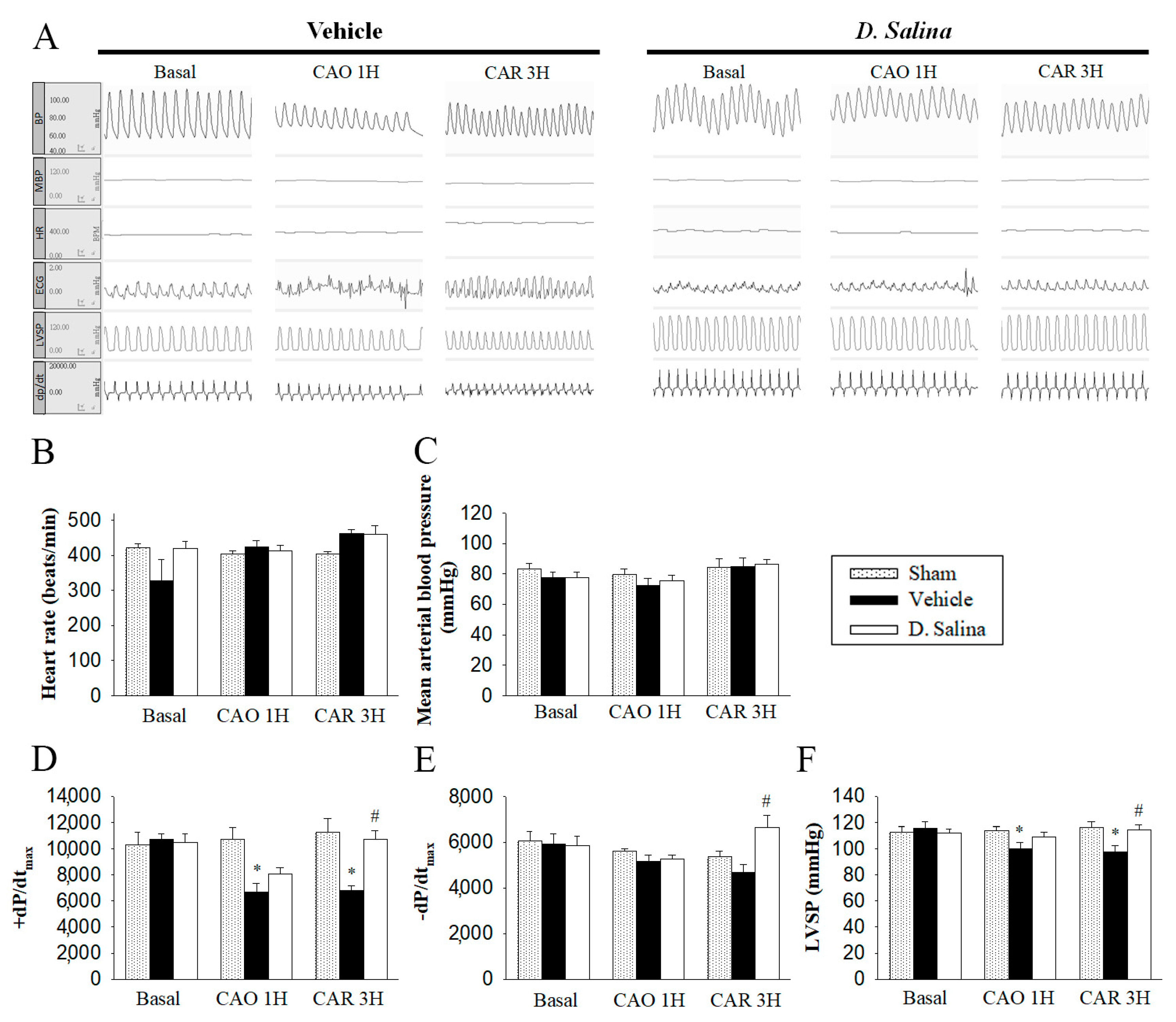
There were no significant differences in the pre-ischemic hemodynamic parameters, including HR, mean BP, ±dp/dtmax, and LVSP among the three groups of rats. Moreover, there were no significant differences in HR and mean BP between the vehicle- and 1 mg/kg D. salina-treated rats during myocardial I/R injury (Figure 2B,C). However, compared with the sham group, the +dP/dtmax and LVSP significantly decreased after 1 h of myocardial ischemia and 3 h of reperfusion in the vehicle group. Compared with the vehicle group, ±dP/dtmax and LVSP significantly increased in the D. salina group (Figure 2D,F). The recovery of ±dP/dtmax and LVSP indicated that 1 mg/kg D. salina could improve the cardiac functional recovery in rats subjected to myocardial I/R injury.

2.3. Effects of D. salina on the Inflammatory Signaling Pathway

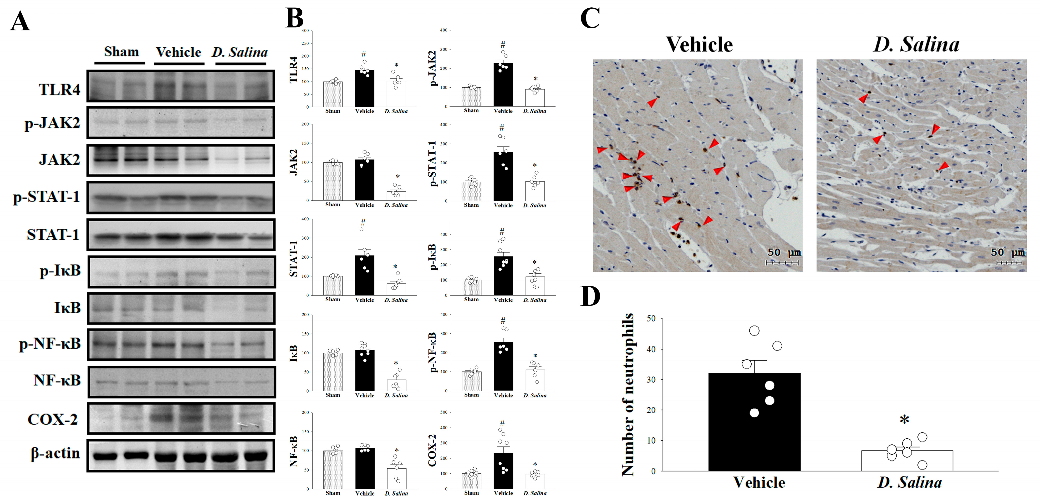
The TLR4 signaling pathway plays a central role in myocardial I/R injury and is involved in the inflammatory pathway after I/R injury. As shown in Figure 3A,B, compared with the vehicle group, the expression of TLR4 significantly decreased in the D. salina-treated group. This indicates that the cardioprotective effect of D. salina may be associated with the TLR4 signaling pathway.
The JAK2–STAT1 pathway is associated with TLR4-mediated inflammatory responses. STAT1 acts as a transcription factor for COX-2; the activation of STAT1 depends on the phosphorylation of JAK2. In the present study, results revealed that compared with the vehicle group, the expression levels of JAK2, p-JAK2, STAT1, and p-STAT1 significantly decreased in the D. salina-treated group after myocardial I/R injury. Similarly, The expression level of COX-2 was significantly suppressed by D. salina treatment in the rats subjected to myocardial I/R injury. Moreover, myocardial I/R injury induced the phosphorylation of IκB, which led to the translocation of NF-κB to the nucleus and mediated proinflammatory gene expression. We also examined whether D. salina could block the activation of IκB to NF-κB during myocardial I/R injury. Compared with the vehicle group, the expression levels of IκB, p-IκB, NF-κB and p-NF-κB significantly decreased in the myocardium of the rats in the D. salina-treated group after myocardial I/R injury (Figure 3A,B). We further examined whether the D. salina-treated group corresponds to less myocardial inflammation as demonstrated by neutrophil infiltration. We used immunohistochemical staining for MPO on the hearts. We found that administration of D. salina significantly reduced I/R-induced neutrophil infiltration in the myocardium compared with the vehicle group (Figure 3C,D). Thus, D. salina may limit myocardial I/R injury-induced inflammation by suppressing not only the JAK2–STAT1 pathway but also the NF-κB-responsive mechanism.

2.4. Effects of D. salina on Apoptosis and Autophagy

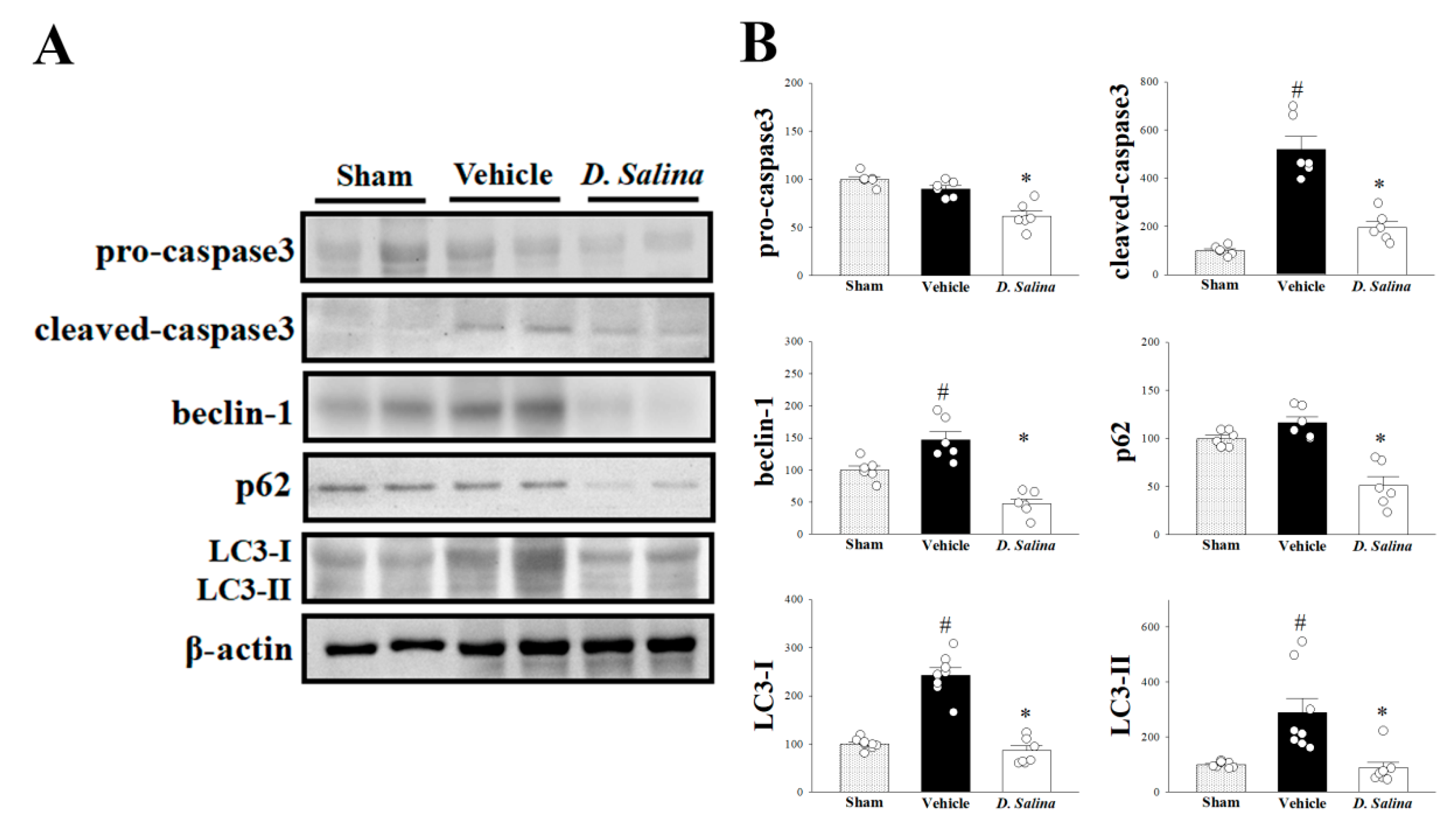
Next, we examined whether caspase-3, a key protein in both extrinsic and intrinsic apoptotic pathways, plays a role in the cardioprotective effect of D. salina in rats subjected to myocardial I/R injury. Compared with the vehicle group, the expression levels of both procaspase-3 (inactive form) and cleaved caspase-3 (active form) significantly decreased in the D. salina-treated group (Figure 4A,B). These findings suggest that D. salina attenuates the apoptotic level in the I/R-injured heart.
To detect whether D. salina regulates myocardial autophagy after I/R injury, we also examined the expression levels of beclin-1, p62 and LC3. Compared with the vehicle group, the expression levels of beclin-1, p62 and LC3-I/II significantly decreased in the D. salina-treated group (Figure 4A,B). These results indicate that D. salina decreases myocardial autophagy after I/R injury.

3. Discussion

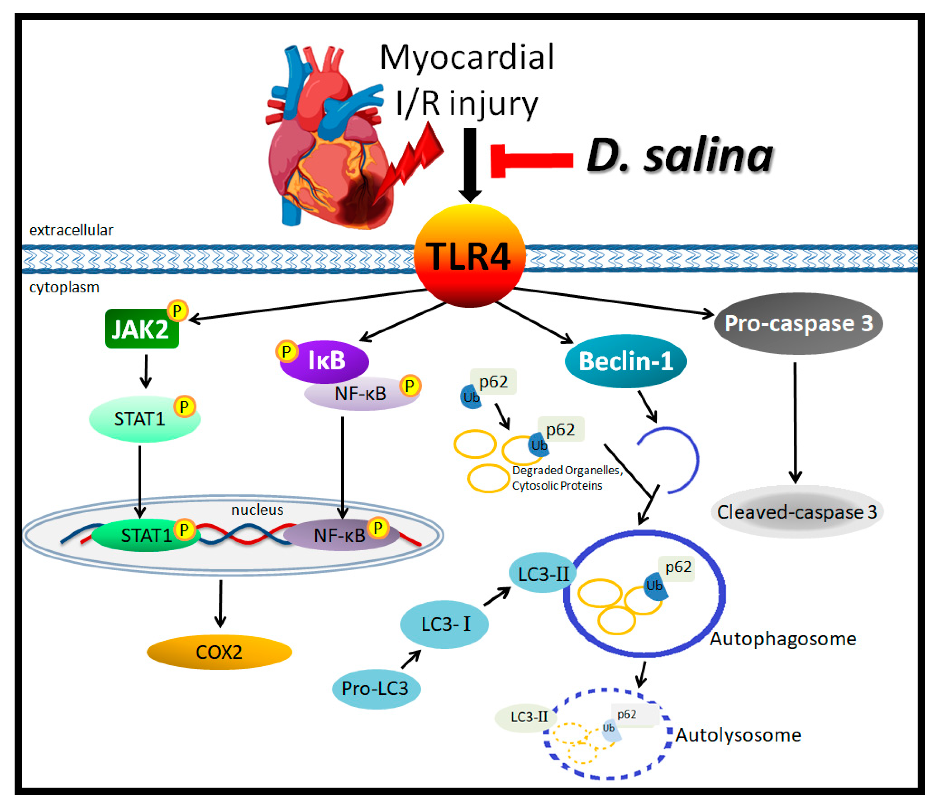
D. salina algae have been widely used as health food components, vitamin A precursor supplements, food colorants and food and cosmetics additives. The present study is the first to reveal the cardioprotective effects of D. salina in rats subjected to myocardial I/R injury. The main findings of the present study are as follows: (1) Compared with the vehicle, D. salina significantly decreased the LDH activity and troponin I level in the plasma and decreased myocardial infarction in the rats after myocardial I/R injury. (2) D. salina improved the cardiac functional recovery in the rats subjected to myocardial I/R injury, as evidenced by significantly higher ±dP/dtmax and LVSP than those in the vehicle group. (3) D. salina significantly attenuated the inflammatory signaling pathway, resulting in decreased expression levels of TLR4, p-JAK2, JAK2, p-STAT1 and STAT1, p-NF-κB, NF-κB, p-IκB, IκB and COX-2. (4) D. salina attenuated apoptosis and autophagy by significantly decreasing the expression levels of caspase-3 and those of beclin-1, p62 and LC3-I/II, respectively. These findings suggest that D. salina extracts play a role in attenuating TLR4 signaling to inhibit inflammatory responses, apoptosis and autophagy to exert potent cardioprotective activities that protect the myocardium against I/R injury (Figure 5).
Dunaliella strains are well known for being rich in β-carotene [18]. However, excess β-carotene levels may cause a variety of adverse effects, and large-scale prospective randomized trials reported that supplementation with 20 to 30 mg β-carotene per day was associated with an increased risk of lung cancer and cardiovascular disease [28]. Moreover, Csepanyi and colleagues revealed that the low-dose of β-carotene treatment (30 mg/kg/day) significantly reduced the infarcted zone relative to the control group, but this protective effect is abolished in the hearts taken from animals treated with a high-dose of β-carotene (150 mg/kg/day) in the Langendorff model [29]. Recently, they also showed that increasing the concentration of β-carotene does not provide an additional benefit in the diabetic I/R myocardium [30]. D. salina has been as well known as the richest source of natural β-carotene, but no study has assessed the potential cardioprotective effect of D. salina on myocardial I/R injury. In the present study, our results showed that 1 mg/kg of D. salina could improve cardiac functional recovery and significantly decrease the infarct size after acute myocardial I/R injury. The effective dosage of D. salina for ameliorating myocardial I/R injury in rats is lower than the dose of β-carotene, which may cause serious side effects in the clinical trials.
Previous reports demonstrated that D. salina had good anti-inflammatory activity. El-Baz et al. declared that D salina attenuated high-fat diet-induced fibrotic cardiac tissue and congestion of myocardial blood vessels by influencing anti-oxidant and anti-inflammatory effects in obese rats [21]. Moreover, D. salina could modulate pseudorabies virus-induced inflammation by inhibition of NF-κB activation via TLR9 dependent-PI3K/Akt inactivation in vitro. In addition, D. salina inhibited reactive oxygen species (ROS) accumulation and inflammation by downregulation of STAT-1/3 dependent NF-κB activation in virus-infected RAW264.7 cells [25]. In this study, we found that D. salina (10 μM) significantly increased cell viability against OGD/R insult in H9c2 cells, and also significantly inhibited ROS accumulation and inflammatory cytokines emancipation (Figure S1). However, the inflammatory signaling pathway is a principal mediator of myocardial injury during ischemia and reperfusion. The unifying pathophysiological mechanism underlying myocardial I/R injury-induced inflammation could be explained by elevated TLR4 levels after myocardial I/R injury, resulting in the direct activation of the inflammatory pathway [31] to enhance tissue injury. In the present study, we found that compared with the vehicle, D. salina significantly decreased the expression level of TLR4. Previous studies have suggested that TLR4 has a direct relationship with the inflammatory signaling pathway (not only the JAK2–STAT1 pathway but also the NF-κB signaling pathway), which finally contributes to the translation of COX-2. The expression levels of many inflammatory cytokines increase after COX-2 upregulation. STAT1 acts as a transcription factor for COX-2; the activation of STAT1 depends on the phosphorylation of JAK2 [32]. NF-kB, an important transcription factor located downstream of the TLR4-mediated signaling pathway, is isolated in the cytoplasm in an inactive form by interacting with IkB under unstimulated conditions. When TLR4 is activated, IκB becomes phosphorylated and degraded, followed by the phosphorylation of NF-κB [33]. Phosphorylated NF-κB enters the nucleus and activates some specific proinflammatory genes, such as COX-2 [34]. Therefore, decreasing the expression level of TLR4 may have an anti-inflammatory effect. We found that compared with the vehicle, D. salina significantly decreased the expression levels of several proteins, including JAK2, p-JAK2, STAT1, p-STAT1 and COX-2. Thus, D. salina has cardioprotective effects that may involve the inhibition of the TLR4-mediated inflammatory signaling pathway.
In addition, the TLR4 cascade can activate cell apoptosis [35]. Apoptosis is the process of programmed cell death that acts under a physiological mechanism to clean up unwanted cells without triggering an inflammatory response. However, myocardial I/R injury can trigger inappropriate apoptosis and cause worse damage to cardiomyocytes [36]. Moreover, Sheu and colleagues declared that D. salina could regulate the antiproliferative effects and induce cell cycle arrest and apoptosis in non-small cell lung cancer cells [37]. In the present study, we assessed the expression levels of procaspase-3 (inactive form) and cleaved caspase-3 (active form) to determine the role of apoptosis in the cardioprotective effect of D. salina. We found that compared with the vehicle, D. salina significantly decreased the expression levels of procaspase-3 and cleaved caspase-3. Hence, D. salina may protect the heart against I/R injury by inhibiting apoptosis.
It is well known that the signaling cascade involved in TLR4-mediated autophagy may play a significant role in the pathogenesis of myocardial I/R injury [10]. Autophagy induces the dysfunction and death of cardiomyocytes, is triggered by myocardial ischemia and is enhanced by reperfusion [38]. However, Beclin-1 is a key factor involved in TLR4-mediated autophagy signaling that can regulate both autophagosome formation and processing [39]. Beclin-1 needs LC3-II as well as p62 for autophagosome formation. Moreover, p62 acts as an autophagy receptor for the degradation of the ubiquitinated substrates. It also facilitates the transport of some degraded organelles and cytosolic proteins to the autophagosome and provides a molecular link between autophagy and ubiquitination [40]. LC3-I, the cytosolic form, is cleaved to form LC3-II during autophagosome formation. Furthermore, using cross species RNA-seq meta-analysis and machine-learning models identified that microalga Dunaliella could regulate chaperone-mediated autophagy, the lipid and nitrogen metabolism and ROS scavenging-related genes against salt stress conditions [41]. In the present study, we found that pretreatment with D. salina significantly decreased the expression levels of Beclin-1, p62, LC3-I and LC3-II in rat heart tissue after myocardial I/R injury. These results suggest that the cardioprotective effect of D. salina is associated with the anti-autophagic pathway.
In conclusion, D. salina has been clinically used as a dietary provitamin A carotenoid supplement in recent years. The present study is the first to demonstrate that pretreatment with D. salina could protect the myocardium against I/R injury. We speculate that the beneficial cardioprotective effects of D. salina are mediated by the attenuation of the TLR4-mediated signaling pathway and play a role in attenuating inflammation, apoptosis, and autophagy in rats subjected to myocardial I/R injury. Our results suggest that a dietary supplement of D. salina might be beneficial for protecting the heart against myocardial I/R injury.

4. Materials and Methods

4.1. Animals

The protocols of the present study conformed to those published in the Guide for the Care and Use of Laboratory Animals by the National Research Council of the National Academies (NIH publication, revised 2011). Six-week-old male Sprague–Dawley rats (LASCO Co., Taipei, Taiwan) were used in the study and were housed in the Animal Center of Chung Shan Medical University at a stable temperature of 25 ± 1 °C and humidity of 55 ± 5% under a 12 h light–dark cycle. The rats were fed normal chow and given water ad libitum. The surgical procedures for inducing myocardial I/R injury were performed under supervision and approved by the Institutional Animal Care and Use Committee of Chung Shan Medical University, Taichung, Taiwan (IACUC 1924). Every effort was made to minimize animal suffering and to reduce the number of rats sacrificed.

4.2. Preparation of D. salina Extract

The dried powder of D. salina cultivated in Taiwan was obtained from Gong Bih Enterprise Co., Ltd. (Wunlin, Taiwan). D. salina extract was prepared according to the method of Lin et al. [25]. Extract the algal sample (10 g) in 250 mL hexane/acetone/ethanol (2:1:1, v/v/v) with a shaker for 2 h at room temperature. Then, saponification was performed by adding 10 mL of 40% methanolic KOH at 25 °C for 16 h. The solvent was evaporated to dryness to yield carotenoid extract. The carotenoids composition in the extract was estimated by high-performance liquid chromatography (HPLC) under the conditions determined in the previous report [25]: column, YMC C30 (250 × 4.6 mm, 5 μm) (Waters Co., Milford, MA, USA); mobile phase, methanol (MeOH)–acetonitrile–H2O (84/14/2, v/v/v)/CH2Cl2 = 75/25 (v/v); flow rate, 1 mL/min; detection, 210–650 nm at a rate of 1.00 spectrum/s. The equipment consisted of a PrimeLine Gradient Model 500G HPLC pump system (Analytical Scientific Instruments, Inc., El Sobrante, CA, USA) and an S-3210 photodiode-array detector (Schambeck SFD GmbH, Bad Honnef, Germany). Moreover, all-trans-β-carotene and 9- or 9′-cis-β-carotene accounted for 92.46% of the major components of the D. salina extract [14]. The extracts was then dissolved in dimethyl sulfoxide (DMSO) for the experiment.

4.3. D. salina Administration

The D. salina extract was freshly prepared before administration. The experimental rats were randomly divided into four groups: (1) sham: rats subjected to all surgical procedures, except for left anterior descending (LAD) coronary artery occlusion, (2) vehicle: rats treated with vehicle (0.1% DMSO in normal saline), (3) D. salina-0.1: rats treated with 0.1 mg/kg D. salina extract, and (4) D. salina-1: rats treated with 1 mg/kg D. salina extract. The D. salina extract or vehicle was intravenously administered 15 min before LAD coronary artery occlusion. Our preliminary results found that 1 mg/kg of D. salina can effectively protect the heart against myocardial I/R injury. In order to reduce the number of animals used in the present study, the dose used to investigate the mechanisms of D. salina cardioprotection was tested from 1 mg/kg. This dose is lower than the long-term safe dose of 100 mg/kg, which is administrated in both genders of rats [19].

4.4. Surgical Protocol

Myocardial I/R injury was induced by temporary occlusion of the LAD coronary artery, as described previously [42]. Briefly, the rats were initially anesthetized with a single intraperitoneal injection of urethane (1.25 g/kg). The jugular vein and femoral artery were cannulated to administer drugs and to continuously monitor the blood pressure (BP) and heart rate [39] during the experiment. Left thoracotomy cut the fourth and fifth ribs to approximately 2 mm of the sternum in order to expose the heart; a 6/0 silk suture was then used to surround the LAD coronary artery. The tightening of the suture occluded the LAD coronary artery for 1 h and the tension of the suture was then released to cause reperfusion for 3 h.

4.5. Evaluation of Cardiac Function

A Millar catheter was inserted into the left ventricular cavity via the right common carotid artery and changes in the left ventricular systolic pressure (LVSP), left ventricular diastolic pressure (LVDP) and maximal slope of systolic pressure increment (+dP/dtmax) and diastolic decrement (−dP/dtmax) were continuously recorded using a Transonic Scisense Pressure Measurement system (SP200, Transonic Scisense Inc., London, ON, Canada).

4.6. Measurement of Myocardial Injury and Collection of Myocardial Samples

Myocardial infarct size was determined by a double staining technique using Evans blue and 2,3,5-triphenyltetrazolium chloride (TTC; Sigma-Aldrich, St. Louis, MO, USA). At the end of the experiment, the coronary artery was re-occluded and 0.1% Evans blue solution was injected intravenously, and the non-ischemic myocardium was stained to determine the area at risk. Hearts were sectioned transversely into 2 mm thick slices using a cardiac slicer matrix (Jacobowitz Systems, Zivic-Miller Laboratories Inc., Allison Park, PA, USA). Heart sections were stained with 2% TTC for 30 min at 37 °C in the dark. Place the sections in a solution containing 10% formalin for 1 day at room temperature. Infarcted tissue sections were scanned and tissue weight was assessed by distinguishing the normal myocardium (stained blue) from the area at risk and infarct area (unstained) in a TTC staining assay. Arterial blood collected from the carotid catheter in rats that survived after 60 min of ischemia and 3 h of reperfusion was centrifuged at 3000× g for 10 min to separate plasma and determine myocardial damage. Myocardial cellular damage was determined using automated clinical analyzers to measure plasma activities of lactate dehydrogenase [43] (ADVIA 1800, Siemens Healthcare Diagnostics Inc., Tarrytown, NY, USA) and the levels of Troponin-I (Centaur, Siemens Healthcare Diagnostics Inc., NY, USA).

4.7. Protein Extraction and Western Blot Analysis

In order to reduce the number of animals used in the present study, 1 mg/kg D. salina was used as the effective dose to investigate the mechanism underlying the cardioprotective effect of D. salina. Protein expression was evaluated by western blot as previously described procedures [44]. The primary antibodies against COX-2 (Cayman Chemical, Ann Arbor, MI, USA) included TLR4, phospho-JAK2 (p-JAK2), JAK2 (Santa Cruz, Dallas, TX, USA), phospho-NF-κB (p-NF-κB), NF-κB, phospho-IκB (p-IκB), IκB, procaspase-3, cleaved caspase-3 (Cell Signaling, Danvers, MA, USA), signal transducer and activator of transcription-1 (STAT1), phospho-STAT1 (p-STAT1) (Abcam, Cambridge, UK), light chain 3 (LC3), p62 and Beclin-1 (Novus, Centennial, CO, USA). β-actin (Abcam, Cambridge, UK) was used as an internal loading control. The membranes were incubated with an HRP-conjugated secondary antibody (Jackson Immuno Research Laboratories, West Grove, PA, USA) prior to chemiluminescence detection (Thermo Scientific, Waltham, MA, USA).

4.8. Immunohistochemical Staining

Rat hearts were individually fixed in 10% neutral buffered formalin for 24 h. They were then dehydrated in a graded ethanol series (50%, 75%, 95% and 100%), cleared in xylene, and embedded in paraffin at 55 °C for 24 h. For immunohistochemical analysis, serial sections of relatively thick (5 μm) wax-embedded hearts were cut, mounted on glass slides, deparaffinized and then rehydrated in the same manner as for histological analysis. Sections were then treated with 3% H2O2 in methanol for 10 min to inactivate any endogenous peroxidases and washed 3 times with PBS for 5 min each. Block with 3% BSA at room temperature for 1 h, then add 1:50 diluted rabbit anti-human MPO polyclonal antibody and incubate at 37 °C for 1 h. After 3 additional washes in PBS, sections were incubated with HRP-conjugated rabbit anti-goat IgG diluted 1:100 in 1% BSA for 1 h at 37 °C, followed by 3 additional washes in PBS. Finally, sections were incubated with 3,3’-diaminobenzidine (0.3 mg/mL) in 100 mM Tris (pH 7.5) containing 0.3 μL H2O2/mL for 3 min at room temperature. After three washes in PBS, the sections were fixed in 50% glycerol in PBS and examined under a light microscope.

4.9. Statistical Analysis

The data are expressed as the mean ± standard error of the mean. Statistical analyses of differences were performed using one-way analysis of variance for combined data, followed by Bonferroni tests. p < 0.05 was considered statistically significant.

Supplementary Materials

The following supporting information can be downloaded at https://www.mdpi.com/article/10.3390/ijms24043871/s1.

Author Contributions

Conceptualization, C.-F.T. and S.-S.H.; Data curation, J.-W.T., C.-S.C., C.-Y.C., H.-H.S., P.-H.L. and Y.-H.W.; Funding acquisition, C.-F.T. and S.-S.H.; Investigation, J.-M.L., J.-W.T., C.-S.C., C.-Y.C. and Y.-C.C.; Methodology, H.-W.L. and M.W.; Project administration, M.W. and S.-S.H.; Resources, H.-W.L., J.-M.L., K.-M.C., M.W. and S.-S.H.; Software, J.-M.L. and K.-M.C.; Supervision, C.-F.T. and S.-S.H.; Validation, H.-H.S., P.-H.L. and Y.-H.W.; Writing—original draft, J.-W.T. and C.-S.C.; Writing—review and editing, C.-F.T., Y.-H.W., M.W. and S.-S.H. All authors have read and agreed to the published version of the manuscript.

Funding

This research was funded by research grants from Chung Shan Medical University Hospital, grant number CSH-2018-C-031 and CSH-2020-C-026 to Chin-Feng Tsai and Shiang-Suo Huang.

Institutional Review Board Statement

The animal study protocol for inducing myocardial I/R injury were performed under supervision and approved by the Institutional Animal Care and Use Committee of Chung Shan Medical University, Taichung, Taiwan (IACUC 1924 of 31 August 2017).

Data Availability Statement

The data presented in this study are available on request from the corresponding author.

Acknowledgments

We thank the Instrument Resource Center of Chung Shan Medical University for the chemiluminescence/fluorescence imaging analyzer.

Conflicts of Interest

The authors declare that there are no conflict of interest.

Abbreviations

AARarea at risk
BParterial blood pressure
I/Rischemia/reperfusion
HRheart rate
LDHlactate dehydrogenase
LADleft anterior descending coronary artery
LVSPleft ventricular systolic pressure
LC3light chain 3
STAT1signal transducer and activator of transcription-1
±dp/dtmaxthe maximal rate of left ventricular pressure increase and decrease
TLR4toll-like receptor 4
VTventricular tachycardia
VFventricular fibrillation

References

  1. Folino, A.; Losano, G.; Rastaldo, R. Balance of Nitric Oxide and Reactive Oxygen Species in Myocardial Reperfusion Injury and Protection. J. Cardiovasc. Pharmacol. 2013, 62, 567–575. [Google Scholar] [CrossRef]
  2. Daud, M.Y.; Awan, M.S.; Khan, M.; Hayat, U.; Tuyyab, F. Procedural Outcomes Of Primary Percutaneous Coronary Intervention In Elderly Patients With Stemi. JAMC 2019, 30, 585–588. [Google Scholar]
  3. Yang, Q.-H.; Yang, M.; Zhang, L.-L.; Xiao, M.-C.; Zhao, Y.; Yan, D.-X. The mechanism of miR-23a in regulating myocardial cell apoptosis through targeting FoxO3. Eur. Rev. Med. Pharmacol. Sci. 2017, 21, 5789–5797. [Google Scholar]
  4. Zhou, T.; Prather, E.; Garrison, D.; Zuo, L. Interplay between ROS and Antioxidants during Ischemia-Reperfusion Injuries in Cardiac and Skeletal Muscle. Int. J. Mol. Sci. 2018, 19, 417. [Google Scholar] [CrossRef] [Green Version]
  5. Jalali, Z.; Khademalhosseini, M.; Soltani, N.; Nadimi, A.E. Smoking, alcohol and opioids effect on coronary microcirculation: An update overview. BMC Cardiovasc. Disord. 2021, 21, 1–17. [Google Scholar] [CrossRef]
  6. Obad, A.; Peeran, A.; Little, J.I.; Haddad, G.E.; Tarzami, S.T. Alcohol-Mediated Organ Damages: Heart and Brain. Front. Pharmacol. 2018, 9, 81. [Google Scholar] [CrossRef] [Green Version]
  7. Zhao, H.; Mayhan, W.G.; Arrick, D.M.; Xiong, W.; Sun, H. Dose-Related Influence of Chronic Alcohol Consumption on Cerebral Ischemia/Reperfusion Injury. Alcohol. Clin. Exp. Res. 2011, 35, 1265–1269. [Google Scholar] [CrossRef] [Green Version]
  8. Yao, L.; Chen, H.; Wu, Q.; Xie, K. Hydrogen-rich saline alleviates inflammation and apoptosis in myocardial I/R injury via PINK-mediated autophagy. Int. J. Mol. Med. 2019, 44, 1048–1062. [Google Scholar] [CrossRef] [Green Version]
  9. Oyama, J.-I.; Blais, C.; Liu, X.; Pu, M.; Kobzik, L.; Kelly, R.A.; Bourcier, T. Reduced Myocardial Ischemia-Reperfusion Injury in Toll-Like Receptor 4-Deficient Mice. Circulation 2004, 109, 784–789. [Google Scholar] [CrossRef] [Green Version]
  10. Guo, X.; Jiang, H.; Yang, J.; Chen, J.; Yang, J.; Ding, J.W.; Li, S.; Wu, H.; Ding, H.S. Radioprotective 105 kDa protein attenuates ischemia/reperfusion-induced myocardial apoptosis and autophagy by inhibiting the activation of the TLR4/NF-kappaB signaling pathway in rats. Int. J. Mol. Med. 2016, 38, 885–893. [Google Scholar] [CrossRef] [Green Version]
  11. Yang, J.; Yang, J.; Ding, J.-W.; Chen, L.-H.; Wang, Y.-L.; Li, S.; Wu, H. Sequential Expression of TLR4 and its Effects on the Myocardium of Rats with Myocardial Ischemia-Reperfusion Injury. Inflammation 2008, 31, 304–312. [Google Scholar] [CrossRef]
  12. Tang, Y.; Zhou, G.; Yao, L.; Xue, P.; Yu, D.; Xu, R.; Shi, W.; Yao, X.; Yan, Z.; Duan, J.A. Protective effect of Ginkgo biloba leaves extract, EGb761, on myocardium injury in ischemia reperfusion rats via regulation of TLR-4/NF-kappaB signaling pathway. Oncotarget 2017, 8, 86671–86680. [Google Scholar] [CrossRef]
  13. Zhang, J.; Chen, F.; Sun, Z.; Sun, P.; Chen, T.; Chen, F. Microalgal carotenoids: Beneficial effects and potential in human health. Food Funct. 2014, 5, 413–425. [Google Scholar] [CrossRef]
  14. Hu, C.-C.; Lin, J.-T.; Lu, F.-J.; Chou, F.-P.; Yang, D.-J. Determination of carotenoids in Dunaliella salina cultivated in Taiwan and antioxidant capacity of the algal carotenoid extract. Food Chem. 2008, 109, 439–446. [Google Scholar] [CrossRef]
  15. Levy, Y.; Zaltsberg, H.; Ben-Amotz, A.; Kanter, Y.; Aviram, M. Dietary Supplementation of a Natural Isomer Mixture of Beta-Carotene Inhibits Oxidation of LDL Derived from Patients with Diabetes mellitus. Ann. Nutr. Metab. 2000, 44, 54–60. [Google Scholar] [CrossRef]
  16. Bechor, S.; Relevy, N.Z.; Harari, A.; Almog, T.; Kamari, Y.; Ben-Amotz, A.; Harats, D.; Shaish, A. 9-cis β-Carotene Increased Cholesterol Efflux to HDL in Macrophages. Nutrients 2016, 8, 435. [Google Scholar] [CrossRef] [Green Version]
  17. Rotenstreich, Y.; Belkin, M.; Sadetzki, S.; Chetrit, A.; Ferman-Attar, G.; Sher, I.; Harari, A.; Shaish, A.; Harats, D. Treatment with 9-cis β-carotene-rich powder in patients with retinitis pigmentosa: A randomized crossover trial. JAMA Ophthalmol. 2013, 131, 985–992. [Google Scholar] [CrossRef] [PubMed] [Green Version]
  18. Jin, E.S.; Melis, A. Microalgal biotechnology: Carotenoid production by the green algae Dunaliella salina. Biotechnol. Bioprocess Eng. 2003, 8, 331–337. [Google Scholar] [CrossRef]
  19. El-Baz, F.K.; Aly, H.F.; Salama, A.A. Toxicity assessment of the green Dunaliella salina microalgae. Toxicol. Rep. 2019, 6, 850–861. [Google Scholar] [CrossRef]
  20. Kiokias, S.; Proestos, C.; Oreopoulou, V. Effect of Natural Food Antioxidants against LDL and DNA Oxidative Changes. Antioxidants 2018, 7, 133. [Google Scholar] [CrossRef] [Green Version]
  21. El-Baz, F.K.; Aly, H.F.; Abd-Alla, H.I. The ameliorating effect of carotenoid rich fraction extracted from Dunaliella salina microalga against inflammation- associated cardiac dysfunction in obese rats. Toxicol. Rep. 2019, 7, 118–124. [Google Scholar] [CrossRef] [PubMed]
  22. El-Baz, F.K.; Hussein, R.A.; Saleh, D.O.; Jaleel, G.A.R.A. Zeaxanthin Isolated from Dunaliella salina Microalgae Ameliorates Age Associated Cardiac Dysfunction in Rats through Stimulation of Retinoid Receptors. Mar. Drugs 2019, 17, 290. [Google Scholar] [CrossRef] [PubMed]
  23. Lin, H.W.; Chen, Y.C.; Liu, C.W.; Yang, D.J.; Chen, S.Y.; Chang, T.J.; Chang, Y.Y. Regulation of virus-induced inflammatory response by Dunaliella salina alga extract in macrophages. Food Chem. Toxicol. 2014, 71, 159–165. [Google Scholar] [CrossRef]
  24. Chitranjali, T.; Anoop Chandran, P.; Muraleedhara Kurup, G. Omega-3 fatty acid concentrate from Dunaliella salina possesses anti-inflammatory properties including blockade of NF-kappaB nuclear translocation. Immunopharmacol. Immunotoxicol. 2015, 37, 81–89. [Google Scholar] [PubMed]
  25. Lin, H.W.; Liu, C.W.; Yang, D.J.; Chen, C.C.; Chen, S.Y.; Tseng, J.K.; Chang, T.J.; Chang, Y.Y. Dunaliella salina alga extract inhibits the production of interleukin-6, nitric oxide, and reactive oxygen species by regulating nuclear factor-κB/Janus kinase/signal transducer and activator of transcription in virus-infected RAW264.7 cells. J. Food Drug Anal. 2017, 25, 908–918. [Google Scholar] [CrossRef] [Green Version]
  26. Cebova, M.; Pechanova, O. Protective Effects of Polyphenols against Ischemia/Reperfusion Injury. Molecules 2020, 25, 3469. [Google Scholar] [CrossRef]
  27. Gammone, M.A.; Riccioni, G.; D’Orazio, N. Carotenoids: Potential allies of cardiovascular health? Food Nutr. Res. 2015, 59, 26762. [Google Scholar]
  28. Blomhoff, R. Vitamin A and Carotenoid Toxicity. Food Nutr. Bull. 2001, 22, 320–334. [Google Scholar] [CrossRef]
  29. Csepanyi, E.; Czompa, A.; Haines, D.; Lekli, I.; Bakondi, E.; Balla, G.; Tosaki, A.; Bak, I. Cardiovascular effects of low versus high-dose beta-carotene in a rat model. Pharmacol. Res. 2015, 100, 148–156. [Google Scholar] [CrossRef] [Green Version]
  30. Csepanyi, E.; Czompa, A.; Szabados-Furjesi, P.; Lekli, I.; Balla, J.; Balla, G.; Tosaki, A.; Bak, I. The Effects of Long-Term, Low- and High-Dose Beta-Carotene Treatment in Zucker Diabetic Fatty Rats: The Role of HO-1. Int. J. Mol. Sci. 2018, 19, 1132. [Google Scholar] [CrossRef] [Green Version]
  31. Zhai, Y.; Ao, L.; Cleveland, J.C.; Zeng, Q.; Reece, T.B.; Fullerton, D.A.; Meng, X. Toll-like Receptor 4 Mediates the Inflammatory Responses and Matrix Protein Remodeling in Remote Non-Ischemic Myocardium in a Mouse Model of Myocardial Ischemia and Reperfusion. PLsS ONE 2015, 10, e0121853. [Google Scholar] [CrossRef]
  32. De Prati, A.C.; Ciampa, A.R.; Cavalieri, E.; Zaffini, R.; Darra, E.; Menegazzi, M.; Suzuki, H.; Mariotto, S. STAT1 as a New Molecular Target of Anti-Inflammatory Treatment. Curr. Med. Chem. 2005, 12, 1819–1828. [Google Scholar] [CrossRef]
  33. Kawai, T.; Akira, S. Signaling to NF-kappaB by Toll-like receptors. Trends Mol. Med. 2007, 13, 460–469. [Google Scholar] [CrossRef]
  34. Surh, Y.J.; Chun, K.S.; Cha, H.H.; Han, S.S.; Keum, Y.S.; Park, K.K.; Lee, S.S. Molecular mechanisms underlying chemopreventive activities of anti-inflammatory phytochemicals: Down-regulation of COX-2 and iNOS through suppression of NF-kappa B activation. Mutat. Res. 2001, 480–481, 243–268. [Google Scholar] [CrossRef]
  35. Zhang, Y.; Peng, T.; Zhu, H.; Zheng, X.; Zhang, X.; Jiang, N.; Cheng, X.; Lai, X.; Shunnar, A.; Singh, M.; et al. Prevention of hyperglycemia-induced myocardial apoptosis by gene silencing of Toll-like receptor-4. J. Transl. Med. 2010, 8, 133. [Google Scholar] [CrossRef] [PubMed]
  36. Hamacher-Brady, A.; Brady, N.; Gottlieb, R.A. The Interplay between Pro-Death and Pro-Survival Signaling Pathways in Myocardial Ischemia/Reperfusion Injury: Apoptosis Meets Autophagy. Cardiovasc. Drugs Ther. 2006, 20, 445–462. [Google Scholar] [CrossRef] [PubMed]
  37. Sheu, M.-J.; Huang, G.-J.; Wu, C.-H.; Chen, J.-S.; Chang, H.-Y.; Chang, S.-J.; Chung, J.-G. Ethanol extract of Dunaliella salina induces cell cycle arrest and apoptosis in A549 human non-small cell lung cancer cells. In Vivo 2008, 22, 369–378. [Google Scholar] [PubMed]
  38. Matsui, Y.; Kyoi, S.; Takagi, H.; Hsu, C.-P.; Hariharan, N.; Ago, T.; Vatner, S.F.; Sadoshima, J. Molecular mechanisms and physiological significance of autophagy during myocardial ischemia and reperfusion. Autophagy 2008, 4, 409–415. [Google Scholar] [CrossRef] [Green Version]
  39. Shi, C.-S.; Kehrl, J.H. Traf6 and A20 differentially regulate TLR4-Induced autophagy by affecting the ubiquitination of Beclin 1. Autophagy 2010, 6, 986–987. [Google Scholar] [CrossRef] [Green Version]
  40. Kirkin, V.; McEwan, D.G.; Novak, I.; Dikic, I. A Role for Ubiquitin in Selective Autophagy. Mol. Cell 2009, 34, 259–269. [Google Scholar] [CrossRef] [PubMed]
  41. Panahi, B.; Frahadian, M.; Dums, J.; Hejazi, M.A. Integration of Cross Species RNA-seq Meta-Analysis and Machine-Learning Models Identifies the Most Important Salt Stress–Responsive Pathways in Microalga Dunaliella. Front. Genet. 2019, 10, 752. [Google Scholar] [CrossRef] [PubMed] [Green Version]
  42. Wang, Y.-H.; Chen, K.-M.; Chiu, P.-S.; Lai, S.-C.; Su, H.-H.; Jan, M.-S.; Lin, C.-W.; Lu, D.-Y.; Fu, Y.-T.; Liao, J.-M.; et al. Lumbrokinase attenuates myocardial ischemia-reperfusion injury by inhibiting TLR4 signaling. J. Mol. Cell. Cardiol. 2016, 99, 113–122. [Google Scholar] [CrossRef] [PubMed]
  43. Folkerts, H.; Wierenga, A.T.; Heuvel, F.A.V.D.; Woldhuis, R.R.; Kluit, D.S.; Jaques, J.; Schuringa, J.J.; Vellenga, E. Elevated VMP1 expression in acute myeloid leukemia amplifies autophagy and is protective against venetoclax-induced apoptosis. Cell Death Dis. 2019, 10, 1–12. [Google Scholar] [CrossRef] [PubMed]
  44. Su, H.H.; Liao, J.M.; Wang, Y.H.; Chen, K.M.; Lin, C.W.; Lee, I.H.; Li, Y.J.; Huang, J.Y.; Tsai, S.K.; Yen, J.C.; et al. Exogenous GDF11 attenuates non-canonical TGF-beta signaling to protect the heart from acute myocardial ischemia-reperfusion injury. Basic Res. Cardiol. 2019, 114, 20. [Google Scholar] [CrossRef] [PubMed]
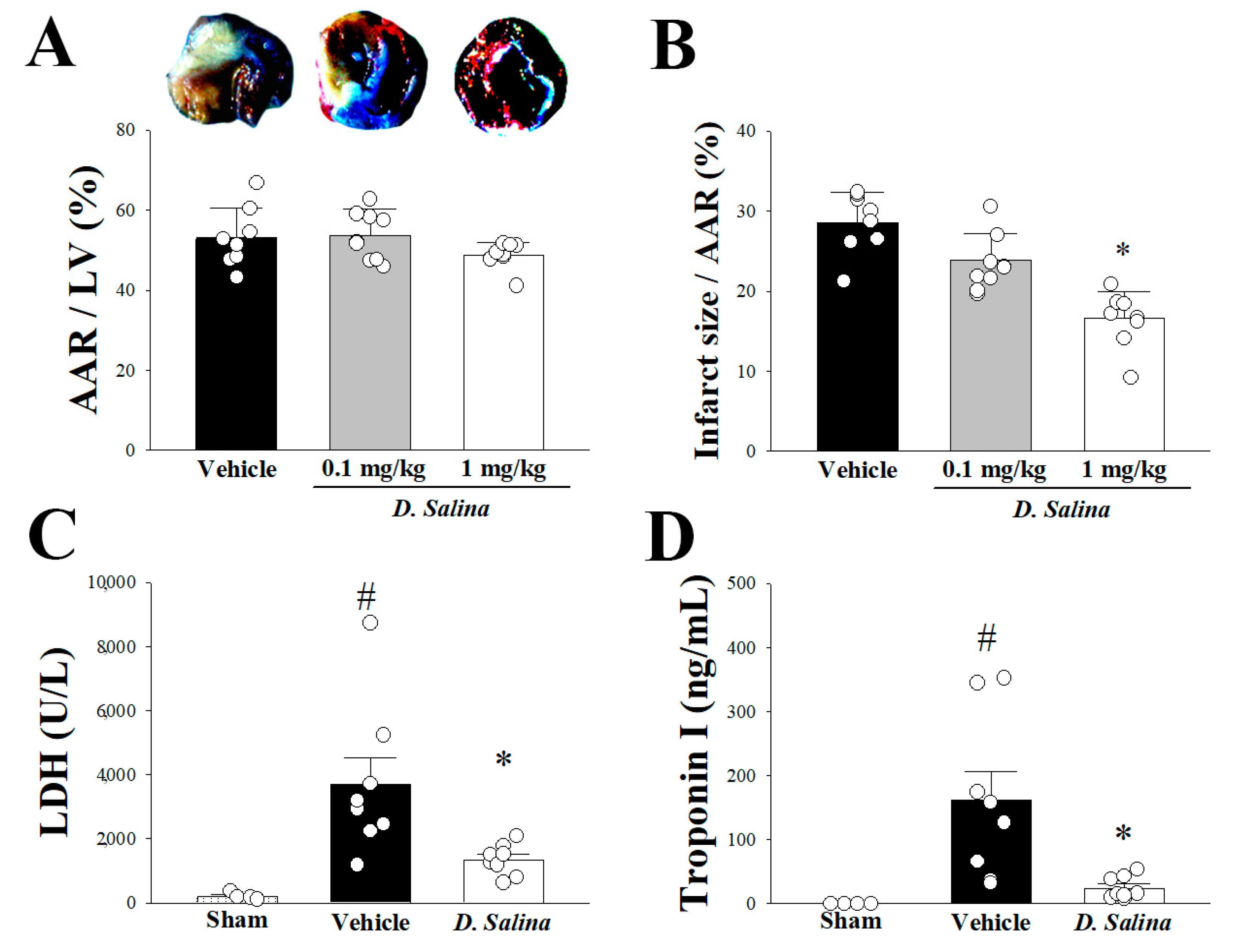
Figure 1. Effect of D. salina on I/R induced myocardial damage in rats. (A) Representative Evans blue/TTC staining of heart sections from vehicle-treated group and different dose (0.1 and 1 mg/kg) of D. salina groups after 1 h of LAD coronary artery occlusion and 3 h of reperfusion. Perfused area is seen in blue; area at risk (AAR) is seen in red and white. Infarction is seen in white. Quantitative AAR/Ventricle % (ratio of non-blue area to total area), (B) the risk zone infarcted % (ratio of white area to AAR), (C) plasma lactate dehydrogenase activity and (D) plasma troponin-I level, in vehicle and 1 mg/kg D. salina-treated rats. The results are shown as mean ± S.E.M. (n = 4–9); # p < 0.05 compared with the sham group; * p < 0.05 compared with the vehicle-treated group.
Ijms 24 03871 g001
Figure 2. Effect of D. salina on hemodynamic parameters in rats subjected to myocardial I/R injury. (A) Representative image of hemodynamic parameters between the vehicle- and 1 mg/kg D. salina-treated rats during myocardial I/R injury. Quantitatively analyzed results of (B) heart rate, (C) mean arterial blood pressure, (D) maximum rates of pressure change in the LV (+dP/dt), (E) minimum rates of pressure change in the LV (−dP/dt) and (F) left ventricular systolic pressure (LVSP) were recorded by physiological parameters (CAO1H, 1 h after LAD coronary artery occlusion; CAR3H, 3 h after LAD coronary artery reperfusion). Values are mean ± S.E.M.; (n = 8 per group); # p < 0.05 compared with the sham group; * p < 0.05 compared with the vehicle-treated group.
Ijms 24 03871 g002
Figure 3. D. salina deceased inflammation after myocardial I/R injury. (A) Representative images of the Western blot results in heart tissue. (B) Graphs represent the quantitative differences of TLR4, p-JAK2 and JAK2, p-STAT1 and STAT1, p-IκB and IκB, p-NF-κB and NF-κB, COX-2 between sham, vehicle and 1 mg/kg D. salina-treated groups in myocardial I/R animals. β-actin was used as a loading control for the blots. (C) Representative areas of MPO immunohistochemical staining slides of rats subjected to myocardial I/R injury. The red arrow indicates the infiltration of neutrophils. (D) Quantitative analysis of MPO positive cells of rat heart slides that were subjected to myocardial I/R injury. Data are normalized with the mean expression from the sham group. # p < 0.05 compared with the sham group; * p < 0.05 compared with the vehicle-treated group. Values are expressed as mean ± S.E.M. (n = 6–8).
Ijms 24 03871 g003
Figure 4. D. salina decreased apoptosis and autophagy after myocardial I/R injury. (A) Representative images of the Western blot results in heart tissue. (B) Graphs represent the quantitative differences of pro-caspase3 and cleaved-caspase3, beclin-1, p62, LC3-I and LC3-II between sham, vehicle and 1 mg/kg D. salina-treated groups in myocardial I/R animals. β-actin was used as a loading control for the blots. Data are normalized with the mean expression from the sham group. # p < 0.05 compared with the sham group; * p < 0.05 compared with the vehicle group. Values are expressed as mean ± S.E.M. (n = 6–8).
Ijms 24 03871 g004
Figure 5. Schematic diagram of the effects of D. salina treatment regulates TLR4 signaling against myocardial I/R injury. Pre-treatment with D. salina mediates anti-inflammatory and anti-apoptotic activities and decreases autophagy through the TLR4-mediated signaling pathway to antagonize myocardial I/R injury.
Ijms 24 03871 g005
Disclaimer/Publisher’s Note: The statements, opinions and data contained in all publications are solely those of the individual author(s) and contributor(s) and not of MDPI and/or the editor(s). MDPI and/or the editor(s) disclaim responsibility for any injury to people or property resulting from any ideas, methods, instructions or products referred to in the content.