12 pages, 1876 KiB  
Article
Untargeted Metabolomics Identifies Potential Hypertrophic Cardiomyopathy Biomarkers in Carriers of MYBPC3 Founder Variants
by Mark Jansen 1,2,*, Maike Schuldt 3, Beau O. van Driel 3, Amand F. Schmidt 4,5, Imke Christiaans 6, Saskia N. van der Crabben 7, Yvonne M. Hoedemaekers 8, Dennis Dooijes 1, Jan D. H. Jongbloed 6, Ludolf G. Boven 6, Ronald H. Lekanne Deprez 7, Arthur A. M. Wilde 9, Judith J. M. Jans 1, Jolanda van der Velden 3, Rudolf A. de Boer 10, J. Peter van Tintelen 1,2, Folkert W. Asselbergs 2,4,5,9,11 and Annette F. Baas 1
1 Department of Genetics, University Medical Centre Utrecht, Utrecht University, 3584CX Utrecht, The Netherlands
2 Netherlands Heart Institute, 3511EP Utrecht, The Netherlands
3 Department of Physiology, Amsterdam UMC, Vrije Universiteit Amsterdam, Amsterdam Cardiovascular Sciences, 1081HZ Amsterdam, The Netherlands
4 Department of Cardiology, University Medical Centre Utrecht, Utrecht University, 3584CX Utrecht, The Netherlands
5 Institute of Cardiovascular Science, Faculty of Population Health Sciences, University College London, London WC1E 6DD, UK
6 Department of Genetics, University Medical Centre Groningen, University of Groningen, 9713GZ Groningen, The Netherlands
7 Department of Human Genetics, Amsterdam UMC, University of Amsterdam, 1105AZ Amsterdam, The Netherlands
8 Department of Clinical Genetics, Radboud University Medical Centre, 6525GA Nijmegen, The Netherlands
9 Heart Centre, Department of Cardiology, Amsterdam UMC, University of Amsterdam, 1081HZ Amsterdam, The Netherlands
10 Department of Cardiology, University Medical Centre Groningen, University of Groningen, 9713GZ Groningen, The Netherlands
11 Health Data Research UK and Institute of Health Informatics, University College London, London NW1 2DA, UK
add Show full affiliation list remove Hide full affiliation list
Int. J. Mol. Sci. 2023, 24(4), 4031; https://doi.org/10.3390/ijms24044031 - 17 Feb 2023
Cited by 3 | Viewed by 2204
Abstract
Hypertrophic cardiomyopathy (HCM) is the most prevalent monogenic heart disease, commonly caused by pathogenic MYBPC3 variants, and a significant cause of sudden cardiac death. Severity is highly variable, with incomplete penetrance among genotype-positive family members. Previous studies demonstrated metabolic changes in HCM. We [...] Read more.
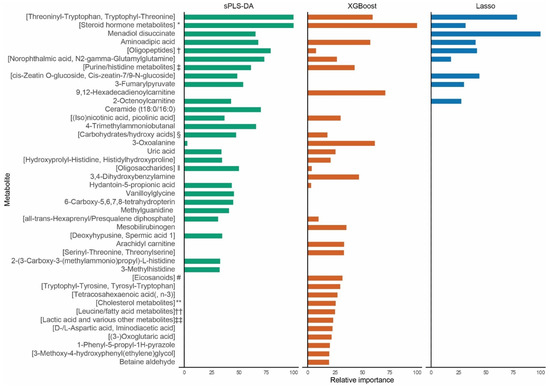
Hypertrophic cardiomyopathy (HCM) is the most prevalent monogenic heart disease, commonly caused by pathogenic MYBPC3 variants, and a significant cause of sudden cardiac death. Severity is highly variable, with incomplete penetrance among genotype-positive family members. Previous studies demonstrated metabolic changes in HCM. We aimed to identify metabolite profiles associated with disease severity in carriers of MYBPC3 founder variants using direct-infusion high-resolution mass spectrometry in plasma of 30 carriers with a severe phenotype (maximum wall thickness ≥20 mm, septal reduction therapy, congestive heart failure, left ventricular ejection fraction <50%, or malignant ventricular arrhythmia) and 30 age- and sex-matched carriers with no or a mild phenotype. Of the top 25 mass spectrometry peaks selected by sparse partial least squares discriminant analysis, XGBoost gradient boosted trees, and Lasso logistic regression (42 total), 36 associated with severe HCM at a p < 0.05, 20 at p < 0.01, and 3 at p < 0.001. These peaks could be clustered to several metabolic pathways, including acylcarnitine, histidine, lysine, purine and steroid hormone metabolism, and proteolysis. In conclusion, this exploratory case-control study identified metabolites associated with severe phenotypes in MYBPC3 founder variant carriers. Future studies should assess whether these biomarkers contribute to HCM pathogenesis and evaluate their contribution to risk stratification. Full article
(This article belongs to the Special Issue Molecular Mechanisms of Cardiac Development and Disease)
Show Figures

Figure 1

19 pages, 3167 KiB  
Review
Unveiling the Machinery behind Chromosome Folding by Polymer Physics Modeling
by Mattia Conte, Andrea Esposito, Francesca Vercellone, Alex Abraham and Simona Bianco *
1 Dipartimento di Fisica, Università di Napoli Federico II, and INFN Napoli, Complesso Universitario di Monte Sant’Angelo, 80126 Naples, Italy
These authors contributed equally to this work.
Int. J. Mol. Sci. 2023, 24(4), 3660; https://doi.org/10.3390/ijms24043660 - 11 Feb 2023
Cited by 1 | Viewed by 2201
Abstract
Understanding the mechanisms underlying the complex 3D architecture of mammalian genomes poses, at a more fundamental level, the problem of how two or multiple genomic sites can establish physical contacts in the nucleus of the cells. Beyond stochastic and fleeting encounters related to [...] Read more.
Understanding the mechanisms underlying the complex 3D architecture of mammalian genomes poses, at a more fundamental level, the problem of how two or multiple genomic sites can establish physical contacts in the nucleus of the cells. Beyond stochastic and fleeting encounters related to the polymeric nature of chromatin, experiments have revealed specific, privileged patterns of interactions that suggest the existence of basic organizing principles of folding. In this review, we focus on two major and recently proposed physical processes of chromatin organization: loop-extrusion and polymer phase-separation, both supported by increasing experimental evidence. We discuss their implementation into polymer physics models, which we test against available single-cell super-resolution imaging data, showing that both mechanisms can cooperate to shape chromatin structure at the single-molecule level. Next, by exploiting the comprehension of the underlying molecular mechanisms, we illustrate how such polymer models can be used as powerful tools to make predictions in silico that can complement experiments in understanding genome folding. To this aim, we focus on recent key applications, such as the prediction of chromatin structure rearrangements upon disease-associated mutations and the identification of the putative chromatin organizing factors that orchestrate the specificity of DNA regulatory contacts genome-wide. Full article
(This article belongs to the Special Issue Chromatin Architecture: A Flexible Foundation for Gene Expression)
Show Figures

Figure 1

19 pages, 6924 KiB  
Article
Eltrombopag Inhibits Metastasis in Breast Carcinoma by Targeting HuR Protein
by Yao Chen 1,†, Rui Zhang 1,†, Liuqing Yang 1, Pei Zhang 1, Feiyun Wang 1, Guoqiang Lin 1, Jiange Zhang 1,2,* and Yuying Zhu 1,3,*
1 The Research Center of Chiral Drugs, Innovation Research Institute of Traditional Chinese Medicine, Shanghai University of Traditional Chinese Medicine, Shanghai 201203, China
2 Shanghai Frontiers Science Center for Traditional Chinese Medicine Chemical Biology, Innovation Research Institute of Traditional Chinese Medicine, Shanghai University of Traditional Chinese Medicine, Shanghai 201203, China
3 Shanghai Institute of Traditional Chinese Medicine, Shanghai 201203, China
These authors contributed equally to this work.
Int. J. Mol. Sci. 2023, 24(4), 3164; https://doi.org/10.3390/ijms24043164 - 5 Feb 2023
Viewed by 2201
Abstract
Eltrombopag is a small molecule TPO-R agonist that has been shown in our previous studies to inhibit tumor growth by targeting Human antigen R (HuR) protein. HuR protein not only regulates the mRNA stability of tumor growth-related genes, but it also regulates the [...] Read more.
Eltrombopag is a small molecule TPO-R agonist that has been shown in our previous studies to inhibit tumor growth by targeting Human antigen R (HuR) protein. HuR protein not only regulates the mRNA stability of tumor growth-related genes, but it also regulates the mRNA stability of a variety of cancer metastasis-related genes, such as Snail, Cox-2, and Vegf-c. However, the role and mechanisms of eltrombopag in breast cancer metastasis have not been fully investigated. The purpose of this study was to investigate whether eltrombopag can inhibit breast cancer metastasis by targeting HuR. Our study first found that eltrombopag can destroy HuR-AU-rich element (ARE) complexes at the molecular level. Secondly, eltrombopag was found to suppress 4T1 cell migration and invasion and inhibit macrophage-mediated lymphangiogenesis at the cellular level. In addition, eltrombopag exerted inhibitory effects on lung and lymph node metastasis in animal tumor metastasis models. Finally, it was verified that eltrombopag inhibited the expressions of Snail, Cox-2, and Vegf-c in 4T1 cells and Vegf-c in RAW264.7 cells by targeting HuR. In conclusion, eltrombopag displayed antimetastatic activity in breast cancer in an HuR dependent manner, which may provide a novel application for eltrombopag, hinting at the multiple effects of HuR inhibitors in cancer therapy. Full article
(This article belongs to the Special Issue Novel Targeted Therapies in Cancer)
Show Figures

Graphical abstract

20 pages, 4523 KiB  
Article
Ephedra foeminea as a Novel Source of Antimicrobial and Anti-Biofilm Compounds to Fight Multidrug Resistance Phenotype
by Shurooq Ismail 1,2, Rosa Gaglione 1,3, Marco Masi 1, Srichandan Padhi 4, Amit K. Rai 4, Ghadeer Omar 2, Alessio Cimmino 1,* and Angela Arciello 1,3,*
1 Department of Chemical Sciences, University of Naples Federico II, Via Cintia 21, I-80126 Naples, Italy
2 Department of Biology and Biotechnology, An-Najah National University, Nablus 97300, Palestine
3 Istituto Nazionale di Biostrutture e Biosistemi (INBB), 00136 Rome, Italy
4 Institute of Bioresources and Sustainable Development, Imphal, Manipur 795004, India
Int. J. Mol. Sci. 2023, 24(4), 3284; https://doi.org/10.3390/ijms24043284 - 7 Feb 2023
Cited by 3 | Viewed by 2200
Abstract
Plants are considered a wealthy resource of novel natural drugs effective in the treatment of multidrug-resistant infections. Here, a bioguided purification of Ephedra foeminea extracts was performed to identify bioactive compounds. The determination of antimicrobial properties was achieved by broth microdilution assays to [...] Read more.
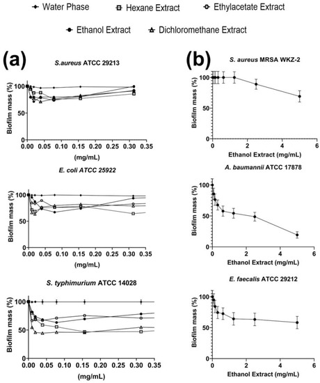
Plants are considered a wealthy resource of novel natural drugs effective in the treatment of multidrug-resistant infections. Here, a bioguided purification of Ephedra foeminea extracts was performed to identify bioactive compounds. The determination of antimicrobial properties was achieved by broth microdilution assays to evaluate minimal inhibitory concentration (MIC) values and by crystal violet staining and confocal laser scanning microscopy analyses (CLSM) to investigate the antibiofilm capacity of the isolated compounds. Assays were performed on a panel of three gram-positive and three gram-negative bacterial strains. Six compounds were isolated from E. foeminea extracts for the first time. They were identified by nuclear magnetic resonance (NMR) spectroscopy and mass spectrometry (MS) analyses as the well-known monoterpenoid phenols carvacrol and thymol and as four acylated kaempferol glycosides. Among them, the compound kaempferol-3-O-α-L-(2″,4″-di-E-p-coumaroyl)-rhamnopyranoside was found to be endowed with strong antibacterial properties and significant antibiofilm activity against S. aureus bacterial strains. Moreover, molecular docking studies on this compound suggested that the antibacterial activity of the tested ligand against S. aureus strains might be correlated to the inhibition of Sortase A and/or of tyrosyl tRNA synthase. Collectively, the results achieved open interesting perspectives to kaempferol-3-O-α-L-(2″,4″-di-E-p-coumaroyl)-rhamnopyranoside applicability in different fields, such as biomedical applications and biotechnological purposes such as food preservation and active packaging. Full article
(This article belongs to the Special Issue Advances in Antimicrobial and Macromolecules Materials)
Show Figures

Figure 1

21 pages, 5741 KiB  
Article
Blood Inflammatory-like and Lung Resident-like Eosinophils Affect Migration of Airway Smooth Muscle Cells and Their ECM-Related Proliferation in Asthma
by Airidas Rimkunas 1, Andrius Januskevicius 1, Egle Vasyle 1, Jolita Palacionyte 2, Ieva Janulaityte 3, Skaidrius Miliauskas 2 and Kestutis Malakauskas 1,2,*
1 Laboratory of Pulmonology, Department of Pulmonology, Lithuanian University of Health Sciences, LT-44307 Kaunas, Lithuania
2 Department of Pulmonology, Lithuanian University of Health Sciences, LT-44307 Kaunas, Lithuania
3 Department of Laboratory Medicine, Lithuanian University of Health Sciences, LT-44307 Kaunas, Lithuania
Int. J. Mol. Sci. 2023, 24(4), 3469; https://doi.org/10.3390/ijms24043469 - 9 Feb 2023
Cited by 2 | Viewed by 2198
Abstract
Airway remodeling is a hallmark feature of asthma, and one of its key structural changes is increased airway smooth muscle (ASM) mass and disturbed extracellular matrix (ECM) homeostasis. Eosinophil functions in asthma are broadly defined; however, we lack knowledge about eosinophil subtypes’ interaction [...] Read more.
Airway remodeling is a hallmark feature of asthma, and one of its key structural changes is increased airway smooth muscle (ASM) mass and disturbed extracellular matrix (ECM) homeostasis. Eosinophil functions in asthma are broadly defined; however, we lack knowledge about eosinophil subtypes’ interaction with lung structural cells and their effect on the airway’s local microenvironment. Therefore, we investigated the effect of blood inflammatory-like eosinophils (iEOS-like) and lung resident-like eosinophils (rEOS-like) on ASM cells via impact on their migration and ECM-related proliferation in asthma. A total of 17 non-severe steroid-free allergic asthma (AA), 15 severe eosinophilic asthma (SEA) patients, and 12 healthy control subjects (HS) were involved in this study. Peripheral blood eosinophils were enriched using Ficoll gradient centrifugation and magnetic separation, subtyped by using magnetic separation against CD62L. ASM cell proliferation was assessed by AlamarBlue assay, migration by wound healing assay, and gene expression by qRT-PCR analysis. We found that blood iEOS-like and rEOS-like cells from AA and SEA patients’ upregulated genes expression of contractile apparatus proteins, COL1A1, FN, TGF-β1 in ASM cells (p < 0.05), and SEA eosinophil subtypes demonstrated the highest effect on sm-MHC, SM22, and COL1A1 gene expression. Moreover, AA and SEA patients’ blood eosinophil subtypes promoted migration of ASM cells and their ECM-related proliferation, compared with HS (p < 0.05) with the higher effect of rEOS-like cells. In conclusion, blood eosinophil subtypes may contribute to airway remodeling by upregulating contractile apparatus and ECM component production in ASM cells, further promoting their migration and ECM-related proliferation, with a stronger effect of rEOS-like cells and in SEA. Full article
(This article belongs to the Special Issue Cell Signaling Regulation in Extracellular Matrix)
Show Figures

Figure 1

15 pages, 2739 KiB  
Article
Neutralising Effects of Different Antibodies on Clostridioides difficile Toxins TcdA and TcdB in a Translational Approach
by Georg Csukovich 1, Nina Kramer 1, Barbara Pratscher 1, Ivana Gotic 2,†, Patricia Freund 1, Rainer Hahn 3, Gottfried Himmler 2, Sabine Brandt 4 and Iwan Anton Burgener 1,*
1 Small Animal Internal Medicine, Department for Companion Animals and Horses, Vetmeduni, 1210 Vienna, Austria
2 The Antibody Lab GmbH, 1210 Vienna, Austria
3 Department for Biotechnology, University of Natural Resources and Life Sciences Vienna, 1190 Vienna, Austria
4 Research Group Oncology (RGO), Clinical Unit of Equine Surgery, Department for Companion Animals and Horses, Vetmeduni, 1210 Vienna, Austria
Current address: Alterras Therapeutics GmbH, 1030 Vienna, Austria.
Int. J. Mol. Sci. 2023, 24(4), 3867; https://doi.org/10.3390/ijms24043867 - 15 Feb 2023
Cited by 3 | Viewed by 2196
Abstract
Given the high prevalence of intestinal disease in humans and animals, there is a strong need for clinically relevant models recapitulating gastrointestinal systems, ideally replacing in vivo models in accordance with the principles of the 3R. We established a canine organoid system and [...] Read more.
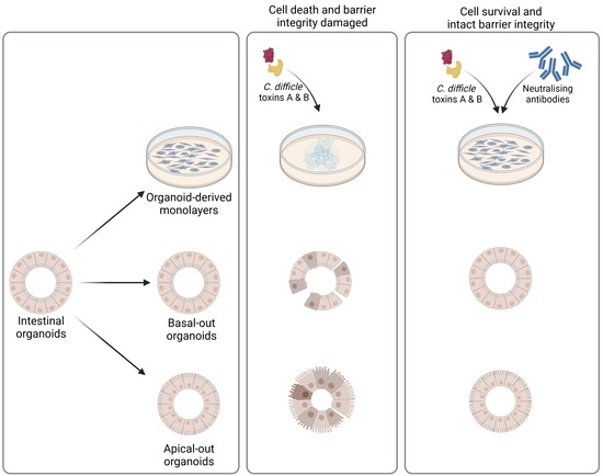
Given the high prevalence of intestinal disease in humans and animals, there is a strong need for clinically relevant models recapitulating gastrointestinal systems, ideally replacing in vivo models in accordance with the principles of the 3R. We established a canine organoid system and analysed the neutralising effects of recombinant versus natural antibodies on Clostridioides difficile toxins A and B in this in vitro system. Sulforhodamine B cytotoxicity assays in 2D and FITC-dextran barrier integrity assays on basal-out and apical-out organoids revealed that recombinant, but not natural antibodies, effectively neutralised C. difficile toxins. Our findings emphasise that canine intestinal organoids can be used to test different components and suggest that they can be further refined to also mirror complex interactions between the intestinal epithelium and other cells. Full article
(This article belongs to the Special Issue Organoids: The New 3D-Frontier to Model Different Diseases In Vitro)
Show Figures

Graphical abstract

14 pages, 1265 KiB  
Article
Melatonin Treatments Reduce Chilling Injury and Delay Ripening, Leading to Maintenance of Quality in Cherimoya Fruit
by Jorge Medina-Santamarina 1, Fabián Guillén 1, Mihaela Iasmina Madalina Ilea 1, María Celeste Ruiz-Aracil 1, Daniel Valero 1, Salvador Castillo 1 and María Serrano 2,*
1 Department of Food Technology-CIAGRO, University Miguel Hernández, Ctra. Beniel, km 3.2, 03312 Orihuela, Alicante, Spain
2 Department of Applied Biology-CIAGRO, University Miguel Hernández, Ctra. Beniel, km 3.2, 03312 Orihuela, Alicante, Spain
Int. J. Mol. Sci. 2023, 24(4), 3787; https://doi.org/10.3390/ijms24043787 - 14 Feb 2023
Cited by 8 | Viewed by 2196
Abstract
Spain is the world’s leading producer of cherimoya, a climacteric fruit highly appreciated by consumers. However, this fruit species is very sensitive to chilling injury (CI), which limits its storage. In the present experiments, the effects of melatonin applied as dipping treatment on [...] Read more.
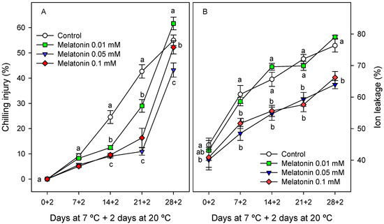
Spain is the world’s leading producer of cherimoya, a climacteric fruit highly appreciated by consumers. However, this fruit species is very sensitive to chilling injury (CI), which limits its storage. In the present experiments, the effects of melatonin applied as dipping treatment on cherimoya fruit CI, postharvest ripening and quality properties were evaluated during storage at 7 °C + 2 days at 20 °C. The results showed that melatonin treatments (0.01, 0.05, 0.1 mM) delayed CI, ion leakage, chlorophyll losses and the increases in total phenolic content and hydrophilic and lipophilic antioxidant activities in cherimoya peel for 2 weeks with respect to controls. In addition, the increases in total soluble solids and titratable acidity in flesh tissue were also delayed in melatonin-treated fruit, and there was also reduced firmness loss compared with the control, the highest effects being found for the 0.05 mM dose. This treatment led to maintenance of fruit quality traits and to increases in the storage time up to 21 days, 14 days more than the control fruit. Thus, melatonin treatment, especially at 0.05 mM concentration, could be a useful tool to decrease CI damage in cherimoya fruit, with additional effects on retarding postharvest ripening and senescence processes and on maintaining quality parameters. These effects were attributed to a delay in the climacteric ethylene production, which was delayed for 1, 2 and 3 weeks for 0.01, 0.1 and 0.05 mM doses, respectively. However, the effects of melatonin on gene expression and the activity of the enzymes involved in ethylene production deserves further research. Full article
Show Figures

Figure 1

13 pages, 1227 KiB  
Review
Zebrafish Models to Study Ectopic Calcification and Calcium-Associated Pathologies
by João M. A. Santos 1,*, Vincent Laizé 1, Paulo J. Gavaia 1,2, Natércia Conceição 1,2,3 and M. Leonor Cancela 1,2,3,*
1 Centre of Marine Sciences, University of Algarve, 8005-139 Faro, Portugal
2 Faculty of Medicine and Biomedical Sciences, University of Algarve, 8005-139 Faro, Portugal
3 Algarve Biomedical Center, University of Algarve, 8005-139 Faro, Portugal
Int. J. Mol. Sci. 2023, 24(4), 3366; https://doi.org/10.3390/ijms24043366 - 8 Feb 2023
Cited by 1 | Viewed by 2196
Abstract
Ectopic calcification refers to the pathological accumulation of calcium ions in soft tissues and is often the result of a dysregulated action or disrupted function of proteins involved in extracellular matrix mineralization. While the mouse has traditionally been the go-to model organism for [...] Read more.
Ectopic calcification refers to the pathological accumulation of calcium ions in soft tissues and is often the result of a dysregulated action or disrupted function of proteins involved in extracellular matrix mineralization. While the mouse has traditionally been the go-to model organism for the study of pathologies associated with abnormal calcium deposition, many mouse mutants often have exacerbated phenotypes and die prematurely, limiting the understanding of the disease and the development of effective therapies. Since the mechanisms underlying ectopic calcification share some analogy with those of bone formation, the zebrafish (Danio rerio)—a well-established model for studying osteogenesis and mineralogenesis—has recently gained momentum as a model to study ectopic calcification disorders. In this review, we outline the mechanisms of ectopic mineralization in zebrafish, provide insights into zebrafish mutants that share phenotypic similarities with human pathological mineralization disorders, list the compounds capable of rescuing mutant phenotypes, and describe current methods to induce and characterize ectopic calcification in zebrafish. Full article
Show Figures

Figure 1

12 pages, 2592 KiB  
Article
Impairment of Endogenous Synthesis of Omega-3 DHA Exacerbates T-Cell Inflammatory Responses
by Emanuela Talamonti 1,2, Anders Jacobsson 2 and Valerio Chiurchiù 3,4,*
1 Department of Biochemistry and Biophysics, Stockholm University, 114 Stockholm, Sweden
2 Department of Molecular Biosciences, The Wenner-Gren Institute, Stockholm University, 114 Stockholm, Sweden
3 Institute of Translational Pharmacology, National Research Council, 00133 Rome, Italy
4 Laboratory of Resolution of Neuroinflammation, IRCCS Santa Lucia Foundation, 00179 Rome, Italy
Int. J. Mol. Sci. 2023, 24(4), 3717; https://doi.org/10.3390/ijms24043717 - 13 Feb 2023
Cited by 1 | Viewed by 2192
Abstract
Omega-3 (ω-3) polyunsaturated fatty acids, including docosahexaenoic acid (DHA), are involved in numerous biological processes and have a range of health benefits. DHA is obtained through the action of elongases (ELOVLs) and desaturases, among which Elovl2 is the key enzyme involved in its [...] Read more.
Omega-3 (ω-3) polyunsaturated fatty acids, including docosahexaenoic acid (DHA), are involved in numerous biological processes and have a range of health benefits. DHA is obtained through the action of elongases (ELOVLs) and desaturases, among which Elovl2 is the key enzyme involved in its synthesis, and can be further metabolized into several mediators that regulate the resolution of inflammation. Our group has recently reported that ELOVL2 deficient mice (Elovl2−/−) not only display reduced DHA levels in several tissues, but they also have higher pro-inflammatory responses in the brain, including the activation of innate immune cells such as macrophages. However, whether impaired synthesis of DHA affects cells of adaptive immunity, i.e., T lymphocytes, is unexplored. Here we show that Elovl2−/− mice have significantly higher lymphocytes in peripheral blood and that both CD8+ and CD4+ T cell subsets produce greater amounts of pro-inflammatory cytokines in both blood and spleen compared to wild type mice, with a higher percentage of cytotoxic CD8+ T cells (CTLs) as well as IFN-γ-producing Th1 and IL-17-producing Th17 CD4+ cells. Furthermore, we also found that DHA deficiency impacts the cross-talk between dendritic cells (DC) and T cells, inasmuch as mature DCs of Elovl2−/− mice bear higher expression of activation markers (CD80, CD86 and MHC-II) and enhance the polarization of Th1 and Th17 cells. Reintroducing DHA back into the diets of Elovl2−/− mice reversed the exacerbated immune responses observed in T cells. Hence, impairment of endogenous synthesis of DHA exacerbates T cell inflammatory responses, accounting for an important role of DHA in regulating adaptive immunity and in potentially counteracting T-cell-mediated chronic inflammation or autoimmunity. Full article
(This article belongs to the Special Issue The Role of Bioactive Lipids in Health and Disease)
Show Figures

Figure 1

13 pages, 4095 KiB  
Article
Development of a CRISPRi Human Retinal Pigmented Epithelium Model for Functional Study of Age-Related Macular Degeneration Genes
by Jiang-Hui Wang 1,2,†, Daniel Urrutia-Cabrera 1,2,†, Jarmon G. Lees 3,4, Santiago Mesa Mora 1,2, Tu Nguyen 1,2, Sandy S. C. Hung 1,2, Alex W. Hewitt 1,2,5, Shiang Y. Lim 3,4, Thomas L. Edwards 1,2 and Raymond C. B. Wong 1,2,*
1 Centre for Eye Research Australia, Royal Victorian Eye and Ear Hospital, Melbourne, VIC 3002, Australia
2 Ophthalmology, Department of Surgery, University of Melbourne, Melbourne, VIC 3010, Australia
3 O’Brien Institute Department, St Vincent’s Institute of Medical Research, Fitzroy, VIC 3065, Australia
4 Departments of Surgery and Medicine, University of Melbourne, Melbourne, VIC 3010, Australia
5 Menzies Institute for Medical Research, School of Medicine, University of Tasmania, Hobart, TAS 7000, Australia
These authors contributed equally to this work.
Int. J. Mol. Sci. 2023, 24(4), 3417; https://doi.org/10.3390/ijms24043417 - 8 Feb 2023
Cited by 4 | Viewed by 2187
Abstract
Age-related macular degeneration (AMD) is a blinding disease characterised by dysfunction of the retinal pigmented epithelium (RPE) which culminates in disruption or loss of the neurosensory retina. Genome-wide association studies have identified >60 genetic risk factors for AMD; however, the expression profile and [...] Read more.
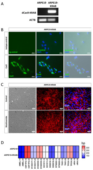
Age-related macular degeneration (AMD) is a blinding disease characterised by dysfunction of the retinal pigmented epithelium (RPE) which culminates in disruption or loss of the neurosensory retina. Genome-wide association studies have identified >60 genetic risk factors for AMD; however, the expression profile and functional role of many of these genes remain elusive in human RPE. To facilitate functional studies of AMD-associated genes, we developed a human RPE model with integrated CRISPR interference (CRISPRi) for gene repression by generating a stable ARPE19 cell line expressing dCas9-KRAB. We performed transcriptomic analysis of the human retina to prioritise AMD-associated genes and selected TMEM97 as a candidate gene for knockdown study. Using specific sgRNAs, we showed that knockdown of TMEM97 in ARPE19 reduced reactive oxygen species (ROS) levels and exerted a protective effect against oxidative stress-induced cell death. This work provides the first functional study of TMEM97 in RPE and supports a potential role of TMEM97 in AMD pathobiology. Our study highlights the potential for using CRISPRi to study AMD genetics, and the CRISPRi RPE platform generated here provided a useful in vitro tool for functional studies of AMD-associated genes. Full article
(This article belongs to the Special Issue Molecular Mechanisms of Retinal Degeneration and How to Avoid It)
Show Figures

Figure 1

12 pages, 2561 KiB  
Article
Development and Evaluation of the Magnetic Properties of a New Manganese (II) Complex: A Potential MRI Contrast Agent
by Giovanni Reale 1, Francesca Calderoni 2, Teresa Ghirardi 3, Francesca Porto 4, Federica Illuminati 5, Lorenza Marvelli 4, Petra Martini 6, Licia Uccelli 1, Eugenia Tonini 2, Lucia Del Bianco 7, Federico Spizzo 7, Martina Capozza 8, Emiliano Cazzola 9, Aldo Carnevale 1, Melchiore Giganti 1, Alessandro Turra 2, Juan Esposito 3 and Alessandra Boschi 4,*
1 Department of Translational Medicine, University of Ferrara, 44121 Ferrara, Italy
2 Medical Physics Unit, University Hospital of Ferrara, 44124 Cona, Italy
3 Legnaro National Laboratories (LNL-INFN), National Institute of Nuclear Physics, 35020 Padua, Italy
4 Department of Chemical, Pharmaceutical and Agricultural Sciences , University of Ferrara, 44121 Ferrara, Italy
5 Radiology Unit, University Hospital of Ferrara, 44124 Cona, Italy
6 Department of Environmental and Prevention Sciences, University of Ferrara, 44121 Ferrara, Italy
7 Department of Physics and Earth Science, University of Ferrara, 44122 Ferrara, Italy
8 Department of Molecular Biotechnologies and Health Sciences, University of Torino, 10126 Torino, Italy
9 IRCCS Sacro Cuore Don Calabria Hospital, Negrar di Valpolicella (VR), 37024 Negrar, Italy
Int. J. Mol. Sci. 2023, 24(4), 3461; https://doi.org/10.3390/ijms24043461 - 9 Feb 2023
Cited by 5 | Viewed by 2185
Abstract
Magnetic resonance imaging (MRI) is a non-invasive powerful modern clinical technique that is extensively used for the high-resolution imaging of soft tissues. To obtain high-definition pictures of tissues or of the whole organism this technique is enhanced by the use of contrast agents. [...] Read more.
Magnetic resonance imaging (MRI) is a non-invasive powerful modern clinical technique that is extensively used for the high-resolution imaging of soft tissues. To obtain high-definition pictures of tissues or of the whole organism this technique is enhanced by the use of contrast agents. Gadolinium-based contrast agents have an excellent safety profile. However, over the last two decades, some specific concerns have surfaced. Mn(II) has different favorable physicochemical characteristics and a good toxicity profile, which makes it a good alternative to the Gd(III)-based MRI contrast agents currently used in clinics. Mn(II)-disubstituted symmetrical complexes containing dithiocarbamates ligands were prepared under a nitrogen atmosphere. The magnetic measurements on Mn complexes were carried out with MRI phantom measurements at 1.5 T with a clinical magnetic resonance. Relaxivity values, contrast, and stability were evaluated by appropriate sequences. Studies conducted to evaluate the properties of paramagnetic imaging in water using a clinical magnetic resonance showed that the contrast, produced by the complex [Mn(II)(L’)2] × 2H2O (L’ = 1.4-dioxa-8-azaspiro[4.5]decane-8-carbodithioate), is comparable to that produced by gadolinium complexes currently used in medicine as a paramagnetic contrast agent. Full article
(This article belongs to the Special Issue Magnetic Materials and Their Various Applications)
Show Figures

Figure 1

16 pages, 323 KiB  
Review
A Comparative Review of Pregnancy and Cancer and Their Association with Endoplasmic Reticulum Aminopeptidase 1 and 2
by Brian Hur 1, Veronica Wong 2 and Eun D. Lee 1,*
1 Department of Microbiology and Immunology, School of Medicine, Virginia Commonwealth University, Richmond, VA 23298, USA
2 Department of Biology, Virginia Commonwealth University, Richmond, VA 23298, USA
Int. J. Mol. Sci. 2023, 24(4), 3454; https://doi.org/10.3390/ijms24043454 - 9 Feb 2023
Cited by 3 | Viewed by 2184
Abstract
The fundamental basis of pregnancy and cancer is to determine the fate of the survival or the death of humanity. However, the development of fetuses and tumors share many similarities and differences, making them two sides of the same coin. This review presents [...] Read more.
The fundamental basis of pregnancy and cancer is to determine the fate of the survival or the death of humanity. However, the development of fetuses and tumors share many similarities and differences, making them two sides of the same coin. This review presents an overview of the similarities and differences between pregnancy and cancer. In addition, we will also discuss the critical roles that Endoplasmic Reticulum Aminopeptidase (ERAP) 1 and 2 may play in the immune system, cell migration, and angiogenesis, all of which are essential for fetal and tumor development. Even though the comprehensive understanding of ERAP2 lags that of ERAP1 due to the lack of an animal model, recent studies have shown that both enzymes are associated with an increased risk of several diseases, including pregnancy disorder pre-eclampsia (PE), recurrent miscarriages, and cancer. The exact mechanisms in both pregnancy and cancer need to be elucidated. Therefore, a deeper understanding of ERAP’s role in diseases can make it a potential therapeutic target for pregnancy complications and cancer and offer greater insight into its impact on the immune system. Full article
(This article belongs to the Special Issue Placental Related Disorders of Pregnancy 2.0.)
18 pages, 2460 KiB  
Review
What’s on the Other Side of the Gate: A Structural Perspective on DNA Gate Opening of Type IA and IIA DNA Topoisomerases
by Vita Vidmar 1,†, Marlène Vayssières 1,† and Valérie Lamour 1,2,*
1 Institut de Génétique et de Biologie Moléculaire et Cellulaire (IGBMC), Université de Strasbourg, CNRS UMR 7104, Inserm U 1258, 67400 Illkirch, France
2 Hôpitaux Universitaires de Strasbourg, 67098 Strasbourg, France
These authors contributed equally to this work.
Int. J. Mol. Sci. 2023, 24(4), 3986; https://doi.org/10.3390/ijms24043986 - 16 Feb 2023
Cited by 6 | Viewed by 2182
Abstract
DNA topoisomerases have an essential role in resolving topological problems that arise due to the double-helical structure of DNA. They can recognise DNA topology and catalyse diverse topological reactions by cutting and re-joining DNA ends. Type IA and IIA topoisomerases, which work by [...] Read more.
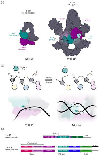
DNA topoisomerases have an essential role in resolving topological problems that arise due to the double-helical structure of DNA. They can recognise DNA topology and catalyse diverse topological reactions by cutting and re-joining DNA ends. Type IA and IIA topoisomerases, which work by strand passage mechanisms, share catalytic domains for DNA binding and cleavage. Structural information has accumulated over the past decades, shedding light on the mechanisms of DNA cleavage and re-ligation. However, the structural rearrangements required for DNA-gate opening and strand transfer remain elusive, in particular for the type IA topoisomerases. In this review, we compare the structural similarities between the type IIA and type IA topoisomerases. The conformational changes that lead to the opening of the DNA-gate and strand passage, as well as allosteric regulation, are discussed, with a focus on the remaining questions about the mechanism of type IA topoisomerases. Full article
Show Figures

Figure 1

15 pages, 1926 KiB  
Article
Steroidogenic Factor 1, a Goldilocks Transcription Factor from Adrenocortical Organogenesis to Malignancy
by Lauriane Relav 1,2, Mabrouka Doghman-Bouguerra 1,2, Carmen Ruggiero 1,2, João C. D. Muzzi 3,4,5, Bonald C. Figueiredo 5,6 and Enzo Lalli 1,2,7,*
1 Institut de Pharmacologie Moleculaire et Cellulaire CNRS UMR 7275, 06560 Valbonne, France
2 Universite Cote d’Azur, 06560 Valbonne, France
3 Laboratório de Imunoquímica (LIMQ), Pós-Graduação em Microbiologia, Parasitologia e Patologia, Departamento de Patologia Básica, Universidade Federal do Paraná (UFPR), Curitiba 81530-990, PR, Brazil
4 Laboratório de Bioinformática e Biologia de Sistemas, Pós-Graduação em Bioinformática, Universidade Federal do Paraná (UFPR), Curitiba 81520-260, PR, Brazil
5 Instituto de Pesquisa Pelé Pequeno Príncipe, Oncology Division, Curitiba 80250-060, PR, Brazil
6 Centro de Genética Molecular e Pesquisa do Câncer em Crianças (CEGEMPAC), Molecular Oncology Laboratory, Curitiba 80030-110, PR, Brazil
7 Inserm, 06560 Valbonne, France
Int. J. Mol. Sci. 2023, 24(4), 3585; https://doi.org/10.3390/ijms24043585 - 10 Feb 2023
Cited by 8 | Viewed by 2182
Abstract
Steroidogenic factor-1 (SF-1, also termed Ad4BP; NR5A1 in the official nomenclature) is a nuclear receptor transcription factor that plays a crucial role in the regulation of adrenal and gonadal development, function and maintenance. In addition to its classical role in regulating the expression [...] Read more.
Steroidogenic factor-1 (SF-1, also termed Ad4BP; NR5A1 in the official nomenclature) is a nuclear receptor transcription factor that plays a crucial role in the regulation of adrenal and gonadal development, function and maintenance. In addition to its classical role in regulating the expression of P450 steroid hydroxylases and other steroidogenic genes, involvement in other key processes such as cell survival/proliferation and cytoskeleton dynamics have also been highlighted for SF-1. SF-1 has a restricted pattern of expression, being expressed along the hypothalamic-pituitary axis and in steroidogenic organs since the time of their establishment. Reduced SF-1 expression affects proper gonadal and adrenal organogenesis and function. On the other hand, SF-1 overexpression is found in adrenocortical carcinoma and represents a prognostic marker for patients’ survival. This review is focused on the current knowledge about SF-1 and the crucial importance of its dosage for adrenal gland development and function, from its involvement in adrenal cortex formation to tumorigenesis. Overall, data converge towards SF-1 being a key player in the complex network of transcriptional regulation within the adrenal gland in a dosage-dependent manner. Full article
Show Figures

Graphical abstract

17 pages, 3102 KiB  
Article
Otoprotective Effects of Fucoidan Reduce Cisplatin-Induced Ototoxicity in Mouse Cochlear UB/OC-2 Cells
by Cheng-Yu Hsieh 1, Jia-Ni Lin 1, Ting-Ya Kang 1, Yu-Hsuan Wen 2,3,4, Szu-Hui Yu 5, Chen-Chi Wu 6,7,8 and Hung-Pin Wu 1,2,*
1 Department of Otolaryngology, Head and Neck Surgery, Taichung Tzu Chi Hospital, Buddhist Tzu Chi Medical Foundation, Taichung 427213, Taiwan
2 School of Medicine, Tzu Chi University, Hualien 970374, Taiwan
3 Department of Otolaryngology, Head and Neck Surgery, Hualien Tzu Chi Hospital, Buddhist Tzu Chi Medical Foundation, Hualien 970473, Taiwan
4 Institute of Medical Sciences, Tzu Chi University, Hualien 970473, Taiwan
5 Department of Music, Tainan University of Technology, Tainan 710302, Taiwan
6 Department of Otolaryngology, National Taiwan University Hospital, Taipei 100225, Taiwan
7 Department of Medical Genetics, National Taiwan University Hospital, Taipei 100225, Taiwan
8 Department of Medical Research, National Taiwan University Hospital Hsin-Chu Branch, Hsinchu 300195, Taiwan
Int. J. Mol. Sci. 2023, 24(4), 3561; https://doi.org/10.3390/ijms24043561 - 10 Feb 2023
Cited by 1 | Viewed by 2181
Abstract
Cisplatin is a widely used standard chemotherapy for various cancers. However, cisplatin treatment is associated with severe ototoxicity. Fucoidan is a complex sulfated polysaccharide mainly derived from brown seaweeds, and it shows multiple bioactivities such as antimicrobial, anti-inflammatory, anticancer, and antioxidant activities. Despite [...] Read more.
Cisplatin is a widely used standard chemotherapy for various cancers. However, cisplatin treatment is associated with severe ototoxicity. Fucoidan is a complex sulfated polysaccharide mainly derived from brown seaweeds, and it shows multiple bioactivities such as antimicrobial, anti-inflammatory, anticancer, and antioxidant activities. Despite evidence of the antioxidant effects of fucoidan, research on its otoprotective effects remains limited. Therefore, the present study investigated the otoprotective effects of fucoidan in vitro using the mouse cochlear cell line UB/OC-2 to develop new strategies to attenuate cisplatin-induced ototoxicity. We quantified the cell membrane potential and analyzed regulators and cascade proteins in the apoptotic pathway. Mouse cochlear UB/OC-2 cells were pre-treated with fucoidan before cisplatin exposure. The effects on cochlear hair cell viability, mitochondrial function, and apoptosis-related proteins were determined via flow cytometry, Western blot analysis, and fluorescence staining. Fucoidan treatment reduced cisplatin-induced intracellular reactive oxygen species production, stabilized mitochondrial membrane potential, inhibited mitochondrial dysfunction, and successfully protected hair cells from apoptosis. Furthermore, fucoidan exerted antioxidant effects against oxidative stress by regulating the Nrf2 pathway. Therefore, we suggest that fucoidan may represent a potential therapeutic agent for developing a new otoprotective strategy. Full article
Show Figures

Figure 1