Article

Hydrophobin Gene Cmhyd4 Negatively Regulates Fruiting Body Development in Edible Fungi Cordyceps militaris

1
State Key Laboratory of Mycology, Institute of Microbiology, Chinese Academy of Sciences, Beijing 100101, China
2
College of Horticulture, Hebei Agricultural University, Baoding 071001, China
3
University of Chinese Academy of Sciences, Beijing 101408, China
*
Author to whom correspondence should be addressed.
Int. J. Mol. Sci. 2023, 24(5), 4586; https://doi.org/10.3390/ijms24054586
Submission received: 5 January 2023 / Revised: 15 February 2023 / Accepted: 20 February 2023 / Published: 27 February 2023
(This article belongs to the Special Issue 21st Anniversary of IJMS: Advances in Molecular Genetics and Genomics)

Abstract

:
A deep understanding of the mechanism of fruiting body development is important for mushroom breeding and cultivation. Hydrophobins, small proteins exclusively secreted by fungi, have been proven to regulate the fruiting body development in many macro fungi. In this study, the hydrophobin gene Cmhyd4 was revealed to negatively regulate the fruiting body development in Cordyceps militaris, a famous edible and medicinal mushroom. Neither the overexpression nor the deletion of Cmhyd4 affected the mycelial growth rate, the hydrophobicity of the mycelia and conidia, or the conidial virulence on silkworm pupae. There was also no difference between the micromorphology of the hyphae and conidia in WT and ΔCmhyd4 strains observed by SEM. However, the ΔCmhyd4 strain showed thicker aerial mycelia in darkness and quicker growth rates under abiotic stress than the WT strain. The deletion of Cmhyd4 could promote conidia production and increase the contents of carotenoid and adenosine. The biological efficiency of the fruiting body was remarkably increased in the ΔCmhyd4 strain compared with the WT strain by improving the fruiting body density, not the height. It was indicated that Cmhyd4 played a negative role in fruiting body development. These results revealed that the diverse negative roles and regulatory effects of Cmhyd4 were totally different from those of Cmhyd1 in C. militaris and provided insights into the developmental regulatory mechanism of C. militaris and candidate genes for C. militaris strain breeding.

1. Introduction

Filamentous fungi have formed molecular adaptations to the physicochemical challenges related to their lifestyle after one billion years of evolution [1]. For instance, filamentous fungi commonly secrete small amphiphilic and highly surface-active protein hydrophobins [2,3]. They are initially secreted in a soluble form, spontaneously localize at the hydrophilic/hydrophobic interface, and then are assembled into amphipathic layers of varying solubility [4,5].
Hydrophobin layers significantly reduce the interfacial tension, decrease wetting, and provide the ability to adapt to environmental pressures, thus letting the hyphae break through the surface of the liquid and grow into the air by forming aerial hyphae, spores, and even fruiting bodies in macro fungi [6,7,8]. In Schizophyllum commune (Agaricales), the transcripts of the hydrophobins SC3 and SC4 markedly increase in vegetative hyphae, mounds, and fruiting bodies [9]. The inactivation of the hydrophobin gene Hyd9 results in a decrease in aerial mycelia and primordia formation in Flammulina filiformis (Agaricales) [7]. Moreover, hydrophobin can encourage both spores and hyphae to adhere to hydrophobic surfaces or interact with symbiotic partners [10,11,12,13] and influence the growth and development of pathogenic fungi [14,15,16]. Luciano-Rosario et al. [13] reported that during competitive pathogenicity tests on apples, the hydrophobin septuple-deletion mutant in Penicillium expansum (Eurotiales) showed a decrease in spore dispersal and was more suitable than the wild type. In the entomopathogenic fungus Beauveria bassiana (Hypocreales), hyd1 was found to positively regulate the hydrophobicity and fungal virulence, while hyd2 played a positive role in the surface hydrophobicity of conidia without affecting virulence [17]. In another entomopathogenic (as well as macro) fungus, C. militaris (Hypocreales), the hydrophobin Cmhyd1 showed increased transcription during fruiting body development compared with the mycelial stage and was proven to play a positive role in conidiation, infection, and primordium differentiation [18,19].
C. militaris, a model organism for the study of Cordyceps spp., can form fruiting bodies on both silkworm pupae and wheat media [20,21]. Its fruiting bodies have been used as food and nutrition tonics worldwide, especially in eastern Asia, due to their various effective constituents, such as carotenoids, adenosine, cordycepin, mannitol, and pentostatin [22,23]. C. militaris has become a research model for the growth and development of edible fungi, owing to its genetic tractability and controllable asexual and sexual developments. The fruiting body formation was demonstrated to be regulated by the photoreceptor genes Cmwc-1 and Cmvvd, as well as the hydrophobin gene Cmhyd1 [18,19,24,25].
Our previous study found that there existed four hydrophobin-encoding genes in C. militaris [19]. There were different transcript patterns among the four hydrophobin genes during fruiting body development, indicating their different roles [19]. Cmhyd4 was found to be only transcribed at the mycelial stage in the WT strain cultured in darkness, with very little transcription after light exposure, pupae infection, and fruiting body development [19]. In this study, the negative regulation of the fruiting body development of a hydrophobin gene was verified by reverse genetics in the edible fungus C. militaris.

2. Results

2.1. Deletion, Complementation, and Overexpression of Cmhyd4 in Cordyceps militaris

The Cmhyd4 knockout (ΔCmhyd4) strain was generated by homologous recombination (Figure 1A). Genetic complementation (ΔCmhyd4c) and overexpression (Cmhyd4oe) strains were produced by introducing Cmhyd4 with the original promoter of Cmhyd4 and a strong promoter of Cmgpda to Cmhyd4 disruption and wild-type (WT) strains, respectively (Figure 1).
Gene-disruption and genetic-complementation strains of Cmhyd4 were initially identified by PCR with the primers listed in Table S1 (Figure 1B, Table S1), then confirmed by Southern blot assay with an amplified probe, as well as RT-PCR. The WT and ΔCmhyd4 strains showed 1.61-kb and 2.67-kb hybridized bands, respectively, whereas 1.61-kb and 2.67-kb hybridized bands were presented by the ΔCmhyd4c strains (Figure 1D). The RT-PCR results revealed that the transcription of Cmhyd4 was inactive in the ΔCmhyd4 strain, the same as that in the WT strain in the two complementation strains (ΔCmhyd4c-1 and ΔCmhyd4c-2), and about 3.0–4.0-fold that in the WT strain in the two Cmhyd4oe strains (Cmhyd4oe-4 and Cmhyd4oe-5) (Figure 1C). One gene-disruption strain (ΔCmhyd4), two gene-complementation strains (ΔCmhyd4c-1 and ΔCmhyd4c-2), and two gene-overexpression strains (Cmhyd4oe-4 and Cmhyd4oe-5) of Cmhyd4 were obtained and used for the phenotype observation.

2.2. Deletion of Cmhyd4 Affects the Aerial mycelia in Darkness and the Colony Color after Light Irradiation

When the WT, ΔCmhyd4, Cmhyd4c, and Cmhyd4oe strains were cultured in a test tube with PDA in darkness, the aerial mycelia of the ΔCmhyd4 strain were the thickest among all the strains (Figure 2A). All the strains, cultured on PDA plates after light irradiation for 4 d, showed obvious differences in colony color. The colony color of the ΔCmhyd4 strain was deeper than that of the WT strain. The color of the ΔCmhyd4c culture was saffron yellow, similar to that of the WT, while the color of the Cmhyd4oe culture was almost white (Figure 2B). These observations implied that Cmhyd4 could be correlated to pigment synthesis in C. militaris.
The growth rates on PDA medium (Figure 2B,E) in the Cmhyd4 mutant strains, as well as the hydrophobicity of the aerial mycelia, stayed the same as in the WT strain (Figure 2C). There was no distinct difference between the hyphal morphology of the ΔCmhyd4 and WT strains according to scanning electron microscopy (SEM) (Figure 3A,B).
The ΔCmhyd4 strain showed a significantly increased conidial yield (p < 0.05) compared with that of the WT strain (Figure 2F). The conidial production of the two complementation strains ΔCmhyd4c-1 and ΔCmhyd4c-2 was similar to that of the WT strain (Figure 2F), while in the two overexpression strains Cmhyd4oe-4 and Cmhyd4oe-5, there was a significant decrease compared to the WT strain (Figure 2F). However, the conidia of the ΔCmhyd4 strain were well-dispersed in 0.1% Tween 80, implying that the deletion of Cmhyd4 did not affect the hydrophobicity of the conidia (Figure 2D). On the surface of the conidia, the granular attachments of the ΔCmhyd4 strain were also the same as those of the WT strain observed by SEM (Figure 3A,B).

2.3. Deletion of Cmhyd4 Affects Carotenoid and Adenosine Production in Cordyceps militaris

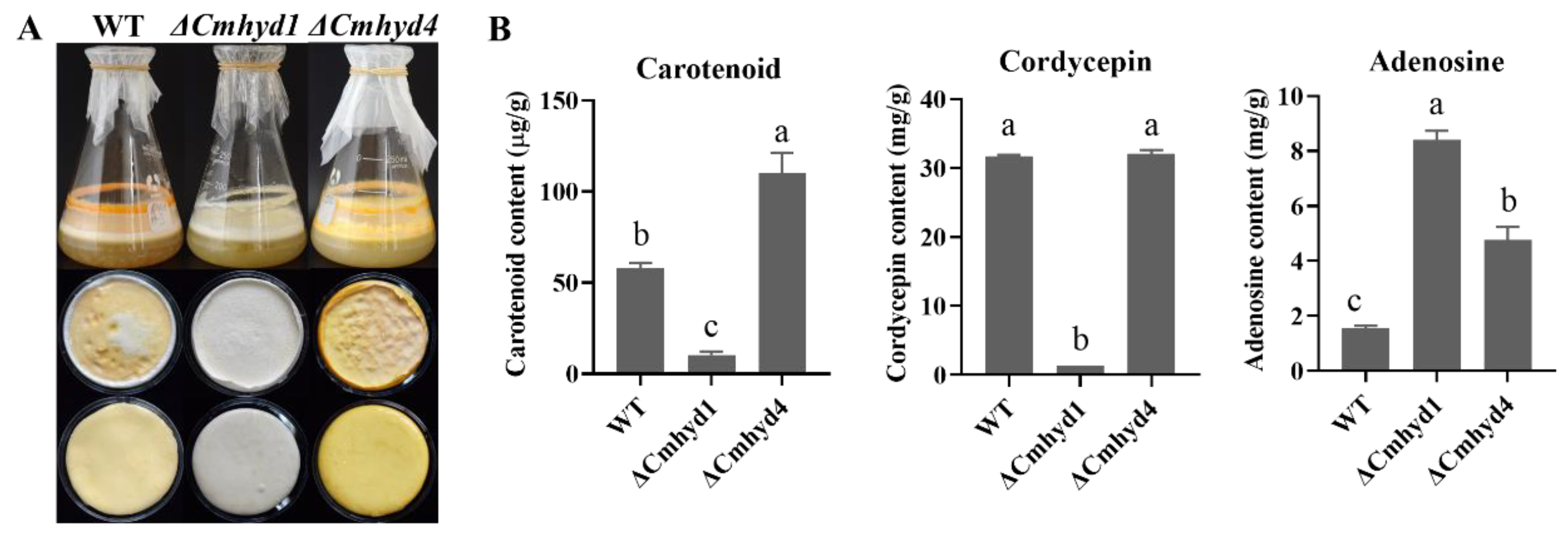
Our previous report in C. militaris found that two-stage culture could effectively increase the natural carotenoid content [26]. Therefore, we determined the secondary metabolite production of the WT, ΔCmhyd1, and ΔCmhyd4 strains by two-stage cultures. When C. militaris was cultured in potato dextrose broth (PDB) in flasks at 150 r.p.m. for 4 d and then statically incubated for another 14 d under a 12 h:12 h light/dark cycle, the vela were formed. The velum color of both the upper and reverse sides in the ΔCmhyd4 strain was the deepest among all the tested strains (Figure 3A). The ΔCmhyd1 strain appeared white for both the upper and reverse sides of the velum. The carotenoid content of the ΔCmhyd4 strain was two-fold higher than that of WT strain, and there was little carotenoid in the ΔCmhyd1 strain, which was consistent with the morphology observations (Figure 4A,B).
In C. militaris, both cordycepin and adenosine are important active compounds [27,28]. The ΔCmhyd4 strain showed a similar cordycepin content to the WT strain and a higher content than that of the ΔCmhyd1 strain, while the adenosine content increased two-fold in the absence of Cmhyd4 and eight-fold in the absence of Cmhyd1 compared with the WT strain (Figure 4B).

2.4. Deletion of Cmhyd4 Affects Stress Responses in Cordyceps militaris

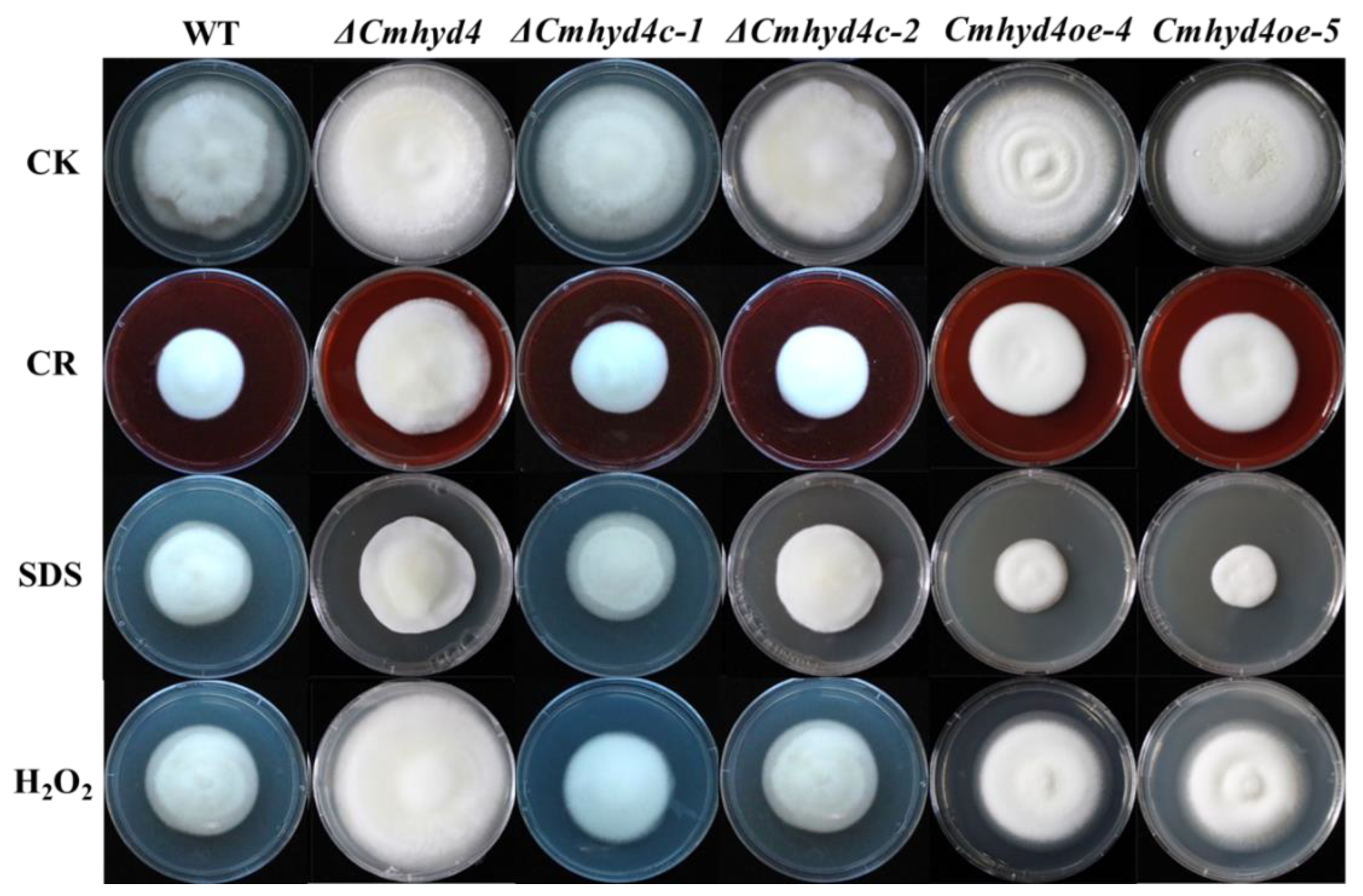
Compared with the WT and complementation strains, the mycelia of the ΔCmhyd4 strain grew more quickly in the presence of 10% CR, 0.1% SDS, and 0.075 mM H2O2, especially in the presence of 0.075 mM H2O2 (Figure 5). It was inferred that the tolerance of the mycelia to oxidant stress was affected after Cmhyd4 deletion.

2.5. Cmhyd4 Plays no Role in Cuticle-Bypassing Infection in Pupae

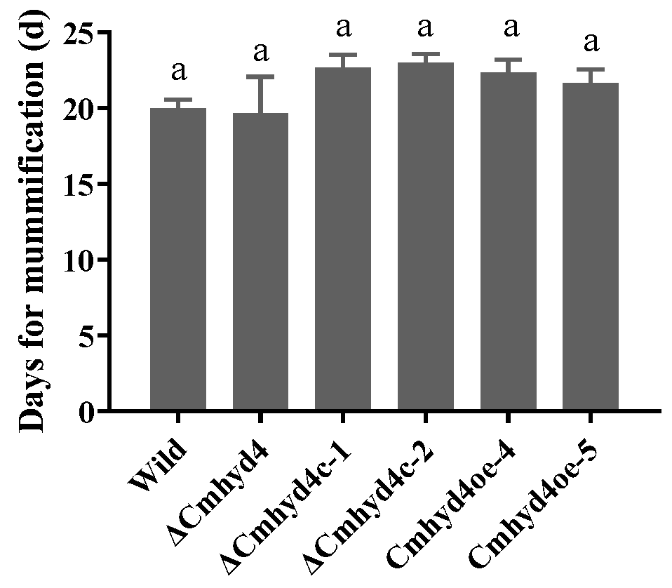
In the field or by artificial cultivation, the club-like fruiting bodies are formed after the conidia of C. militaris infecting pupae. It was found that Cmhyd1 showed a crucial role in cuticle-bypassing infection [18]. However, when the Cmhyd4 mutant strains inoculated the silkworm pupae by infection, there was no difference in the days for mummification between all the mutants and the WT strain (Figure 6), implying that Cmhyd4 played no role in cuticle-bypassing infection.

2.6. Cmhyd4 Negatively Regulates Fruiting Body Development in Cordyceps militaris

C. militaris is a well-known edible and medicinal mushroom, and the fruiting body is the main edible part. The ΔCmhyd4 and ΔCmhyd4c strains could form primordia and normal fruiting bodies, while the Cmhyd4oe strains developed malformed fruiting bodies (Figure 7A). Moreover, the biological efficiency of the ΔCmhyd4 strain was significantly increased compared to that of the WT strain. Further analysis found that the deletion of Cmhyd4 increased the biological conversion rate by improving the fruiting body density, not the height (Figure 7B).
Our previous study showed that the MAPK signaling pathway played a role in fruiting body development and formation in C. militaris [18]. The transcription levels of Cmhyd4 and seven genes in the MAPK signaling pathway, namely, the Pth11-like G-protein-coupled receptor gene (Pth11, CCM_03015); the Ras small GTPase gene (CDC42, CCM_00979); STE7 (MAPKK, CCM_03428); STE11 (MAPKKK, CCM_02296); STE20 (MAPKKKK, CCM_09268); and the mitogen-activated protein kinase gene (ERK, CCM_01235, CCM_09637) [18,29], were compared between the stages of mycelia and primordia in the WT and ΔCmhyd4 strains. At the stage of the primordium, Cmhyd4 showed a very low transcription level compared with the mycelial stage in the WT strain (Figure 7C). Compared with the mycelial stage in the WT strain, the transcription of all the tested genes in the MAPK signaling pathway except ERK1 and ERK2 experienced a significant upregulation at the primordium stage, which was consistent with our previous results [18]. In the ΔCmhyd4 strain, the transcription levels of Pth11, CDC42, STE7, STE11, and STE20 at the primordial stage exhibited a similar upregulation compared to the WT strain (Figure 7C, Supplementary Materials Figure S1).

3. Discussion

In C. militaris, four hydrophobin-encoding genes have been recovered. The class Ⅱ hydrophobin Cmhyd1, expressed highly during fruiting body formation and development compared with the mycelial stage, was confirmed to positively regulate aerial mycelial growth, conidia production, the hydrophobicity of the mycelia and conidia, pigment synthesis, pupae infection, and primordium formation [18,19]. The class Ⅰ hydrophobin Cmhyd4 only presented a high transcription level at the mycelial stage under dark condition [19], implying a different biological function from Cmhyd1 in C. militaris. In this study, it was found that Cmhyd4 negatively regulated aerial mycelial growth; conidiation; carotenoid and adenosine synthesis; resistance to oxidant stress, and fruiting body development by gene deletion, overexpression, and complementation.
Studies in pathogenic fungi have indicated that hydrophobins may be important in fungal–host interactions [17,30]. In the human pathogenic fungus Aspergillus fumigatus, hydrophobin rodA was reported to regulate the rodlet structure of conidia, play an appreciable role in insect hemocyte immune evasion, and be involved in the resistance of conidia to host cells [31]. In the plant pathogenic fungus Magnaporthe grisea (Magnaporthales), MHP1 has a key role in hydrophobicity and infection-related fungal development and is a requisite for pathogenicity [32]. In the entomogenous fungus Beauveria bassiana (Hypocreales), it has been reported that both hyd1 and hyd2 play no appreciable role in insect hemocyte immune evasion, despite their effects on conidia hydrophobicity and rodlet layers [17]. ΔCmhyd1 strains in the entomogenous fungus C. militaris exhibited lower infection rates than the WT strain over the whole silkworm pupae infection period via cuticle-bypassing infection [18]. However, there was no difference between the infection ability in pupae between the WT and ΔCmhyd4 strains in this study. Moreover, the deletion of Cmhyd4, in contrast to the deletion of Cmhyd1, had no influence on the hydrophobicity or granular attachments on the conidial surface (Figure 2C, 2D; Figure 3). Although there were no rodlets observed on the outer surface of the conidia in the WT, ΔCmhyd1, and ΔCmhyd4 strains, it is possible that the hydrophobin in C. militaris regulated the conidial virulence by affecting granular attachments on the conidial surface. Alternatively, the combined effects of conidial clumping (lowered germination) could have been due to the lower hydrophobicity and lowered hyphal growth rate in ΔCmhyd1. However, it should be noted that the silkworm pupae could not be mummified completely after being infected with the conidia of ΔCmhyd1 via cuticle-bypassing infection but were mummified completely after adding heterogenous expression to CmHYD1. Thus, the hydrophobins in C. militaris play an appreciable role in insect hemocyte immune evasion, unlike the hydrophobins in B. bassiana [17], implying the involvement of birth-and-death-type hydrophobin gene family evolution, resulting in functionally diverse proteins.
Most of the functional genes affected fruiting body development and the vegetative growth rate. For example, the hydrophobin CmHYD1 and photoreceptor CmWC-1 in C. militaris [18,24]; the hydrophobin Hyd9 [7], Gβ-like protein FvCPC2 [33], and PDD1 in F. filiformis [34]; and GlSwi6 in Ganoderma lucidum [35] affected both fruiting body development and the vegetative growth rate. Other genes have opposite roles in fruiting body development and the vegetative growth rate. The transcription factors Fst4, Hom2, Tea1, and WC-2 in S. commune repressed the vegetative growth rate but promoted fruiting body development, while Hom1 promoted the vegetative growth rate but suppressed fruiting body development [36,37,38]. As far as we know, there are few genes that specifically regulate fruiting body development, and they have no influence on the vegetative growth rate—these include fst3 and gat1 in S. commune [36,37,38] and lfc1 in F. filiformis [39]. In this study, the hydrophobin Cmhyd4 belonged to this class of functional genes.
Regarding the transcription regulation of hydrophobins, there are very few reports on the direct regulation of upstream gene transcription, and most research stops at the level of gene expression. During hydrophobin synthesis, the nutrition-linked regulatory mechanism in Ma. grisea [40,41], Metarhizium anisopliae [42], and Me. robertsii [43,44] may regulate the transcription of hydrophobin genes. A previous study in our laboratory showed that the nitrogen regulation transcription factor CmAreA was recruited to the promoter of Cmhyd1 and activated the transcription of Cmhyd1 with the coactivation of CmOTam [18]. In Trichoderma guizhouense, hydrophobin production was related to blue light receptors [45]. We confirmed that the transcription of Cmhyd1 was not regulated by light and photoreceptors, but the transcription of Cmhyd4 was regulated negatively by light and the photoreceptor Cmwc-1 [19]. We tried to verify whether CmWC-1 could directly bind the promoter of Cmhyd4 but failed. In our latest study involving ChIP-seq, a 12 bp region AAATCAGACCAC/GTGGTCTGATTT, predominated by AAATCA in positions 1–6 and CCAC in positions 9–12, was identified as a binding site for CmWC-1. There was no binding site in the promoter of Cmhyd4, and Cmhyd4 was not among the 427 target genes directly regulated by CmWC-1 [46]. Cmwc-1 was assumed to regulate the transcription of Cmhyd4 by indirect binding and, possibly, other unknown factors.
After Cmhyd4 deletion, the density and biological conversion rate of the fruiting body were remarkably increased compared with the WT strain (Figure 7). In addition, the carotenoid and adenosine contents also increased one- and two-fold compared to those of the WT strain (Figure 4). When a mushroom cultivar has these features, it presents a higher yield with more nutritional contents. Moreover, highly efficient marker-free gene editing by the effective AMA1-based CRISPR/Cas9 system in C. militaris has been realized [47]. In the future, Cmhyd4 could be useful as a target gene for C. militaris strain breeding.

4. Materials and Methods

4.1. Strains and Culture Conditions

The C. militaris strain (No. CGMCC 3.16323) was stored on potato dextrose agar (PDA) at 20 °C in darkness. The Escherichia coli strain DH5α and Agrobacterium tumefaciens strain AGL-1 (Tiangen Biotech Co., Ltd., Beijing, China) were maintained at −80 °C and cultured with Luria Bertani medium supplemented with kanamycin (50 μg/mL) and yeast extract beef (YEB) medium supplemented with carbenicillin (50 μg/mL) and kanamycin (50 μg/mL), respectively, for resistant label screening.
During phenotype observation, the C. militaris strains were cultured in constant darkness at 20 °C for 21 d and then placed under dark/light (12 h/12 h) condition at 20 °C for 4 d. After the colonies were cultured on PDA medium for 21 d, the diameters of the colony were measured to evaluate the growth rates by the cross-line method. The conidial production of each colony was calculated from six replicate plates with three counts for each strain after exposure to dark/light (12 h/12 h) condition at 20 °C for 4 d. Liquid cultures were performed in media containing 200 g/L potato, 20 g/L glucose, 3 g/L peptone, 1 g/L KH2PO4, and 0.5 g/L MgSO4 as two-stage cultures.

4.2. Gene Deletion, Complementation, and Overexpression

According to the C. militaris strain CM01 genome (accession: SRA047932), the ORF cassette of Cmhyd4 with 1381 bp upstream and 1328 bp downstream was obtained [29]. Based on vector pAg1-H3 (provided by Prof. Xingzhong Liu from Nankai University), the deletion vectors pAg1-hyg-Cmhyd4 were cloned with upstream hygromycin-resistance gene (Hyg) cassette and downstream insertion. After A. tumefaciens-mediated transformation, mutant strains were obtained and verified by polymerase chain reaction (PCR), Southern blot, and reverse transcription PCR (RT-PCR).
For gene complementation, the 1, 226 bp Cmhyd4-coding sequence containing its putative promoter and terminator regions was amplified from the WT strain and ligated into vector pAg1-bar (this lab) to generate pAg1-bar-Cmhyd4. The pAg1-bar-Cmhyd4 was introduced into the ΔCmhyd4 strain. The Cmhyd4 complementation mutants were screened with 2 mg/mL glufosinate (bar) and then verified by PCR, Southern blot, and RT-PCR.
Cmhyd4 overexpression vector pAg1-hyg-PCmgpda-Cmhyd4 was obtained with the strong promoter Cmgpda from C. militaris, the 374 bp Cmhyd4 ORF sequence, and the terminator TtrpC from A. nidulans, which were inserted into pAg1-H3. The Cmhyd4 overexpression strains were screened and verified by PCR and RT-PCR. All the vector constructions were generated with a CloneExpress® Ultra One Step Cloning Kit (C115, Vazyme Biotech Co., Ltd., Beijing, China). Table S1 lists the primer sequences used in this study.

4.3. Hydrophobicity Determination for the Mycelia and Conidia

At the surface of the mycelia, we added 20 μL of 0.1% Tween 80 and observed the diffusion capacity for 30 min to check their hydrophobicity. As for the conidial hydrophobicity, we observed the conidial suspensions containing 0.1% Tween 80 solution using a light microscope for 30 min.

4.4. Hyphae and Conidia Observation by Scanning Electron Microscopy

Under 20 °C condition, the WT and all mutant strains were grown in constant darkness for 21 d and transferred to dark/light (12/12 h) condition for 2 d. We observed the hyphae and conidia using a scanning electron microscope (SEM) (Hitachi SU8010, Tokyo, Japan). The experimental procedures were described in detail in our previous study [19].

4.5. Determination of Carotenoid, Cordycepin, and Adenosine Contents

The WT, ΔCmhyd1, and ΔCmhyd4 strains were cultured in PDB using a two-stage culture method. Firstly, we conducted the culture at 150 r.p.m. in darkness at 20 °C for 4 d. Secondly, we transferred the flasks to an illumination incubator (MGC-450BP, Yiheng Instruments, Shanghai, China) at 20 °C in light for 14 d for velum forming. We collected and dried the velums and then determined the carotenoid content using the method we published in [48]. Cordycepin and adenosine were quantified by high-performance liquid chromatography, as previously published in [49].

4.6. Assays for Stress Adaptation

We determined the effects of osmotic and oxidative stresses on growth by the method described by Li et al. [18]. After culturing in constant darkness at 20 °C for 21 d, the colony was photographed.

4.7. Assays for Fungal Virulence in Silkworm Pupae

For cuticle-bypassing infection, a 100 μL conidia suspension at 107/mL was injected into each silkworm pupa in three groups (6 pupae per group). All treated pupae were maintained at 20 °C, and we monitored the conidial infection rate daily until all the pupae were mummified completely.

4.8. Fruiting Body Formation in Cordyceps militaris

We cultivated the strains for fruiting in wheat medium according to our previously described method [19]. The fresh weights of the fruiting bodies were recorded after harvest. The formula for calculating the biological efficiency (BE) of each strain was BE (%) = (weight of fresh stroma/weight of dry substrate) × 100. The biological efficiency, number, and height of the fruiting bodies in each vessel were measured among the three groups (5 vessels per group). These fruiting experiments were conducted three times independently.

4.9. RNA Extraction and RT-qPCR Analysis

An E.Z.N.A.™ Plant RNA Kit (Omega, Stamford, CT, USA) was used to isolate the total RNA from frozen mycelia or fruiting bodies. A HiScript III 1st Strand cDNA Synthesis Kit (+gDNA wiper) (Vazyme Biotech Co., Ltd., Beijing, China) was used to synthesize the first-strand cDNA from the total RNA. RT-qPCR was conducted using a CFX Connect Real-Time System (Bio-Rad, Singapore) with 10 µL qPCR solutions containing 5 μL AceQ® qPCR SYBR® Green Master Mix, 2 ng cDNA, and 0.1 μM primers. The rpb1 (CCM_05485) was used as an internal standard [50]. The 2−ΔΔCt method was used to calculate the relative gene expression levels [51]. Table S1 lists all the primer sequences used in this study. The data provided represent three biological replicates and three technical replicates.

5. Conclusions

This study revealed negative roles for the novel putative hydrophobin gene Cmhyd4 in aerial mycelia, mycelia stress resistance, conidiation, secondary metabolites, and primordium formation in C. militaris. Furthermore, Cmhyd4 had no effects on the mycelial growth rate, hydrophobicity, microphenotype of hyphae and conidia or conidial virulence in silkworm pupae. Since the deletion of Cmhyd4 could promote fruiting body development and increase yields, CmHYD4 and its homologous encoding genes could be useful in C. militaris breeding.

Supplementary Materials

The following supporting information can be downloaded at: https://www.mdpi.com/article/10.3390/ijms24054586/s1.

Author Contributions

X.L.: conceptualization, visualization, methodology, investigation, writing (original draft), writing (review and editing), project administration, funding acquisition. M.L.: investigation, methodology, writing (review and editing). C.D.: methodology, supervision, project administration, funding acquisition, conceptualization, writing (review and editing). All authors have read and agreed to the published version of the manuscript.

Funding

This research was funded by the National Natural Science Foundation of China (grant numbers 31872163 and 32202569).

Institutional Review Board Statement

Not applicable.

Informed Consent Statement

Not applicable.

Data Availability Statement

Data is contained within the article or supplementary material.

Acknowledgments

The authors are grateful to Xingzhong Liu from Nankai University and Linqi Wang from the Institute of Microbiology, Chinese Academy of Sciences. We are also grateful to Huishan Guo from the Institute of Microbiology, Chinese Academy of Sciences for the support with the heterogenous expression system and Southern blot.

Conflicts of Interest

The authors declare no conflict of interest.

References

  1. Taylor, J.W.; Berbee, M.L. Dating divergences in the Fungal Tree of Life: Review and new analyses. Mycologia 2006, 98, 838–849. [Google Scholar] [CrossRef] [PubMed]
  2. Elliot, M.A.; Talbot, N.J. Building filaments in the air: Aerial morphogenesis in bacteria and fungi. Curr. Opin. Microbiol. 2004, 7, 594–601. [Google Scholar] [CrossRef] [PubMed]
  3. Linder, M.B.; Szilvay, G.R.; Nakari-Setälä, T.; Penttilä, M.E. Hydrophobins: The protein-amphiphiles of filamentous fungi. FEMS Microbiol. Rev. 2005, 29, 877–896. [Google Scholar] [CrossRef] [PubMed] [Green Version]
  4. Wösten, H.A. Hydrophobins: Multipurpose proteins. Annu. Rev. Microbiol. 2001, 55, 625–646. [Google Scholar] [CrossRef] [Green Version]
  5. Gebbink, M.F.; Claessen, D.; Bouma, B.; Dijkhuizen, L.; Wösten, H.A. Amyloids—A functional coat for microor-ganisms. Nat. Rev. Microbiol. 2005, 3, 333–341. [Google Scholar] [CrossRef] [Green Version]
  6. Dubey, M.K.; Jensen, D.F.; Karlsson, M. Hydrophobins are required for conidial hydrophobicity and plant root colonization in the fungal biocontrol agent Clonostachys rosea. BMC Microbiol. 2014, 14, 18. [Google Scholar] [CrossRef] [Green Version]
  7. Tao, Y.X.; Chen, R.L.; Yan, J.J.; Long, Y.; Tong, Z.J.; Song, H.B.; Xie, B.G. A hydrophobin gene, Hyd9, plays an important role in the formation of aerial hyphae and primordia in Flammulina filiformis. Gene 2019, 706, 84–90. [Google Scholar] [CrossRef]
  8. Xu, D.; Wang, Y.; Keerio, A.A.; Ma, A. Identification of hydrophobin genes and their physiological functions related to growth and development in Pleurotus ostreatus. Microbiol. Res. 2021, 247, 126723. [Google Scholar] [CrossRef]
  9. Banerjee, G.; Robertson, D.L.; Leonard, T.J. Hydrophobins Sc3 and Sc4 gene expression in mounds, fruiting bodies and vegetative hyphae of Schizophyllum commune. Fungal. Genet. Biol. 2008, 45, 171–179. [Google Scholar] [CrossRef]
  10. Aimanianda, V.; Bayry, J.; Bozza, S.; Kniemeyer, O.; Perruccio, K.; Elluru, S.R.; Clavaud, C.; Paris, S.; Brakhage, A.A.; Kaveri, S.V.; et al. Surface hydrophobin prevents immune recognition of airborne fungal spores. Nature 2009, 460, 1117–1121. [Google Scholar] [CrossRef]
  11. Plett, J.M.; Gibon, J.; Kohler, A.; Duffy, K.; Hoegger, P.J.; Velagapudi, R.; Han, J.; Kües, U.; Grigoriev, I.V.; Martin, F. Phylogenetic, genomic organization and expression analysis of hydrophobin genes in the ectomycorrhizal basidiomycete Laccaria bicolor. Fungal. Genet. Biol. 2012, 49, 199–209. [Google Scholar] [CrossRef] [PubMed] [Green Version]
  12. Vila, T.; Nazir, R.; Rozental, S.; Dos Santos, G.M.; Calixto, R.O.; Barreto-Bergter, E.; Wick, L.Y.; van Elsas, J.D. The Role of hydrophobicity and surface receptors at hyphae of Lyophyllum sp. strain karsten in the interaction with Burkholderia terrae BS001-implications for interactions in soil. Front. Microbiol. 2016, 7, 1689. [Google Scholar] [CrossRef] [PubMed] [Green Version]
  13. Luciano-Rosario, D.; Eagan, J.L.; Aryal, N.; Dominguez, E.G.; Hull, C.M.; Keller, N.P. The Hydrophobin gene family confers a fitness trade-off between spore dispersal and host colonization in Penicillium expansum. mBio 2022, 13, e0275422. [Google Scholar] [CrossRef] [PubMed]
  14. Askolin, S.; Penttila, M.; Wosten, H.A.; Nakari-Setala, T. The Trichoderma reesei hydrophobin genes hfb1 and hfb2 have diverse functions in fungal development. FEMS Microbiol. Lett. 2005, 253, 281–288. [Google Scholar] [CrossRef] [PubMed] [Green Version]
  15. Grünbacher, A.; Throm, T.; Seidel, C.; Gutt, B.; Röhrig, J.; Strunk, T.; Vincze, P.; Walheim, S.; Schimmel, T.; Wenzel, W.; et al. Six hydrophobins are involved in hydrophobin rodlet formation in Aspergillus nidulans and contribute to hydrophobicity of the spore surface. PLoS ONE 2014, 9, e94546. [Google Scholar] [CrossRef] [Green Version]
  16. Cai, F.; Zhao, Z.; Gao, R.; Chen, P.; Ding, M.; Jiang, S.; Fu, Z.; Xu, P.; Chenthamara, K.; Shen, Q.; et al. The pleiotropic functions of intracellular hydrophobins in aerial hyphae and fungal spores. PLoS Genet. 2021, 17, e1009924. [Google Scholar] [CrossRef]
  17. Zhang, S.; Xia, Y.X.; Kim, B.; Keyhani, N.O. Two hydrophobins are involved in fungal spore coat rodlet layer assembly and each play distinct roles in surface interactions, development and pathogenesis in the entomopathogenic fungus, Beauveria bassiana. Mol. Microbiol. 2011, 80, 811–826. [Google Scholar] [CrossRef]
  18. Li, X.; Wang, F.; Liu, M.Q.; Dong, C.H. Hydrophobin CmHYD1 is involved in conidiation, infection and primordium formation, and regulated by nitrogen regulatory transcription factor CmAreA in edible fungus, Cordyceps militaris. J. Fungi. 2021, 7, 674. [Google Scholar] [CrossRef]
  19. Li, X.; Wang, F.; Xu, Y.Y.; Dong, C.H. Cysteine-rich hydrophobin gene family: Genome wide analysis, phylogeny and putative function in Cordyceps militaris. Int. J. Mol. Sci. 2020, 22, 643. [Google Scholar] [CrossRef]
  20. Shrestha, B.; Zhang, W.; Zhang, Y.; Liu, X. The medicinal fungus Cordyceps militaris: Research and development. Mycol. Prog. 2012, 11, 599–614. [Google Scholar] [CrossRef]
  21. Guo, M.M.; Guo, S.P.; Yan, H.J.; Bu, N.; Dong, C.H. Comparison of major bioactive compounds of the caterpillar medicinal mushroom, Cordyceps militaris (Ascomycetes), fruiting bodies cultured on wheat substrate and pupae. Int. J. Med. Mushrooms 2016, 18, 327–336. [Google Scholar] [CrossRef]
  22. Xia, Y.; Luo, F.; Shang, Y.; Chen, P.; Lu, Y.; Wang, C. Fungal cordycepin biosynthesis is coupled with the production of the safeguard molecule pentostatin. Cell Chem. Biol. 2017, 24, 1479–1489.e4. [Google Scholar] [CrossRef] [Green Version]
  23. Dong, J.Z.; Wang, S.H.; Ai, X.R.; Yao, L.; Sun, Z.W.; Lei, C.; Wang, Y.; Wang, Q. Composition and characterization of cordyxanthins from Cordyceps militaris fruit bodies. J. Funct. Foods. 2013, 5, 1450–1455. [Google Scholar] [CrossRef]
  24. Yang, T.; Guo, M.M.; Yang, H.; Guo, S.; Dong, C.H. The blue-light receptor CmWC-1 mediates fruit body development and secondary metabolism in Cordyceps militaris. Appl. Microbiol. Biotechnol. 2016, 100, 743–755. [Google Scholar] [CrossRef]
  25. Zhang, J.J.; Wang, F.; Yang, Y.; Wang, Y.; Dong, C.H. CmVVD is involved in fruiting body development and carotenoid production and the transcriptional linkage among three blue-light receptors in edible fungus Cordyceps militaris. Environ. Microbiol. 2020, 22, 466–482. [Google Scholar] [CrossRef]
  26. Yang, Y.; Bu, N.; Wang, S.; Zhang, J.; Wang, Y.; Dong, C.H. Carotenoid production by caterpillar medicinal mushrooms, Cordyceps militaris (Ascomycetes), under different culture conditions. Int. J. Med. Mushrooms 2020, 22, 1191–1201. [Google Scholar] [CrossRef]
  27. Jin, Y.; Meng, X.; Qiu, Z.; Su, Y.; Yu, P.; Qu, P. Anti-tumor and anti-metastatic roles of cordycepin, one bioactive compound of Cordyceps militaris. Saudi. J. Biol. Sci. 2018, 25, 991–995. [Google Scholar] [CrossRef]
  28. Dong, J.Z.; Liu, M.R.; Lei, C.; Zheng, X.J.; Wang, Y. Effects of selenium and light wavelengths on liquid culture of Cordyceps militaris Link. Appl. Biochem. Biotech. 2012, 166, 2030–2036. [Google Scholar] [CrossRef] [PubMed]
  29. Zheng, P.; Xia, Y.L.; Xiao, G.H.; Xiong, C.H.; Hu, X.; Zhang, S.W.; Zheng, H.J.; Huang, Y.; Zhou, Y.; Wang, S.Y.; et al. Genome sequence of the insect pathogenic fungus Cordyceps militaris, a valued traditional Chinese medicine. Genome Biol. 2011, 12, R116. [Google Scholar] [CrossRef] [PubMed] [Green Version]
  30. Xiao, G.H.; Ying, S.H.; Zheng, P.; Wang, Z.L.; Zhang, S.W.; Xie, X.Q.; Shang, Y.F.; St Leger, R.J.; Zhao, G.P.; Wang, C.S.; et al. Genomic perspectives on the evolution of fungal entomopathogenicity in Beauveria bassiana. Sci. Rep. 2012, 2, 483. [Google Scholar] [CrossRef] [PubMed] [Green Version]
  31. Paris, S.; Debeaupuis, J.P.; Crameri, R.; Carey, M.; Charlès, F.; Prévost, M.C.; Schmitt, C.; Philippe, B.; Latgé, J.P. Conidial hydrophobins of Aspergillus fumigatus. Appl. Environ. Microb. 2003, 69, 1581–1588. [Google Scholar] [CrossRef] [PubMed] [Green Version]
  32. Kim, S.; Ahn, I.P.; Rho, H.S.; Lee, Y.H. MHP1, a Magnaporthe grisea hydrophobin gene, is required for fungal development and plant colonization. Mol. Microbiol. 2005, 57, 1224–1237. [Google Scholar] [CrossRef] [PubMed]
  33. Wu, T.; Zhang, Z.; Hu, C.; Zhang, L.; Wei, S.; Li, S. A WD40 protein encoding gene Fvcpc2 positively regulates mushroom development and yield in Flammulina velutipes. Front. Microbiol. 2020, 11, 498. [Google Scholar] [CrossRef] [PubMed]
  34. Wu, T.; Hu, C.; Xie, B.; Zhang, L.; Yan, S.; Wang, W.; Tao, Y.; Li, S. A single transcription factor (PDD1) determines development and yield of winter mushroom (Flammulina velutipes). Appl. Environ. Microb. 2019, 85, e01735-19. [Google Scholar] [CrossRef] [PubMed]
  35. Zhang, G.; Ren, A.; Shi, L.; Zhu, J.; Jiang, A.; Shi, D.; Zhao, M. Functional analysis of an APSES transcription factor (GlSwi6) involved in fungal growth, fruiting body development and ganoderic-acid biosynthesis in Ganoderma lucidum. Microbiol. Res. 2018, 207, 280–288. [Google Scholar] [CrossRef]
  36. Ohm, R.A.; de Jong, J.F.; Lugones, L.G.; Aerts, A.; Kothe, E.; Stajich, J.E.; de Vries, R.P.; Record, E.; Levasseur, A.; Baker, S.E.; et al. Genome sequence of the model mushroom Schizophyllum commune. Nat. Biotechnol. 2010, 28, 957–963. [Google Scholar] [CrossRef] [Green Version]
  37. Ohm, R.A.; de Jong, J.F.; de Bekker, C.; Wösten, H.A.; Lugones, L.G. Transcription factor genes of Schizophyllum commune involved in regulation of mushroom formation. Mol. Microbiol. 2011, 81, 1433–1445. [Google Scholar] [CrossRef]
  38. Pelkmans, J.F.; Patil, M.B.; Gehrmann, T.; Reinders, M.J.; Wösten, H.A.; Lugones, L.G. Transcription factors of Schizophyllum commune involved in mushroom formation and modulation of vegetative growth. Sci. Rep. 2017, 7, 310. [Google Scholar] [CrossRef] [Green Version]
  39. Wu, T.; Hu, C.; Xie, B.; Wei, S.; Zhang, L.; Zhu, Z.; Zhang, Z.; Li, S. A putative transcription factor LFC1 negatively regulates development and yield of winter mushroom. Appl. Microbiol. Biot. 2020, 104, 5827–5844. [Google Scholar] [CrossRef]
  40. Lau, G.; Hamer, J.E. Regulatory genes controlling MPG1 expression and pathogenicity in the rice blast fungus Magnaporthe grisea. Plant Cell. 1996, 8, 771–781. [Google Scholar] [CrossRef] [Green Version]
  41. Soanes, D.M.; Kershaw, M.J.; Cooley, R.N.; Talbot, N.J. Regulation of the MPG1 hydrophobin gene in the rice blast fungus Magnaporthe grisea. Mol. Plant-Microbe. Interact. 2002, 15, 1253–1267. [Google Scholar] [CrossRef] [PubMed] [Green Version]
  42. Fang, W.G.; Pei, Y.; Bidochka, M.J. A regulator of a G protein signalling (RGS) gene, cag8, from the insect-pathogenic fungus Metarhizium anisopliae is involved in conidiation, virulence and hydrophobin synthesis. Microbiology 2007, 153, 1017–1025. [Google Scholar] [CrossRef] [PubMed] [Green Version]
  43. Ries, L.N.; Beattie, S.R.; Espeso, E.A.; Cramer, R.A.; Goldman, G.H. Diverse regulation of the CreA carbon catabolite repressor in Aspergillus nidulans. Genetics 2016, 203, 335–352. [Google Scholar] [CrossRef] [PubMed] [Green Version]
  44. Lai, Y.L.; Cao, X.; Chen, J.J.; Wang, L.L.; Wei, G.; Wang, S.B. Coordinated regulation of infection-related morphogenesis by the KMT2-Cre1-Hyd4 regulatory pathway to facilitate fungal infection. Sci. Adv. 2020, 6, eaaz1659. [Google Scholar] [CrossRef] [PubMed] [Green Version]
  45. Li, Y.; Sun, T.; Guo, D.; Gao, J.; Zhang, J.; Cai, F.; Fischer, R.; Shen, Q.; Yu, Z. Comprehensive analysis of the regulatory network of blue-light-regulated conidiation and hydrophobin production in Trichoderma guizhouense. Environ. Microbiol. 2021, 23, 6241–6256. [Google Scholar] [CrossRef] [PubMed]
  46. Zhang, J.J.; Wang, F.; Liu, M.Q.; Fu, M.; Dong, C.H. Dynamic genome-wide transcription profiling and direct target genes of CmWC-1 reveal hierarchical light signal transduction in Cordyceps militaris. J. Fungi. 2022, 8, 624. [Google Scholar] [CrossRef] [PubMed]
  47. Meng, G.L.; Wang, X.; Liu, M.Q.; Wang, F.; Liu, Q.; Dong, C.H. Efficient CRISPR/Cas9 system based on autonomously replicating plasmid with an AMA1 sequence and precisely targeted gene deletion in the edible fungus, Cordyceps militaris. Microbial. Biotechnol. 2022, 15, 2594–2606. [Google Scholar] [CrossRef]
  48. Yang, T.; Dong, C.H. Photo morphogenesis and photo response of the blue-light receptor gene Cmwc-1 in different strains of Cordyceps militaris. FEMS Microbiol. Lett. 2014, 352, 190–197. [Google Scholar] [CrossRef] [Green Version]
  49. Dong, C.H.; Yao, Y.J. Comparison of some metabolites among cultured mycelia of Ophiocordyceps sinensis from different geographical regions. Int. J. Med. Mushrooms 2010, 12, 287–297. [Google Scholar] [CrossRef]
  50. Lian, T.T.; Yang, T.; Liu, G.J.; Sun, J.D.; Dong, C.H. Reliable reference gene selection for Cordyceps militaris gene expression studies under different developmental stages and media. FEMS Microbiol. Lett. 2014, 356, 97–104. [Google Scholar] [CrossRef] [Green Version]
  51. Livak, K.J.; Schmittgen, T.D. Analysis of relative gene expression data using real-time quantitative PCR and the 2−ΔΔCT method. Methods 2001, 25, 402–408. [Google Scholar] [CrossRef] [PubMed]
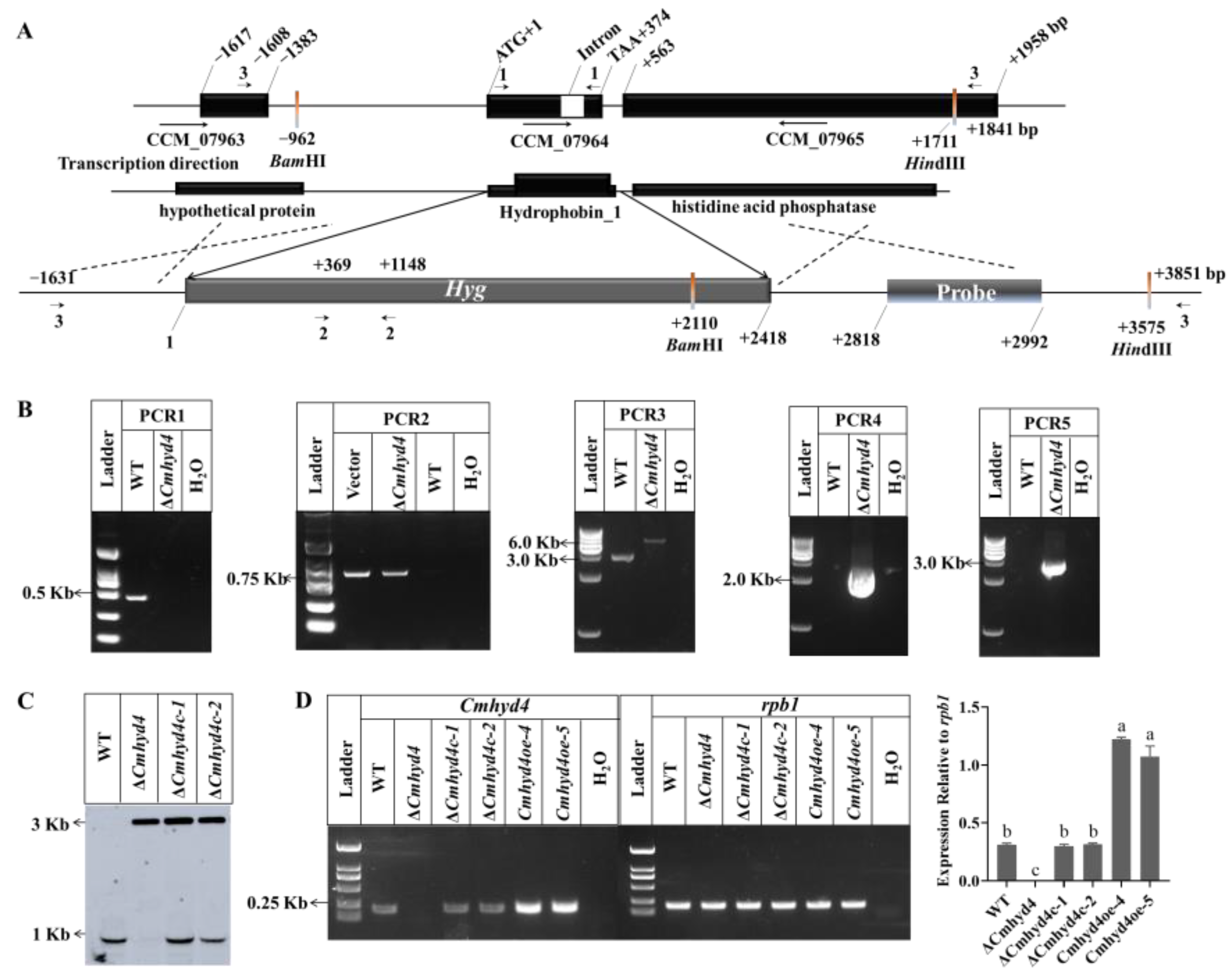
Figure 1. Generation and identification of the Cmhyd4 mutants. (A) Scheme for the ΔCmhyd4 strain obtained via homologous recombination; Cmhyd4 was replaced by the hygromycin resistance gene. (B) Confirmation of Cmhyd4 deletion by PCR. The primers 1, 2, and 3 were used to perform PCR1, PCR2, and PCR3, respectively; PCR4 and PCR5 were performed with the primers 3F/2R and 2F/3R, respectively. (C) Southern blot assay with double-digest sites BamHI and HindIII validating the Cmhyd4 gene deletion and complementation strains. Lane 1: WT; lane 2: ΔCmhyd4 strain; lanes 3 and 4: ΔCmhyd4c-1 and ΔCmhyd4c-2 strains, respectively. (D) The transcription levels of Cmhyd4 in the WT and Cmhyd4 mutants according to RT-PCR. The transcription levels of Cmhyd4 relative to rpb1 are presented as the optical density ratio obtained using ImageJ v.1, and the different letters over the histogram indicate significant differences at p < 0.05.
Ijms 24 04586 g001
Figure 2. Morphology, growth rate, hydrophobicity, and conidiation in the WT and mutant strains. (A) Aerial mycelia: Aerial mycelia were cultured in a test tube with PDA medium at 20 °C in darkness. We observed them after culturing for 7 d. (B) Colony morphology: The colonies were cultured on PDA medium in darkness for 21 d at 20 °C. We observed them after exposure to light/dark (12/12 h) condition for 4 d. (C) The hydrophobicity of mycelia. (D) The hydrophobicity of conidia. (E) Growth rates. (F) Conidia production. Error bars represent the standard deviation (SD) of three replicates. Different letters above the bars represent significant differences (p < 0.05).
Ijms 24 04586 g002
Figure 3. Hyphae and conidia of WT (A) and ΔCmhyd4 (B) strains observed by SEM. A1 and B1 showed the overview of the hyphae and conidia; A2 and B2 showed the surface of the hyphae; A3 and B3 showed the surface of the conidia. Bars: A1, B1 = 1 μm; others = 10 μm.
Ijms 24 04586 g003
Figure 4. The morphology and secondary metabolite production of WT, ΔCmhyd1 and ΔCmhyd4 strains by two-stage cultivation. (A) Fermentation broth, upper and reverse sides of the vela from the WT, ΔCmhyd1 and ΔCmhyd4 strains after two-stage cultivation for 18 d; (B) Contents of carotenoid in the vela, cordycepin, and adenosine in fermentation broth for the WT, ΔCmhyd1, and ΔCmhyd4 strains. Error bars represent the SD of three biological replicates with three technical replicates. Different letters above the bars represent significant differences (p < 0.05).
Ijms 24 04586 g004
Figure 5. The stress response of mycelia in the WT and mutant strains. As the control, CK was inoculated on PDA medium. CR, SDS, and H2O2 represent the PDA with 10% congo red, 0.1% SDS, and 0.075 mM H2O2. All the strains were cultured at 20 °C for 21 d in darkness and then photographed. The experiments were repeated three times with similar results.
Ijms 24 04586 g005
Figure 6. Conidial infecting ability of WT and mutant strains. Error bars represent the SD of three biological replicates with three technical replicates. The same “a” above the bars indicates that there is no significant difference (p < 0.05).
Ijms 24 04586 g006
Figure 7. Cmhyd4 negatively regulated fruiting body development in Cordyceps militaris. (A) Fruiting body formation and development in WT and mutants—PR: primordium formation (stroma < 1 cm); MF: mature fruiting body. (B) Mushroom yield of WT and mutants. After inoculation for 45 d, fruiting bodies were harvested and evaluated. (C) The relative transcription levels of Cmhyd4 and fruiting-body-development-related genes at the primordial stage. Error bars represent the SD of three biological replicates with three technical replicates. The different letters over the histogram represent significant differences at p < 0.05.
Ijms 24 04586 g007
Disclaimer/Publisher’s Note: The statements, opinions and data contained in all publications are solely those of the individual author(s) and contributor(s) and not of MDPI and/or the editor(s). MDPI and/or the editor(s) disclaim responsibility for any injury to people or property resulting from any ideas, methods, instructions or products referred to in the content.