


Comparison of SARS-CoV-2 Detection in Nasopharyngeal Swab and Saliva Samples from Patients Infected with Omicron Variant
 ,
,  ,
,  , ,
, ,  , , and
Abstract
:1. Introduction
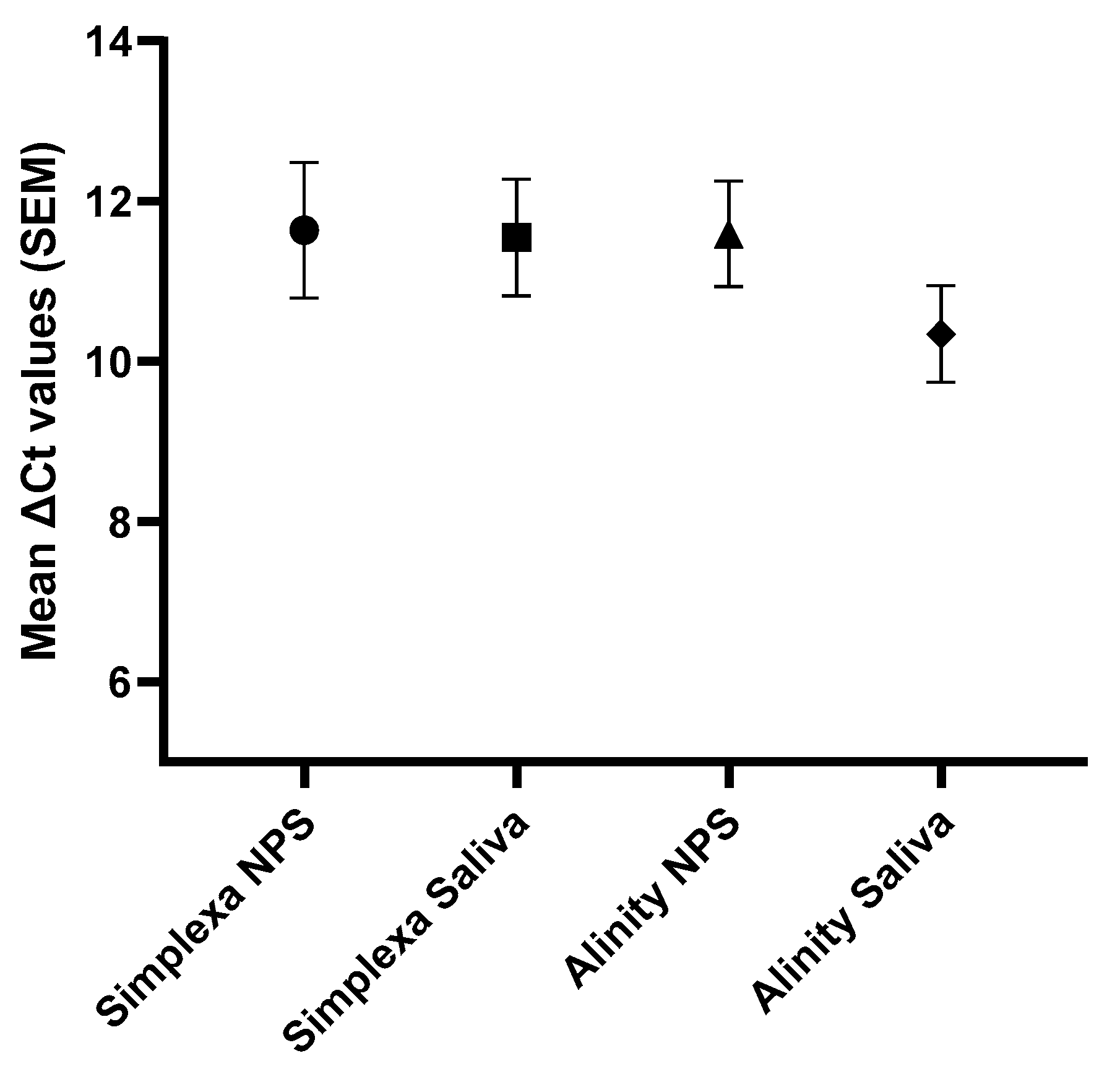
2. Results
3. Discussion
4. Materials and Methods
4.1. Study Population and Sample Collection
4.2. SARS-CoV-2 Real-Time PCR and Sequence Analysis
4.3. Statistical Analysis
Author Contributions
Funding
Institutional Review Board Statement
Informed Consent Statement
Data Availability Statement
Conflicts of Interest
References
- World Health Organization. WHO Coronavirus (COVID-19) Dashboard. Available online: https://covid19.who.int/ (accessed on 3 October 2022).
- Tegally, H.; Wilkinson, E.; Althaus, C.L.; Giovanetti, M.; San, J.E.; Giandhari, J.; Pillay, S.; Naidoo, Y.; Ramphal, U.; Msomi, N.; et al. Rapid replacement of the Beta variant by the Delta variant in South Africa. medRxiv 2021, medRxiv:2021.09.23.21264018. [Google Scholar] [CrossRef]
- Dhar, M.S.; Marwal, R.; Vs, R.; Ponnusamy, K.; Jolly, B.; Bhoyar, R.C.; Sardana, V.; Naushin, S.; Rophina, M.; Mellan, T.A.; et al. Genomic characterization and epidemiology of an emerging SARS-CoV-2 variant in Delhi, India. Science 2021, 374, 995–999. [Google Scholar] [CrossRef]
- Viana, R.; Moyo, S.; Amoako, D.G.; Tegally, H.; Scheepers, C.; Althaus, C.L.; Anyaneji, U.J.; Bester, P.A.; Boni, M.F.; Chand, M.; et al. Rapid epidemic expansion of the SARS-CoV-2 Omicron variant in southern Africa. Nature 2022, 603, 679–686. [Google Scholar] [CrossRef] [PubMed]
- National Institute for Communicable Diseases. Available online: https://www.nicd.ac.za/wp-content/uploads/2021/12/COVID-19-Effective-Reproductive-Number-in-South-Africa-week-51.pdf (accessed on 15 October 2022).
- Pulliam, J.R.C.; van Schalkwyk, C.; Govender, N.; von Gottberg, A.; Cohen, C.; Groome, M.J.; Dushoff, J.; Mlisana, K.; Moultrie, H. Increased risk of SARS-CoV-2 reinfection associated with emergence of Omicron in South Africa. Science 2022, 376, eabn4947. [Google Scholar] [CrossRef]
- Bordi, L.; Sberna, G.; Lalle, E.; Piselli, P.; Colavita, F.; Nicastri, E.; Antinori, A.; Boumis, E.; Petrosillo, N.; Marchioni, L.; et al. Frequency and Duration of SARS-CoV-2 Shedding in Oral Fluid Samples Assessed by a Modified Commercial Rapid Molecular Assay. Viruses 2020, 12, 1184. [Google Scholar] [CrossRef] [PubMed]
- Butler-Laporte, G.; Lawandi, A.; Schiller, I.; Yao, M.; Dendukuri, N.; McDonald, E.G.; Lee, T.C. Comparison of Saliva and Nasopharyngeal Swab Nucleic Acid Amplification Testing for Detection of SARS-CoV-2: A Systematic Review and Meta-analysis. JAMA Intern. Med. 2021, 181, 353–360. [Google Scholar] [CrossRef] [PubMed]
- Caixeta, D.C.; Oliveira, S.W.; Cardoso-Sousa, L.; Cunha, T.M.; Goulart, L.R.; Martins, M.M.; Marin, L.M.; Jardim, A.C.G.; Siqueira, W.L.; Sabino-Silva, R. One-Year Update on Salivary Diagnostic of COVID-19. Front. Public Health 2021, 9, 589564. [Google Scholar] [CrossRef]
- Chu, C.Y.; Marais, G.; Opperman, C.; Doolabh, D.; Iranzadeh, A.; Marais, C.; Cox, H.; Williamson, C.; Hardie, D.; Brink, A. Performance of saliva and mid-turbinate swabs for detection of the beta variant in South Africa. Lancet Infect. Dis. 2021, 21, 1354. [Google Scholar] [CrossRef] [PubMed]
- Ott, I.M.; Strine, M.S.; Watkins, A.E.; Boot, M.; Kalinich, C.C.; Harden, C.A.; Vogels, C.B.F.; Casanovas-Massana, A.; Moore, A.J.; Muenker, M.C.; et al. Stability of SARS-CoV-2 RNA in Nonsupplemented Saliva. Emerg. Infect. Dis. 2021, 27, 1146–1150. [Google Scholar] [CrossRef]
- Tan, S.H.; Allicock, O.; Armstrong-Hough, M.; Wyllie, A.L. Saliva as a gold-standard sample for SARS-CoV-2 detection. Lancet Respir. Med. 2021, 9, 562–564. [Google Scholar] [CrossRef]
- Khiabani, K.; Amirzade-Iranaq, M.H. Are saliva and deep throat sputum as reliable as common respiratory specimens for SARS-CoV-2 detection? A systematic review and meta-analysis. Am. J. Infect. Control 2021, 49, 1165–1176. [Google Scholar] [CrossRef] [PubMed]
- Marais, G.; Hsiao, N.Y.; Iranzadeh, A.; Doolabh, D.; Joseph, R.; Enoch, A.; Chu, C.Y.; Williamson, C.; Brink, A.; Hardie, D. Improved oral detection is a characteristic of Omicron infection and has implications for clinical sampling and tissue tropism. J. Clin. Virol. 2022, 152, 105170. [Google Scholar] [CrossRef] [PubMed]
- Hui, K.P.Y.; Ng, K.C.; Ho, J.C.W.; Yeung, H.W.; Ching, R.H.H.; Gu, H.; Chung, J.C.K.; Chow, V.L.Y.; Sit, K.Y.; Hsin, M.K.Y.; et al. Replication of SARS-CoV-2 Omicron BA.2 variant in ex vivo cultures of the human upper and lower respiratory tract. EBioMedicine 2022, 83, 104232. [Google Scholar] [CrossRef] [PubMed]
- Bordi, L.; Piralla, A.; Lalle, E.; Giardina, F.; Colavita, F.; Tallarita, M.; Sberna, G.; Novazzi, F.; Meschi, S.; Castilletti, C.; et al. Rapid and sensitive detection of SARS-CoV-2 RNA using the Simplexa™ COVID-19 direct assay. J. Clin. Virol. 2020, 128, 104416. [Google Scholar] [CrossRef]
- Bordi, L.; Parisi, G.; Sberna, G.; Amendola, A.; Mariani, B.; Meoni, G.; Orazi, D.; Bartoletti, P.; Lombardozzi, L.; Barca, A.; et al. Effective screening strategy against SARS-CoV-2 on self-collected saliva samples in primary school setting: A pilot project. J. Infect. 2021, 83, e8–e10. [Google Scholar] [CrossRef]
- Sberna, G.; Lalle, E.; Capobianchi, M.R.; Bordi, L.; Amendola, A. Letter of concern re: Immunochromatographic test for the detection of SARS-CoV-2 in saliva. J. Infect. Chemother. 2021, 27, 384–386. [Google Scholar] [CrossRef]
- Amendola, A.; Sberna, G.; Lalle, E.; Colavita, F.; Castilletti, C.; Menchinelli, G.; Posteraro, B.; Sanguinetti, M.; Ippolito, G.; Bordi, L.; et al. Saliva Is a Valid Alternative to Nasopharyngeal Swab in Chemiluminescence-Based Assay for Detection of SARS-CoV-2 Antigen. J. Clin. Med. 2021, 10, 1471. [Google Scholar] [CrossRef]
- Uršič, T.; Kogoj, R.; Šikonja, J.; Roškarič, D.; Jevšnik Virant, M.; Bogovič, P.; Petrovec, M. Performance of nasopharyngeal swab and saliva in detecting Delta and Omicron SARS-CoV-2 variants. J Med Virol. 2022, 94, 4704–4711. [Google Scholar] [CrossRef]
- Cornette, M.; Decaesteker, B.; Martens, G.A.; Vandecandelaere, P.; Jonckheere, S. From Delta to Omicron SARS-CoV-2 variant: Switch to saliva sampling for higher detection rate. J. Clin. Virol. Plus 2022, 2, 100090. [Google Scholar] [CrossRef]
- Lin, J.; Frediani, J.K.; Damhorst, G.L.; Sullivan, J.A.; Westbrook, A.; McLendon, K.; Baugh, T.J.; O’Sick, W.H.; Roback, J.D.; Piantadosi, A.L.; et al. Where is Omicron? Comparison of SARS-CoV-2 RT-PCR and Antigen Test Sensitivity at Commonly Sampled Anatomic Sites Over the Course of Disease. medRxiv 2022, medRxiv:2022.02.08.22270685. [Google Scholar] [CrossRef]
- Castillo-Bravo, R.; Lucca, N.; Lai, L.; Marlborough, K.; Brychkova, G.; Sakhteh, M.S.; Lonergan, C.; O’Grady, J.; Alikhan, N.F.; Trotter, A.J.; et al. Clinical Performance of Direct RT-PCR Testing of Raw Saliva for Detection of SARS-CoV-2 in Symptomatic and Asymptomatic Individuals. Microbiol. Spectr. 2022, 10, e0222922. [Google Scholar] [CrossRef] [PubMed]
- Migueres, M.; Mansuy, J.M.; Vasseur, S.; Claverie, N.; Lougarre, C.; Soulier, F.; Trémeaux, P.; Izopet, J. Omicron Wave SARS-CoV-2 Diagnosis: Evaluation of Saliva, Anterior Nasal, and Nasopharyngeal Swab Samples. Microbiol. Spectr. 2022, 10, e0252122. [Google Scholar] [CrossRef] [PubMed]
- Sberna, G.; Fabeni, L.; Berno, G.; Carletti, F.; Specchiarello, E.; Colavita, F.; Meschi, S.; Matusali, G.; Garbuglia, A.R.; Bordi, L.; et al. Rapid and qualitative identification of SARS-CoV-2 mutations associated with variants of concern using a multiplex RT-PCR assay coupled with melting analysis. Int. J. Infect. Dis. 2022, 122, 401–404. [Google Scholar] [CrossRef] [PubMed]


Disclaimer/Publisher’s Note: The statements, opinions and data contained in all publications are solely those of the individual author(s) and contributor(s) and not of MDPI and/or the editor(s). MDPI and/or the editor(s) disclaim responsibility for any injury to people or property resulting from any ideas, methods, instructions or products referred to in the content. |
© 2023 by the authors. Licensee MDPI, Basel, Switzerland. This article is an open access article distributed under the terms and conditions of the Creative Commons Attribution (CC BY) license (https://creativecommons.org/licenses/by/4.0/).
Share and Cite
Bordi, L.; Sberna, G.; Lalle, E.; Fabeni, L.; Mazzotta, V.; Lanini, S.; Corpolongo, A.; Garbuglia, A.R.; Nicastri, E.; Girardi, E.; et al. Comparison of SARS-CoV-2 Detection in Nasopharyngeal Swab and Saliva Samples from Patients Infected with Omicron Variant. Int. J. Mol. Sci. 2023, 24, 4847. https://doi.org/10.3390/ijms24054847
Bordi L, Sberna G, Lalle E, Fabeni L, Mazzotta V, Lanini S, Corpolongo A, Garbuglia AR, Nicastri E, Girardi E, et al. Comparison of SARS-CoV-2 Detection in Nasopharyngeal Swab and Saliva Samples from Patients Infected with Omicron Variant. International Journal of Molecular Sciences. 2023; 24(5):4847. https://doi.org/10.3390/ijms24054847
Chicago/Turabian StyleBordi, Licia, Giuseppe Sberna, Eleonora Lalle, Lavinia Fabeni, Valentina Mazzotta, Simone Lanini, Angela Corpolongo, Anna Rosa Garbuglia, Emanuele Nicastri, Enrico Girardi, and et al. 2023. "Comparison of SARS-CoV-2 Detection in Nasopharyngeal Swab and Saliva Samples from Patients Infected with Omicron Variant" International Journal of Molecular Sciences 24, no. 5: 4847. https://doi.org/10.3390/ijms24054847
APA StyleBordi, L., Sberna, G., Lalle, E., Fabeni, L., Mazzotta, V., Lanini, S., Corpolongo, A., Garbuglia, A. R., Nicastri, E., Girardi, E., Vaia, F., Antinori, A., & Maggi, F. (2023). Comparison of SARS-CoV-2 Detection in Nasopharyngeal Swab and Saliva Samples from Patients Infected with Omicron Variant. International Journal of Molecular Sciences, 24(5), 4847. https://doi.org/10.3390/ijms24054847