Int. J. Mol. Sci. 2023, 24(6), 5678; https://doi.org/10.3390/ijms24065678 - 16 Mar 2023
Cited by 4 | Viewed by 2204
Abstract
Current 3D cancer models (in vitro) fail to reproduce complex cancer cell extracellular matrices (ECMs) and the interrelationships occurring (in vivo) in the tumor microenvironment (TME). Herein, we propose 3D in vitro colorectal cancer microtissues (3D CRC μTs), which reproduce the TME more
[...] Read more.
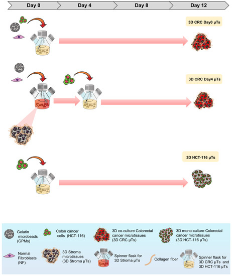
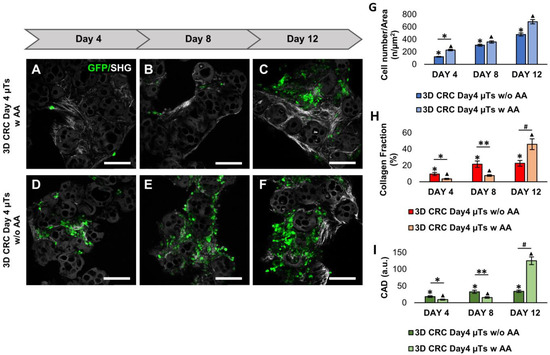
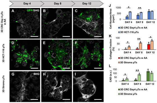
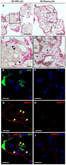
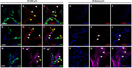
Current 3D cancer models (in vitro) fail to reproduce complex cancer cell extracellular matrices (ECMs) and the interrelationships occurring (in vivo) in the tumor microenvironment (TME). Herein, we propose 3D in vitro colorectal cancer microtissues (3D CRC μTs), which reproduce the TME more faithfully in vitro. Normal human fibroblasts were seeded onto porous biodegradable gelatin microbeads (GPMs) and were continuously induced to synthesize and assemble their own ECMs (3D Stroma μTs) in a spinner flask bioreactor. Then, human colon cancer cells were dynamically seeded onto the 3D Stroma μTs to achieve the 3D CRC μTs. Morphological characterization of the 3D CRC μTs was performed to assess the presence of different complex macromolecular components that feature in vivo in the ECM. The results showed the 3D CRC μTs recapitulated the TME in terms of ECM remodeling, cell growth, and the activation of normal fibroblasts toward an activated phenotype. Then, the microtissues were assessed as a drug screening platform by evaluating the effect of 5-Fluorouracil (5-FU), curcumin-loaded nanoemulsions (CT-NE-Curc), and the combination of the two. When taken together, the results showed that our microtissues are promising in that they can help clarify complex cancer–ECM interactions and evaluate the efficacy of therapies. Moreover, they may be combined with tissue-on-chip technologies aimed at addressing further studies in cancer progression and drug discovery.
Full article
(This article belongs to the Special Issue Recent Advance in 3D Cultures)
►
Show Figures

Graphical abstract


