Article

The Impact of Light Wavelength and Darkness on Metabolite Profiling of Korean Ginseng: Evaluating Its Anti-Cancer Potential against MCF-7 and BV-2 Cell Lines

1
Smart Farm Research Center, Korea Institute of Science and Technology, Gangneung 25451, Republic of Korea
2
Department of Plant Science, Gangneung-Wonju National University, Gangneung 25457, Republic of Korea
3
Natural Product Research Center, Korea Institute of Science and Technology, Gangneung 25451, Republic of Korea
4
Department of Botany, Abdul Wali Khan University Mardan, Mardan 23200, Pakistan
5
Department of Medical Biotechnology, College of Life Science and Biotechnology, Dongguk University, Seoul 04620, Republic of Korea
6
Korean Ginseng Company (KGC), 71 Beotkkot-gil, Daedeok-gu, Daejeon 34337, Republic of Korea
7
Division of Bio-Medical Science and Technology, KIST School, University of Science and Technology (UST), Daejeon 34113, Republic of Korea
*
Authors to whom correspondence should be addressed.
Int. J. Mol. Sci. 2023, 24(9), 7768; https://doi.org/10.3390/ijms24097768
Submission received: 25 March 2023 / Revised: 13 April 2023 / Accepted: 20 April 2023 / Published: 24 April 2023
(This article belongs to the Special Issue Natural Products: Antioxidants and Neuroprotection)

Abstract

:
Korean ginseng is a source of functional foods and medicines; however, its productivity is hindered by abiotic stress factors, such as light. This study investigated the impacts of darkness and different light wavelengths on the metabolomics and anti-cancer activity of ginseng extracts. Hydroponically-grown Korean ginseng was shifted to a light-emitting diodes (LEDs) chamber for blue-LED and darkness treatments, while white fluorescent (FL) light treatment was the control. MCF-7 breast cancer and lipopolysaccharide (LPS)-induced BV-2 microglial cells were used to determine chemo-preventive and neuroprotective potential. Overall, 53 significant primary metabolites were detected in the treated samples. The levels of ginsenosides Rb1, Rb2, Rc, Rd, and Re, as well as organic and amino acids, were significantly higher in the dark treatment, followed by blue-LED treatment and the FL control. The dark-treated ginseng extract significantly induced apoptotic signaling in MCF-7 cells and dose-dependently inhibited the NF-κB and MAP kinase pathways in LPS-induced BV-2 cells. Short-term dark treatment increased the content of Rd, Rc, Rb1, Rb2, and Re ginsenosides in ginseng extracts, which promoted apoptosis of MCF-7 cells and inhibition of the MAP kinase pathway in BV-2 microglial cells. These results indicate that the dark treatment might be effective in improving the pharmacological potential of ginseng.

1. Introduction

Ginseng is a medicinal herb used worldwide for its pharmacological and therapeutic potential [1]. The plant is commonly referred to as “Panax”, a term derived from the Greek word “Panacea”, meaning “cure-all” [2,3,4]. For centuries, Korean ginseng has been used in traditional medicine to treat various ailments, including fatigue [5], stress [6], anxiety [7], and inflammation [8]. It is believed to possess adaptogenic properties, helping the body adapt to stress and improving overall health. Additionally, Korean ginseng has gained attention in modern medicine for its potential pharmacological properties. It contains several active compounds, including ginsenosides, which exhibit anti-inflammatory [9], anti-cancer [10], and antioxidant effects. Research has also suggested that Korean ginseng may have potential benefits for cognitive functioning [11], cardiovascular health [12], and immune system functioning [13].
Ginseng can be successfully grown using hydroponics, a water-nutrient-based system used to grow plants in the absence of soil. Ginseng growth can be accelerated using nutrient-enriched hydroponic media. For example, four months of cultivation in a hydroponic system can easily produce roots equal in size to those of two-year-old ginseng plants grown using conventional methods [14].
Light is one of the major abiotic factors that influence the biosynthesis of secondary metabolites in plants [15]. Light-dependent processes, such as photosynthesis, can be modified under controlled lighting conditions, where the intensity, wavelength, and photoperiod are regulated [16]. Light-emitting diodes (LEDs) are mostly used as an artificial light source, one which improves the quality of food products in commercial agriculture [17] and upregulates the biosynthesis of defense-related secondary metabolites [18]. LEDs are efficient sources of light for growing plants and have many benefits, including improvement of photosynthesis in plants, specific bandwidth selection, long life, and reduced heat production [19]. White, blue, and red lights improve signal transduction and enhance the production of important secondary metabolites [20]. Specific wavelengths, such as red and blue, are good elicitors for the improved production of bioactive compounds, including phenols, carotenoids, and antioxidants [21]. Meanwhile, blue light is an effective pre-harvest treatment that delays fruit ripening and softening, thus improving the shelf life of different climacteric crops [22]. LED use is also recommended during storage or transportation to minimize post-harvest losses and maintain product quality [23].
In women, breast cancer is the most common type of malignant cancer found worldwide, with approximately 1.8–2 million new cases reported each year globally, and >60% of deaths occurring in underdeveloped countries [24,25]. Moreover, breast cancer is the most commonly-diagnosed cancer in developed countries, accounting for 30% of the total cancer diagnoses, and has an estimated death rate > 15% in women [26]. TNBC (triple negative breast cancer) is the most fatal form of breast cancer, as it has a high rate of proliferation and heterogeneity and no approved targeted molecular therapies are available against this cancer subtype [27]. In recent years, natural products have proven to be the most effective and safe compounds for the treatment of cancer. Natural products modulate the cancer microenvironment and intervene within cell signaling cascades [28]. Previous reports have suggested that ginsenosides, such as Rg3, Rh2, and Rh4, exhibit anti-tumor potential by inducing apoptosis in tumor cells and inhibiting cell proliferation and signaling pathways [29].
Microglial cells are macrophage-like cells that belong to the central nervous immune system. The main task of the microglial cells is to protect the brain by producing various neurotrophic factors to support neuronal cells during brain damage [30]. The overactivation of microglial cells in the central nervous system results in the overproduction of inflammatory mediators, such as pro-inflammatory cytokines and reactive oxygen species, which can be associated with the development of various neurodegenerative diseases, including Parkinson’s disease, multiple sclerosis, Huntington’s disease, and Alzheimer’s disease [31]. Several studies have reported that ginsenosides have neuroprotective effects against lipopolysaccharide (LPS)-induced microglial cells [32,33].
In ginseng plants, the quantity, quality, and total number of ginsenosides vary depending on both biotic and abiotic factors [34,35]. In addition, processed ginseng with higher content of Rg5, Rg3, and Rd ginsenosides exhibits higher anti-tumor potential than does unprocessed ginseng [36,37]. Therefore, this study aimed to determine the impact of different light treatments (i.e., blue-LED, dark, and white fluorescent (FL) light) on the anti-cancer potential of Korean ginseng extracts. MCF-7 (human breast cancer) and BV-2 (microglial cancer cells) cell lines were used to determine the effect of the treatments on the anti-cancer potential of ginseng plants cultivated in a hydroponic system. Ginsenosides and primary metabolites were profiled to reveal the complex interactions between primary metabolites, ginsenosides content, and therapeutic effects. The metabolic changes in ginseng plants growing under different light conditions might determine the specific characteristics of each light treatment.

2. Results

2.1. Analysis of Ginseng Metabolome by GC-TOF-MS

Korean ginseng is a source of proteins and terpenoids, particularly ginsenosides. A discriminatory holistic approach was applied in this study to compare the metabolite profiling of ginseng samples from the dark treatment with that of samples from the blue-LED treatment and FL control via GC-TOF-MS. Biological variance among treatments was assessed using three independent biological replicates performed under uniform conditions. Metabolite changes were observed among the hydroponically-grown ginseng plants from the different treatments (Table 1). A total of 101 metabolites were identified in the three treatments, including organic and amino acids, carbohydrates, and lactones (Table 1).
The dark-treatment samples were enriched in organic acids (36.62%), carbohydrates (33.96%), lactones (14.81%), and amino acids (13.47%). The blue-LED samples were rich in carbohydrates (46.16%), organic acids (24.57%), lactones (21.24%), and amino acids (6.10%). Meanwhile, the FL-control samples were enriched in carbohydrates (57.28%), organic acids (20.99%), lactones (15.61%), and amino acids (4.80%). Specific markers of the treatments were revealed after the multivariate analysis (Figure 1). Metabolite changes occurred during the biosynthesis of ginsenosides and their accumulation in hydroponically-grown ginseng under short-term exposure to darkness and various light wavelengths. The metabolite profiling of the dark-treated ginseng in this study was similar to that reported previously by Park et al. [38]. Carbohydrate content was previously reported as a specific marker of the white FL treatment, as it is higher in Korean ginseng than in American ginseng [39].
A principal component analysis (PCA) model was utilized to extract the most important variables or features from a large dataset of ginseng extract compounds obtained through GC-TOF-MS analysis. By reducing the dimensionality of the data and identifying the most significant compounds that had contributed to the variances in the dataset, the PCA model can aid in interpreting and visualizing complex data structures. Figure 1A shows the score plot of PCA derived from the GC–MS profiling of the treated ginseng plants. The first and second components of the PCA score plot explained 70.5% and 18.9% of the variation in the metabolite content among the different ginseng samples, respectively (Figure 1A). The dark-treated ginseng samples were fully distinguished in the PCA plot compared to the blue-LED and FL-control samples. The blue-LED samples were close to those of the FL control, suggesting that the blue-LED treatment and FL control shared considerable similarities when compared to the dark treatment.
The PCA scatter plot was used to identify the metabolites that segregated the dark and blue-LED treatments from the FL control (Figure 1B). The dark treatment could be differentiated from the FL control by its high levels of propanedioic acid, propanoic acid, L-glutamine, ferulic acid, L-aspartic acid, L-lysine, talonic acid, L-alanine, methionine, D-mannose, and ethyl methyl malonate. Moreover, the levels of serine, fructose, and tartaric, mannoic, pentanoic, and ribonic acids were higher in the blue-LED treatment than in the FL control. Correlation analysis was performed to understand the relevance of the metabolite compositions of the different treatments (Figure 1C). A heat map was used for the quantitative analysis of metabolites by using a z-score, and metabolites were plotted on a red-blue color scale (Figure 1D). The PCA and heat map patterns of the dark-treated samples were significantly different from that of the blue-LED and FL-control samples.
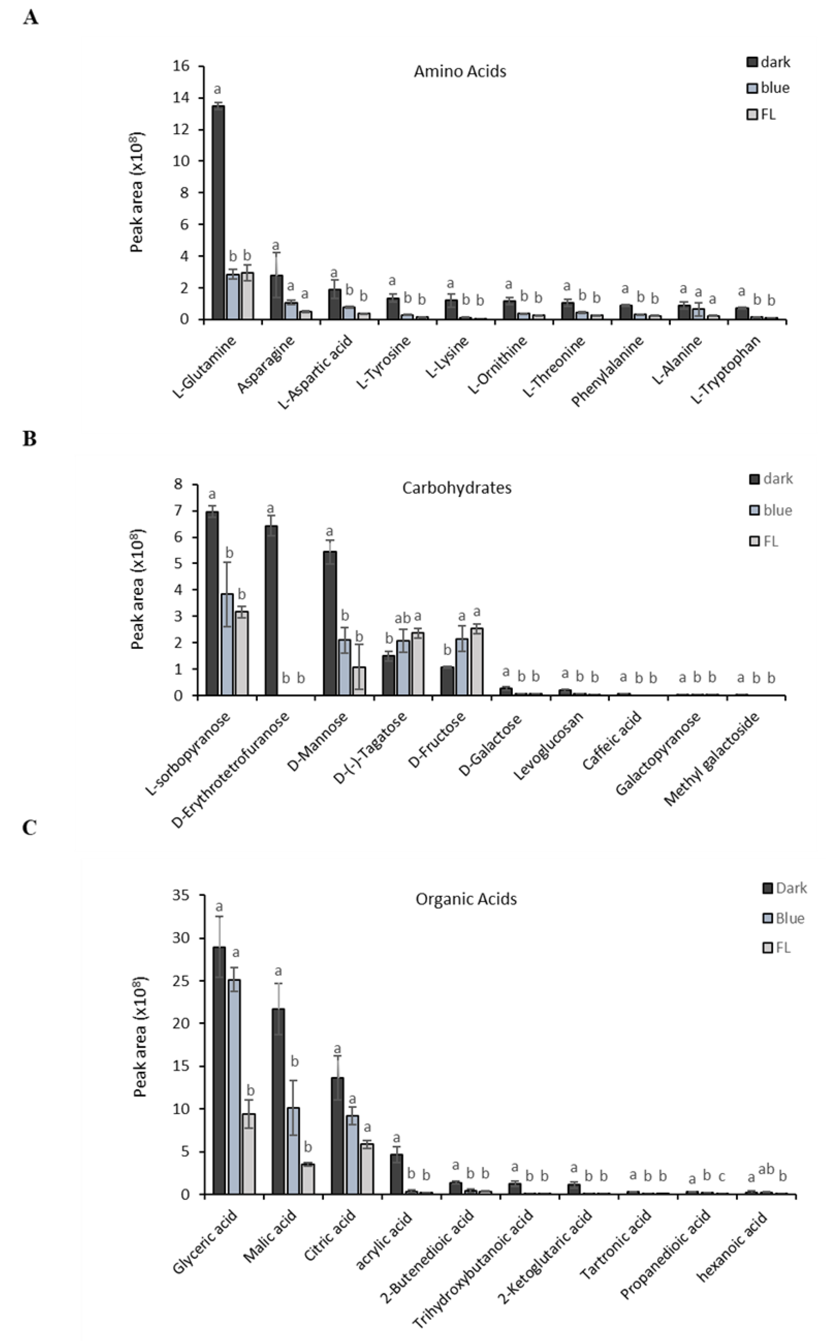
The quantitative differences in metabolites among treatments are shown in Figure 2. The content of amino acids, such as L-glutamine, asparagine, L-aspartic acid, and L-tryptophan, was significantly higher in the dark treatment than in the FL control. Amino acids (Figure 2A) play an important role in the conversion of major ginsenosides to minor ginsenosides [40]. Similarly, the content level of sugars (Figure 2B), including D-mannose, D-erythrotetrofuranose, and L-sorbopyranose, was significantly higher in the dark treatment than in the FL control. Among the organic acids (Figure 2C), glyceric, malic, and citric acids were higher in the dark treatment than in the FL control, which is consistent with the findings of Seong et al. [38].

2.2. Ginsenosides Composition under Different Treatments

We determined the ginsenosides content of the ginseng extracts used in each treatment. Total ginsenosides, including Rg1, Re, Rb1, Rc, Rb2, Rd, Rg6, F4, and F2, were quantified using HPLC-UV-VIS. Each compound was identified by comparing its retention time with that obtained from the chromatograms of the mixed ginsenoside standards. FL-control samples contained Rb1, Rb2, Rc, Rd, Re, Rg1, Rg6, F2, and F4 as the major ginsenosides (Figure 3A). After seven days of dark treatment, the contents of Rg1, Re, Rb1, Rc, Rb2, and Rd remarkably increased when compared with those of the blue-LED treatment and FL control. Upregulation of β-glucosidase enzymatic activities has been effective in increasing the Rb1, Rb2, Rc, and Rd content in ginseng [41]. The content of Rd ginsenosides has been found to be higher in red ginseng when compared to FL [42]. In this study, F2 levels were higher in the FL control than in the blue-LED treatment (Figure 3B). Moreover, Re and F2 were the most abundant ginsenosides among all treatments. The abundance of Re in ginseng was also reported by Xu et al. [43]. Therefore, the exposure to short-term dark treatment improved the overall ginsenoside content in ginseng plants.

2.3. Dark-Treated Ginseng Extract Reduces the Viability of MCF-7 Cancer Cells

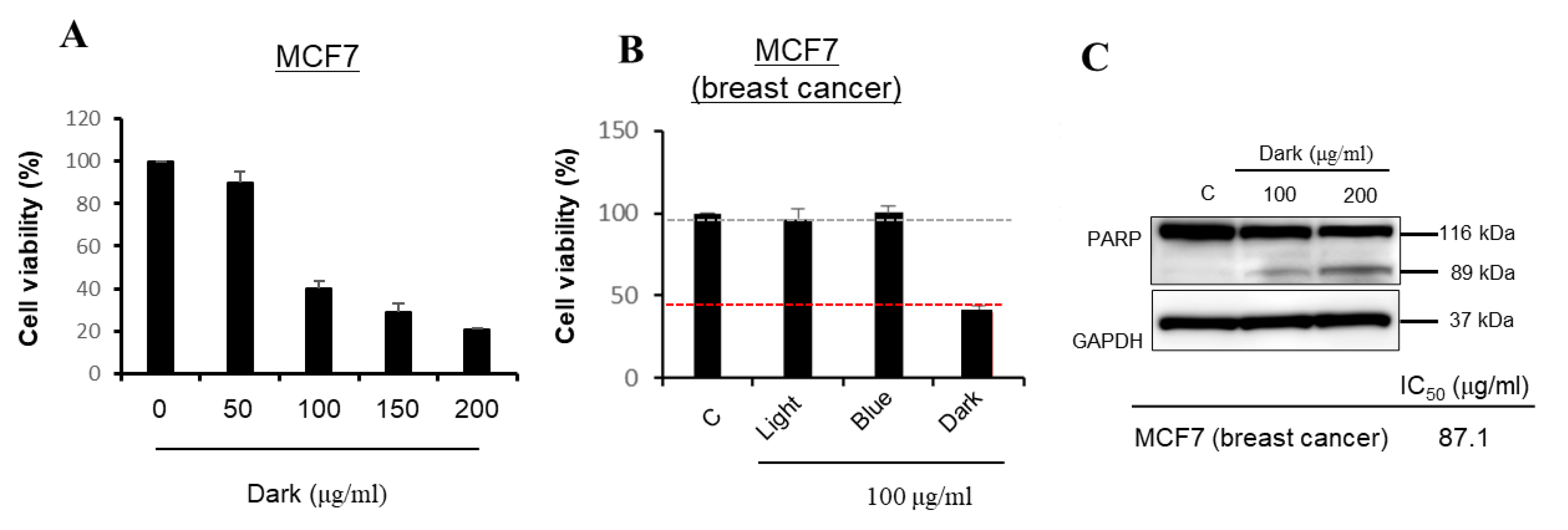
The anti-cancer activity of the various ginseng extracts was tested on the MCF-7 cell line. The MCF-7 cell line contains progesterone, estrogen, and glucocorticoid receptors [44]. Due to its ability to retain the characteristics of the mammary epithelium, it is mostly used as a model system for the apoptosis of cancer cells [45,46]. The dark-treated ginseng extract had a strong potential to induce apoptosis in the MCF-7 cancer cells among all treatments. To select a suitable nontoxic range of drug concentrations in cell culture, the ginseng extracts from all treatments were assessed for their effects on the viability of MCF-7 cells by MTT reduction assay for 24 h (Figure 4A). The cytotoxic effects of ginseng extracts were first measured at a concentration of 100 μg/mL (Figure 4A). The dark-treated ginseng extract showed significant cytotoxicity (IC50 = 87.1 μg/mL) in the MCF-7 cell line. In general, an extract concentration that reduces cell viability to <50% is considered to be an effective concentration, and this was only achieved by the dark-treated ginseng extract (Figure 4B). To examine whether the anti-proliferative effect of the dark-treated ginseng extract was involved in apoptotic cell death, we examined PARP protein status in MCF-7 cells exposed to the extracts. Apoptosis is a crucial process in various biological events, including development, immune response, and maintenance of cellular balance. This process is characterized by significant alterations in cellular morphology, such as chromatin condensation, membrane blebbing, nuclear breakdown, and the emergence of membrane-bound apoptotic bodies. Additionally, it involves inter-nucleosomal DNA fragmentation and cleavage of poly(ADP-ribose) polymerase (PARP) [47]. PARP is an enzyme that catalyzes poly(ADP-ribose) conversion of several nuclear proteins, using NAD as a substrate [48]. It was suggested that PARP contributes to cell death by depleting the cell of NAD and ATP, since it is activated by binding to DNA ends or strand breaks [49]. Furthermore, PARP is cleaved into two fragments of 89 and 24-kDa during drug-induced apoptosis in various cells. These fragments contain the active site and DNA-binding domain of the enzyme, respectively, and essentially inactivate the enzyme by destroying its ability to respond to DNA strand breaks [50].
As shown in Figure 4C, the dark-treated ginseng extracts induced PARP cleavage in a dose-dependent manner. These findings suggested that the dark-treated ginseng extract inhibited cell viability by inducing apoptotic signaling in breast cancer cells. Western blotting clearly showed the ability of the dark-treated ginseng extract to induce PARP cleavage, as indicated by the diminution of its parent band (116 kDa) and the concomitant accumulation of its cleavage fragment (89 kDa) (Figure 4C). The interrelationship among caspase-3 activation, PARP degradation, DNA fragmentation, and apoptosis induction has been well defined in the literature [51,52,53]. PARP degradation and apoptosis induction in MCF-7 cells provided additional insights into the mechanisms of action of the dark-treated ginseng extract. GAPDH was used as an internal control.

2.4. Dark-Treated Ginseng Extract Inhibits the Pathway of NF-κB Signaling

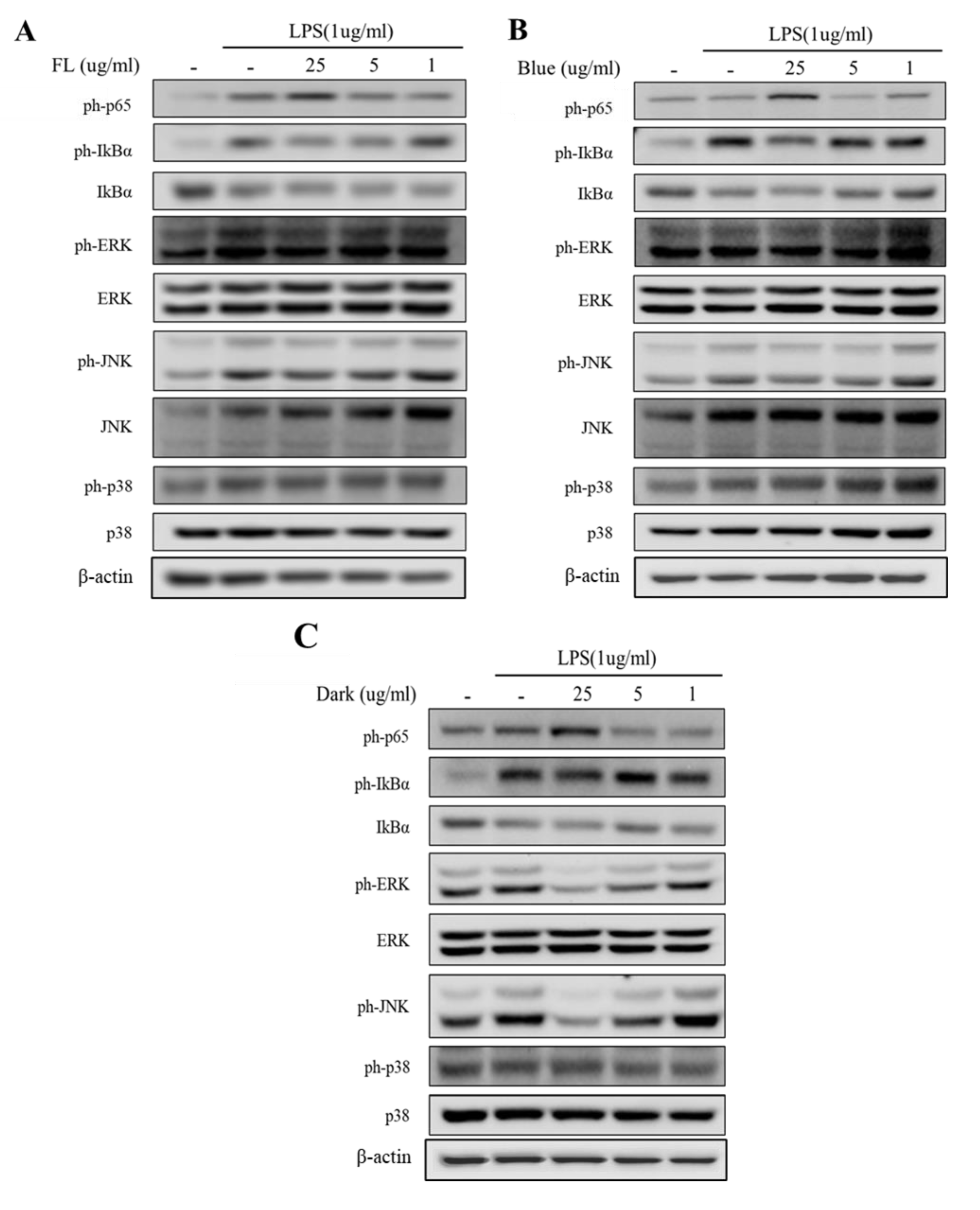
We examined a key transcription factor that modulates the expression of a wide range of pro-inflammatory genes to investigate the impact of the treatments on the effectiveness of ginseng extracts against the activation of the NF–κB signaling pathway. As shown in Figure 5, the ph-p65 levels were highly induced upon LPS administration; however, the level of p65 phosphorylation was significantly inhibited by the dark-treated ginseng extract in a dose-dependent manner (1–25 µg/mL). The dark treatment displayed a stronger inhibitory activity on p-65 protein expression than did the blue-LED treatment or the FL control. Next, the effects of the treatments on the IκB–NF–κB signaling pathway were measured using western blot analysis. Cells exposed to the dark-treated ginseng extract showed increased expression levels of IκB-α and reduced phosphorylation levels of IκB-α compared to LPS-stimulated microglia, suggesting that the dark treatment suppressed the LPS-induced IκB-α phosphorylation and degradation (Figure 5). Our results showed similar suppressive effects on cancer cells than those reported in the literature for other treatments [54]. The dark-treated ginseng extracts decreased LPS-induced NF-κB p65 nuclear translocation at concentrations ranging from 1–25 μg/mL.

2.5. Activation of MAPK Signaling Was Inhibited by Dark Treated Ginseng Extract

A crucial mediator in the activation of the microglia’s secreted pro-inflammatory cytokine production is the MAPK signaling pathway. We examined the effects of the dark-treated ginseng extracts on phosphorylated MAPK in LPS-treated microglia in order to assess their impact on the activation of inflammatory mediators through MAPK. Stimulation of BV-2 microglia with LPS increased the phosphorylation levels of ERK, JNK, and p38 MAPKs. Dark-treated ginseng extracts significantly attenuated the LPS-induced phosphorylation of representative MAPKs, including ERK, JNK, and p38, in a dose-dependent manner (Figure 5A,B). Activation of MAPK signaling was also inhibited by the dark-treated ginseng extracts (Supplementary Figure S2)

3. Discussion

The advancement of injury, infection, toxicity, or autoimmunity in the central nervous system (CNS) is heavily influenced by neuroinflammation [55]. This occurs when microglia become activated and release neurotoxic molecules and pro-inflammatory cytokines, causing progressive neuronal cell degeneration [56]. Therefore, reducing the excessive activation of microglia is a promising approach for the alleviation of the inflammatory response in the nervous system [57]. The use of natural compounds as a potential source for the development of new medicines to alleviate neuroinflammation and treat neurological disorders has gained significant recognition.
The anti-inflammatory properties of ginseng extracts have been established through the use of purified ginsenosides, including ginsenosides Rb1, Rg1, Rg3, Rh2, and compound K. These ginsenosides have been found to negatively regulate the expression of pro-inflammatory cytokines such as TNF-α, IL-1β, and IL-6, as well as enzymes including iNOS and COX-2, thus demonstrating their anti-inflammatory mechanism in M1-polarized macrophages and microglia [9]. Ginsenoside Rg3, one of the most important minor ginsenosides found in Korean ginseng, can significantly reduce the inflammatory response induced by LPS, as well as mediate TNF-α, and IL-6 in BV2 and primary microglial cells [30]. Ginsenoside Rh2 (GRh2) is a well-known constituent found in red ginseng, a plant which has been widely used in Korea and China for various medical purposes and as a dietary supplement. GRh2 has been reported to exhibit several beneficial biological effects, such as anti-inflammatory, antioxidant, and neuroprotective properties, among others. Its potential therapeutic applications have been studied extensively in recent years [58]. Rh3 is a bacterial byproduct of heat-processed ginseng’s main component, Rg5. Rh3 has anti-inflammatory effects by means of inhibiting the expression of iNOS and pro-inflammatory cytokines such as TNF-α and IL-6 and enhancing the expression of anti-inflammatory hemeoxygenase-1. Rh3 also inhibits NF-κB through SIRT1 upregulation and enhances Nrf2 DNA-binding activities. It activates AMPK phosphorylation and inhibits Akt and JAK1/STAT1 pathways induced by LPS [59].
In our study, we investigated the impact of darkness and blue LED treated ginseng extract on five different cell lines (Supplementary Figure S1) and found that dark-treated hydroponic ginseng could inhibit cytokine expression in LPS-stimulated microglia to exert neuroprotective activity. Our results suggest that different experimental environments and conditions may affect pharmacological potential; thus, we will study the impact of different LED treatments in future studies. LPS is a widely used method for studying neuroinflammation in laboratory experiments, both in vitro and in vivo. The LPS model is advantageous due to its simplicity and consistent activation of inflammation. Following LPS administration, an excess of pro-inflammatory cytokines can soon be detected [60]. In our research, we found that a short period of dark treatment in hydroponic systems resulted in the high level of anti-inflammatory activity in BV-2 microglia cells exposed to LPS through various signaling pathways. Our findings indicate that an ethanolic extract obtained from ginseng subjected to dark treatment was effective in inhibiting the phosphorylation of NF-κB P65 and the expression of NF-κB P50 in the nucleus of microglia cells induced by LPS. Additionally, the phosphorylation of MAPKs was suppressed in a concentration-dependent manner.
In conclusion, the present study revealed that LED treatment can improve the overall pharmacological potential of Korean ginseng, with dark treatment being the most effective, and that these treatments could inhibit the expression of cytokines in LPS-induced microglia cells in in-vitro conditions. Further research found that dark-treated ginseng extract expressed higher anti-inflammation activity by inhibiting the NF–κB and MAPK signaling pathways. Ginsenosides content plays an important role in the improved neuroprotective potential of dark-treated ginseng, as the content of ginsenosides Rd, Rb2, and Rc were significantly improved. Ginsenoside Rd is further converted to ginsenosides Rg3 and F2, which already reported having higher neuroprotective potential [61]. Moreover, hydroponically-grown ginseng within improved environmental conditions can reduce the neuron damage induced by neuroinflammation and can be the best natural remedy for curing cancers such as breast cancer.

4. Materials and Methods

4.1. Plant Material

One-year-old ginseng (Panax ginseng cv. Geumpung) root seedlings were obtained from the Korean Ginseng Company and grown in a hydroponic system. The roots were first soaked in water and incubated for 48 h in moist conditions to germinate the bud. Then, they were transplanted to a nutrient bath under white light in a vertical farming facility. Light intensity was set at 65–70 µmol/m2/s with a photoperiod of 16 h/8 h (day/night) at 24 °C and 18 °C, respectively. Polyurethane sponge was used as a supporting medium for the deep-water culture-type hydroponic system. RDA (Rural Development Administration) approved nutrient solution for ginseng was supplied to the seedlings. The nutrient solution was composed of NO3-N 12.5, NH4-N 1.3, K 8.5, Ca 4.5, Mg 2.3, PO4-P 3.5, SO4-S 2.0, Fe-EDTA 0.8, Mn 0.6, B 0.6, Cu 0.02, Mo 0.06, and Zn 0.06 mg/L. The electrical conductivity of the media was maintained at 2.5 mS/cm with pH 6.8. After 4 weeks of growth, the ginseng seedlings were shifted to a LED chamber to conduct light treatments [62].

4.2. Light Treatments and Ginsenosides Extraction

Ginseng plants underwent three different treatments: dark, blue-LED (465–470 nm), and white FL light. Light treatments lasted for 10 days, with the application of a light intensity of 65–70 µmol on a 24/7 schedule, whereas the dark treatment lasted for 7 days. The whole ginseng plants, including both aerial parts and roots, were harvested after each treatment and immediately stored at −80 °C, followed by freeze-drying for 48 h in a freeze-dryer (FDCF-12006 Operon Co., Ltd., Gimpo, Republic of Korea). Freeze-dried plant powder was extracted thrice with 50 mL of extraction solvent (70% MeOH) by sonication (UCP02, Jeiotech, Daejeon, Republic of Korea) for 60 min [63]. After filtration using filter paper (0.2 μm, ADVANTEC, Dublin, CA, USA), evaporation method was used to remove solvent, and the dried residue was dissolved in 100% dimethyl sulfoxide (DMSO) at 40 mg/mL concentration. Each treatment had three biological replicates.

4.3. Ginsenosides Analysis by HPLC—Evaporative Light Scattering Detection (ELSD)

To determine the ginsenoside content, the ginseng extracts were diluted to 1 mg/mL using 100% MeOH [64,65,66]. Using a SIL-9A auto-injector, a volume of 10 L was injected into the HPLC system (Hitachi L-6200 pump, Tokyo, Japan) connected to a Sedex 75 ELSD (Sedere, Vitry-sur-Seine, France) (Shimadzu, Japan). All separations were performed using an Agilent Technologies (Palo Alto, CA, USA) Zorbax SB-Aq C18 column (4.6 mm 150 mm, 5 m particle size). The high-pressure liquid chromatographic (HPLC) conditions were as follows: solvent A, water; solvent B, acetonitrile. B 20–22% (0–5 min); 22–25% (5–7 min); 26–30% (27–30 min); 30–35% (30–40 min); 50–70% (45–60 min); 70–85% (60–61 min); 85–100% (61–90 min). Nebulizer for nitrogen gas was adjusted to 2.5 bar, and ELSD was set to a probe temperature of 75 °C. A total of 5 μg of each ginsenoside standard was injected for HPLC analysis [66].

4.4. Sample Preparation for Primary Metabolites Profiling

Freeze-dried sample powders (10 mg) were dissolved in a GC–MS extraction solvent containing water, methanol, and chloroform in ration of 2.5:1:1, V/V/V, ribitol and 5α-cholestane as internal standards. The samples were extracted at 37 °C, 1200 rpm for 30 min, followed by centrifugation at 13,000 rpm for 3 min. Top part, (polar phase 8 mL) was transferred to a new tube, and 4 mL double distilled water was added, centrifuged for 3 min at 13,000 rpm and were dried using a freezing dryer. Derivatization process was performed based on standard procedure using trimethylsilyl and methoxyamine hydrochloride [67,68].

4.5. Profiling of Primary Metabolites

Gas chromatography (GC) in conjunction with a time-of-flight mass spectrometer (TOF-MS) were used to investigate primary metabolites (LECO Pegasus GC HRT, Leco Corp., St. Joseph, MI, USA). At 230 °C, 1 µL by volume sample was injected into the machine with the mode being split with 1:25 ratio. Helium, the carrier gas, with 1 mL/min flow rate RTX-5MS (30 m 0.25 mm; 0.25 m film thickness) was the column used. The oven temp was first set at 80 °C for 2 min before rising at a rate of 15 °C/min to 320 °C for 10 min. The ion source and transfer line were both adjusted at 200 °C and 250 °C, respectively. A total of 25 spectra were acquired every second at a rate of TOF-MS acquisition that was delayed for 200 s. The electron energy and detector voltage were set at 70 eV and 1600 V, respectively [69].

4.6. Cell Cultures

In accordance with the five most common cancer varieties in Korea, we used five established human cancer cell lines. The American Type Culture Collection (ATCC, Manassas, VA, USA) provided the human lung carcinoma cell line A549, human breast cancer cell line MCF-7, human colorectal carcinoma cell line HCT116, and human gastric cancer cell line AGS (ATCC, Manassas, VA, USA). The Korean Cell Line Bank sold the human hepatocellular carcinoma cell line Huh7 (Seoul, Korea). RPMI 1640 (Gibco, Waltham, MA, USA) media was used for regular subcultures of cell with 15% bovine fetal serum, 150 U/mL of penicillin, and 100 nanograms per milliliter of streptomycin in a humid environment with 37 °C and 5% carbon dioxide [70].

4.7. Cell Viability Assay

According to the manufacturer’s instructions, the EZ-Cytox test kit (Daeil Lab Service, Seoul, Korea) was used to assess the cytotoxic effects of the ginseng extracts on the examined cell lines. In a 96-well plate, triplicates of exponentially developing cells were planted at a density of 1.0 104 cells per well. The cells were given 1, 5, and 25 g/mL of the extracts the following day. The cells were cultured for 1 h with 10 L of the kit reagent after 24 h of incubation. A microplate reader operating at a 450 nm wavelength was used to scan cell cultures to determine cell viability. A quantum of 0.5% (v/v) DMSO-containing culture solutions were used to expose control cells to. With extract concentration on the x-axis and the relative cell viability on the y-axis of a non-linear regression curve, the concentration needed to inhibit cell viability by 50% (IC50) was graphically estimated [71].

4.8. Preparation of Nuclear Extract, Whole-Cell Lysates and Immunoblot Analysis

Ginseng extracts from the various light treatments were given to MCF-7 cells that had been cultured in 6-well plates for 24 h. Then, using RIPA buffer (Cell Signaling Technology, CST, Danvers, MA, USA) together with a 1 protease inhibitor cocktail (Roche, Mannheim, Germany) and 1 mM phenylmethylsulfonyl fluoride, whole-cell lysates were created in accordance with the manufacturer’s instructions. Proteins (whole-cell extracts, 30 g/lane) were electrophoretically separated from one another on NuPAGE 4–12% Bis-Tris gels (Invitrogen, Waltham, MA, USA), blotted onto PVDF transfer membranes, and examined with primary and secondary antibodies that are specific for the epitopes in question. Western blotting detection reagents from Thermo Fisher Scientific, Waltham, Massachusetts, USA, and a LAS 4000 imaging system were used to detect bound antibodies (Fujifilm, Tokyo, Japan). Rabbit anti-PARP (9542; CST) and rabbit anti-GAPDH were the principal antibodies employed (2118; CST). The HRP-linked secondary antibody was goat anti-rabbit IgG (7074, CST) [72].
Before starting nuclear and cytoplasmic extraction, two lysate buffers were prepared namely buffer A (10 mM HEPES, 10 mM KCl, 1 mM EDTA, 0.2 mM EGTA, 1 mM PMSF, 1 mM DDT, 0.5% NP40, and a protease inhibitor cocktail [Sigma-Aldrich, St. Louis, MO, USA] in HPLC grade water) and buffer B (0.1 mM EDTA, 0.1 mM EGTA, 20 mM HEPES, 420 mM NaCl, 1 mM PMSF, 1 mM DDT, and protease inhibitor cocktail in RIPA buffer [Sigma-Aldrich]) [73]. For cytoplasmic extraction cells were treated with varying concentrations of isodojaponin D (1, 5, or 10 μg/mL) in the presence or absence of LPS (1 μg/mL) for 24 h. After LPS stimulation, buffer A was used for cell lyses. Homogenate were vortexed for 1 min followed by centrifugation for 5 min at 20,000× g, supernatant was collected as cytoplasmic extract. Resulting residual pellets were used for nuclear extraction using 50 μL of buffer B. After homogenization and centrifugation (20,000× g for 5 min) supernatant was collected as nuclear extract. Both extracts were stored at −70 °C [74].

4.9. MTT Assay

The MTT assay was used to determine cell cytotoxicity. A quantum of 0.2 × 10⁴ cells/well of cells were sown in 96-well culture plates, and the cells were left to adhere for 24 h. Different concentrations of ginseng extracts, such as dark, blue-LED, and FL (e.g., 5 mg/mL, 10 mg/mL, 15 mg/mL, and 20 mg/mL), were applied to each well of culture media. A total of 30 µL of MTT (3-(4,5-dimethylthiazol-2-yl)-2,5-diphenyl tetrazolium bromide) stock solution (3 mg/mL) were applied to each well at the appropriate time intervals. DMSO was applied to the cells after two hours of culture at 37 °C to dissolve the formazan crystals. Using a microplate reader (EL800, Biotek Instruments Inc., Winooski, VT, USA), the absorbance of the cell culture was measured at a 540 nm wavelength [75].

4.10. Western Blot Analysis

Bradford method (Bio-Rad, Hercules, CA, USA) was used to assay total proteins in cell extracts or nuclear and cytoplasmic extracts. For SDS-PAGE, 30 μg of protein from cell lysates was separated according to size. Equal amounts of protein for each sample were heated with 2X Laemmli sample buffer and loaded onto 12% or 7.5% polyacrylamide gels. Proteins were then transferred onto a PVDF (Millipore, Billerica, MA, USA) membrane for 2.4 h at 400 mA. The membrane was blocked with 5% nonfat dry milk and incubated with primary antibody overnight at 4 °C. The antibodies used were NF-κB p50 (1:1000, Santa Cruz, Santa Cruz, CA, USA), NF-κB p65 (1:1000, Santa Cruz), c-Jun Nterminal kinase (JNK, 1:1000, Cell Signaling), p-JNK (1:1000, Cell Signaling), p38 (1:1000, Santa Cruz), Phospho-p38 MAPK (1:1000, Cell Signaling), extracellular-signal-regulated kinase (ERK, 1:5000, Cell Signaling), pERK (1:1000, Cell Signaling), and β-actin (1:100,000, Sigma Aldrich). After incubation, the membranes were washed with TBS-T buffer and incubated again with the respective secondary antibodies conjugated with peroxidase (Sigma-Aldrich). Secondary antibodies conjugated to horseradish peroxidase (1:2500, Santa Cruz), followed by enhanced chemiluminescence (ECL Western Blotting Systems, GE Healthcare, Little Chalfont, BKM, UK) reagents (Amersham Biosciences, Piscataway, NJ, USA), were used for the detection of proteins. The western blot scanning results showed a linear curve in the range used for each antibody. The bands observed on the membrane were representative of the groups. To quantify the proteins, normalization with a control group was performed [76].

4.11. Statistical Analysis

Data were expressed as mean ± standard error (SEM) of the mean of n observations. Principal component analysis (PCA) was performed using MetaboAnalyst version 5.0 to identify the different metabolites produced in each treatment. Orthogonal partial least squares discriminant analysis (OPLS-DA) was performed using SIMCA-P, version 15.0.2 (Umetrics, Umea, Sweden). All experiments were repeated at least thrice, and all data were compiled from a minimum of three replicates. Significant differences between treated and untreated cell lines were analyzed by post-hoc Tukey’s test followed by two-way ANOVA. p-values < 0.05 were considered significantly different (for three biological replicates) between each compared pair groups.

5. Conclusions

A GC–MS-based metabolomics approach revealed trends in the accumulation of various metabolites in ginseng in response to darkness and different light treatments. The short-term dark treatment increased the organic acid content in ginseng and decreased carbohydrate content by converting monosaccharides into complex secondary metabolites, such as ginsenosides Rd and Rb. The abundant Rb1 and Rb2 in the dark treatment served as major substrates for the cell enzymatic conversion to Rg3, which, together with other minor ginsenosides, increased the chemo-preventive and neuroprotective potential of the dark-treated ginseng. Our investigation revealed that the dark-treated ginseng extract induced apoptosis in MCF-7 cells. Western blot analysis was used to evaluate the expression and cleavage of PARP protein as well as caspase 3 expression, which was regulated in a dose-dependent manner. Dark-treated ginseng extracts prevented LPS-induced IκB-α/β phosphorylation, which, in turn, inhibited the translocation of NF-κB-dependent pathways. This finding suggests that the dark treatment is effective in improving the overall metabolite profiling of ginseng. Moreover, the short-term dark treatment is highly effective in enhancing the biological conversion of major ginsenosides to minor ginsenosides, which in turn enhances the pharmacological potential of ginseng. The GC-MS-based metabolomics approach is an efficient tool for metabolite profiling in plants. Metabolomics-based profiling tools may be useful for improving the quality of ginseng plants. Further in-depth investigations are recommended to identify specific genes, whose upregulation could increase the content of minor ginsenosides in dark-treated ginseng.

Supplementary Materials

The following supporting information can be downloaded at: https://www.mdpi.com/1422-0067/24/9/7768/s1.

Author Contributions

Conceptualization and writing–original draft was composed by H.-Y.K. and N.B.S.; methodology and experimental procedure were composed by N.B.S.; correspondence with journal was performed by H.-Y.K.; software and data analysis was performed by S.N.K.; in vivo based study of cell line was performed by H.K. and S.-W.J. helped to provide plant material and identification of cultivar. M.H., J.-H.J., N.I.P. and S.-H.Y. helped in writing–review & editing. Reviewing was performed by H.-Y.K. and Y.-J.K. All authors have read and agreed to the published version of the manuscript.

Funding

This work was financially supported by an intramural grant (2Z06831, 2Z06821) from the Korea Institute of Science and Technology (KIST).

Institutional Review Board Statement

Not applicable.

Informed Consent Statement

Not applicable.

Data Availability Statement

The data presented in this study are available in supplementary form.

Conflicts of Interest

There are no conflict of interest declared by authors with respect to publication or/and authorship of this research article.

References

  1. Ratan, Z.A.; Haidere, M.F.; Hong, Y.H.; Park, S.H.; Lee, J.-O.; Lee, J.; Cho, J.Y. Pharmacological potential of ginseng and its major component ginsenosides. J. Ginseng Res. 2020, 45, 199–210. [Google Scholar] [CrossRef] [PubMed]
  2. Lim, S.W.; Luo, K.; Quan, Y.; Cui, S.; Shin, Y.J.; Ko, E.J.; Chung, B.H.; Yang, C.W. The safety, immunological benefits, and efficacy of ginseng in organ transplantation. J. Ginseng Res. 2020, 44, 399–404. [Google Scholar] [CrossRef] [PubMed]
  3. Unlu, A.; Nayir, E.; Kirca, O.; Ay, H.; Ozdogan, M. Ginseng and cancer. J. BUON 2016, 21, 1383–1387. [Google Scholar] [PubMed]
  4. Shi, Z.-Y.; Zeng, J.-Z.; Wong, A.S.T. Chemical structures and pharmacological profiles of ginseng saponins. Molecules 2019, 24, 2443. [Google Scholar] [CrossRef] [Green Version]
  5. Sadeghian, M.; Rahmani, S.; Zendehdel, M.; Hosseini, S.A.; Zare Javid, A. Ginseng and cancer-related fatigue: A systematic review of clinical trials. Nutr. Cancer 2021, 73, 1270–1281. [Google Scholar] [CrossRef]
  6. Farh, M.E.-A.; Kim, Y.-J.; Sukweenadhi, J.; Singh, P.; Yang, D.-C. Aluminium resistant, plant growth promoting bacteria induce overexpression of Aluminium stress related genes in Arabidopsis thaliana and increase the ginseng tolerance against Aluminium stress. Microbiol. Res. 2017, 200, 45–52. [Google Scholar] [CrossRef]
  7. Lee, S.; Rhee, D.-K. Effects of ginseng on stress-related depression, anxiety, and the hypothalamic–pituitary–adrenal axis. J. Ginseng Res. 2017, 41, 589–594. [Google Scholar] [CrossRef]
  8. Bae, M.; Jang, S.; Lim, J.W.; Kang, J.; Bak, E.J.; Cha, J.-H.; Kim, H. Protective effect of Korean Red Ginseng extract against Helicobacter pylori-induced gastric inflammation in Mongolian gerbils. J. Ginseng Res. 2014, 38, 8–15. [Google Scholar] [CrossRef] [Green Version]
  9. Im, D.-S. Pro-resolving effect of ginsenosides as an anti-inflammatory mechanism of Panax ginseng. Biomolecules 2020, 10, 444. [Google Scholar] [CrossRef] [Green Version]
  10. Lee, D.-Y.; Park, C.W.; Lee, S.J.; Park, H.-R.; Kim, S.H.; Son, S.-U.; Park, J.; Shin, K.-S. Anti-cancer effects of Panax ginseng berry polysaccharides via activation of immune-related cells. Front. Pharmacol. 2019, 10, 1411. [Google Scholar] [CrossRef] [Green Version]
  11. Park, K.-C.; Jin, H.; Zheng, R.; Kim, S.; Lee, S.-E.; Kim, B.-H.; Yim, S.-V. Cognition enhancing effect of panax ginseng in Korean volunteers with mild cognitive impairment: A randomized, double-blind, placebo-controlled clinical trial. Transl. Clin. Pharmacol. 2019, 27, 92–97. [Google Scholar] [CrossRef] [Green Version]
  12. Hyun, S.H.; Bhilare, K.D.; In, G.; Park, C.-K.; Kim, J.-H. Effects of Panax ginseng and ginsenosides on oxidative stress and cardiovascular diseases: Pharmacological and therapeutic roles. J. Ginseng Res. 2022, 46, 33–38. [Google Scholar] [CrossRef]
  13. You, L.; Cha, S.; Kim, M.-Y.; Cho, J.Y. Ginsenosides are active ingredients in Panax ginseng with immunomodulatory properties from cellular to organismal levels. J. Ginseng Res. 2022, 46, 711–721. [Google Scholar] [CrossRef]
  14. Hwang, C.R.; Lee, S.H.; Jang, G.Y.; Hwang, I.G.; Kim, H.Y.; Woo, K.S.; Lee, J.; Jeong, H.S. Changes in ginsenoside compositions and antioxidant activities of hydroponic-cultured ginseng roots and leaves with heating temperature. J. Ginseng Res. 2014, 38, 180–186. [Google Scholar] [CrossRef] [Green Version]
  15. Alrifai, O.; Hao, X.; Marcone, M.F.; Tsao, R. Current review of the modulatory effects of LED lights on photosynthesis of secondary metabolites and future perspectives of microgreen vegetables. J. Agric. Food Chem. 2019, 67, 6075–6090. [Google Scholar] [CrossRef]
  16. Al Murad, M.; Razi, K.; Jeong, B.R.; Samy, P.M.A.; Muneer, S. Light emitting diodes (LEDs) as agricultural lighting: Impact and its potential on improving physiology, flowering, and secondary metabolites of crops. Sustainability 2021, 13, 1985. [Google Scholar] [CrossRef]
  17. Pennisi, G.; Blasioli, S.; Cellini, A.; Maia, L.; Crepaldi, A.; Braschi, I.; Spinelli, F.; Nicola, S.; Fernandez, J.A.; Stanghellini, C. Unraveling the role of red: Blue LED lights on resource use efficiency and nutritional properties of indoor grown sweet basil. Front. Plant Sci. 2019, 10, 305. [Google Scholar] [CrossRef] [Green Version]
  18. Handa, N.; Kohli, S.K.; Sharma, A.; Thukral, A.K.; Bhardwaj, R.; Abd_Allah, E.F.; Alqarawi, A.A.; Ahmad, P. Selenium modulates dynamics of antioxidative defence expression, photosynthetic attributes and secondary metabolites to mitigate chromium toxicity in Brassica juncea L. plants. Environ. Exp. Bot. 2019, 161, 180–192. [Google Scholar] [CrossRef]
  19. Maroga, G.M.; Soundy, P.; Sivakumar, D. Different postharvest responses of fresh-cut sweet peppers related to quality and antioxidant and phenylalanine ammonia lyase activities during exposure to light-emitting diode treatments. Foods 2019, 8, 359. [Google Scholar] [CrossRef] [Green Version]
  20. Lian, T.T.; Cha, S.-Y.; Moe, M.M.; Kim, Y.J.; Bang, K.S. Effects of different colored LEDs on the enhancement of biologically active ingredients in callus cultures of Gynura procumbens (Lour.) Merr. Molecules 2019, 24, 4336. [Google Scholar] [CrossRef] [Green Version]
  21. Bantis, F.; Smirnakou, S.; Ouzounis, T.; Koukounaras, A.; Ntagkas, N.; Radoglou, K. Current status and recent achievements in the field of horticulture with the use of light-emitting diodes (LEDs). Sci. Hortic. 2018, 235, 437–451. [Google Scholar] [CrossRef]
  22. Dhakal, R.; Baek, K.-H. Short period irradiation of single blue wavelength light extends the storage period of mature green tomatoes. Postharvest Biol. Technol. 2014, 90, 73–77. [Google Scholar] [CrossRef]
  23. Taulavuori, E.; Taulavuori, K.; Holopainen, J.K.; Julkunen-Tiitto, R.; Acar, C.; Dincer, I. Targeted use of LEDs in improvement of production efficiency through phytochemical enrichment. J. Sci. Food Agric. 2017, 97, 5059–5064. [Google Scholar] [CrossRef] [PubMed]
  24. Amrane, M.; Oukid, S.; Gagaoua, I.; Ensari, T. Breast cancer classification using machine learning. In Proceedings of the 2018 Electric Electronics, Computer Science, Biomedical Engineerings’ Meeting (EBBT), Istanbul, Turkey, 18–19 April 2018; IEEE: Piscataway, NJ, USA, 2018; pp. 1–4. [Google Scholar]
  25. Miller, K.D.; Nogueira, L.; Mariotto, A.B.; Rowland, J.H.; Yabroff, K.R.; Alfano, C.M.; Jemal, A.; Kramer, J.L.; Siegel, R.L. Cancer treatment and survivorship statistics, 2019. CA A Cancer J. Clin. 2019, 69, 363–385. [Google Scholar] [CrossRef] [PubMed] [Green Version]
  26. Sharma, R. Global, regional, national burden of breast cancer in 185 countries: Evidence from GLOBOCAN 2018. Breast Cancer Res. Treat. 2021, 187, 557–567. [Google Scholar] [CrossRef]
  27. Camorani, S.; Granata, I.; Collina, F.; Leonetti, F.; Cantile, M.; Botti, G.; Fedele, M.; Guarracino, M.R.; Cerchia, L. Novel aptamers selected on living cells for specific recognition of triple-negative breast cancer. iScience 2020, 23, 100979. [Google Scholar] [CrossRef]
  28. Dutta, S.; Mahalanobish, S.; Saha, S.; Ghosh, S.; Sil, P.C. Natural products: An upcoming therapeutic approach to cancer. Food Chem. Toxicol. 2019, 128, 240–255. [Google Scholar] [CrossRef]
  29. Xia, T.; Zhang, J.; Zhou, C.; Li, Y.; Duan, W.; Zhang, B.; Wang, M.; Fang, J. 20 (S)-Ginsenoside Rh2 displays efficacy against T-cell acute lymphoblastic leukemia through the PI3K/Akt/mTOR signal pathway. J. Ginseng Res. 2020, 44, 725–737. [Google Scholar] [CrossRef]
  30. Lee, K.-W.; Jung, S.Y.; Choi, S.-M.; Yang, E.J. Effects of ginsenoside Re on LPS-induced inflammatory mediators in BV2 microglial cells. BMC Complement. Altern. Med. 2012, 12, 196. [Google Scholar] [CrossRef] [Green Version]
  31. Lee, Y.Y.; Park, J.-S.; Jung, J.-S.; Kim, D.-H.; Kim, H.-S. Anti-inflammatory effect of ginsenoside Rg5 in lipopolysaccharide-stimulated BV2 microglial cells. Int. J. Mol. Sci. 2013, 14, 9820–9833. [Google Scholar] [CrossRef] [Green Version]
  32. Kim, M.; Choi, S.Y.; Kim, K.-T.; Rhee, Y.K.; Hur, J. Ginsenoside Rg18 suppresses lipopolysaccharide-induced neuroinflammation in BV2 microglia and amyloid-β-induced oxidative stress in SH-SY5Y neurons via nuclear factor erythroid 2-related factor 2/heme oxygenase-1 induction. J. Funct. Foods 2017, 31, 71–78. [Google Scholar] [CrossRef]
  33. Lu, D.; Zhu, L.-H.; Shu, X.-M.; Zhang, C.-J.; Zhao, J.-Y.; Qi, R.-B.; Wang, H.-D.; Lu, D.-X. Ginsenoside Rg1 relieves tert-Butyl hydroperoxide-induced cell impairment in mouse microglial BV2 cells. J. Asian Nat. Prod. Res. 2015, 17, 930–945. [Google Scholar] [CrossRef]
  34. Jang, S.-w.; Sadiq, N.B.; Hamayun, M.; Jung, J.; Lee, T.; Yang, J.-S.; Lee, B.; Kim, H.-Y. Silicon foliage spraying improves growth characteristics, morphological traits, and root quality of Panax ginseng CA Mey. Ind. Crops Prod. 2020, 156, 112848. [Google Scholar] [CrossRef]
  35. CHU, L.L.; Hanhong, B. Bacterial endophytes from ginseng and their biotechnological application. J. Ginseng Res. 2021, 46, 1–10. [Google Scholar] [CrossRef]
  36. Hong, H.; Baatar, D.; Hwang, S.G. Anticancer activities of ginsenosides, the main active components of ginseng. Evid. Based Complement. Altern. Med. 2021, 2021, 8858006. [Google Scholar] [CrossRef]
  37. Choi, P.; Park, J.Y.; Kim, T.; Park, S.-H.; Kim, H.-k.; Kang, K.S.; Ham, J. Improved anticancer effect of ginseng extract by microwave-assisted processing through the generation of ginsenosides Rg3, Rg5 and Rk1. J. Funct. Foods 2015, 14, 613–622. [Google Scholar] [CrossRef]
  38. Park, S.-E.; Seo, S.-H.; Lee, K.I.; Na, C.-S.; Son, H.-S. Metabolite profiling of fermented ginseng extracts by gas chromatography mass spectrometry. J. Ginseng Res. 2018, 42, 57–67. [Google Scholar] [CrossRef]
  39. Cui, S.; Wu, J.; Wang, J.; Wang, X. Discrimination of American ginseng and Asian ginseng using electronic nose and gas chromatography–mass spectrometry coupled with chemometrics. J. Ginseng Res. 2017, 41, 85–95. [Google Scholar] [CrossRef] [Green Version]
  40. Liu, Z.; Wen, X.; Wang, C.-Z.; Li, W.; Huang, W.-H.; Xia, J.; Ruan, C.-C.; Yuan, C.-S. Remarkable impact of amino acids on ginsenoside transformation from fresh ginseng to red ginseng. J. Ginseng Res. 2020, 44, 424–434. [Google Scholar] [CrossRef]
  41. Kim, H.W.; Han, S.H.; Lee, S.W.; Choi, H.S.; Suh, H.J.; Hong, K.B. Enzymatic hydrolysis increases ginsenoside content in Korean red ginseng (Panax ginseng CA Meyer) and its biotransformation under hydrostatic pressure. J. Sci. Food Agric. 2019, 99, 6806–6813. [Google Scholar] [CrossRef]
  42. Choi, M.-K.; Jin, S.; Jeon, J.-H.; Kang, W.Y.; Seong, S.J.; Yoon, Y.-R.; Han, Y.-H.; Song, I.-S. Tolerability and pharmacokinetics of ginsenosides Rb1, Rb2, Rc, Rd, and compound K after single or multiple administration of red ginseng extract in human beings. J. Ginseng Res. 2020, 44, 229–237. [Google Scholar] [CrossRef] [PubMed]
  43. Xu, X.Y.; Yi, E.S.; Kang, C.H.; Liu, Y.; Lee, Y.-G.; Choi, H.S.; Jang, H.B.; Huo, Y.; Baek, N.-I.; Yang, D.C. Whitening and inhibiting NF-κB-mediated inflammation properties of the biotransformed green ginseng berry of new cultivar K1, ginsenoside Rg2 enriched, on B16 and LPS-stimulated RAW 264.7 cells. J. Ginseng Res. 2021, 45, 631–641. [Google Scholar] [CrossRef] [PubMed]
  44. Chen, D.; Reierstad, S.; Lu, M.; Lin, Z.; Ishikawa, H.; Bulun, S.E. Regulation of breast cancer-associated aromatase promoters. Cancer Lett. 2009, 273, 15–27. [Google Scholar] [CrossRef]
  45. Ahmad, G.; Mir, S.A.; Anand, L.K.; Pottoo, F.H.; Dhiman, N.; Malik, F.; Ali, A. Myricanol-9-acetate, a novel naturally occurring derivative of myricanol, induces ROS-dependent mitochondrial-mediated Apoptosis in MCF-7 cancer cells. Curr. Top. Med. Chem. 2021, 21, 1418–1427. [Google Scholar] [CrossRef]
  46. Lee, G.H.; Jin, S.W.; Kim, S.J.; Pham, T.H.; Choi, J.H.; Jeong, H.G. Tetrabromobisphenol A induces MMP-9 expression via NADPH oxidase and the activation of ROS, MAPK, and Akt pathways in human breast cancer MCF-7 cells. Toxicol. Res. 2019, 35, 93–101. [Google Scholar] [CrossRef] [PubMed] [Green Version]
  47. Boulares, A.H.; Yakovlev, A.G.; Ivanova, V.; Stoica, B.A.; Wang, G.; Iyer, S.; Smulson, M. Role of poly (ADP-ribose) polymerase (PARP) cleavage in apoptosis: Caspase 3-resistant PARP mutant increases rates of apoptosis in transfected cells. J. Biol. Chem. 1999, 274, 22932–22940. [Google Scholar] [CrossRef] [Green Version]
  48. Mullen, P. PARP cleavage as a means of assessing apoptosis. In Cancer Cell Culture; Springer: Berlin/Heidelberg, Germany, 2004; pp. 171–181. [Google Scholar]
  49. Leisching, G.; Loos, B.; Botha, M.; Engelbrecht, A.-M. Bcl-2 confers survival in cisplatin treated cervical cancer cells: Circumventing cisplatin dose-dependent toxicity and resistance. J. Transl. Med. 2015, 13, 328. [Google Scholar] [CrossRef]
  50. Vo, P.H.T.; Nguyen, T.D.T.; Tran, H.T.; Nguyen, Y.N.; Doan, M.T.; Nguyen, P.H.; Lien, G.T.K.; To, D.C.; Tran, M.H. Cytotoxic components from the leaves of Erythrophleum fordii induce human acute leukemia cell apoptosis through caspase 3 activation and PARP cleavage. Bioorg. Med. Chem. Lett. 2021, 31, 127673. [Google Scholar] [CrossRef]
  51. Sayed, A.E.-D.H.; Hamed, H.S. Induction of apoptosis and DNA damage by 4-nonylphenol in African catfish (Clarias gariepinus) and the antioxidant role of Cydonia oblonga. Ecotoxicol. Environ. Saf. 2017, 139, 97–101. [Google Scholar] [CrossRef]
  52. Ajji, P.K.; Binder, M.J.; Walder, K.; Puri, M. Balsamin induces apoptosis in breast cancer cells via DNA fragmentation and cell cycle arrest. Mol. Cell. Biochem. 2017, 432, 189–198. [Google Scholar] [CrossRef]
  53. Villanueva, P.J.; Martinez, A.; Baca, S.T.; DeJesus, R.E.; Larragoity, M.; Contreras, L.; Gutierrez, D.A.; Varela-Ramirez, A.; Aguilera, R.J. Pyronaridine exerts potent cytotoxicity on human breast and hematological cancer cells through induction of apoptosis. PLoS ONE 2018, 13, e0206467. [Google Scholar]
  54. Wang, H.-M.; Yang, H.-L.; Thiyagarajan, V.; Huang, T.-H.; Huang, P.-J.; Chen, S.-C.; Liu, J.-Y.; Hsu, L.-S.; Chang, H.-W.; Hseu, Y.-C. Coenzyme Q0 enhances ultraviolet B–induced apoptosis in human estrogen receptor–positive breast (MCF-7) cancer cells. Integr. Cancer Ther. 2017, 16, 385–396. [Google Scholar] [CrossRef] [Green Version]
  55. Shabab, T.; Khanabdali, R.; Moghadamtousi, S.Z.; Kadir, H.A.; Mohan, G. Neuroinflammation pathways: A general review. Int. J. Neurosci. 2017, 127, 624–633. [Google Scholar] [CrossRef]
  56. Block, M.L.; Zecca, L.; Hong, J.-S. Microglia-mediated neurotoxicity: Uncovering the molecular mechanisms. Nat. Rev. Neurosci. 2007, 8, 57–69. [Google Scholar] [CrossRef]
  57. Smith, J.A.; Das, A.; Ray, S.K.; Banik, N.L. Role of pro-inflammatory cytokines released from microglia in neurodegenerative diseases. Brain Res. Bull. 2012, 87, 10–20. [Google Scholar] [CrossRef]
  58. Vinoth Kumar, R.; Oh, T.W.; Park, Y.-K. Anti-inflammatory effects of ginsenoside-Rh2 inhibits LPS-induced activation of microglia and overproduction of inflammatory mediators via modulation of TGF-β1/Smad pathway. Neurochem. Res. 2016, 41, 951–957. [Google Scholar] [CrossRef]
  59. Lee, Y.Y.; Park, J.-S.; Lee, E.-J.; Lee, S.-Y.; Kim, D.-H.; Kang, J.L.; Kim, H.-S. Anti-inflammatory mechanism of ginseng saponin metabolite Rh3 in lipopolysaccharide-stimulated microglia: Critical role of 5′-adenosine monophosphate-activated protein kinase signaling pathway. J. Agric. Food Chem. 2015, 63, 3472–3480. [Google Scholar] [CrossRef]
  60. Yu, R.; Li, Q.; Feng, Z.; Cai, L.; Xu, Q. m6A reader YTHDF2 regulates LPS-induced inflammatory response. Int. J. Mol. Sci. 2019, 20, 1323. [Google Scholar] [CrossRef] [Green Version]
  61. Chen, Y.-Y.; Liu, Q.-P.; An, P.; Jia, M.; Luan, X.; Tang, J.-Y.; Zhang, H. Ginsenoside Rd: A promising natural neuroprotective agent. Phytomedicine 2022, 95, 153883. [Google Scholar] [CrossRef]
  62. Kim, G.-S.; Lee, S.-E.; Noh, H.-J.; Kwon, H.; Lee, S.-W.; Kim, S.-Y.; Kim, Y.-B. Effects of natural bioactive products on the growth and ginsenoside contents of Panax ginseng cultured in an aeroponic system. J. Ginseng Res. 2012, 36, 430. [Google Scholar] [CrossRef] [Green Version]
  63. Wei, Y.; Hou, B.; Fang, H.; Sun, X.; Ma, F. Salting-out extraction of ginsenosides from the enzymatic hydrolysates of Panax quinquefolium based on ethanol/sodium carbonate system. J. Ginseng Res. 2020, 44, 44–49. [Google Scholar] [CrossRef] [PubMed]
  64. Li, P.; Zhao, P.; Liu, W.; Jiang, Y.; Wang, W.; Bao, L.; Jin, Y.; Li, X. Determination of common ginsenosides in Kang’ai injection by aqueous two-phase extraction with deep eutectic solvents and HPLC-UV/DAD. Microchem. J. 2018, 137, 302–308. [Google Scholar] [CrossRef]
  65. Xu, L.; Xu, J.; Shi, G.; Xiao, S.; Dai, R.; Wu, S.; Sun, B.; Zhang, X.; Zhao, Y. Optimization of flash extraction, separation of ginsenosides, identification by HPLC-FT-ICR-MS and determination of rare ginsenosides in mountain cultivated ginseng. RSC Adv. 2020, 10, 44050–44057. [Google Scholar] [CrossRef] [PubMed]
  66. Song, H.; Song, K.-W.; Hong, S.-P. Simultaneous quantification of six nonpolar ginsenosides in white ginseng by reverse-phase high-performance liquid chromatography coupled with integrated pulsed amperometric detection. J. Ginseng Res. 2020, 44, 563–569. [Google Scholar] [CrossRef]
  67. Kim, T.J.; Choi, J.; Kim, K.W.; Ahn, S.K.; Ha, S.H.; Choi, Y.; Park, N.I.; Kim, J.K. Metabolite profiling of peppers of various colors reveals relationships between tocopherol, carotenoid, and phytosterol content. J. Food Sci. 2017, 82, 2885–2893. [Google Scholar] [CrossRef]
  68. Ghosson, H.; Schwarzenberg, A.; Jamois, F.; Yvin, J.-C. Simultaneous untargeted and targeted metabolomics profiling of underivatized primary metabolites in sulfur-deficient barley by ultra-high performance liquid chromatography-quadrupole/time-of-flight mass spectrometry. Plant Methods 2018, 14, 62. [Google Scholar] [CrossRef] [Green Version]
  69. Al-Rubaye, A.F.; Hameed, I.H.; Kadhim, M.J. A review: Uses of gas chromatography-mass spectrometry (GC-MS) technique for analysis of bioactive natural compounds of some plants. Int. J. Toxicol. Pharmacol. Res. 2017, 9, 81–85. [Google Scholar] [CrossRef]
  70. Lim, W.-C.; Kim, H.; Kim, Y.-J.; Jeon, B.-N.; Kang, H.-B.; Ko, H. Catechol inhibits epidermal growth factor-induced epithelial-to-mesenchymal transition and stem cell-like properties in hepatocellular carcinoma cells. Sci. Rep. 2020, 10, 7620. [Google Scholar] [CrossRef]
  71. Kumar, P.; Nagarajan, A.; Uchil, P.D. Analysis of cell viability by the MTT assay. Cold Spring Harb. Protoc. 2018, 2018, prot095505. [Google Scholar] [CrossRef]
  72. Litovchick, L. Preparing whole-cell lysates for immunoblotting. Cold Spring Harb. Protoc. 2018, 2018, prot098400. [Google Scholar] [CrossRef]
  73. Gessi, S.; Borea, P.A.; Bencivenni, S.; Fazzi, D.; Varani, K.; Merighi, S. The activation of μ-opioid receptor potentiates LPS-induced NF-kB promoting an inflammatory phenotype in microglia. FEBS Lett. 2016, 590, 2813–2826. [Google Scholar] [CrossRef] [Green Version]
  74. Santa-Cecília, F.V.; Socias, B.; Ouidja, M.O.; Sepulveda-Diaz, J.E.; Acuna, L.; Silva, R.L.; Michel, P.P.; Del-Bel, E.; Cunha, T.M.; Raisman-Vozari, R. Doxycycline suppresses microglial activation by inhibiting the p38 MAPK and NF-kB signaling pathways. Neurotox. Res. 2016, 29, 447–459. [Google Scholar] [CrossRef]
  75. Karakaş, D.; Ari, F.; Ulukaya, E. The MTT viability assay yields strikingly false-positive viabilities although the cells are killed by some plant extracts. Turk. J. Biol. 2017, 41, 919–925. [Google Scholar] [CrossRef]
  76. Sousa, N.A.; Oliveira, G.A.; de Oliveira, A.P.; Lopes, A.L.F.; Iles, B.; Nogueira, K.M.; Araújo, T.S.; Souza, L.K.; Araújo, A.R.; Ramos-Jesus, J. Novel ocellatin peptides mitigate LPS-induced ROS formation and NF-kB activation in microglia and hippocampal neurons. Sci. Rep. 2020, 10, 2696. [Google Scholar] [CrossRef] [Green Version]
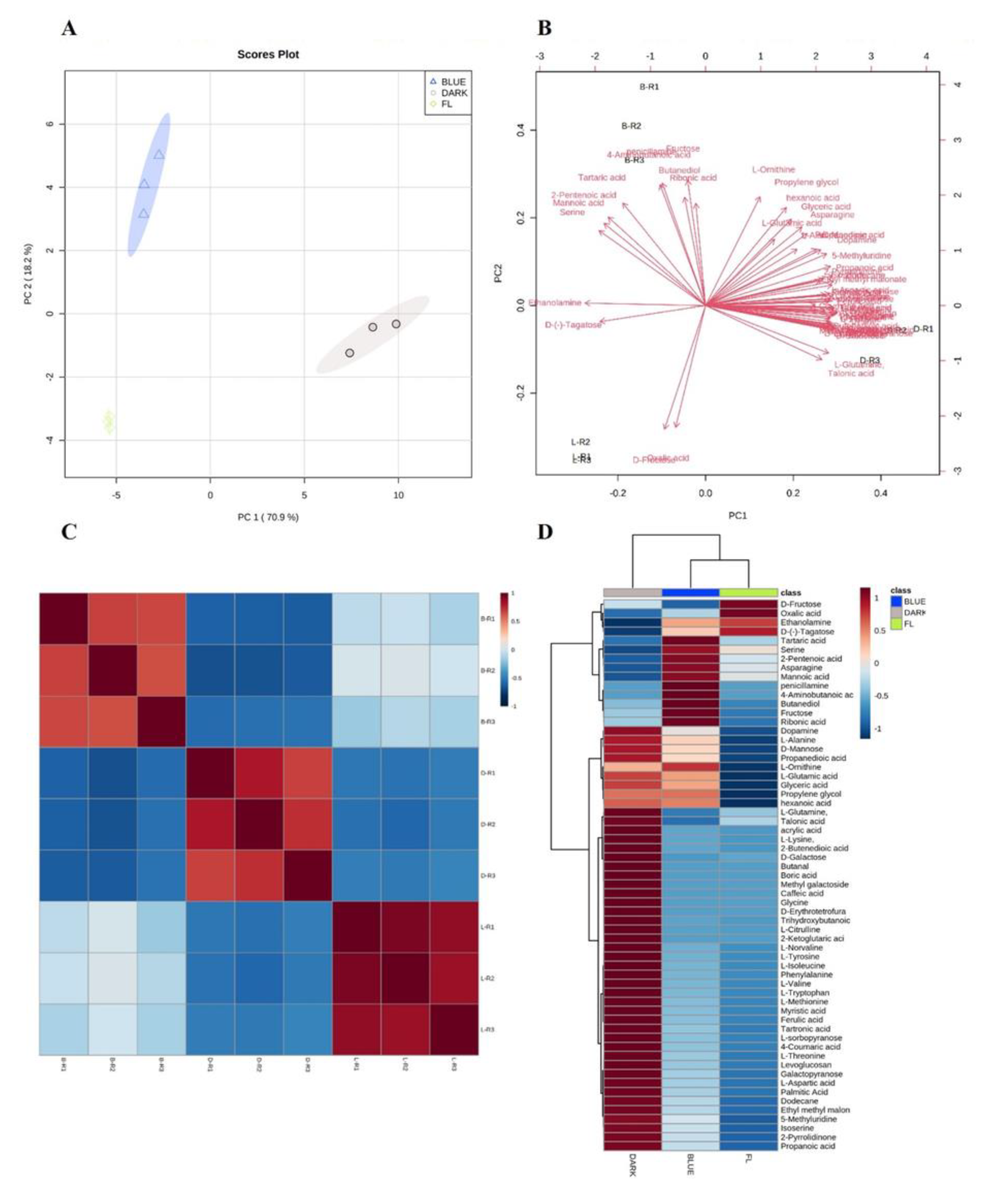
Figure 1. (A) Principal component analysis score; (B) loading plot; (C) correlation analysis; and (D) heat map from 53 significantly different detected compounds, as compared to control. Blue, gray, and green dots represent blue, dark, and FL treatment in the PCA plot, respectively. The heat map in D panel was presented with a red—blue color scale with red representing a higher metabolites level and blue for lower metabolites levels. A value of p < 0.05 was used to determine statistically significant difference.
Ijms 24 07768 g001
Figure 2. Primary metabolites contents in dark treatment: (A) amino acids, (B) carbohydrates and (C) organic acids. Values are means± standard deviation. Values with the same superscript letter are not significantly different at p < 0.05 level.
Ijms 24 07768 g002
Figure 3. HPLC chromatograms compression showing: (A) TIC of total ginsenosides in FL, blue—LED, and dark treatment; and (B) ginsenosides content in dark, FL and blue-LED treated ginseng. Column and error bars represent the means and standard deviation (n = 5). Columns with different letters for each specific ginsenosides are significantly different (p < 0.05).
Ijms 24 07768 g003
Figure 4. Anticancer effect of LED-treated ginseng on MCF7 cell: (A) cell viability assay for dark treatment; (B) comparison of cell viability among three different LED treatments; and (C) immunoblot analysis revealed the protein levels of PARP. GAPDH was used as an internal control. IC50 value of extract on MCF7 cell line. Each value represents the mean ± SD of three independent experiments.
Ijms 24 07768 g004
Figure 5. The inhibitory effect of LED-treated ginseng on MAPKs in LPS-stimulated BV2 cells: (A) FL as control, (B) blue-LED, (C) dark treatment. Western blot analysis on p38, ERK1/2 and JNK phosphorylation level in BV-2 cells. Cells were pre-treated with dark-, blue-, and FL-treated ginseng extract (1–25 µg/mL) for 2 h prior to LPS (1 µg/mL) exposure for 24 h. β-actin used as housekeeping protein.
Ijms 24 07768 g005
Table 1. Significantly different primary metabolites as compared to control in dark and blue-LED treated samples by GC-TOF-MS (p < 0.05). Three biological replications were analyzed. ND means “not detected”.
NORT(SEC)Identified CompoundFormulaTreatmentPeak Areap-Value
Organic Acids
1274.6Boric acidH3BO3FLND
Dark55,158 ± 49790.003
BlueND
2326.8Ethyl methyl malonateC6H10O4FLND
Dark2,648,561 ± 77,9050.000
Blue907,763 ± 450,128
3414.5Propanedioic acidC3H4O4FL11,837,242 ± 963,277
Dark26,345,064 ± 807,5970.000
Blue20,531,543 ± 1,296,0590.01
44714-Aminobutanoic acidC4H9NO2FLND
DarkND
Blue35,057,390 ± 5,863,5360.001
5490Glyceric acidC3H6O4FL941,334,205 ± 162,244,741
Dark2,894,792,786 ± 755,578,224
Blue2,514,331,459 ± 142,931,8240.01
6495.12-Butenedioic acidC6H8O4FL3,388,837 ± 3,149,704
Dark138,224,506 ± 14,064,4240.008
Blue38,551,250 ± 20,640,438
7603Trihydroxybutanoic acidC4H8O5FL6,946,451 ± 1,197,940
Dark102,284,588.33 ± 3,523,5810.000
Blue9,506,910 ± 901,734.6
8628Oxalic acidC2H2O4FL15,000,978 ± 2,279,165
DarkND
Blue5,048,996 ± 997,7370.03
9666.3Hexanoic acidC6H12O2FL6,361,231 ± 2,906,165
Dark25,147,663 ± 3,163,9630.03
Blue23,949,708 ± 4,094,7440.003
10703.3Ribonic acidC5H9O6FL10,727,733 ± 6,149,856
Dark13,826,780 ± 866,318
Blue23,317,685 ± 7,507,8670.01
11745.72-Ketoglutaric acidC5H6O5FL5,572,062 ± 1,486,034
Dark110,162,880 ± 33,201,3270.04
Blue6,303,697 ± 4,026,419
12775.5Acrylic acidC3H4O2FL17,604,061 ± 1,032,006
Dark463,504,027 ± 92,918,7120.01
Blue3,5014,339 ± 14,354,648
13780.44-Coumaric acidC9H8O3FLND
Dark8,441,648 ± 4,174,601
Blue1,794,541 ± 258,199.40.008
14785.4Tartronic acidC3H4O5FL2,751,360 ± 304,388
Dark27,093,984 ± 4,230,2080.001
Blue7,538,675 ± 1,832,0290.05
15826Tartaric acidC4H6O6FL3,5980,414 ± 2,407,682
DarkND
Blue109,798,116 ± 3,291,8420.000
16854.5Ferulic acidC10H10O4FLND
Dark19,429,576 ± 7,087,9940.05
Blue3,751,615 ± 607,3320.01
178712-Pentenoic acidC5H8O2FL2,427,381 ± 154,542
DarkND
Blue5,276,122 ± 243,4890.005
18936.2Propanoic acidC₃H₆O₂FLND
Dark4,142,119 ± 68,4300.000
Blue1,682,445 ± 172,7120.004
191484Mannoic acidC6H12O7FL103,339,880 ± 7,836,722
DarkND
Blue204,832,544 ± 15,541,7220.01
Amino Acids
20341.2L-ValineC5H11NO2FL648,024 ± 38,340
Dark21,271,780 ± 5,902,2370.03
Blue3,254,685 ± 1,830,534
21350.5L-AlanineC3H7NO2FL22,921,160 ± 2,208,680
Dark98,293,721 ± 15,568,0390.01
Blue64,064,382 ± 40,670,416
22506.2SerineC3H7NO3FL38,823,176 ± 3,976,486
DarkND
Blue66,865,059 ± 8,216,4460.01
23512.4IsoserineC3H7NO3FL1,655,877 ± 625,229
Dark5,951,890 ± 19,0490.01
Blue3,295,161 ± 1,544,319
24521.4L-ThreonineC4H9NO3FL26,596,544 ± 3,132,799
Dark106,533,538 ± 20,954,1600.03
Blue4,396,874 ± 792,5040.02
25562.2L-CitrullineC6H13N3O3FLND
Dark4,730,208 ± 335,0830.002
Blue68,736 ± 53,704
26589.9L-Aspartic acidC4H7NO4FL36,974,682 ± 3,990,717
Dark189,220,043 ± 58,130,9370.01
Blue76,458,966 ± 3,877,7460.006
27591.1L-MethionineC4H7NO4FL3,918,664 ± 755,737
Dark25,062,068 ± 579,2090.000
Blue6,590,229 ± 635,920
28634.6L-OrnithineC5H12N2O2FL25,776,495 ± 2,926,910
Dark114,425,820 ± 21,676,4070.02
Blue36,284,134 ± 3,763,759
29636.9L-Glutamic acidC5H9NO4FLND
Dark30,258,688 ± 4,385,8700.008
Blue22,155,999 ± 26,019,046
30589.9L-Aspartic acidC4H7NO4FL36,974,682 ± 3,990,717
Dark155,886,710 ± 18,716,0990.01
Blue76,458,966 ± 3,877,7460.006
31644.7PhenylalanineC9H11NO2FL22,839,808 ± 2,262,238
Dark89,290,303 ± 6,606,8220.005
Blue30,596,843 ± 2,963,0830.04
32676.7L-LysineC6H14N2O2FL6,874,649 ± 415,258
Dark120,874,869 ± 42,373,3900.05
Blue12,535,775 ± 3,810,434
33707.9L-GlutamineC5H10N2O3FL294,061,961 ± 52,227,860
Dark1,347,690,969 ± 20,051,9350.000
Blue285,096,879 ± 30,214,602
34662.9AsparagineC4H8N2O3FL51,785,180 ± 5,177,064
Dark150,042,799 ± 25,531,5610.02
Blue105,437,258 ± 14,148,7320.03
35781.7L-TyrosineC9H11NO3FL16,119,127 ± 1,204,190
Dark135,002,280 ± 26,479,4540.02
Blue28,514,941 ± 3,558,7000.02
36932.3L-TryptophanC11H12N2O2FL8,223,776 ± 1,547,891
Dark70,186,274 ± 6,300,2780.003
Blue16,435,122 ± 782,2770.02
Carbohydrates
37725.7D-MannoseC6H12O6FL107,738,818 ± 84,582,603
Dark543,957,084 ± 46,193,9390.02
Blue209,192,752 ± 48,618,900
38733.3L-sorbopyranoseC6H12O6FL316,158,562 ± 20,999,619
Dark697,213,182 ± 20,642,8080.001
Blue383,562,829 ± 121,668,384
39755.2D-FructoseC6H12O6FL253,630,327 ± 18,247,145
Dark107,158,254 ± 3,685,0790.009
Blue214,588,479 ± 48,192,251
40759.7D-(-)-TagatoseC6H12O6FL235,944,910 ± 19,541,216
Dark149,426,526 ± 7,315,2720.03
Blue205,866,117 ± 43,378,085
41809.3D-ErythrofuranoseC4H8O4FLND
Dark643,197,453 ± 38,582,7220.001
BlueND
42851D-GalactoseC6H12O6FL6,796,773 ± 416,588
Dark25,968,973 ± 686,27560.04
Blue6,104,887 ± 607,559
43876.4Caffeic acidC9H8O4FLND
Dark6,097,960 ± 405,3900.001
BlueND
44908.9Methyl galactosideC7H14O6FLND
Dark1,748,082 ± 248,7000.008
BlueND
451020.5LevoglucosanC6H10O5FL3,822,565 ± 81,039
Dark18,672,292 ± 536,75400.05
Blue7,096,685 ± 264,6840.001
461160.6GalactopyranoseC6H12O6FL1,243,798 ± 314,085
Dark3,954,698 ± 428,9170.01
Blue1,937,122 ± 233,8530.02
Others
d47410DodecaneC12H26FLND
Dark2,089,678 ± 289,3950.007
Blue750,133 ± 109,0950.08
48453.8EthanolamineC2H7NOFL27,440,780 ± 624,097
Dark1,463,234 ± 97,7210.000
Blue22,216,551 ± 8,680,885
49533DopamineC8H11NO2FLND
Dark1,463,234 ± 97,7210.000
Blue2,322,461 ± 76,4350.000
50608PenicillamineC5H11NO2SFLND
DarkND
Blue2,036,085 ± 189,3340.003
51823.4Palmitic AcidC16H32O2FL12,165,495 ± 236,559
Dark30,359,205 ± 4,795,8210.02
Blue16,927,244 ± 2,657,439
52927Myristic acidC14H28O2FL7,061,525 ± 491,473
Dark12,418,666 ± 1,559,4380.02
Blue7,921,258 ± 489,889
531467.75-MethyluridineC10H14N2O6FLND
Dark8,401,912 ± 758,2320.003
Blue3,838,136 ± 443,1750.005
Disclaimer/Publisher’s Note: The statements, opinions and data contained in all publications are solely those of the individual author(s) and contributor(s) and not of MDPI and/or the editor(s). MDPI and/or the editor(s) disclaim responsibility for any injury to people or property resulting from any ideas, methods, instructions or products referred to in the content.