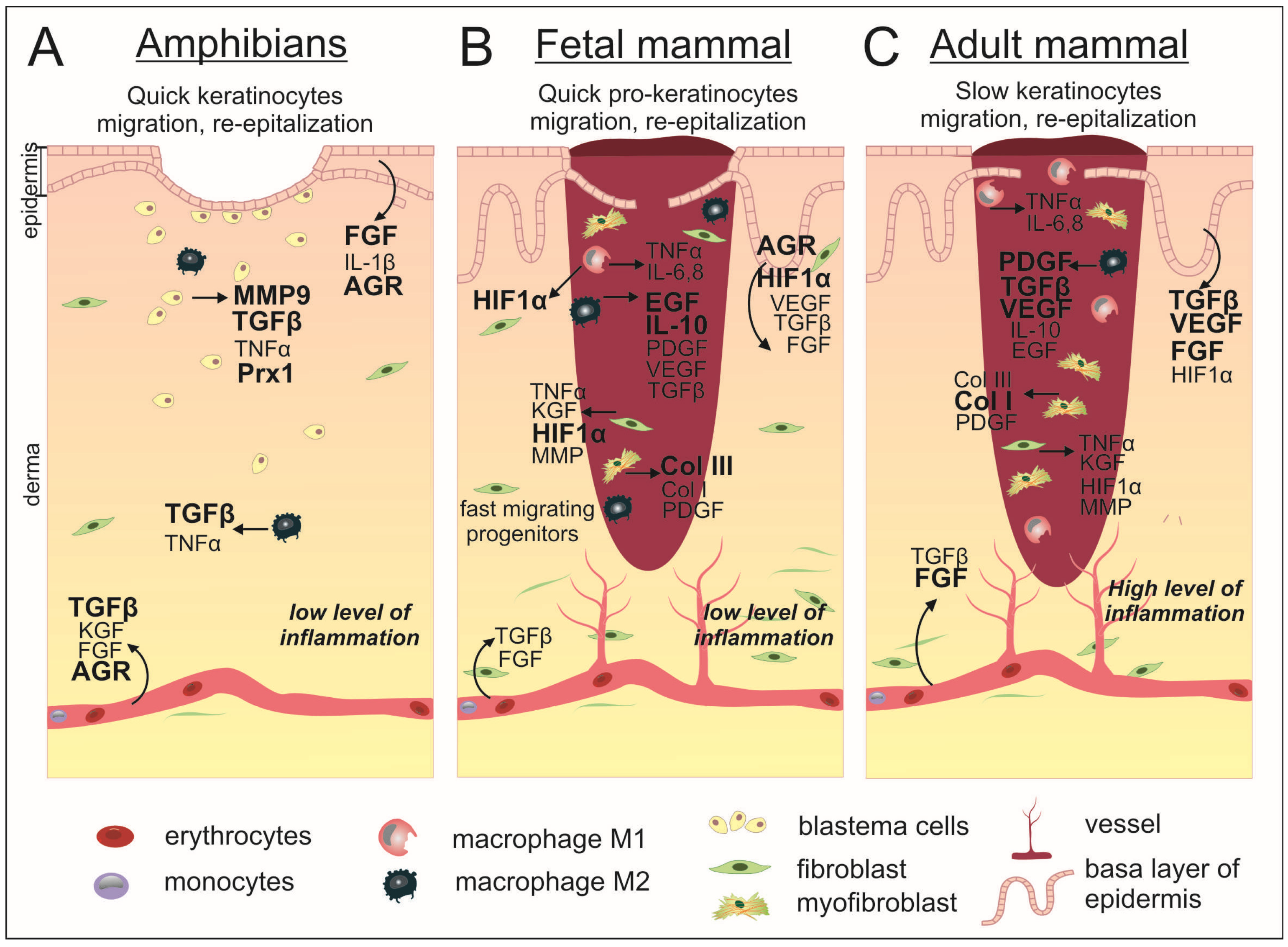
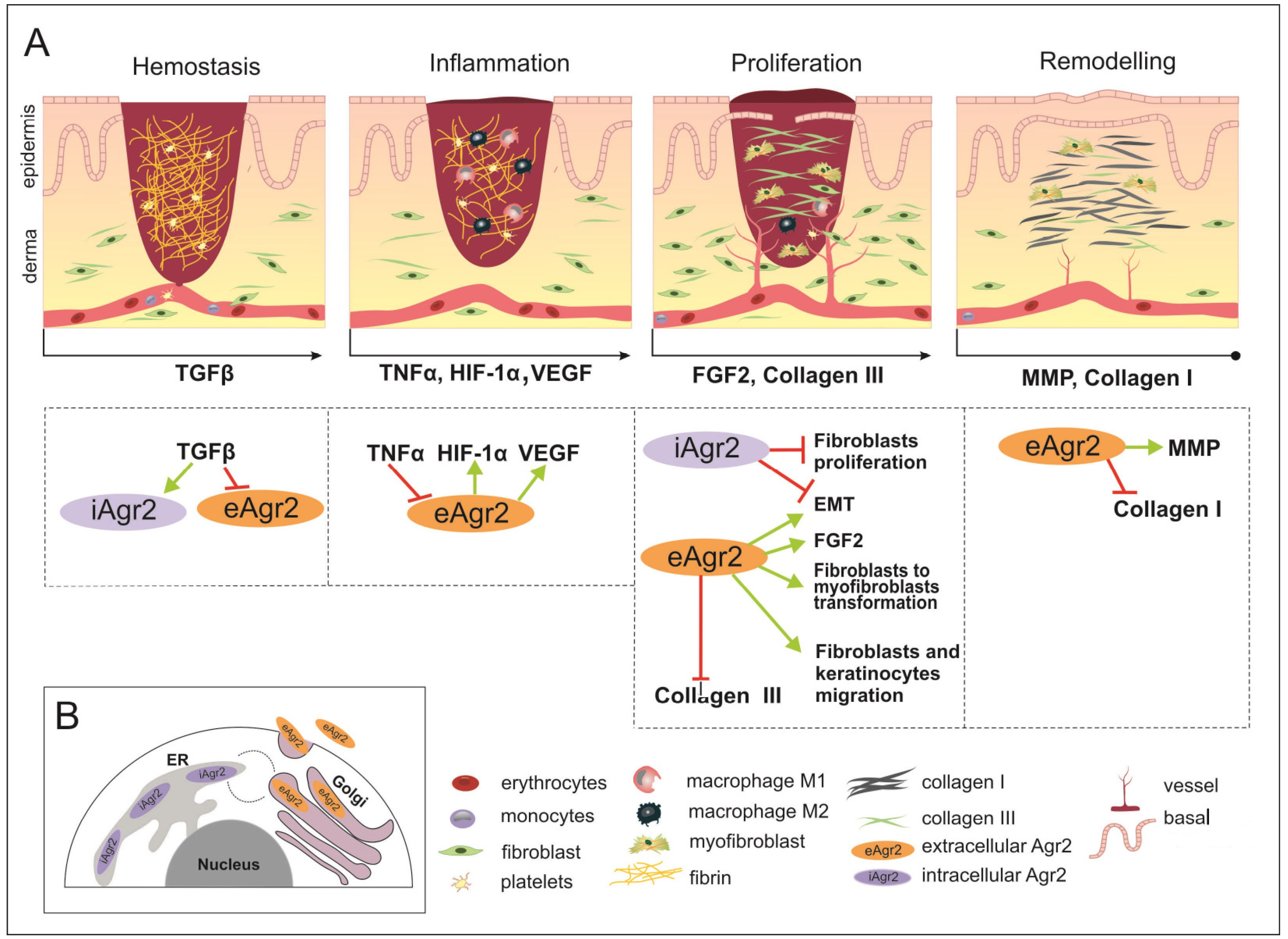
Potential Role of AGR2 for Mammalian Skin Wound Healing
Abstract
:1. Introduction
2. AGR Family of Protein Disulfide Isomerases (PDI)
2.1. AGR Protein Structural Characteristics
2.2. AGR Proteins in Anamniotes Regeneration
2.3. Ambivalent Mammalian AGR Proteins
3. Mammalian Acute Wound Healing: Stages and Molecular Regulators
3.1. Hemostasis
3.2. Inflammation
3.3. Proliferation
3.4. Remodeling
4. Conclusions
Author Contributions
Funding
Institutional Review Board Statement
Informed Consent Statement
Data Availability Statement
Acknowledgments
Conflicts of Interest
References
- Lévesque, M.; Villiard, E.; Roy, S. Skin Wound Healing in Axolotls: A Scarless Process. J. Exp. Zool. B Mol. Dev. Evol. 2010, 314, 684–697. [Google Scholar] [CrossRef] [PubMed]
- Yokoyama, H.; Maruoka, T.; Aruga, A.; Amano, T.; Ohgo, S.; Shiroishi, T.; Tamura, K. Prx-1 Expression in Xenopus Laevis Scarless Skin-Wound Healing and Its Resemblance to Epimorphic Regeneration. J. Investig. Dermatol. 2011, 131, 2477–2485. [Google Scholar] [CrossRef] [PubMed]
- Bertolotti, E.; Malagoli, D.; Franchini, A. Skin Wound Healing in Different Aged Xenopus Laevis. J. Morphol. 2013, 274, 956–964. [Google Scholar] [CrossRef] [PubMed]
- Rowlatt, U. Intrauterine Wound Healing in a 20 Week Human Fetus. Virchows Arch. A Pathol. Anat. Histol. 1979, 381, 353–361. [Google Scholar] [CrossRef]
- Whitby, D.J.; Ferguson, M.W. The Extracellular Matrix of Lip Wounds in Fetal, Neonatal and Adult Mice. Dev. Camb. Engl. 1991, 112, 651–668. [Google Scholar] [CrossRef]
- Ferguson, M.W.J.; O’Kane, S. Scar-Free Healing: From Embryonic Mechanisms to Adult Therapeutic Intervention. Philos. Trans. R. Soc. Lond. B. Biol. Sci. 2004, 359, 839–850. [Google Scholar] [CrossRef]
- Reinke, J.M.; Sorg, H. Wound Repair and Regeneration. Eur. Surg. Res. 2012, 49, 35–43. [Google Scholar] [CrossRef]
- Cass, D.L.; Bullard, K.M.; Sylvester, K.G.; Yang, E.Y.; Longaker, M.T.; Adzick, N.S. Wound Size and Gestational Age Modulate Scar Formation in Fetal Wound Repair. J. Pediatr. Surg. 1997, 32, 411–415. [Google Scholar] [CrossRef]
- Marshall, C.D.; Hu, M.S.; Leavitt, T.; Barnes, L.A.; Lorenz, H.P.; Longaker, M.T. Cutaneous Scarring: Basic Science, Current Treatments, and Future Directions. Adv. Wound Care 2018, 7, 29–45. [Google Scholar] [CrossRef]
- Lorenz, H.P.; Whitby, D.J.; Longaker, M.T.; Adzick, N.S. Fetal Wound Healing The Ontogeny of Scar Formation in the Non-Human Primate. Ann. Surg. 1993, 217, 391–396. [Google Scholar] [CrossRef]
- Colwell, A.S.; Krummel, T.M.; Longaker, M.T.; Lorenz, H.P. An in Vivo Mouse Excisional Wound Model of Scarless Healing. Plast. Reconstr. Surg. 2006, 117, 2292–2296. [Google Scholar] [CrossRef]
- Kuliyev, E.; Doherty, J.R.; Mead, P.E. Expression of Xenopus Suppressor of Cytokine Signaling 3 (XSOCS3) Is Induced by Epithelial Wounding. Dev. Dyn. Off. Publ. Am. Assoc. Anat. 2005, 233, 1123–1130. [Google Scholar] [CrossRef]
- Suzuki, M.; Satoh, A.; Ide, H.; Tamura, K. Nerve-Dependent and -Independent Events in Blastema Formation during Xenopus Froglet Limb Regeneration. Dev. Biol. 2005, 286, 361–375. [Google Scholar] [CrossRef]
- Liu, Y.; Ilinski, A.; Gerstenfeld, L.C.; Bragdon, B. Prx1 Cell Subpopulations Identified in Various Tissues with Diverse Quiescence and Activation Ability Following Fracture and BMP2 Stimulation. Front. Physiol. 2023, 14, 1106474. [Google Scholar] [CrossRef]
- Gong, W.; Ekmu, B.; Wang, X.; Lu, Y.; Wan, L. AGR2-Induced Glucose Metabolism Facilitated the Progression of Endometrial Carcinoma via Enhancing the MUC1/HIF-1α Pathway. Hum. Cell 2020, 33, 790–800. [Google Scholar] [CrossRef]
- Aberger, F.; Weidinger, G.; Grunz, H.; Richter, K. Anterior Specification of Embryonic Ectoderm: The Role of the Xenopus Cement Gland-Specific Gene XAG-2. Mech. Dev. 1998, 72, 115–130. [Google Scholar] [CrossRef]
- Ivanova, A.S.; Tereshina, M.B.; Araslanova, K.R.; Martynova, N.Y.; Zaraisky, A.G. The Secreted Protein Disulfide Isomerase Ag1 Lost by Ancestors of Poorly Regenerating Vertebrates Is Required for Xenopus Laevis Tail Regeneration. Front. Cell Dev. Biol. 2021, 9, 738940. [Google Scholar] [CrossRef]
- Ivanova, A.S.; Tereshina, M.B.; Ermakova, G.V.; Belousov, V.V.; Zaraisky, A.G. Agr Genes, Missing in Amniotes, Are Involved in the Body Appendages Regeneration in Frog Tadpoles. Sci. Rep. 2013, 3, 1279. [Google Scholar] [CrossRef]
- Gupta, A.; Wodziak, D.; Tun, M.; Bouley, D.M.; Lowe, A.W. Loss of Anterior Gradient 2 (Agr2) Expression Results in Hyperplasia and Defective Lineage Maturation in the Murine Stomach. J. Biol. Chem. 2013, 288, 4321–4333. [Google Scholar] [CrossRef]
- Patel, P.; Clarke, C.; Barraclough, D.L.; Jowitt, T.A.; Rudland, P.S.; Barraclough, R.; Lian, L.-Y. Metastasis-Promoting Anterior Gradient 2 Protein Has a Dimeric Thioredoxin Fold Structure and a Role in Cell Adhesion. J. Mol. Biol. 2013, 425, 929–943. [Google Scholar] [CrossRef]
- Murawala, P.; Tanaka, E.M.; Currie, J.D. Regeneration: The Ultimate Example of Wound Healing. Semin. Cell Dev. Biol. 2012, 23, 954–962. [Google Scholar] [CrossRef] [PubMed]
- Kumar, A.; Godwin, J.W.; Gates, P.B.; Garza-Garcia, A.A.; Brockes, J.P. Molecular Basis for the Nerve Dependence of Limb Regeneration in an Adult Vertebrate. Science 2007, 318, 772–777. [Google Scholar] [CrossRef] [PubMed]
- Ivanova, A.S.; Shandarin, I.N.; Ermakova, G.V.; Minin, A.A.; Tereshina, M.B.; Zaraisky, A.G. The Secreted Factor Ag1 Missing in Higher Vertebrates Regulates Fins Regeneration in Danio Rerio. Sci. Rep. 2015, 5, 8123. [Google Scholar] [CrossRef] [PubMed]
- Zhu, Q.; Mangukiya, H.B.; Mashausi, D.S.; Guo, H.; Negi, H.; Merugu, S.B.; Wu, Z.; Li, D. Anterior Gradient 2 Is Induced in Cutaneous Wound and Promotes Wound Healing through Its Adhesion Domain. FEBS J. 2017, 284, 2856–2869. [Google Scholar] [CrossRef]
- Komiya, T.; Tanigawa, Y.; Hirohashi, S. Cloning of the Gene Gob-4, Which Is Expressed in Intestinal Goblet Cells in Mice. Biochim. Biophys. Acta 1999, 1444, 434–438. [Google Scholar] [CrossRef]
- Park, S.-W.; Zhen, G.; Verhaeghe, C.; Nakagami, Y.; Nguyenvu, L.T.; Barczak, A.J.; Killeen, N.; Erle, D.J. The Protein Disulfide Isomerase AGR2 Is Essential for Production of Intestinal Mucus. Proc. Natl. Acad. Sci. USA 2009, 106, 6950–6955. [Google Scholar] [CrossRef]
- Coufalova, D.; Remnant, L.; Hernychova, L.; Muller, P.; Healy, A.; Kannan, S.; Westwood, N.; Verma, C.S.; Vojtesek, B.; Hupp, T.R.; et al. An Inter-Subunit Protein-Peptide Interface That Stabilizes the Specific Activity and Oligomerization of the AAA+ Chaperone Reptin. J. Proteom. 2019, 199, 89–101. [Google Scholar] [CrossRef]
- Hong, X.; Li, Z.-X.; Hou, J.; Zhang, H.-Y.; Zhang, C.-Y.; Zhang, J.; Sun, H.; Pang, L.-H.; Wang, T.; Deng, Z.-H. Effects of ER-Resident and Secreted AGR2 on Cell Proliferation, Migration, Invasion, and Survival in PANC-1 Pancreatic Cancer Cells. BMC Cancer 2021, 21, 33. [Google Scholar] [CrossRef]
- Zhao, F.; Edwards, R.; Dizon, D.; Mastroianni, J.R.; Geyfman, M.; Ouellette, A.J.; Andersen, B.; Lipkin, S.M. Disruption of Paneth and Goblet Cell Homeostasis and Increased Endoplasmic Reticulum Stress in Agr2−/− Mice. Dev. Biol. 2010, 338, 270–279. [Google Scholar] [CrossRef]
- Liu, D.; Rudland, P.S.; Sibson, D.R.; Platt-Higgins, A.; Barraclough, R. Human Homologue of Cement Gland Protein, a Novel Metastasis Inducer Associated with Breast Carcinomas. Cancer Res. 2005, 65, 3796–3805. [Google Scholar] [CrossRef]
- Edgell, T.A.; Barraclough, D.L.; Rajic, A.; Dhulia, J.; Lewis, K.J.; Armes, J.E.; Barraclough, R.; Rudland, P.S.; Rice, G.E.; Autelitano, D.J. Increased Plasma Concentrations of Anterior Gradient 2 Protein Are Positively Associated with Ovarian Cancer. Clin. Sci. 2010, 118, 717–725. [Google Scholar] [CrossRef]
- Bai, Z.; Ye, Y.; Liang, B.; Xu, F.; Zhang, H.; Zhang, Y.; Peng, J.; Shen, D.; Cui, Z.; Zhang, Z.; et al. Proteomics-Based Identification of a Group of Apoptosis-Related Proteins and Biomarkers in Gastric Cancer. Int. J. Oncol. 2011, 38, 375–383. [Google Scholar] [CrossRef]
- de Moraes, C.L.; Cruz e Melo, N.; Valoyes, M.A.V.; Naves do Amaral, W. AGR2 and AGR3 Play an Important Role in the Clinical Characterization and Prognosis of Basal like Breast Cancer. Clin. Breast Cancer 2022, 22, e242–e252. [Google Scholar] [CrossRef]
- Brychtova, V.; Hermanova, M.; Karasek, P.; Lenz, J.; Selingerova, I.; Vojtesek, B.; Kala, Z.; Hrstka, R. Anterior Gradient 2 and Mucin 4 Expression Mirrors Tumor Cell Differentiation in Pancreatic Adenocarcinomas, but Aberrant Anterior Gradient 2 Expression Predicts Worse Patient Outcome in Poorly Differentiated Tumors. Pancreas 2014, 43, 75–81. [Google Scholar] [CrossRef]
- Delom, F.; Mohtar, M.A.; Hupp, T.; Fessart, D. The Anterior Gradient-2 Interactome. Am. J. Physiol. Cell Physiol. 2020, 318, C40–C47. [Google Scholar] [CrossRef]
- Ryu, J.; Park, S.G.; Lee, P.Y.; Cho, S.; Lee, D.H.; Kim, G.H.; Kim, J.-H.; Park, B.C. Dimerization of Pro-Oncogenic Protein Anterior Gradient 2 Is Required for the Interaction with BiP/GRP78. Biochem. Biophys. Res. Commun. 2013, 430, 610–615. [Google Scholar] [CrossRef]
- Boisteau, E.; Posseme, C.; Di Modugno, F.; Edeline, J.; Coulouarn, C.; Hrstka, R.; Martisova, A.; Delom, F.; Treton, X.; Eriksson, L.A.; et al. Anterior Gradient Proteins in Gastrointestinal Cancers: From Cell Biology to Pathophysiology. Oncogene 2022, 41, 4673–4685. [Google Scholar] [CrossRef]
- Clarke, D.J.; Murray, E.; Faktor, J.; Mohtar, A.; Vojtesek, B.; MacKay, C.L.; Smith, P.L.; Hupp, T.R. Mass Spectrometry Analysis of the Oxidation States of the Pro-Oncogenic Protein Anterior Gradient-2 Reveals Covalent Dimerization via an Intermolecular Disulphide Bond. Biochim. Biophys. Acta 2016, 1864, 551–561. [Google Scholar] [CrossRef]
- Maurel, M.; Obacz, J.; Avril, T.; Ding, Y.-P.; Papadodima, O.; Treton, X.; Daniel, F.; Pilalis, E.; Hörberg, J.; Hou, W.; et al. Control of Anterior GRadient 2 (AGR2) Dimerization Links Endoplasmic Reticulum Proteostasis to Inflammation. EMBO Mol. Med. 2019, 11, e10120. [Google Scholar] [CrossRef]
- Wang, Z.; Hao, Y.; Lowe, A.W. The Adenocarcinoma-Associated Antigen, AGR2, Promotes Tumor Growth, Cell Migration, and Cellular Transformation. Cancer Res. 2008, 68, 492–497. [Google Scholar] [CrossRef]
- Fletcher, G.C.; Patel, S.; Tyson, K.; Adam, P.J.; Schenker, M.; Loader, J.A.; Daviet, L.; Legrain, P.; Parekh, R.; Harris, A.L.; et al. HAG-2 and HAG-3, Human Homologues of Genes Involved in Differentiation, Are Associated with Oestrogen Receptor-Positive Breast Tumours and Interact with Metastasis Gene C4.4a and Dystroglycan. Br. J. Cancer 2003, 88, 579–585. [Google Scholar] [CrossRef] [PubMed]
- Gray, T.A.; Alsamman, K.; Murray, E.; Sims, A.H.; Hupp, T.R. Engineering a Synthetic Cell Panel to Identify Signalling Components Reprogrammed by the Cell Growth Regulator Anterior Gradient-2. Mol. Biosyst. 2014, 10, 1409–1425. [Google Scholar] [CrossRef] [PubMed]
- Ramachandran, V.; Arumugam, T.; Wang, H.; Logsdon, C.D. Anterior Gradient 2 Is Expressed and Secreted during the Development of Pancreatic Cancer and Promotes Cancer Cell Survival. Cancer Res. 2008, 68, 7811–7818. [Google Scholar] [CrossRef] [PubMed]
- Park, K.; Chung, Y.J.; So, H.; Kim, K.; Park, J.; Oh, M.; Jo, M.; Choi, K.; Lee, E.-J.; Choi, Y.-L.; et al. AGR2, a Mucinous Ovarian Cancer Marker, Promotes Cell Proliferation and Migration. Exp. Mol. Med. 2011, 43, 91–100. [Google Scholar] [CrossRef] [PubMed]
- Guo, H.; Zhu, Q.; Yu, X.; Merugu, S.B.; Mangukiya, H.B.; Smith, N.; Li, Z.; Zhang, B.; Negi, H.; Rong, R.; et al. Tumor-Secreted Anterior Gradient-2 Binds to VEGF and FGF2 and Enhances Their Activities by Promoting Their Homodimerization. Oncogene 2017, 36, 5098–5109. [Google Scholar] [CrossRef]
- Wodziak, D.; Dong, A.; Basin, M.F.; Lowe, A.W. Anterior Gradient 2 (AGR2) Induced Epidermal Growth Factor Receptor (EGFR) Signaling Is Essential for Murine Pancreatitis-Associated Tissue Regeneration. PLoS ONE 2016, 11, e0164968. [Google Scholar] [CrossRef]
- Takada, Y.; Ye, X.; Simon, S. The Integrins. Genome Biol. 2007, 8, 215. [Google Scholar] [CrossRef]
- Fitridge, R.; Thompson, M. (Eds.) Mechanisms of Vascular Disease: A Reference Book for Vascular Specialists; University of Adelaide Press: Adelaide, Australia, 2011. [Google Scholar]
- Clark, R.A. Regulation of Fibroplasia in Cutaneous Wound Repair. Am. J. Med. Sci. 1993, 306, 42–48. [Google Scholar] [CrossRef]
- Walraven, M.; Gouverneur, M.; Middelkoop, E.; Beelen, R.H.J.; Ulrich, M.M.W. Altered TGF-β Signaling in Fetal Fibroblasts: What Is Known about the Underlying Mechanisms? Wound Repair Regen. 2014, 22, 3–13. [Google Scholar] [CrossRef]
- Hart, J. Inflammation. 1: Its Role in the Healing of Acute Wounds. J. Wound Care 2002, 11, 205–209. [Google Scholar] [CrossRef]
- Beanes, S.R.; Dang, C.; Soo, C.; Wang, Y.; Urata, M.; Ting, K.; Fonkalsrud, E.W.; Benhaim, P.; Hedrick, M.H.; Atkinson, J.B.; et al. Down-Regulation of Decorin, a Transforming Growth Factor-Beta Modulator, Is Associated with Scarless Fetal Wound Healing. J. Pediatr. Surg. 2001, 36, 1666–1671. [Google Scholar] [CrossRef]
- Olutoye, O.O.; Yager, D.R.; Cohen, I.K.; Diegelmann, R.F. Lower Cytokine Release by Fetal Porcine Platelets: A Possible Explanation for Reduced Inflammation after Fetal Wounding. J. Pediatr. Surg. 1996, 31, 91–95. [Google Scholar] [CrossRef] [PubMed]
- Vieujean, S.; Hu, S.; Bequet, E.; Salee, C.; Massot, C.; Bletard, N.; Pierre, N.; Quesada Calvo, F.; Baiwir, D.; Mazzucchelli, G.; et al. Potential Role of Epithelial Endoplasmic Reticulum Stress and Anterior Gradient Protein 2 Homologue in Crohn’s Disease Fibrosis. J. Crohn’s Colitis 2021, 15, 1737–1750. [Google Scholar] [CrossRef] [PubMed]
- Wagers, A.J.; Christensen, J.L.; Weissman, I.L. Cell Fate Determination from Stem Cells. Gene Ther. 2002, 9, 606–612. [Google Scholar] [CrossRef] [PubMed]
- Mizuuchi, Y.; Aishima, S.; Ohuchida, K.; Shindo, K.; Fujino, M.; Hattori, M.; Miyazaki, T.; Mizumoto, K.; Tanaka, M.; Oda, Y. Anterior Gradient 2 Downregulation in a Subset of Pancreatic Ductal Adenocarcinoma Is a Prognostic Factor Indicative of Epithelial-Mesenchymal Transition. Lab. Investig. 2015, 95, 193–206. [Google Scholar] [CrossRef]
- Mori, R.; Kondo, T.; Ohshima, T.; Ishida, Y.; Mukaida, N. Accelerated Wound Healing in Tumor Necrosis Factor Receptor P55-Deficient Mice with Reduced Leukocyte Infiltration. FASEB J. Off. Publ. Fed. Am. Soc. Exp. Biol. 2002, 16, 963–974. [Google Scholar] [CrossRef]
- Eming, S.A.; Krieg, T.; Davidson, J.M. Inflammation in Wound Repair: Molecular and Cellular Mechanisms. J. Investig. Dermatol. 2007, 127, 514–525. [Google Scholar] [CrossRef]
- Braun, S.; Hanselmann, C.; Gassmann, M.G.; auf dem Keller, U.; Born-Berclaz, C.; Chan, K.; Kan, Y.W.; Werner, S. Nrf2 Transcription Factor, a Novel Target of Keratinocyte Growth Factor Action Which Regulates Gene Expression and Inflammation in the Healing Skin Wound. Mol. Cell. Biol. 2002, 22, 5492–5505. [Google Scholar] [CrossRef]
- Ye, X.; Sun, M. AGR2 Ameliorates Tumor Necrosis Factor-α-Induced Epithelial Barrier Dysfunction via Suppression of NF-ΚB P65-Mediated MLCK/p-MLC Pathway Activation. Int. J. Mol. Med. 2017, 39, 1206–1214. [Google Scholar] [CrossRef]
- Vihersaari, T.; Kivisaari, J.; Ninikoski, J. Effect of Changes in Inspired Oxygen Tension on Wound Metabolism. Ann. Surg. 1974, 179, 889–895. [Google Scholar] [CrossRef]
- Bishop, A. Role of Oxygen in Wound Healing. J. Wound Care 2008, 17, 399–402. [Google Scholar] [CrossRef]
- Wang, X.; Shen, K.; Wang, J.; Liu, K.; Wu, G.; Li, Y.; Luo, L.; Zheng, Z.; Hu, D. Hypoxic Preconditioning Combined with Curcumin Promotes Cell Survival and Mitochondrial Quality of Bone Marrow Mesenchymal Stem Cells, and Accelerates Cu-taneous Wound Healing via PGC-1α/SIRT3/HIF-1α Signaling. Free Radic. Biol. Med. 2020, 159, 164–176. [Google Scholar] [CrossRef]
- Luo, J.; Qiao, F.; Yin, X. Hypoxia Induces FGF2 Production by Vascular Endothelial Cells and Alters MMP9 and TIMP1 Ex-pression in Extravillous Trophoblasts and Their Invasiveness in a Cocultured Model. J. Reprod. Dev. 2011, 57, 84–91. [Google Scholar] [CrossRef]
- Kuwabara, K.; Ogawa, S.; Matsumoto, M.; Koga, S.; Clauss, M.; Pinsky, D.J.; Lyn, P.; Leavy, J.; Witte, L.; Joseph-Silverstein, J. Hypoxia-Mediated Induction of Acidic/Basic Fibroblast Growth Factor and Platelet-Derived Growth Factor in Mononuclear Phagocytes Stimulates Growth of Hypoxic Endothelial Cells. Proc. Natl. Acad. Sci. USA 1995, 92, 4606–4610. [Google Scholar] [CrossRef]
- Scheid, A.; Wenger, R.H.; Christina, H.; Camenisch, I.; Ferenc, A.; Stauffer, U.G.; Gassmann, M.; Meuli, M. Hypoxia-Regulated Gene Expression in Fetal Wound Regeneration and Adult Wound Repair. Pediatr. Surg. Int. 2000, 16, 232–236. [Google Scholar] [CrossRef]
- Zweitzig, D.R.; Smirnov, D.A.; Connelly, M.C.; Terstappen, L.W.M.M.; O’Hara, S.M.; Moran, E. Physiological Stress Induces the Metastasis Marker AGR2 in Breast Cancer Cells. Mol. Cell. Biochem. 2007, 306, 255–260. [Google Scholar] [CrossRef]
- Hong, X.-Y.; Wang, J.; Li, Z. AGR2 Expression Is Regulated by HIF-1 and Contributes to Growth and Angiogenesis of Glio-blastoma. Cell Biochem. Biophys. 2013, 67, 1487–1495. [Google Scholar] [CrossRef]
- Bao, P.; Kodra, A.; Tomic-Canic, M.; Golinko, M.S.; Ehrlich, H.P.; Brem, H. The Role of Vascular Endothelial Growth Factor in Wound Healing. J. Surg. Res. 2009, 153, 347–358. [Google Scholar] [CrossRef]
- Sorg, H.; Krueger, C.; Vollmar, B. Intravital Insights in Skin Wound Healing Using the Mouse Dorsal Skin Fold Chamber. J. Anat. 2007, 211, 810–818. [Google Scholar] [CrossRef]
- Wilgus, T.A.; Ferreira, A.M.; Oberyszyn, T.M.; Bergdall, V.K.; Dipietro, L.A. Regulation of Scar Formation by Vascular Endothelial Growth Factor. Lab Investig. 2008, 88, 579–590. [Google Scholar] [CrossRef]
- Jia, M.; Guo, Y.; Zhu, D.; Zhang, N.; Li, L.; Jiang, J.; Dong, Y.; Xu, Q.; Zhang, X.; Wang, M.; et al. Pro-Metastatic Activity of AGR2 Interrupts Angiogenesis Target Bevacizumab Efficiency via Direct Interaction with VEGFA and Activation of NF-ΚB Pathway. Biochim. Biophys. Acta Mol. Basis Dis. 2018, 1864 Pt 5A, 1622–1633. [Google Scholar] [CrossRef]
- Buchanan, E.P.; Longaker, M.T.; Lorenz, H.P. Fetal Skin Wound Healing. Adv. Clin. Chem. 2009, 48, 137–161. [Google Scholar] [CrossRef] [PubMed]
- Dang, C.M.; Beanes, S.R.; Soo, C.; Ting, K.; Benhaim, P.; Hedrick, M.H.; Lorenz, H.P. Decreased Expression of Fibroblast and Keratinocyte Growth Factor Isoforms and Receptors during Scarless Repair. Plast. Reconstr. Surg. 2003, 111, 1969–1979. [Google Scholar] [CrossRef] [PubMed]
- Hinz, B. Formation and Function of the Myofibroblast during Tissue Repair. J. Investig. Dermatol. 2007, 127, 526–537. [Google Scholar] [CrossRef] [PubMed]
- Al-Qattan, M.M.; Shier, M.K.; Abd-Alwahed, M.M.; Mawlana, O.H.; El-Wetidy, M.S.; Bagayawa, R.S.; Ali, H.H.; Al-Nbaheen, M.S.; Aldahmash, A.M. Salamander-Derived, Human-Optimized NAG Protein Suppresses Collagen Synthesis and Increases Collagen Degradation in Primary Human Fibroblasts. BioMed Res. Int. 2013, 2013, 384091. [Google Scholar] [CrossRef]
- Al-Qattan, M.M.; Abd-Al Wahed, M.M.; Hawary, K.; Alhumidi, A.A.; Shier, M.K. Recombinant NAG (a Salamander-Derived Protein) Decreases the Formation of Hypertrophic Scarring in the Rabbit Ear Model. BioMed Res. Int. 2014, 2014, 121098. [Google Scholar] [CrossRef]
- Suda, S.; Williams, H.; Medbury, H.J.; Holland, A.J.A. A Review of Monocytes and Monocyte-Derived Cells in Hypertrophic Scarring Post Burn. J. Burn Care Res. 2016, 37, 265–272. [Google Scholar] [CrossRef]
- Gimenez, A.; Kopkin, R.; Chang, D.K.; Belfort, M.; Reece, E.M. Advances in Fetal Surgery: Current and Future Relevance in Plastic Surgery. Semin. Plast. Surg. 2019, 33, 204–212. [Google Scholar] [CrossRef]
- Martin, P. Wound Healing--Aiming for Perfect Skin Regeneration. Science 1997, 276, 75–81. [Google Scholar] [CrossRef]
- Werner, S.; Grose, R. Regulation of Wound Healing by Growth Factors and Cytokines. Physiol. Rev. 2003, 83, 835–870. [Google Scholar] [CrossRef]
- Jacinto, A.; Martinez-Arias, A.; Martin, P. Mechanisms of Epithelial Fusion and Repair. Nat. Cell Biol. 2001, 3, E117–E123. [Google Scholar] [CrossRef]
- Nauta, A.; Gurtner, G.C.; Longaker, M.T. Wound Healing and Regenerative Strategies. Oral Dis. 2011, 17, 541–549. [Google Scholar] [CrossRef] [PubMed]
- Jaguin, M.; Houlbert, N.; Fardel, O.; Lecureur, V. Polarization Profiles of Human M-CSF-Generated Macrophages and Com-parison of M1-Markers in Classically Activated Macrophages from GM-CSF and M-CSF Origin. Cell. Immunol. 2013, 281, 51–61. [Google Scholar] [CrossRef]
- Kim, S.Y.; Nair, M.G. Macrophages in Wound Healing: Activation and Plasticity. Immunol. Cell Biol. 2019, 97, 258–267. [Google Scholar] [CrossRef]
- Assoian, R.K.; Fleurdelys, B.E.; Stevenson, H.C.; Miller, P.J.; Madtes, D.K.; Raines, E.W.; Ross, R.; Sporn, M.B. Expression and Secretion of Type Beta Transforming Growth Factor by Activated Human Macrophages. Proc. Natl. Acad. Sci. USA 1987, 84, 6020–6024. [Google Scholar] [CrossRef] [PubMed]
- Barrientos, S.; Stojadinovic, O.; Golinko, M.S.; Brem, H.; Tomic-Canic, M. Growth Factors and Cytokines in Wound Healing. Wound Repair Regen. 2008, 16, 585–601. [Google Scholar] [CrossRef]
- Walraven, M.; Talhout, W.; Beelen, R.H.J.; van Egmond, M.; Ulrich, M.M.W. Healthy Human Second-Trimester Fetal Skin Is Deficient in Leukocytes and Associated Homing Chemokines. Wound Repair Regen. 2016, 24, 533–541. [Google Scholar] [CrossRef]
- Sommerova, L.; Ondrouskova, E.; Vojtesek, B.; Hrstka, R. Suppression of AGR2 in a TGF-β-Induced Smad Regulatory Pathway Mediates Epithelial-Mesenchymal Transition. BMC Cancer 2017, 17, 546. [Google Scholar] [CrossRef]
- Thiery, J.P. Epithelial-Mesenchymal Transitions in Tumour Progression. Nat. Rev. Cancer 2002, 2, 442–454. [Google Scholar] [CrossRef]
- Hrstka, R.; Brychtova, V.; Fabian, P.; Vojtesek, B.; Svoboda, M. AGR2 Predicts Tamoxifen Resistance in Postmenopausal Breast Cancer Patients. Dis. Markers 2013, 35, 207–212. [Google Scholar] [CrossRef]
- Tsuji, T.; Satoyoshi, R.; Aiba, N.; Kubo, T.; Yanagihara, K.; Maeda, D.; Goto, A.; Ishikawa, K.; Yashiro, M.; Tanaka, M. Agr2 Mediates Paracrine Effects on Stromal Fibroblasts That Promote Invasion by Gastric Signet-Ring Carcinoma Cells. Cancer Res. 2015, 75, 356–366. [Google Scholar] [CrossRef] [PubMed]
- Chanda, D.; Lee, J.H.; Sawant, A.; Hensel, J.A.; Isayeva, T.; Reilly, S.D.; Siegal, G.P.; Smith, C.; Grizzle, W.; Singh, R.; et al. Anterior Gradient Protein-2 Is a Regulator of Cellular Adhesion in Prostate Cancer. PLoS ONE 2014, 9, e89940. [Google Scholar] [CrossRef] [PubMed]
- Bosurgi, L.; Cao, Y.G.; Cabeza-Cabrerizo, M.; Tucci, A.; Hughes, L.D.; Kong, Y.; Weinstein, J.S.; Licona-Limon, P.; Schmid, E.T.; Pelorosso, F.; et al. Macrophage Function in Tissue Repair and Remodeling Requires IL-4 or IL-13 with Apoptotic Cells. Science 2017, 356, 1072–1076. [Google Scholar] [CrossRef] [PubMed]
- Chester, D.; Brown, A.C. The Role of Biophysical Properties of Provisional Matrix Proteins in Wound Repair. Matrix Biol. 2017, 60-61, 124–140. [Google Scholar] [CrossRef] [PubMed]
- Park, J.E.; Barbul, A. Understanding the Role of Immune Regulation in Wound Healing. Am. J. Surg. 2004, 187, 11S–16S. [Google Scholar] [CrossRef] [PubMed]
- Potteti, H.R.; Noone, P.M.; Tamatam, C.R.; Ankireddy, A.; Noel, S.; Rabb, H.; Reddy, S.P. Nrf2 Mediates Hypoxia-Inducible HIF1α Activation in Kidney Tubular Epithelial Cells. Am. J. Physiol. Renal Physiol. 2021, 320, F464–F474. [Google Scholar] [CrossRef]
- Sadok, A.; Marshall, C.J. Rho GTPases: Masters of Cell Migration. Small GTPases 2014, 5, e29710. [Google Scholar] [CrossRef]
- Lorenz, H.P.; Longaker, M.T.; Perkocha, L.A.; Jennings, R.W.; Harrison, M.R.; Adzick, N.S. Scarless Wound Repair: A Human Fetal Skin Model. Dev. Camb. Engl. 1992, 114, 253–259. [Google Scholar] [CrossRef]
- Plikus, M.V.; Guerrero-Juarez, C.F.; Ito, M.; Li, Y.R.; Dedhia, P.H.; Zheng, Y.; Shao, M.; Gay, D.L.; Ramos, R.; Hsi, T.-C.; et al. Regeneration of Fat Cells from Myofibroblasts during Wound Healing. Science 2017, 355, 748–752. [Google Scholar] [CrossRef]
- Rolfe, K.J.; Richardson, J.; Vigor, C.; Irvine, L.M.; Grobbelaar, A.O.; Linge, C. A Role for TGF-Beta1-Induced Cellular Responses during Wound Healing of the Non-Scarring Early Human Fetus? J. Investig. Dermatol. 2007, 127, 2656–2667. [Google Scholar] [CrossRef]
- Toma, A.I.; Fuller, J.M.; Willett, N.J.; Goudy, S.L. Oral Wound Healing Models and Emerging Regenerative Therapies. Transl. Res. J. Lab. Clin. Med. 2021, 236, 17–34. [Google Scholar] [CrossRef]


| Factors | WH Function | Embryo Important Differencies | Interaction with AGR2 |
|---|---|---|---|
| PDGF | monocytes recruitment, stimulation of fibroblasts proliferation and migration, promotion of fibroblast-to-myofibroblast differentiation [49,51,52] | lower levels in serum compared with adult serum [53] | eAGR2 promotes fibroblast-to-myofibroblast differentiation [24,54] |
| TGF-β | neutrophils, macrophages, and fibroblasts recruitment; stimulation of angiogenesis and fibroplasia; initiation of the proliferative phase; stimulation of fibroblasts and pericytes; promotion of contraction of the wound bed; fibroblast-to-myofibroblast differentiation and ECM remodeling [50,51,52,55] | lower levels in serum compared with adult serum and no in intact skin [24,53] | TGF-β inhibits eAGR2 expression; eAGR2 promotes fibroblast-to-myofibroblast differentiation; endoplasmic reticulum stress activates iAGR2 expression with TGF-β1 and AGR2 secretion; iAGR2 is activated in fetal post-wound skin [24,50,54,56] |
| TNF-α | Promotion of inflammation, cell proliferation and angiogenesis [51] | no significant differences were found | TNF-α inhibit eAGR2 expression in combination with epithelial permeability [56,57,58,59,60] |
| HIF-1α | Stimulation of angiogenesis, VEGF expression; control of endothelial cells migration, fibroblast growth and collagen synthesis; recruitment of MSCs [61,62,63,64,65] | constitutive presence in intact skin [66] | eAGR2 induces lactate production, glucose uptake and HIF-1α expression; eAGR2 expression is upregulated by HIF-1α [15,67,68] |
| VEGF | Stimulation of angiogenesis, activation of endothelial cells, proteolytic enzymes secretion, releasing of MMPs for further proliferation and migration [69,70] | lower levels [71] | eAGR2 enhances VEGF homodimerization, thus controlling angiogenesis, endothelial cells and fibroblasts invasion [45,72] |
| FGFs | control cell proliferation, differentiation and migration; FGF 1, 2, 5, 7, 10 upregulation during cutaneous WH; in scarless WH FGF signaling downregulates; FGF2 expression increases under the hypoxia [51,64,65,73,74] | constitutive presence in intact skin as morphogens; FGF7 and FGF10 downregulate in fetal scarless wounds; FGF2 expression decrease in both scarless and scarring fetal wounds [73,74] | eAGR2 enhances FGF2 homodimerization, thus controlling angiogenesis, endothelial cells and fibroblasts invasion [45] |
| MMPs | Regulation of matrix stiffness, lysing surrounding tissues, control of ECM remodeling [70,75] | no significant differences were found | nAG activates MMP, increase collagen degradation [76,77] |
| Collagen I, III | collagen III (proliferation phase)-thin fibrils, part of the provisional matrix replaced by the collagen I, orientating along the epidermal surface; low ratio of type III (10%) to type I (20%) collagen [78,79] | higher ratio of collagen III (60%) to collagen I (30%) [79] | nAG suppresses collagen I and III synthesis, increases collagen degradation [76,77] |
| Cell capabilities | keratinocytes-proliferation, migration along the fibrin of the blood clot, closure of the wound surface [7,80,81,82]; fibroblasts-recruitment to the wound bed, transformation to myofibroblast, secretion ECM [52,55,75,83]; M1-macrophages migration, secrete pro-inflammatory mediators, switch to M2 form [84]; M2-macrophages secrete TGF-β1, PDGF [85,86,87] | lower number of M1, M2-macrophages, higher ratio of M2-macrophages to M1, reduced presentation time [88] | eAGR2 accelerates fibroblasts and keratinocytes migration; eAGR2 promotes epithelial morphogenesis, disrupts cell–cell contact and basal laminin; iAGR2 prevents EMT induction [24,89,90,91,92,93] |
Disclaimer/Publisher’s Note: The statements, opinions and data contained in all publications are solely those of the individual author(s) and contributor(s) and not of MDPI and/or the editor(s). MDPI and/or the editor(s) disclaim responsibility for any injury to people or property resulting from any ideas, methods, instructions or products referred to in the content. |
© 2023 by the authors. Licensee MDPI, Basel, Switzerland. This article is an open access article distributed under the terms and conditions of the Creative Commons Attribution (CC BY) license (https://creativecommons.org/licenses/by/4.0/).
Share and Cite
Kosykh, A.V.; Tereshina, M.B.; Gurskaya, N.G. Potential Role of AGR2 for Mammalian Skin Wound Healing. Int. J. Mol. Sci. 2023, 24, 7895. https://doi.org/10.3390/ijms24097895
Kosykh AV, Tereshina MB, Gurskaya NG. Potential Role of AGR2 for Mammalian Skin Wound Healing. International Journal of Molecular Sciences. 2023; 24(9):7895. https://doi.org/10.3390/ijms24097895
Chicago/Turabian StyleKosykh, Anastasiya V., Maria B. Tereshina, and Nadya G. Gurskaya. 2023. "Potential Role of AGR2 for Mammalian Skin Wound Healing" International Journal of Molecular Sciences 24, no. 9: 7895. https://doi.org/10.3390/ijms24097895
APA StyleKosykh, A. V., Tereshina, M. B., & Gurskaya, N. G. (2023). Potential Role of AGR2 for Mammalian Skin Wound Healing. International Journal of Molecular Sciences, 24(9), 7895. https://doi.org/10.3390/ijms24097895


