Article

Staphylococcal Resistance Patterns, blaZ and SCCmec Cassette Genes in the Nasopharyngeal Microbiota of Pregnant Women

by
Sylwia Andrzejczuk
1,*,
Monika Cygan
2,
Dominik Dłuski
3,
Dagmara Stępień-Pyśniak
4 and
Urszula Kosikowska
1,*
1
Department of Pharmaceutical Microbiology, Medical University of Lublin, W. Chodźki Str. 1, 20-093 Lublin, Poland
2
Student Research Group at the Department of Pharmaceutical Microbiology, Medical University of Lublin, W. Chodźki Str. 1, 20-093 Lublin, Poland
3
Department of Obstetrics and Perinatology, Medical University of Lublin, Jaczewskiego Str. 8, 20-090 Lublin, Poland
4
Department of Veterinary Prevention and Avian Diseases, Faculty of Veterinary Medicine, University of Life Sciences in Lublin, 20-950 Lublin, Poland
*
Authors to whom correspondence should be addressed.
Int. J. Mol. Sci. 2023, 24(9), 7980; https://doi.org/10.3390/ijms24097980
Submission received: 28 February 2023 / Revised: 21 April 2023 / Accepted: 25 April 2023 / Published: 28 April 2023
(This article belongs to the Special Issue State-of-the-Art Molecular Genetics and Genomics in Poland 2.0)

Abstract

:
Antimicrobial resistance in Staphylococcus spp. colonising the nasopharynx can create risk factors of therapeutic treatment failure or prophylaxis in pregnant women. Resistance is mostly encoded on plasmids (e.g., blaZ gene for penicillinase synthesis) or chromosomes (e.g., mecA and mecC for methicillin resistance). The mecA gene is part of the chromosomal mec gene cassette (SCCmec), which is also located on the plasmid. The disc diffusion method for the selected drugs (beta-lactams, fluoroquinolones, streptogramins, aminoglicosides, macrolides, oxasolidinones, tetracyclines and other groups) was used. PCR for blaZ, mecA and mecC genes and SCCmec cassette detection and typing were performed. S. aureus (54.4%) and S. epidermidis (27.9%) were the most prevalent and showed the highest diversity of resistance profiles. The blaZ, mecA and mecC genes were reported in 95.6%, 20.6% and 1.5% of isolates, respectively. The highest resistance was found to beta-lactams, commonly used during pregnancy. Resistance to a variety of antimicrobials, including benzylpenicillin resistance in blaZ-positive isolates, and the existence of a very high diversity of SCCmec cassette structures in all staphylococci selected from the nasopharyngeal microbiota of pregnant women were observed for the first time. Knowledge of the prevalence of antimicrobial-resistant staphylococci in the nasopharynx of pregnant women may be important for the appropriate treatment or prophylaxis of this group of patients.

1. Introduction

During pregnancy, antibiotics should only be used for legitimate indications where untreated bacterial infection can adversely affect maternal health and fetal development [1]. The main indications for initiating antibiotic therapy in a pregnant woman are urinary tract infections, including asymptomatic bacteriuria; pyelonephritis; bacterial vaginosis; bacterial respiratory tract infections (including pneumonia); gonorrhoea; rheumatoid arthritis; syphilis; toxoplasmosis; premature rupture of the membranes of the fetus; group B streptococci (prophylaxis during childbirth); and other serious conditions (including endocarditis) [1,2].
Antibiotics are the most commonly prescribed class of drugs during pregnancy and lactation (80% of all prescribed drugs) [1]. It is estimated that more than 40% of women took an antibiotic during pregnancy as part of perinatal prophylaxis for early neonatal sepsis due to Streptococcus agalactiae (GBS) or for prophylaxis of surgical site infection after a caesarean section [2]. When considering other antimicrobial therapies in pregnancy, such as the treatment of asymptomatic bacteriuria or bacterial vaginosis (BV), and the overuse of antibiotics to treat respiratory and genital tract infections, it is reasonable to assume that the vast majority of fetuses are now exposed to antibiotics before birth [1,2,3]. The following antibiotics may be used during pregnancy: penicillins (amoxicillin and ampicillin), cephalosporins (cefazolin, cephalexin, ceftriaxone and cefuroxime), macrolides (erythromycin) and cotrimoxazole (contraindicated in the first trimester of pregnancy) [2,4,5]. According to the Polish recommendations, all women undergoing an elective or emergency caesarean section should receive antibiotic prophylaxis, and a single dose of first-generation cephalosporins is the drug of choice [2]. If the patient is allergic to penicillin, clindamycin or erythromycin may be used [2,4,5]. Further research is needed on antibiotic prophylaxis in operative vaginal delivery [2]. The World Health Organisation (WHO) recommends the use of antibiotic prophylaxis despite the lack of scientific evidence of its effectiveness and emphasises the need for appropriate research, particularly in caesarean sections and other manipulative intrauterine procedures [1]. To reduce the risk of GBS infection, perinatal prophylaxis is given with penicillin or ampicillin and, in cases of penicillin allergy, with cefazolin (without the risk of anaphylaxis), clindamycin, erythromycin or vancomycin [1,4,5].
Staphylococci are an important component of healthy human microbiota. They can occur within the skin glands and on the skin as major colonisers, as well as on the surface of the mucous membranes of the respiratory and urogenital systems [6,7,8,9]. In the study by Fujiwara et al. [10], determining the composition of the oral microbiota in both women at different pregnancy periods (early, 7–16 weeks gestation; middle, 17–28 weeks; and late, 29–39 weeks) and healthy non-pregnant women, the genus Streptococcus was found to be the most abundant, followed by Staphylococcus. Staphylococcal infections are classified as invasive, with a severe course, especially in immunodeficient individuals. Staphylococcus aureus has long been known to cause a wide range of infections, from mild to fatal [7]. Coagulase-negative Staphylococcus epidermidis (CoNSE) and other coagulase-negative staphylococci (non-SE CoNS) in recent years have become one of the most important causes of nosocomial infections [8,9,11,12,13,14,15,16]. It is also known that in 1.5-month-old infants, Staphylococcus spp. is predominant in the nose and nasopharynx [17].
Over the last few years, their resistance to antimicrobials has increased significantly. Many of them have become known as multidrug-resistant strains [11]. The resistance is most often encoded on plasmids, as in the case of the blaZ gene, which determines the synthesis of penicillinases [18,19,20]. The second type of resistance is encoded on the bacterial chromosome, as in the case of the mecA and mecC genes responsible for methicillin resistance [8,9,16,21,22]. The mecA gene is part of the staphylococcal chromosomal mec gene cassette (SCCmec), located within the plasmid [23,24]. The origin of the SCCmec cassette is not precisely known; there are indications that S. epidermidis may be its reservoir of resistance genes, mainly for the more pathogenic S. aureus [12,14]. SCCmec cassettes differ from one another. The cassette consists of the mecA operon, the ccr gene complex, and three joining regions. It is bounded at both of its ends by repeptide structures. These elements are the constants of the basic backbone of such a gene cassette. The International Expert Group for Cassette Classification SCCmec has distinguished eleven types of cassettes. The criterion for belonging to a given subtype is the diversity of the ccr gene and the mec gene. The structural diversity of the cassettes is so great that many of them cannot be typed at all, which brings many diagnostic difficulties [21,23,24]. Genes, whether blaZ, mecA or mecC, can undergo horizontal transfer. This transfer can occur within the same or different bacterial species. This is a very dangerous phenomenon, leading to the spread of antimicrobial resistance of microorganisms and difficulties in outpatient care [13,19,23].
In view of the above, the aim of this study was to investigate the prevalence and antimicrobial susceptibility profile of staphylococci to a variety of antimicrobial agents (benzylpenicillin, ciprofloxacin, norfloxacin, quinupristin-dalfopristin, amikacin, erythromycin, linezolid, trimethoprim-sulfamethoxazole, tetracycline and chloramphenicol) in the nasopharyngeal microbiota of healthy pregnant women. The presence of common beta-lactam resistance genes (blaZ, mecA and mecC) as well as ccr gene types, a class of mec cassette and SCCmec cassette types was also determined. The highest levels of resistance were found to beta-lactams, commonly used for prophylaxis or treatment during pregnancy, including in daily outpatient practice or short hospital stays. Resistance to a variety of antimicrobials was observed, including benzylpenicillin resistance in blaZ-positive isolates and the existence of a very high diversity of SCCmec cassette structures in all staphylococci selected from the nasopharyngeal microbiota of pregnant women with a physiological pregnancy course. The present study is, to our knowledge, one of the first studies to evaluate the presence of antimicrobial-resistant staphylococci phenotypes and genotypes in the nasopharyngeal microbiota of pregnant women. This knowledge may be important for the appropriate treatment or prophylaxis of this group of patients, as well as for the fetus.

2. Results

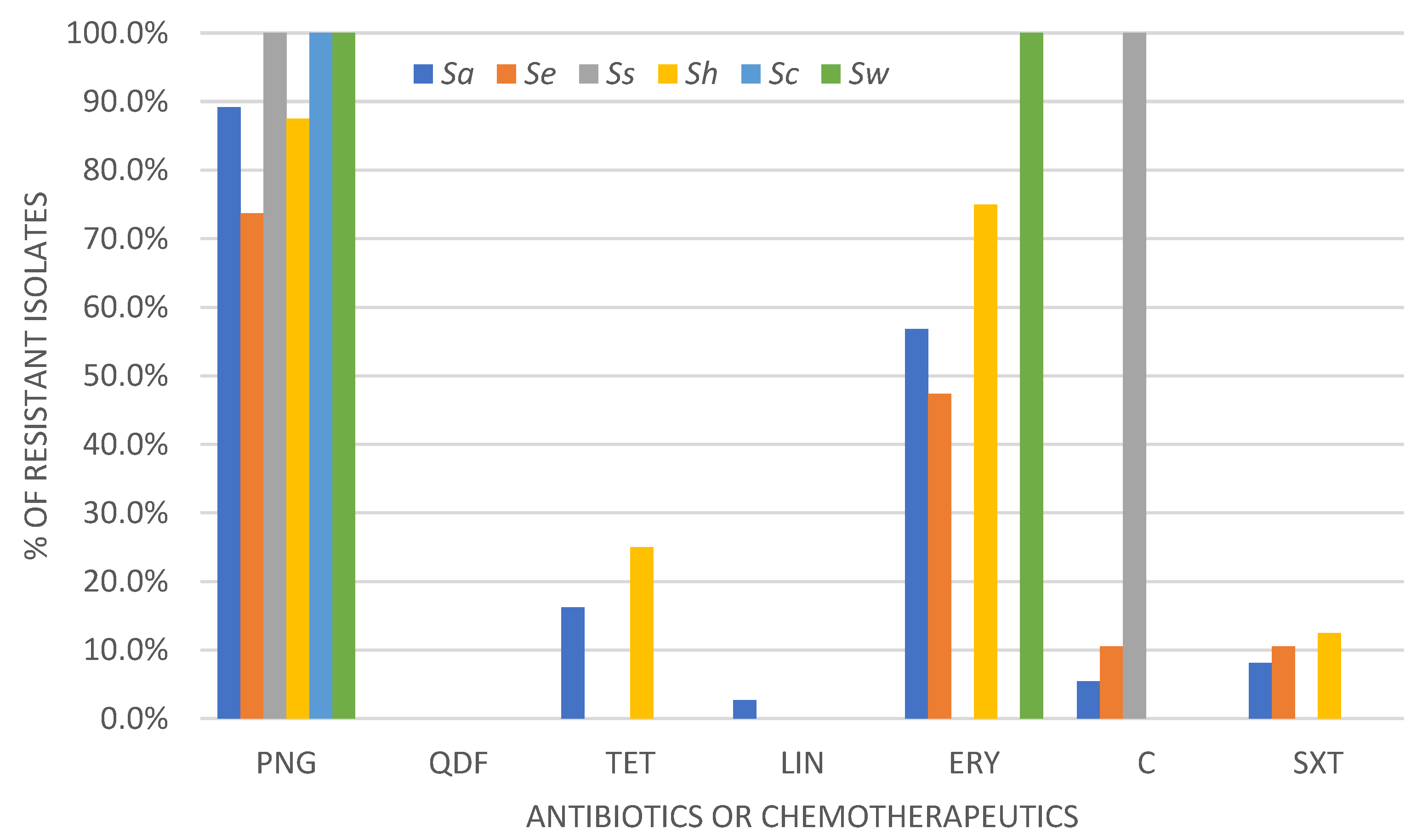
2.1. Determination of Drug Susceptibility Profile by Disc-Diffusion Method

The study included a total of 68 bacterial isolates of the Staphylococcaceae family, with the most prevalent being S. aureus 54.4% (37/68) and S. epidermidis 27.9% (19/68), followed by S. hominis 11.7% (8/68), S. capitis 2.9% (2/68), S. warneri and S. saprothyticus 1.5% (1/68) each.
The disk diffusion method was conducted to determine the susceptibility profile to selected antimicrobials in accordance with the current recommendations of EUCAST [25]. The obtained results are shown in Figure 1, which indicates the highest overall resistance to penicillin and erythromycin. The overall analysis of the obtained data showed that the largest number of Staphylococcus spp. isolates showed in vitro susceptibility to aminoglycosides, quinolones and oxazolidinones, with the highest prevalence of all susceptible isolates to quinupristin-dalfopristin.
The percentage analysis of the drug susceptibility profile for each tested staphylococcal species is shown below (Figure 2). It was observed that S. aureus, S. epidermidis and S. hominis showed the highest diversity of drug susceptibility profiles. Both S. aureus and S. epidermidis showed the highest resistance frequency to beta-lactam antibiotics.
We found 23.5% (16/68) of methicillin-resistant S. aureus (MRSA) and 7.4% (5/68) of coagulase-negative S. epidermidis (MRCoNSE) and methicillin-resistant coagulase-negative staphylococci other than S. epidermidis (nonSE-MRCoNS) each. Penicillin resistance was reported in all staphylococci isolates from the nasopharynx of pregnant women, with the highest frequency of 13.6% (20/68) in methicillin-susceptible S. aureus (MSSA) isolates and resistance to erythromycin observed in 7.5% (11/68) of them (Table 1). Resistance to linezolid and norfloxacin was found in only 0.7% (1/68) of MRSA isolates each. Susceptibility, increased exposure of isolates was noted in all isolates to ciprofloxacin in a frequency range of 3.4–12.3%, with the highest percentage noted in MSSA isolates (12.3%, 19/68).

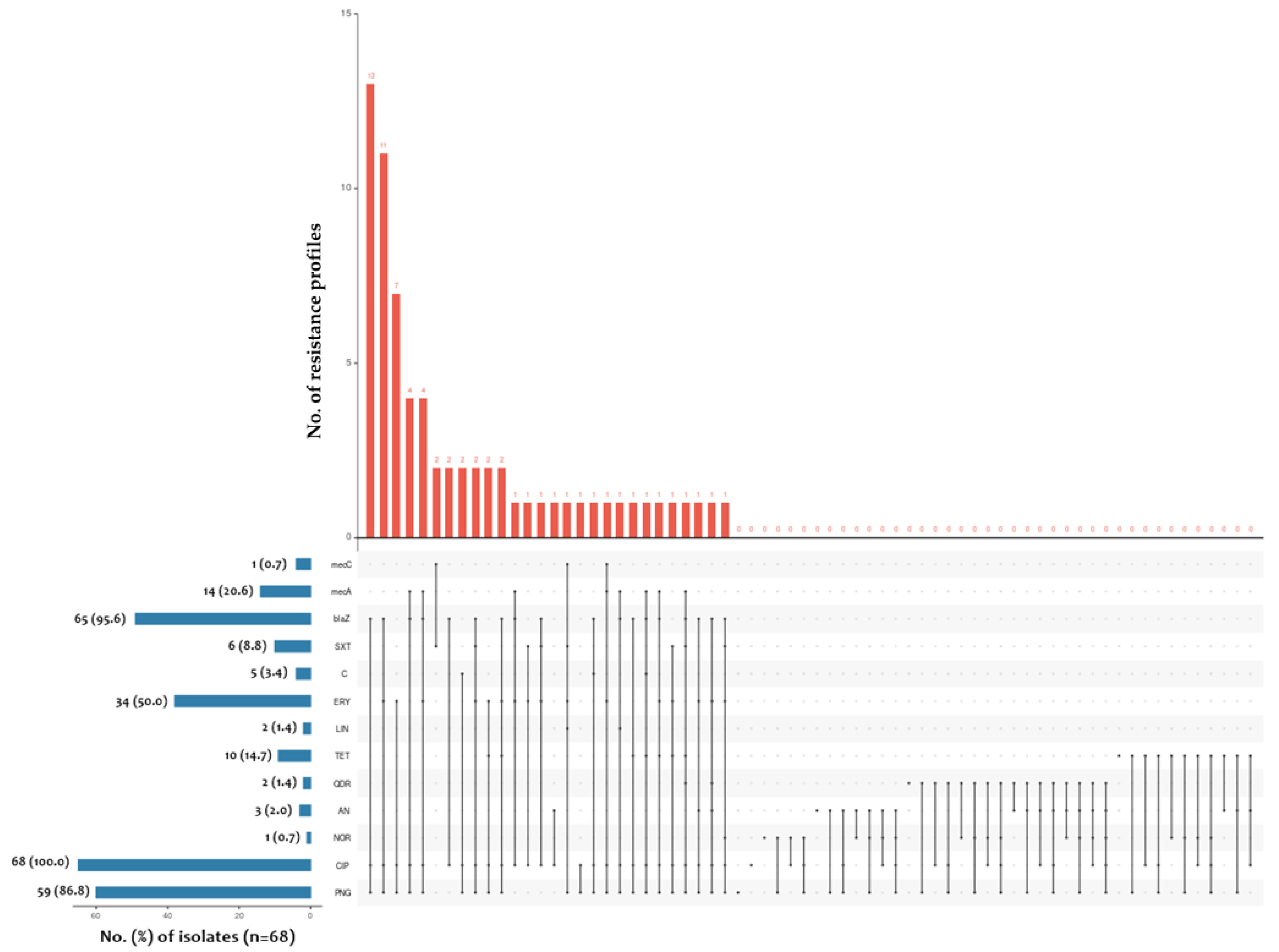
2.2. Determination of blaZ, mecA and mecC Gene Prevalence among Tested Staphylococcus spp. Isolates

For each isolate, a PCR by using specific primers for blaZ, mecA and mecC genes was performed.
According to the analysis of the obtained data, the blaZ gene was reported in 95.6% (65/68) of the tested isolates, followed by 51.5% (35/68) and 27.9% (19/68) for S. aureus and S. epidermidis, respectively. The mecA gene was detected in 20.6% (14/68) of all tested isolates, in 13.2% (9/68), 4.4% (3/68) and 2.9% (2/68) of S. aureus, S. hominis and S. epidermidis isolates, respectively (Table 2). In addition, the mecC gene was detected in only one (1.5%, 1/68) S. aureus isolate. The species with the highest frequency of blaZ gene presence was S. epidermidis, while the mecA gene was predominant in S. hominis isolates.

2.3. Determination of SCCmec Gene Cassette Type

For mecA-positive isolates, the presence of either the ccr gene type or the mec complex type was confirmed by PCR (Table 3). The presence of the ccr gene was detected in 11.8% (8/68) of the tested bacteria. Further analysis identified 2.9% (2/68) of S. hominis isolates with the ccrA1 gene (type I ccr complex); 5.9% (4/68) with ccrA4 (type IV ccr complex), including two strains of S. epidermidis and S. aureus; and 4.4% (3/68) with ccrC (type V ccr complex), including one S. epidermidis and two S. aureus isolates. One (1.5%, 1/68) of all tested strains was characterised by a double ccr allotype (ccrA4 + ccrC detected in S. aureus isolate). Further analysis of the obtained results allowed the identification of 5.9% (4/68) of the isolates with a specific mec complex class, comprising 4.4% (3/68) S. hominis with the presence of the mecI gene (class A mec complex). The presence of the IS1272 sequence (class B mec complex) was observed in 1.5% (1/68) of S. epidermidis strains. For the remaining analysed isolates, the classification was not specified.
The data presented above allowed us to assign the tested isolates to the different types of SCCmec cassettes. The results of these tests are shown in Table 4. As a result, only one tested strain could be assigned to SCCmec cassette type VI; it was S. epidermidis. The remaining strains were classified to UT5v type according to Zong et al. [14]. The final result was that only three tested strains could be assigned to a specific SCCmec cassette type, including one S. epidermidis and two S. hominis isolates.
The frequency of each gene detected in this study in relation to the staphylococcal mecA-positive species and the number of isolates obtained from a pregnant patient is shown in Table S3.
The following diagnostic test reliability parameters were calculated: specificity, sensitivity and positive and negative predictive values for blaZ and mecA gene detection compared with phenotypic methods performed for beta-lactam resistance testing (Table 5).
The obtained data showed 100% diagnostic test sensitivity and NPV for mecA gene detection and over 80% sensitivity and PPV for blaZ gene detection in comparison with phenotypic methods used to determine beta-lactam resistance in staphylococci isolated from the respiratory microbiota of pregnant women. Detection of the blaZ gene had a higher specificity than mecA gene detection (43.75% vs. 18.18%) and a lower NPV value (70.0% vs. 100%).

3. Discussion

There are some serious risks associated with giving antibiotics to pregnant women, so there are a number of rules to follow determined by the condition of the patients, the type of examinations and procedures performed and the mode of delivery [1,2,4,5]. Antibiotics with the lowest possible risk of adverse effects should be chosen, which often means choosing older antibiotics with documented safety in pregnancy [1,3]. The beta-lactam group of antibiotics was chosen for this study because the studied isolates were exclusively from women in a physiological state of pregnancy when the choice of treatment is clinically very limited. The negligible risk of pregnancy-related complications, low organ toxicity and easy penetration of the blood–placental barrier make these antibiotics the most commonly used group of antimicrobial drugs during pregnancy [1,3]. Różanska et al. [3] conducted a study to analyse the use of antibiotics prescribed by gynaecologists during pregnancy in one province of Poland. The authors concluded that monotherapy was used in almost 96% of the studied women, and beta-lactams were used in combination with beta-lactamase inhibitors in more than 67% of the patients. These findings are in line with recommendations for antibiotic therapy in pregnant women [1,2]. However, there is still no clear information on which stages of pregnancy are critical in terms of the adverse effects of drugs on the developing foetus [28]. The risks to the foetus from maternal use of antibiotics are also not fully understood. Therefore, the use of antimicrobial drugs in pregnancy is still controversial [1,2,3]. Another limitation to the use of drugs in this group in clinical practice is the problem of drug resistance among microorganisms. Hospital and outpatient strains of S. epidermidis are usually characterised by multidrug resistance, including lack of sensitivity to beta-lactam antibiotics [12,13,14].
Our study included 68 staphylococcal isolates from 43 pregnant women. The study included a drug susceptibility profile for fluoroquinolones (ciprofloxacin and norfloxacin), streptogramins (quinupristin-dalfopristin), aminoglycosides (amikacin), macrolides (erythromycin), oxasolidinones (linezolid), tetracyclines (tetracycline) and other groups (trimethoprim-sulfamethoxazole and chloramphenicol). The data were interpreted and analysed according to EUCAST recommendations [25]. Of all staphylococcal isolates identified in this study, 30.9% and 23.5% were methicillin-susceptible and methicillin-resistant S. aureus (MSSA and MRSA) isolates, respectively, with the most common resistance to penicillin being 13.6% and 9.5%, respectively. Among coagulase-negative S. epidermidis (CoNSE) isolates, the highest frequency of susceptible, increased exposure isolates was 9.5% to ciprofloxacin and 6.1% to penicillin, while among non-coagulase-negative staphylococci (non-CoNS) isolates, it was 4.8% each. No resistance to chinupristin-dalfopristin was found in this study. Interestingly, 38.2%, 20.6% and 1.5% of the isolates phenotypically resistant to beta-lactams carried the blaZ, mecA and mecC genes, respectively, whereas 25.0% carried none of the tested genes. The results obtained in this study are consistent with data from other authors. In a total of more than 180 S. epidermidis strains, Aubry et al. and Ferreira et al. found penicillin resistances of 30% and 72%, respectively [12,20]. It is now reported that the prevalence of S. epidermidis strains resistant to penicillin can exceed 90%, as confirmed by Guo et al. (95.5% in clinical strains and 82.1% in colonised strains) [29] and Wang et al. (80–100% of strains analysed from different provinces in China) [30]. The phenomenon of acquiring resistance to antimicrobial agents is very dynamic within this genus. Penicillins, once used as first-line drugs in staphylococcal infections, are now of little use. This phenomenon has led to a situation where strains are resistant to semi-synthetic penicillins, beta-lactamase inhibitors, cephalosporins and even carbapenems [2,3,8,16,28]. The treatment of infections caused by staphylococci presents many difficulties, both in terms of the diagnosis and in choosing the right treatment.
Based on data from other authors and our own, PCR detection of the blaZ gene was considered the gold standard for confirming the presence of penicillinase in the tested staphylococci isolates. This choice was also motivated by the Clinical and Laboratory Standards Institute (CLSI) position, which suggests that detection of this gene should be considered in serious cases of infection with S. aureus aetiology requiring penicillin therapy [31]. In this study, 95.6% of blaZ-positive isolates were confirmed, of which 38.2% were resistant to benzylpenicillin. Less than 11% of all tested isolates lacked the blaZ gene and were not resistant to any of the tested antibiotics. These results differ to some extent from those reported by other authors. In the study by Aubry et al. aimed at comparing different phenotypic methods for the detection of penicillinases in S. epidermidis, the presence of the blaZ gene was detected in a total of 30% of the strains [12]. Similar studies using the disc diffusion method have been reported by Ferreira et al. [20]. Of more than one hundred staphylococcal strains of different tested species, approximately 72% of CNS isolates resistant to benzylpenicillin possessed the blaZ gene. Olsen et al. [7] analysed the diversity and frequency of the blaZ gene between human and bovine CoNS and S. aureus. The authors detected this gene in 143 strains of penicillin-resistant S. aureus and CoNS. They found that all penicillin-resistant strains carried blaZ and showed a similar organisation of blaR1 and blaZ genes. Similarly, Bagcigil et al. [18] investigated the carriage of the blaZ gene in 78 beta-lactamase-positive S. aureus isolates from bovine mastitis. They found that all isolates carried the blaZ gene. Similar to our study, Soares et al. [13] evaluated the phenotypical and genotypical antimicrobial resistance profile of 100 CoNS species isolated from dairy cow milk. They found that only 6% of staphylococcal isolates were phenotypically susceptible to penicillin and ampicillin, with S. xylosus being the most common species, followed by S. cohnii, S. hominis, S. capitis and S. haemolyticus.
Other studies have reported the occurrence of the so-called blaZ and mecA gene correlation phenomena in staphylococci [13,19,22,32]. There is therefore an ongoing debate about the usefulness of detecting both genes simultaneously in S. aureus. In our study, we found that both the blaZ and mecA genes were present in 11.8% of the S. aureus isolates and 7.4% of the CoNS isolates, exclusively from S. epidermidis, isolated from the respiratory microbiota of healthy women at different stages of pregnancy. Others reported that 4% of mecA+ S. xylosus isolates also carried the blaZ gene, which was also detected in 16% of the isolates and all mecA+ isolates [13]. Authors assumed that the presence of both genes was correlated with phenotypic beta-lactam resistance. Co-occurrence of the mecA and blaZ genes has also been reported in MRSA, as studied by Okiki et al. in 135 pregnant women with vaginitis [22]. Of the MRSA isolates, 54.5% carried the mecA gene, and none of the MSSA or methicillin-resistant or methicillin-susceptible coagulase-negative staphylococci (MRCoNS or MSCoNS) isolates had the mecA gene with a prevalence of 30%. The blaZ gene was detected in 65% of the staphylococcal isolates, including 72.7% and 22.2% of MRSA and MSSA, respectively, and 15% of MRCoNS; 54.5% of MRSA isolated from women with vaginal discharge and itching had both mecA and blaZ genes [22].
Infections caused by various species of staphylococci, e.g., S. aureus and S. epidermidis in particular, can be a problem for infection due to the ever-increasing drug resistance of these bacteria [7,8,16]. In staphylococci, resistance to several antimicrobial drugs may be associated with insensitivity to multiple chemical groups of antibiotics, giving the character of multidrug-resistant strains. Drug resistance, except plasmid-encoded, can also be found on the bacterial chromosome, as in the case of methicillin resistance, conditioned most often by the presence of the mecA and/or mecC gene [8,14,19,21,22]. This gene occurs within the so-called SCCmec gene cassettes [23], classified into individual types I-XI, thanks to the use of different methods for typing SCCmec cassettes, usually based on multiplex PCR reaction [23,27,33]. One of the most effective methods, other than whole-genome sequencing, is proposed by Kondo et al. [27] and is still recommended by the International Working Group for SCCmec elements (IWG-SCC) [24]. The PCR reaction proposed by Chen et al. [33] allows the determination of the mec complex and ccr allotype after the reaction in two panels: the first to detect mecA, mecI, insertion sequence IS1272 (class B of the mec gene) and ccrB2 to detect type II and IV of the SCCmec cassette and the second to identify ccrC, ccrB1-B4 and insertion sequence IS431 (class C2 of the mec gene), allowing the determination of cassette types I, III, V and VI. Both methods have their own limitations as they are not yet sufficiently effective in determining the remaining cassette types, including VII, IX, X, XI and the newest types [23]. This study showed that among mecA-positive staphylococci isolated from the respiratory microbiota of pregnant women, taken for further analysis of ccr complex types and SCCmec cassettes, 21.5% of isolates had type IV, 14.3% had type I or V and 7.1% had type IV + V, while 42.9% of strains contained an undetermined ccr complex. Of the staphylococcal strains tested for the mec gene complex, 21.4% and 7.1% were class A and B, respectively. We also found that only one S. epidermidis isolate had the VIth SCCmec cassette type, 14.3% and 7.1% of the UT5v and VI types, respectively, according to Zong et al. [14]. The literature data indicate that S. epidermidis strains may contain SCCmec I-VIII cassettes, while S. aureus may contain any of the eleven types [23]. Our data are in agreement with those of Sign-Moodley et al. [21], who demonstrated that not all unknown isolates could be definitively assigned to clearly defined types using the SCCmec typing method. Most of these isolates were not conventional types; untypeable elements appeared to be composite SCCmec elements consisting of multiple ccr gene complexes. This may be due to the ease of transfer of these genes between different bacterial species. It is assumed that only one class of the mec complex is involved, although strains with several different mec allotypes have been reported in the literature. Many authors have also shown that the majority of untypeable isolates carry more than one ccr gene complex [8,21], which is consistent with our data. A significant proportion of MRSA and MRSE have unclassifiable cassettes. This may be the result of an unusual combination of the mec complex and ccr genes. Such complexes cannot be assigned to any of the known types. Sequencing of the entire element would be required to determine whether these elements represent a single SCCmec element with two ccr gene complexes or whether the element consists of two separate integrated SCCmec elements [21,24].
The classification of strains of S. epidermidis and coagulase-negative staphylococci (CoNS) is very important given the ever-increasing drug resistance of these bacteria, including isolates from the respiratory microbiota where staphylococci may change their role from opportunistic to pathogenic [8,16]. The increasing multidrug resistance of methicillin-resistant strains of staphylococci, either S. aureus or CoNS, associated with the presence of additional resistance genes to antibiotics is becoming an additional problem for clinicians [8,28]. Increasing resistance to beta-lactams is making antimicrobials, including beta-lactams, an increasingly ineffective therapy for treating staphylococcal infections. This phenomenon significantly limits the therapeutic options available to patients, including pregnant women, for whom there are relatively few treatment options [8,16,28,34]. This in turn may contribute to prolonged antibiotic therapy or discomfort associated with taking antibiotics and/or chemotherapeutics and thus an increased risk of complications. There are also major diagnostic difficulties with coagulase-negative microorganisms as there are no clear diagnostic recommendations for detecting resistance mechanisms in these microorganisms [8,16,23,25,31,34]. The issue of genetically determined resistance and drawing appropriate conclusions especially about CoNS resistance to penicillin is complicated by its production mechanism [16,34]. It occurs in an inducible manner, i.e., only when a beta-lactam antibiotic is present in the bacterial environment [12].
The lack of clear recommendations and reliable methods for the detection of penicillinases in CoNS presents many diagnostic difficulties. The possibility of detecting beta-lactam antibiotic resistance mechanisms by blaZ gene detection increases the role of molecular testing in favour of traditional phenotypic methods and their use in daily clinical practice. In the available literature, it is still difficult to find publications that address the issue of the possible consequences of the presence of resistance genes on molecular elements carried by microorganisms belonging to the human microbiota. As shown by the studies carried out in this work and others [12,13,14], CoNS staphylococci, in addition to co-forming the human respiratory microbiota, may represent a potential reservoir of genetic determinants determining phenotypic resistance to antibiotics and/or chemotherapeutics. Furthermore, the overuse and prevalence of antibiotics and their presence in the human body make them optimal carriers of resistance genes.

4. Materials and Methods

4.1. Bacterial Isolates and Culture

Sixty-eight isolates of Staphylococcaceae were used in this study as the clinical material. The bacteria were cultured and identified from nasopharyngeal swabs of 43 adult (19–41 ± 4.6, mean age 29.8 years) healthy women at different stages of pregnancy (2.15%, 5/43 in the first; 3.9%, 9/43 in the second; and 15.9%, 37/43 in the third trimester). The exclusion criteria for patient participation in our study were as follows: the absence of infection at the time of collection, the absence of chronic and acute diseases of the respiratory tract and the absence of antimicrobial use within a 2-year retrospective period. All tested isolates were not an etiological agent of any infection but were derived from nasopharyngeal microbiota as colonizers. All staphylococcal isolates were identified phenotypically based on their properties, e.g., with colony morphology and Gram-staining effect, biochemical features and protein profile detection by the matrix-assisted laser desorption-ionization time of flight mass spectrometry (MALDI-TOF MS) technique. MALDI-TOF mass spectroscopy was performed as described by Bucka-Kolendo et al. [26] on the basis of the protein profile at the Department of Epidemiology and Infectious Diseases, University of Life Sciences, Lublin, Poland. A few colonies of fresh 24 h microbial cultures were aspirated into 1.5 mL Eppendorf tubes containing 150 µL ultrapure water. The tubes were vortexed. Ethanol was added to the suspension at 450 µL, vortexed and centrifuged at 13,000 × g for 5 min. Microbial pellets were suspended in 40 µL of 70% formic acid (Fluka Analytical, Munich, Germany). After vortexing, the sample was centrifuged (13,000 × g, 2 min) with the addition of 40 µL of acetonitrile (Fluka Analytical, Munich, Germany). For identification, an equal volume of α-cyano-4-hydroxycinnamic acid (HCCA) solution (Bruker Daltonics, Billerica, MA, USA) was added to 1 µL of the protein extract on a MALDI target plate. The MALDI Biotyper 3.1 database—Build (Bruker, Billerica, USA) was used to process the mass spectra obtained for each isolate. The probability of correct identification was expressed in the form of a score index with a range of values: 2.300–3.000, reliable identification of the microorganism to the species level; 2.000–2.299, reliable identification of the microorganism to the genus level and probable identification result to the species level; 1.700–1.999, probable identification result to the genus level; and 0.0–1.699, unreliable identification result.
After that, the bacteria were subcultured on Mueller–Hinton agar medium (MHA, Oxoid, Hampshire, United Kingdom) and incubated for 18 ± 2 h at 35 ± 2 °C under aerobic conditions. The same medium was also used for antimicrobial drug susceptibility testing using the disc diffusion method according to the recommendations of the European Committee on Antimicrobial Susceptibility Testing (EUCAST) [25]. The following American Type Culture Collection (ATCC) reference strains were used: methicillin-resistant S. aureus ATCC BAA-1707 (MRSA), S. aureus ATCC 29213 (EUCAST quality control) and S. epidermidis ATCC 12228.

4.2. Disk Diffusion Method

Staphylococcal drug susceptibility was assessed using the disk diffusion method, according to current criteria and EUCAST recommendations [25]. All isolates were analysed for their susceptibility profiles to the selected antimicrobials benzylpenicillin (1 U), ciprofloxacin (5 μg), norfloxacin (10 μg), quinupristin-dalfopristin (15 μg), amikacin (30 μg), erythromycin (15 μg), linezolid (10 μg), trimethoprim-sulfamethoxazole (1.25–23.75 μg), tetracycline (30 μg) and chloramphenicol (30 μg). All isolates were also tested for the production of methicillin resistance mechanisms using a cefoxitin disc (30 μg) [25]. Isolates were defined as cefoxitin-resistant if the diameter of their growth inhibition zone around the antibiotic disc was <22 mm. Such staphylococcal isolates were classified as methicillin-resistant strains [25]. For this purpose, bacterial inoculum of 0.5 McFarland scale in sterile 0.85% NaCl solution from fresh, 24 h culture was prepared. Inoculated plates, inoculated in three directions, with a disc impregnated with a known concentration of antimicrobial compounds placed on top, were pre-incubated at room temperature for 15 min and incubated under the conditions described above. The size of the growth inhibition zone [mm] for each antibiotic/chemotherapeutic disc as a result of the susceptibility testing was used to classify the staphylococcal isolates into the following three categories: susceptible, standard dosing regimen (S); sensitive, increased exposure (I); and resistant (R) [25].

4.3. DNA Extraction and PCR Reaction

4.3.1. Specific PCR Reactions

Genomic DNA from each bacterial isolate was extracted by using a lysostaphin-based extraction protocol with the Genomic Mini Kit (A&A Biotechnology, Gdansk, Poland). Detection of the genes blaZ, mecA and mecC was performed by PCR reaction in a total volume of 15 µL, containing 7.5 µL REDTaq® ReadyMix™ PCR Reaction Mix (Merck, Darmstadt, Germany) and 1 µL of bacterial genomic DNA, by using specific primers (Table S1) and temperature–time procedures as previously published [12,32]. The following controls were included in all amplification reactions: S. aureus ATCC 29213 (blaZ-positive S. aureus), S. aureus ATCC BAA-1707 (mecC-positive S. aureus), S. aureus ATCC 25923 (mecA, blaZ-negative S. aureus), S. epidermidis ATCC 12228 (mecA, mecC-negative S. epidermidis) and negative control (water).

4.3.2. SCCmec Cassette Detection and Typing

Further PCR reactions were then performed on all mecA-positive (mecA+) isolates to detect specific sequences and products to determine the type of ccr gene complex and the class of mec gene complex [27]. Three separate multiplex-PCR (M-PCR) reactions were performed for this purpose (Table S2). The identification of five types of SCCmec elements was performed by M-PCR reactions in a total volume of 25 µL containing 12.5 µL of REDTaq® ReadyMix™ PCR Reaction Mix (Merck, Darmstadt, Germany), 5 µL of DNA, 1 µL of each 10 µM specific primer and the missing volume of molecular-use H2O (EURx, Gdansk, Poland).
The types of SCCmec cassette were detected in reactions consisting of a pre-denaturation of 94 °C for 5 min and final extension of 72 °C for 2 min and 30 cycles (consisting of the following steps: denaturation of 94 °C for 2 min, annealing of 57–60 °C for 1 min and extension of 72 °C for 2 min), followed by 1.5–2.0% gel electrophoresis. Each gel was stained with SimplySafe dye (EURx, Gdansk, Poland), documented with Quantum VilberLourmat ST4 system and visualised with Quantum-Capt ST4 (Vilber Lourmat, Collégien, France). The following controls were included in all amplification reactions: S. aureus ATCC BAA-1707 (SCCmec: Type IV-positive S. aureus), S. aureus ATCC 25923 (mecA-negative S. aureus), S. epidermidis ATCC 12228 (mecA-negative S. epidermidis) and negative control (water).

4.4. Data Analysis

An UpSet plot was constructed to show the antimicrobial resistance pattern of all staphylococcal isolates using online tools (https://asntech.shinyapps.io/intervene/ (accessed 2 February 2023)). An UpSet plot showed the number of shared phenotypes with gene hits that were found in each isolate. In the binary input file, each column represented a set, and each row represented an element. If a name was in the set, it was represented as a 1; otherwise, it was represented as a 0, based on the manufacturer’s instructions.

5. Conclusions

Among the tested isolates, the highest resistance was shown to beta-lactam antibiotics, commonly used in outpatient clinics. The presence of the blaZ gene determining the synthesis of penicillinases is common in bacteria of the genus Staphylococcus. Phenotypic and genotypic tests showed that blaZ-positive isolates were resistant to benzylpenicillin. Detection of the blaZ gene had a higher specificity than mecA detection (44% vs. 18%) and a lower either sensitivity (94% vs. 100%) or NPV value (70% vs. 100%). All staphylococci were found to have a very high diversity of SCCmec cassette structures. Knowledge of methicillin-resistant staphylococci in humans, especially the nasopharyngeal microbiota of pregnant women, needs to be further developed. Our findings are of cognitive importance as there is little knowledge of the resistance of bacteria isolated from the respiratory microbiota, including the nasopharyngeal cavity, of women during the physiological course of pregnancy. A better understanding of this phenomenon, given the global increase in resistance, the large number of opportunistic strains currently circulating in the community, and the presence of opportunistic pathogens resistant to antibiotics and other antimicrobials in the maternal respiratory microbiota, may be helpful in preparing treatment or prevention that may be useful in improving pregnancy outcomes.

Supplementary Materials

The following supporting information can be downloaded at: https://www.mdpi.com/article/10.3390/ijms24097980/s1.

Author Contributions

Conceptualization, S.A. and U.K.; methodology, S.A.; software, S.A.; validation, S.A., U.K., D.D. and D.S.-P.; formal analysis, S.A., M.C. and D.D.; investigation, S.A. and M.C.; resources, U.K.; data curation, D.S.-P. and U.K.; writing—original draft preparation, S.A. and D.D.; writing—review and editing, S.A. and U.K.; visualization, S.A.; supervision, U.K.; project administration, D.D.; funding acquisition, U.K. and D.D. All authors have read and agreed to the published version of the manuscript.

Funding

This research received no external funding. This study was supported by the Medical University of Lublin, Poland (grant no. DS31).

Institutional Review Board Statement

The study was conducted in accordance with the Declaration of Helsinki and approved by the Medical University of Lublin, Poland Bioethical Commission (No. KE-0254/75/2011 28 April 2011 and No. KE-0254/59/2016 25 February 2016).

Informed Consent Statement

Not applicable.

Data Availability Statement

Not applicable.

Conflicts of Interest

The authors declare no conflict of interest.

References

  1. World Health Organization. WHO Recommendations for Prevention and Treatment of Maternal Peripartum Infections; World Health Organization: Geneva, Switzerland, 2015. [Google Scholar]
  2. Żukowska, A.; Hryniewicz, W. Recommendations for Diagnosis, Antibiotic Therapy and Prevention Hospital-Acquired Infections; National Medicines Institute: Warsaw, Poland, 2020. [Google Scholar]
  3. Różańska, A.; Pac, A.; Jachowicz, E.; Jaślan, D.; Siewierska, M.; Wójkowska-Mach, J. Outpatient Antibiotic Prescriptions in Pregnant Women in Małopolska Province. Antibiotics 2020, 10, 14. [Google Scholar] [CrossRef] [PubMed]
  4. Prevention of Group B Streptococcal Early-Onset Disease in Newborns: ACOG Committee Opinion, Number 797. Obstet. Gynecol. 2020, 135, e51–e72. [CrossRef] [PubMed]
  5. Schafer, R.; Phillippi, J.C. Group B Streptococcal Bacteriuria in Pregnancy: An Evidence-Based, Patient-Centered Approach to Care. J. Midwifery Womens Health 2020, 65, 376–381. [Google Scholar] [CrossRef]
  6. Pan, H.; Cui, B.; Huang, Y.; Yang, J.; Ba-Thein, W. Nasal Carriage of Common Bacterial Pathogens among Healthy Kindergarten Children in Chaoshan Region, Southern China: A Cross-Sectional Study. BMC Pediatr. 2016, 16, 161. [Google Scholar] [CrossRef] [PubMed] [Green Version]
  7. Chen, H.; Zhang, J.; He, Y.; Lv, Z.; Liang, Z.; Chen, J.; Li, P.; Liu, J.; Yang, H.; Tao, A.; et al. Exploring the Role of Staphylococcus aureus in Inflammatory Diseases. Toxins 2022, 14, 464. [Google Scholar] [CrossRef]
  8. Michels, R.; Last, K.; Becker, S.L.; Papan, C. Update on Coagulase-Negative Staphylococci—What the Clinician Should Know. Microorganisms 2021, 9, 830. [Google Scholar] [CrossRef]
  9. Alharbi, N.S. Screening of Antibiotic-Resistant Staphylococci in the Nasal Cavity of Patients and Healthy Individuals. Saudi J. Biol. Sci. 2020, 27, 100–105. [Google Scholar] [CrossRef]
  10. Fujiwara, N.; Tsuruda, K.; Iwamoto, Y.; Kato, F.; Odaki, T.; Yamane, N.; Hori, Y.; Harashima, Y.; Sakoda, A.; Tagaya, A.; et al. Significant Increase of Oral Bacteria in the Early Pregnancy Period in Japanese Women. J. Investig. Clin. Dent. 2017, 8, e12189. [Google Scholar] [CrossRef]
  11. Stratmann, J.A.; Lacko, R.; Ballo, O.; Shaid, S.; Gleiber, W.; Vehreschild, M.J.G.T.; Wichelhaus, T.; Reinheimer, C.; Göttig, S.; Kempf, V.A.J.; et al. Colonization with Multi-Drug-Resistant Organisms Negatively Impacts Survival in Patients with Non-Small Cell Lung Cancer. PLoS ONE 2020, 15, e0242544. [Google Scholar] [CrossRef]
  12. Aubry, B.; Lemarié, C.; Chenouard, R.; Kempf, M.; Eveillard, M.; Pailhoriès, H. Performance of Penicillinase Detection Tests in Staphylococcus Epidermidis: Comparison of Different Phenotypic Methods. BMC Microbiol. 2020, 20, 240. [Google Scholar] [CrossRef]
  13. Soares, L.C.; Pereira, I.A.; Pribul, B.R.; Oliva, M.S.; Coelho, S.M.O.; Souza, M.M.S. Antimicrobial Resistance and Detection of MecA and BlaZ Genes in Coagulase-Negative Staphylococcus Isolated from Bovine Mastitis. Pesqui. Veterinária Bras. 2012, 32, 692–696. [Google Scholar] [CrossRef] [Green Version]
  14. Zong, Z.; Peng, C.; Lü, X. Diversity of SCCmec Elements in Methicillin-Resistant Coagulase-Negative Staphylococci Clinical Isolates. PLoS ONE 2011, 6, e20191. [Google Scholar] [CrossRef] [PubMed]
  15. Olsen, J.E.; Christensen, H.; Aarestrup, F.M. Diversity and Evolution of BlaZ from Staphylococcus aureus and Coagulase-Negative Staphylococci. J. Antimicrob. Chemother. 2006, 57, 450–460. [Google Scholar] [CrossRef] [PubMed]
  16. Marincola, G.; Liong, O.; Schoen, C.; Abouelfetouh, A.; Hamdy, A.; Wencker, F.D.R.; Marciniak, T.; Becker, K.; Köck, R.; Ziebuhr, W. Antimicrobial Resistance Profiles of Coagulase-Negative Staphylococci in Community-Based Healthy Individuals in Germany. Front. Public Health 2021, 9, 684456. [Google Scholar] [CrossRef] [PubMed]
  17. Biesbroek, G.; Tsivtsivadze, E.; Sanders, E.A.M.; Montijn, R.; Veenhoven, R.H.; Keijser, B.J.F.; Bogaert, D. Early Respiratory Microbiota Composition Determines Bacterial Succession Patterns and Respiratory Health in Children. Am. J. Respir. Crit. Care Med. 2014, 190, 1283–1292. [Google Scholar] [CrossRef] [PubMed]
  18. Bagcigil, A.F.; Taponen, S.; Koort, J.; Bengtsson, B.; Myllyniemi, A.-L.; Pyörälä, S. Genetic Basis of Penicillin Resistance of S. aureus Isolated in Bovine Mastitis. Acta Vet. Scand. 2012, 54, 69. [Google Scholar] [CrossRef] [Green Version]
  19. Rocha, G.D.; Nogueira, J.F.; Gomes dos Santos, M.V.; Boaventura, J.A.; Nunes Soares, R.A.; José de Simoni Gouveia, J.; Matiuzzi da Costa, M.; Gouveia, G.V. Impact of Polymorphisms in BlaZ, BlaR1 and BlaI Genes and Their Relationship with β-Lactam Resistance in S. Aureus Strains Isolated from Bovine Mastitis. Microb. Pathog. 2022, 165, 105453. [Google Scholar] [CrossRef]
  20. Ferreira, A.M.; Martins, K.B.; da Silva, V.R.; Mondelli, A.L.; de Lourdes Ribeiro de Souza da Cunha, M. Correlation of Phenotypic Tests with the Presence of the BlaZ Gene for Detection of Beta-Lactamase. Braz. J. Microbiol. 2017, 48, 159–166. [Google Scholar] [CrossRef] [Green Version]
  21. Singh-Moodley, A.; Strasheim, W.; Mogokotleng, R.; Ismail, H.; Perovic, O. Unconventional SCCmec Types and Low Prevalence of the Panton-Valentine Leukocidin Exotoxin in South African Blood Culture Staphylococcus aureus Surveillance Isolates, 2013–2016. PLoS ONE 2019, 14, e0225726. [Google Scholar] [CrossRef]
  22. Okiki, P.A.; Eromosele, E.S.; Ade-Ojo, P.; Sobajo, O.A.; Idris, O.O.; Agbana, R.D. Occurrence of MecA and BlaZ Genes in Methicillin-Resistant Staphylococcus aureus Associated with Vaginitis among Pregnant Women in Ado-Ekiti, Nigeria. New Microbes New Infect. 2020, 38, 100772. [Google Scholar] [CrossRef]
  23. Uehara, Y. Current Status of Staphylococcal Cassette Chromosome Mec (SCCmec). Antibiotics 2022, 11, 86. [Google Scholar] [CrossRef] [PubMed]
  24. International Working Group on the Staphylococcal Cassette Chromosome Elements (IWG-SCC). Available online: https://www.sccmec.org/index.php/en/ (accessed on 5 December 2022).
  25. The European Committee on Antimicrobial Susceptibility Testing. Breakpoint Tables for Interpretation of MICs and Zone Diameters. Version 12.0; The European Committee on Antimicrobial Susceptibility Testing: Växjö, Sweden, 2022. [Google Scholar]
  26. Bucka-Kolendo, J.; Sokołowska, B.; Winiarczyk, S. Influence of High Hydrostatic Pressure on the Identification of Lactobacillus by MALDI-TOF MS- Preliminary Study. Microorganisms 2020, 8, 813. [Google Scholar] [CrossRef] [PubMed]
  27. Kondo, Y.; Ito, T.; Ma, X.X.; Watanabe, S.; Kreiswirth, B.N.; Etienne, J.; Hiramatsu, K. Combination of Multiplex PCRs for Staphylococcal Cassette Chromosome Mec Type Assignment: Rapid Identification System for Mec, Ccr, and Major Differences in Junkyard Regions. Antimicrob. Agents Chemother. 2007, 51, 264–274. [Google Scholar] [CrossRef] [PubMed] [Green Version]
  28. Qu, Y.; Daley, A.J.; Istivan, T.S.; Garland, S.M.; Deighton, M.A. Antibiotic Susceptibility of Coagulase-Negative Staphylococci Isolated from Very Low Birth Weight Babies: Comprehensive Comparisons of Bacteria at Different Stages of Biofilm Formation. Ann. Clin. Microbiol. Antimicrob. 2010, 9, 16. [Google Scholar] [CrossRef] [PubMed] [Green Version]
  29. Guo, Y.; Song, G.; Sun, M.; Wang, J.; Wang, Y. Prevalence and Therapies of Antibiotic-Resistance in Staphylococcus aureus. Front. Cell. Infect. Microbiol. 2020, 10, 107. [Google Scholar] [CrossRef] [Green Version]
  30. Wang, B.; Xu, Y.; Zhao, H.; Wang, X.; Rao, L.; Guo, Y.; Yi, X.; Hu, L.; Chen, S.; Han, L.; et al. Methicillin-Resistant Staphylococcus aureus in China: A Multicentre Longitudinal Study and Whole-Genome Sequencing. Emerg. Microbes Infect. 2022, 11, 532–542. [Google Scholar] [CrossRef]
  31. Clinical and Laboratory Standards Institute (CLSI). Performance Standards for Antimicrobial Susceptibility Testing, 32nd ed.; CLSI Supplement M100le; Clinical and Laboratory Standards Institute: Berwyn, PA, USA, 2022. [Google Scholar]
  32. Shifa Meharaj, S.H.; Jayanthi, S.; Danisvijay, D.; Sujhithra, A.; Perumal, J. Multiplex PCR Based Detection of MecA, MecC and PVL Gene in Analysis of Prevalence, Circulation, Transmission of MSSA/MRSA Strains in a Tertiary Care Hospital. IP Int. J. Med. Microbiol. Trop. Dis. 2019, 5, 155–159. [Google Scholar]
  33. Chen, L.; Mediavilla, J.R.; Oliveira, D.C.; Willey, B.M.; de Lencastre, H.; Kreiswirth, B.N. Multiplex Real-Time PCR for Rapid Staphylococcal Cassette Chromosome Mec Typing. J. Clin. Microbiol. 2009, 47, 3692–3706. [Google Scholar] [CrossRef] [Green Version]
  34. Sigudu, T.T.; Oguttu, J.W.; Qekwana, D.N. Prevalence of Staphylococcus spp. from Human Specimens Submitted to Diagnostic Laboratories in South Africa, 2012–2017. S. Afr. J. Infect. Dis. 2023, 38, 477. [Google Scholar] [CrossRef]
Figure 1. Species-dependent resistance of all tested Staphylococcus spp. isolates selected from nasopharyngeal microbiota of pregnant women to individual antibiotics/chemotherapeutics on the basis of the disc-diffusion method. Abbreviations: Sa—Staphylococcus aureus (n = 37); Se—Staphylococcus epidermidis (n = 19); Ss—Staphylococcus saprophyticus (n = 1); Sh—Staphylococcus hominis (n = 8); Sc—Staphylococcus capitis (n = 2); Sw—Staphylococcus warneri (n = 1); PNG—benzylopenicillin (1 U); QDF—quinupristin-dalfopristin (15 µg); TET—tetracycline (30 µg); LIN—linezolid (10 µg); ERY—erythromycin (15 µg); C—chloramphenicol (30 µg); SXT—trimethoprim-sulfamethoxazole (1.25 µg–23.75 µg).
Ijms 24 07980 g001
Figure 2. Resistance pattern of all tested Staphylococcus spp. isolates selected from the nasopharyngeal microbiota of pregnant women to individual antibiotics. Abbreviations: PNG—benzylopenicillin (1 U); CIP—ciprofloxacin (5 μg); NOR—norfloxacin (10 μg); AN—amikacin (30 μg); QDR—quinupristin-dalfopristin (15 µg); TET—tetracycline (30 µg); LIN—linezolid (10 µg); ERY—erythromycin (15 µg); C—chloramphenicol (30 µg); SXT—trimethoprim-sulfamethoxazole (1.25 µg–23.75 µg); blaZ—presence of the blaZ gene; mecA—presence of the mecA gene; mecC—presence of the mecC gene; left barplot—the total number (%) of resistance to each antimicrobial agent and/or resistance gene represented by studied isolates (n = 68); top barplot—every possible resistance profile and its occurrence in the studied isolates.
Ijms 24 07980 g002
Table 1. Patterns of resistance to all tested antimicrobials in different phenotypes of staphylococci isolated from pregnant women.
Antimicrobial Drug% (Number) of Resistant/Susceptible, Increased Exposure Isolates (n = 68)
SA (n = 37)CoNSE (n = 19)nonSE-CoNS (n = 12)
MSSA
(n = 21)
MRSA
(n = 16)
MRCoNSE
(n = 5)
MSCoNSE
(n = 14)
nonSE-MRCoNS
(n = 5)
nonSE-MSCoNS
(n = 7)
PNG13.6 (20)9.5 (14)3.4 (5)6.1 (9)2.7 (4)4.8 (7)
CIP1.4 (2)/12.3 (19)3.4 (5)/7.5 (11)0.0 (0)/3.4 (5)0.0 (0)/9.5 (14)0.0 (0)/3.4 (5)0.0 (0)/4.8 (7)
NOR0.0 (0)0.7 (1)0.0 (0)0.0 (0)0.0 (0)0.0 (0)
AN0.0 (0)0.7 (1)0.0 (0)1.4 (2)0.0 (0)0.0 (0)
QDR0.0 (0)/0.7 (1)0.0 (0)/0.7 (1)0.0 (0)0.0 (0)0.0 (0)0.0 (0)
TET2.7 (4)/1.4 (2)1.4 (2)0.0 (0)0.0 (0)1.4 (2)0.0 (0)
LIN0.0 (0)0.7 (1)0.0 (0)0.0 (0)0.0 (0)0.0 (0)
ERY7.5 (11)6.8 (10)0.7 (1)5.4 (8)2.7 (4)0.0 (0)
C1.4 (2)0.0 (0)1.4 (2)0.0 (0)0.0 (0)0.7 (1)
SXT0.7 (1)1.4 (2)0.0 (0)1.4 (2)0.7 (1)0.0 (0)
Abbreviations: SA—Staphylococcus aureus; MSSA—methicillin-susceptible Staphylococcus aureus; MRSA—methicillin-resistant Staphylococcus aureus; CoNSE—coagulase-negative Staphylococcus epidermidis; MRCoNSE—methicillin-resistant Staphylococcus epidermidis; MSCoNSE—methicillin-susceptible Staphylococcus epidermidis; nonSE-CoNS—coagulase-negative staphylococci other than Staphylococcus epidermidis; nonSE-MRCoNS—methicillin-resistant coagulase-negative staphylococci other than Staphylococcus epidermidis; nonSE-MSCoNS—methicillin-susceptible coagulase-negative staphylococci other than Staphylococcus epidermidis; PNG—benzylopenicillin (1 U); CIP—ciprofloxacin (5 μg); NOR—norfloxacin (10 μg); AN—amikacin (30 μg); QDR—quinupristin-dalfopristin (15 µg); TET—tetracycline (30 µg); LIN—linezolid (10 µg); ERY—erythromycin (15 µg); C—chloramphenicol (30 µg); SXT—trimethoprim-sulfamethoxazole (1.25 µg–23.75 µg).
Table 2. The occurrence and relationship of the blaZ, mecA and mecC gene within staphylococci isolates (n = 68) selected from the nasopharyngeal microbiota of pregnant women and phenotypic resistance to beta-lactams.
SpeciesNo. (%) of Isolates Phenotypically Beta-Lactam ResistantNo. (%) of Isolates with Each Resistance Gene Occurring
blaZ GenemecA GenemecC Gene
SAS. aureus3 (4.4%)35 (51.5%)9 (13.2%)1 (1.5%)
CoNSES. epidermidis5 (7.4%)19 (27.9%)2 (2.9%)0
nonSE-CoNSS. hominis07 (10.3%)3 (4.4%)0
S. capitis02 (2.9%)00
S. warneri01 (1.5%)00
S. saprophyticus01 (1.5%)00
Total 65 (95.6%)14 (20.6%)1 (1.5%)
Abbreviations: SA—Staphylococcus aureus; CoNSE—coagulase-negative Staphylococcus epidermidis; nonSE-CoNS—coagulase-negative staphylococci other than Staphylococcus epidermidis.
Table 3. The ccr gene complex types among mecA-positive staphylococci.
Type of ccr Complex According to [24,26]% (Number) of Isolates (n = 68)
Type I2.9 (2)
Type II0.0 (0)
Type III0.0 (0)
Type IV5.9 (4)
Type V4.4 (3)
Type IV + type V1.5 (1)
Not defined8.8 (6)
Total23.5 (16)
Table 4. Types of SCCmec gene cassettes.
StrainType of ccr ComplexClass of mec Gene ComplexSCCmec Cassette
Type According to [27]
Type According to [14]
Staphylococcus aureus
1.V-ND-
2.IV-ND-
3.--ND-
4.IV-ND-
5.--ND-
6.--ND-
7.--ND-
8.V-ND-
9.IANDUT5v
Staphylococcus epidermidis
10.IVBVIVI
11.IV + V-ND-
Staphylococcus hominis
12.-AND-
13.IANDUT5v
ND—not determined.
Table 5. Diagnostic performance of all methods testing for beta-lactam resistance genes.
blaZmecA
Specificity43.75%18.18%
Sensitivity94.23%100.00%
PPV82.76%23.73%
NPV70.00%100.00%
Abbreviations: PPV—positive prediction value; NPV—negative prediction value.
Disclaimer/Publisher’s Note: The statements, opinions and data contained in all publications are solely those of the individual author(s) and contributor(s) and not of MDPI and/or the editor(s). MDPI and/or the editor(s) disclaim responsibility for any injury to people or property resulting from any ideas, methods, instructions or products referred to in the content.

