Abstract
The oocyte transcriptome follows a tightly controlled dynamic that leads the oocyte to grow and mature. This succession of distinct transcriptional states determines embryonic development prior to embryonic genome activation. However, these oocyte maternal mRNA regulatory events have yet to be decoded in humans. We reanalyzed human single-oocyte RNA-seq datasets previously published in the literature to decrypt the transcriptomic reshuffles ensuring that the oocyte is fully competent. We applied trajectory analysis (pseudotime) and a meta-analysis and uncovered the fundamental transcriptomic requirements of the oocyte at any moment of oogenesis until reaching the metaphase II stage (MII). We identified a bunch of genes showing significant variation in expression from primordial-to-antral follicle oocyte development and characterized their temporal regulation and their biological relevance. We also revealed the selective regulation of specific transcripts during the germinal vesicle-to-MII transition. Transcripts associated with energy production and mitochondrial functions were extensively downregulated, while those associated with cytoplasmic translation, histone modification, meiotic processes, and RNA processes were conserved. From the genes identified in this study, some appeared as sensitive to environmental factors such as maternal age, polycystic ovary syndrome, cryoconservation, and in vitro maturation. In the future, the atlas of transcriptomic changes described in this study will enable more precise identification of the transcripts responsible for follicular growth and oocyte maturation failures.
1. Introduction
Obtaining a mature oocyte is a lengthy and complex process. It results from two processes, folliculogenesis and oogenesis, which set the stage for early embryonic development. Interestingly, these multistep procedures are characterized by specific transcriptional states and are associated with notable transcriptomic transitions. To date, there is still much we do not know about the tight regulation of oogenesis.
The long journey of an oocyte is initiated in the primordial germ cells during fetal life when oogonia enter meiosis (approximately at week 9 in humans) and is subsequently arrested at the diplotene stage of prophase I (germinal vesicle stage, GV) [1,2]. The nuclear maturation is stopped for several years and resumes after puberty at each menstrual cycle, when a surge of luteinizing hormone triggers the final maturation of a dominant follicle recruited to develop until the oocyte reaches the metaphase of meiosis II (MII) stage. The oocyte has then completed both cytoplasmic and nuclear maturation. This maturation is manifested by transcriptional and physiological/morphological changes that should provide optimal conditions for fertilization and the transport of essential maternal transcripts that will control the first embryonic divisions [3,4,5]. Concomitantly, oocyte nuclear maturation is also conditioned by two other biological modifications: organelle maturation, which situates mitochondria/ribosomes/endoplasmic reticulum/cortical granules and the Golgi apparatus, and epigenetic maturation, because important epigenetic reprogramming takes place at the same time (de novo methylation, histone modifications and exchanges) [6]. Thus, oocyte growth and maturation are the main factors determining early embryonic competence.
In the pre-antral follicles, the chromatin is decondensed, making the transcriptional activity high and fitting the needs required by the follicle to grow [7,8]. As the tertiary follicle develops, gradual condensation of chromatin occurs, and the transcriptional activity is finally stopped when oocytes reach the GV breakdown (GVBD) and could initiate drastic mRNA degradation, as found in the mouse model [9,10]. The degradation of maternal transcripts is needed to pave the way for the embryonic transcriptome and occurs in two big waves. Maternal decay (M-decay) takes place during final oocyte maturation and is thought to eliminate 30–50% of maternal factors, while zygotic decay (Z-decay) occurs after fertilization and clears the remaining material in such a way that approximately 80% of polyadenylated (poly(A)) mRNAs stored at the GV stage are not present in the eight-cell mouse embryo [10,11,12,13,14]. This degradation is notably governed by RNA-binding proteins, microRNAs, nonsense-mediated decay factors, or siRNA [15,16,17,18]. Reaching the MII stage does not guarantee that the embryo will fully develop, and oocyte competence should be decoded from their transcriptome [5,19,20]. The key process in oocyte maturation that will determine embryo quality may lie (i) in the selective degradation and conservation of maternal transcripts from the fully grown GV to MII stage and (ii) in the appropriate and timely regulation of a set of specific transcripts at each stage of oocyte development.
Advances in the knowledge of oocyte maturation and folliculogenesis may also be beneficial for the success of fertility preservation. It may lead to the optimization of in vitro maturation and culture processes in fertility clinics by identifying key factors conditioning the favorable outcome of oocyte maturation. In addition, it may lead to a better understanding of what causes deficient maturation, because excessive transcript accumulation and defects in the degradation machinery were shown to be associated with embryonic genome activation failure [11,15,21,22]. Moreover, the development of RNA-seq techniques has expanded our knowledge of the transcriptome-wide landscape of tissues and even single cells (scRNA-seq). Capturing polyadenylated RNA in a sample provides a snapshot of the existing mRNA population and reflects its gene expression identity. Over the last few years, RNA-seq has been used to profile the transcriptome of growing and maturing oocytes, but no studies have comprehensively aggregated the resulting data.
In the current review, we comprehensively evaluated these data to better identify key processes involved in human oocyte growth and maturation. We first applied a Gene Ontology and a pseudotime analysis to identify the fundamental transcriptomic requirements of the oocyte at any stage of folliculogenesis and the resultant mRNA regulation. Secondly, we performed a meta-analysis of studies comparing fully grown GV and MII oocytes to decode the accumulation/degradation of maternal transcripts occurring during this final maturation. In closing, we evaluated whether the transcripts supposedly having a role in the oocyte growth and maturation processes are sensitive to environmental factors, which could help us better understand the origin of oocyte maturation defects.
2. Results
2.1. Folliculogenesis
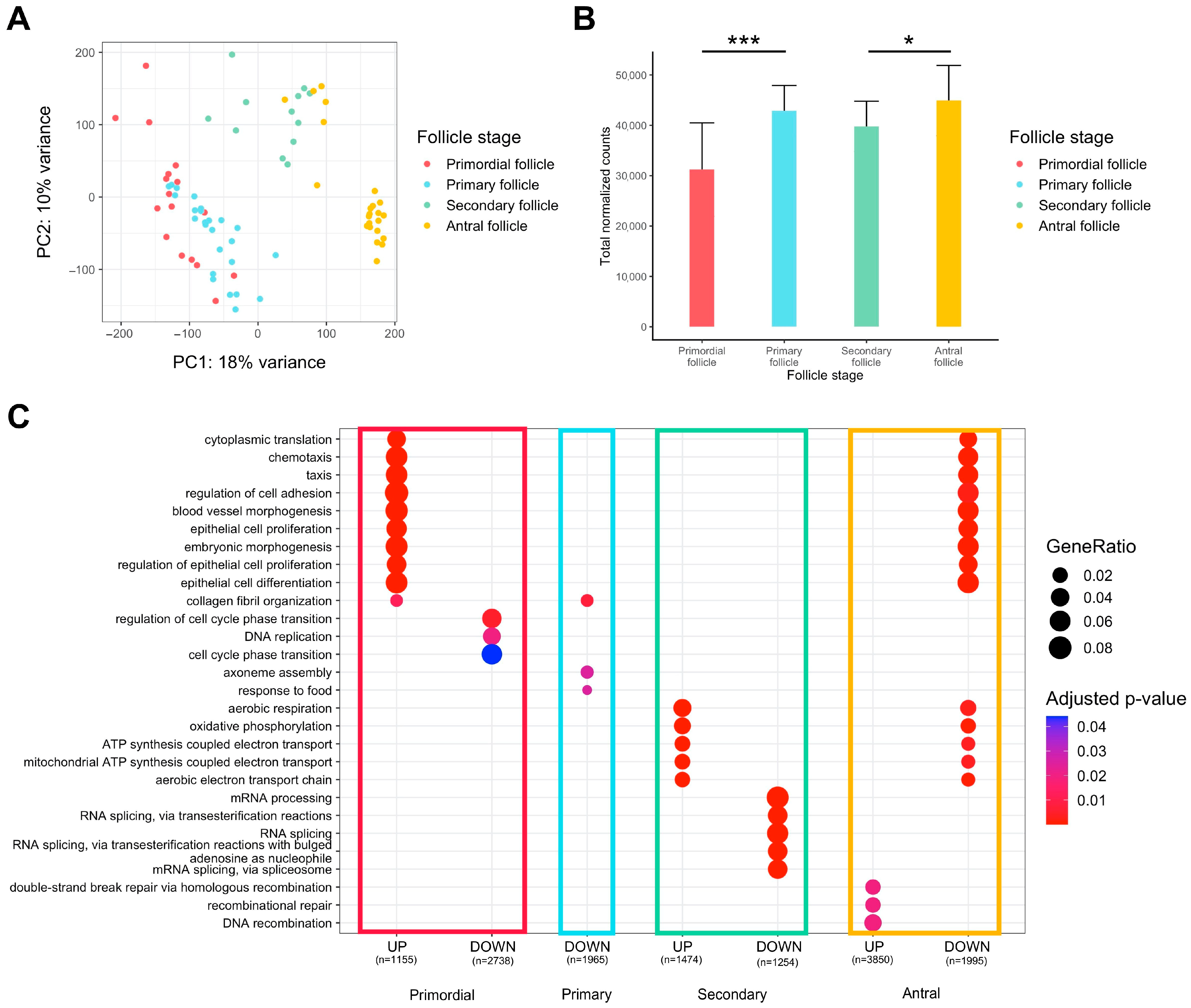
Global observation via principal component analysis (PCA) of transcriptomes from oocytes originating from primordial to antral follicles indicated overall differences between the different groups, but there was little difference between primordial and primary follicles, suggesting high transcriptional profile proximity for those cells (Figure 1A). The accumulation of maternal transcripts in the oocyte throughout folliculogenesis was confirmed, but there was no significant variation from the primary to the secondary follicle stage (Figure 1B).
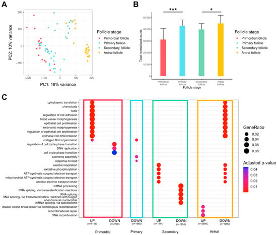
Figure 1.
Differential expression analysis of transcriptomic signature changes along the different stages of folliculogenesis (dataset from Zhang et al. (2018) [23]). (A) Principal component analysis of the whole expression dataset removing the 10% genes with the lowest variance. (B) Comparison of the normalized read counts total between the different follicle stages (significance assessed with a t-test, * p < 0.05, *** p < 0.005). (C) Comparative Gene Ontology enrichment analysis according to the up- or downregulation of transcripts between all four follicle stages. GeneRatio corresponds to the fraction of differentially expressed genes (DEGs) found in each Gene Ontology set.
We carried out a gene ontology over-representation analysis on genes differentially expressed between one stage and all others to reveal the biological signatures of oocytes from each follicle type (Figure 1C, Supplementary Table S1).
Oocytes of primordial follicles are characterized by the upregulation of genes involved in cytoplasmic translation, chemotaxis, regulation of cell adhesion, and regulation of epithelial cells, while the cell cycle phase transition is downregulated. Primary follicle oocytes do not display strong transcript identity compared to oocytes from other stages of follicle development, since few pathways are significantly downregulated and there is low biological function linked to oocyte development. Oocytes of secondary follicles manifest a rise in aerobic respiration, mitochondrial ATP synthesis, and oxidative phosphorylation, while RNA processes (mRNA processing, RNA splicing) tend to be slowed down. Finally, oocytes of antral follicles reveal a pattern that is opposite to primordial follicles (downregulation of cytoplasmic translation, chemotaxis, epithelial cell regulation, upregulation of cell cycle transition regulation) and secondary follicles (downregulation in aerobic respiration, mitochondrial ATP synthesis, and oxidative phosphorylation). Oocytes of antral follicles are also characterized by higher expression of genes involved in recombinational repair.
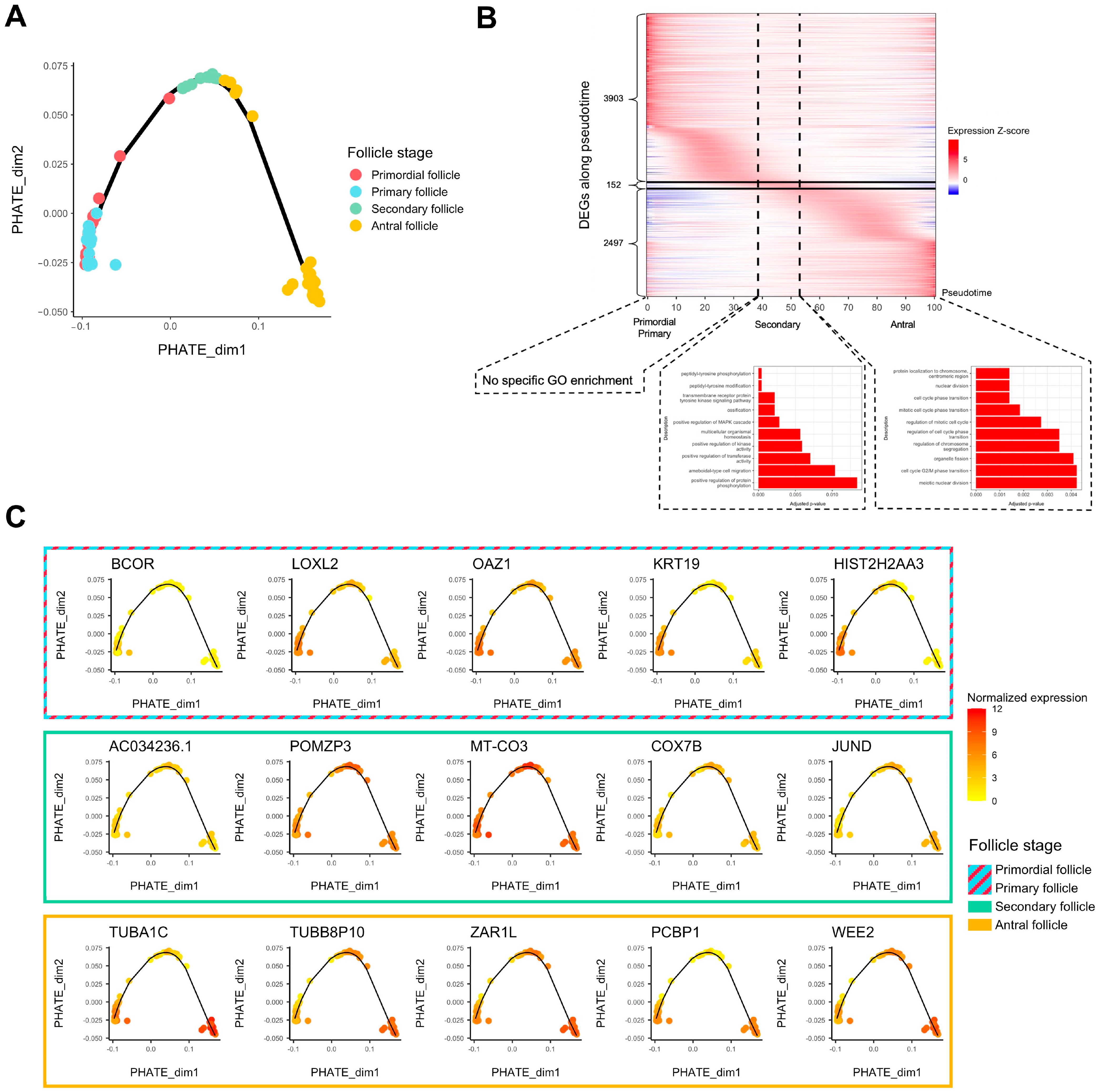
Folliculogenesis is likely a dynamic process since oocytes in follicles undergo continuous transcriptomic modifications and are not arrested at definite stages. As such, we performed a pseudotime analysis to temporally assess which genes show variation of expression in the window of folliculogenesis and to characterize the activation or repression behavior of these transcripts throughout follicular development. We first applied PHATE dimension reduction to normalized counts and discerned a transcriptomic trajectory (Figure 2A). We observed that primordial and primary follicles were almost indistinguishable in the first two dimensions (Figure 2A). A heatmap comparing differentially expressed genes all in one stage versus all other stages confirmed this observation considering that hierarchical clustering hardly separates primordial from primary follicles, revealing their transcriptomic proximity (Supplementary Figure S1A). We also observed that in the same follicle stage group, the transcriptome could be temporally different. Specifically, some antral follicles were close to secondary follicles, while the remaining antral follicles formed a clearly separated cluster later in the follicle developmental trajectory (Figure 2A), which turned out to be related to a patient effect (Patients E and G in Supplementary Figure S1B). The patients included in this study were notably healthy or suffering from reproductive pathologies of varying degrees of severity.
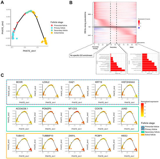
Figure 2.
Trajectory analysis of transcriptomic changes along folliculogenesis. (A) PHATE dimensionality reduction analysis of the whole expression dataset. (B) Heatmap of inferred expression of the most variable significant genes along folliculogenesis (6552 genes showing average expression changed by at least logFC > 1 within the oocyte developmental trajectory). Pseudotime is a metric that could be interpreted as a timing distance between one cell and its precursor cell and helps identify the ordering of cells along a lineage based on their gene expression profile. In this analysis, pseudotime 0 represents oocytes in the beginning of the window of the oocyte developmental trajectory (early follicles), while pseudotime 100 represents oocytes in the end of the window (antral follicles). Vertical lines delimit oocytes from the primordial/primary, secondary, and antral follicle groups. Horizontal lines delimit significantly differentially expressed genes along folliculogenesis reaching their peak expression during the primordial/primary (3903), secondary (152), and antral follicle groups (2497). (C) Variation of expression, for each follicle stage group, of the top 5 genes differentially expressed along folliculogenesis reaching their peak expression at this stage.
Then, we generated a list of 6552 transcripts whose expression varied temporally during folliculogenesis according to our pseudotime analysis (FDR < 0.05, logFC > 1) (Supplementary Table S2). The heatmap in Figure 2B highlights the sequential waves of transcriptional activity modifications from the primordial to the antral stage (Figure 2B). Successive activation and repression of these genes may drive folliculogenesis and regulate the transition from early to late stages, which is particularly the case for transcription factors (Supplementary Figure S2). Most of the 6552 transcripts are highly expressed inside the primordial and primary follicles and show lower expression in the following stages (3903 transcripts) (Figure 2B) but they are not enriched for a specific gene ontology. The transition to a secondary follicle oocyte is marked by the activation of a small set of genes (152) related to peptidyl-tyrosine modification and kinase activity, while final follicle oocyte growth is associated with the upregulation of 2497 transcripts enriched in cell cycle pathway genes (Figure 2B). The top five genes for each follicle stage group (primordial/primary, secondary, and antral) are displayed in Figure 2C, and their function is described in Table 1.

Table 1.
Description, for each follicle stage group, of the top 5 genes differentially expressed along folliculogenesis reaching their peak expression at this stage. Their function was retrieved and simplified from Uniprot database.
2.2. Oocyte Maturation
- Re-analysis of individual studies
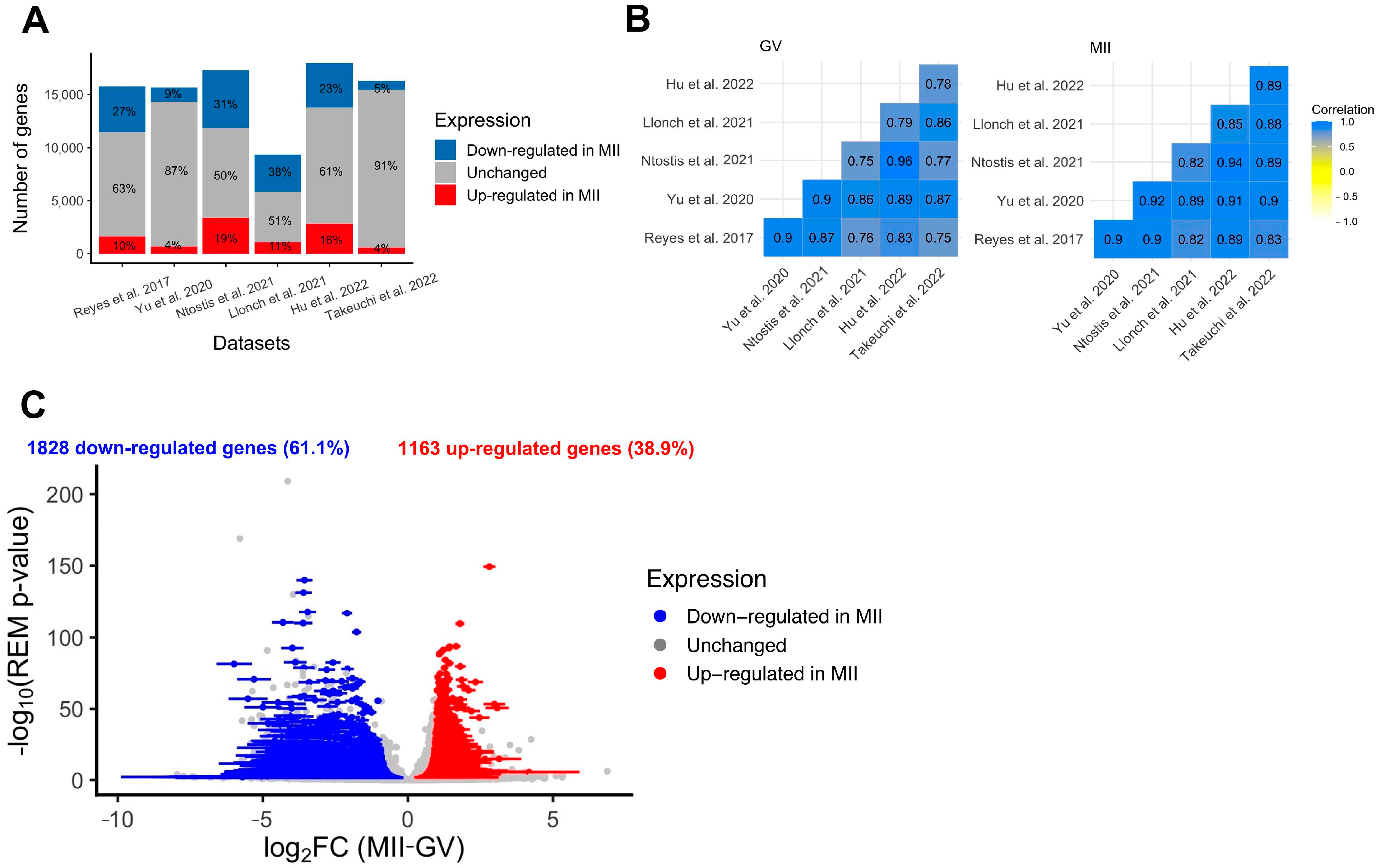
Standard re-analysis of six studies comparing the transcriptome of fully grown GV and MII oocytes obtained after controlled ovarian stimulation confirmed the existence of intense modulation of maternal transcript abundance [24,25,26,27,28,29]. First, a clear separation between fully grown GV and MII oocytes was observed in all datasets using dimension reduction (PCA method) (Supplementary Figure S3). Secondly, the number of differentially expressed genes between GV and MII oocytes was high in all datasets, with an average of 33% (9–51%) of transcripts undergoing modification between the two stages (Figure 3A). According to several independent studies, the main pathways modified were related to mitochondrial processes (downregulated in MII compared to fully grown GV), cytoplasmic translation, and chromatin organization (upregulated in MII compared to fully grown GV) (Supplementary Figure S4A,B). Correlations in all transcript expressions were high between datasets, but there were some differences, likely due to technical variation, justifying the need for a meta-analysis (Figure 3B). The six RNA-seq datasets showed high correlation in all expressed gene counts (Pearson’s correlation coefficient > 0.75, Figure 3B), but as seen on PCA on merged datasets, the various techniques have an influence on transcript abundance even after batch correction (Supplementary Figure S5).
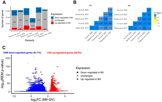
Figure 3.
Differential expression analysis of genes during the GV-to-MII oocyte transition. (A) Results of individual differential expression analysis for the six datasets evaluated in this study. Numbers represent the percentage of genes upregulated, downregulated, or stable in MII versus GV oocytes. (B) Correlation of the normalized expression of all expressed genes for the six datasets evaluated in this study, respectively, for GV and MII oocytes. (C) Volcanoplot of the meta-analysis result for the six datasets evaluated in this study. A random effects model based on effect size (log2FC, difference of expression between MII and GV oocytes) and p-values from each dataset was applied. One gene is represented by one dot. A total of 2991 significant DEGs were identified (summary adjusted p-value from the random effect model < 0.05 and sign of log2FC consistent in all 6 studies). Blue dots represent significant DEGs downregulated in MII oocytes compared to GV. Similarly, red dots represent significant DEGs upregulated in MII oocytes compared to GV oocytes. The interval confidence of the summary log2FC is represented by the bars associated with the dots for each DEG [24,25,26,27,28,29].
- Meta-analysis
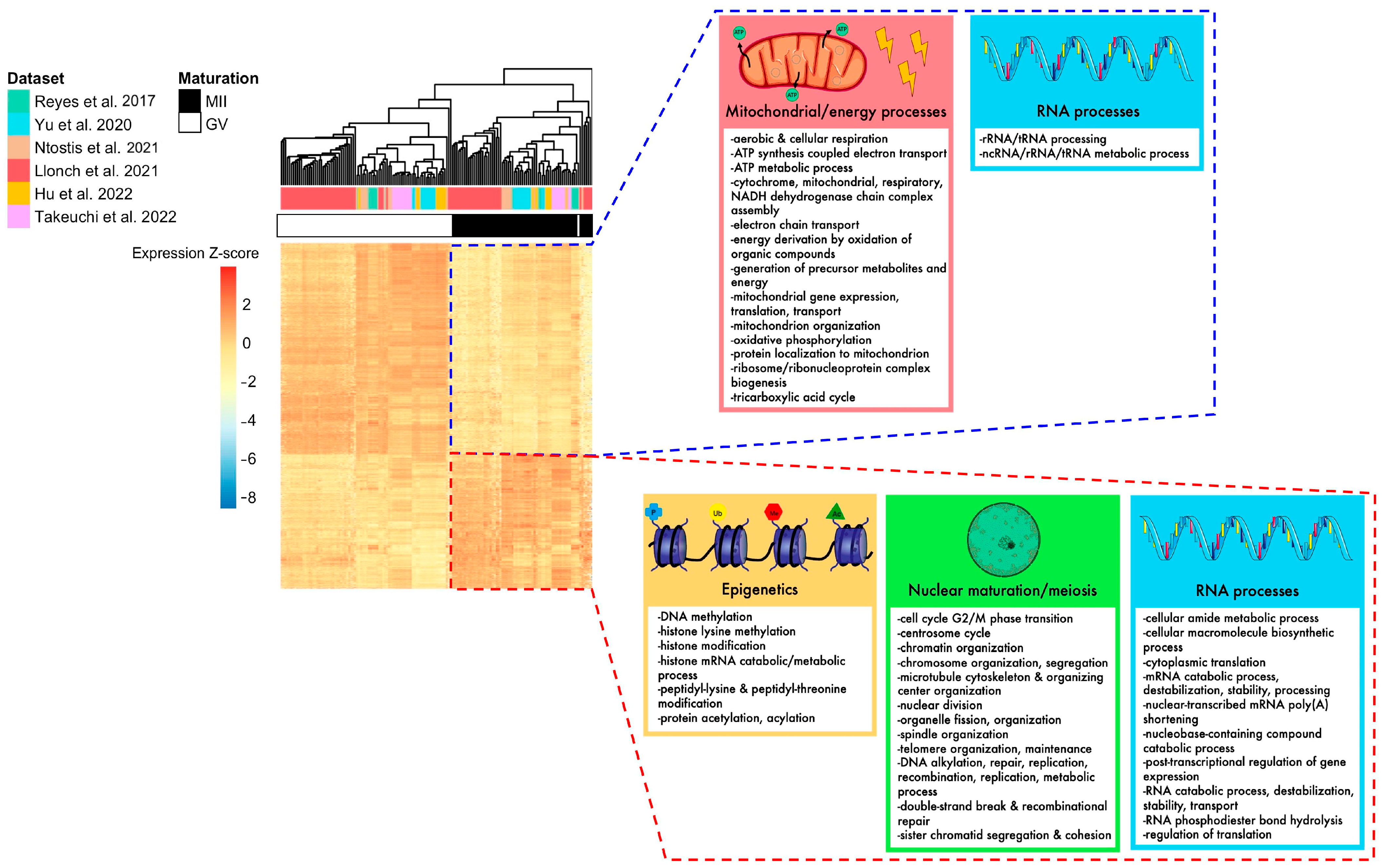
To eliminate parameters likely to influence biological effects (such as age or health condition) and increase statistical power, we conducted a meta-analysis of differentially expressed genes (DEGs) between fully grown GV and MII oocytes. In total, the six datasets included 87 fully grown GV and 70 MII oocytes, collected from patients ranging from young to advanced maternal ages (18–44 yo). The random effects model meta-analysis identified 2991 DEGs with consistent modifications across all six datasets (Figure 3C, Supplementary Table S3). While most were downregulated in MII oocytes (61%) (Figure 3C), some displayed a higher level of transcripts in MII oocytes. Upregulated pathways were related to histone modification, nuclear maturation, and RNA processes, while downregulated pathways were related to mitochondrial processes (Figure 4). Transcript annotation revealed that DEGs were mainly protein-coding genes and, in small proportions, transposable elements or long non-coding RNAs (lncRNA) (Supplementary Figure S6A). Long terminal repeats (LTR) were mostly upregulated in MII, while long interspersed nuclear elements (LINE) elements were downregulated (Supplementary Figure S6B).
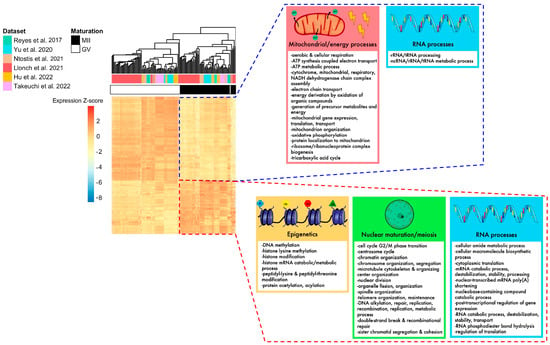
Figure 4.
Summary of biological pathways enriched for DEGs identified in the meta-analysis, according to their up- or downregulation. The heatmap of DEGs identified with the meta-analysis shows that MII and GV oocytes are correctly segregated according to their expression for these genes. Mitochondrial processes, energy processes, and some RNA processes were downregulated in MII oocytes (framed in blue), while nuclear maturation/meiosis, epigenetics, and some other RNA processes were upregulated in MII oocytes (framed in red) [24,25,26,27,28,29].
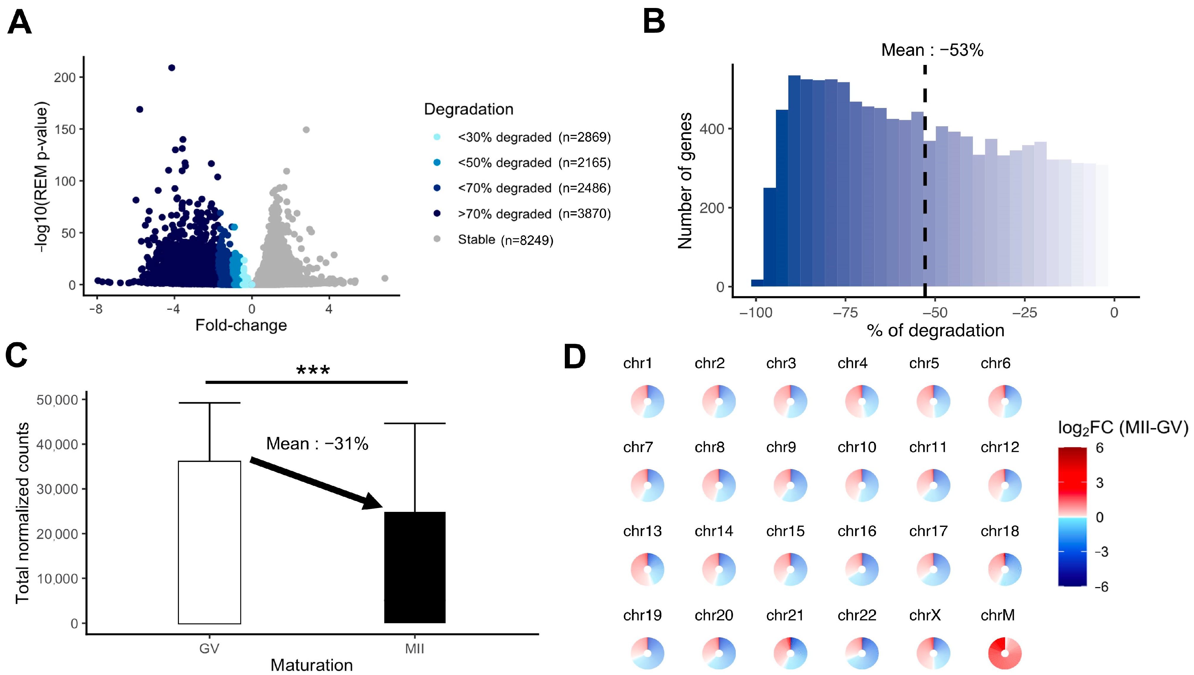
We evaluated the extent of mRNA degradation during the transition from fully grown GV to MII oocyte stage according to the meta-analysis results. A substantial proportion of the transcripts was highly degraded (>70% loss) (Figure 5A). The mean degradation per transcript was 53% for downregulated transcripts (Figure 5B). Between fully grown GV and MII stages, there was a significant drop in total maternal mRNA quantity, which could be evaluated at 31% (Figure 5C). The degradation concerned transcripts from all chromosomes, whereas mtDNA gene expression was increased in MII oocytes (Figure 5D).
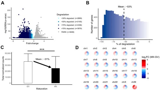
Figure 5.
Measurement of the degradation of the maternal mRNA content in MII oocytes. (A) Volcanoplot of the meta-analysis result (MII versus GV) colored by the intensity of degradation between GV and MII stages (stable, <30% of degradation, <50% of degradation, <70% of degradation, or >70% of degradation). (B) Histogram focusing on the extent of degradation in downregulated transcripts in MII versus GV oocytes. (C) Comparison of total normalized counts between the GV and MII oocytes (significance assessed with a t-test, *** p < 0.005). (D) Assessment of degradation by chromosome. Each circle is divided into sections according to the number of genes on the chromosome and each section, representing one gene, is colored by the summary log2FC resulting from the meta-analysis result (MII versus GV) corresponding to the gene. ChrM: mitochondrial chromosome.
2.3. Influence of the Environment on Human Oogenesis/Folliculogenesis
We identified transcripts differentially expressed during folliculogenesis or the GV-to-MII transition; these transcripts are also putative transcripts susceptible to environmental stressors occurring in the window of oocyte development (maternal age, polycystic ovary syndrome (PCOS), in vitro maturation (IVM), cryopreservation). A list of these genes is available in Table 2 and a summary of studies on the factors evaluated can be found in Supplementary Table S4.

Table 2.
Comparison of folliculogenesis and oogenesis-specific genes identified in this review with previous putative genes sensitive to the environment (PCOS, maternal age, IVM, cryoconservation). Only DEGs retrieved in >2 studies for each factor were considered to increase robustness of the analysis.
3. Discussion
The oocyte is the largest human cell and one of the richest in mRNA quantity [30]. It determines early embryonic competence and its transcriptome contains many secrets waiting to be revealed [31]. In this study, we comprehensively gathered scRNA-seq datasets from human oocytes at multiple stages during the long process of oocyte growth and maturation and decrypted the transcriptomic reorganization required to ensure that the oocyte becomes fully competent. We believe it is important to focus on human oocyte regulation exclusively, as previous studies comparing human oocyte maturation with other mammalian species revealed very few overlaps in the individual transcripts modified, even if major biological processes activation/repression were similar [18].
Folliculogenesis and oocyte maturation imply specific regulation during oocyte development. First, variation in chromatin accessibility determines the activity of transcription, which is high within early follicles but absent after GV breakdown [7,32]. Secondly, transcription factor activity shapes the oocyte transcriptome during folliculogenesis by upregulating or downregulating specific transcripts to allow the transition from a primary to a pre-ovulatory follicle oocyte [33]. Thirdly, selective degradation or translation of maternal transcripts is a prerequisite for cytoplasmic maturation, leading to a fertilizable MII oocyte that can ensure the first embryonic divisions [14].
Our chronological gene ontology comparison analysis revealed signature pathways activated and repressed in oocytes from the different follicle stages. The primordial stage may reflect strong communication between the oocyte and its surrounding cells, manifested by an upregulation of genes related to cell adhesion, epithelial cell proliferation, and collagen fibril organization pathways, which tend to be reduced afterwards. Communication with the surrounding cells, such as granulosa, plays a pivotal role in oocyte growth via the action of paracrine factors [34]. Oocytes mobilize specific transcripts such as GDF9, TGFB1, and ACTN1, to act on granulosa cell proliferation and maintain gap junctions [35,36]. Moreover, downregulation of cytoplasmic translation was seen in antral follicles while the highest activity was observed in primordial follicles, which corroborates the findings that progressively maternal mRNAs are not translated but stored until they are degraded or translated after meiosis resumption following GVBD. Oocytes from primordial follicles also show downregulation of cell cycle pathways which agrees with their arrest at the diplotene stage of meiosis I until their activation [37]. In the antral follicle, cell cycle phase transition pathways are upregulated, possibly correlating with the acquisition of meiotic competence to sustain proximate oocyte maturation [38]. One of our conclusions would be that the oocytes at the primary follicle stage do not display a highly specific transcriptomic profile compared to the primordial follicle. The transition between both stages is likely to be mRNA quantity-specific rather than pathway-specific, meaning that primary follicle oocytes accumulate mRNA drastically but not selectively. This primary-to-primordial transition may be driven by a specific regulatory network involving transcription factors and long non-coding RNAs according to previous evidence [23,39]. Then, the transition from an early follicle oocyte to a secondary follicle oocyte is marked by a switch in energy consumption. Activation of mitochondrial ATP synthesis, aerobic respiration, and oxidative phosphorylation sustain major structural and biochemical evolution, which are energy-intensive processes [40]. Furthermore, mRNA processing and notably splicing are slowed down in secondary follicle oocytes compared to other stages. Finally, oocytes in antral follicles turn off energy activity possibly passively (downregulation of aerobic respiration and oxidative phosphorylation pathways) because of the difficulty of accessing O2 in a restricted micro-environment, according to mathematical modelling in humans [41,42,43]. We also observed that oocytes in antral follicles show an increased ability to repair DNA alterations compared to other stages (upregulation of recombinational repair pathway), which is confirmed by previous observations [44].
Taking advantage of the single-cell nature of the Zhang et al. dataset [23], we then applied trajectory analysis along folliculogenesis to characterize continuous transcriptomic changes. We identified a bunch of genes showing significant variation in expression from primordial-to-antral follicle oocyte development and characterized their temporal regulation. This revealed that a large majority of them were highly upregulated during the early follicle stages, seemingly biologically unspecific as no Gene Ontology showed enrichment for them. This confirms our hypothesis of an unspecific accumulation of transcripts during the primary follicle stage. Surprisingly, only about a hundred folliculogenesis variable genes show a peak of expression in the window of the secondary follicle stage, which appeared to be related to peptidyl-tyrosine modification and regulation of kinase (notably the MAPK cascade) and transferase activity. Following transcriptional waves of upregulation may involve the preparation for meiosis resumption in the antral follicle oocyte.
Furthermore, we observed that depending on the patients, the transcriptome dynamic displayed different kinetics of progression. Differences between patients in the previously published dataset used herein could stem from the inclusion of patients with particular pathological conditions (cervical cancer, endometrial cancer, lymphoma), which may affect reproductive functions. This could lead to unwanted bias in the interpretation of the results as a small number of patients for each follicle stage were included.
Next, we better characterized the downfall undergone by maternal mRNAs during late oogenesis. We found an average decline of 30% in total mRNA from fully grown GV to the MII stage across studies. Previous studies indicated that 60% of maternal transcripts are degraded from the fully grown GV to MII stage, using mass measures [45,46]. The difference observed may be a direct consequence of the fact that mRNAs with short polyA tails are not captured by the employed sequencing technologies. Therefore, our analysis mirrors the biological pathways that are recruited in a timely manner (mRNAs with long polyA tails on the verge of being degraded or translated) rather than how degraded all maternal transcripts are. However, this approach displays the selective downregulation of specific transcripts. Indeed, transcripts associated with energy production and mitochondrial functions were extensively downregulated, while those associated with cytoplasmic translation, histone modification, meiotic processes, and RNA processes were conserved. The downregulation of mitochondrial processes is highly conserved across mammals and may be a protective system with respect to oxidative stress via ROS production and the inability of the early embryo to protect against these stressors as reported in rhesus monkey, mouse, and cow models [18,47]. Concomitantly, MII oocytes should have already synthesized sufficient mtDNA to support key developmental events such as epigenetic programming [48]. The new meta-analysis we proposed found that genes involved in mobilization of cytoplasmic translation, histone modification, meiotic processes, and RNA processes are upregulated in MII oocytes, which are metabolic pathways crucial for the multiplicity of maturations (cytoplasmic-nuclear-epigenetic) required by the oocyte.
Interestingly, the transition from fully grown GV to MII oocytes displayed upregulation of a substantial number of transcripts, whereas oocytes are thought to be transcriptionally quiescent. This could be explained by several hypotheses. First, as explained, the scRNA-sequencing methods used in the meta-analyzed studies captured mRNA with long polyA tails, thus reflecting the amount of mRNA that can be recruited for translation and not silenced or set to degrade mRNAs with short polyA tails. Some transcripts are still enrolled for translation during the GV-MII transition via polyA lengthening, showing them to be upregulated in MII oocytes. A second hypothesis that still needs investigation is that transcriptional quiescence after the GVBD stage is not total [49,50]. This theory is contrasted by a third hypothesis, only proven in C. elegans, assuming that oocytes after GVBD cannot synthetize mRNA but can accumulate transcripts from surrounding somatic cells, supposedly via extracellular vesicles [51].
Furthermore, we intended to re-analyze studies comparing failed-to-mature oocytes versus mature oocytes. In Pietroforte et al., the authors did not identify any DEG between both groups, while the low number of samples in Li et al. (three oocytes per group) would be too low to detect expression differences with satisfactory power [52,53]. Then, it remains crucial to assess the biological reasons behind oocyte maturation failure and whether transcriptomic abnormalities would be responsible for it. In some cases, defects in the maternal mRNA degradation machinery would be involved, notably an alteration in the expression of Btg4 or Msy2 in mice [22,54,55]. This would lead to the unwanted maintenance of certain categories of transcripts detrimental to oocyte fertility or embryonic development.
In addition, throughout growth and maturation, oocytes are exposed to multiple environmental stressors, which can alter the sequential gene expression changes governing oocyte development [56]. Our identification of transcripts expressed along oocyte development that are highly susceptible to those factors is of major importance, as they may be the key to understanding the apparition of oocyte defects and deficiencies and overcoming these outcomes [27,28,29,57,58,59,60,61,62,63,64,65,66,67]. For oocyte in vitro maturation and cryoconservation, regaining expected levels of expression of those transcripts by adapting assisted reproductive protocols may become a path of research to increase mature oocyte rates in the future. For age and PCOS, identification of those transcripts is also important to understand the precise underlying mechanisms. In future studies, it would be highly interesting to observe transcriptomic modifications in the same patient from the primordial follicle stage and compare the dynamic of transcriptional changes between several donors, particularly under pathological conditions (pathology versus control).
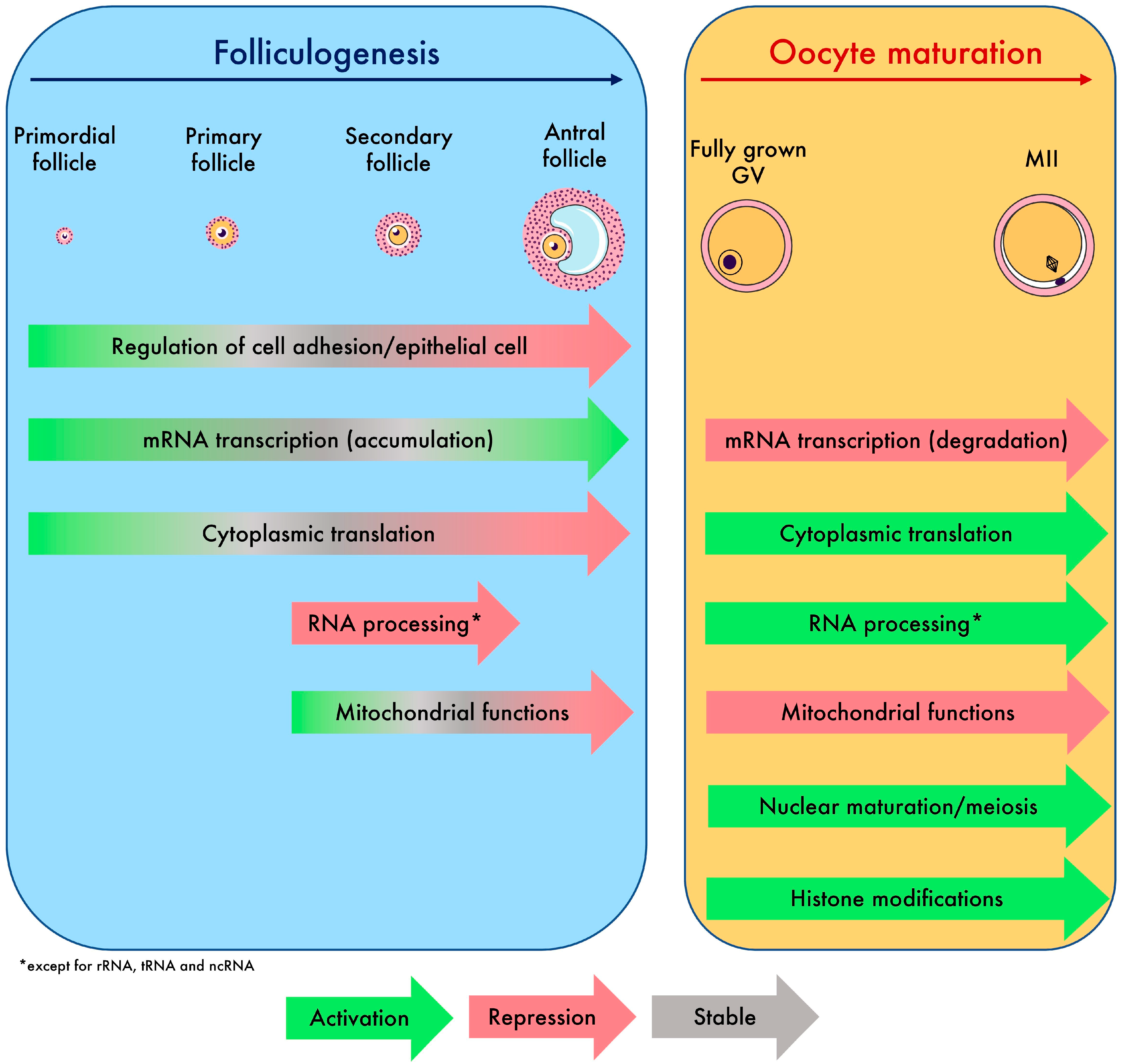
To conclude, the oocyte transcriptome follows a dynamic that is tightly controlled. Overall, we have decoded the regulatory events of maternal mRNAs that lead the oocyte to grow and undergo multiple types of maturation, which we synthetized in Figure 6. The atlas of transcriptomic modifications described here will facilitate the precise identification of the transcripts involved in the failure of oocyte growth and maturation in future research.
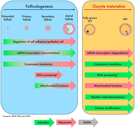
Figure 6.
Summary of biological processes and global transcriptomic events driving oocyte growth and maturation identified in this study.
4. Materials and Methods
4.1. Population
To examine the transcriptomic modifications experienced by the oocyte along human folliculogenesis, we included datasets comprising human primary oocytes from primordial to antral follicles (at least two different stages). To examine the transcriptomic modifications experienced by the oocyte throughout human oogenesis, we included datasets comprising human immature fully grown GV (prophase I arrested), MI (metaphase of meiosis I), and MII oocytes collected from controlled ovarian hyperstimulation protocols. Inclusion was also conditional on the sequencing method, which had to be single-oocyte RNA-seq. Only single-denuded oocytes were considered, meaning that oocytes surrounding cells were excluded from this review. Oocytes recovered in all clinical contexts were included, whether they were collected from donors or patients seeking infertility treatment, from in vitro or in vivo maturation, and at all maternal ages. No restrictions were placed on the publication date of datasets. A summary of the included studies with information on population characteristics, study design, and sequencing protocol is presented in Table 3.

Table 3.
Characteristics of studies re-analyzed in this review.
4.2. Dataset Collection
All the datasets used in this study are available in public repositories. Raw data (.fastq) were downloaded from European Nucleotide Archive accessions: PRJNA666614, PRJNA508772, PRJNA377237, PRJNA811110, PRJNA701233, PRJNA690226 and PRJNA421274.
4.3. Dataset Processing
For each dataset, adapters and low-quality sequences were trimmed using TrimGalore! V0.6.6. Next, paired-end alignment was performed onto human reference genome (hg38) with STAR v2.7.9a, reporting randomly one position and allowing 6% of mismatches. Transposable element (TE) annotation was downloaded from RepeatMasker and joined with Gencode v19 gene annotation [68] to measure their expression level as TEs are crucial in the regulation of gametes and embryos gene expression. Finally, gene expression was quantified with featureCounts v2.0.1.
All datasets were processed following a standard procedure. Genes with low counts were removed with the filterByExpr function from the edgeR R package. Counts were normalized using the limma-voom method. When clinical information was available, we used duplicateCorrelation to account for samples from the same patients (considered as technical replicates).
For comparison of folliculogenesis stages (oocytes from primordial, primary, secondary, and antral follicles), differential analysis was performed between oocytes from one stage versus all stages using limma (differentially expressed if FDR < 0.05 and FC > 2 or FC < 0.5). This analysis was designed to identify a specific signature for each follicular stage. Because folliculogenesis is likely a dynamic process, a pseudotime analysis was implemented using tradeSeq using PHATE reduction to find differentially expressed genes throughout folliculogenesis rather than separating oocytes into distinct clusters [69,70]. This will identify precisely in time which genes are activated and repressed in oocytes during the development of follicles, from the primordial to the antral stage. The advantage of PHATE reduction is that it relies on diffusion dynamics and is thus particularly suitable to assess developmental trajectories such as oocyte growth.
For the comparison between GV, MI, and MII oocytes, differential analysis was performed between stage groups for each independent dataset using limma. Maternal age was incorporated as a covariate when available. Genes were considered differentially expressed when FDR < 0.05 and FC > 2 or FC < 0.5.
All PCAs in this article were performed using PCAtools on normalized expression (log2(cpm + 1)) removing the 10% genes with the lowest variance. Pathway analyses were run with clusterProfiler using the enrichGO function. For the purpose of visualization, independent datasets were corrected for batch effects using ComBat. All heatmaps were created with the pheatmap R package.
4.4. Meta-Analysis
A meta-analysis between all datasets comparing fully grown GV, MI, and MII oocytes was performed with MetaVolcanoR, which relies on the metafor package [71,72]. We summarized the expression fold change for each gene across the six independent datasets, implementing a random effects model using effect size (log2FC) and raw p-values from each dataset as input. The resulting estimation of a summary p-value for each gene indicates the probability of the summary expression fold-change being equal to zero. The advantage of the random effects model is the consideration of heterogeneity between studies (between-study effect variance) in addition to the within-study effect variance. For each gene, we considered significant expression change with an adjusted summary p-value < 0.05 and consistency (same sign of log2FC) in all studies.
Degradation or accumulation of mRNA quantity for each transcript through final oocyte maturation was calculated by converting the summary log(fold-change) to the percentage difference of normalized expression between baseline and the compared group. Transcripts with higher level of transcripts in MII than in fully grown GV were considered stable due to the assumed transcriptional quiescence of oocytes during the GV-to-MII transition (difference in polyadenylation and not quantity).
Because of the low number of studies that included MI oocytes (n = 2) and their reduced sample size (respectively, 6 and 7 MI), results including the comparison of MI with fully grown GV and MII oocytes are not discussed further (high risk of bias).
4.5. Influence of the Environment on Human Oogenesis/Folliculogenesis
In a previous review on the transcriptomic integrity of human oocytes used in assisted reproductive techniques (ARTs), we discussed the biological mechanisms likely to be altered in the context of ART interventions, maternal aging, a suboptimal lifestyle, or reproductive health issues [56]. We believe it would be highly interesting to identify oocyte growth and maturation-specific transcripts dysregulated by those factors. Thereby, available lists of DEGs from each study discussed in our previous review were retrieved and cross-checked for each environmental factor considered (endometriosis, PCOS, maternal age, smoking, body mass index (BMI), ovarian stimulation, IVM, and cryopreservation) to find common transcripts dysregulated between at least two studies. Those lists were in turn cross-checked with lists of DEGs identified in our folliculogenesis and oocyte maturation differential expression analyses. Previous studies on granulosa and cumulus cells were not considered here.
Supplementary Materials
The following supporting information can be downloaded at: https://www.mdpi.com/article/10.3390/ijms25010033/s1.
Author Contributions
Conceptualization, B.D. and P.F.; Methodology, B.D.; Software, B.D.; Formal Analysis, B.D.; Investigation, B.D. and P.F.; Data Curation, B.D.; Writing—Original Draft Preparation, B.D. and P.F.; Writing—Review and Editing, B.D. and P.F.; Visualization, B.D.; Supervision, P.F.; Critical Revision and Editing, L.F. and C.P. All authors have read and agreed to the published version of the manuscript.
Funding
This work was supported by funding from the Agence Nationale pour la Recherche (‘CARE’-ANR JCJC 2017).
Informed Consent Statement
Not applicable.
Data Availability Statement
Data are contained within the article and supplementary materials.
Acknowledgments
We thank Suzanne Rankin for her help in proofreading the manuscript.
Conflicts of Interest
The authors declare no conflict of interest.
References
- Bendsen, E.; Byskov, A.G.; Andersen, C.Y.; Westergaard, L.G. Number of Germ Cells and Somatic Cells in Human Fetal Ovaries during the First Weeks after Sex Differentiation. Hum. Reprod. 2006, 21, 30–35. [Google Scholar] [CrossRef] [PubMed]
- Tripathi, A.; Prem Kumar, K.V.; Chaube, S.K. Meiotic Cell Cycle Arrest in Mammalian Oocytes. J. Cell Physiol. 2010, 223, 592–600. [Google Scholar] [CrossRef] [PubMed]
- Eppig, J.J. Coordination of Nuclear and Cytoplasmic Oocyte Maturation in Eutherian Mammals. Reprod. Fertil. Dev. 1996, 8, 485–489. [Google Scholar] [CrossRef] [PubMed]
- Watson, A.J. Oocyte Cytoplasmic Maturation: A Key Mediator of Oocyte and Embryo Developmental Competence. J. Anim. Sci. 2007, 85, E1–E3. [Google Scholar] [CrossRef] [PubMed]
- Conti, M.; Franciosi, F. Acquisition of Oocyte Competence to Develop as an Embryo: Integrated Nuclear and Cytoplasmic Events. Hum. Reprod. Update 2018, 24, 245–266. [Google Scholar] [CrossRef]
- Fan, H.-Y.; Sun, Q.-Y. Oocyte Meiotic Maturation. In The Ovary; Elsevier: Amsterdam, The Netherlands, 2019; pp. 181–203. [Google Scholar]
- Gu, C.; Liu, S.; Wu, Q.; Zhang, L.; Guo, F. Integrative Single-Cell Analysis of Transcriptome, DNA Methylome and Chromatin Accessibility in Mouse Oocytes. Cell Res. 2019, 29, 110–123. [Google Scholar] [CrossRef]
- Luciano, A.M.; Lodde, V.; Franciosi, F.; Tessaro, I.; Corbani, D.; Modina, S.C. Large-Scale Chromatin Morpho-Functional Changes during Mammalian Oocyte Growth and Differentiation. Eur. J. Histochem. 2012, 56, 231–235. [Google Scholar] [CrossRef]
- Wu, D.; Dean, J. EXOSC10 Sculpts the Transcriptome during the Growth-to-Maturation Transition in Mouse Oocytes. Nucleic Acids Res. 2020, 48, 5349–5365. [Google Scholar] [CrossRef]
- Jiang, Y.; Adhikari, D.; Li, C.; Zhou, X. Spatiotemporal Regulation of Maternal MRNAs during Vertebrate Oocyte Meiotic Maturation. Biol. Rev. 2023, 98, 900–930. [Google Scholar] [CrossRef]
- Sha, Q.Q.; Zheng, W.; Wu, Y.W.; Li, S.; Guo, L.; Zhang, S.; Lin, G.; Ou, X.H.; Fan, H.Y. Dynamics and Clinical Relevance of Maternal MRNA Clearance during the Oocyte-to-Embryo Transition in Humans. Nat. Commun. 2020, 11, 4917. [Google Scholar] [CrossRef]
- Cornet-Bartolomé, D.; Barragán, M.; Zambelli, F.; Ferrer-Vaquer, A.; Tiscornia, G.; Balcells, S.; Rodriguez, A.; Grinberg, D.; Vassena, R. Human Oocyte Meiotic Maturation Is Associated with a Specific Profile of Alternatively Spliced Transcript Isoforms. Mol. Reprod. Dev. 2021, 88, 605–617. [Google Scholar] [CrossRef]
- Tora, L.; Vincent, S.D. What Defines the Maternal Transcriptome? Biochem. Soc. Trans. 2021, 49, 2051–2062. [Google Scholar] [CrossRef] [PubMed]
- Su, Y.Q.; Sugiura, K.; Woo, Y.; Wigglesworth, K.; Kamdar, S.; Affourtit, J.; Eppig, J.J. Selective Degradation of Transcripts during Meiotic Maturation of Mouse Oocytes. Dev. Biol. 2007, 302, 104–117. [Google Scholar] [CrossRef] [PubMed]
- Jiang, Z.Y.; Fan, H.Y. Five Questions toward MRNA Degradation in Oocytes and Preimplantation Embryos: When, Who, to Whom, How, and Why? Biol. Reprod. 2022, 107, 62–75. [Google Scholar] [CrossRef] [PubMed]
- Christou-Kent, M.; Dhellemmes, M.; Lambert, E.; Ray, P.F.; Arnoult, C. Diversity of RNA-Binding Proteins Modulating Post-Transcriptional Regulation of Protein Expression in the Maturing Mammalian Oocyte. Cells 2020, 9, 662. [Google Scholar] [CrossRef] [PubMed]
- Svoboda, P.; Franke, V.; Schultz, R.M. Sculpting the Transcriptome During the Oocyte-to-Embryo Transition in Mouse. In Current Topics in Developmental Biology; Academic Press Inc.: Cambridge, MA, USA, 2015; Volume 113, pp. 305–349. ISBN 9780124095236. [Google Scholar]
- Schall, P.Z.; Latham, K.E. Essential Shared and Species-Specific Features of Mammalian Oocyte Maturation-Associated Transcriptome Changes Impacting Oocyte Physiology. Am. J. Physiol. Cell Physiol. 2021, 321, C3–C16. [Google Scholar] [CrossRef] [PubMed]
- Labrecque, R.; Sirard, M.A. The Study Ofmammalian Oocyte Competence by Transcriptome Analysis: Progress and Challenges. Mol. Hum. Reprod. 2014, 20, 103–116. [Google Scholar] [CrossRef] [PubMed]
- Suo, L.; Zhou, Y.X.; Jia, L.L.; Wu, H.B.; Zheng, J.; Lyu, Q.F.; Sun, L.H.; Sun, H.; Kuang, Y.P. Transcriptome Profiling of Human Oocytes Experiencing Recurrent Total Fertilization Failure. Sci. Rep. 2018, 8, 17890. [Google Scholar] [CrossRef]
- Rong, Y.; Ji, S.Y.; Zhu, Y.Z.; Wu, Y.W.; Shen, L.; Fan, H.Y. ZAR1 and ZAR2 Are Required for Oocyte Meiotic Maturation by Regulating the Maternal Transcriptome and MRNA Translational Activation. Nucleic Acids Res. 2019, 47, 11387–11402. [Google Scholar] [CrossRef]
- Medvedev, S.; Pan, H.; Schultz, R.M. Absence of MSY2 in Mouse Oocytes Perturbs Oocyte Growth and Maturation, RNA Stability, and the Transcriptome. Biol. Reprod. 2011, 85, 575–583. [Google Scholar] [CrossRef]
- Zhang, Y.; Yan, Z.; Qin, Q.; Nisenblat, V.; Chang, H.M.; Yu, Y.; Wang, T.; Lu, C.; Yang, M.; Yang, S.; et al. Transcriptome Landscape of Human Folliculogenesis Reveals Oocyte and Granulosa Cell Interactions. Mol. Cell 2018, 72, 1021–1034.e4. [Google Scholar] [CrossRef] [PubMed]
- Takeuchi, H.; Yamamoto, M.; Fukui, M.; Inoue, A.; Maezawa, T.; Nishioka, M.; Kondo, E.; Ikeda, T.; Matsumoto, K.; Miyamoto, K. Single-Cell Profiling of Transcriptomic Changes during in Vitro Maturation of Human Oocytes. Reprod. Med. Biol. 2022, 21, e12464. [Google Scholar] [CrossRef] [PubMed]
- Hu, W.; Zeng, H.; Shi, Y.; Zhou, C.; Huang, J.; Jia, L.; Xu, S.; Feng, X.; Zeng, Y.; Xiong, T.; et al. Single-Cell Transcriptome and Translatome Dual-Omics Reveals Potential Mechanisms of Human Oocyte Maturation. Nat. Commun. 2022, 13, 5114. [Google Scholar] [CrossRef] [PubMed]
- Yu, B.; Jayavelu, N.D.; Battle, S.L.; Mar, J.C.; Schimmel, T.; Cohen, J.; Hawkins, R.D. Single-Cell Analysis of Transcriptome and DNA Methylome in Human Oocyte Maturation. PLoS ONE 2020, 15, e0241698. [Google Scholar] [CrossRef] [PubMed]
- Llonch, S.; Barragán, M.; Nieto, P.; Mallol, A.; Elosua-Bayes, M.; Lorden, P.; Ruiz, S.; Zambelli, F.; Heyn, H.; Vassena, R.; et al. Single Human Oocyte Transcriptome Analysis Reveals Distinct Maturation Stage-Dependent Pathways Impacted by Age. Aging Cell 2021, 20, e13360. [Google Scholar] [CrossRef] [PubMed]
- Ntostis, P.; Iles, D.; Kokkali, G.; Vaxevanoglou, T.; Kanavakis, E.; Pantou, A.; Huntriss, J.; Pantos, K.; Picton, H.M. The Impact of Maternal Age on Gene Expression during the GV to MII Transition in Euploid Human Oocytes. Hum. Reprod. 2021, 37, 80–92. [Google Scholar] [CrossRef] [PubMed]
- Reyes, J.M.; Silva, E.; Chitwood, J.L.; Schoolcraft, W.B.; Krisher, R.L.; Ross, P.J. Differing Molecular Response of Young and Advanced Maternal Age Human Oocytes to IVM. Hum. Reprod. 2017, 32, 2199–2208. [Google Scholar] [CrossRef]
- Kataruka, S.; Modrak, M.; Kinterova, V.; Malik, R.; Zeitler, D.M.; Horvat, F.; Kanka, J.; Meister, G.; Svoboda, P. MicroRNA Dilution during Oocyte Growth Disables the MicroRNA Pathway in Mammalian Oocytes. Nucleic Acids Res. 2020, 48, 8050–8062. [Google Scholar] [CrossRef]
- Kocabas, A.M.; Crosby, J.; Ross, P.J.; Otu, H.H.; Beyhan, Z.; Can, H.; Tam, W.L.; Rosa, G.J.M.; Halgren, R.G.; Lim, B.; et al. The Transcriptome of Human Oocytes. Proc. Natl. Acad. Sci. USA 2006, 103, 14027–14032. [Google Scholar] [CrossRef]
- De La Fuente, R.; Eppig, J.J. Transcriptional Activity of the Mouse Oocyte Genome: Companion Granulosa Cells Modulate Transcription and Chromatin Remodeling. Dev. Biol. 2001, 229, 224–236. [Google Scholar] [CrossRef]
- Hamazaki, N.; Kyogoku, H.; Araki, H.; Miura, F.; Horikawa, C.; Hamada, N.; Shimamoto, S.; Hikabe, O.; Nakashima, K.; Kitajima, T.S.; et al. Reconstitution of the Oocyte Transcriptional Network with Transcription Factors. Nature 2021, 589, 264–269. [Google Scholar] [CrossRef] [PubMed]
- Emori, C.; Sugiura, K. Role of Oocyte-Derived Paracrine Factors in Follicular Development. Anim. Sci. J. 2014, 85, 627–633. [Google Scholar] [CrossRef] [PubMed]
- Mcginnis, L.K.; Kinsey, W.H. Role of Focal Adhesion Kinase in Oocyte-Follicle Communication. Mol. Reprod. Dev. 2015, 82, 90–102. [Google Scholar] [CrossRef] [PubMed]
- Gilchrist, R.B.; Ritter, L.J.; Myllymaa, S.; Kaivo-Oja, N.; Dragovic, R.A.; Hickey, T.E.; Ritvos, O.; Mottershead, D.G. Molecular Basis of Oocyte-Paracrine Signalling That Promotes Granulosa Cell Proliferation. J. Cell Sci. 2006, 119, 3811–3821. [Google Scholar] [CrossRef] [PubMed]
- Adhikari, D.; Liu, K. Molecular Mechanisms Underlying the Activation of Mammalian Primordial Follicles. Endocr. Rev. 2009, 30, 438–464. [Google Scholar] [CrossRef] [PubMed]
- Mehlmann, L.M. Stops and Starts in Mammalian Oocytes: Recent Advances in Understanding the Regulation of Meiotic Arrest and Oocyte Maturation. Reproduction 2005, 130, 791–799. [Google Scholar] [CrossRef] [PubMed]
- Ernst, E.H.; Nielsen, J.; Ipsen, M.B.; Villesen, P.; Lykke-Hartmann, K. Transcriptome Analysis of Long Non-Coding RNAs and Genes Encoding Paraspeckle Proteins during Human Ovarian Follicle Development. Front. Cell Dev. Biol. 2018, 6, 78. [Google Scholar] [CrossRef]
- Kirillova, A.; Smitz, J.E.J.; Sukhikh, G.T.; Mazunin, I. The Role of Mitochondria in Oocyte Maturation. Cells 2021, 10, 2484. [Google Scholar] [CrossRef]
- Thompson, J.G.; Brown, H.M.; Kind, K.L.; Russell, D.L. The Ovarian Antral Follicle: Living on the Edge of Hypoxia or Not? Biol. Reprod. 2015, 92, 153. [Google Scholar] [CrossRef]
- Lim, M.; Thompson, J.G.; Dunning, K.R. Hypoxia and Ovarian Function: Follicle Development, Ovulation, Oocyte Maturation. Reproduction 2021, 161, F33–F40. [Google Scholar] [CrossRef]
- Redding, G.P.; Bronlund, J.E.; Hart, A.L. Theoretical Investigation into the Dissolved Oxygen Levels in Follicular Fluid of the Developing Human Follicle Using Mathematical Modelling. Reprod. Fertil. Dev. 2008, 20, 408–417. [Google Scholar] [CrossRef] [PubMed]
- Winship, A.L.; Stringer, J.M.; Liew, S.H.; Hutt, K.J. The Importance of DNA Repair for Maintaining Oocyte Quality in Response to Anti-Cancer Treatments, Environmental Toxins and Maternal Ageing. Hum. Reprod. Update 2018, 24, 119–134. [Google Scholar] [CrossRef] [PubMed]
- Bachvarova, R. Gene Expression During Oogenesis and Oocyte Development. In Developmental Biology; Springer: Boston, MA, USA, 1985; Volume 1, pp. 453–524. [Google Scholar]
- Paynton, B.V.; Rempel, R.; Bachvarova, R. Changes in State of Adenylation and Time Course of Degradation of Maternal MRNAs during Oocyte Maturation and Early Embryonic Development in the Mouse. Dev. Biol. 1988, 129, 304–314. [Google Scholar] [CrossRef] [PubMed]
- Ruebel, M.L.; Zambelli, F.; Schall, P.Z.; Barragan, M.; Vandevoort, C.A.; Vassena, R.; Latham, K.E. Shared Aspects of Mrna Expression Associated with Oocyte Maturation Failure in Humans and Rhesus Monkeys Indicating Compromised Oocyte Quality. Physiol. Genom. 2021, 53, 137–149. [Google Scholar] [CrossRef] [PubMed]
- St John, J.C.; Okada, T.; Andreas, E.; Penn, A. The Role of MtDNA in Oocyte Quality and Embryo Development. Mol. Reprod. Dev. 2023, 90, 621–633. [Google Scholar] [CrossRef] [PubMed]
- Reyes, J.M.; Chitwood, J.L.; Ross, P.J. RNA-Seq Profiling of Single Bovine Oocyte Transcript Abundance and Its Modulation by Cytoplasmic Polyadenylation. Mol. Reprod. Dev. 2015, 82, 103–114. [Google Scholar] [CrossRef] [PubMed]
- Mamo, S.; Carter, F.; Lonergan, P.; Leal, C.L.V.; Al Naib, A.; McGettigan, P.; Mehta, J.P.; Evans, A.C.O.; Fair, T. Sequential Analysis of Global Gene Expression Profiles in Immature and in Vitro Matured Bovine Oocytes: Potential Molecular Markers of Oocyte Maturation. BMC Genom. 2011, 12, 151. [Google Scholar] [CrossRef]
- Trimmer, K.A.; Zhao, P.; Seemann, J.; Chen, S.Y.; Mondal, S.; Ben-Yakar, A.; Arur, S. Spatial Single-Cell Sequencing of Meiosis I Arrested Oocytes Indicates Acquisition of Maternal Transcripts from the Soma. Cell Rep. 2023, 42, 112544. [Google Scholar] [CrossRef]
- Pietroforte, S.; Barragan Monasterio, M.; Ferrer-Vaquer, A.; Irimia, M.; Ibáñez, E.; Popovic, M.; Vassena, R.; Zambelli, F. Specific Processing of Meiosis-Related Transcript Is Linked to Final Maturation in Human Oocytes. Mol. Hum. Reprod. 2023, 29, gaad021. [Google Scholar] [CrossRef]
- Li, J.; Lu, M.; Zhang, P.; Hou, E.; Li, T.; Liu, X.; Xu, X.; Wang, Z.; Fan, Y.; Zhen, X.; et al. Aberrant Spliceosome Expression and Altered Alternative Splicing Events Correlate with Maturation Deficiency in Human Oocytes. Cell Cycle 2020, 19, 2182–2194. [Google Scholar] [CrossRef]
- Yu, C.; Ji, S.Y.; Sha, Q.Q.; Dang, Y.; Zhou, J.J.; Zhang, Y.L.; Liu, Y.; Wang, Z.W.; Hu, B.; Sun, Q.Y.; et al. BTG4 Is a Meiotic Cell Cycle-Coupled Maternal-Zygotic-Transition Licensing Factor in Oocytes. Nat. Struct. Mol. Biol. 2016, 23, 387–394. [Google Scholar] [CrossRef] [PubMed]
- Pasternak, M.; Pfender, S.; Santhanam, B.; Schuh, M. The BTG4 and CAF1 Complex Prevents the Spontaneous Activation of Eggs by Deadenylating Maternal MRNAs. Open Biol. 2016, 6, 160184. [Google Scholar] [CrossRef] [PubMed]
- Ducreux, B.; Patrat, C.; Trasler, J.; Fauque, P. Transcriptomic Integrity of Human Oocytes Used in ARTs: Technical and Intrinsic Factor Effects. Hum. Reprod. Update 2023. [Google Scholar] [CrossRef]
- Li, J.; Chen, H.; Gou, M.; Tian, C.; Wang, H.; Song, X.; Keefe, D.L.; Bai, X.; Liu, L. Molecular Features of Polycystic Ovary Syndrome Revealed by Transcriptome Analysis of Oocytes and Cumulus Cells. Front. Cell Dev. Biol. 2021, 9, 735684. [Google Scholar] [CrossRef] [PubMed]
- Wood, J.R.; Dumesic, D.A.; Abbott, D.H.; Strauss, J.F. Molecular Abnormalities in Oocytes from Women with Polycystic Ovary Syndrome Revealed by Microarray Analysis. J. Clin. Endocrinol. Metab. 2007, 92, 705–713. [Google Scholar] [CrossRef]
- Grøndahl, M.L.; Yding Andersen, C.; Bogstad, J.; Nielsen, F.C.; Meinertz, H.; Borup, R. Gene Expression Profiles of Single Human Mature Oocytes in Relation to Age. Hum. Reprod. 2010, 25, 957–968. [Google Scholar] [CrossRef]
- Jones, G.M.; Cram, D.S.; Song, B.; Magli, M.C.; Gianaroli, L.; Lacham-Kaplan, O.; Findlay, J.K.; Jenkin, G.; Trounson, A.O. Gene expression profiling of human oocytes following in vivo or in vitro maturation. Hum. Reprod. 2008, 23, 1138–1144. [Google Scholar] [CrossRef]
- Lee, A.W.T.; Ng, J.K.W.; Liao, J.; Luk, A.C.; Suen, A.H.C.; Chan, T.T.H.; Cheung, M.Y.; Chu, H.T.; Tang, N.L.S.; Zhao, M.P.; et al. Single-Cell RNA Sequencing Identifies Molecular Targets Associated with Poor in Vitro Maturation Performance of Oocytes Collected from Ovarian Stimulation. Hum. Reprod. 2021, 36, 1907–1921. [Google Scholar] [CrossRef]
- Virant-Klun, I.; Bauer, C.; Ståhlberg, A.; Kubista, M.; Skutella, T. Human Oocyte Maturation in Vitro Is Improved by Co-Culture with Cumulus Cells from Mature Oocytes. Reprod. Biomed. Online 2018, 36, 508–523. [Google Scholar] [CrossRef]
- Barberet, J.; Ducreux, B.; Bruno, C.; Guilleman, M.; Simonot, R.; Lieury, N.; Guilloteau, A.; Bourc’his, D.; Fauque, P. Comparison of Oocyte Vitrification Using a Semi-Automated or a Manual Closed System in Human Siblings: Survival and Transcriptomic Analyses. J. Ovarian Res. 2022, 15, 128. [Google Scholar] [CrossRef]
- Chamayou, S.; Bonaventura, G.; Alecci, C.; Tibullo, D.; di Raimondo, F.; Guglielmino, A.; Barcellona, M.L. Consequences of Metaphase II Oocyte Cryopreservation on MRNA Content. Cryobiology 2011, 62, 130–134. [Google Scholar] [CrossRef] [PubMed]
- Huo, Y.; Yuan, P.; Qin, Q.; Yan, Z.; Yan, L.; Liu, P.; Li, R.; Yan, J.; Qiao, J. Effects of Vitrification and Cryostorage Duration on Single-Cell RNA-Seq Profiling of Vitrified-Thawed Human Metaphase II Oocytes. Front. Med. 2021, 15, 144–154. [Google Scholar] [CrossRef] [PubMed]
- Monzo, C.; Haouzi, D.; Roman, K.; Assou, S.; Dechaud, H.; Hamamah, S. Slow Freezing and Vitrification Differentially Modify the Gene Expression Profile of Human Metaphase II Oocytes. Hum. Reprod. 2012, 27, 2160–2168. [Google Scholar] [CrossRef] [PubMed]
- Stigliani, S.; Moretti, S.; Anserini, P.; Casciano, I.; Venturini, P.L.; Scaruffi, P. Storage Time Does Not Modify the Gene Expression Profile of Cryopreserved Human Metaphase II Oocytes. Hum. Reprod. 2015, 30, 2519–2526. [Google Scholar] [CrossRef] [PubMed]
- Teissandier, A.; Servant, N.; Barillot, E.; Bourc’His, D. Tools and Best Practices for Retrotransposon Analysis Using High-Throughput Sequencing Data. Mob. DNA 2019, 10, 52. [Google Scholar] [CrossRef] [PubMed]
- Moon, K.R.; van Dijk, D.; Wang, Z.; Gigante, S.; Burkhardt, D.B.; Chen, W.S.; Yim, K.; van den Elzen, A.; Hirn, M.J.; Coifman, R.R.; et al. Visualizing Structure and Transitions in High-Dimensional Biological Data. Nat. Biotechnol. 2019, 37, 1482–1492. [Google Scholar] [CrossRef]
- van den Berge, K.; Roux de Bézieux, H.; Street, K.; Saelens, W.; Cannoodt, R.; Saeys, Y.; Dudoit, S.; Clement, L. Trajectory-Based Differential Expression Analysis for Single-Cell Sequencing Data. Nat. Commun. 2020, 11, 1201. [Google Scholar] [CrossRef]
- Prada, C.; Lima, D.; Nakaya, H. MetaVolcanoR: Gene Expression Meta-Analysis Visualization Tool. R Package Version 2020, 1, 2020. [Google Scholar]
- Viechtbauer, W. Conducting Meta-Analyses in R with the Metafor Package. J. Stat. Softw. 2010, 36, 1–48. [Google Scholar] [CrossRef]
Disclaimer/Publisher’s Note: The statements, opinions and data contained in all publications are solely those of the individual author(s) and contributor(s) and not of MDPI and/or the editor(s). MDPI and/or the editor(s) disclaim responsibility for any injury to people or property resulting from any ideas, methods, instructions or products referred to in the content. |
© 2023 by the authors. Licensee MDPI, Basel, Switzerland. This article is an open access article distributed under the terms and conditions of the Creative Commons Attribution (CC BY) license (https://creativecommons.org/licenses/by/4.0/).

