Article

The Bmtret1 Gene Family and Its Potential Role in Response to BmNPV Stress in Bombyx mori

1
Jiangsu Key Laboratory of Sericultural Biology and Biotechnology, School of Biotechnology, Jiangsu University of Science and Technology, Zhenjiang 212100, China
2
Key Laboratory of Silkworm and Mulberry Genetic Improvement, Ministry of Agriculture and Rural Affairs, The Sericultural Research Institute, Chinese Academy of Agricultural Sciences, Zhenjiang 212100, China
*
Authors to whom correspondence should be addressed.
These authors contributed equally to this work.
Int. J. Mol. Sci. 2024, 25(1), 402; https://doi.org/10.3390/ijms25010402
Submission received: 2 November 2023 / Revised: 15 December 2023 / Accepted: 19 December 2023 / Published: 28 December 2023
(This article belongs to the Section Biochemistry)

Abstract

:
Trehalose is a non-reducing disaccharide and participates in physiological activities such as organ formation, energy metabolism, and stress resistance in insects. The Bmtret1 gene family is mainly involved in in the sugar metabolism of silkworm. In the present study, phylogenetic analysis divided 21 Bmtret1 orthologs into three clades. These genes are equally distributed on the nine chromosomes. The cis-elements in the promoter regions of Bmtret1s indicated the possible function of Bmtret1s in response to hormones and environmental stimulus. The qPCR analysis showed the significantly different expression levels of Bmtret1s in different tissues and organs, indicating possible functional divergence. In addition, most Bmtret1s showed disturbed expression levels in response to silkworm nuclear polyhedrosis virus (BmNPV) stresses. Our results provide a clue for further functional dissection of the Tret1s in Bombyx mori and implicate them as potential regulators of antiviral responses.

1. Introduction

The silkworm, considered a model invertebrate creature, was the first insect used for silk production in human history and was widely used throughout domestication [1]. However, many viral diseases can pose a serious threat to the growth and development of silkworm [2,3]. Silkworm nuclear polyhedrosis virus (BmNPV) infection is a major threat to sericulture and can cause serious economic losses [4,5,6]. When BmNPV infects a host, two types of virus particles are produced: early budding virions (BVSs), which are transmitted primarily between cells, and late occlusion-derived virions (ODVs), which are transmitted primarily between hosts [7,8]. ODV virus particles are packaged in the polyhedra of a highly symmetric covalent crosslinked lattice [9]. BmNPV mainly infects silkworm larvae through the mouth. The polyhedron is alkaline-lysed by the host intestinal environment, and the enteric membrane is destroyed by viruses to form pores [10]. The nucleocapsid protein of the virus enters the columnar epithelial cells of the host midgut through envelope-mediated membrane fusion, triggering primary infection. The nucleocapsid protein enters the nucleus under the traction of actin, undergoes transcription, and completes the assembly of the progeny viral nucleocapsid in the nucleus [11]. The mature progeny nucleocapsid enters the cytoplasm through the nuclear pore, obtains the host cell membrane structure under the traction of capsid protein, completes the growth process, and forms a new progeny virus. Late during infection, the progeny ODV is re-embedded into the polyhedron and released into the environment after the death and disintegration of the host. In recent decades, extensive research has been conducted to enrich our understanding of the molecular mechanisms of silkworm resistance to BmNPV infection [12,13]. However, the molecular mechanism of its antiviral activity has not been fully elucidated.
Trehalose, also known as fungose, is a non-reducing disaccharide formed by connecting two glucose molecules via an α, α-1, 1-glucoside bond. It is found in a wide variety of organisms, including bacteria, fungi, insects, plants, and invertebrates [14,15]. Due to its unique chemical properties, trehalose has the advantage of protecting organisms from a variety of environmental stresses such as cold, oxidation, hypoxia, and drying [16]. Trehalose is the main hemolymph sugar of most insects [17], accounting for 80%-90% of the total hemolymph sugar content. It is synthesized in the adipose body, an organ similar to the mammalian liver and adipose tissue, and is released into the hemolymph [18,19]. Trehalose plays an important role in the growth and stress resistance of organisms, so some people call trehalose “the sugar of life”.
The trehalose metabolic pathways (synthesis, transport, and decomposition) have been extensively studied in insects. After insects feed, sucrose can be hydrolyzed into fructose and glucose in the gut [20]. Insects tend to ingest excessive amounts of sucrose, most of which is converted into long-chain oligosaccharides and excreted as honeydew [21]. The remaining sucrose is used for energy metabolism and the maintenance of osmotic balance [22]. Glucose is transported into the fat body via glucose transporters (GLUT) and participates in the synthesis of trehalose by trehalose-6-phosphate synthase/trehalose-6-phosphate phosphatase (TPS/TPP) [23]. Trehalose cannot directly cross the cell membrane [24], but depends on the specific trehalose transporter TRET1 for facilitated diffusion into the cell [25]. Kanamori et al., showed that the tret1 gene family is relatively conserved in insects, encoding proteins with different dynamic properties and participating in the release of trehalose from adipose bodies and its introduction into other tissues [26]. The Tret1 gene has been cloned from Polypedilum vanderplanki, Anopheles gambiae, and Nilaparvata lugens. Kikawada et al., isolated and characterized Tret1 from insects and found that the trehalose synthesized in the fat bodies was transported into the hemolymph [27]. Trehalose is hydrolyzed into two glucose monomers by alginase in the hemolymph and transported into tissues in the hemolymph to meet energy requirements [28]. Studies on trehalose transporters have mainly focused on energy metabolism and stress resistance, but there are few studies on the antiviral mechanisms underpinning insect trehalose transport [29].
In this study, we conducted transcriptomic profiling and bioinformatics analysis of the silkworm trehalose transporter Bmtret1 gene family and found it a candidate key gene family for silkworm BmNPV resistance in BmNPV susceptible species (Baiyu, BY). This information prompted us to analyze the expression of the sugar transporter gene in susceptible cultivars and its relationship to viral susceptibility. We also analyzed the homologous genes of Bmtret1 and their phylogenetic relationships to investigate their function in Bombyx mori. Using bioinformatic approaches, this study explores the functions of the silkworm Bmtret1 family, and provides a data reference for studying the molecular mechanisms behind insect virus resistance.

2. Results

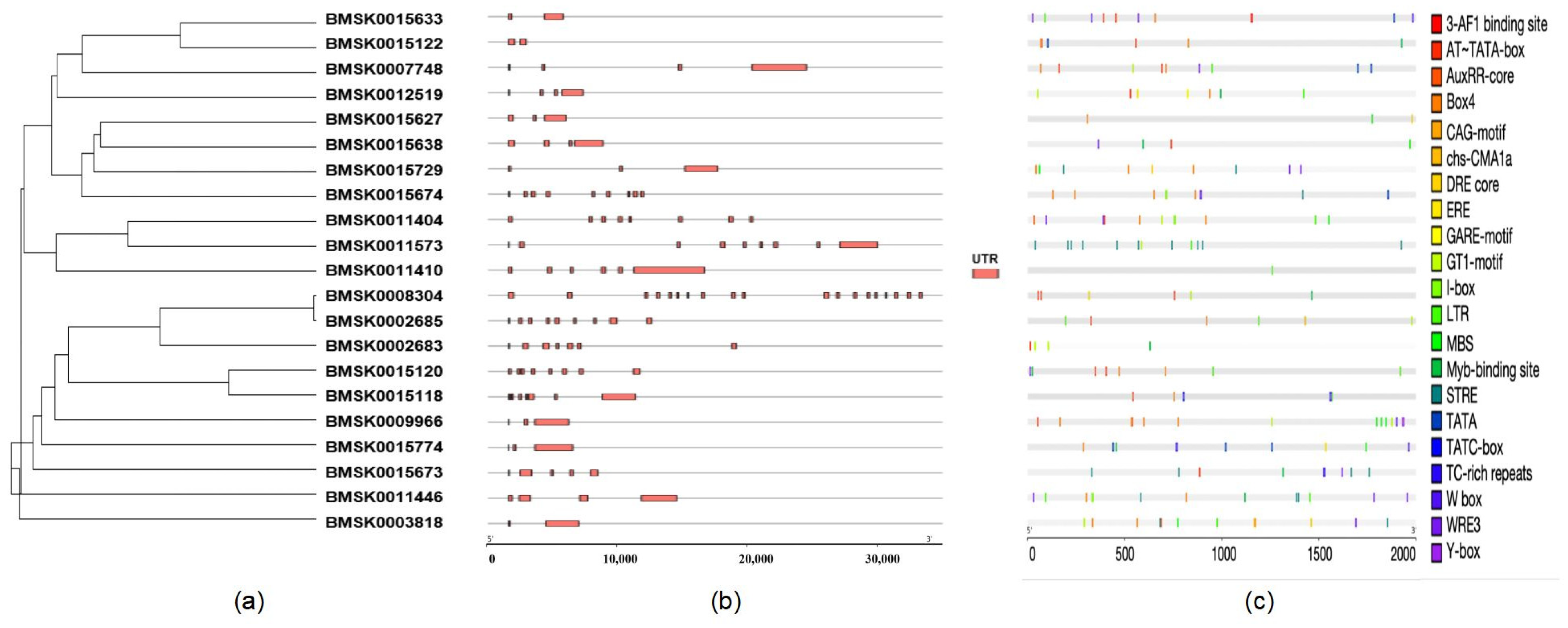
2.1. Genome-Wide Identification and Phylogenetic Analysis of the Bmtret1s in B. mori

Based on the silkworm genome information, 21 Bmtret1 homologs were identified. These Bmtret1 homologs encode proteins of 204 to 591 amino acids with molecular weights of 23.18 to 65.47 kDa and theoretical isoelectric points of 4.83 to 9.48. Based on WoLF PSORT prediction of the subcellular localization, most of them (18/21) likely localize to the plasma membrane (PM) (Table 1). According to the amino acid sequence, each sample is divided into three clades, and samples within the same clade are highly related (Figure 1).

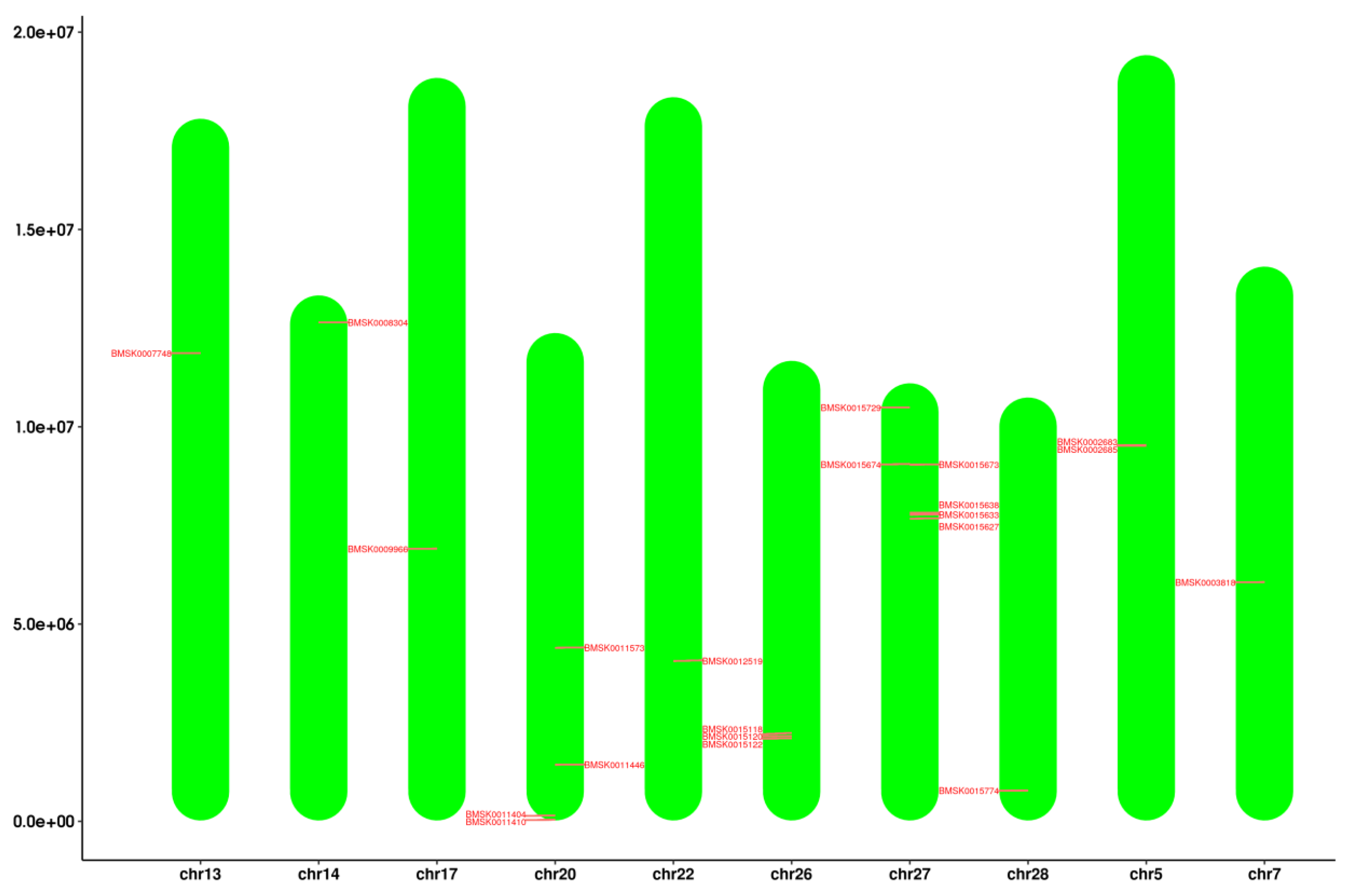
2.2. Chromosomal Localization of Bmtret1s

The target genes were mainly distributed on chromosomes 5, 7, 13, 14, 17, 20, 26, 27, and 28. Chromosome 27 has the most target genes, followed by chromosomes 20 and 26 (Figure 2). No active tandem genes and gene replication pairs were found in the preliminary screening, while the results need to be further verified.

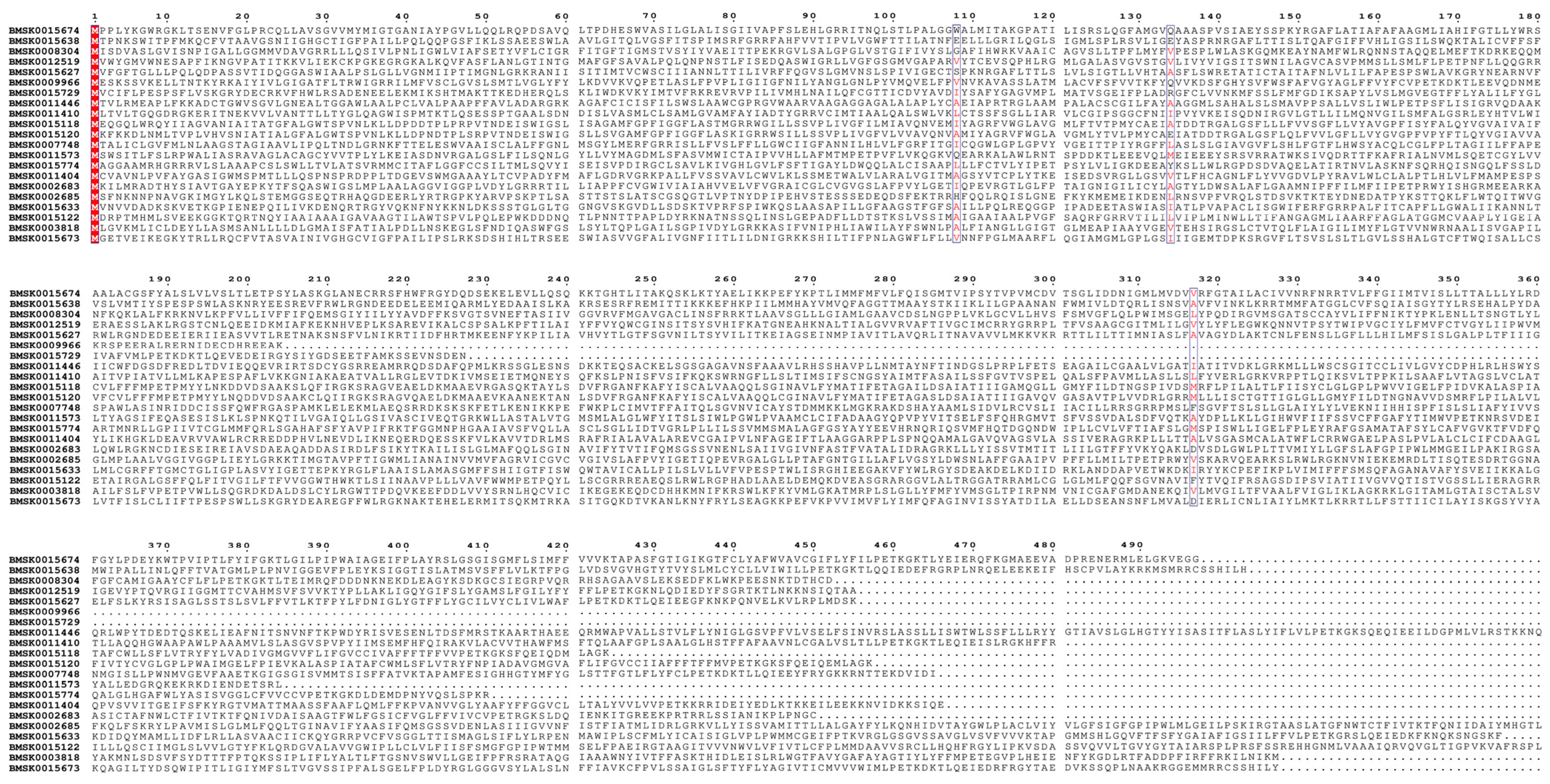
2.3. Sequence Analysis of the Bmtret1s

Conserved regions in the Bmtret1 proteins were identified using multiple sequence alignment analysis of the amino acid sequence. The alignment and conserved motif analysis showed that the tret1 gene family retained four conserved sites, indicated by red highlighting (Figure 3).

2.4. Gene Organization and Promoter Analysis of Bmtret1s

Cis-acting elements are present in the peripheral sequences of genes that affect gene expression. Cis-acting elements include promoters, enhancers, regulatory sequences, and inducible elements, which are involved in the regulation of gene expression. The cis-acting element itself does not encode any protein, but merely provides an action site that interacts with trans-acting factors. According to the annotation information of the silkworm genome, the molecular characteristics of the Bmtret1 genes were analyzed, and a phylogenetic tree was constructed to identify possible functional elements in each gene. The Tret1’s exon was located, and the sample sequence was basically located in the exon region, suggesting that it was involved in the regulation of this gene expression. As can be seen in the distribution map of cis-elements in the Tret1 promoter region (Figure 4), stress-response-related elements were identified in multiple samples.

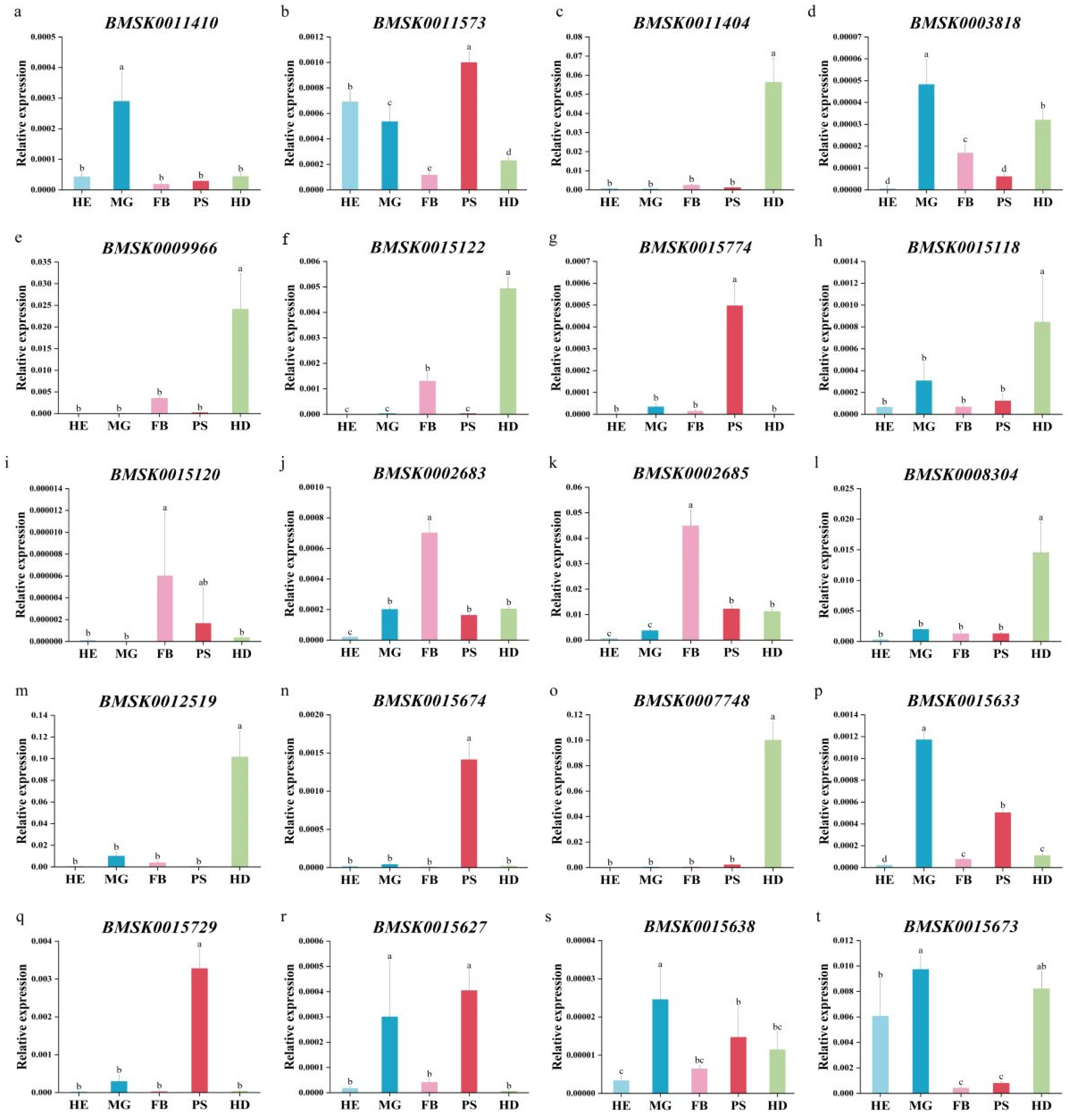
2.5. Expression Profile of Bmtret1s in Different Tissues of Silkworm

To determine the tissue and organ expression profile of Bmtret1s, the relative expression levels of the Bmtret1 genes in the silkworm hemolymph (HE), midgut (MG), fat body (FB), posterior silk gland (PS), and head (HD) were measured using qRT-PCR with silkworm actin 3 as the internal reference. The expression of BMSK0011446 in the phylogenetic branch in these tissues was relatively low. The gene expression patterns in phylogenetic branch I varied greatly, with BMSK0011410 having the highest expression in the midgut (MG); BMSK0011573 having the highest expression in the posterior silk gland (PS), hemolymph (HE), and posterior midgut (MG); and BMSK0011404 having the highest expression in the head (HD) (Figure 5a–c).
While the expression patterns of Bmtret1 in the phylogenetic clade is more diverse, the highest expressed Bmtret1s in the midgut (MG) were BMSK0003818, BMSK0015729, BMSK0015638, and BMSK0015673, where BMSK0015729 and BMSK0015638 had similar expression patterns, The expression levels of each tissue were ordered from high to low: midgut (MG), posterior silk gland (PS), head (HD), fat body (FB), and hemolymph (HE) (Figure 5d,q,s,t); BMSK0015120, BMSK0002683, and BMSK0002685 were the highest in the fat body (FB), with the latter two having similar expression patterns (Figure 5i–k). Four genes (BMSK0015774, BMSK0015674, BMSK0015633, and BMSK0015627) had the highest expression levels in the posterior silk glands (PS). With the exception of the high expression of BMSK0015627 in the midgut (MG), the expression patterns were very similar. All other tissues had relatively low expression levels (Figure 5g,n,p,r). The remaining genes have the highest expression in the head (HD). There are two patterns of expression. The expression pattern of BMSK0009966, BMSK0015122, and BMSK0007748 was similar to others in the phylogenetic branch I, except for having higher expression in the head (HD) and fat body (FB) (Figure 5e,f,o,c). The other type of Bmtret1s (BMSK0015118, BMSK0008304, and BMSK0012519) were the highest expressed in the fat body (FB) and midgut (MG) (Figure 5h,l,m).

2.6. Transcriptional-Level Responses of Bmtret1s to BmNPV Stress

The relative expression of Bmtret1 genes in the hemolymph (HE), midgut (MG), and fat body (FB) 24 h after infection with BmNPV was determined using qRT-PCR. BmNPV is a viral disease caused by infection with the polyhydrosis virus. All genes responded to infection with BmNPV. In the fat body (FB), the expression level of 16/20 Bmtret1s was increased and 4/20 was decreased. In the midgut (MG), 15/20 Bmtret1 genes were upregulated, 3/20 downregulated, and 2/20 unchanged. A different pattern was observed in the hemolymph, where 12/20 Bmtret1s were downregulated. Only one gene (BMSK0015729) showed an upregulated level in the hemolymph after infection (Figure 6).
Further, 9 out of the 20 genes (BMSK0011410, BMSK0011404, BMSK0003818, BMSK0015122, BMSK0015774, BMSK0015120, BMSK0002683, BMSK0002685, and BMSK0008034) had a similar expression pattern, being upregulated in both the midgut (MG) and the fat body (FB). These genes are also closely related in the phylogenetic tree (Figure 6a,c,d,f,g,i–l). With differences in their expression levels in the hemolymph (HE), the nine genes can be divided into two expression pattern groups. Five genes (BMSK0011404, BMSK0003818, BMSK0015122, BMSK0015120, and BMSK0002685) showed an insignificant response in the hemolymph (HE), while the remaining four genes were downregulated. Five other genes (BMSK0009966, BMSK0015118, BMSK0012519, BMSK0015627, and BMSK0015638) showed similar expression profiles to each other after BmNPV biological stress, with their expression levels being upregulated in the midgut and downregulated in the fat body (Figure 6e,h,m,r,s).

3. Discussion

Nuclear polyhedrosis virus disease of silkworms is highly infectious and harmful. It is the most common and most harmful silkworm disease in silkworm rearing and production, and causes serious economic losses every year [30,31]. Over the years, many researchers have been committed to screening and breeding resistant silkworm varieties and discovering resistance genes to elucidate the molecular mechanisms of silkworm resistance to BmNPV [32]. With the development of biotechnology, more achievements have been made in the study of B. mori’s resistance to the BmNPV virus at the molecular level, and further studies have been made on genes or proteins that may be involved in the antiviral mechanism. After the completion of silkworm genome sequencing, gene chip technology has become an important gene expression analysis method, which has the advantages of a large throughput and high accuracy. Zhou et al., detected 92 differentially expressed genes in the intestinal tissues of silkworm varieties BC8 and 306 after 12 h of toxic treatment with nucleic acid probes. They further analyzed 10 upregulated genes using fluorescent quantitative PCR. Fluorescence quantitative PCR technology can quickly compare and analyze the expression of all genes in the sample [33]. BmS3A is related to the inhibition of the apoptosis of infected cells, which inhibits the replication of viruses [34]. The SOP2 gene may promote the actin polymerization process and affect virus replication [35]. We selected the midgut tissues of a conventional susceptible strain of silkworm Baiyu infected with BmNPV for second-generation RNA-Seq transcriptome sequencing, for systematic screening of candidate differentially expressed genes involved in BmNPV infection resistance in the Bmtret1 gene family.
Trehalose, as a type of natural sugar, can be used as a protective factor to protect the organism from external environmental stresses or internal metabolic disorders. TRET, the trehalose transporter, can transport trehalose from the fat body to the hemolymph, and plays an important role in insect stress resistance [26,27]. While TRET plays an important role in the resistance to numerous insect stresses, there are very few studies on the effects of TRET on virus infection. Some studies have speculated that the trehalose transprotein-1 (Tret1) gene may be related to the transport of the virus during the interaction between gray planthoppers and the rice stripe virus [36]. In addition, the trehalose transprotein-1 (BmTret1-like) gene of silkworm plays a specific role in the mechanism of BmNPV virus resistance [37]. Recent studies have shown that the expression of the BmTret1-X1 gene has a clear inhibitory effect on the expression of viral genes in BmNPV [38]. The transcriptome results found that the expression level of Bmtret1s significantly responded to BmNPV biological stress, and we speculated that Bmtret1s may play an important role in the infection of BmNPV.
In this study, it was found that the Bmtret1 gene family varied greatly in different tissues with possible functional differences. Because trehalose is involved in the process of silkworm epidermis formation, it is speculated that the high expression level in the head may be associated with ecdysone and juvenile hormone. The higher expression in the two detoxifying organs of the midgut and fat body indicates that Bmtret1s may participate in the molecular mechanisms of disease resistance. In BmNPV-susceptible varieties of white jade silkworm, the vast majority of Bmtret1 genes are downregulated in response to BmNPV oral infection. We speculate that the downregulation of the trehalose transporter gene in the hemolymph allows for BmNPV invasion and is the cause of susceptibility. Furthermore, the high expression of Bmtret1s in the midgut and fat body correlates with viral resistance in these two detoxification organs. Some of the genes being downregulated in the detoxification organs suggest that not every gene in the Bmtret1 gene family is involved in the defense against BmNPV.

4. Materials and Methods

4.1. Sericulture Breeding and Virus Preparation

The Baiyu were acquired from the Resources Center of the Silviculture Research Institute, the Chinese Academy of Agricultural Sciences. Baiyu silkworms have a strong desire to eat mulberries, have a larger body and a bluish–white color, and are susceptible to BmNPV. All silkworm larvae were raised on fresh mulberry leaves. The worms were raised at 27 ± 1 °C at 75 ± 5% relative humidity under a 12 h day and night cycle. The BmNPV strain was maintained in our laboratory and purified according to the protocol reported by Rahman et al. [8]. After starvation treatment, the experimental group was fed 7 μL of BmNPV suspension (2 × 10⁸ polyhedra/mL), and the control group was fed normally. The hemolymph, midgut, and fat body of the larvae from both groups were taken 24 h after infection. After 72 h, the hemolymph, midgut, fat body, posterior silk gland, and head of the control group were collected. There were three biological replicates, and five silkworms constitute one biological replicate. They were taken and stored at −80 °C after infection.

4.2. Identification of the Bmtret1 Gene Family in B. mori

Sequences homologous to the Bmtret1 genes were downloaded from the silkworm genome database, SilkDB 3.0 (https://silkdb.bioinfotoolkits.net/, accessed on 1 May 2023). Their chromosomal distribution and homology relationships were analyzed. The Biological Toolbox v1.098774 was used to analyze the sequence length, molecular weight, and theoretical isoelectric point (PI) values for each homologous gene. The distribution of TM helices was determined using TMHMM Server v.2.0 (http://www.cbs.dtu.dk/services/TMHMM/, accessed on 18 May 2023) Forecast. Subcellular localization of the Bmtret1s protein was predicted using the online tool WoLF PSORT (https://wolfpsort.hgc.jp/, accessed on 21 May 2023).

4.3. Chromosomal Localization and Homology Analysis of Bmtret1s

The chromosomal location information of the Bmtret1 gene family was extracted from the silkworm genome annotation file. This operation was performed and visualized in Tbtools v1.098774.

4.4. Sequence Alignment

The Bmtret1 protein sequences were aligned using clustalW and assembled in MEGA 11.0.

4.5. Gene Structure and Promoter Analysis of Bmtret1s

The gene structure of each Bmtret1 was displayed based on the genome sequence and its annotation file using Gene Structure View assembled in Tbtools v1.098774. The upstream 2000 bp sequences were extracted for the promoter region analysis. The cis-acting elements were predicted using PlantCARE (http://bioinformatics.psb.ugent.be/webtools/plantcare/html/, accessed on 25 May 2023).

4.6. Phylogenetic Analysis of Bmtret1s

A neighbor-joining (NJ) phylogenetic tree was constructed using full-length Bmtret1 protein sequences from B. mori and using MEGA 11.0 and JTT + G models, with bootstrap tests with 1000 replicates.

4.7. RNA Extraction and Quantitative Real-Time PCR (qRT-PCR) Analysis

According to the manufacturer’s instructions, the EASYspin Tissue/Cell RNA Rapid Extraction Kit (Aidlab, Beijing, China) and HiScript III 1st Strand cDNA Synthesis Kit (+gDNA wiper) were used for the RNA extraction and cDNA synthesis.
The ChamQ SYBR qPCR Master Mix from Vazyme was used. Actin3 was used as a reference gene. The RT-qPCR was performed using an ABI StepOnePlus™ Real-Time PCR System (Shanghai, China) to verify the expression patterns of Bmtret1 in different tissues at different times. GraphPad Prism 8.0 was used to visualize the RT-qPCR results and to perform Duncan’s test and Student’s t-test. In Figure 5, a p-value <0.05 was marked as significant. In Figure 6, a p-value < 0.001 was marked with ***, p-value < 0.01 was marked with ** and p-value < 0.05 was marked with *. At least three biological replicates were performed, respectively, for the RT-qPCR. The primers used are shown in Appendix A Table A1. The relative expression level of these genes was estimated according to the 2−ΔΔCt method.

5. Conclusions

We conducted transcriptome and phylogenetic analysis of the silkworm Bmtret gene family and performed expression profiling and transcript-level analysis after infection with BmNPV. The Bmtret1 gene family has been implicated in silkworm resistance against BmNPV, and the high expression of most Bmtret1s in the midgut and fat body may inhibit the gene transcription of BmNPV and DNA replication, and thus reduce the assembly efficiency of virions to resist BmNPV infection. The Bmtret1 gene family of silkworm trehalose transporter was preliminarily identified as a key candidate gene family of silkworm BmNPV resistance, and the specific mechanism needs further study.

Author Contributions

G.Z. and H.Q. conceived and designed the experiments; M.L., Y.Q., E.C., and M.W. performed the experiments. G.O. and Y.X. analyzed the data. M.L. and G.Z. wrote the manuscript. All authors have read and agreed to the published version of the manuscript.

Funding

This research was funded by the Foundation of Post Scientists in the National Sericultural System (CARS-18-ZJ0101) and the Natural Science Foundation of Jiangsu Province (BK20201229).

Institutional Review Board Statement

Not applicable.

Informed Consent Statement

Not applicable.

Data Availability Statement

The data presented in this study are available in Appendix A.

Conflicts of Interest

The authors declare no conflicts of interest.

Appendix A

Table A1. Primer sequence.
Primer NameSequence(5′ to 3′)
BMSK0015674-FACAATAGCATTCGCATTCG
BMSK0015674-RAGTCTCCAGAGTCAGTGAA
BMSK0015638-FACCTTACGATGCGATGTG
BMSK0015638-RCTCTTCCAACTCCTGTCTG
BMSK0008304-FGCAAGGAACAGCAGATGA
BMSK0008304-RCTATGATGGAAGCCGTGAA
BMSK0012519-FGTTCGCCTTCTGCTCTAA
BMSK0012519-RGGTCTCCGTCCATATCTTC
BMSK0015627-FCGGATTCACTGGACTGTTA
BMSK0015627-RTGGTAAGATTGTTGGCGATA
BMSK0009966-FCTGTGAAGGAATTGCTAACC
BMSK0009966-RAATAGTGACCGAACGAATCT
BMSK0015729-FTTGTTGCCTTCGTTATGTTG
BMSK0015729-RCACTGTTCACTTCGCTACT
BMSK0011446-FGCACGCATTGTCCTTATC
BMSK0011446-RGCACGCATTGTCCTTATC
BMSK0011410-FAGCCAAGACAACATCAGAG
BMSK0011410-RTCAGCATCAACAACACAGT
BMSK0015118-FGCAGTATGTCGGAGTAGC
BMSK0015118-RGAGATGTAGAACGCCTTGA
BMSK0015120-FAATGGCAACGCAGTAGTC
BMSK0015120-RACAGAAGGCAGTGGCTAT
BMSK0007748-FCGTCCTTATACCACAACTGA
BMSK0007748-RAACACCAACTGCGATTCC
BMSK0011573-FACAAGAGACCTGCGGATA
BMSK0011573-RGACACTGGAGAAGATGATGA
BMSK0015774-FGTCCACCGTAACCAGAGA
BMSK0015774-RGATGCGTATAACCAGAATGC
BMSK0015122-FGGACTATGCGTAGGATTGAT
BMSK0015122-RTGATATGAATGTGCCGAGAA
BMSK0011404-FGCTTCTGTTCGTCTCCTC
BMSK0011404-RAGTCCTCGCTGATCTCTT
BMSK0003818-FCATCTATCGCACGCTACA
BMSK0003818-RCGCTGAAGACGCTATTATC
BMSK0002683-FTAGGAAGGCGTAGAACTATAC
BMSK0002683-RCACCAACACATAATCCACAA
BMSK0002685-FTGCCTCGTTATCTATGTGTT
BMSK0002685-RTCCAGTTGAAGCCAGTTG
BMSK0015633-FTATGGCGATGCTCCTAATAG
BMSK0015633-RTCTCCTGTAGACTCCTTCC
BMSK0015673-FGCTGGTTCCTGTTTCTAC
BMSK0015673-RTTGGGTCCGTCATTTCTC

References

  1. Li, K.; Dong, Z.; Pan, M. Common strategies in silkworm disease resistance breeding research. Pest Manag. Sci. 2023, 10, 1002. [Google Scholar] [CrossRef] [PubMed]
  2. Luan, J.B.; Li, J.M.; Varela, N.; Wang, Y.L.; Li, F.F.; Bao, Y.Y.; Zhang, C.X.; Liu, S.S.; Wang, X.W. Global analysis of the transcriptional response of whitefly to tomato yellow leaf curl China virus reveals the relationship of coevolved adaptations. J. Virol. 2011, 85, 3330–3340. [Google Scholar] [CrossRef] [PubMed]
  3. Chen, H.Q.; Yao, Q.; Bao, F.; Chen, K.P.; Liu, X.Y.; Li, J.; Wang, L. Comparative proteome analysis of silkworm in its susceptibility and resistance responses to Bombyx mori densonucleosis virus. Intervirology 2011, 55, 21–28. [Google Scholar] [CrossRef] [PubMed]
  4. Zhang, M.; Fei, S.; Xia, J.; Wang, Y.; Wu, H.; Li, X.; Guo, Y.; Swevers, L.; Sun, J.; Feng, M. Sirt5 Inhibits BmNPV replication by promoting a relish-Mediated antiviral pathway in Bombyx mori. Front. Immunol. 2022, 13, 906738. [Google Scholar] [CrossRef] [PubMed]
  5. Huang, L.; Dong, Z.Q.; Dong, F.F.; Yu, X.B.; Hu, Z.G.; Liao, N.C.; Chen, P.; Lu, C.; Pan, M.H. Gene editing the BmNPV inhibitor of apoptosis protein 2 (iap2) as an antiviral strategy in transgenic silkworm. Int. J. Biol. Macromol. 2021, 166, 529–537. [Google Scholar] [CrossRef] [PubMed]
  6. Cao, H.H.; Zhang, S.Z.; Zhu, L.B.; Wang, J.; Liu, Y.X.; Wang, Y.L.; Kong, X.; You, L.L.; Toufeeq, S.; Liu, S.H.; et al. The digestive proteinase trypsin, alkaline A contributes to anti-BmNPV activity in silkworm (Bombyx mori). Dev. Comp. Immunol. 2021, 119, 104035. [Google Scholar] [CrossRef] [PubMed]
  7. Katsuma, S.; Mita, K.; Shimada, T. ERK and JNK-dependent signaling pathways contribute to Bombyx mori nucleopolyhedrovirus infection. J. Virol. 2007, 81, 13700–13709. [Google Scholar] [CrossRef]
  8. Rahman, M.M.; Gopinathan, K.P. Systemic and in vitro infection process of Bombyx mori nucleopolyhedrovirus. Virus Res. 2004, 101, 109–118. [Google Scholar] [CrossRef]
  9. Braunagel, S.C.; Summers, M.D. Molecular biology of the baculovirus occlusion-derived virus envelope. Curr. Drug Targets 2007, 8, 1084–1095. [Google Scholar] [CrossRef]
  10. Blissard, G.W. Baculovirus—Insect cell interactions. Cytotechnology 1996, 20, 73–93. [Google Scholar] [CrossRef]
  11. Wang, Y.; Wang, Q.; Liang, C.; Song, J.; Li, N.; Shi, H.; Chen, X. Autographa californica multiple nucleopolyhedrovirus nucleocapsid protein BV/ODV-C42 mediates the nuclear entry of P78/83. J. Virol. 2008, 82, 4554–4561. [Google Scholar] [CrossRef]
  12. Bao, Y.Y.; Tang, X.D.; Lv, Z.Y.; Wang, X.Y.; Tian, C.H.; Xu, Y.P.; Zhang, C.X. Gene expression profiling of resistant and susceptible Bombyx mori strains reveals nucleopolyhedrovirus-associated variations in host gene transcript levels. Genomics 2009, 82, 138–145. [Google Scholar] [CrossRef]
  13. Mei, X.; Li, C.; Peng, P.; Wang, J.; He, E.; Qiu, Z.; Xia, D.; Zhao, Q.; Shen, D. Bombyx mori C-Type lectin (BmIML-2) inhibits the proliferation of B.mori nucleopolyhedrovirus (BmNPV) through involvement in apoptosis. Int. J. Mol. Sci. 2022, 23, 8369. [Google Scholar] [CrossRef]
  14. Elbein, A.D.; Pan, Y.T.; Pastuszak, I.; Carroll, D. New insights on trehalose: A multifunctional molecule. Glycobiology 2003, 13, 17–27. [Google Scholar] [CrossRef]
  15. Chen, A.; Tapia, H.; Goddard, J.M.; Gibney, P.A. Trehalose and its applications in the food industry. Compr. Rev. Food Sci. Food Saf. 2022, 21, 5004–5037. [Google Scholar] [CrossRef]
  16. Crowe, J.H.; Carpenter, J.F.; Crowe, L.M. The role of vitrification in anhydrobiosis. Annu. Rev. Physiol. 1998, 60, 73–103. [Google Scholar] [CrossRef]
  17. Wyatt, G.R.; Kale, G.F. The chemistry of insect hemolymph.II.Trehalose and other carbohydrates. J. Gen. Physiol. 1957, 40, 833–847. [Google Scholar] [CrossRef]
  18. Candy, D.J.; Kilby, B.A. Site and mode of trehalose biosynthesis in the locust. Nature 1959, 183, 1594–1595. [Google Scholar] [CrossRef]
  19. Murphy, T.A.; Wyatt, G.R. The enzymes of glycogen and trehalose synthesis in silk moth fat body. J. Biol. Chem. 1965, 240, 1500–1508. [Google Scholar] [CrossRef]
  20. Kikuta, S.; Nakamura, Y.; Hattori, M.; Sato, R.; Kikawada, T.; Noda, H. Herbivory-induced glucose transporter gene expression in the brown planthopper, Nilaparvata lugens. Insect Biochem. Mol. Biol. 2015, 64, 60–67. [Google Scholar] [CrossRef] [PubMed]
  21. Ashford, D.A.; Smith, W.A.; Douglas, A.E. Living on a high sugar diet: The fate of sucrose ingested by a phloem-feeding insect, the pea aphid acyrthosiphon pisum. J. Insect Physiol. 2000, 46, 335–341. [Google Scholar] [CrossRef] [PubMed]
  22. Fraga, A.; Ribeiro, L.; Lobato, M.; Santos, V.; Silva, J.R.; Gomes, H.; da Cunha Moraes, J.L.; de Souza Menezes, J.; de Oliveira, C.J.; Campos, E.; et al. Glycogen and glucose metabolism are essential for early embryonic development of the red flour beetle Tribolium castaneum. PLoS ONE 2013, 8, e65125. [Google Scholar] [CrossRef] [PubMed]
  23. Tang, B.; Wang, S.; Wang, S.G.; Wang, H.J.; Zhang, J.Y.; Cui, S.Y. Invertebrate trehalose-6-phosphate synthase gene: Genetic architecture, biochemistry, physiological function, and potential applications. Front. Physiol. 2018, 9, 30. [Google Scholar] [CrossRef] [PubMed]
  24. García de Castro, A.; Tunnacliffe, A. Intracellular trehalose improves osmotolerance but not desiccation tolerance in mammalian cells. FEBS Lett. 2000, 487, 199–202. [Google Scholar] [CrossRef]
  25. Stambuk, B.U.; Panek, A.D.; Crowe, J.H.; Crowe, L.M.; de Araujo, P.S. Expression of high-affinity trehalose-H+ symport in Saccharomyces cerevisiae. Biochim. Biophys. Acta 1998, 1379, 118–128. [Google Scholar] [CrossRef]
  26. Kanamori, Y.; Saito, A.; Hagiwara-Komoda, Y.; Tanaka, D.; Mitsumasu, K.; Kikuta, S.; Watanabe, M.; Cornette, R.; Kikawada, T.; Okuda, T. The trehalose transporter 1 gene sequence is conserved in insects and encodes proteins with different kinetic properties involved in trehalose import into peripheral tissues. Insect Biochem. Mol. Biol. 2010, 40, 30–37. [Google Scholar] [CrossRef]
  27. Kikawada, T.; Saito, A.; Kanamori, Y.; Nakahara, Y.; Iwata, K.-I.; Tanaka, D.; Watanabe, M.; Okuda, T. Trehalose transporter 1, a facilitated and high-capacity trehalose transporter, allows exogenous trehalose uptake into cells. Proc. Natl. Acad. Sci. USA 2007, 104, 11585–11590. [Google Scholar] [CrossRef]
  28. Takiguchi, M.; Niimi, T.; Su, Z.H.; Yaginuma, T. Trehalase from male accessory gland of an insect, Tenebrio molitor. cDNA sequencing and developmental profile of the gene expression. Biochem. J. 1992, 266, 19–22. [Google Scholar] [CrossRef]
  29. Tang, B.; Yang, M.; Shen, Q.; Xu, Y.; Wang, H.; Wang, S. Suppressing the activity of trehalase with validamycin disrupts the trehalose and chitin biosynthesis pathways in the rice brown planthopper, Nilaparvata lugens. Pestic. Biochem. Physiol. 2017, 137, 81–90. [Google Scholar] [CrossRef]
  30. Harrison, R.L.; Herniou, E.A.; Jehle, J.A.; Theilmann, D.A.; Burand, J.P.; Becnel, J.J.; Krell, P.J.; van Oers, M.M.; Mowery, J.D.; Bauchan, G.R.; et al. ICTV virus taxonomy profile: Baculoviridae. J. Gen. Virol. 2018, 99, 1185–1186. [Google Scholar] [CrossRef]
  31. Lange, M.; Wang, H.; Zhihong, H.; Jehle, J.A. Towards a molecular identification and classification system of lepidopteran-specific baculoviruses. Virology 2004, 325, 36–47. [Google Scholar] [CrossRef]
  32. Zafar, B.; Shabir, A.W.; Malik, M.A.; Ganai, M.A. A review: Disease resistance in mulberry silkworm Bombyx mori L. Asian J. Sci. Technol. 2013, 4, 157–166. [Google Scholar]
  33. Zhou, Y.; Gao, L.; Shi, H.; Xia, H.; Gao, L.; Lian, C.; Chen, L.; Yao, Q.; Chen, K.; Liu, X. Microarray analysis of gene expression profile in resistant and susceptible Bombyx mori strains reveals resistance-related genes to nucleopolyhedrovirus. Genomics 2013, 101, 256–262. [Google Scholar] [CrossRef]
  34. Jia-Ping, X.; Ke-Ping, C.; Ming-Hui, L.; Qin, Y.; Gui-Tian, G.; Yuan, Z. Identification and characterization of Bms3a in Bombyx mori L. Afr. J. Biotechnol. 2008, 34, 24–30. [Google Scholar]
  35. Xu, J.P.; Chen, K.P.; Yao, Q.; Liu, M.H.; Gao, G.T.; Zhao, Y. Identification and characterization of an NPV infection-related gene Bmsop2 in Bombyx mori L. J. Appl. Entomol. 2005, 129, 425–431. [Google Scholar] [CrossRef]
  36. Yu, W.D.; Pan, B.Y.; Qiu, L.Y.; Huang, Z.; Zhou, T.; Ye, L.; Tang, B.; Wang, S.G. The structure characteristics and biological functions on regulating trehalose metabolism of two NlTret1s in Nilaparvata lugens. Sci. Agric. Sin. 2020, 53, 4802–4812. [Google Scholar]
  37. Yang, J. Transcriptome Analysis of Midgut Tissue Infected by BmNPV and Functional Identification of BmTret1-like Gene. Master’s Thesis, Jiangsu University of Science and Technology, Zhenjiang, China, 2017. [Google Scholar]
  38. Song, Q. Identification of Resistance of Silkworm BmTret1-X1 Gene to Bombyx mori nucleopolyhedrovirus (BmNPV). Master’s Thesis, Jiangsu University of Science and Technology, Zhenjiang, China, 2022. [Google Scholar]
Figure 1. Phylogenetic relationships of the TRET1 family genes in Bombyx mori. The sequences of the 21 TRET1 proteins from the above insect were aligned using Clustal Omega, and the phylogenetic tree was constructed using MEGA 11.0 using the NJ method with 1000 bootstrap replicates.
Ijms 25 00402 g001
Figure 2. Distribution of Bmtret1 genes in Bombyx mori chromosomes. The scale is provided in megabases (Mb).
Ijms 25 00402 g002
Figure 3. Multiple sequence alignment of Bmtret1 proteins. Conserved sites are indicated by red highlights.
Ijms 25 00402 g003
Figure 4. Gene organization of Bmtret1s and cis-elements in promoter regions of Bmtret1s. (a) Phylogenetic tree using 21 Bmtret1s. (b) Exon/intron structures of Bmtret1s. (c) Cis-element distribution in the promoter regions of Bmtret1s.
Ijms 25 00402 g004
Figure 5. Transcript levels of Bmtret1s in hemolymph, midgut, fat body, posterior silk gland, and head of Bombyx mori. Three biological replicates were analyzed. Different letters indicate statistically significant differences (Duncan’s test, p < 0.05). Values are presented as mean ± SEM. The first three genes belong to branch I, and the expression level of gene of branch II is relatively low. The other genes belong to branch III. (a) BMSK0011410; (b) BMSK0011573; (c) BMSK0011404; (d) BMSK0003818; (e) BMSK0009966; (f) BMSK0015122; (g) BMSK0015774; (h) BMSK0015118; (i) BMSK0015120; (j) BMSK0002683; (k) BMSK0002685; (l) BMSK0008304; (m) BMSK0012519; (n) BMSK0015674; (o) BMSK0007748; (p) BMSK0015633; (q) BMSK0015729; (r) BMSK0015627; (s) BMSK0015638; (t) BMSK0015673.
Ijms 25 00402 g005
Figure 6. Expression levels of 20 Bmtret1 genes in response to BmNPV stress conditions. Three biological replicates were analyzed. Asterisks indicate significant differences as determined using Student’s t-test (* p < 0.05; ** p < 0.01; *** p < 0.001). Values are presented as mean ± SEM. C is the control group and T is the treatment group. The first three genes belong to branch I, and the expression level of gene of branch II is relatively low. The other genes belong to branch III. (a) BMSK0011410; (b) BMSK0011573; (c) BMSK0011404; (d) BMSK0003818; (e) BMSK0009966; (f) BMSK0015122; (g) BMSK0015774; (h) BMSK0015118; (i) BMSK0015120; (j) BMSK0002683; (k) BMSK0002685; (l) BMSK0008304; (m) BMSK0012519; (n) BMSK0015674; (o) BMSK0007748; (p) BMSK0015633; (q) BMSK0015729; (r) BMSK0015627; (s) BMSK0015638; (t) BMSK0015673.
Ijms 25 00402 g006
Table 1. TRET1 gene family in Bombyx mori.
Gene IDCDS Size (bp)Protein Physicochemical CharacteristicsTMHsSubcellular
Localization *
Length (aa)MW (kDa)pIAliphatic Index
BMSK0011410144348051.749.31116.6712PM
BMSK0011573115538442.906.02107.167PM
BMSK0011404140146650.778.31111.3312PM
BMSK0011446163254358.857.84100.729PM
BMSK0003818152450756.447.55112.7611PM
BMSK000996661520423.188.28101.184EX
BMSK0015122163554458.669.48113.8611PM
BMSK0015774123341044.818.17108.2410PM
BMSK0015118127542446.074.83115.2611PM
BMSK0015120137445749.346.59117.1312PM
BMSK0002683135345049.198.61116.4211PM
BMSK0002685177659165.479.1598.6510PM
BMSK0008304135945249.908.74102.2310PM
BMSK0012519136845549.999.42100.2610PM
BMSK0015674149449754.729.05107.9510MT
BMSK0007748139846551.749.05108.0412PM
BMSK0015633160853558.768.92103.2012PM
BMSK001572968422726.005.2192.692CY
BMSK0015627136845550.329.08125.3411PM
BMSK0015638151250356.549.28104.1012PM
BMSK0015673151550455.879.08113.5710PM
* The subcellular localizations were predicted using WoLF PSORT. MT, Mitochondrial; PM, Plasma Membrane; EX, Extracellular; CY, Cytoplasmic.
Disclaimer/Publisher’s Note: The statements, opinions and data contained in all publications are solely those of the individual author(s) and contributor(s) and not of MDPI and/or the editor(s). MDPI and/or the editor(s) disclaim responsibility for any injury to people or property resulting from any ideas, methods, instructions or products referred to in the content.

