Article

A Testis-Specific DMRT1 (Double Sex and Mab-3-Related Transcription Factor 1) Plays a Role in Spermatogenesis and Gonadal Development in the Hermaphrodite Boring Giant Clam Tridacna crocea

1
Key Laboratory of Tropical Marine Bio-Resources and Ecology, Guangdong Provincial Key Laboratory of Applied Marine Biology, South China Sea Institute of Oceanology, Chinese Academy of Sciences, Guangzhou 510301, China
2
University of Chinese Academy of Sciences, Beijing 100049, China
3
Southern Marine Science and Engineering Guangdong Laboratory (Zhuhai), Zhuhai 519015, China
4
Hainan Provincial Key Laboratory of Tropical Marine Biology Technology, Sanya Institute of Oceanology Chinese Academy of Sciences, Sanya 572024, China
5
Animal Science and Technology College, Guangxi University, Nanning 530004, China
*
Authors to whom correspondence should be addressed.
These authors contributed equally to this work.
Int. J. Mol. Sci. 2024, 25(11), 5574; https://doi.org/10.3390/ijms25115574
Submission received: 12 April 2024 / Revised: 11 May 2024 / Accepted: 16 May 2024 / Published: 21 May 2024

Abstract

:
The testis-specific double sex and mab-3-related transcription factor 1 (DMRT1) has long been recognized as a crucial player in sex determination across vertebrates, and its essential role in gonadal development and the regulation of spermatogenesis is well established. Here, we report the cloning of the key spermatogenesis-related DMRT1 cDNA, named Tc-DMRT1, from the gonads of Tridacna crocea (T. crocea), with a molecular weight of 41.93 kDa and an isoelectric point of 7.83 (pI). Our hypothesis is that DMRT1 machinery governs spermatogenesis and regulates gonadogenesis. RNAi-mediated Tc-DMRT1 knockdown revealed its critical role in hindering spermatogenesis and reducing expression levels in boring giant clams. A histological analysis showed structural changes, with normal sperm cell counts in the control group (ds-EGFP) but significantly lower concentrations of sperm cells in the experimental group (ds-DMRT1). DMRT1 transcripts during embryogenesis exhibited a significantly high expression pattern (p < 0.05) during the early zygote stage, and whole-embryo in-situ hybridization confirmed its expression pattern throughout embryogenesis. A qRT-PCR analysis of various reproductive stages revealed an abundant expression of Tc-DMRT1 in the gonads during the male reproductive stage. In-situ hybridization showed tissue-specific expression of DMRT1, with a positive signal detected in male-stage gonadal tissues comprising sperm cells, while no signal was detected in other stages. Our study findings provide an initial understanding of the DMRT1 molecular machinery controlling spermatogenesis and its specificity in male-stage gonads of the key bivalve species, Tridacna crocea, and suggest that DMRT1 predominantly functions as a key regulator of spermatogenesis in giant clams.

1. Introduction

Giant clams are widely distributed in the tropical marine waters of the Indo-Pacific region, with prominent species identified in the South China Sea, living at a depth of approximately 20 m embedded in shallow coral reefs. Only thirteen species of giant clams are known today, with only eight species identified in the South China Sea. Tridacna plays a crucial biological role in preserving and sustaining coral reef ecosystems [1,2]. They are considered synchronized bisexuals, with both male and female gonads present in the same animal and same organ, with male gonads maturing first and female gonads maturing later. During the spawning season, they typically release sperm first, followed by eggs after a brief period, and both gametes fertilize outside the body [3]. In recent decades, due to the over-exploitation of clam resources and habitat loss caused by human activities, giant clam populations have been highly threatened throughout their geographic ranges [4]. While efforts have been made to restock and induce breeding, which may help alleviate the situation to some degree, the molecular machinery controlling gonadal development needs further evaluation [5]. To genuinely support restoration efforts, it is essential to understand the fundamental molecular pathways that govern gonadal development and gametogenesis.
It has been investigated that, in the majority of animal species, sex determination and differentiation are commonly assisted by signal transduction cascades. In the fruit fly (Drosophila melanogaster), the signal transduction cascade was studied as the sex-lethal (Sxl)-transformer (Sxl)-transformer (Affairs)-doublesex (Dsx) pathway, controlled by a set of genes [6]. The sex-determining cascade in the nematode Caenorhabditis elegans consists of an ordered series of regulatory genes, including Xol-1, Her-1, Fem-1,2,3, and Tra-1,2,3 [7]. The assigned genes ensure gonadal development, differentiation, and gametogenesis. In decapod crustaceans, the development of male sex characteristics is regulated by the insulin-like androgenic gland hormone (IAG), which is thought to be produced and secreted by a male-specific endocrine organ, the androgenic gland (AG) [8,9,10,11]. Furthermore, mollusks, particularly bivalves, have developed diverse reproductive strategies, including dioecy and hermaphroditism, and some species exhibit sex reversal capabilities. However, a significant knowledge gap remains in understanding the precise molecular pathways involved in sex determination in mollusk species. Notably, the DMRT1 gene, which is highly conserved across various animal taxa, plays a crucial role in regulating testis differentiation and development [12,13,14,15]. In Medaka (Oryzias latipes), the discovery of DMRT reveals a male sex-determining gene called dmY/dmrtY, which is considered functionally parallel to the mammalian SRY. DMRT family genes are highly conserved in vertebrate species and are considered to be vertebrate transcription factors, comprising a DNA motif (DM domain). In invertebrates, the doublesex (dsx) DNA-binding motif is also conserved, including in the nematode male-biased gene, male abnormal 3 (mab-3). Based on the outcomes of previous studies, it is confirmed that the functions of DM domain protein in sex determination are conserved and active within a wide scope of metazoans [16,17,18,19].
It is believed that among DMRT genes, only DMRT1 is expressed in the gonad, which regulates gametogenesis, gonadal differentiation, and sex determination in various vertebrates [20,21,22,23]. Furthermore, all members of the DMRT family genes control sex determination in numerous vertebrate phyla [14,22,24]. For instance, in one or both sexes of the mouse fetal gonad, several DMRT genes are expressed, and it has been observed that solely DMRT1 is involved in gonadal function before birth [25]. The DMRT1 protein is expressed and becomes male-specific by approximately E14.5, when ovarian and testicular structures are examined in mice [21,26,27,28]. These findings suggest that genes for sex differentiation and determination are highly conserved among various phyla. In reproduction, sex determination is a fundamental and crucial process for the survival and evolution of vertebrate species. However, for bivalve mollusks, especially giant clams, there are very little data on the validation of DMRT1 gene-related functions. Therefore, it is particularly important to pursue a comprehensive understanding of the role of DMRT1 in gonadal development and spermatogenesis in giant clams.
In this study, we successfully cloned the full-length sequence of the Tc-DMRT1 gene from the giant clam gonad. To evaluate the functional attributes of DMRT1 in the giant clam, we conducted an RNAi experiment. We explored the tissue-specific expression of Tc-DMRT1 in different reproductive stages using in-situ hybridization and qRT-PCR. Additionally, we examined the expression pattern of Tc-DMRT1 in various embryonic stages using whole-embryo in-situ hybridization. Our research provides valuable insights into the reproductive biology of giant clams, shedding light on key processes, such as spermatogenesis, gonadal development, and maternal gene expression. These findings offer important theoretical references for future breeding programs and conservation efforts.

2. Results

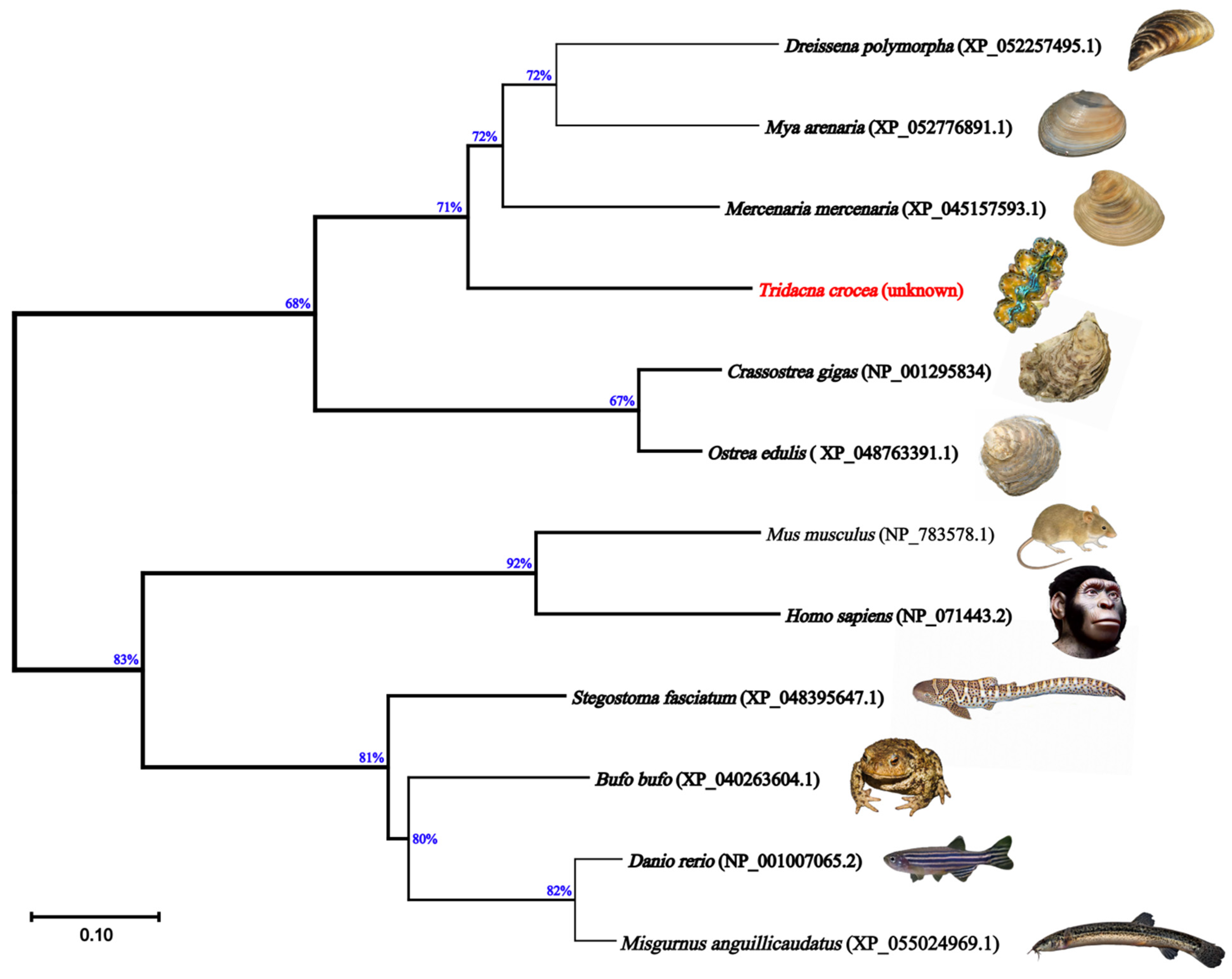
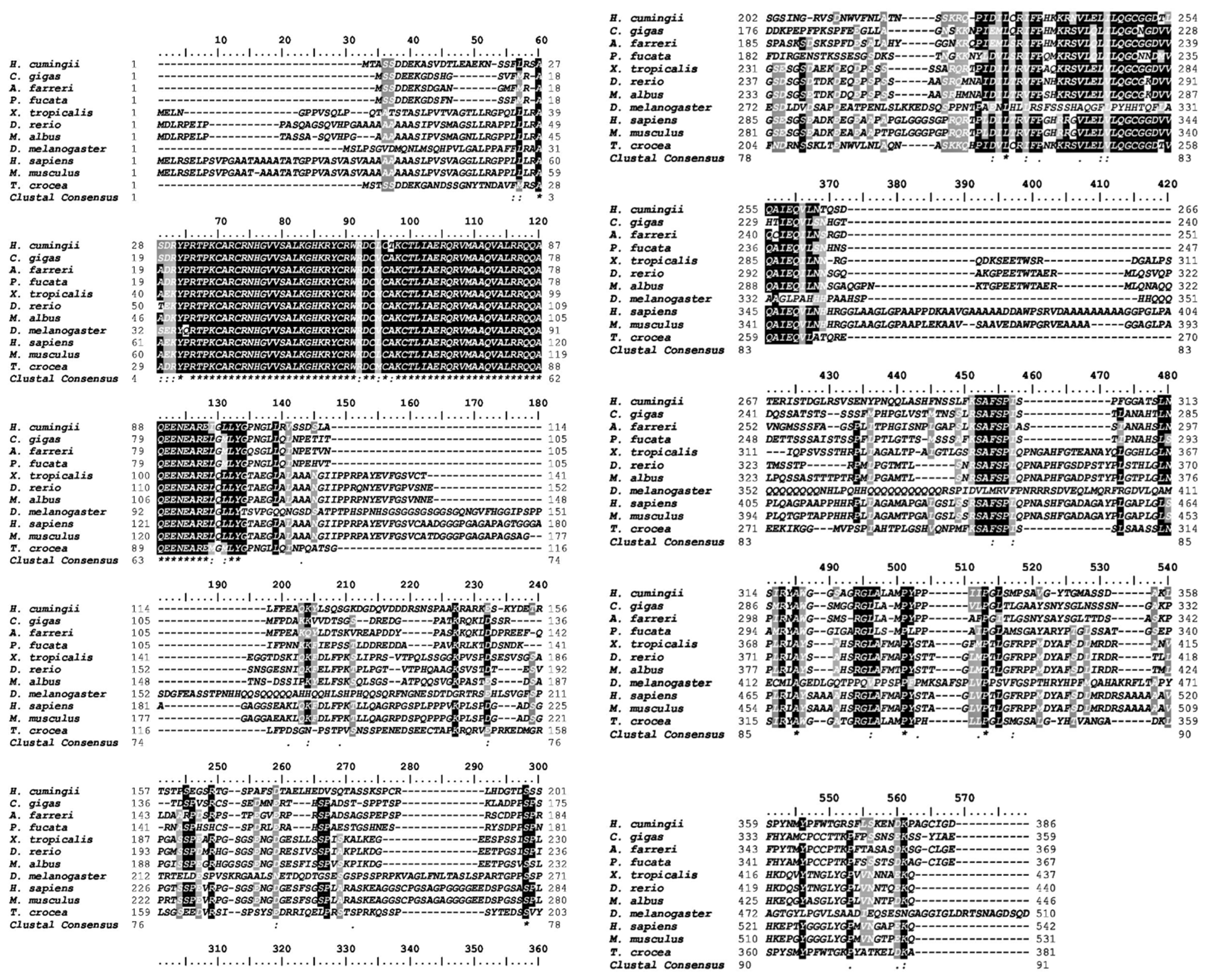
2.1. Nucleotides, Translated Amino Acid Profile, and Phylogeny of Tc-DMRT1 Gene

The full-length sequence of the Tc-DMRT1 gene was retrieved from the gonads of T. crocea. The 5′-untranslated region (5′-UTR) consists of 178 bp, while the 3′-UTR consists of 269 bp. The open reading frame (ORF) sequence of 1146 bp encodes a polypeptide of 381 amino acid residues, with a predicted molecular weight of approximately 41.93 kDa. The full-length gene spans 1589 bp, including UTRs obtained from T. crocea gonadal tissues through PCR amplification. A polyadenylation signal (AATAAA) is located 39 bp upstream of the poly-A tail of Tc-DMRT1. The putative amino acid sequence contains the conserved DM domain and CUE-like DMA (Figure 1). Phylogenetic analysis of the deduced amino acid sequence of Tc-DMRT1 revealed two major clades: one comprising vertebrates, including Danio rerio, Bufo bufo, Mus musculus, and Stegostoma fasciatum DMRT1 sequences, and another comprising invertebrates, including Crassostrea gigas, Pinctada fucata, and Dreissena polymorpha. T. crocea clustered in the bivalve clade with Mercenaria mercenaria (Figure 2). Multiple sequence alignment revealed the conserved DM domain in Tc-DMRT1, which shows complete conservation with other species, including higher vertebrates, and shares the highest sequence similarity with its closely related mollusk species (Figure 3). These results indicate the conserved status of Tc-DMRT1 in giant clams and rest of the animals in the clades.

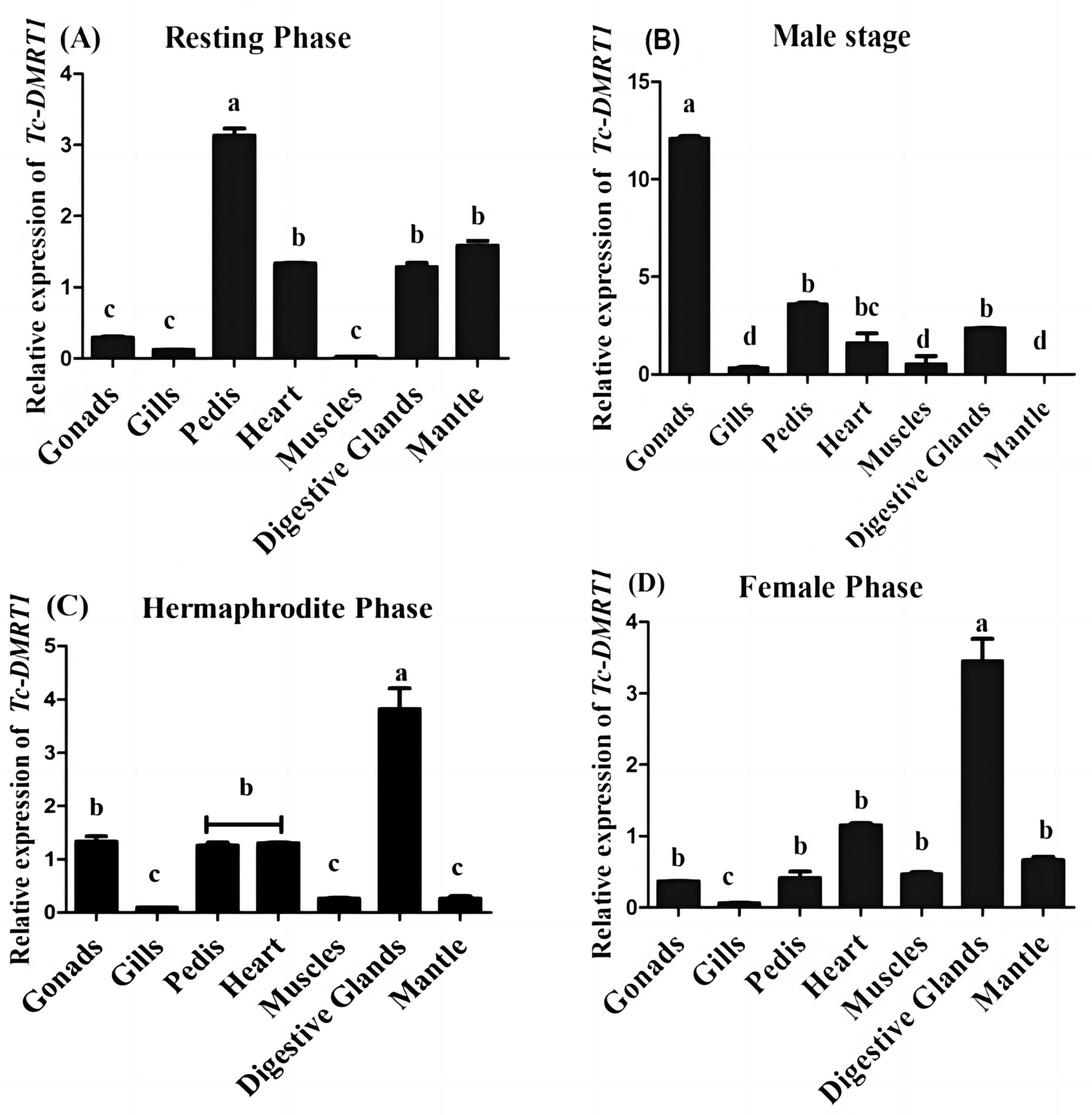
2.2. Expression Pattern of Tc-DMRTI during Various Reproductive Stages

The expression pattern of Tc-DMRT1 in giant clam gonads was evaluated across various stages, including resting, male, hermaphrodite, and female stage. Quantitative PCR analysis revealed that Tc-DMRT1 expression is significantly up-regulated during the resting stage in the pedis, followed by lower expression in the heart tissues. However, expression remains relatively low in the gonads, gills, and muscles. Notably, during the hermaphroditic stage, the gene shows significant up-regulation in the digestive glands (p < 0.05), while expression remains low in the gills, pedis, heart, muscles, gonads, and mantle tissues. In contrast, the male stage exhibits an exacerbated expression level in the gonads, with the highest overall expression observed. Meanwhile, the female stage shows elevated expression levels in the digestive tract (p < 0.05), with negligible concentrations of Tc-DMRT1 transcript observed in the ctenidium (Figure 4). These results suggest subtle differences in Tc-DMRT1 expression across various stages and organs, with the male stage of gonads exhibiting the highest overall expression.

2.3. In-Situ Hybridization for the Localization of Tc-DMRT1 mRNA during Various Reproductive Stages

Tissues from the male stage, which contain sperm cells, primarily spermatogonia, exhibited positive signals when probed with the antisense probe, confirming the expression of Tc-DMRT1 in sperm cells. In contrast, no signal was detected when using the sense probe, serving as a negative control. Repeated in-situ hybridization (ISH) experiments using the antisense probe on female stage tissues, comprising oocyte cells, failed to show a positive signal, indicating the absence of Tc-DMRT1 expression in oocyte cells of the gonadal tissues. Similarly, no cross-hybridization signal was observed in the resting stage when using both sense and antisense probes, confirming the absence of Tc-DMRT1 expression in the connective tissue of the gonads. Therefore, ISH experiments demonstrated that Tc-DMRT1 is exclusively expressed in the sperm cells of giant clams, supporting its role in male reproduction (Figure 5).

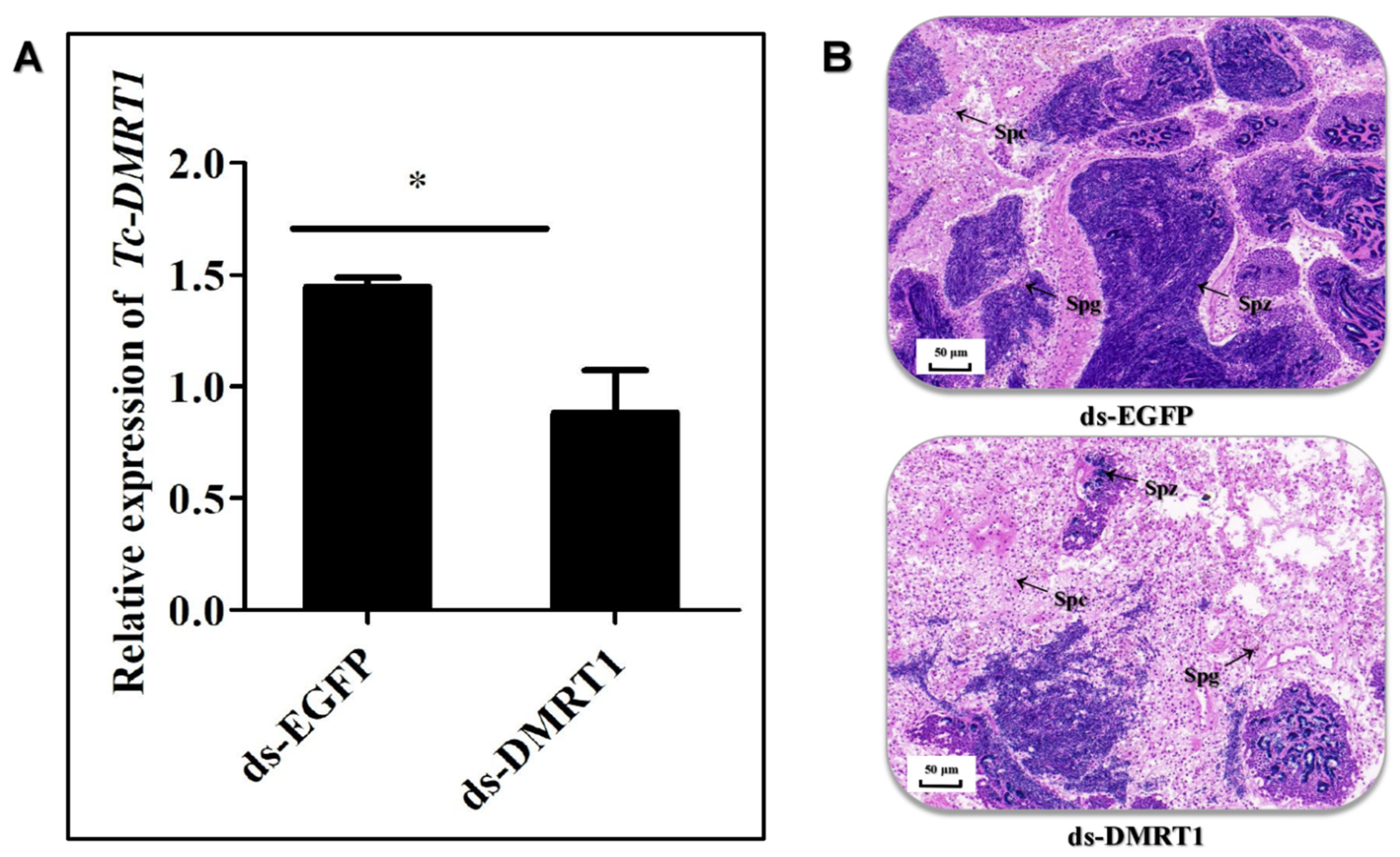
2.4. ds-DMRT1 Injection

Fourteen days after administering dsRNA injections, we quantified the mRNA transcripts of Tc-DMRT1 in giant clams using qRT-PCR. The results showed significant down-regulation of Tc-DMRT1 expression (p < 0.05) in the experimental group receiving in-vivo ds-DMRT1 injections, validating the inhibitory effect of RNA interference. In contrast, the control group (EGFP) showed an upstream expression profile of the Tc-DMRT1 transcript. The injection of dsRNA into giant clams resulted in the blocking of Tc-DMRT1, leading to reduced spermatogenesis and a significantly lower number of sperm cells in the gonadal tissues of the experimental group (Figure 6A). Histological sections from the EGFP group showed a large aggregation of sperm cells in the middle and outer ridges, whereas the ds-DMRT1-treated group had fewer sperm cells in the outer ridges, indicating a clear histological differentiation between the two groups (Figure 6B).

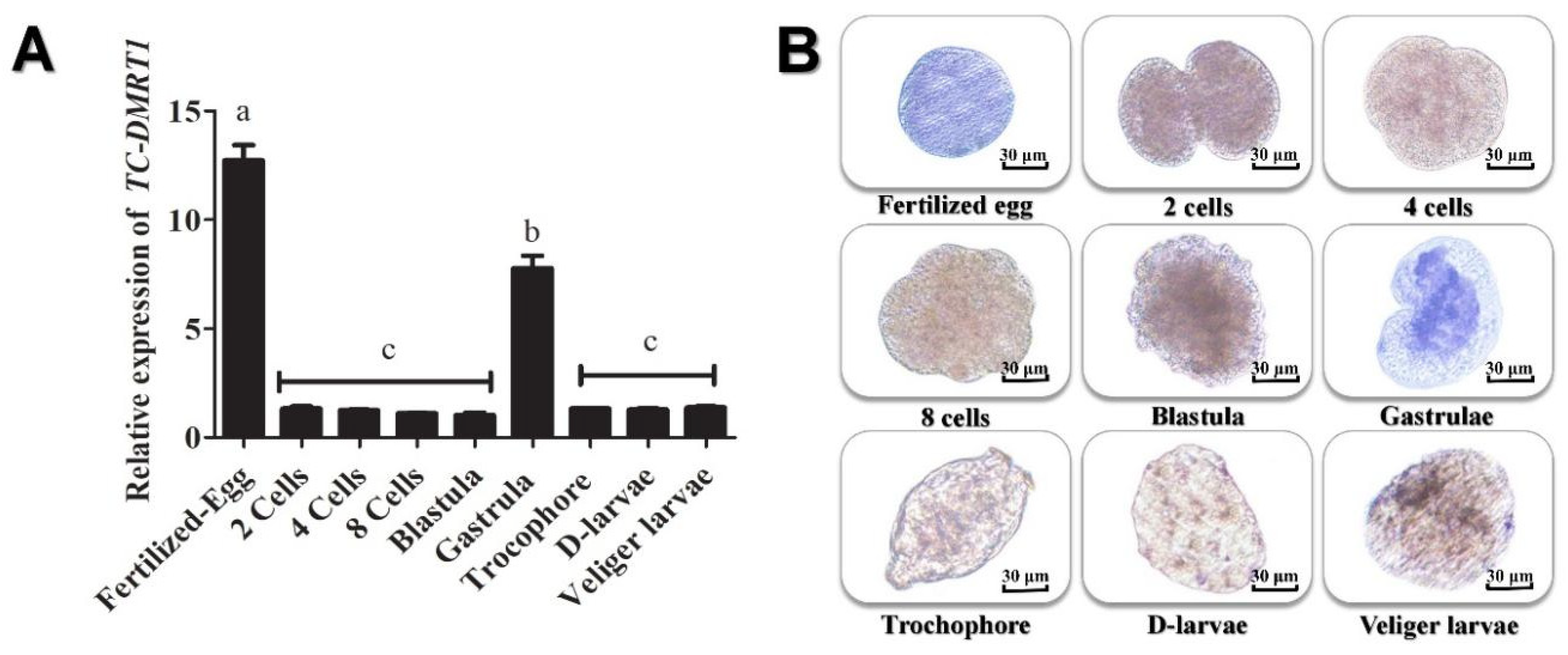
2.5. Spatial and Temporal Expression Pattern of Tc-DMRT1 during Embryogenesis

To investigate the DMRT1 mRNA level during early embryo development and metamorphosis in giant clams, we analyzed different developmental stages for differential transcript levels. The results revealed a peak in DMRT1 expression during early embryogenesis, followed by a gradual decline in 2, 4, and 8-cells blastomeres. However, DMRT1 expression increased during gastrulation and then gradually decreased during the D-larvae, trochophore, and juvenile stages (Figure 7A). To further examine DMRT1 expression during embryogenesis, we employed whole-embryo in-situ hybridization using an antisense probe to detect the mRNA nucleotides of the Tc-DMRT1 gene in various embryonic stages. The fertilized egg showed a strong positive signal with the antisense probe, followed by faint signals in different cleavage stages. The blastula exhibited weak signals, while strong expression was observed in the gastrula, and the positive signal remained stable until the veliger stage (Figure 7B).

3. Discussion

The role of DMRT1 orthologs has long been deemed crucial in sex determination, gonad development, and maintenance throughout evolutionary history. It acts as a functional regulator of mammalian gonads and as a differentiation and determination factor for sex, and engages in the normal progression of spermatogenesis in major phyla. DMRT1 is imperative in Sertoli cells to prevent female reprogramming of the adult animal’s testes [29] and is requisite in spermatogonia to maintain their proliferative phase by evading meiotic entry [30]. Another study has shown that DMRT1 presumably acts as a bifunctional transcriptional regulator by activating certain pivotal genes necessary for the absolute development of the male phenotype, while blocking the expression of others responsible for female gonad differentiation [31]. These studies register the pivotal role of Tc-DMRT1 in the spermatogenesis of boring giant clams.
Elucidating the regulatory role of Tc-DMRT1 during spermatogenesis would be fascinating, as its dynamics likely contribute significantly to the phenotype. Notably, this study is the first to explore the functional properties of Tc-DMRT1 in giant clams. The full-length sequence obtained was 1589 bp, with an open reading frame (ORF) length of 1146 bp. The phylogenetic analysis revealed that the DM region is a conserved sequence among vertebrates, including humans, zebrafish, and mice, while the Tc-DMRT1 protein clusters closely with its mollusk analogs. The conserved DM domain comprises cysteine residues, which likely form a zinc-finger motif. Similarly, a study on Japanese eel fish DMRT1 found a conserved DM domain, zinc finger motif, and six conserved cysteine residues [32]. Haliotis asinine, a common marine mollusk, has a complete DMRT1 length of 1740 bp, with a predicted ORF range of 732 bp, translating to 243 amino acids. Multiple sequence alignment confirmed the conserved status of the regulatory DM domain in the giant clam (Figure 1).
A transcriptome analysis of Pinctada margaritifera revealed the presence of testis-specific genes encoding proteins involved in sex determination and gonad differentiation, such as fem-1-like and DMRT1, which are exclusively expressed in male testes [33]. This finding supports our conclusion regarding the testis-specific expression of DMRT1 and its crucial role in sex determination. Furthermore, a comparative analysis of giant clams identified candidate genes responsible for gametogenesis, with DMRT1 emerging as a testis-specific gene potentially involved in male gonadal development (Figure 4) [23].
The expression level of DMRT1 remains high during spermatogenesis in rainbow trout, followed by a significant decrease during spermiation. However, its expression remains low during gonadogenesis [34]. Previous studies have shown that DMRT1 expression is exclusively restricted to the testes and varies according to different spermatogenetic phases in pejerrey fish (Odontesthes bonariensis). Fernandino et al. (2006) confirmed that DMRT1 expression is crucial during the initiation and maintenance stages of spermatogenesis. Similarly, in air-breathing catfish, DMRT1 concentrations remain dormant during post-spawning stages. The sustained expression of DMRT1 throughout spermatogenesis leads to the down-regulation of SoxE and another ovarian regulator, foxl2 [35]. These studies highlight the positive role of DMRT1 as a co-regulator of SoxE and reinforce its key role in testicular development. Furthermore, RNAi injection in giant clams resulted in the down-regulation of Tc-DMRT1, confirmed by testis histology, which showed significantly lower sperm cell counts in the RNAi-injected group compared to the EGFP group, which had prominent sperm counts [36]. The RNAi experiment endorses the functional role of Tc-DMRT1 in giant clams, and the low sperm counts suggest that Tc-DMRT1 knockdown impedes spermatogenesis (Figure 6A).
In the gonads of Pinctada margaritifera, the male-specific gene DMRT1 shows an increasing trend throughout spermatogenesis, from early development to mature phases [33]. Similarly, DMRT1 is exclusively expressed in the testis of Haliotis asinine [37]. We observed high amplification trends of DMRT1 only in the testes, with a lower trend in hemocytes. The tissue-specific expression in Haliotis asinine gonads independently confirms its exclusive expression in gonadal tissues, suggesting its role in testicular development. In vertebrates, such as chickens, elevated DMRT1 concentrations have been detected in female-to-male sex-reversed embryos [38]. The hermaphroditic fish species Epinephelus coioides, which has gonads that mature as ovaries before reverting to the testes, also expresses DMRT1 during spermatogenesis [39]. A Northern blot analysis revealed testis-specific functions of DMRT1 in protogynous wrasse fish, indicating its key role in sex determination and sex-related functions [40]. Other studies demonstrate DMRT1 expression in both germ and somatic cells in animals like chickens [41], frog Xenopus laevis, [42], and mice [27]. Mouse DMRT1 is suggested to play indispensable roles, including germ-cell development and somatic cell masculinization [30]. Although robust male-biased expression of DMRT1 is a general trend, it has also been detected in the ovaries of various animal species. For instance, DMRT1 expression was observed in spermatogonia of Atlantic cod, maturing oocytes, and immature ovary oogonia [43]. Similarly, in developing germ cells of zebrafish, DMRT1 mRNA expression is detected in testes, developing oocytes, and various stages of oocyte development, including early peri-nucleolus stage oocytes, oil drop stage oocytes, and late yolk vesicle stage oocytes, as revealed by in-situ hybridization [44]. Another isoform of the DMRT1 family, DMRT3, in C. elegans, is exclusively expressed in male gonads, where it promotes maleness [13,22,45]. Based on current findings, Tc-DMRT1 can be assumed to be a testis-specific gene that likely enhances testis development and controls sperm development. While its expression may vary according to the developmental stage of giant clams, it is sustained during developmental stages, including fertilized eggs, 2-cells and 4-cells embryos, with a decreasing trend noticed post-gastrulation. Moreover, its testis-specific expression is confirmed by both qRT-PCR amplification and in-situ hybridization (ISH), collectively reaffirming its specificity in the testes.

4. Materials and Methods

4.1. Giant Clam Procurements and Maintenance Conditions

Adult Tridacna crocea species, with a wet mass of 451 ± 111 g, were obtained from the Tropical Marine Research Station in Sanya, Hainan Island, China. A total of 30 animals were used for the experiment, all of which were from the same generation and approximately three years old. The animals were maintained in indoor rearing tanks equipped with an aerated flow-through, sand-filtered water system, and all rearing conditions were artificially optimized to ensure their health and well-being [46]. They were maintained at a salinity of 20–30‰ and a temperature of 25.5–26.6 °C. Samples were collected in accordance with the guidelines set by the Guangzhou Animal Care and Use Committee, which granted an exemption from approval for the use of giant clams in this study.

4.2. Tissue Collection

After a brief 14-day acclimatization period, the giant clams were sampled for various experiments and analyses. For analytical purposes, seven targeted organs tissues were collected, including the mantle, heart, gonads, pedis, digestive glands, muscles, and ctenidium (gills) [47]. The animal shell was forcibly opened by invasive cutting of the adductor muscles to collect the samples. Additionally, different developmental stages, including fertilized eggs, 2-cells, 4-cells, 8-cells, blastula, gastrula, trochophore, D-larvae, and veliger larvae, were confirmed by microscopy and selected accordingly for further analysis. After removal, tissue samples were snap-frozen in liquid nitrogen and stored at −80 °C until further analysis [48].

4.3. Total RNA Extraction, cDNA Synthesis, PCR, Cloning, and RACE-PCR

Total RNA was extracted from various adult giant clam tissues using Triazole reagent (Invitrogen, Waltham, MA, USA) and then reverse transcribed into cDNA using the PrimeScriptTM RT reagent Kit (with gDNA Eraser) (TaKaRa, Kusatsu, Japan). A total of 2-5 µg of RNA was transcribed into 40 μL of cDNA. Fresh RNA samples isolated from giant clam gonads were pooled in triplicate and used as templates for RACE-PCR (Rapid Amplification of cDNA Ends Polymerase Chain Reaction) with the SMARTer RACE 5′/3′ Kit (TaKaRa, Japan).
Using the T. crocea transcriptome database, we retrieved the partial sequence of Tc-DMRT1 and obtained the ORF (ORFfinder, NCBI, Bethesda, MD, USA) and 5′ and 3′ UTRs through PCR amplifications. Primer 5.0 software was used for primer design. All primers used for amplification are listed in Table 1. The unknown sequences at the 3′ and 5′ ends of Tc-DMRT1 were retrieved using the following PCR protocol: initial denaturation at 97 °C for 3 min, followed by 35 cycles of amplification at 97 °C for 30 s, 60–55 °C for 30 s, and 72 °C for 1 min, and a final elongation at 72 °C for 10 min. The PCR product was then diluted and used as a template for a second PCR, following the manufacturer’s protocol: denaturation at 94 °C for 5 min, followed by 35 amplifications at 94 °C for 15 s, 55 °C for 30 s, and 72 °C for 1 min. Finally, the desired product was gel purified, ligated into the T-vector pMD19-(TaKaRa, Japan), transfected into α5-competent E. coli cells, and grown on an Luria Broth (LB) plate overnight. Clean colonies were grown in a LB solution and sent for sequencing.

4.4. Quantitative Polymerase Chain Reaction

A quantitative polymerase chain reaction was used for the expression of DMRT1 in various tissues of T. crocea. I.e., the mantle, heart, gonads, pedis, digestive glands, muscles, and ctenidium (gills) using a set of primers listed in Table 1. The 96-well Light Cycler 480II system (Roche, Indianapolis, IN, USA) was used for the qPCR. Templates were diluted ten times before further analysis by qRT-PCR. The reaction was run on a 96-well thermal cycler in a light Cycler 480II system (Roche, USA); 20 μL PCR volume contained 1 μL of cDNA templates, 0.5 μL of forward and reverse primers (10 μM), 8 μL of PCR-grade water, and 10 μL of 2 × SYBR Green PCR Master Mix (Roche, USA). The following steps prescribed by the manufacturer were used for qRT-PCR: the denaturation process at 94 °C for 1 min; 40 cycles of amplification briefly for 15 s performed at 95 °C, 57 °C for a further 30 s, 72 °C for 20 s, and finally 1 min is required for signal collection at 85 °C for 15 s in every cycle. Each sample of experimental and control reactions (qRPL5) was performed in triplicates, and the relative expression level of Tc-DMRT1 was calculated by adopting the 2−ΔΔct method [49].

4.5. Synthesis of dsRNA

This study aimed to investigate the functional role of DMRT1 in spermatogenesis and gonadal development. We designed and amplified a sequence from the ORF region of Tc-DMRT1 as a dsRNA target site, which was confirmed by sequencing using the gene-specific primers ds-F and ds-R. We then incorporated T7 promoter sequence into the primers to synthesize ds-DMRT1. Two separate PCRs were run using sequence-specific primers and T7 promoter-incorporated sequence primers, and the target band was gel-purified and transcribed using the Promega RiboMAXTM Express RNAi System. The transcription products were mixed in vitro and purified according to the manufacturer’s guidelines. As a control, ds-GFP was obtained similarly, with a 400 bp fragment of GFP initially amplified from pEGFP-N1 and then transcribed to obtain the product. An in-vivo experiment was conducted to evaluate the interference phenomenon by ds-DMRT1. Twelve adult giant clams were randomly divided into control and treated groups, receiving intramuscular injections of ds-DMRT1 and ds-EGFP into the adductor muscles. The experimental group received 50 μL dsRNA injections on day one, while the control group received an equal volume of ds-EGFP. On day seven, both groups received the same dsRNA concentrations injected into their adductor muscles. Finally, on day fourteen, the giant clams were dissected to obtain gonads, which were examined for potential effects of ds-DMRT1 on spermatogenesis, validated through qPCR and histopathology.

4.6. In-Situ Hybridization (ISH) and Whole-Mount In-Situ Hybridization (WISH)

This study aimed to confirm the stage-specific and tissue-specific expression of DMRT1 in gonadal tissues. Freshly sampled giant clams’ gonads were fixed with 4% paraformaldehyde and incubated at 4 °C overnight. The following day, tissues were thoroughly washed thrice with PBS and dehydrated using a 30% sucrose solution. Tissue sections were placed in a cube-shaped box made of aluminum foil, then fixed in OCT compound (Optimal cutting temperature) and snap-frozen at −80 °C. The rest of the procedure was followed for tissue sections, as described by [48], while for the whole embryo, methods adopted by [50] were used with specific alterations. Finally, positive signals were observed under a microscope. The expression of DMRT1 in various tissues is visualized by the blue/purple color, which indicates the presence of the specific nucleotide sequence. This is achieved through the activity of Nitrotetrazolium Blue chloride (NBT), which causes the specific cells to glow, thereby validating the expression of DMRT1 in those tissues.

4.7. Histology Sections

Samples collected for the ds-RNA experiment and qPCR were also used for the histology analysis to visualize exact reproductive stages. Histology sections were prepared from various gonadal stages, selected based on germ-cell development, including the resting stage, male, hermaphrodite, and female stages. The samples were dehydrated using a series of ethanol concentrations and then embedded in paraffin. Thin sections (6 μm) were cut using a microtome, stained with hematoxylin-eosin, and examined under a microscope [48].

4.8. Data Analysis

The data obtained were compiled from biological triplicates, and the statistical analysis was performed using SPSS 18.0 and GraphPad Prism 5 to identify significant differences. All data are presented as means ± SEM. A one-way ANOVA was followed by post-hoc tests, and a significance level of p < 0.05 was chosen to determine statistically significant variations. A neighbor-joining tree was constructed using MEGA11, with node values representing the percent bootstrap confidence derived from 1000 replicates. This tree provides a visual representation of the evolutionary relationships among the sequences, with bootstrap values indicating the confidence level of each node.

5. Conclusions

The present study aimed to isolate the full-length DMRT1 from the boring giant clam, which is thought to be functionally responsible for gonadal development and spermatogenesis in vertebrates. Based on its functional properties in vertebrates, we successfully isolated and characterized Tc-DMRT1, which shows functional similarities with its vertebrate species and a conserved domain structure. RNAi knockdown of DMRT1 confirmed its functional role in spermatogenesis by blocking sperm production due to its lower expression level. Tissue specificity revealed by in-situ hybridization (ISH) showed its expression in the male stage, while whole-mount in-situ hybridization (WISH) experiments confirmed the maternal expression of DMRT1. This study’s outcomes highlighted the significance of DMRT1 in giant clams, which is essential for spermatogenesis. This study provides molecular data for further evaluations of DMRT1 and its role in gonadogenesis and gametogenesis.

Author Contributions

Conceptualization, Z.N.; Methodology, Z.N. and Z.Z.; Validation, Z.N., S.G., Z.W., B.C. and H.M.; Formal analysis, Z.Z.; Investigation, Z.N., Z.Z. and Y.Q.; Data curation, Z.Z.; Writing—original draft, Z.N.; Writing—review & editing, Z.N., J.L. and Y.Z.; Visualization, S.G., Z.W., B.C. and Z.Y.; Supervision, Y.Z.; Project administration, Y.Z. All authors have read and agreed to the published version of the manuscript.

Funding

This research was supported by the National Key Research and Development Program of China (2022YFC310200; 2022YFC3103403; 2021YFC3100504); Guangdong Provincial Key Research and Development Program (2021B0202020003); Guangzhou Science and Technology Project (202206010133); the National Science Foundation of China (32002387); the Project of Sanya Yazhou Bay Science and Technology City; Science and Technology Project of Guangdong Provincial Department of Natural Resources (GDNRC [2022]40); Guangdong Basic and Applied Basic Research Foundation (2023A1515010944; 2021A1515011181; 2022A1515010203); the Open Foundation of the State Key Laboratory of Loess and Quaternary Geology (SKLLQG2213); National Marine Genetic Resource Center; the earmarked fund for CARS-49; and the Science and Technology Planning Project of Guangdong Province, China (2020B1212060058); CAS-TWAS President’s Fellowship Programme support throughout the study.

Institutional Review Board Statement

Not applicable.

Informed Consent Statement

Not applicable.

Data Availability Statement

The data are only available on request and are saved in our repository.

Conflicts of Interest

The authors declare no conflict of interest.

References

  1. Ramah, S.; Taleb-Hossenkhan, N.; Todd, P.A.; Neo, M.L.; Bhagooli, R.J.M.B. Drastic decline in giant clams (Bivalvia: Tridasscninae) around Mauritius Island, Western Indian Ocean: Implications for conservation and management. Mar. Biodivers. 2019, 49, 815–823. [Google Scholar] [CrossRef]
  2. Zhou, Z.; Li, J.; Ma, H.; Qin, Y.; Zhou, Y.; Wei, J.; Deng, Y.; Li, X.; Xiao, S.; Xiang, Z.; et al. Artificial interspecific hybridization of two giant clams, Tridacna squamosa and Tridacna crocea, in the south China sea. Aquaculture 2019, 515, 734581. [Google Scholar] [CrossRef]
  3. Soo, P.; Todd, P.A.J.M.b. The behaviour of giant clams (Bivalvia: Cardiidae: Tridacninae). Mar. Biol. 2014, 161, 2699–2717. [Google Scholar] [CrossRef] [PubMed]
  4. Pandolfi, J.M.; Bradbury, R.H.; Sala, E.; Hughes, T.P.; Bjorndal, K.A.; Cooke, R.G.; McArdle, D.; McClenachan, L.; Newman, M.J.H.; Paredes, G.; et al. Global Trajectories of the Long-Term Decline of Coral Reef Ecosystems. Science 2003, 301, 955–958. [Google Scholar] [CrossRef] [PubMed]
  5. Zhang, Y.; Zhou, Z.; Qin, Y.; Li, X.; Ma, H.; Wei, J.; Zhou, Y.; Xiao, S.; Xiang, Z.; Noor, Z.; et al. Phenotypic traits of two boring giant clam (Tridacna crocea) populations and their reciprocal hybrids in the South China Sea. Aquaculture 2019, 519, 734890. [Google Scholar] [CrossRef]
  6. Billeter, J.-C.; Rideout, E.J.; Dornan, A.J.; Goodwin, S.F. Control of Male Sexual Behavior in Drosophila by the Sex Determination Pathway. Curr. Biol. 2006, 16, R766–R776. [Google Scholar] [CrossRef] [PubMed]
  7. Hansen, D.; Pilgrim, D. Sex and the single worm: Sex determination in the nematode C. elegans. Mech. Dev. 1999, 83, 3–15. [Google Scholar] [CrossRef]
  8. Fu, C.; Li, F.; Wang, L.; Wu, F.; Wang, J.; Fan, X.; Liu, T. Molecular characteristics and abundance of insulin-like androgenic gland hormone and effects of RNA interference in Eriocheir sinensis. Anim. Reprod. Sci. 2020, 215, 106332. [Google Scholar] [CrossRef]
  9. Liu, Z.; Ma, K.; Qiu, G. Identification of an insulin-like androgenic gland hormone transcript highly expressed in male precociousness of the Chinese mitten crab Eriocheir sinensis. Aquac. Res. 2018, 50, 304–311. [Google Scholar] [CrossRef]
  10. Ma, K.-Y.; Lin, J.-Y.; Guo, S.-Z.; Chen, Y.; Li, J.-L.; Qiu, G.-F. Molecular characterization and expression analysis of an insulin-like gene from the androgenic gland of the oriental river prawn, Macrobrachium nipponense. Gen. Comp. Endocrinol. 2013, 185, 90–96. [Google Scholar] [CrossRef]
  11. Ventura, T.; Manor, R.; Aflalo, E.D.; Weil, S.; Raviv, S.; Glazer, L.; Sagi, A. Temporal Silencing of an Androgenic Gland-Specific Insulin-Like Gene Affecting Phenotypical Gender Differences and Spermatogenesis. Endocrinology 2008, 150, 1278–1286. [Google Scholar] [CrossRef] [PubMed]
  12. Naimi, A.; Martinez, A.-S.; Specq, M.-L.; Mrac, A.; Diss, B.; Mathieu, M.; Sourdaine, P. Identification and expression of a factor of the DM family in the oyster Crassostrea gigas. Comp. Biochem. Physiol. Part A Mol. Integr. Physiol. 2009, 152, 189–196. [Google Scholar] [CrossRef] [PubMed]
  13. Kopp, A.J.T.i.G. Dmrt genes in the development and evolution of sexual dimorphism. Trends Genet. 2012, 28, 175–184. [Google Scholar] [CrossRef] [PubMed]
  14. Matson, C.K.; Zarkower, D. Sex and the singular DM domain: Insights into sexual regulation, evolution and plasticity. Nat. Rev. Genet. 2012, 13, 163–174. [Google Scholar] [CrossRef] [PubMed]
  15. Picard, M.A.-L.; Cosseau, C.; Mouahid, G.; Duval, D.; Grunau, C.; Toulza, E.; Allienne, J.-F.; Boissier, J. The roles of Dmrt (Double sex/Male-abnormal-3 Related Transcription factor) genes in sex determination and differentiation mechanisms: Ubiquity and diversity across the animal kingdom. Comptes Rendus Biol. 2015, 338, 451–462. [Google Scholar] [CrossRef]
  16. Baker, B.S.; Ridge, K.A.J.G. Sex and the single cell. I. On the action of major loci affecting sex determination in Drosophila melanogaster. Genetics 1980, 94, 383–423. [Google Scholar] [CrossRef]
  17. Burtis, K.; Coschigano, K.; Baker, B.; Wensink, P. The doublesex proteins of Drosophila melanogaster bind directly to a sex-specific yolk protein gene enhancer. EMBO J. 1991, 10, 2577–2582. [Google Scholar] [CrossRef]
  18. Raymond, C.S.; Shamu, C.E.; Shen, M.M.; Seifert, K.J.; Hirsch, B.; Hodgkin, J.; Zarkower, D. Evidence for evolutionary conservation of sex-determining genes. Nature 1998, 391, 691–695. [Google Scholar] [CrossRef]
  19. Shen, M.M.; Hodgkin, J. mab-3, a gene required for sex-specific yolk protein expression and a male-specific lineage in C. elegans. Cell 1988, 54, 1019–1031. [Google Scholar] [CrossRef]
  20. Masuyama, H.; Yamada, M.; Kamei, Y.; Fujiwara-Ishikawa, T.; Todo, T.; Nagahama, Y.; Matsuda, M. Dmrt1 mutation causes a male-to-female sex reversal after the sex determination by Dmy in the medaka. Chromosom. Res. 2011, 20, 163–176. [Google Scholar] [CrossRef]
  21. Raymond, C.S.; Murphy, M.W.; O’Sullivan, M.G.; Bardwell, V.J.; Zarkower, D. Dmrt1, a gene related to worm and fly sexual regulators, is required for mammalian testis differentiation. Genes Dev. 2000, 14, 2587–2595. [Google Scholar] [CrossRef] [PubMed]
  22. Smith, C.A.; Roeszler, K.N.; Ohnesorg, T.; Cummins, D.M.; Farlie, P.G.; Doran, T.J.; Sinclair, A.H. The avian Z-linked gene DMRT1 is required for male sex determination in the chicken. Nature 2009, 461, 267–271. [Google Scholar] [CrossRef] [PubMed]
  23. Li, J.; Zhou, Y.; Zhou, Z.; Lin, C.; Wei, J.; Qin, Y.; Xiang, Z.; Ma, H.; Zhang, Y.; Zhang, Y.; et al. Comparative transcriptome analysis of three gonadal development stages reveals potential genes involved in gametogenesis of the fluted giant clam (Tridacna squamosa). BMC Genom. 2020, 21, 872. [Google Scholar] [CrossRef] [PubMed]
  24. Yoshimoto, S.; Okada, E.; Umemoto, H.; Tamura, K.; Uno, Y.; Nishida-Umehara, C.; Matsuda, Y.; Takamatsu, N.; Shiba, T.; Ito, M. A W-linked DM-domain gene, DM-W, participates in primary ovary development in Xenopus laevis. Proc. Natl. Acad. Sci. USA 2008, 105, 2469–2474. [Google Scholar] [CrossRef] [PubMed]
  25. Kim, S.; Kettlewell, J.R.; Anderson, R.C.; Bardwell, V.J.; Zarkower, D. Sexually dimorphic expression of multiple doublesex-related genes in the embryonic mouse gonad. Gene Expr. Patterns 2003, 3, 77–82. [Google Scholar] [CrossRef] [PubMed]
  26. Krentz, A.D.; Murphy, M.W.; Sarver, A.L.; Griswold, M.D.; Bardwell, V.J.; Zarkower, D. DMRT1 promotes oogenesis by transcriptional activation of Stra8 in the mammalian fetal ovary. Dev. Biol. 2011, 356, 63–70. [Google Scholar] [CrossRef] [PubMed]
  27. Lei, N.; Hornbaker, K.I.; Rice, D.A.; Karpova, T.; Agbor, V.A.; Heckert, L.L. Sex-Specific Differences in Mouse DMRT1 Expression Are Both Cell Type- and Stage-Dependent During Gonad Development1. Biol. Reprod. 2007, 77, 466–475. [Google Scholar] [CrossRef] [PubMed]
  28. Matson, C.K.; Murphy, M.W.; Griswold, M.D.; Yoshida, S.; Bardwell, V.J.; Zarkower, D. The Mammalian Doublesex Homolog DMRT1 Is a Transcriptional Gatekeeper that Controls the Mitosis versus Meiosis Decision in Male Germ Cells. Dev. Cell 2010, 19, 612–624. [Google Scholar] [CrossRef]
  29. Matson, C.K.; Murphy, M.W.; Sarver, A.L.; Griswold, M.D.; Bardwell, V.J.; Zarkower, D. DMRT1 prevents female reprogramming in the postnatal mammalian testis. Nature 2011, 476, 101–104. [Google Scholar] [CrossRef]
  30. Zarkower, D. DMRT genes in vertebrate gametogenesis. Curr. Top. Dev. Biol. 2013, 102, 327–356. [Google Scholar]
  31. Murphy, M.W.; Sarver, A.L.; Rice, D.; Hatzi, K.; Ye, K.; Melnick, A.; Heckert, L.L.; Zarkower, D.; Bardwell, V.J. Genome-wide analysis of DNA binding and transcriptional regulation by the mammalian Doublesex homolog DMRT1 in the juvenile testis. Proc. Natl. Acad. Sci. USA 2010, 107, 13360–13365. [Google Scholar] [CrossRef] [PubMed]
  32. Jeng, S.-R.; Wu, G.-C.; Yueh, W.-S.; Kuo, S.-F.; Dufour, S.; Chang, C.-F. Dmrt1 (doublesex and mab-3-related transcription factor 1) expression during gonadal development and spermatogenesis in the Japanese eel. Gen. Comp. Endocrinol. 2019, 279, 154–163. [Google Scholar] [CrossRef] [PubMed]
  33. Teaniniuraitemoana, V.; Huvet, A.; Levy, P.; Klopp, C.; Lhuillier, E.; Gaertner-Mazouni, N.; Gueguen, Y.; Le Moullac, G. Gonad transcriptome analysis of pearl oyster Pinctada margaritifera: Identification of potential sex differentiation and sex determining genes. BMC Genom. 2014, 15, 491. [Google Scholar] [CrossRef] [PubMed]
  34. Marchand, O.; Govoroun, M.; D’cotta, H.; McMeel, O.; Lareyre, J.-J.; Bernot, A.; Laudet, V.; Guiguen, Y. DMRT1 expression during gonadal differentiation and spermatogenesis in the rainbow trout, Oncorhynchus mykiss. Biochim. Et Biophys. Acta (BBA)-Gene Struct. Expr. 2000, 1493, 180–187. [Google Scholar] [CrossRef]
  35. Fernandino, J.I.; Guilgur, L.G.; Somoza, G.M. Dmrt1 expression analysis during spermatogenesis in pejerrey, Odontesthes bonariensis. Fish Physiol. Biochem. 2006, 32, 231–240. [Google Scholar] [CrossRef]
  36. Liu, B.Z.; Cong, J.J.; Su, W.Y.; Hao, Z.L.; Sun, Z.H.; Chang, Y.Q. Identification and functional analysis of the Dmrt1 gene and the SoxE gene in the sexual development of sea cucumber, Apostichopus japonicus. Front. Genet. 2023, 14, 1097825. [Google Scholar] [CrossRef] [PubMed]
  37. Klinbunga, S.; Amparyup, P.; Khamnamtong, B.; Hirono, I.; Aoki, T.; Jarayabhand, P. Isolation and Characterization of Testis-Specific DMRT1 in the Tropical Abalone (Haliotis asinina). Biochem. Genet. 2008, 47, 66–79. [Google Scholar] [CrossRef] [PubMed]
  38. Smith, C.A.; Katz, M.; Sinclair, A.H. DMRT1 is upregulated in the gonads during female-to-male sex reversal in ZW chicken embryos. Biol. Reprod. 2003, 68, 560–570. [Google Scholar] [CrossRef] [PubMed]
  39. Xia, W.; Zhou, L.; Yao, B.; Li, C.-J.; Gui, J.-F. Differential and spermatogenic cell-specific expression of DMRT1 during sex reversal in protogynous hermaphroditic groupers. Mol. Cell. Endocrinol. 2007, 263, 156–172. [Google Scholar] [CrossRef] [PubMed]
  40. Jeong, H.-B.; Park, J.-G.; Park, Y.-J.; Takemura, A.; Hur, S.-P.; Lee, Y.-D.; Kim, S.-J. Isolation and characterization of DMRT1 and its putative regulatory region in the protogynous wrasse, Halichoeres tenuispinis. Gene 2009, 438, 8–16. [Google Scholar] [CrossRef]
  41. Omotehara, T.; Smith, C.A.; Mantani, Y.; Kobayashi, Y.; Tatsumi, A.; Nagahara, D.; Hashimoto, R.; Hirano, T.; Umemura, Y.; Yokoyama, T.; et al. Spatiotemporal expression patterns of doublesex and mab-3 related transcription factor 1 in the chicken developing gonads and Müllerian ducts. Poult. Sci. 2014, 93, 953–958. [Google Scholar] [CrossRef] [PubMed]
  42. Mawaribuchi, S.; Musashijima, M.; Wada, M.; Izutsu, Y.; Kurakata, E.; Park, M.K.; Takamatsu, N.; Ito, M. Molecular Evolution of Two Distinct dmrt1 Promoters for Germ and Somatic Cells in Vertebrate Gonads. Mol. Biol. Evol. 2016, 34, 724–733. [Google Scholar] [CrossRef] [PubMed]
  43. Johnsen, H.; Seppola, M.; Torgersen, J.S.; Delghandi, M.; Andersen, Ø. Sexually dimorphic expression of dmrt1 in immature and mature Atlantic cod (Gadus morhua L.). Comp. Biochem. Physiol. Part B Biochem. Mol. Biol. 2010, 156, 197–205. [Google Scholar] [CrossRef] [PubMed]
  44. Guo, Y.; Cheng, H.; Huang, X.; Gao, S.; Yu, H.; Zhou, R. Gene structure, multiple alternative splicing, and expression in gonads of zebrafish Dmrt1. Biochem. Biophys. Res. Commun. 2005, 330, 950–957. [Google Scholar] [CrossRef]
  45. Yi, W.; Zarkower, D. Similarity of DNA binding and transcriptional regulation by Caenorhabditis elegans MAB-3 and Drosophila melanogaster DSX suggests conservation of sex determining mechanisms. Development 1999, 126, 873–881. [Google Scholar] [CrossRef] [PubMed]
  46. Zhou, Y.; Li, Y.; Liao, Q.; Shi, G.; Qin, Y.; Zhang, Y.; Ma, H.; Li, J.; Yu, Z. Developmental Expression Pattern of the Piwi1 Gene, Timing of Sex Differentiation and Maturation in Artificially Produced Juvenile Boring Giant Clam, Tridacna crocea. Front. Mar. Sci. 2022, 9, 883661. [Google Scholar] [CrossRef]
  47. Poo, J.S.T.; Choo, C.Y.L.; Hiong, K.C.; Boo, M.V.; Wong, W.P.; Chew, S.F.; Ip, Y.K. Phototrophic potential and form II ribulose-1,5-bisphosphate carboxylase/oxygenase expression in five organs of the fluted giant clam, Tridacna squamosa. Coral Reefs 2020, 39, 361–374. [Google Scholar] [CrossRef]
  48. Livak, K.J.; Schmittgen, T.D. Analysis of Relative Gene Expression Data Using Real-Time Quantitative PCR and the 2 2 DD C T Method. Methods 2001, 25, 402–408. [Google Scholar] [CrossRef]
  49. Noor, Z.; Guo, S.; Zhao, Z.; Qin, Y.; Shi, G.; Ma, H.; Zhang, Y.; Li, J.; Yu, Z. Identification and involvement of DAX1 gene in spermatogenesis of boring giant clam Tridacna crocea. Gene 2024, 911, 148338. [Google Scholar] [CrossRef]
  50. Fabioux, C.; Pouvreau, S.; Le Roux, F.; Huvet, A. The oyster vasa-like gene: A specific marker of the germline in Crassostrea gigas. Biochem. Biophys. Res. Commun. 2004, 315, 897–904. [Google Scholar] [CrossRef]
Figure 1. The full-length nucleotide sequence of T. crocea DMRT1 cDNA and the predicted protein sequence are shown below. The start and stop codons are highlighted with red and bold. The DM and CUE-like DMA domain are represented in the enclosed box pattern. The tail signal is highlighted yellow.
Ijms 25 05574 g001
Figure 2. A neighbor-joining tree of Tc-DMRT1 was constructed for comparison with vertebrates and bivalve species based on amino acid sequences from Mercenaria mercenaria (XP_045157593.1), Crassostrea gigas (NP001295834), Dreissena polymorpha, (XP_052257495.1), Ostrea edilus (XP_048763391.1), Mya arenaria (XP_052776891.1), Danio rerio (NP001007065.2), Homo sapiens (NP 1154486.1), Mus musculus (NP_783578.1), Bufo bufo (XP_040263604.1), Misguruns anguillicaudatus (XP_055024969.1), and Stegostoma fasciatum (XP_048395647.1). The tree includes bootstrap values (1000 replicates) indicating the credibility of each clade. Valves denote bootstrap scores, providing a measure of the reliability of each branch.
Ijms 25 05574 g002
Figure 3. Multiple sequence alignments of the putative amino acid sequence of Tc-DMRT1 cloned from Tridacna crocea, aligned with the sequences from other animals and bivalve species. The key DM domain with the highest likeness is highlighted grey and indicated in the box. Asterisk symbols correspond to 100%, two dots to 80%, and one dot to 60% similarity, respectively. Gene accession numbers are given as Crassostrea gigas (NP_001295834), Danio rerio (NP_001007065.2), Drosophila melanogaster (NP_524549.1), Homo sapiens (NP 1154486.1), Mus musculus (NP758500.2), and Tridacna crocea (unknown).
Ijms 25 05574 g003
Figure 4. The transcript level of Tc-DMRT1 is represented as mean ± SD (N = 5) in the bar graph. The different reproductive stages of giant clams are shown in the montage pictures. (A) Resting phase: the absence of male and female gametes in gonadal tissues. (B) Male stage: the presence of male gametes (sperm) Only. (C) Hermaphrodite stage: the presence of both sperm and ova. (D) Female phase: the presence of ova only. The different alphabetical symbols (a, b, c, and d) represent significant differences between various tissues (p < 0.05).
Ijms 25 05574 g004
Figure 5. Localization of a specific segment of Tc-DMRT1 was achieved using an antisense probe within histological sections of gonads at different stages. The figure shows the gonadal histology sections of the male stage (sperm cells), female stage (egg cells), and resting stage (connective tissues). Tissue sections of gonads treated with an antisense Tc-DMRT1 probe and a sense probe (control). Various reproductive stages of Tc-DMRT1 expression in spermatocytes (SPCs), spermatogonia (SPG), spermatozoa (SPZ), oocytes (OCs), and connective tissues (CTs).
Ijms 25 05574 g005
Figure 6. RNAi knockdown of Tc- DMRT1. (A) Tc-DMRT1 expression pattern after ds-DMRT1 injection. The post-injection profile of Tc-DMRT1 was analyzed by qPCR in gonadal tissues, showing a significant reduction in Tc-DMRT1 expression. The bar graph represents mean ± SD (N = 5), with a significant difference between Tc-DMRT1 and ds-GFP (* p < 0.05). (B) Histological attributes of gonadal tissues of giant clams after ds-RNA injections, showing changes in gonadal tissue.
Ijms 25 05574 g006
Figure 7. Expression trend of Tc-DMRT1 in various embryonic stages of Tridacna crocea. (A) Comparative analysis of Tc-DMRT1 transcript levels during different embryological stages. Results represent means ± SEM (n = 5) in the bar graphs, with a significance level of p < 0.05.The different alphabetical symbols (a, b, and c) represent significant differences between various tissues (p < 0.05). (B) A whole-embryo in-situ hybridization technique was used to localize the nucleotide sequence of Tc-DMRT1 during early embryonic stages. Antisense probe-treated samples were visualized with an NBT solution, appearing blue/purple. The figure shows various embryonic stages, including a fertilized egg, 2-cell stage, 4-cell stage, 8-cell stage, blastula, gastrula, Trochophore larvae, D-shaped larvae, and Veliger larvae. This figure demonstrates the spatial and temporal expression pattern of Tc-DMRT1 during Tridacna crocea embryonic development.
Ijms 25 05574 g007
Table 1. Primers used for the amplification of various transcripts of Tc-DMRT1.
PrimerSequence (5′-3′)Purpose
Dmrt1_5′ RACE1CCATCTCTCTTGCCTCGTTTTCCT5′ RACE of Tc-DMRT1
DMRT1_5′ RACE2GTCAGTGGCTCCTTTTTCGTCATC
DMRT1_3′ RACE1ATCTTCGCTCAGTGCTGCCAGTTCTC3′ RACE of Tc-DMRT1
DMRT1_3′ RACE2CTGGAAAACCGTATGCTACCAAGGA
qDMRT1 5′CTTGGCATTTATGACAATCAAGCQuantification of DMRT1
qDMRT1 3′ACGGCGTCGTTCGTGTAGTT
qRPL5_5′CATACTCACACGAACTGCCTCReference ribosomal protein L5 gene for reproductive stages
qRPL5_3′CTCCGTCTACTTCTGTCTGTCC
DMRT1_RNAi_5′TCAGCAGCAGACCGTTATCCGGATCCTAATACGACTCACTATAGGRNA interference
DMRT1_RNAi_3′GGATCCTAATACGACTCACTATAGGTCGGCACCATTCCACCTTT
Disclaimer/Publisher’s Note: The statements, opinions and data contained in all publications are solely those of the individual author(s) and contributor(s) and not of MDPI and/or the editor(s). MDPI and/or the editor(s) disclaim responsibility for any injury to people or property resulting from any ideas, methods, instructions or products referred to in the content.

