Article

The ArgR-Regulated ADI Pathway Facilitates the Survival of Vibrio fluvialis under Acidic Conditions

National Key Laboratory of Intelligent Tracking and Forecasting for Infectious Diseases, National Institute for Communicable Disease Control and Prevention, Chinese Center for Disease Control and Prevention, Beijing 102206, China
*
Authors to whom correspondence should be addressed.
These authors contributed equally to this work.
Int. J. Mol. Sci. 2024, 25(11), 5679; https://doi.org/10.3390/ijms25115679
Submission received: 27 March 2024 / Revised: 15 May 2024 / Accepted: 21 May 2024 / Published: 23 May 2024
(This article belongs to the Special Issue Current Insights into Nucleic Acids)

Abstract

:
Vibrio fluvialis is an emerging foodborne pathogenic bacterium that can cause severe cholera-like diarrhea and various extraintestinal infections, posing challenges to public health and food safety worldwide. The arginine deiminase (ADI) pathway plays an important role in bacterial environmental adaptation and pathogenicity. However, the biological functions and regulatory mechanisms of the pathway in V. fluvialis remain unclear. In this study, we demonstrate that L-arginine upregulates the expression of the ADI gene cluster and promotes the growth of V. fluvialis. The ADI gene cluster, which we proved to be comprised of two operons, arcD and arcACB, significantly enhances the survival of V. fluvialis in acidic environments both in vitro (in culture medium and in macrophage) and in vivo (in mice). The mRNA level and reporter gene fusion analyses revealed that ArgR, a transcriptional factor, is necessary for the activation of both arcD and arcACB transcriptions. Bioinformatic analysis predicted the existence of multiple potential ArgR binding sites at the arcD and arcACB promoter regions that were further confirmed by electrophoretic mobility shift assay, DNase I footprinting, or point mutation analyses. Together, our study provides insights into the important role of the ArgR-ADI pathway in the survival of V. fluvialis under acidic conditions and the detailed molecular mechanism. These findings will deepen our understanding of how environmental changes and gene expression interact to facilitate bacterial adaptations and virulence.

1. Introduction

During evolution, bacterial pathogens have developed different strategies to adapt to diverse environmental stresses and resist host immune defenses [1,2]. These defense strategies enable bacteria to survive infections and exploit nutrient-rich environments in diverse hosts to enhance their survival [1,3,4,5]. Arginine, a semi-essential amino acid, produces the antimicrobial metabolite nitric oxide (NO), which plays a crucial role in both innate and adaptive immune responses [6,7]. In bacterial pathogens, arginine can be metabolized by various pathways [8,9,10,11,12], among which the arginine deaminase (ADI) pathway, which is widely observed in microorganisms, helps bacteria adapt to hostile ecological niches and evade host defenses [13,14,15,16,17,18,19].
The ADI pathway is essential for bacterial survival under acidic conditions [12,20,21,22,23]. In this pathway, arginine is metabolized to generate ATP, carbon dioxide, and ammonia. As a metabolite of arginine, ammonia produces NH4+, which increases the pH of the cytoplasm and thus protects the bacteria from being killed by hostile acidic conditions [20,23]. In addition, another metabolite, ATP, provides energy to bacteria and facilitates the translocation of protons within the cytoplasm, preserving the homeostasis of cytosolic pH [20,24]. Moreover, the ADI pathway plays an important role in bacterial virulence and resistance to environmental stress [20,22,25,26]. In Salmonella typhimurium, the ADI pathway has been identified as a virulence factor because deletion of the ADI gene significantly reduces bacterial replication in murine macrophages and attenuates bacterial survival in mouse models [25]. ADI is also required for the survival of Listeria monocytogenes within macrophages and in the spleen of mouse models [20].
The ADI pathway is a crucial multienzyme pathway primarily composed of arc operons, including arcA (arginine deaminase), arcB (ornithine carbamoyltransferase), and arcC (carbamate kinase). Many ADI pathways also involve additional genes encoding arginine-ornithine antiporter (arcD) and a putative aminopeptidase [20,22]. The expression of the ADI pathway is tightly regulated by various environmental stresses, including carbon catabolite repression [27,28], low pH [17,29], anaerobiosis [14,30], temperature [31], and extracellular L-arginine levels [14,19,22]. In addition, the transcription factor ArgR, which is independent of the arc operons, significantly contributes to the regulation of the ADI pathway [14,18,20,22,32]. ArgR belongs to the ArgR/AhrC family of transcriptional regulators and normally binds to specific DNA sequences in the promoter region, known as the ARG box, through a conserved DNA binding motif at the N-terminus to exert regulatory effects [33,34,35]. ArgR in Escherichia coli forms a hexamer through two trimers, binding to an incomplete palindromic 18-nucleotide ARG box with two tandem repeats. In contrast, in Bacillus licheniformis [36] and Streptococcus suis [22], the binding site may only have one repeat. The pattern of ArgR binding varies in different bacteria.
Vibrio fluvialis is a saline, Gram-negative, facultative anaerobic bacterium commonly found in rivers and coastal waters. It is classified as a novel foodborne enteric pathogen that causes cholera-like acute diarrhea and a variety of extraintestinal infections [37,38,39,40]. Outbreaks of acute gastroenteritis resulting from V. fluvialis infection have been reported worldwide. In addition, V. fluvialis infects a range of fishes and aquatic animals, resulting in considerable economic losses in the aquaculture industry. Therefore, V. fluvialis is considered an emerging threat to public health, food safety, and economic development.
ADI positivity is one of the crucial biochemical characteristics that distinguishes V. fluvialis from Vibrio cholerae. Apart from the presence of bloody stool [41], V. fluvialis is almost indistinguishable from V. cholerae due to its similar clinical symptoms and colony morphology on Thiosulfate-Citrate-Bile-Sucrose agar [38]. In V. fluvialis, the ADI pathway comprises a cluster of arcDACB genes, whereas in V. cholerae, the absence of arcC and arcD leads to the inactivation of ADI pathway function [42]. The physiological function and regulatory mechanism of ADI in V. fluvialis are still unclear. In this study, the survival adaptation of V. fluvialis wild-type (WT) and isogenic ADI mutants in an acidic environment and the detailed regulatory mechanism of the ADI pathway by ArgR were investigated.

2. Results

2.1. Bioinformatic Analysis of ADI Gene Clusters and Regulatory Genes in the Genus Vibrio

The ADI pathway in V. fluvialis appears to be clustered in an operon-like structure, which includes arginine deiminase (ArcA), ornithine carbamoyltransferase (ArcB), carbamate kinase (ArcC), and an arginine-ornithine antiporter (ArcD). Together with the regulatory protein ArgR, these proteins can functionally reverse the negative phenotype of arginine dihydrolase in V. cholerae, as we have demonstrated [42]. The ArcD, ArcA, ArcC, and ArcB coding genes are organized as an arc cluster approximately 5600 bp long, with noncoding regions 685 bp before arcD, 853 bp before arcA, 66 bp before arcC, and 90 bp before arcB (Figure 1A). The argR gene is located at a distant position downstream of arcB (Figure 1A). Reverse transcription (RT)-PCR using gene-specific and intergenic region-specific primers revealed that the arc cluster is organized into two operons, cotranscribed arcACB and separately transcribed arcD (Figure 1B). Subsequently, 5′-RACE was applied to determine the transcription start sites (TSSs) of the two operons. The TSSs were found to be located 274 bp upstream of the arcD start codon and 63 bp upstream of the arcA start codon (Figure 1A). Sequence analysis of the arcD promoter region revealed a putative −10 motif, TATCAC, and a −35 motif, TTAACG, with an 18 bp interval. Each putative motif has two mismatches (underlined bases) from the typical consensus motifs TATAAT and TTGACA. For the arcA promoter, sequence analysis predicted the elements TAAATT and ATGAAT as possible −10 and −35 motifs, which individually have two or three mismatches (underlined bases) with typical consensus sequences and are spaced at 15 bp intervals.
Then, we investigated the prevalence of the ADI gene cluster in the genus Vibrio using a tBLASTn search and compared its genetic organization. Although arcA and arcB homologs were identified in all Vibrio species with high similarity (>75%), no arcC or arcD homologs were found in V. cholerae, V. parahaemolyticus, V. harveyi, V. alginolyticus, or V. vulnificus, which all showed the species-specific negative phenotype of L-arginine utilization in the biochemical tests [43] (Figure 1C). A total of 36 known Vibrio species and 15 unknown Vibrio species in the NCBI database have complete arcDACB homologs, the amino acid sequences of which were mapped to the amino acid sequence of the arc cluster of V. fluvialis 33809. A phylogenetic tree based on the ArcDACB proteins from 36 species revealed that the arc gene cluster of V. fluvialis is closely related to those of Vibrio furnissii and Vibrio gallicus (Figure 1C). It is known that V. fluvialis, V. furnissii, and V. anguillarum have arginine dihydrolase-positive phenotypes [44,45,46]. Based on the high homology of ArcDACB proteins and similar genetic organization, we inferred that the other 33 known species may also exhibit an arginine dihydrolase-positive phenotype, which still needs biochemical confirmation. Together, our results showed the high conservation of arc gene clusters in different Vibrio species.

2.2. L-Arginine Enhances the Growth of V. fluvialis at Low pH

Since L-arginine promotes the growth and expression of the ADI operon in other bacterial species [14,19,22,28], the role of L-arginine in V. fluvialis growth was investigated. As expected, exogenous L-arginine in LB medium greatly enhanced growth, especially in the late exponential growth phase of V. fluvialis (Figure 2A).
Because the conversion of arginine to ammonia by the ADI pathway could cause changes in culture pH, we next investigated the impact of L-arginine on pH in the culture supernatant (CS) of V. fluvialis. The WT was incubated at 37 °C in LB medium with an initial pH of 5. As shown in Figure 2B, the pH of CS increased in an arginine concentration-dependent pattern. The pH of L-arginine-supplemented CS increased rapidly at 4 h compared to that of the nonarginine control and increased to a greater value at 8 h, with the 10 mM group reaching nearly neutral and the 25 mM group even above a pH of 7.5. In contrast, the pH of the nonarginine control CS remained below 6 at 44 h and roughly reached neutral at 72 h. These data suggest that the ability of L-arginine to enhance the growth of V. fluvialis at low pH is correlated with the rapid increase in pH resulting from the production of ammonia from arginine by the ADI pathway. This finding was further supported by qRT-PCR analysis, which showed that the expression of the ADI gene cluster was strongly induced in response to the presence of arginine, with an 8-fold increase in the expression of arcD, a 20-fold increase in the expression of arcA, a 34-fold increase in the expression of arcC and a 28-fold increase in the expression of arcB (p-value < 0.0001) (Figure 2C).

2.3. Role of the ADI Gene Cluster and argR in Acid Resistance in V. fluvialis

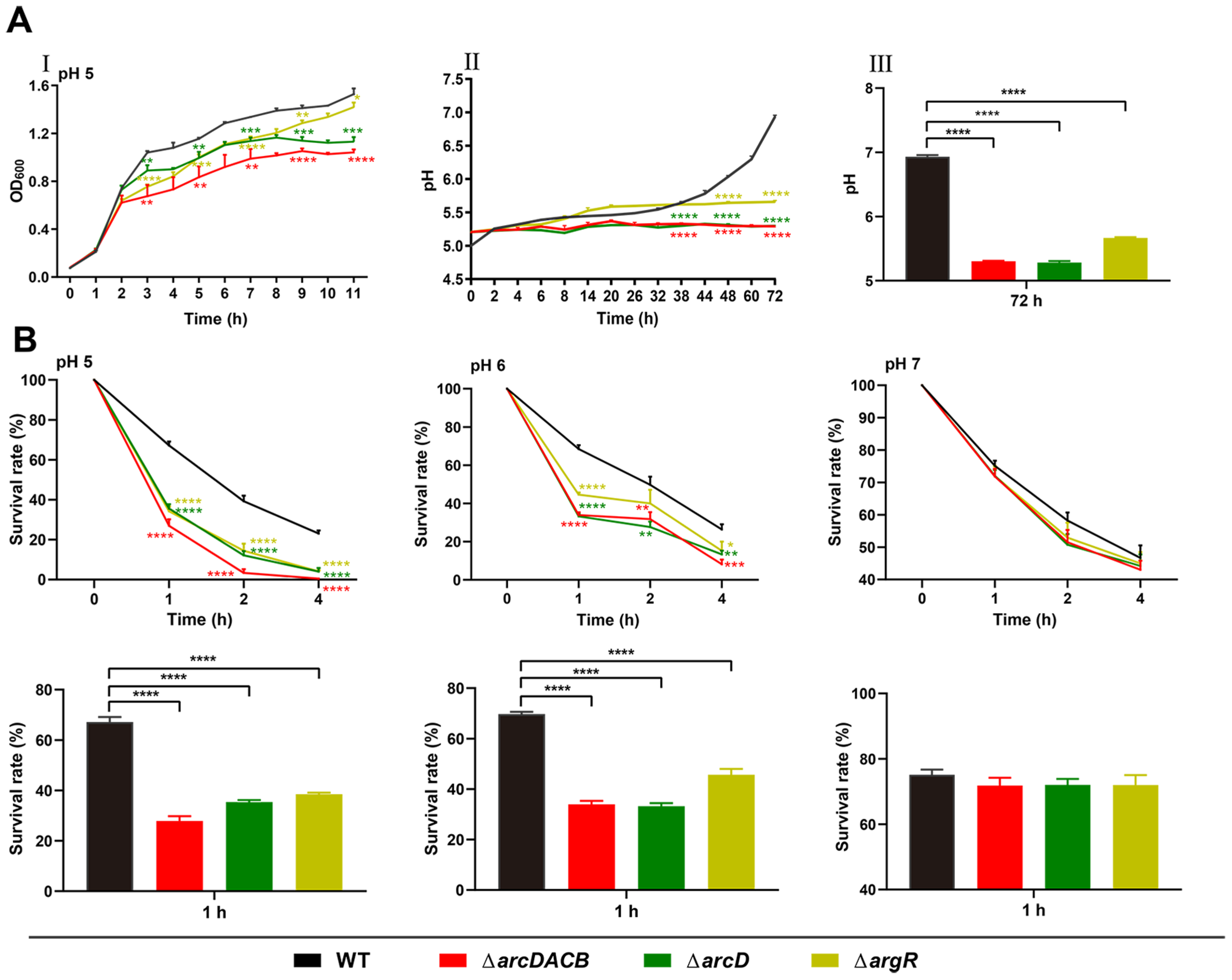
To better understand the role of the ADI cluster in acid resistance, the in-frame deletion mutants of arcDACB, arcD, and argR were constructed. First, the WT, ΔarcDACB, ΔarcD, and ΔargR strains were cultured in acidic LB media (pH = 5) to explore their growth curves. As shown in Figure 3A(I), the ΔarcDACB, ΔarcD, and ΔargR strains all grew at lower rates than did the WT strain; ΔarcDACB grew at the slowest rate, followed by the ΔarcD and ΔargR strains which grew at roughly similar rates. Notably, there was no difference in their growth trends under neutral conditions (pH = 7) (Supplementary Figure S1A). Then, we monitored the pH of the CS of the WT strain and each mutant strain under acidic conditions (pH = 5) for 72 h (Figure 3A(I,III)). The pH of the WT CS slowly and gradually increased with incubation time and finally reached 7, while the pH of the CSs of ΔargR, ΔarcDACB, and ΔarcD remained below 5.5 during the whole incubation period, although the pH of the ΔargR exhibited a minor increase compared to that of the ΔarcDACB and ΔarcD, which may indicate that the ADI pathway still has some weak activity in the argR-negative background. These data indicate that the argR, arcD, and arcACB operons are individually necessary to maintain the function of the ADI pathway in V. fluvialis.
Subsequently, we investigated the acid resistance of the WT and each mutant strain in acidic solutions at pH 5 and 6, and the neutral condition (pH = 7) was used as the control. In general, all strains demonstrated high sensitivity to acidic conditions and showed a lower survival rate during longer incubation times. However, the survival rate of the WT strain was always notably greater than that of the arc mutants (Figure 3B). Additionally, the survival defects became more severe with decreased pH. At a pH of 5, much less survival of the ΔarcDACB, ΔarcD, and ΔargR was observed than that at a pH of 6, especially at both the 2 h and 4 h time points. Comparatively, under neutral conditions (pH = 7), the viabilities of the WT and the acr deletion mutants did not significantly differ, consistent with the similar growth curves presented in Supplementary Figure S1A. These findings corroborate that although not indispensable for growth, the ADI pathway and ArgR play a role in the acid resistance of V. fluvialis, and their absence significantly attenuates the survival of V. fluvialis under acidic conditions.

2.4. ArgR Activates arcD at the Transcriptional Level by Directly Binding to Its Promoter Region

To determine whether ArgR regulates arcD expression, we examined the mRNA levels of arcD in the WT and ΔargR mutant strains. Figure 4A reveals a significant reduction in the arcD mRNA level in the ΔargR strain compared to that in the WT strain. To further elucidate the impact of ArgR on arcD promoter activity, the pBBR-lux vector carrying a promoterless luxCDABE reporter gene cluster was used to construct the fusion reporter plasmid parcD-lux, which was transformed into the WT and ΔargR mutant strains. Compared to that of the WT strain, the luminescence activity of parcD-lux was much lower in the ΔargR strain (Figure 4B), implying that ArgR activates arcD transcription.
ArgR is a regulator of the arginine pathway that binds to a conserved 14–20 bp refolding sequence named the ARG box in the target gene promoter region to regulate its expression [47]. The ArgR consensus in Vibrio comprises 18 nucleotides with relatively high AT content, and the sequence is as follows: 5′-WWTGMATWWWWATKCANW-3′ (where W = A or T, M = A or C, K = G or T, R = A or G, Y = T or C, N = any nucleotide) (https://regprecise.lbl.gov/sites.jsp?regulog_id=2347, accessed on 24 July 2023). To explore whether ArgR directly binds to the arcD promoter region, we initially analyzed the arcD promoter sequences for potential ARG boxes. Based on the conserved motif, three potential binding sites were identified at the −515 to −498, −282 to −265, and −209 to −192 positions relative to the arcD start codon (Figure 4C).
To validate the direct binding of ArgR to the predicted ArgR binding sites in the arcD promoter region, EMSA was performed with purified recombinant ArgR-His6 protein. We generated two biotin-labeled probes, namely, arcD1 (373 bp long with three potential binding sites) and arcD2 (177 bp long with two potential binding sites) (Figure 4C). Different amounts of ArgR-His6 proteins were incubated with 15 ng of the arcD1 or arcD2 probe. As shown in Figure 4D, four clearly shifted bands appeared for the arcD1 probe and two for the arcD2 probe, with the band intensity increasing with increasing ArgR-His6 concentration, indicating a direct interaction between the arcD probes and the recombinant ArgR-His6 proteins. For the arcD probe, the appearance of an extra band than expected may imply another potential binding site or non-specific binding at the highest protein concentration.
To further define the binding sequences of ArgR, we conducted a DNase I footprinting assay with a fluorescent FAM-labeled arcD probe containing the three predicted binding sites. DNase I footprinting assay revealed a nondigested ArgR-protected region comprising a 35 bp long sequence extending from the −521 to −486 positions relative to the arcD start codon (Figure 4E). This region encompasses the predicted ArgR binding site 1 (−515 to −498). Although the EMSA results for the arcD2 probe revealed two ArgR-shifted bands, indicating the presence of the predicted ArgR binding sites 2 and 3, these bands were not detected by DNase I footprinting analysis. This difference might be attributed to the lower affinity of ArgR for sites 2 and 3 than for site 1.
To further confirm and dissect the individual contributions of ArgR binding sites 1, 2, and 3 to arcD promoter activity, site mutations were performed in parcD-lux to generate parcDmu1-lux, parcDmu2-lux, and parcDmu3-lux. Each binding site underwent four-point mutation, substituting conserved G-AT with A-CC in all binding sites, the fourth-to-last C with A in sites 1 and 3, and the seventh-to-last A with C in site 2 (Figure 4F). These point-mutated recombinant plasmids were then transformed into the WT and ΔargR mutant strains to measure the luminescence. In the WT background, the luminescence of parcDmu1-lux dramatically decreased by 22-fold compared to that of parcD-lux, while the luminescence of parcDmu2-lux and parcDmu3-lux decreased 2-fold (p value < 0.0001). No significant difference in luminescence was observed for the ΔargR mutant (Figure 4G). Collectively, these results indicated that the introduced mutation in the ArgR binding sites disrupted the binding of ArgR, resulting in reduced luminescence activity. Furthermore, although all three predicted ArgR binding sites contribute to arcD transcriptional activation, the effect of ArgR binding site 1 is considerably greater than these of ArgR binding sites 2 and 3.

2.5. ArgR Activates the arcACB Operon by Physically Binding to the Promoter Region

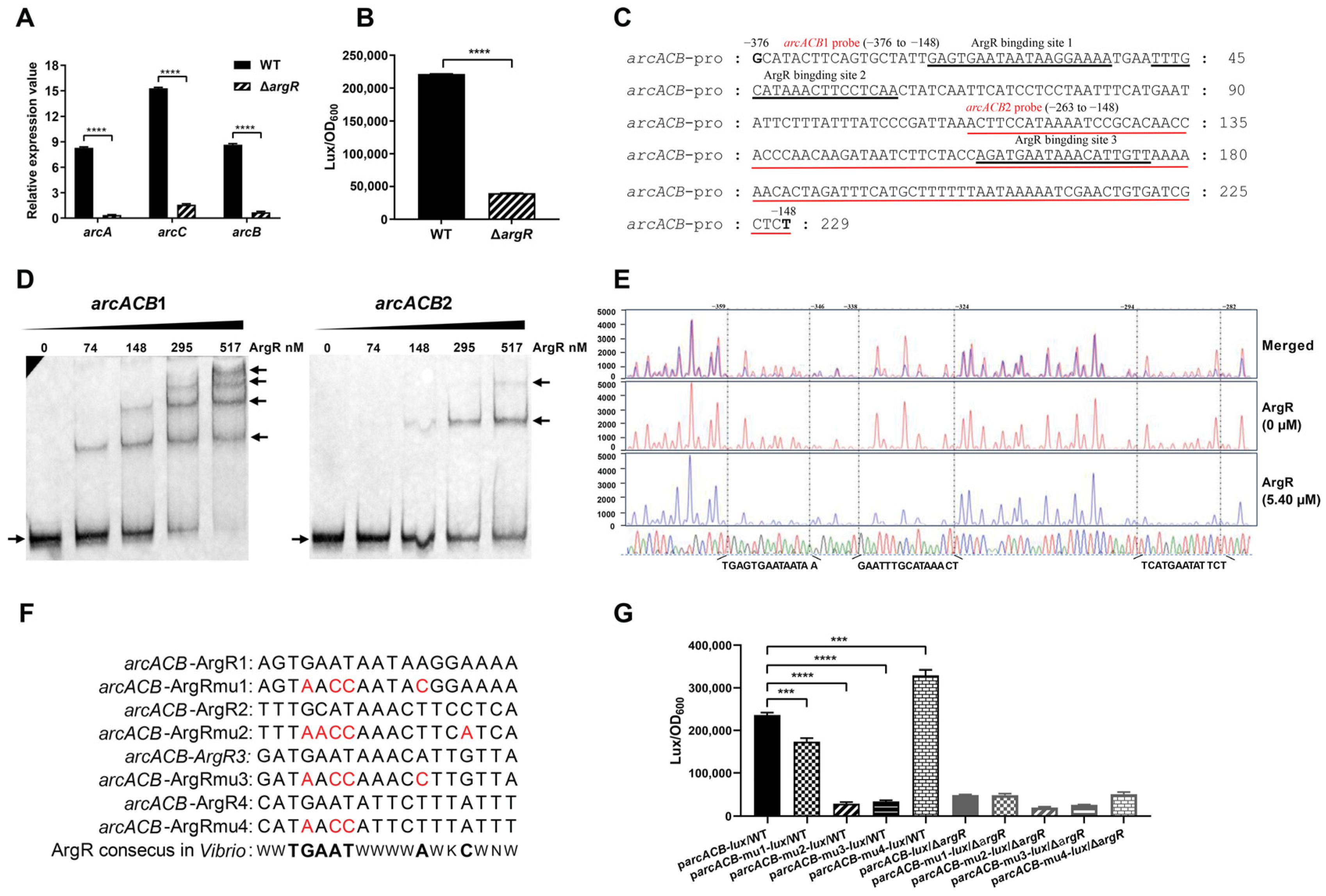
To clarify whether ArgR regulates the arcACB operon, the mRNA levels of arcA, arcC, and arcB were examined in the WT and ΔargR mutant strains. As displayed in Figure 5A, the relative mRNA abundance of these genes was significantly lower in the ΔargR mutant than in the WT. To verify that the regulation occurred at the transcriptional level, a fusion reporter plasmid, parcACB-lux, was constructed and introduced into the WT and ΔargR mutant. The luminescence activity of parcACB-lux was notably lower in the ΔargR mutant than in the WT (Figure 5B). These findings suggest that ArgR plays a crucial role in activating the transcription of the arcACB gene cluster.
With the same strategy as arcD, the sequence of the promoter region of arcACB was analyzed in line with the ArgR consensus sequence in Vibrio (Figure 4C). Three potential ArgR binding sites were identified, ArgR binding site 1 (5′-AGTGAATAATAAGGAAAA-3′), binding site 2 (5′-TTTGCATAAACTTCC TCA-3′), and binding site 3 (5′-GATGAATAAACATTGTTA-3′), which are located at the −357 to −340, −335 to −318, and −217 to −200 positions, respectively, relative to the arcA start codon (Figure 5C). Next, we investigated the direct binding of ArgR to these binding sites using the EMSA and DNase I footprinting methods described above. EMSA revealed four shifted bands detected by the arcACB1 probes (229 bp, encompassing 3 predicted binding sites) and two shifted bands detected by the arcACB2 probes (116 bp, with the third binding site) (Figure 5D). DNase I footprinting analysis (Figure 5E) revealed three ArgR-protected regions located at positions −359 to −346 (5′-TGAGTGAATAATAA-3′), −338 to −324 (5′-GAATTTGCATAAACT-3′), and −294 to −282 (5′-TCATGAATATTCT-3′) relative to the arcA start codon. The percentages of the overlap between the first two protected regions and the predicted ArgR binding sites 1 and 2 were 86% and 80%, respectively. In contrast to EMSA, the DNase I footprinting method did not detect ArgR binding site 3 but detected an unpredicted new binding site, therefore, and we referred to ArgR binding site 4 below.
To functionally confirm these results, mutations were introduced into each predicted ArgR binding site in the arcACB promoter region using the same mutation strategy described above to generate parcACBmu1-lux, parcACBmu2-lux, parcACBmu3-lux, and parcACBmu4-lux based on parcACB-lux (Figure 5F). These were then transformed into the WT and ΔargR mutant. The luminescence activities of parcACBmu1-lux, parcACBmu2-lux and parcACBmu3-lux were 1.36-, 8.10-, and 6.83-fold lower than these of parcACB-lux in the WT, while parcACBmu4-lux showed enhanced activity (Figure 5G). In comparison, no significant differences were observed in the luminescence activities of these recombinant plasmids in the ΔargR mutant. The barely detectable luminescence activity of parcACBmu3-lux in the WT, together with the EMSA results of the arcACB2 probe, suggested that the predicted ArgR binding site 3 is involved in the regulation of arcACB by ArgR. Additionally, parcACBmu2-lux showed the lowest luminescence activity, suggesting that it may be the most functional ArgR binding site. The contrasting result of parcACBmu4-lux is unexpected and needs to be investigated in the future.

2.6. Effects of the ADI Gene Cluster and argR on In Vivo Colonization of V. fluvialis

The acidic conditions of low pH in gastric fluid are often considered unfavorable for bacterial growth [48]. To further elucidate the role of the ADI pathway and its transcription factor ArgR in the in vivo infection of V. fluvialis, competitive colonization experiments were performed in mice to observe the colonization of V. fluvialis. Female C57BL/6 mice (n = 5) were infected by gavage with a mixture (1 × 10⁸ CFU each) of ΔlacZ and one of the competitive strains (WT, ΔarcDACB, ΔarcD or ΔargR), after which fecal and intestinal samples were collected for bacterial calculations as described in the Materials and Methods Section. As depicted in Figure 6, the trends for both the feces and large intestine results were generally consistent. Taking the 24 h results as an example, the CI values for the feces samples were 2.63 for WT, 1.08 for ΔarcDACB, 1.23 for ΔarcD, and 0.55 for ΔargR. For the large intestine samples, the CI values were 2.02 for WT, 1.30 for ΔarcDACB, 1.44 for ΔarcD, and 0.30 for ΔargR. Despite the similar growth abilities of the WT, ΔarcDACB, ΔarcD, and ΔargR strains in LB or M9 media (Supplementary Figure S1), deletion of either the ADI gene cluster or the argR gene severely attenuated the colonization of V. fluvialis in mice, suggesting a positive correlation between arginine metabolism and colonization.

2.7. ADI Pathway Deficiency Enhances the Phagocytosis of V. fluvialis by Macrophages

Since the ADI pathway can protect bacteria from lethal acidity in macrophages by increasing the cytoplasmic pH [49], we explored the effect of the ADI pathway on the survival of V. fluvialis in macrophages. WT and ΔarcDACB mutant strains were separately incubated with RAW 264.7 macrophages. Compared with the WT strain, the ΔarcDACB mutant strain exhibited enhanced macrophage adhesion and invasion, whereas intracellular survival was significantly reduced (Figure 7A–C).
NO is synthesized by nitric oxide synthase (iNOS) from arginine, and the bioavailability of arginine is one of the rate-limiting factors for intracellular NO production [50,51]. The ADI pathway utilizes intracellular arginine, creating competition. To further clarify the role of the ADI pathway in the infection of macrophages by V. fluvialis, we examined the mRNA levels of intracellular iNOS post infection. After 0 h of invasion into macrophages, the relative mRNA abundance of iNOS increased more in the ΔarcDACB-infected macrophage group than in the WT group at 2 h and 4 h post infection, with the increase being directly proportional to the incubation duration (Figure 7D). Taking the 4 h results as an example, the mRNA levels of iNOS were upregulated 106-fold in the ΔarcDACB-infected group, whereas they were upregulated 70-fold in the WT-infected group (Figure 7D). These preliminary results suggest that the ADI pathway might attenuate the phagocytic and immune responses of macrophages during V. fluvialis infection, thereby increasing the survival of V. fluvialis within macrophages.

3. Discussion

The ADI pathway, a prominent pathway for arginine catabolism in various microorganisms, has received extensive attention from researchers [14,17,25,27,28]. Bacteria expressing the ADI pathway can better adapt to hostile environments and evade host defenses, which has important implications for bacterial survival [28,29,52]. Specifically, the generation of ATP by the conversion of arginine to ornithine constitutes a major source of energy for bacteria during nutrient starvation. The NH4+ in the final product can change the pH in the environmental niche and enhance the fitness of bacteria. In addition, the intermediate metabolite ornithine contributes to the synthesis of polyamines, which are essential for tissue repair, anti-inflammatory responses [7], and also important for both the host (macrophages) and various intracellular pathogens like Salmonella typhimurium and Mycobacterium tuberculosis, etc. [6]. leading to investigations of new drag targets in the related pathways [53]. The ADI-positive (commonly called arginine dihydrolase-positive) phenotype is an important biochemical indicator for the identification of V. fluvialis species. In this study, we investigated the genetic organization, biological function, and regulatory mechanism of ADI in V. fluvialis, an emerging foodborne pathogen of public health concern. Our results showed that the deletion of the arc gene cluster significantly reduced the growth capacity of V. fluvialis under acidic conditions both in vitro (in culture medium and macrophages) and in vivo (in mice). Moreover, we revealed a positive regulatory mechanism of ArgR on arcD and arcACB.
The gene arrangement of the arc cluster differs among species [22], e.g., in Streptococcus suis [22], the arrangement was arcABCD, whereas in S. typhimurium [25], it was arcACBD; in Pseudomonas aeruginosa [54], it was arcDABC. In V. fluvialis, the arc gene cluster arrangement was arcDACB, and it was found to be highly conserved in the other Vibrio species, differing only in Vibrio quintilis and Vibrio tapetis (Figure 1C). Here, we showed that in V. fluvialis, the arcDACB gene cluster is organized into two transcriptional units. Specifically, the arcD is a monocistron, while the arcA, arcC and arcB are in one polycistron. 5′-Race revealed that arcD mRNA has a 274 bp long 5′ untranslated region (5′UTR) and that arcACB polycistronic mRNA has a short one with 63 bp long.
The comparison of the genetic content and organization of arc gene clusters in V. fluvialis and other Vibrio species revealed two major differences. In alignment with the biochemical arginine dihydrolase-negative phenotype, V. mimicus, V. cholerae, V. parahaemolyticus, V. harveyi, V. alginolyticus, and V. vulnificus lack the arcD, arcC and adjacent argR genes. The rest of the analyzed species contained the complete arcD gene, arcACB gene, and adjacent argR gene, of which the majority had the same genetic arrangement as V. fluvialis, V. furnissii and V. anguillarum, they are all arginine dihydrolase-positive phenotypes. Additionally, the pyrB and pyrI genes, which encode the aspartate carbamoyltransferase catalytic subunit and aspartate carbamoyltransferase regulatory chain, respectively, are associated with the core ADI cluster in Vibrio species without affecting the phenotype of arginine dihydrolase. Therefore, we reasoned that the presence of arcD, arcACB, and argR could be a preliminary indicator of the phenotype of positive arginine degradation in specific Vibrio species before experimental verification, and the genomic integrity could benefit the survival of Vibrio in nutrient-limited water niches.
It has been reported that arc catabolic gene expression is positively regulated by the regulator ArgR [20,32]. In V. fluvialis, ArgR also activates ADI expression through direct binding to the promoters of arcD and arcACB (Figure 4 and Figure 5). DNase I footprinting revealed one and three ArgR-protected DNA sequences in the arcD and arcACB promoter regions, respectively. These sequences were compared with the ArgR consensus sequences (ARG boxes) in Vibrio (Figure 4C) and other bacteria, such as E. coli [55], Bacillus subtilis [56], and Streptococcus [57]. These ArgR binding sites show highly conserved sequences of TGMAT but not strictly conserved palindromes, implying that the DNA-binding structural domains of ArgR may show different levels of conservation in various bacteria, but the DNA sequences they recognize remain relatively conserved.
ARG boxes typically achieve recognition specificity through cooperative interactions between tandem sites [58,59,60], and as such, these interactions usually occur in pairs, although single binding sites also exist [22,36,57,59]. The reported cooperation combined with the results of EMSA, DNase I footprinting, and point mutation (Figure 4D,E,G and Figure 5D,E,G), we also confirmed the presence of multiple ArgR binding sites on arcD and arcACB. We noticed that these binding sites show genomic position specificity. As for arcD, binding site 1 is the most critical one and is located far away from the TSS, while site 2 overlaps with the arcD TSS region, and site 3 is at the downstream of the TSS. Considering the long 5′UTR of arcD (−273 to the ATG start codon) and the locations of these binding sites, complex and fine modulation probably exist in arcD gene expression. As for arcACB, ArgR binding sites 2 and 3 were functionally confirmed to be the key cis-regulatory elements, which are all present upstream of the TSS of arcA. However, unlike site 2, EMSA confirmed site 3 but was not verified by DNase I footprinting analysis. This variation is probably due to the difference in the stability of the ArgR-DNA complex under the reaction conditions of the two assays but remains to be determined. Additionally, DNase I footprinting revealed an extra binding site 4 and was confirmed to inhibit arcACB expression by promoter-reporter fusion analysis, but the underlying mechanism and biological significance require more exploration in the following study. Considering that the arcD and arcACB promoters contain multiple ArgR binding sites with different affinities, whether the interactions between these sites increase the complexity of the regulatory mechanisms needs to be further investigated.
The ADI pathway, which is prevalent in various Streptococcus [22,28,61] and P. aeruginosa [14,54] strains, enables bacterial survival under lethal acidification by producing ammonia to increase the environmental pH [62]. Macrophage infection may further confirm this result, as the formation of phagocytic lysosomes in macrophages reduces the environmental pH to 4.4–4.7 [63]. Consistent with the literature, V. fluvialis WT exhibited faster pH regulation than the ΔarcDACB, ΔarcD, and ΔargR mutant strains under acidic conditions in vitro (Figure 3A), and this ability was further amplified by the utilization of L-arginine (Figure 2B). Deletion of the ADI pathway resulted in a significant reduction in acid survival (Figure 3B). During V. fluvialis infection of macrophages, the ΔarcDACB strain showed greater adhesion and invasion but lower intracellular survival than did the WT strain (Figure 7A–C). In addition to the lower acidic survival of ΔarcDACB, as we demonstrated above, we speculate that the lower intracellular survival is also related to the NO-mediated killing of macrophages. The ADI pathway competes for intracellular arginine; theoretically, ΔarcDACB-infected cells maintain higher intracellular arginine levels due to the loss of arginine catabolism. The infection of macrophages by the ΔarcDACB strain triggered a surge in intracellular iNOS expression (Figure 7D), which may enhance the throughput of intracellular iNOS to synthesize NO using arginine as a substrate and increase macrophage immunocompetence, leading to reduced survival of ΔarcDACB.
In vivo, competitive coinfection experiments in mice further demonstrated that the WT strain exhibited greater colonization ability in the feces and large intestine than did the mutant strains (Figure 6). This may be due to the greater acid resistance of the WT strain when it passed through the acidic environment of the stomach. An acid resistance assay also confirmed the high survival rate of the WT strain at a pH of 5 at 1 h, which was approximately three times greater than that of the ΔarcDACB strain (Figure 3B). The ΔarcDACB and ΔarcD strains exhibited similar growth rates in acidic environments and in a mouse competition assay, albeit significantly lower than that of the WT strain (Figure 3). This finding suggested a critical role of the transporter protein ArcD in V. fluvialis growth under acidic conditions, consistent with its role in maintaining cellular homeostasis and survival adaptation in S. pneumoniae [18] and S. suis [64]. However, the growth of the ΔargR strain in vivo differed from that in vitro. ArgR, as a global regulator, not only regulates the ADI pathway but also regulates other functional pathways, such as alcohol dehydrogenase and the ABC transporter system [18]. The weaker colonization of the ΔargR strains in vivo may be related to its broad regulatory capacity, which needs to be further investigated.
In conclusion, our study revealed that the ADI pathway significantly enhanced the growth capacity of V. fluvialis in acidic culture environments, in macrophages, and in the mouse large intestine, proving that the transcription factor ArgR is involved in the regulation of ADI through direct binding to the promoter regions of arcD and arcACB. These findings greatly enrich our understanding of arginine metabolism and regulation in V. fluvialis, providing a crucial basis for future research on the mechanisms of pathogenicity and environmental adaptation of V. fluvialis.

4. Materials and Methods

4.1. Bacterial Strains, Culture Conditions, and Plasmids

All bacterial strains and plasmids used in this study are listed in Supplementary Table S1, and the primers used are listed in Supplementary Table S2. The V. fluvialis 85003, isolated in 1985 from an adult patient with diarrhea in Xinjiang Province, China [42], was used as the WT strain, and the mutant strains ΔarcDACB, ΔarcD, and ΔargR were generated in this study. E. coli Top10 and SM10λpir were used for cloning and conjugation, respectively, and Rosetta (DE3) was used as the host for the expression and purification of ArgR-His6 which was amplified by PCR and inserted into the pET30a vector. All strains were grown in Luria–Bertani (LB) broth (Oxoid, Basingstoke, UK) containing 1% NaCl at 37 °C. For growth experiments, a 2.5 M L-arginine master mixture was added to the growth media at the indicated concentrations. Antibiotics were used at the following final concentrations (wt/vol) if necessary: ampicillin (Amp, 100 µg/mL); chloramphenicol (Cm, 10 µg/mL for E. coli, 3 µg/mL for V. fluvialis); streptomycin (Sm, 100 µg/mL); kanamycin (Kan, 50 μg/mL); isopropyl-b-D-thiogalac-topyranoside (IPTG) at a final concentration of 0.4 mM.

4.2. Distribution and Phylogenetic Analysis of the ADI Cluster in Vibrio Species

The amino acid sequences of the arginine deiminase ArcA (arcA, AMF95908), ornithine carbamoyltransferase ArcB (arcB, AMF95906), carbamate kinase ArcC (arcC, AMF95907), arginine-ornithine antiporter ArcD (arcD, AMF95909), and regulatory protein ArgR (argR, AMF95903) were retrieved from the NCBI database (NZ_CP014035.2). The homology of the five proteins in the NCBI nucleotide database (26–28 February 2023) was compared using a protein alignment-based nucleotide search method (tBLASTn, https://blast.ncbi.nlm.nih.gov/Blast.cgi, accessed on 26–28 February 2023), with a minimum of 60% coverage and 40% identity as the retrieval criteria for reference genome selection for different Vibrio species. Sequence information for the arc gene cluster was extracted from 42 different Vibrio species, and the specific Vibrio strain names are shown in Supplementary Table S3. The arc gene clusters were then concatenated to construct a neighbor-joining phylogenetic tree via MEGA7 [65]. iTOL (https://itol.embl.de/, accessed on 28 February 2023) was used for visualization, and clinker (https://github.com/gamcil/clinker, accessed on 28 February 2023) for gene cluster comparison.

4.3. Construction of in-Frame Deletion Mutants

In-frame deletion mutants of the WT strains ΔarcDACB, ΔarcD, and ΔargR were generated by allelic exchange. Briefly, approximately 1000 bp of the upstream and downstream flanking fragments of the targeting ORFs were amplified using the corresponding primers (Supplementary Table S2) and stitched together by an overlapping PCR method as described previously [66]. The 1779 bp ΔarcDACB, 1736 bp ΔarcD, and 1998 bp ΔargR fragments were cloned into the pWM91 suicide plasmid at the XbaI-SacI sites, resulting in pWM91ΔarcDACB, pWM91ΔarcD, and pWM91ΔargR, respectively. Then, the recombinant suicide plasmids were introduced into V. fluvialis WT (recipient) strain from E. coli SM10λpir (donor) by conjugation. The transformants were screened in LB media supplemented with Amp and Sm and then counterselected by streaking on NaCl-free and sucrose-containing (10%) LB agar plates. Sucrose-resistant and Amp-sensitive strains were verified by PCR and confirmed by DNA sequencing.

4.4. RNA Extraction and Quantitative Real-Time PCR (qRT-PCR)

The WT and its derivative mutants were harvested at the late-logarithmic phase (OD600 ~1.0) for total RNA extraction, and cDNA synthesis was subsequently performed as previously described [66]. Three biological replicates were conducted for each sample. Nonreverse transcription RNA served as a negative control, recA served as a reference gene for V. fluvialis and β-actin served as a reference gene for RAW 264.7 cells. The relative expression values (R) were calculated using the equation R = 2−(ΔCT target−ΔCT reference). The relevant primers used are listed in Supplementary Table S2, with iNOS and β-actin primers from Xu YW et al. [67].

4.5. 5′-Rapid Amplification of cDNA End (5′-RACE)

5′-RACE was performed using a SMARTer® RACE 5′/3′ kit (Takara Bio Inc., Kusatsu, Japan). Total RNA extraction was the same as that for qRT-PCR. Following the manufacturer’s guidelines, 5′-RACE-ready cDNA samples were generated utilizing a random primer mixture. Then, 5′-RACE PCR was performed using a universal primer (UPM) and a gene-specific primer (GSP). The resulting PCR amplicon was then ligated into the pMD20 TA cloning vector and transformed into the Top 10 competent cells. Subsequently, the integrity of the inserted sequence was determined by DNA sequencing.

4.6. Growth Analysis

Growth curves were examined by microtiter plates as follows: overnight cultures of V. fluvialis strains were washed once in 1 volume of phosphate-buffered saline (PBS) and then diluted (1:100) into fresh LB medium with different L-arginine concentrations. Triplicates of 200 µL of diluted cultures were transferred to a 100-well microtiter plate and incubated at 37 °C with constant shaking at 200 rpm. The OD600 was measured every hour using a Bioscreen (Oy Growth Curve, Helsinki, Finland). The OD600 values at each time point were averaged and plotted against time to generate growth curves.

4.7. Acid Resistance of V. fluvialis Strains

The growth of V. fluvialis strains in low-pH LB was performed as follows: fresh overnight cultures of WT and its derivative mutants were diluted (1:30) in 40 mL of LB (pH 5). At different intervals up to 72 h, 2 mL of supernatant from each strain was collected for pH measurement. The survival of V. fluvialis was examined as described previously [22]. Briefly, 100 µL of fresh bacterial culture (1.5 × 10⁸ CFU/mL) was inoculated in an acidic solution (20 mM Na2HPO4, 1 mM MgCl2, 25 mM arginine-HCl) with a pH of 5, 6, or 7 and incubated at 37 °C for 1 h, 2 h, or 4 h. The surviving bacteria were quantified by the plate count method. Survival rates are presented as the ratios of surviving bacteria to input bacteria.

4.8. Analysis of the Transcriptional Activity of Promoters with a Luciferase Reporter Gene Assay

The promoter regions of arcD and arcACB were amplified and inserted into the upstream of the promoterless luxCDABE operon in pBBRlux. The resultant lux reporter fusion plasmids parcD-lux and parcACB-lux were introduced into the WT and ∆argR strains by conjugation from SM10λpir. Site mutations at the predicted ArgR binding sites were generated by PCR-based site-directed mutagenesis using the corresponding template. For example, parcD-lux was used for parcDmu1-lux, parcDmu2-lux, and parcDmu3-lux, while parcACB-lux was used for parcACBmu1-lux, parcACBmu2-lux, parcACBmu3-lux, and parcACBmu4-lux. Overnight cultures of V. fluvialis strains harboring lux reporter fusion plasmids were diluted (1:100) in fresh LB media. Triplicates of 200 µL of diluted cultures were transferred to opaque 96-well microtiter plates and incubated at 37 °C with shaking at 200 rpm. Luminescence and OD600 were measured every hour using a spectrophotometer (Infinite M200 Pro, Tecan, Grödig, Austria). Luciferase activity was calculated as previously described [68].

4.9. Cloning, Expression, and Purification of ArgR-His6

The coding region of ArgR was PCR amplified and inserted into pET30a at the NdeI/XhoI sites. The resulting pETargR was subsequently transformed into Rosetta (DE3) for ArgR-His overexpression. The E. coli strain was cultured in LB with Kana at 37 °C with shaking at 200 rpm before being induced by 0.4 mM IPTG when the OD600 reached 0.4–0.6. Then, the culture was incubated at 16 °C for 20 h with shaking at 100 rpm. The ArgR-His6 protein was purified using affinity chromatography Ni2+ resin (Thermo Fisher Scientific, Waltham, MA, USA) and concentrated with an Ultra10 centrifugal filter (Merck, Darmstadt, Germany) according to the manufacturer’s instructions.

4.10. Electrophoretic Mobility Shift Assay (EMSA)

The promoter regions of arcD and arcACB were amplified with biotin-labeled primer pairs (Supplementary Table S2) using the parcD-lux and parcACB-lux plasmids, as templates, respectively. A 20 μL reaction mixture of 15 ng of biotin-labeled amplicons with increasing amounts of purified ArgR-His6 protein in binding buffer (20 mM Tris/HCl [pH 7.6], 50 mM KCl, 1 mM EDTA, 5% [vol/vol] glycerol) containing 200 ng of BSA and 500 ng of dI-dC was incubated at 30 °C for 20 min, separated in a native 8% polyacrylamide gel, transferred to a nylon membrane and visualized by a Chemiluminescent Nucleic Acid Detection Module (Thermo Fisher Scientific, Waltham, MA, USA).

4.11. DNase I Footprinting Assay

DNase I footprinting and sequencing assays were performed as previously described [69]. Briefly, the promoter regions of arcD and arcACB of the WT were amplified with a high-fidelity PCR kit (Sangon Biotech, Shanghai, China) using the primer pairs parcD-F (FAM)/parcD-R and parcACB-F (FAM)/parcACB-R (Supplementary Table S2) to generate the arcD and arcACB probes, respectively. The probes, 371 ng of arcACB and 468 ng of arcD, were mixed with 5.40 μM ArgR-His6 in a 40 μL final reaction volume. After incubation at 37 °C for 0.5 h, the reaction mixture was digested with 0.015 units of RNase-Free DNase I (Promega, Madison, WI, USA), sequenced with an Applied Biosystems 3500XL DNA Analyzer (Thermo Fisher Scientific, Waltham, MA, USA) and analyzed with Peak Scanner software v1.0 (Thermo Fisher Scientific, Waltham, MA, USA).

4.12. Mouse Competition Assay

The competition assay was performed based on the principle that the color of colonies on X-gal chromogenic plates changes from blue to white for the V. fluvialis 85003 lacZ deletion mutant. Overnight cultures of the WT, ΔarcDACB, ΔarcD, ΔargR, and ΔlacZ strains were diluted (1:100) in fresh LB media and incubated for 3 h at 37 °C until an OD600 of 0.3 was reached. The bacterial suspensions were centrifuged at 5000 rpm for 3 min before the pellets were resuspended in PBS. An equal volume of bacteria (1 × 10⁹ CFU/mL for each) was mixed, serially diluted, and spread on LB agar plates supplemented with Sm and X-gal for live bacteria plate counting. The input ratios of each strain to ΔlacZ were calculated and adjusted to a ratio of 1:1 for mouse infection. Groups of five Sm-pretreated six-week-old female C57BL/6 mice (Beijing Vital River Laboratory Animal Technology Co., Ltd., Beijing, China) were infected with 100 µL of the mixed suspension (1 × 10⁸ CFU for each) by intragastric gavage. After 24 h, all mice underwent cervical dislocation following carbon dioxide euthanasia. Output ratios of bacteria in feces and large intestines (colon and cecum) were determined from homogenated feces at 8 h, 16 h, and 24 h or tissues at 24 h postinfection by serial dilution plate counting for live bacteria. The competitive index (CI) is defined as the output ratio divided by the input ratio, with CI values less than 0.5 considered significant.
All experiments involving animals were performed according to protocols approved by the Animal Care and Use Committee of the National Institute for Communicable Disease Control and Prevention and according to the medical research regulations of the National Health Commission, China (protocol number 2023-014).

4.13. Adhesion, Invasion, and Intracellular Survival of V. fluvialis in RAW 264.7 Cells

Murine macrophage RAW 264.7 cells were cultured in high glucose (4.5 g/L) Dulbecco’s modified Eagle’s medium (DMEM) supplemented with 10% heat-inactivated fetal bovine serum (Gibco, Grand Island, NY, USA) at 37 °C with 5% CO2. The gentamicin protection assay was used for invasion and intracellular survival tests [70]. For this aim, RAW 264.7 cells (2 × 10⁵ CFU/well) were seeded into 24-well plates and incubated in an antibiotic-free medium. For the adhesion assay, fresh bacteria at an OD600 of 0.3 were prepared as described previously, and these bacteria were serially diluted and counted on LB plates to serve as the original pre-infection bacterial count (CFU/mL). Then, the bacteria were washed with PBS and resuspended in DMEM for RAW 264.7 cell infection at a multiplicity of infection (MOI) of 1:8 and seeded into a 24-well plate. The plate was centrifuged at 1000× g for 5 min and incubated at 37 °C for 1 h. Then, the infected cells were washed three times with PBS to remove unattached bacteria and lysed with lysis buffer (0.1% SDS and 1% Triton X-100 in PBS). The lysate was then serially diluted and spread on LB agar plates. The plates were incubated at 37 °C overnight for live bacteria plate counting (CFU/mL). For invasion, RAW 264.7 cells were infected as described for the adhesion assay, except for treating with gentamicin (500 μg/mL) for 1 h to kill extracellular bacteria immediately after the PBS wash step. For intracellular survival, after gentamicin treatment, the infected cells were washed with PBS and incubated for an additional 4 h in fresh DMEM containing 10 μg/mL gentamicin. Survival rates are presented as the ratios of the CFU in the intracellular survival assay to the CFU in the invasion assay.

4.14. Statistical Analysis

GraphPad Prism software, version 8, was used for statistical analysis. Statistical significance was determined by unpaired two-tailed Student’s t-test and was defined as * if the p-value was less than 0.05, ** if the p-value was less than 0.01, *** if the p-value was less than 0.001, **** if the p-value was less than 0.0001, and ns for no significant difference.

Supplementary Materials

The supporting information can be downloaded at: https://www.mdpi.com/article/10.3390/ijms25115679/s1.

Author Contributions

Conceptualization, B.K. and W.L.; Funding acquisition, B.K. and W.L.; Investigation, Q.C., Y.H., and S.J.; Methodology, Z.L. and A.Q.; Project administration, B.K. and W.L.; Resources, Z.L., A.Q., and W.L.; Software, Q.C. and Y.H.; Supervision, W.L.; Validation, Q.C., Y.H., and S.J.; Writing—original draft, Q.C., Y.H., and Y.X.; Writing—review and editing, Q.C., Y.H., Y.X., A.Q., and W.L. All authors have read and agreed to the published version of the manuscript.

Funding

This research was funded by the National Key Research and Development Program of China under Grant 2023YFC2604400 and 2021YFC2300302, and the National Natural Science Foundation of China under Grant 81772242.

Institutional Review Board Statement

The animal study protocol was approved by the Animal Care and Use Committee of the National Institute for Communicable Disease Control and Prevention and according to the medical research regulations of the National Health Commission, China (protocol number 2023-014, 1 March 2023).

Informed Consent Statement

Not applicable.

Data Availability Statement

No datasets were generated or analyzed during the current study. The original contributions presented in the study are included in the article/Supplementary Materials, further inquiries can be directed to the corresponding authors.

Conflicts of Interest

The authors declare no conflicts of interest.

References

  1. Hornef, M.W.; Wick, M.J.; Rhen, M.; Normark, S. Bacterial strategies for overcoming host innate and adaptive immune responses. Nat. Immunol. 2002, 3, 1033–1040. [Google Scholar] [CrossRef]
  2. Tlaskalová-Hogenová, H.; Štěpánková, R.; Hudcovic, T.; Tučková, L.; Cukrowska, B.; Lodinová-Žádníková, R.; Kozáková, H.; Rossmann, P.; Bártová, J.; Sokol, D.; et al. Commensal bacteria (normal microflora), mucosal immunity and chronic inflammatory and autoimmune diseases. Immunol. Lett. 2004, 93, 97–108. [Google Scholar] [CrossRef]
  3. Donnenberg, M.S.; Whittam, T.S. Pathogenesis and evolution of virulence in enteropathogenic and enterohemorrhagic Escherichia coli. J. Clin. Investig. 2001, 107, 539–548. [Google Scholar] [CrossRef]
  4. Hooper, L.V.; Gordon, J.I. Commensal Host-Bacterial Relationships in the Gut. Science 2001, 292, 1115–1118. [Google Scholar] [CrossRef]
  5. Lynch, J.P.; Lesser, C.F. A host metabolite promotes Salmonella survival. Science 2021, 371, 344–345. [Google Scholar] [CrossRef]
  6. Manchester, M.; Das, P.; Lahiri, A.; Lahiri, A.; Chakravortty, D. Modulation of the Arginase Pathway in the Context of Microbial Pathogenesis: A Metabolic Enzyme Moonlighting as an Immune Modulator. PLoS Pathog. 2010, 6, e1000899. [Google Scholar] [CrossRef]
  7. Gogoi, M.; Datey, A.; Wilson, K.T.; Chakravortty, D. Dual role of arginine metabolism in establishing pathogenesis. Curr. Opin. Microbiol. 2016, 29, 43–48. [Google Scholar] [CrossRef]
  8. Abdelal, A.T. Arginine Catabolism by Microorganisms. Annu. Rev. Microbiol. 1979, 33, 139–168. [Google Scholar] [CrossRef]
  9. Stadelmann, B.; Hanevik, K.; Andersson, M.K.; Bruserud, O.; Svärd, S.G. The role of arginine and arginine-metabolizing enzymes during Giardia—Host cell interactions in vitro. BMC Microbiol. 2013, 13, 256. [Google Scholar] [CrossRef]
  10. Xiong, L.F.; Teng, J.L.L.; Botelho, M.G.; Lo, R.C.; Lau, S.K.P.; Woo, P.C.Y. Arginine Metabolism in Bacterial Pathogenesis and Cancer Therapy. Int. J. Mol. Sci. 2016, 17, 363. [Google Scholar] [CrossRef]
  11. Hernández, V.M.; Arteaga, A.; Dunn, M.F. Diversity, properties and functions of bacterial arginases. Fems Microbiol. Rev. 2021, 45, fuab034. [Google Scholar] [CrossRef]
  12. Xiong, L.; Teng, J.L.L.; Watt, R.M.; Kan, B.; Lau, S.K.P.; Woo, P.C.Y. Arginine deiminase pathway is far more important than urease for acid resistance and intracellular survival in Laribacter hongkongensis: A possible result of arc gene cassette duplication. BMC Microbiol. 2014, 14, 42. [Google Scholar] [CrossRef]
  13. Weathers, P.J.; Chee, H.L.; Allen, M.M. Arginine catabolism in Aphanocapsa 6308. Arch. Microbiol. 1978, 118, 1–6. [Google Scholar] [CrossRef] [PubMed]
  14. Lu, C.-D.; Winteler, H.; Abdelal, A.; Haas, D. The ArgR Regulatory Protein, a Helper to the Anaerobic Regulator ANR during Transcriptional Activation of the arcD Promoter in Pseudomonas aeruginosa. J. Bacteriol. 1999, 181, 2459–2464. [Google Scholar] [CrossRef]
  15. Minic, Z.; Hervé, G. Arginine Metabolism in the Deep Sea Tube Worm Riftia pachyptila and Its Bacterial Endosymbiont. J. Biol. Chem. 2003, 278, 40527–40533. [Google Scholar] [CrossRef]
  16. Caldara, M.; Charlier, D.; Cunin, R. The arginine regulon of Escherichia coli: Whole-system transcriptome analysis discovers new genes and provides an integrated view of arginine regulation. Microbiology 2006, 152, 3343–3354. [Google Scholar] [CrossRef]
  17. Liu, Y.; Dong, Y.; Chen, Y.-Y.M.; Burne, R.A. Environmental and Growth Phase Regulation of the Streptococcus gordonii Arginine Deiminase Genes. Appl. Environ. Microbiol. 2008, 74, 5023–5030. [Google Scholar] [CrossRef]
  18. Schulz, C.; Gierok, P.; Petruschka, L.; Lalk, M.; Mäder, U.; Hammerschmidt, S.; Thornton, J.A.; McDaniel, L.S. Regulation of the Arginine Deiminase System by ArgR2 Interferes with Arginine Metabolism and Fitness of Streptococcus pneumoniae. mBio 2014, 5, e01858-14. [Google Scholar] [CrossRef]
  19. Coburn, J.; Richards, C.L.; Raffel, S.J.; Bontemps-Gallo, S.; Dulebohn, D.P.; Herbert, T.C.; Gherardini, F.C. The arginine deaminase system plays distinct roles in Borrelia burgdorferi and Borrelia hermsii. PLOS Pathog. 2022, 18, e1010370. [Google Scholar] [CrossRef]
  20. Ryan, S.; Begley, M.; Gahan, C.G.M.; Hill, C. Molecular characterization of the arginine deiminase system in Listeria monocytogenes: Regulation and role in acid tolerance. Environ. Microbiol. 2009, 11, 432–445. [Google Scholar] [CrossRef]
  21. Lindgren, J.K.; Thomas, V.C.; Olson, M.E.; Chaudhari, S.S.; Nuxoll, A.S.; Schaeffer, C.R.; Lindgren, K.E.; Jones, J.; Zimmerman, M.C.; Dunman, P.M.; et al. Arginine Deiminase in Staphylococcus epidermidis Functions to Augment Biofilm Maturation through pH Homeostasis. J. Bacteriol. 2014, 196, 2277–2289. [Google Scholar] [CrossRef]
  22. Gruening, P.; Fulde, M.; Valentin-Weigand, P.; Goethe, R. Structure, Regulation, and Putative Function of the Arginine Deiminase System of Streptococcus suis. J. Bacteriol. 2006, 188, 361–369. [Google Scholar] [CrossRef]
  23. Xu, B.; Yang, X.Y.; Zhang, P.; Ma, Z.; Lin, H.X.; Fan, H.J. The arginine deiminase system facilitates environmental adaptability of Streptococcus equi ssp. zooepidemicus through pH adjustment. Res. Microbiol. 2016, 167, 403–412. [Google Scholar] [CrossRef] [PubMed]
  24. Cotter, P.D.; Gahan, C.G.M.; Hill, C. Analysis of the role of the Listeria monocytogenes F0F1-ATPase operon in the acid tolerance response. Int. J. Food Microbiol. 2000, 60, 137–146. [Google Scholar] [CrossRef]
  25. Choi, Y.; Choi, J.; Groisman, E.A.; Kang, D.-H.; Shin, D.; Ryu, S.; Camilli, A. Expression of STM4467-Encoded Arginine Deiminase Controlled by the STM4463 Regulator Contributes to Salmonella enterica Serovar Typhimurium Virulence. Infect. Immun. 2012, 80, 4291–4297. [Google Scholar] [CrossRef]
  26. Chakraborty, B.; Burne, R.A. Effects of Arginine on Streptococcus mutans Growth, Virulence Gene Expression, and Stress Tolerance. Appl. Environ. Microbiol. 2017, 83, e00496-17. [Google Scholar] [CrossRef] [PubMed]
  27. Makhlin, J.; Kofman, T.; Borovok, I.; Kohler, C.; Engelmann, S.; Cohen, G.; Aharonowitz, Y. Staphylococcus aureus ArcR controls expression of the arginine deiminase operon. J. Bacteriol. 2007, 189, 5976–5986. [Google Scholar] [CrossRef]
  28. Hitzmann, A.; Bergmann, S.; Rohde, M.; Chhatwal, G.S.; Fulde, M. Identification and characterization of the arginine deiminase system of Streptococcus canis. Vet. Microbiol. 2013, 162, 270–277. [Google Scholar] [CrossRef] [PubMed]
  29. Chantratita, N.; Tandhavanant, S.; Wikraiphat, C.; Trunck, L.A.; Rholl, D.A.; Thanwisai, A.; Saiprom, N.; Limmathurotsakul, D.; Korbsrisate, S.; Day, N.P.J.; et al. Proteomic analysis of colony morphology variants of Burkholderia pseudomallei defines a role for the arginine deiminase system in bacterial survival. J. Proteom. 2012, 75, 1031–1042. [Google Scholar] [CrossRef]
  30. Maghnouj, A.; Abu-Bakr, A.A.W.; Baumberg, S.; Stalon, V.; Wauven, C. Regulation of anaerobic arginine catabolism in Bacillus licheniformis by a protein of the Crp/Fnr family. FEMS Microbiol. Lett. 2000, 191, 227–234. [Google Scholar] [CrossRef]
  31. Winterhoff, N.; Goethe, R.; Gruening, P.; Rohde, M.; Kalisz, H.; Smith, H.E.; Valentin-Weigand, P. Identification and Characterization of Two Temperature-Induced Surface-Associated Proteins of Streptococcus suis with High Homologies to Members of the Arginine Deiminase System of Streptococcus pyogenes. J. Bacteriol. 2002, 184, 6768–6776. [Google Scholar] [CrossRef] [PubMed]
  32. Fulde, M.; Willenborg, J.; de Greeff, A.; Benga, L.; Smith, H.E.; Valentin-Weigand, P.; Goethe, R. ArgR is an essential local transcriptional regulator of the arcABC operon in Streptococcus suis and is crucial for biological fitness in an acidic environment. Microbiology 2011, 157, 572–582. [Google Scholar] [CrossRef]
  33. Grandori, R.; Lavoie, T.A.; Pflumm, M.; Tian, G.; Niersbach, H.; Maas, W.K.; Fairman, R.; Carey, J. The DNA-binding Domain of the Hexameric Arginine Repressor. J. Mol. Biol. 1995, 254, 150–162. [Google Scholar] [CrossRef] [PubMed]
  34. Garnett, J.A.; Marincs, F.; Baumberg, S.; Stockley, P.G.; Phillips, S.E.V. Structure and Function of the Arginine Repressor-Operator Complex from Bacillus subtilis. J. Mol. Biol. 2008, 379, 284–298. [Google Scholar] [CrossRef] [PubMed]
  35. Cherney, L.T.; Cherney, M.M.; Garen, C.R.; James, M.N.G. Crystal Structure of the Intermediate Complex of the Arginine Repressor from Mycobacterium tuberculosis Bound with Its DNA Operator Reveals Detailed Mechanism of Arginine Repression. J. Mol. Biol. 2010, 399, 240–254. [Google Scholar] [CrossRef] [PubMed]
  36. Maghnouj, A.; de Sousa Cabral, T.F.; Stalon, V.; Vander Wauven, C. The arcABDC Gene Cluster, Encoding the Arginine Deiminase Pathway of Bacillus licheniformis, and Its Activation by the Arginine Repressor ArgR. J. Bacteriol. 1998, 180, 6468–6475. [Google Scholar] [CrossRef] [PubMed]
  37. Chatterjee, B.; Thawani, G.; Sanyal, S. Etiology of acute childhood diarrhoea in Calcutta. Trop Gastroenterol 1989, 10, 158–166. [Google Scholar] [PubMed]
  38. Chowdhury, G.; Pazhani, G.P.; Dutta, D.; Guin, S.; Dutta, S.; Ghosh, S.; Izumiya, H.; Asakura, M.; Yamasaki, S.; Takeda, Y.; et al. Vibrio fluvialisin Patients with Diarrhea, Kolkata, India. Emerg. Infect. Dis. 2012, 18, 1868–1871. [Google Scholar] [CrossRef]
  39. Kobayashi, K.; Taguchi, M.; Shimada, T.; Sakazaki, R. Ten cases of gastroenteritis possibly caused by Vibrio fluvialis and its enterotoxigenicity. J. Jpn. Assoc. Infect. Dis. 1983, 57, 375–382. [Google Scholar] [CrossRef]
  40. Igbinosa, E.O.; Obi, L.C.; Okoh, A.I. Occurrence of potentially pathogenic Vibrios in final effluents of a wastewater treatment facility in a rural community of the Eastern Cape Province of South Africa. Res. Microbiol. 2009, 160, 531–537. [Google Scholar] [CrossRef]
  41. Huq, M.I.; Alam, A.K.; Brenner, D.J.; Morris, G.K. Isolation of Vibrio-like group, EF-6, from patients with diarrhea. J. Clin. Microbiol. 1980, 11, 621–624. [Google Scholar] [CrossRef]
  42. Lu, X.; Liang, W.; Wang, Y.; Xu, J.; Zhu, J.; Kan, B. Identification of Genetic Bases of Vibrio fluvialis Species-Specific Biochemical Pathways and Potential Virulence Factors by Comparative Genomic Analysis. Appl. Environ. Microbiol. 2014, 80, 2029–2037. [Google Scholar] [CrossRef]
  43. Xiao, D.; Yang, W.; Wang, H.; Bi, Z.; Lu, L.; Kan, B. Vibrio Cholera Prevention and Control Manual, 6th ed.; Wang, L., Wang, D., Wang, M., Wang, G., Feng, Z., Bi, Z., Su, H., Yang, W., Xiao, D., Wang, H., et al., Eds.; People’s Health Publishing House: Beijing, China, 2013; p. 91. (in Chinese) [Google Scholar]
  44. Brenner, D.J.; Hickman-Brenner, F.W.; Lee, J.V.; Steigerwalt, A.G.; Fanning, G.R.; Hollis, D.G.; Farmer, J.J.; Weaver, R.E.; Joseph, S.W.; Seidler, R.J. Vibrio furnissii (formerly aerogenic biogroup of Vibrio fluvialis), a new species isolated from human feces and the environment. J. Clin. Microbiol. 1983, 18, 816–824. [Google Scholar] [CrossRef]
  45. Liang, P.; Cui, X.; Du, X.; Kan, B.; Liang, W. The virulence phenotypes and molecular epidemiological characteristics of Vibrio fluvialis in China. Gut Pathog. 2013, 5, 6. [Google Scholar] [CrossRef]
  46. Kala, K.; Shahi, N.; Singh, S.; Rawat, S.; Patiyal, R.S.; Pande, V.; Mallik, S.K. New host record of Vibrio anguillarum associated with haemorrhagic septicaemia in golden mahseer, Tor putitora (Hamilton, 1822) from India. Indian J. Comp. Microbiol. Immunol. Infect. Dis. 2021, 42, 71–83. [Google Scholar] [CrossRef]
  47. Makarova, K.S.; Mironov, A.A.; Gelfand, M.S. Conservation of the binding site for the arginine repressor in all bacterial lineages. Genome Biol. 2001, 2, RESEARCH0013. [Google Scholar] [CrossRef]
  48. Franzosa, E.A.; Morgan, X.C.; Segata, N.; Waldron, L.; Reyes, J.; Earl, A.M.; Giannoukos, G.; Boylan, M.R.; Ciulla, D.; Gevers, D.; et al. Relating the metatranscriptome and metagenome of the human gut. Proc. Natl. Acad. Sci. USA 2014, 111, E2329–E2338. [Google Scholar] [CrossRef]
  49. Degnan, B.A.; Tuomanen, E.I.; Fontaine, M.C.; Doebereiner, A.H.; Lee, J.J.; Mastroeni, P.; Dougan, G.; Goodacre, J.A.; Kehoe, M.A. Characterization of an Isogenic Mutant of Streptococcus pyogenes Manfredo Lacking the Ability To Make Streptococcal Acid Glycoprotein. Infect. Immun. 2000, 68, 2441–2448. [Google Scholar] [CrossRef]
  50. Alderton, W.K.; Cooper, C.E.; Knowles, R.G. Nitric oxide synthases: Structure, function and inhibition. Biochem. J. 2001, 357, 593–615. [Google Scholar] [CrossRef] [PubMed]
  51. Mori, M.; Gotoh, T. Arginine Metabolic Enzymes, Nitric Oxide and Infection. J. Nutr. 2004, 134, 2820S–2825S. [Google Scholar] [CrossRef] [PubMed]
  52. Cusumano, Z.T.; Watson, M.E.; Caparon, M.G.; Camilli, A. Streptococcus pyogenes Arginine and Citrulline Catabolism Promotes Infection and Modulates Innate Immunity. Infect. Immun. 2014, 82, 233–242. [Google Scholar] [CrossRef]
  53. Krysenko, S.; Emani, C.S.; Bäuerle, M.; Oswald, M.; Kulik, A.; Meyners, C.; Hillemann, D.; Merker, M.; Wohlers, I.; Hausch, F.; et al. GlnA3Mt is able to glutamylate spermine but it is not essential for the detoxification of spermine in Mycobacterium tuberculosis. bioRxiv 2023. [Google Scholar] [CrossRef]
  54. Gamper, M.; Zimmermann, A.; Haas, D. Anaerobic regulation of transcription initiation in the arcDABC operon of Pseudomonas aeruginosa. J. Bacteriol. 1991, 173, 4742–4750. [Google Scholar] [CrossRef]
  55. Maas, W.K. The arginine repressor of Escherichia coli. Microbiol. Rev. 1994, 58, 631–640. [Google Scholar] [CrossRef]
  56. Klingel, U.; Miller, C.M.; North, A.K.; Stockley, P.G.; Baumberg, S. A binding site for activation by the Bacillus subtilis AhrC protein, a repressor/activator of arginine metabolism. Mol. Gen. Genet. MGG 1995, 248, 329–340. [Google Scholar] [CrossRef]
  57. Rodríguez-García, A.; Ludovice, M.; Martín, J.F.; Liras, P. Arginine boxes and the argR gene in Streptomyces clavuligerus: Evidence for a clear regulation of the arginine pathway. Mol. Microbiol. 2003, 25, 219–228. [Google Scholar] [CrossRef]
  58. Charlier, D.; Roovers, M.; Van Vliet, F.; Boyen, A.; Cunin, R.; Nakamura, Y.; Glansdorff, N.; Piérard, A. Arginine regulon of Escherichia coli K-12. J. Mol. Biol. 1992, 226, 367–386. [Google Scholar] [CrossRef]
  59. Tian, G.; Lim, D.; Carey, J.; Maas, W.K. Binding of the arginine repressor of Escherichia coli K12 to its operator sites. J. Mol. Biol. 1992, 226, 387–397. [Google Scholar] [CrossRef]
  60. Miller, C.M.; Baumberg, S.; Stockley, P.G. Operator interactions by the Bacillus subtilis arginine repressor/activator, AhrC: Novel positioning and DNA-mediated assembly of a transcriptional activator at catabolic sites. Mol. Microbiol. 2003, 26, 37–48. [Google Scholar] [CrossRef] [PubMed]
  61. Zeng, L.; Dong, Y.; Burne, R.A. Characterization of cis-acting sites controlling arginine deiminase gene expression in Streptococcus gordonii. J. Bacteriol. 2006, 188, 941–949. [Google Scholar] [CrossRef] [PubMed]
  62. Marquis, R.E.; Bender, G.R.; Murray, D.R.; Wong, A. Arginine deiminase system and bacterial adaptation to acid environments. Appl. Environ. Microbiol. 1987, 53, 198–200. [Google Scholar] [CrossRef] [PubMed]
  63. Bassoe, C.F.; Bjerknes, R. Phagocytosis by Human Leukocytes, Phagosomal pH and Degradation of Seven Species of Bacteria Measured by Flow Cytometry. J. Med. Microbiol. 1985, 19, 115–125. [Google Scholar] [CrossRef] [PubMed]
  64. Fulde, M.; Willenborg, J.; Huber, C.; Hitzmann, A.; Willms, D.; Seitz, M.; Eisenreich, W.; Valentin-Weigand, P.; Goethe, R. The arginine-ornithine antiporter ArcD contributes to biological fitness of Streptococcus suis. Front. Cell. Infect. Microbiol. 2014, 4, 107. [Google Scholar] [CrossRef] [PubMed]
  65. Nguyen, L.-T.; Schmidt, H.A.; von Haeseler, A.; Minh, B.Q. IQ-TREE: A Fast and Effective Stochastic Algorithm for Estimating Maximum-Likelihood Phylogenies. Mol. Biol. Evol. 2015, 32, 268–274. [Google Scholar] [CrossRef] [PubMed]
  66. Wu, R.; Zhao, M.; Li, J.; Gao, H.; Kan, B.; Liang, W. Direct regulation of the natural competence regulator gene tfoX by cyclic AMP (cAMP) and cAMP receptor protein (CRP) in Vibrios. Sci. Rep. 2015, 5, 14921. [Google Scholar] [CrossRef] [PubMed]
  67. Xu, Y.W.; Xing, R.X.; Zhang, W.H.; Li, L.; Wu, Y.; Hu, J.; Wang, C.; Luo, Q.L.; Shen, J.L.; Chen, X. Toxoplasma ROP16I/III ameliorated inflammatory bowel diseases via inducing M2 phenotype of macrophages. World J. Gastroenterol. 2019, 25, 6634–6635. [Google Scholar] [CrossRef] [PubMed]
  68. Pan, J.J.; Zhao, M.; Huang, Y.M.; Li, J.; Liu, X.S.; Ren, Z.H.; Kan, B.; Liang, W.L. Integration Host Factor Modulates the Expression and Function of T6SS2 in Vibrio fluvialis. Front. Microbiol. 2018, 9, 00962. [Google Scholar] [CrossRef]
  69. Zianni, M.; Tessanne, K.; Merighi, M.; Laguna, R.; Tabita, F.R. Identification of the DNA bases of a DNase I footprint by the use of dye primer sequencing on an automated capillary DNA analysis instrument. J. Biomol. Tech. 2006, 17, 103–113. [Google Scholar]
  70. Monack, D.M.; Raupach, B.; Hromockyj, A.E.; Falkow, S. Salmonella typhimurium invasion induces apoptosis in infected macrophages. Proc. Natl. Acad. Sci. USA 1996, 93, 9833–9838. [Google Scholar] [CrossRef]
Figure 1. The genomic organizations of the arc gene cluster in V. fluvialis and in the genus Vibrio. (A) Genomic organization of ADI operons of V. fluvialis: arcD, arginine-ornithine antiporter; arcA, arginine deaminase; arcC, carbamate kinase; arcB, ornithine carbamoyltransferase; argR, arginine regulatory factor. The TSSs of arcD and arcA and the length of the spacer region between ORFs are shown above. The AI, AII, and AIII fragments with black lines of different lengths represent the arc gene sequences and the intergenic sequences amplified by PCR in panel (B). (B) RT-PCR analysis of the transcripts of the arc operon. Genomic DNA (gDNA) and unreversed RNA served as positive and negative controls, respectively. recA: internal reference. M: marker. (C) Composition and distribution of ADI gene clusters and their affinities in the genus Vibrio. The phylogenetic tree on the left shows the genetic relationships of the four proteins of the arc gene cluster in different Vibrio species. The tree scale represented a nucleotide substitution rate of 0.01 for each site. The genetic organization on the right shows the composition of the arc gene clusters, with different genes indicated by different colors. The length scale represents 2.5 kb. The presence of the arginine dihydrolase phenotype was represented as follows: “−”: negative; “+”, positive; “N”, unknown.
Ijms 25 05679 g001
Figure 2. V. fluvialis responds to the presence of L-arginine during in vitro growth. (A,B) The V. fluvialis WT strain was grown in conventional LB (A) and acidic LB (pH = 5) (B) supplemented with 0, 10 mM, or 25 mM L-arginine, after which the OD600 of the culture (A) and the pH of the CS (B) were determined at indicated time points. (C) The WT strain was grown in LB supplemented with or without 25 mM L-arginine, and the transcription of the arc gene clusters was analyzed via qRT-PCR. The results are presented as the means ± SDs of three biological replicates, with the error bar highlighted in pink in (A). * p< 0.05, *** p < 0.001, and **** p < 0.0001.
Ijms 25 05679 g002
Figure 3. The effect of ADI and ArgR on the growth and survival of V. fluvialis under different pH environments. (A) I: Fresh overnight cultures of the WT and mutant strains were diluted (1:100) in fresh LB media at an initial pH of 5 and incubated at 37 °C for 12 h. The OD600 was measured every hour and the time points of 3, 5, 7, 9 and 11 h were used for the statistical analysis. II: Fresh overnight cultures of the WT and mutant strains were diluted (1:30) in fresh LB media at an initial pH of 5 and incubated at 37 °C for 72 h. The pH of the supernatant was measured for 72 h and the time points of 38, 48, and 72 h were used for statistical analysis. III: pH values of the WT and mutant strains at 72 h in II. (B) Acid resistance of the WT and mutant strains to different pH were monitored as described in the Materials and Methods Section. The results are presented as percentages of survival (CFU counts at corresponding time points compared to those in the initial inoculum). Line graphs from left to right show the survival rates of the WT strain and each mutant strain at different time points (1, 2, and 4 h) at pH 5, 6, and 7. The bar graphs below show the survival rates of the WT strain and each mutant strain at the corresponding pH at a 1 h time point. The results are presented as the means ± SDs of three biological replicates. * p< 0.05, ** p < 0.01, *** p < 0.001, and **** p < 0.0001.
Ijms 25 05679 g003
Figure 4. ArgR physically binds to the promoter region of arcD. (A) Determination of the mRNA level of arcD in the WT and ΔargR mutant strains. (B) Luminescence activity of parcD-lux in the WT and ΔargR mutant strains. (C) The ArgR consensus sequence of Vibrio and the characterization of the arcD promoter region. The three potential ArgR binding sites are underlined, and the EMSA probe sequences (arcD1 probe: position −530 to −158; arcD2 probe: position −334 to −158) are marked and underlined in red. The TSS of arcD is indicated by an arrow and labeled with a red “A”. The numbers above the sequence indicate the positions of nucleotides relative to the arcD start codon. (D) The binding of ArgR to the promoter region of arcD is determined by EMSA. Biotin-labeled arcD1 (373 bp) or arcD2 (177 bp) DNA probes (15 ng for each) were incubated with purified ArgR-His6 protein. The arrows on the right indicated the shift bands of probes binding to the proteins, and the arrows on the left indicated the unbound probes. (E) DNase I footprinting assay of ArgR binding to the promoter region of arcD. The region protected by ArgR is marked with the box with a dotted line, and the nucleotide positions in the arcD promoter are labeled. The red and blue sequence traces represent different concentrations of ArgR (0 μM and 5.40 μM, respectively). The sequence traces of the four different colors representing the sequence of the arcD promoter are shown at the bottom. (F) The parcDmu1-lux, parcDmu2-lux, and parcDmu3-lux were constructed by introducing 4-bp substitutions in the ArgR consensus site at the arcD promoter region. The red font represents the mutation site. (G) Luminescence activities of parcD-lux, parcDmu1-lux, parcDmu2-lux, and parcDmu3-lux in the WT and ΔargR strains. The luminescence activities are reported as light units/OD600. The results are presented as the means ± SDs of three biological replicates. **** p < 0.0001.
Ijms 25 05679 g004
Figure 5. ArgR directly binds to the promoter region of arcACB operon. (A) Examination of the mRNA levels of arcA, arcC, and arcB in the WT and ΔargR mutant strains. (B) Luminescence activity of parcACB-lux in the WT and ΔargR mutant strains. (C) Characterization of the arcACB operon promoter region. The four potential ArgR binding sites are underlined. The EMSA probe sequences (arcACB1 probe: position −376 to −148; arcACB2 probe: position −263 to −148) are marked and underlined in red. The numbers above the sequence indicate the positions of the nucleotides relative to the arcA start codon. (D) The binding of ArgR to the promoter region of arcACB, as determined by EMSA. The method and labeling were the same as those in Figure 4D. The arrows on the right of the gel pictures indicated the shift bands of probes binding to the proteins, and the arrows on the left of the gel pictures indicated the unbound probes. (E) The binding of ArgR to the promoter region of arcACB was determined by a DNase I footprinting assay. The method and labeling were the same as those in Figure 4E. The regions protected by ArgR are marked with three boxes with dotted line, and the nucleotide positions in the arcACB promoter are labeled. The red and blue sequence traces represent different concentrations of ArgR (0 μM and 5.40 μM, respectively). The sequence traces of four different colors representing the sequence of the arcACB promoter are shown at the bottom. (F) The parcACBmu1-lux, parcACBmu2-lux, parcACBmu3-lux and parcACBmu4-lux were constructed by introducing 4-bp changes in the ArgR consensus site at the arcACB promoter region. The red font represents the mutation site. (G) Luminescence activities of parcACB-lux, parcACBmu1-lux, parcACBmu2-lux, parcACBmu3-lux and parcACBmu4-lux in the WT and ΔargR strains. The luminescent activities were measured and are presented as light units/OD600. The results are presented as the means ± SDs of three biological replicates. *** p < 0.001, **** p < 0.0001.
Ijms 25 05679 g005
Figure 6. The deficiency of ArcD antiporter, the whole ADI or ArgR attenuates V. fluvialis colonization in mice. Female C57BL/6 mice (n = 5) were infected with equal amounts (1 × 10⁸ CFU each) of ΔlacZ and one of the competitive strains (WT, ΔarcDACB, ΔarcD, or ΔargR). (A) CI values of the WT, ΔarcDACB, ΔarcD, and ΔargR strains in the feces of the mice at 8 h, 16 h and 24 h postinfection; (B) CI values of the WT, ΔarcDACB, ΔarcD and ΔargR strains in the large intestine of the mice at 24 h postinfection after euthanasia. CI values above 1 indicate faster growth of the competitive strain than that of the ΔlacZ strain. * p< 0.05, ** p < 0.01, *** p < 0.001, **** p < 0.0001, and ns for no significant difference.
Ijms 25 05679 g006
Figure 7. Role of the ADI in V. fluvialis phagocytosis by macrophages. Murine macrophages (RAW 264.7) were infected with WT or arcΔDACB at an MOI of 8 using a gentamicin protection assay. The percentages of bacteria in adhesion (A), invasion (B), and intracellular survival (C) were calculated as the final CFU/mL postinfection divided by the original CFU/mL before infection. (D) The mRNA levels of iNOS in RAW 264.7 cells infected with WT or ΔarcDACB. Cell samples from the invasion (0 h), 2 and 4 h postinfection groups were collected for RNA extraction. qRT-PCR analysis is described in the Materials and Methods Section, with β-actin serving as an intracellular reference and uninfected RAW 264.7 cells serving as a control. The results are presented as the means ± SDs of three biological replicates. ** p < 0.01, *** p < 0.001, and **** p < 0.0001.
Ijms 25 05679 g007
Disclaimer/Publisher’s Note: The statements, opinions and data contained in all publications are solely those of the individual author(s) and contributor(s) and not of MDPI and/or the editor(s). MDPI and/or the editor(s) disclaim responsibility for any injury to people or property resulting from any ideas, methods, instructions or products referred to in the content.