Article

Genome-Wide Identification and Expression Analysis of the YTH Domain-Containing RNA-Binding Protein Family in Cinnamomum camphora

1
State Key Laboratory of Tree Genetics and Breeding, Nanjing Forestry University, Nanjing 210037, China
2
Key Open Laboratory of Forest Genetics and Gene Engineering of National Forestry & Grassland, Nanjing Forestry University, Nanjing 210037, China
3
Co-Innovation Center for Sustainable Forestry in Southern China, Nanjing Forestry University, Nanjing 210037, China
4
Beijing National Laboratory for Molecular Sciences, Beijing 100190, China
*
Author to whom correspondence should be addressed.
Int. J. Mol. Sci. 2024, 25(11), 5960; https://doi.org/10.3390/ijms25115960
Submission received: 15 April 2024 / Revised: 20 May 2024 / Accepted: 25 May 2024 / Published: 29 May 2024
(This article belongs to the Special Issue Latest Epigenetic Research in Plants)

Abstract

:
N6-methyladenosine (m6A) is one of the most abundant chemical modifications on mRNA in eukaryotes. RNA-binding proteins containing the YT521-B (YTH) domain play crucial roles in post-transcriptional regulation of plant growth, development, and stress response by reading the m6A mark. However, the YTH domain-containing RNA-binding protein family has not been studied in a valuable and medicinal tree such as Cinnamomum camphora (C. camphora) yet. In this study, we identified 10 YTH genes in C. camphora, located on eight out of 12 chromosomes. Phylogenetic analysis revealed that these genes can be classified into two major classes, YTHDF (CcDF) and YTHDC (CcDC). Closely related CcYTHs within the same class exhibited a similar distribution of conserved motifs and domain organization, suggesting functional similarities among these closely related CcYTHs. All CcYTH proteins possessed a highly conserved YTH domain, with CcDC1A containing an additional CCCH domain. The liquid–liquid phase separation (LLPS) predictions indicate that CcDC1A, CcDF1A, CcDF1C, CcDF3C, CcDF4C, and CcDF5C may undergo phase transitions. Quantitative expression analysis revealed that tissue-specific expression was observed fo CcYTHs. Notably, there were two genes, CcDF1A and CcDF5C; both exhibited significantly higher expression levels in various tissues than other genes, indicating that the m6A-YTH regulatory network in C. camphora might be quite distinct from that in most plants such as Arabidopsis thaliana (A. thaliana) with only one abundant YTH protein. According to the analysis of the up-stream cis-regulatory elements of these YTH genes, these genes could be closely related to stress, hormones, and development. The following stress response experiments further verified that their expression levels indeed changed under both PEG and NaCl treatments. These findings not only provide a foundation for future functional analysis of CcYTHs in C. camphora, but also provide insights into the functions of epigenetic mark m6A in forest trees.

1. Introduction

To date, over 170 types of RNA modifications have been reported [1]. N6-methyladenosine (m6A), as one of the most abundant RNA modifications in eukaryotes, plays a crucial role in gene regulation and maintenance of genomic stability [2]. Recent research has highlighted the diverse functions of m6A modification in RNA metabolism, such as splicing, translation, stability, and decay, thereby regulating various biological processes including gene expression, cell differentiation, embryonic development, and sex determination, as well as diseases [3,4,5]. Like for mammals, m6A is also important and necessary for plants, which can be dynamically written, erased, and read by m6A methyltransferases, demethylases, and m6A-binding proteins. In plants, MTA (homolog of human METTL3), MTB (homolog of human METTL14), VIR (homolog of human VIRMA), HAKA (homolog of human ZC3H13), FIP (homolog of human WTAP), and FIONA1 (homolog of human MELLT16) have been identified as writers of m6A [6,7,8,9]. ALKBH9B, ALKBH10B, and SlALKBH2 (homolog of human ALKBH5) have been characterized as m6A demethylases [10,11]. The readers have been named as the evolutionarily conserved C-terminal region (ECT) family proteins (homolog of human YTH-domain family proteins) [12]. The function of m6A modification depends mainly on its readers to specifically recognize the m6A marks on RNA to determine the RNA fate. Therefore, the identification of m6A readers is crucial for understanding the regulatory mechanisms of m6A on various physiological and molecular processes in cells.
The YTH protein family, a class of RNA-binding proteins that contains the YTH domain, has been first reported to serve as an m6A reader [13]. In 1998, YTH521, a member of the YTH protein family, was reported to function as an RNA splicing-related protein in rats [14]. In 2002, a novel YTH domain consisting of approximately 150 amino acids rich in aromatic residues was discovered within nuclear proteins and named as the YTH521-B homologous domain, which is a highly conserved in eukaryotes [15]. This domain folds into a conserved α/β fold, which includes 3α helices and 6β strands. The 6β strands form a barrel-like fold, and the 3α helices are packed against the β strands to create a hydrophobic core [16,17]. Within the YTH domain, an aromatic cage forms a hydrophobic binding pocket that is capable of recognizing buried methyl groups with a cavity insertion mode. This complex structure lays the foundation for the specific m6A recognition of YTH domain. Similar to most RNA-binding domains that are surrounded by structured domains or low-complexity regions with diverse functions, the YTH domain is also encircled by several disordered regions [18]. These disordered regions are indispensable for YTH family proteins to modulate the function of m6A-modified RNA by influencing the subcellular localization of YTH family proteins and their partners. Furthermore, these regions also impact their potential for liquid–liquid phase separation [19]. However, the study of m6A readers in plants remains limited. So far, only ECT2/3/4 and CPSF30-L, out of the 13 YTH-domain proteins that mainly come from the ECT family, have been identified as m6A readers in A. thaliana; the rest still remain unknown [20,21,22,23]. In vitro and in vivo experiments have confirmed that ECT2 could directly bind m6A, and its m6A binding activity is essential for normal trichome morphology [20]. Further research indicates that the functions of ECT2 are redundant with those of ECT3 and ECT4, suggesting that ECT3 and ECT4 also act as m6A readers [24,25]. CPSF30-L, the long isoform of the polyadenylation factor CLEAVAGE AND POLYADENYLATION SPECIFICITY FACTOR30 (CPSF30), consists of CPSF30-S and a YTH domain. This protein has been identified as a novel m6A reader in A. thaliana, and is the homolog of YTHDC1 located in the nucleus. It has been reported that CPSF30-L play a vital role in flowering transition and abscisic acid (ABA) response via recognizing m6A-modified RNA to enhance liquid–liquid phase separation and the formation of CPSF30-L nuclear bodies, then regulating alternative polyadenylation (APA) and affecting mRNA degradation, thereby regulating the flowering transition and ABA hypersensitivity, which provides a new insight into m6A regulation in plant RNA metabolism and phase separation [19,26]. According to the current various studies on A. thaliana and other plant species, it has been revealed that YTH proteins play vital roles in many biological processes including trichome branching, flowering transition, vegetative growth, reproductive development, nitrate signaling, and abiotic and biotic stresses. Consequently, it is necessary to study the YTH proteins in plants for better understanding the regulatory mechanisms of m6A on plant growth and development.
C. camphora is an important economic tree species with multiple applications in wood collection, medicine, fragrance, and ecology. As its branches and leaves are rich in essential oils and its seeds are abundant in medium carbon chain fatty acids, it is an important resource for camphor, linalool, natural flavors and medium carbon chain fatty acids. Therefore, it is worth it to study the function of m6A in such valuable tree. However, no m6A readers, the YTH domain-containing RNA-binding protein family, have been studied in C. camphora yet. In this study, we focused on the camphor tree and identified 10 CcYTH genes at the high-quality chromosome level in its genome through analyzing their chromosome localization, phylogenetic analysis, conserved motifs, gene structure, and cis-regulatory elements in the promoter regions. Additionally, the expression patterns of these CcYTH genes as well as their responses to stress treatments were also investigated. According to the results of these analysis, this study aimed to provide insights into the potential roles of CcYTH genes in the growth and development of C. camphora, which might bring a new viewpoint, from an epigenetic perspective, to research on this important tree breed.

2. Results

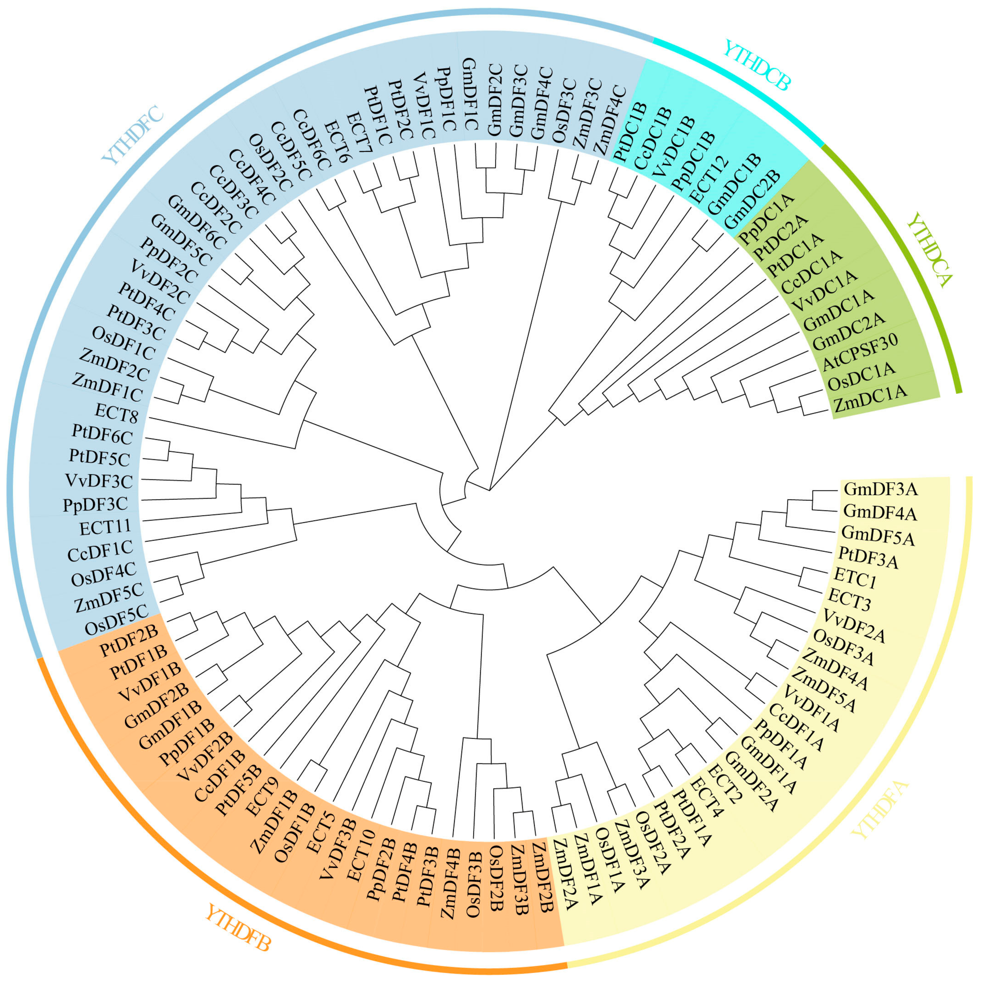
2.1. Identification and Phylogenetic Analysis of the C. camphora YTH Gene Family

Through HMMER searches, we ultimately identified 10 CcYTH genes from the camphor tree genome. (Figure 1; Table 1 and Table S1). In order to analyze the phylogenetic relationship of the CcYTH proteins in C. camphora and obtain the classification of the camphor tree YTH gene family, the neighbor-joining (NJ) method of MEGA 11 software was used to construct the phylogenetic tree of 102 YTH domain proteins from A. thaliana (13 members), Glycine max (G. max) (17 members), Zea mays (Z. mays) (15 members), Oriza sativa (O. sativa) (12 members), Populus trichocarpa (P. trichocarpa) (17 members), Vitis vinefera (V. vinefera) (10 members), Prunus persica (P. persica) (eight members), and C. camphora (10 members).The phylogenetic analysis showed that the YTH gene can be divided into two major groups: YTHDF (CcDF) and YTHDC (CcDC). The YTHDF family is composed of three subgroups: YTHDFA, YTHDFB, and YTHDFC, with one, one, and six members, respectively. The YTHDC family is divided into two subgroups: YTHDCA and YTHDCB, each with one member.

2.2. Synteny Analysis of the CcYTH Genes

To further infer the evolutionary mechanism of the CcYTH family in C. camphora, we constructed a collinearity analysis map of C. camphora, A. thaliana, and P. trichocarpa. The results showed that only one CcYTH gene exhibited collinearity with A. thaliana, while seven genes demonstrated collinearity with P. trichocarpa (Figure 2). This indicates that the CcYTH genes family has a closer evolutionary relationship with P. trichocarpa than with A. thaliana.

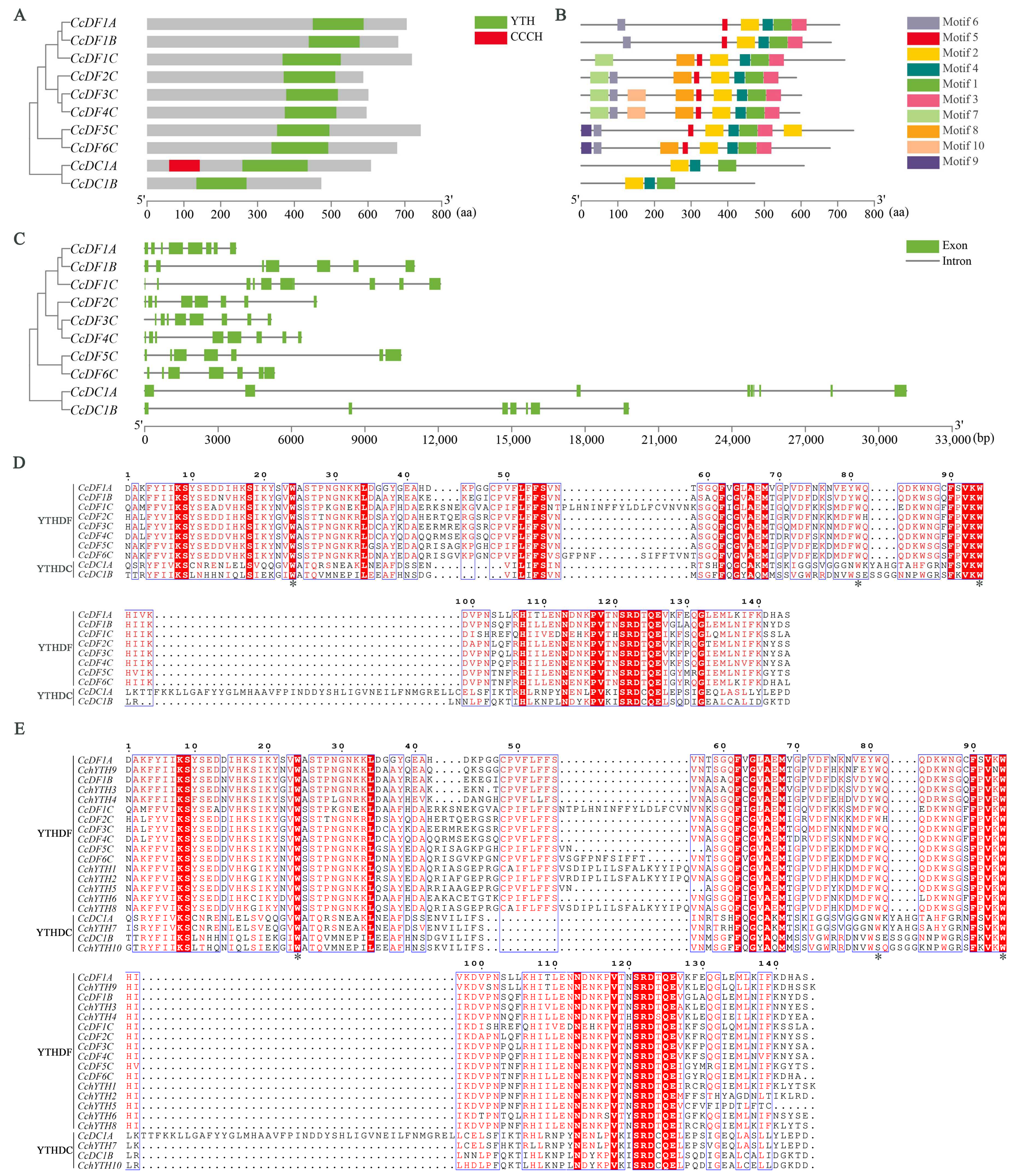
2.3. Protein Features and Gene Structure of the CcYTH Genes

The physicochemical properties of the CcYTH proteins were calculated using the ProtParam software (Table 1). The full length of the CcYTH proteins ranges from 473 amino acids (CcDC1B) to 743 amino acids (CcDF5C), with a molecular weight of 53.5 kDa (CcDC1B) to 81.29 kDa (CcDF5C) and an isoelectric point of 5.21 (CcDF4C) to 8.80 (CcDF1C). The subcellular localization prediction results indicated that, except for CcDF1C, CcDF5C, and CcDF6C, the other seven CcYTH proteins are likely predominantly localized in the nucleus. To further analyze the structural diversity of CcYTH proteins, the gene structure, conserved domain, and motifs of CcYTH proteins were analyzed. From the analysis of conserved domain, the results showed that a typical functional YTH domain exists in each CcYTH proteins and that the b-type YTHDC (CcDC1B) lacks any additional structural domains apart from the YTH domain (Figure 3A). Moreover, the N-terminus of CcDC1A in camphor trees contains a CCCH domain, while that of CcDC1B does not. According to the prediction of the CcYTH proteins motif performed by using the MEME tool, a total of 10 conserved motifs were identified from the YTH proteins of camphor trees, and motifs 1, 2, and 4 were present in all CcYTH proteins (Figure 3B and Table S3). Moreover, similar distribution of conserved motifs has been observed within the same group of CcYTH proteins, indicating that they may have similar functions within same group. Gene structure analysis showed that all members of the CcYTH genes family belong to split genes containing at least seven introns (Figure 3C). Members of the CcYTH genes family exhibit completely different gene structures in terms of the size and arrangement of exons and introns, indicating the independent evolution of these genes. Interestingly, in the gene structure of most YTHDF subfamily members, there are two longer exon segments in the middle position. It is worth noting that in both C. camphora and C. chekiangoleosa, except for the second tryptophan (W) residue replaced by serine (S) in CcDC1B of the YTHDC family and CchYTH10, all other aromatic cages of YTH consist of tryptophan residues (WWW) (Figure 3D,E).

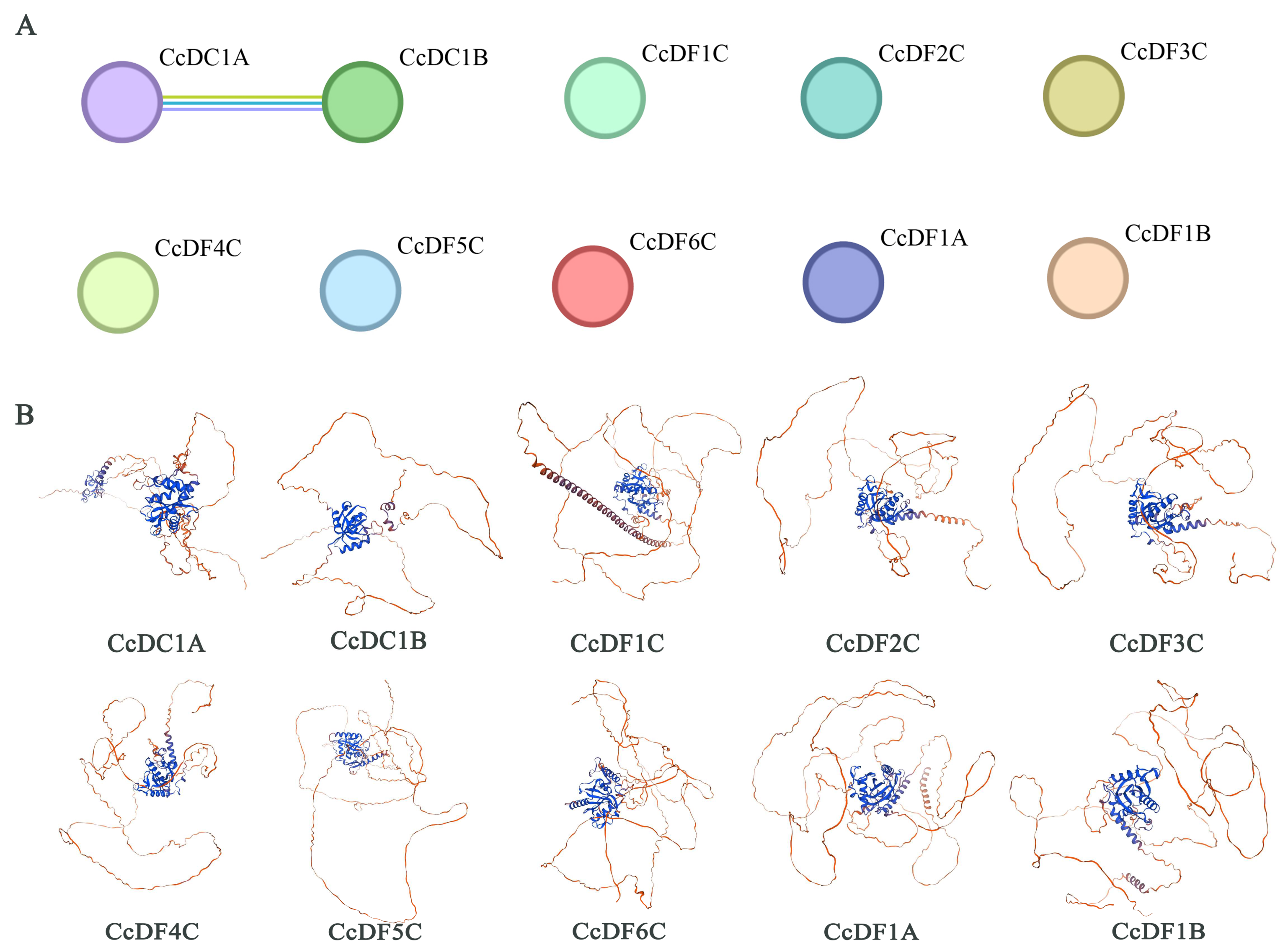
2.4. The Interaction and Tertiary Structure Prediction of CcYTH Proteins

To further understand the interaction relationships among CcYTH protein members, protein–protein interaction prediction was performed on the amino acid sequences of 10 members. The results show that there is only an interaction between CcDC1A and CcDC1B, while no interactions were found among the other CcYTH proteins (Figure 4A). However, the specific interaction network mechanism between CcDC1A and CcDC1B members is not clear and requires further investigation. Homology modeling was conducted on the amino acid sequences of 10 C. camphora YTH proteins. The results indicate that the majority of CcYTH protein sequences share highly similar three-dimensional structures, with higher similarity observed within subfamily members compared to those between subfamilies. Overall, these YTH domains typically adopt a specific mixed α-helix-β-sheet fold, where β-sheets are arranged into a β-barrel structure and surrounded by α-helices (Figure 4B). A well-defined conserved aromatic cage is observed in all YTH domains, endowing them with the ability to distinguish and recognize m6A-modified RNA.

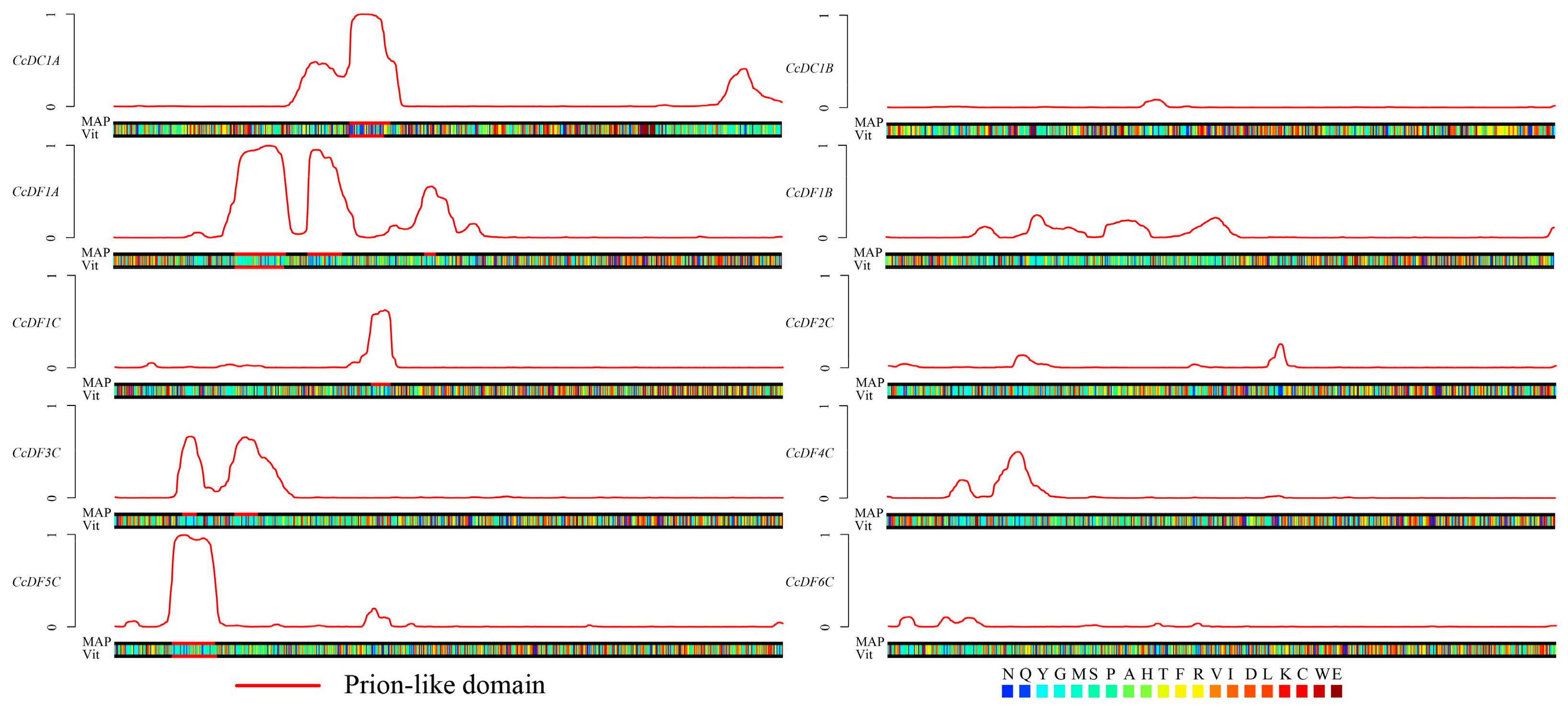
2.5. Prion Subsequences Analysis of the CcYTH Proteins Family

The disorder region of prion-like amino acid composition is a prerequisite for the protein to have phase transition ability [27]. By predicting whether YTH protein family members contain disorder regions, their potential for phase transition can be determined. PLAAC (Prion-Like Amino Acid Composition) is a powerful bioinformatics tool that has been used to identify prion-like domains in a wide range of proteins, including those involved in RNA binding, transcriptional regulation, and signal transduction [28]. Recent research has reported that some members of the YTH gene family in mammals or Arabidopsis can enhance the phase transition ability when binding to m6A sites [26,29]. The results showed that CcDC1A, CcDF1A, CcDF1C, CcDF3C, CcDF4C, and CcDF5C possess at least one the disorder region of prion-like amino acid composition, while no disorder region was found in the rest of CcYTH proteins, indicating these six CcYTH proteins might have the ability to undergo phase transition (Figure 5).

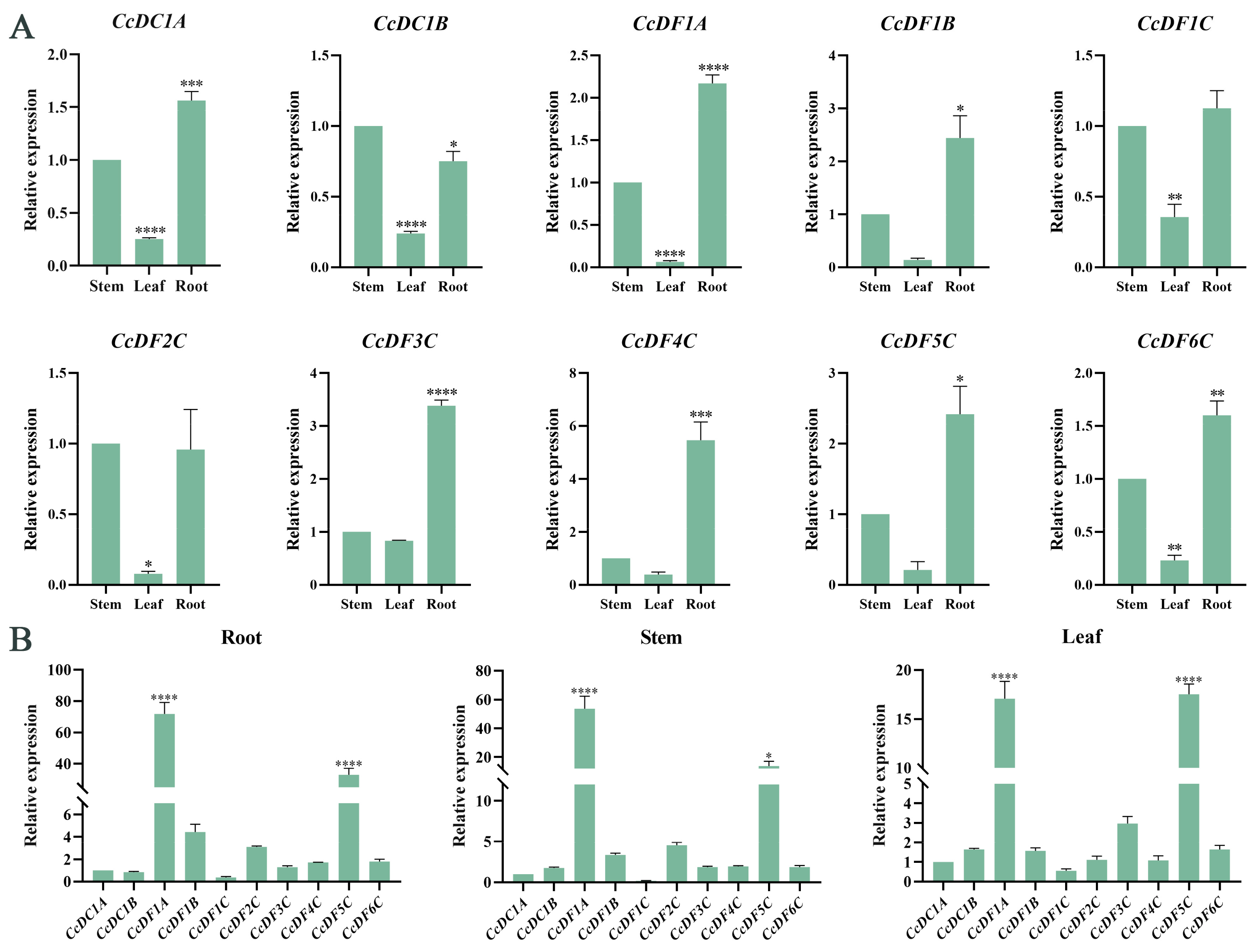
2.6. Tissue Expression Patterns of CcYTH Genes

To investigate the potential physiological roles of the CcYTH genes in the growth and development of C. camphora, the tissue-specific expression patterns of CcYTH genes surveyed across different tissues revealed distinct expression levels of the 10 genes in roots, stems, and leaves (Figure 6A). Interestingly, most genes exhibited the highest expression levels in roots, moderate expression levels in stems, and the lowest expression levels in leaves. Notably, CcDF1A and CcDF5C were expressed at much higher levels than other CcYTH genes in these three tissues (Figure 6B), which is quite different from the findings regarding other plants, such as A. thaliana and C. chekiangoleosa, with only one dominantly high-expressed YTH gene across all tissues. It indicates that C. camphora has a unique YTH regulatory network to achieve downstream biological functions of m6A modification, which is worthy of further investigation into the functions of these two genes.

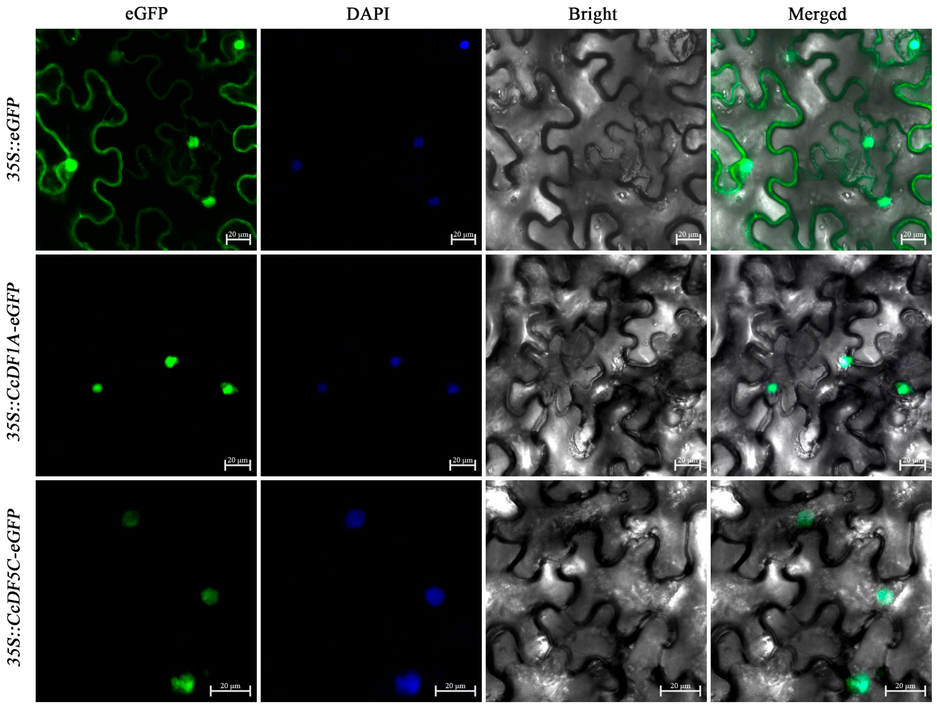
2.7. Subcellular Localization of CcDF1A and CcDF5C Proteins

The subcellular localization prediction results indicated that most of CcYTH proteins are likely predominantly localized in the nucleus. In order to verify the accuracy of these prediction, we used transient expression experiment on tobacco to investigate the subcellular localization of CcYTH proteins. Due to the high expression levels in the roots, stems, and leaves among these CcYTH genes, CcDF1A and CcDF5C were selected for subcellular localization experiments. The results showed that both CcDF1A and CcDF5C exhibit green fluorescence exclusively within the cell nucleus (Figure 7), which confirmed the predicted localization of these two proteins in the nucleus.

2.8. Analysis of Cis-Regulatory Elements in the Promoter Region of the CcYTH Genes

The promoter analysis can provide a deep understanding of gene function. In order to investigate the cis-regulatory elements in the promoter region of the CcYTH genes, 2.0 kb upstream sequences of each CcYTHs translation start site were analyzed. The results showed that there are a large number of stresses, hormone, and development-related cis-regulatory elements in the promoters of CcYTHs (Figure 8 and Table S4). The hormone-responsive elements include methyl jasmonate (MeJA)-responsiveness elements (CcDC1A, CcDC1B, CcDF1A, CcDF1C, CcDF4C, CcDF5C, and CcDF6C), abscisic acid responsiveness elements (CcDC1A, CcDF1A, CcDF1C, CcDF2C, CcDF3C, CcDF4C, and CcDF6C), auxin-responsive elements (CcDC1B, CcDF1A, CcDF1C, CcDF2C, and CcDF3C), salicylic acid responsiveness elements (CcDC1B, CcDF1A, CcDF2C, CcDF3C, CcDF4C, and CcDF5C), and gibberellin-responsiveness elements (CcDC1B, CcDF1A, CcDF1B, CcDF1C, CcDF2C, CcDF3C, and CcDF6C), and each gene promoter contains at least one plant hormone-responsive element. In addition to the plant hormone-related elements, several cis-regulatory elements involved in abiotic stress were also identified, including drought-inducibility (CcDC1B, CcDF1B, CcDF1C, CcDF2C, CcDF4C, CcDF5C, and CcDF6C), defense and stress-responsiveness (CcDF2C, CcDF3C, CcDF4C, CcDF5C, and CcDF6C), and low-temperature responsiveness (CcDF1A, CcDF1B, CcDF2C, CcDF3C, CcDF4C, and CcDF6C) elements. Circadian rhythm control elements were found in the promoters of CcDF2C and CcDF3C. Additionally, there are numerous elements related to light response. These elements can be categorized into four main classes: stress response, plant growth and development, light response, and hormone response elements. In summary, the presence of these cis-regulatory elements in the CcYTHs promoter suggests that the CcYTHs family may be involved in regulating plant responses to hormones and abiotic stresses in C. camphora.

2.9. Expression Patterns of CcYTH Genes under Stress Conditions

The promoter regions of 10 CcYTH genes in C. camphora contain a large number of elements related to stress response. To comprehend the response patterns of YTH genes under stress, we conducted drought and salt stress treatments on C. camphora. The results indicated that after PEG simulated drought treatment, CcDC1A, CcDC1B, CcDF1A, CcDF1C, CcDF2C, CcDF3C, and CcDF4C all exhibited significant changes in expression patterns within 24 h. Among them, the relative expression level of CcDF3C increased significantly at 3 h, showing a 5.1-fold change (Figure 9A). With the exception of CcDF4C, the expression of other genes showed an initial increase followed by a decrease within 0 to 24 h after NaCl treatment. In contrast, CcDF4C showed a decrease initially, followed by a highly significant upregulation in expression at 24 h (Figure 9B). Interestingly, under both PEG and NaCl treatments, the CcDF3C gene displayed a similar expression pattern of initially increasing and then decreasing, with its expression level increasing approximately 5-fold within 3 h. This gene exhibited the most significant upregulation among the 10 CcYTH genes.

3. Discussion

m6A modification is the most common post-transcriptional modification of RNA in eukaryotes, and it is extensively involved in biological processes such as gene regulation, post-transcriptional regulation, RNA metabolism, and disease occurrence [5]. In recent years, research on m6A in plants has uncovered that it plays an important role in plant growth, development, and stress response, making it become an important hotspot in plant molecular breeding and functional genomics research [3]. Revealing the molecular regulatory mechanisms of m6A in various organs or developmental processes is a promising direction for future research. The recognition of m6A sites by reader proteins is critical for executing the multifunctional roles of m6A modifications [12]. m6A readers determine the fate of their target RNAs, thereby exerting physiological impacts. Hence, investigating m6A readers could serve as a valuable entry point to explore how m6A modifications exert their influence on specific organs or biological processes [30].
It has been demonstrated that YTH family members function as m6A reader proteins [31]. These family proteins contain highly conserved YTH domains, which enable them to specifically bind to m6A modified RNA and participate in various biological processes. In humans, RNA-binding proteins with YTH domains have been extensively studied and five YTH proteins have been identified—YTHDC1, YTHDC2, YTHDF1, YTHDF2, and YTHDF3—that employ conserved mechanisms to recognize m6A modifications but lead to different fates of RNAs [2]. YTHDC1 mediates pre-mRNA splicing through interaction with splicing factors. YTHDC2 can regulate spermatogenesis in mammals. YTHDF1 interacts with the translation initiation machinery and enhances the translation efficiency of its target RNAs. YTHDF2 accelerates the degradation of m6A-modified transcripts and YTHDF3 can mediate mRNA decay by directly interacting with YTHDF2 [32,33,34,35,36].
Although there is extensive research on YTH proteins in animals, the study of YTH proteins and their functions in plants remains limited. YTH domain-containing RNA-binding proteins were initially identified in A. thaliana and O. sativa, with 13 and 12 members, respectively [23]. The biochemical studies on some YTH proteins found that recombinant AtYTH05 can bind to single-stranded RNA molecules, suggesting that AtYTH05 protein possesses RNA binding activity in vitro. This is similar to what has been previously demonstrated for YT521-B, which binds to single-stranded RNA sequences [13]. However, only the functions of ECT2/3/4 and CPSF30-L have been detailed and reported as m6A readers in regulating trichome morphology, vegetative growth, reproductive development, flowering transition, and abscisic acid (ABA) response in A. thaliana; the rest still remain unknown [19,20,21,22,26,37]. Therefore, the role of plant YTH proteins deserves further investigation. Compared to A. thaliana and O. sativa, a much larger number of YTH genes, 39 in total, have been identified in Triticum aestivum (T. aestivum) [38]. The aromatic cage in TaDFs is composed of tryptophan, tryptophan, and tryptophan (WWW), while in TaDCs, the aromatic cage consists of tryptophan, tryptophan, and tyrosine (WWY). In the genome of woody plant apple, a total of 26 putative YTH genes were identified [39]. Among them, nine YTH genes are involved in the senescence response of apple, and the expression of these genes is upregulated with leaf aging. So far, there are still many plant YTH proteins in the plant kingdom that have not been studied, especially in woody plants.
In the analysis of the camphor tree genome, a total of 10 YTH domain-containing proteins were identified, which have important effects on the growth and adaptation of C. camphora. Compared to the five YTH genes found in mammals, there are 10 YTH genes present in C. camphora, including one DFA subfamily, one DFB subfamily, six DFC subfamilies, one DCA subfamily, and one DCB subfamily, implying a more complex regulatory mechanism or functional redundancy among YTH family members. (Figure 1 and Table 1). For example, in A. thaliana, ECT2/3/4 exhibit redundant functions [22]. Except for the presence of two CcYTH genes on chromosomes 04 and 05, most CcYTH genes are dispersed across different chromosomes (Table 1). Additionally, the two CcYTH genes on chromosomes 04 and 05 are located far apart, positioned at the ends of the chromosomes, indicating that the evolution of the C. camphora CcYTH genes family does not involve tandem duplication or segmental duplication events. The syntenic map of C. camphora, A. thaliana, and P. trichocarpa indicates that apart from CcDF5C, other CcYTH genes do not show collinearity with A. thaliana, whereas as many as seven CcYTH genes exhibit collinearity with P. trichocarpa (Figure 2). This suggests a closer evolutionary relationship between C. camphora and P. trichocarpa and similar functions of YTH proteins in woody plants compared to herbaceous plants, which indicates that it is necessary to investigate the m6A readers in woody plants to reveal the distinctive roles of m6A in the plant kingdom. The results of the structural diversity analysis of CcYTH proteins indicate the presence of a typical functional YTH domain in each CcYTH proteins. Similar conserved motifs are found in the same group of CcYTH proteins, while they exhibit completely different gene structures in terms of the size and arrangement of exons and introns, suggesting that these genes have evolved independently, and similar functions might be discovered in same group (Figure 3). The predicted interaction relationship among CcYTH proteins members indicates that only CcDC1A and CcDC1B interact with each other. The results of the tertiary structural homology modeling show that the YTH domains of CcYTH proteins typically adopt a specific mixed α-helix-β-sheet structure (Figure 4). The m6A modification can enhance the phase separation potential of mRNA [29]. Prion subsequences analysis of the CcYTH proteins family suggests that CcDC1A, CcDF1A, CcDF1C, CcDF3C, CcDF4C, and CcDF5C may undergo phase transition in its recognition of m6A to regulate plant biological processes (Figure 5).
Different plant organs have evolved different functions in plants. For example, root is the important organ for plants to absorb water and nutrients. Many genes have tissue and organ specific expression patterns in plants which can effectively regulate the growth and development as well as the response to stress of plants in time. The results of tissue-specific expression patterns of CcYTH genes in different tissues indicate that seven CcYTH genes exhibit significantly higher expression levels in roots than in other tissues (Figure 6), suggesting that CcYTH genes might have distinct potential functions in different tissue and play a significant role in the growth and development of roots in C. camphora. From the expression levels of CcYTH genes across all the tissues, it is obvious that the expression levels of CcDF1A and CcDF5C are higher than the others, indicating that they might hold a main position in the recognition of m6A among all the YTH domain containing m6A readers. This discovery that both these two YTH proteins including YTHDF protein and YTHDC protein are abundant in C. camphora is quite different from the research reported regarding Arabidopsis and other plants that possess only one YTH protein, usually YTHDF protein with highest expression, indicating the unique m6A regulatory network in C. camphora. Subcellular localization of protein can provide the possible transport mechanisms and their roles in cellular activities and signal transduction. The two genes with the highest expression levels in C. camphora, CcDF1A and CcDF5C, are both localized in the nucleus (Figure 7), which suggests that the m6A recognition process may primarily occur in the nucleus along with phase–phase separation to regulate the downstream physiological and biological progress in C. camphora. Hence, it is deserved to further uncover the regulatory mechanism of these m6A readers in C. camphora. The cis-regulatory element analysis of C. camphora promoters showed that the promoter regions of the 10 CcYTH genes contain numerous cis-regulatory elements associated with stress responses (Figure 8), which is similar to the findings in various plants such as Arabidopsis, rice, poplar, and so on [23,30,40,41,42]. The expression levels of genes related to development and stress response are generally high or significantly increased during the developmental stage or under stress treatments. With regard to the enrichments of cis-regulatory elements related to stress and the encountering threaten from drought or salinity to C. camphora, we took PEG and NaCl treatments into account to investigate the changes of these CcYTH genes after treatment to unveil the relationship between stress response and m6A. Under treatment with PEG and NaCl, the expression levels of the CcYTH genes exhibited different trends of variation, suggesting that each member of CcYTH genes family might have a specific function in response to stress (Figure 9).
While identifying the YTH genes family in C. camphora, we also identified the YTH genes family in Camellia chekiangoleosa (C. chekiangoleosa), one of the main cultivated tree species with a short maturation period, easy harvest, high oil content in seeds, and superior quality of tea oil [43]. According to the results, 10 randomly distributed YTH genes belonging to the five YTHDFA, YTHDFB, YTHDFC, YTHDCA, and YTHDCB subfamilies were identified in both C. camphora and C. chekiangoleosa. The collinearity analysis results show that compared to A. thaliana, both plants have a closer evolutionary relationship with P. trichocarpa. Conservative domain analysis results showed that each YTH protein contains a typical functional YTH domain. In both C. camphora and C. chekiangoleosa, apart from the second tryptophan (W) residue replaced by serine (S) in CcDC1B of the YTHDC family and CchYTH10, all other aromatic cages of YTH comprise tryptophan residues (WWW). Prion subsequences prediction results showed that there may be seven and nine YTH genes that might have the ability to undergo phase transition in C. camphora and C. chekiangoleosa, respectively. Analysis of tissue-specific expression results showed that the expression levels of genes in the YTHDFA subfamily (CcDF1A and CchYTH9) in C. camphora and C. chekiangoleosa were significantly higher in all tissues compared to other genes. Interestingly, unlike oil tea, there is also a gene, CcDF5C, whose expression level is significantly higher than other genes except for CcDF1A in C. camphora. Analysis of cis-regulatory elements in the promoter regions showed that both C. camphora and C. chekiangoleosa contain a large number of elements related to hormone, light, stress, or plant growth and development response in their promoter regions. When facing stress, most YTH genes in both C. camphora and C. chekiangoleosa showed significantly upregulated or downregulated expression levels. This indicates that YTH genes play a positive regulatory role in plants under stress conditions.
All these findings demonstrate the important and special roles of m6A in the developmental and stress responsive processes of C. camphora as well as the predicted distinctive and unique m6A-YTH regulatory mechanism in C. camphora.

4. Materials and Methods

4.1. Plant Materials and Treatments

Two-year-old camphor seedlings were provided by the Jiangxi Provincial Engineering Research Center for Seed-breeding and Utilization of Camphor Trees of Nanchang Institute of Technology. They were cultivated in nutrient soil under conditions of 25 °C and a 16 h light/8 h dark cycle at the State Key Laboratory of Tree Genetics and Breeding, Nanjing Forestry University (N 32°04′43.05″, E 118°49′1.70″). Fresh root, stem, and leaf samples were collected for tissue-specific expression analysis. For stress experiments, 100 mM NaCl solution and 20% polyethylene glycol (PEG6000) solution were used to water the soil. Leaf samples were collected from each treated seedling at 0 h, 3 h, 6 h, 12 h, and 24 h after the stress was applied, along with untreated leaf samples at 0 h as a control. The collected samples were immediately frozen in liquid nitrogen and stored at −80 °C for subsequent RNA extraction. All treatments were carried out in three biological replicates.

4.2. Identification of the YTH Genes in C. camphora

To identify genes containing the YTH domain in C. camphora, the gene annotation and genome files of C. camphora were downloaded from the National Genomics Data Center (https://ngdc.cncb.ac.cn/, accessed on 27 March 2023) [44]. Subsequently, the Hidden Markov Model (HMM) from the HMMER 3.0 program was employed to search for the YTH521-B domain (PF04146) within the C. camphora genome database, with a cutoff e-value of 1 × 10⁻⁵ [45]. The amino acid properties, molecular weight (MW), aliphatic index, grand average of hydropathicity (GRAVY) and isoelectric point (pI) were determined using the ProtParam tool (http://web.expasy.org/protparam/, accessed on 27 March 2023). The subcellular localizations were predicted by using the Cell-PLoc online tool (http://www.csbio.sjtu.edu.cn/bioinf/Cell-PLoc/, accessed on 28 March 2023).

4.3. Phylogenetic Analysis, Chromasomal Location and Synteny Analysis

To investigate the phylogenetic relationships among YTH proteins, the YTH protein sequences of A. thaliana, G. max, Z. mays, O. sativa, P. trichocarpa, V. vinefera, P. persica, and C. camphora were compared. The phylogenetic tree was generated using MEGA 11 software (https://megasoftware.net/, accessed on 29 May 2023) with neighbor-joining (NJ) method for adjacency, Jones–Taylor–Thornton (JTT) model, pairwise deletion and bootstrap (1000 repetitions). The protein sequences and their gene names (ID) are listed in Supplementary Materials Table S1. The chromosomal location information of the CcYTH genes was extracted from the camphor tree GFF file. Then, TBtools (https://github.com/CJ-Chen/TBtools/releases, accessed on 30 May 2023) were utilized for generating the chromosome position map of the CcYTH genes. For synteny analysis, a homology analysis map was constructed by using TBtools.

4.4. Gene Structure, Conserved Motifs and Consevered Domain Analyses

Gene structure analysis was performed to identify exons and introns. The corresponding GFF data were extracted from the GFF file in C. camphora Genome Database. Then, the gene structure was analyzed and visualized by TBtools. The conserved motifs of CcYTH proteins were analyzed using MEME (https://meme-suite.org/meme/, accessed on 31 May 2023). The maximum number of protein motifs was set to 10, the length of the motifs ranged from 15 to 50, and the output MEME file was further modified with TBtools. The domains of CcYTH proteins were analyzed using Conserved Domains Database (CDD) (https://www.ncbi.nlm.nih.gov/Structure/cdd/wrpsb.cgi, accessed on 31 May 2023).

4.5. Identification of Protein-Protein Interactions, Tertiary Structures, and Prion-like Subsequences of the CcYTH Proteins Family

We used STRING (https://string-db.org/, accessed on 17 May 2024) for predicting the interactions among the C. camphora YTH protein members, with Cinnamomum micranthum, a member of the same Lauraceae family, selected as a reference. The protein’s three-dimensional structure models were established using the SWISS-MODEL (https://swissmodel.expasy.org/, accessed on 17 May 2024). The PLAAC online website (http://plaac.wi.mit.edu/, accessed on 1 June 2023) uses Hidden Markov Model (HMM) algorithm to search CcYTH proteins sequences to identify possible prion sub-sequences [46]. The minimum length of the hidden Markov model for the prion-like domain is set to 60, with a background frequency of 100%.

4.6. Cis-Regulatory Elements Analysis of the CcYTH Genes Promoter

The 2000 bp genomic sequence upstream of transcription start site of each gene was chosen as its promoter sequence. The cis-regulatory elements in the region of promoter were identified using PlantCARE (https://bioinformatics.psb.ugent.be/webtools/plantcare/html/, accessed on 3 June 2023), and then visualized using TBtools.

4.7. Subcellular Localization of CcDF1A and CcDF5C Proteins

The primers CcDF1A-F, CcDF1A-R, and CcDF5C-F, CcDF5C-R were used to amplify the coding sequences of CcDF1A and CcDF5C, respectively (Table S5). The coding region of CcDF1A and CcDF5C was cloned into the pCAMBIA1305 vector between XbaI and SalI restriction sites with C-terminal eGFP, driven by the CaMV35S promoter, respectively. The vectors carrying p35S:: CcDF1A-eGFP and p35S:: CcDF5C-eGFP were introduced into Agrobacterium cells EHA105, which co-cultured with the p19 (RNA silencing suppressor) according to previous report [47]. Agrobacterium strain was infiltrated with a syringe into the leaves of Nicotiana benthamiana for transient expression. Two days later, the GFP signals in the infiltrated areas were captured using a confocal microscope (LSM710, Zeiss, Jena, Germany) at an excitation wavelength of 488 nm, while the DAPI signals were imaged at an excitation wavelength of 405 nm.

4.8. Total RNA Extraction, and Expression of the CcYTH Genes Analyzed by qRT-PCR

The total RNA samples were extracted from the tissues of two-year-old C. camphora according to the manufacturer’s protocol by using the Polysaccharides and Polyphenolics-rich Plant Total RNA Isolation Kit (Vazme Biotechnology, Nanjing, China). The concentration and purity of total RNA were measured using a Nano-Drop 2000 (Thermo Fisher Scientific, Waltham, MA, USA), and its integrity was confirmed through 1.2% agarose gel electrophoresis. The cDNA was synthesized from 1 µg of total RNA using the 1st Strand cDNA Synthesis Kit (Yeasen Biotechnology, Shanghai, China).
The qPCR reactions were carried out via a StepOne Plus Real-Time PCR System (Applied Biosystems, Foster City, CA, USA). The reaction mixture was as follows: 1 µL of cDNA after 20-fold dilution, 0.4 µL of each forward and reverse primer (10 µM), 10 µL of SYBR Green Master Mix (Yeasen Biotechnology, Shanghai, China), and 6.4 µL ddH2O, to reach a final volume of 20 µL. Primer sequences can be found in the Supplementary Materials (Table S6). The amplification was set as pre-denaturation at 95 °C for 2 min, 40 cycles including denaturation at 95 °C for 10 s, and extension at 60 °C for 30 s. The melting procedure followed the instrument’s default settings. The relative expression levels of genes were determined using the 2−∆∆CT method.

4.9. Statistical Analysis

All experimental data were obtained from three replicates, and data are presented as mean ± standard error (SE). Significance differences in the data were evaluated using one-way analysis of variance (ANOVA). * p < 0.05, ** p < 0.01, *** p < 0.001, **** p < 0.0001. p < 0.05 (*) was considered statistically significant.

5. Conclusions

In this study, we have identified 10 CcYTH genes in camphor trees and conducted a comprehensive and systematic investigation of these m6A-binding proteins in the C. camphora genome, including phylogenetic analysis, chromosome localization, gene structure, and conserved motif and promoter analysis, as well as expression profiling. Our results demonstrate that the m6A-binding proteins in camphor trees exhibit a high degree of evolutionary conservation, especially within the woody plants. The identified genes exhibit tissue-specific expression patterns in leaves, stems, and roots. Additionally, the expression of CcYTH genes also undergoes changes in response to various abiotic stresses. Our findings establish a foundation for future functional analysis of these genes, providing a new perspective for future molecular breeding of C. camphora by applying epitransciptomic engineering.

Supplementary Materials

The following supporting information can be downloaded at: https://www.mdpi.com/article/10.3390/ijms25115960/s1.

Author Contributions

Conceptualization, Q.Y.; methodology, J.Z.; software, J.Z., S.Y. and X.C.; validation, J.Z.; formal analysis, J.Z., Y.Z., W.Y. and X.R.; data curation, J.Z.; writing—original draft preparation, J.Z.; writing—review and editing, J.Z., K.J. and Q.Y.; All authors have read and agreed to the published version of the manuscript.

Funding

This research was funded by the Natural Science Foundation of China (grant numbers 32171809 to Q.Y.), Beijing National Laboratory for Molecular Sciences (grant numbers BNLMS202202 to Q.Y.), the Natural Science Foundation of the Jiangsu Higher Education Institutions of China (grant numbers 22KJB220002 to Q.Y.), Postgraduate Research & Practice Innovation Program of Jiangsu Province (grant numbers SJCX23_0346 to Q.Y.) and the Priority Academic Program Development of Jiangsu Higher Education Institutions (PAPD).

Institutional Review Board Statement

Not applicable.

Informed Consent Statement

Not applicable.

Data Availability Statement

Data are contained within the article.

Acknowledgments

The authors extend their sincere gratitude to Beihong Zhang of the Jiangxi Provincial Engineering Research Center for Seed-breeding and Utilization of Camphor Trees at Nanchang Institute of Technology for generously providing the camphor tree seedlings.

Conflicts of Interest

The authors declare no conflicts of interest.

References

  1. Cappannini, A.; Ray, A.; Purta, E.; Mukherjee, S.; Boccaletto, P.; Moafinejad, S.N.; Lechner, A.; Barchet, C.; Klaholz, B.P.; Stefaniak, F.; et al. MODOMICS: A database of RNA modifications and related information. 2023 update. Nucleic Acids Res. 2024, 52, D239–D244. [Google Scholar] [CrossRef]
  2. Yang, Y.; Hsu, P.J.; Chen, Y.S.; Yang, Y.G. Dynamic transcriptomic m6A decoration: Writers, erasers, readers and functions in RNA metabolism. Cell Res. 2018, 28, 616–624. [Google Scholar] [CrossRef]
  3. Ambrosone, A.; Costa, A.; Leone, A.; Grillo, S. Beyond transcription: RNA-binding proteins as emerging regulators of plant response to environmental constraints. Plant Sci. 2012, 182, 12–18. [Google Scholar] [CrossRef]
  4. Harvey, R.; Dezi, V.; Pizzinga, M.; Willis, A.E. Post-transcriptional control of gene expression following stress: The role of RNA-binding proteins. Biochem. Soc. Trans. 2017, 45, 1007–1014. [Google Scholar] [CrossRef] [PubMed]
  5. Mittal, N.; Scherrer, T.; Gerber, A.P.; Janga, S.C. Interplay between Posttranscriptional and Posttranslational Interactions of RNA-Binding Proteins. J. Mol. Biol. 2011, 409, 466–479. [Google Scholar] [CrossRef] [PubMed]
  6. Zhong, S.L.; Li, H.Y.; Bodi, Z.; Button, J.; Vespa, L.; Herzog, M.; Fray, R.G. MTA is an Arabidopsis messenger RNA adenosine methylase and interacts with a homolog of a sex-specific splicing factor. Plant Cell 2008, 20, 1278–1288. [Google Scholar] [CrossRef]
  7. Shen, L.S.; Liang, Z.; Yu, H. N6-Methyladenosine RNA Modification Regulates Shoot Stem Cell Fate in Arabidopsis. Mech. Dev. 2017, 145, S171. [Google Scholar] [CrossRef]
  8. Ruzicka, K.; Zhang, M.; Campilho, A.; Bodi, Z.; Kashif, M.; Saleh, M.; Eeckhout, D.; El-Showk, S.; Li, H.Y.; Zhong, S.L.; et al. Identification of factors required for m6A mRNA methylation in Arabidopsis reveals a role for the conserved E3 ubiquitin ligase HAKAI. New Phytol. 2017, 215, 157–172. [Google Scholar] [CrossRef] [PubMed]
  9. Zhang, M.; Bodi, Z.; Mackinnon, K.; Zhong, S.L.; Archer, N.; Mongan, N.P.; Simpson, G.G.; Fray, R.G. Two zinc finger proteins with functions in m6A writing interact with HAKAI. Nat. Commun. 2022, 13, 15. [Google Scholar] [CrossRef]
  10. Martinez-Perez, M.; Aparicio, F.; Lopez-Gresa, M.P.; Belles, J.M.; Sanchez-Navarro, J.A.; Pallas, V. Arabidopsis m6A demethylase activity modulates viral infection of a plant virus and the m(6)A abundance in its genomic RNAs. Proc. Natl. Acad. Sci. USA 2017, 114, 10755–10760. [Google Scholar] [CrossRef]
  11. Duan, H.C.; Wei, L.H.; Zhang, C.; Wang, Y.; Chen, L.; Lu, Z.K.; Chen, P.R.; He, C.; Jia, G.F. ALKBH10B Is an RNA N6-Methyladenosine Demethylase Affecting Arabidopsis Floral Transition. Plant Cell 2017, 29, 2995–3011. [Google Scholar] [CrossRef] [PubMed]
  12. Patil, D.P.; Pickering, B.F.; Jaffrey, S.R. Reading m6A in the Transcriptome: m6A-Binding Proteins. Trends Cell Biol. 2018, 28, 113–127. [Google Scholar] [CrossRef] [PubMed]
  13. Zhang, Z.Y.; Theler, D.; Kaminska, K.H.; Hiller, M.; de la Grange, P.; Pudimat, R.; Rafalska, I.; Heinrich, B.; Bujnicki, J.M.; Allain, F.H.T.; et al. The YTH Domain Is a Novel RNA Binding Domain. J. Biol. Chem. 2010, 285, 14701–14710. [Google Scholar] [CrossRef] [PubMed]
  14. Imai, Y.; Matsuo, N.; Ogawa, S.; Tohyama, M.; Takagi, T. Cloning of a gene, YT521, for a novel RNA splicing-related protein induced by hypoxia/reoxygenation. Brain Res. Mol. Brain Res. 1998, 53, 33–40. [Google Scholar] [CrossRef] [PubMed]
  15. Stoilov, P.; Rafalska, I.; Stamm, S. YTH: A new domain in nuclear proteins. Trends Biochem. Sci. 2002, 27, 495–497. [Google Scholar] [CrossRef]
  16. Xu, C.; Wang, X.; Liu, K.; Roundtree, I.A.; Tempel, W.; Li, Y.J.; Lu, Z.K.; He, C.; Min, J.R. Structural basis for selective binding of m6A RNA by the YTHDC1 YTH domain. Nat. Chem. Biol. 2014, 10, 927–929. [Google Scholar] [CrossRef]
  17. Liao, S.H.; Sun, H.B.; Xu, C. YTH Domain: A Family of N6-methyladenosine (m6A) Readers. Genom. Proteom. Bioinform. 2018, 16, 99–107. [Google Scholar] [CrossRef]
  18. Achsel, T.; Bagni, C. Cooperativity in RNA-protein interactions: The complex is more than the sum of its partners. Curr. Opin. Neurobiol. 2016, 39, 146–151. [Google Scholar] [CrossRef]
  19. Hou, Y.F.; Sun, J.; Wu, B.X.; Gao, Y.Y.; Nie, H.B.; Nie, Z.T.; Quan, S.X.; Wang, Y.; Cao, X.F.; Li, S.S. CPSF30-L-mediated recognition of mRNA m6A modification controls alternative polyadenylation of nitrate signaling-related gene transcripts in Arabidopsis. Mol. Plant. 2021, 14, 688–699. [Google Scholar] [CrossRef]
  20. Wei, L.H.; Song, P.Z.; Wang, Y.; Lu, Z.K.; Tang, Q.; Yu, Q.; Xiao, Y.; Zhang, X.; Duan, H.C.; Jia, G.F. The m6A Reader ECT2 Controls Trichome Morphology by Affecting mRNA Stability in Arabidopsis. Plant Cell 2018, 30, 968–985. [Google Scholar] [CrossRef]
  21. Arribas-Hernandez, L.; Bressendorff, S.; Hansen, M.H.; Poulsen, C.; Erdmann, S.; Brodersen, P. An m6A-YTH Module Controls Developmental Timing and Morphogenesis in Arabidopsis. Plant Cell 2018, 30, 952–967. [Google Scholar] [CrossRef] [PubMed]
  22. Arribas-Hernandez, L.; Simonini, S.; Hansen, M.H.; Botterweg Paredes, E.; Bressendorff, S.; Dong, Y.; Ostergaard, L.; Brodersen, P. Recurrent requirement for the m6A-ECT2/ECT3/ECT4 axis in the control of cell proliferation during plant organogenesis. Development 2020, 147, 189134. [Google Scholar] [CrossRef] [PubMed]
  23. Li, D.Y.; Zhang, H.J.; Hong, Y.B.; Huang, L.; Li, X.H.; Zhang, Y.F.; Ouyang, Z.G.; Song, F.M. Genome-Wide Identification, Biochemical Characterization, and Expression Analyses of the YTH Domain-Containing RNA-Binding Protein Family in Arabidopsis and Rice. Plant Mol. Biol. Rep. 2014, 32, 1169–1186. [Google Scholar] [CrossRef]
  24. Arribas-Hernandez, L.; Rennie, S.; Koster, T.; Porcelli, C.; Lewinski, M.; Staiger, D.; Andersson, R.; Brodersen, P. Principles of mRNA targeting via the Arabidopsis m6A-binding protein ECT2. eLife 2021, 10, 33. [Google Scholar] [CrossRef] [PubMed]
  25. Arribas-Hernandez, L.; Rennie, S.; Schon, M.; Porcelli, C.; Enugutti, B.; Andersson, R.; Nodine, M.D.; Brodersen, P. The YTHDF proteins ECT2 and ECT3 bind largely overlapping target sets and influence target mRNA abundance, not alternative polyadenylation. eLife 2021, 10, 27. [Google Scholar] [CrossRef] [PubMed]
  26. Song, P.Z.; Yang, J.B.; Wang, C.L.; Lu, Q.; Shi, L.Q.; Tayier, S.; Jia, G.F. Arabidopsis N6-methyladenosine reader CPSF30-L recognizes FUE signals to control polyadenylation site choice in liquid-like nuclear bodies. Mol. Plant. 2021, 14, 571–587. [Google Scholar] [CrossRef] [PubMed]
  27. Boeynaems, S.; Alberti, S.; Fawzi, N.L.; Mittag, T.; Polymenidou, M.; Rousseau, F.; Schymkowitz, J.; Shorter, J.; Wolozin, B.; Van den Bosch, L.; et al. Protein Phase Separation: A New Phase in Cell Biology. Trends Cell Biol. 2018, 28, 420–435. [Google Scholar] [CrossRef]
  28. Lancaster, A.K.; Nutter-Upham, A.; Lindquist, S.; King, O.D. PLAAC: A web and command-line application to identify proteins with prion-like amino acid composition. Bioinformatics 2014, 30, 2501–2502. [Google Scholar] [CrossRef] [PubMed]
  29. Ries, R.J.; Zaccara, S.; Klein, P.; Olarerin-George, A.; Namkoong, S.; Pickering, B.F.; Patil, D.P.; Kwak, H.; Lee, J.H.; Jaffrey, S.R. m6A enhances the phase separation potential of mRNA. Nature 2019, 571, 424–428. [Google Scholar] [CrossRef]
  30. Yin, S.Q.; Ao, Q.J.; Tan, C.Y.; Yang, Y.W. Genome-wide identification and characterization of YTH domain-containing genes, encoding the m6A readers, and their expression in tomato. Plant Cell Rep. 2021, 40, 1229–1245. [Google Scholar] [CrossRef]
  31. Liu, N.; Dai, Q.; Zheng, G.Q.; He, C.; Parisien, M.; Pan, T. N6-methyladenosine-dependent RNA structural switches regulate RNA-protein interactions. Nature 2015, 518, 560–564. [Google Scholar] [CrossRef] [PubMed]
  32. Xiao, W.; Adhikari, S.; Dahal, U.; Chen, Y.S.; Hao, Y.J.; Sun, B.F.; Sun, H.Y.; Li, A.; Ping, X.L.; Lai, W.Y.; et al. Nuclear m6A Reader YTHDC1 Regulates mRNA Splicing. Mol. Cell 2016, 61, 507–519. [Google Scholar] [CrossRef] [PubMed]
  33. Hsu, P.J.; Zhu, Y.F.; Ma, H.H.; Guo, Y.H.; Shi, X.D.; Liu, Y.Y.; Qi, M.J.; Lu, Z.K.; Shi, H.L.; Wang, J.Y.; et al. YTHDC2 is an N6-methyladenosine binding protein that regulates mammalian spermatogenesis. Cell Res. 2017, 27, 1115–1127. [Google Scholar] [CrossRef] [PubMed]
  34. Wang, X.; Zhao, B.S.; Roundtree, I.A.; Lu, Z.K.; Han, D.L.; Ma, H.H.; Weng, X.C.; Chen, K.; Shi, H.L.; He, C. N6-methyladenosine Modulates Messenger RNA Translation Efficiency. Cell 2015, 161, 1388–1399. [Google Scholar] [CrossRef] [PubMed]
  35. Du, H.; Zhao, Y.; He, J.Q.; Zhang, Y.; Xi, H.R.; Liu, M.F.; Ma, J.B.; Wu, L.G. YTHDF2 destabilizes m6A-containing RNA through direct recruitment of the CCR4-NOT deadenylase complex. Nat. Commun. 2016, 7, 11. [Google Scholar] [CrossRef] [PubMed]
  36. Shi, H.L.; Wang, X.; Lu, Z.K.; Zhao, B.X.S.; Ma, H.H.; Hsu, P.J.; Liu, C.; He, C. YTHDF3 facilitates translation and decay of N6-methyladenosine-modified RNA. Cell Res. 2017, 27, 315–328. [Google Scholar] [CrossRef] [PubMed]
  37. Scutenaire, J.; Deragon, J.M.; Jean, V.; Benhamed, M.; Raynaud, C.; Favory, J.J.; Merret, R.; Bousquet-Antonelli, C. The YTH Domain Protein ECT2 Is an m6A Reader Required for Normal Trichome Branching in Arabidopsis. Plant Cell 2018, 30, 986–1005. [Google Scholar] [CrossRef] [PubMed]
  38. Sun, J.; Bie, X.M.; Wang, N.; Zhang, X.S.; Gao, X.Q. Genome-wide identification and expression analysis of YTH domain-containing RNA-binding protein family in common wheat. BMC Plant Biol. 2020, 20, 14. [Google Scholar] [CrossRef]
  39. Wang, N.; Yue, Z.Y.; Liang, D.; Ma, F.W. Genome-wide identification of members in the YTH domain-containing RNA-binding protein family in apple and expression analysis of their responsiveness to senescence and abiotic stresses. Gene 2014, 538, 292–305. [Google Scholar] [CrossRef]
  40. Sun, X.C.; Wu, W.L.; Yang, Y.F.; Wilson, I.; Shao, F.J.; Qiu, D.Y. Genome-Wide Identification of m6A Writers, Erasers and Readers in Poplar 84K. Genes 2022, 13, 16. [Google Scholar] [CrossRef]
  41. Zhou, Y.; Hu, L.F.; Jiang, L.W.; Liu, S.Q. Genome-wide identification and expression analysis of YTH domain-containing RNA-binding protein family in cucumber (Cucumis sativus). Genes Genom. 2018, 40, 579–589. [Google Scholar] [CrossRef] [PubMed]
  42. Yao, S.; Zhang, J.J.; Cheng, X.; Wang, D.B.; Yu, W.Y.; Ji, K.S.; Yu, Q. Genome-Wide Identification and Characterization of the YTH Domain-Containing RNA-Binding Protein Family in Liriodendron chinense. Int. J. Mol. Sci. 2023, 24, 17. [Google Scholar] [CrossRef] [PubMed]
  43. Cheng, X.; Yao, S.; Zhang, J.J.; Wang, D.B.; Xu, S.J.; Yu, Q.; Ji, K.S. Genome-Wide Identification and Expression Analysis of YTH Gene Family for Abiotic Stress Regulation in Camellia chekiangoleosa. Int. J. Mol. Sci. 2024, 25, 16. [Google Scholar] [CrossRef] [PubMed]
  44. Sun, W.H.; Xiang, S.; Zhang, Q.G.; Xiao, L.; Zhang, D.Y.; Zhang, P.L.; Chen, D.Q.; Hao, Y.; Liu, D.K.; Ding, L.; et al. The camphor tree genome enhances the understanding of magnoliid evolution. J. Genet. Genom. 2022, 49, 249–253. [Google Scholar] [CrossRef] [PubMed]
  45. Eddy, S.R. Accelerated Profile HMM Searches. PLoS Comput. Biol. 2011, 7, 16. [Google Scholar] [CrossRef] [PubMed]
  46. Lee, Y.; Rio, D.C. Mechanisms and Regulation of Alternative Pre-mRNA Splicing. Annu. Rev. Biochem. 2015, 84, 291–323. [Google Scholar] [CrossRef]
  47. Sparkes, I.A.; Runions, J.; Kearns, A.; Hawes, C. Rapid, transient expression of fluorescent fusion proteins in tobacco plants and generation of stably transformed plants. Nat. Protoc. 2006, 1, 2019–2025. [Google Scholar] [CrossRef]
Figure 1. Phylogenetic tree of CcYTH genes. Different colored backgrounds and surrounding letters represent different groups. The phylogenetic tree resolved YTH genes into five groups. The YTH domain sequences are shown in Table S2.
Ijms 25 05960 g001
Figure 2. Synteny analysis of the CcYTH genes. Synteny analysis of YTH genes between C. camphora, A. thaliana and P. trichocarpa. Gray lines in the background indicate the collinear blocks within the genomes of C. camphora and other plants, and the blue lines indicate the syntenic YTH gene pairs.
Ijms 25 05960 g002
Figure 3. Conserved YTH domains, motifs, and gene structures of CcYTH proteins. (A) YTH domains of CcYTH proteins. (B) Distributions of conserved motifs in CcYTH proteins. Ten putative motifs are indicated in different colored boxes. A detailed description of the 10 motifs is provided in Supplementary Materials. (C) Exon/intron organizations of CcYTH genes. Green boxes represent exons, and black lines represent introns. (D) Sequence comparison of YTH domain of CcYTH proteins. The position of tryptophan is indicated by an asterisk. (E) Sequence comparison of YTH domain of CcYTH and CchYTH proteins. The position of tryptophan is indicated by an asterisk.
Ijms 25 05960 g003
Figure 4. The interaction and three-dimensional structure of CcYTH proteins. (A) The protein–protein interaction of CcYTH proteins. (B) The tertiary structure of CcYTH family proteins.
Ijms 25 05960 g004
Figure 5. Identified probable prion subsequences of CcYTH genes family in C. camphora. The red line is the prediction of the prion structure region. If the red line is in the non-baseline region, it indicates that the prion structure region exists at that location and the phase transition is highly likely.
Ijms 25 05960 g005
Figure 6. Tissue expression specificity analysis of CcYTH genes. (A) Expression patterns of CcYTH genes under different tissues. The relative expression in “Stem” was set as 1. (B) Expression patterns of different CcYTH genes in the same tissue. Using the expression of the CcDC1A gene as a control, the relative expression levels were determined. Each value represents mean  ±  standard error (SE) of three replicates. Asterisks indicate significant differences in transcript abundance compared to the control group (* p < 0.05, ** p < 0.01, *** p < 0.001, **** p < 0.0001).
Ijms 25 05960 g006
Figure 7. Subcellular localization experiments of CcDF1A and CcDF5C proteins. Transient expression of eGFP (control), CcDF1A, and CcDF5C eGFP in tobacco leaves. The scale bar in the images of is 20 μm.
Ijms 25 05960 g007
Figure 8. The cis-regulatory elements in the promoter of the CcYTH genes. The online software PlantCARE was used to analyze the 2 kb sequence upstream of the transcription start site of each CcYTH genes.
Ijms 25 05960 g008
Figure 9. Expression Patterns of the CcYTH genes in Response to Abiotic Stresses. (A) Expression patterns of CcYTH genes under PEG stress. (B) Expression patterns of CcYTH genes under NaCl stress. For NaCl and PEG stress, leaves were sampled at 0 h, 3 h, 6 h, 12 h, and 24 h. Asterisks indicate significant differences in transcript abundance in the treated group compared to the control group (0 h) (* p < 0.05, ** p < 0.01, *** p < 0.001, **** p < 0.0001).
Ijms 25 05960 g009
Table 1. Characteristics of CcYTH proteins family in C. camphora.
Gene NameGene IDLocusCDS (bp)Protein Length (aa)MW
(kDa)
Aliphatic IndexGRAVYpISubcellular Localization
CcDC1AGWHGBGXC018157Chr05182760866.761.64−0.5395.91Nucleus
CcDC1BGWHGBGXC008948Chr02142247353.568.20−0.6178.62Nucleus
CcDF1AGWHGBGXC015419Chr04211870576.960.26−0.6767.95Nucleus
CcDF1BGWHGBGXC020092Chr05204968274.556.06−0.6185.63Nucleus
CcDF1CGWHGBGXC025982Chr08216071980.759.64−0.8118.8Cell membrane or Nucleus
CcDF2CGWHGBGXC023812Chr07176458764.960.29−0.5805.9Nucleus
CcDF3CGWHGBGXC017636Chr04180660166.556.76−0.6765.42Nucleus
CcDF4CGWHGBGXC008047Chr12179159665.861.16−0.6445.21Nucleus
CcDF5CGWHGBGXC005246Chr11223274381.259.84−0.6556.64Nucleus
CcDF6CGWHGBGXC005110Chr10204067974.373.03−0.5138.17Chloroplast or Nucleus
Disclaimer/Publisher’s Note: The statements, opinions and data contained in all publications are solely those of the individual author(s) and contributor(s) and not of MDPI and/or the editor(s). MDPI and/or the editor(s) disclaim responsibility for any injury to people or property resulting from any ideas, methods, instructions or products referred to in the content.

