Article

The Influence of Oxidized Imino-Allantoin in the Presence of OXOG on Double Helix Charge Transfer: A Theoretical Approach

by
Boleslaw T. Karwowski
DNA Damage Laboratory of Food Science Department, Faculty of Pharmacy, Medical University of Lodz, ul. Muszynskiego 1, 90-151 Lodz, Poland
Int. J. Mol. Sci. 2024, 25(11), 5962; https://doi.org/10.3390/ijms25115962
Submission received: 14 April 2024 / Revised: 19 May 2024 / Accepted: 28 May 2024 / Published: 29 May 2024
(This article belongs to the Special Issue Molecular Advances in DNA Repair and Damage Research)

Abstract

:
The genome is continuously exposed to a variety of harmful factors that result in a significant amount of DNA damage. This article examines the influence of a multi-damage site containing oxidized imino-allantoin (OXIa) and 7,8-dihydro-8-oxo-2′-deoxyguanosine (OXOdG) on the spatial geometry, electronic properties, and ds-DNA charge transfer. The ground stage of a d[A1OXIa2A3OXOG4A5]*d[T5C4T3C2T1] structure was obtained at the M06-2X/6-D95**//M06-2X/sto-3G level of theory in the condensed phase, with the energies obtained at the M06-2X/6-31++G** level. The non-equilibrated and equilibrated solvent-solute interactions were also considered. Theoretical studies reveal that the radical cation prefers to settle on the OXOG moiety, irrespective of the presence of OXIa in a ds-oligo. The lowest vertical and adiabatic ionization potential values were found for the OXOG:::C base pair (5.94 and 5.52 [eV], respectively). Conversely, the highest vertical and adiabatic electron affinity was assigned for OXIaC as follows: 3.15 and 3.49 [eV]. The charge transfers were analyzed according to Marcus’ theory. The highest value of charge transfer rate constant for hole and excess electron migration was found for the process towards the OXOGC moiety. Surprisingly, the values obtained for the driving force and activation energy of electro-transfer towards OXIa2C4 located this process in the Marcus inverted region, which is thermodynamically unfavorable. Therefore, the presence of OXIa can slow down the recognition and removal processes of other DNA lesions. However, with regard to anticancer therapy (radio/chemo), the presence of OXIa in the structure of clustered DNA damage can result in improved cancer treatment outcomes.

1. Introduction

Genetic information is encoded in the DNA matrix, which consists of3.2 × 109 nucleotides. The distribution of nucleobases in DNA has been found as follows: guanine (G) 20%, cytidine (C) 20%, adenine (A) 30%, and thymine (T) 30% [1]. According to the isochore theory, the double helix is a mosaic of different levels of GC ranging from 37% to 52% [2]. This fragile material, in which the secret of life is written, is continuously exposed to harmful factors, both exocellular and endocellular [3]. Regardless of their nature—whether physical or chemical—their interaction with the genome can induce DNA damage, which, if left unrepaired, can lead to mutations and subsequently the onset of carcinogenesis. Over the course of 24 h, in each of the 1014 human cells, 105 DNA lesion events take place [4]. Of all nucleobases, guanine exhibits a lower oxidation potential of 1.29 [V] vs. NHE in comparison to adenine (1.42 [V]), thymine (1.7 V), and cytidine (1.6 V) [5]. As a result, dG is the most susceptible to one-electron oxidation. Reactive oxygen/nitrogen species such as the hydroxyl radical (OH) and singlet oxygen (1O2) react preferentially with dG depending on its location and the neighboring nucleotides in ds-DNA [6]. In the vast majority of nucleobase lesions, 7,8-dihydro-8-oxo-2′-deoxyguanosine (OXOdG) has been found to be the most frequent.
It is commonly accepted that OXOdG, in a cell, is present at a level of 8 × 10⁻⁶ bases. Moreover, its ionization potential has been assigned as 0.75 [eV], which makes it much more predisposed than dG towards further oxidation [7]. As presented in Figure 1, dG can transform into OXOG, guanidinohydantoin (Gh), and finally into oxidized imino-alantoin (OXIa) in the two-, four-, and six-electron oxidation cascade, which is triggered by one-electron abstraction or the addition of a hydroxyl radical. The hydantoin lesions are less abundant by one order of magnitude than OXOG [8,9]. During evolution, to avoid unwanted changes to genetic information, cells developed a DNA damage repair response system [10]. The above-presented DNA lesions are removed from the genome by base excision repair (BER), which is initiated by a mono- or bifunctional glycosylase [11]. These enzymes can recognize, in the vast majority of nucleosides, the lesioned moiety and activate other BER proteins for subsequent action. However, failure in the guanosine DNA lesion recognition/repair system causes transversion in the genome GC→AT or GC→CG [12]. Recently, it has been proposed that glycosylases such as MutYh utilize electron transfer through the double helix to detect DNA damage very effectively [13]. This process has been discussed in the context of isolated damage; however, clustered lesions consisting of the final dG oxidation product, i.e., IaOX, have yet to be taken into consideration. Both diastereomers of guanidinohydantoin can easily rearrange to iminoalantoin (at pH > 8.2) and finally to OXIa [14]. Unlike spiroiminodihydantoin (Sp), which primarily forms in single-stranded DNA, Gh predominantly forms in ds-DNA [15,16,17]. The amount of guanidinohydantoin was determined at the limit of detection, i.e., 1–7 per 108 nucleosides, using HPLC-ESI-MS/MS [18]. Further Gh oxidation leads to its oxidation, with subsequent conversion to OXIa [8]. It should be noted that six-electron oxidation is a rare event under typical physiological conditions. However, during anticancer therapy treatment (e.g., ionization radiation), the occurrence of six-electron oxidation can significantly increase, which can promote OXIa formation. Moreover, identification and quantification of the second oxidation products, such as Gh, Sp, and OXIa, pose serious challenges for analytical techniques. This is because of their instability, high hydrophilicity, and the fact that they appear as a mixture of diastereomers [19]. Even in low abundance, Gh and its oxidized derivative can play a significant role in mutagenesis. It has been found that unlike OXOdG, which yields 3% of G→T transversion, Gh results in almost 100%, with G→C preferences [20,21,22]. Barrows et al. have found that imino-alantoine forms predominantly the base pair with dGTP, which leads to G→C transversion [14]. Therefore, even though OXIa appears in the genome in low amounts, its presence can have a significant impact on genetic information stability, particularly in the context of defects in the NEIL family of glycosylases [23,24]. Furthermore, the presence of OXIa in the structure of the double helix can lead to a protein communication process via charge transfer [25]. It has been found that after electron loss, the binding of MutYh to ds-DNA increases 1000 times [13,26,27]. If no reduction takes place, it can hinder other proteins’ movement, such as OGG1. Until now, the 4[FeS] cluster has not been found in the structure of OGG1, which would appear to indicate that this protein scans ds-DNA in classical mode, i.e., base by base. However, the presence of iron and sulfur is not necessary for the glycolytic activity of MutY. This does not rule out the possible presence of a 4[FeS] cluster in the OGG1 structure, though. The above indicates that the cumulation of DNA damage over a short distance renders the damage repair process difficult with a longer repair time and decreased fidelity [28]. On the other hand, it should be mentioned here that during radio/chemo or combined therapy, the number of clustered DNA lesions (CDL) increases, making the DNA repair process much more complicated and laborious. It should be pointed out here that CDLs are generated not only within (targeted) cancer cells but also in healthy ones. In light of this, the recognition and repair processes of CDLs are an important consideration in terms of the effectiveness and safety of anticancer therapy [29,30]. Hence, in this article, the influence of oxidized imino-alantoin on the charge transfer through ds-DNA in the presence of OXOdG has been investigated and is discussed theoretically at the M06-2x/6-31++G** level of theory in the aqueous phase using equilibrated and non-equilibrated solvent-solute interaction.

2. Results

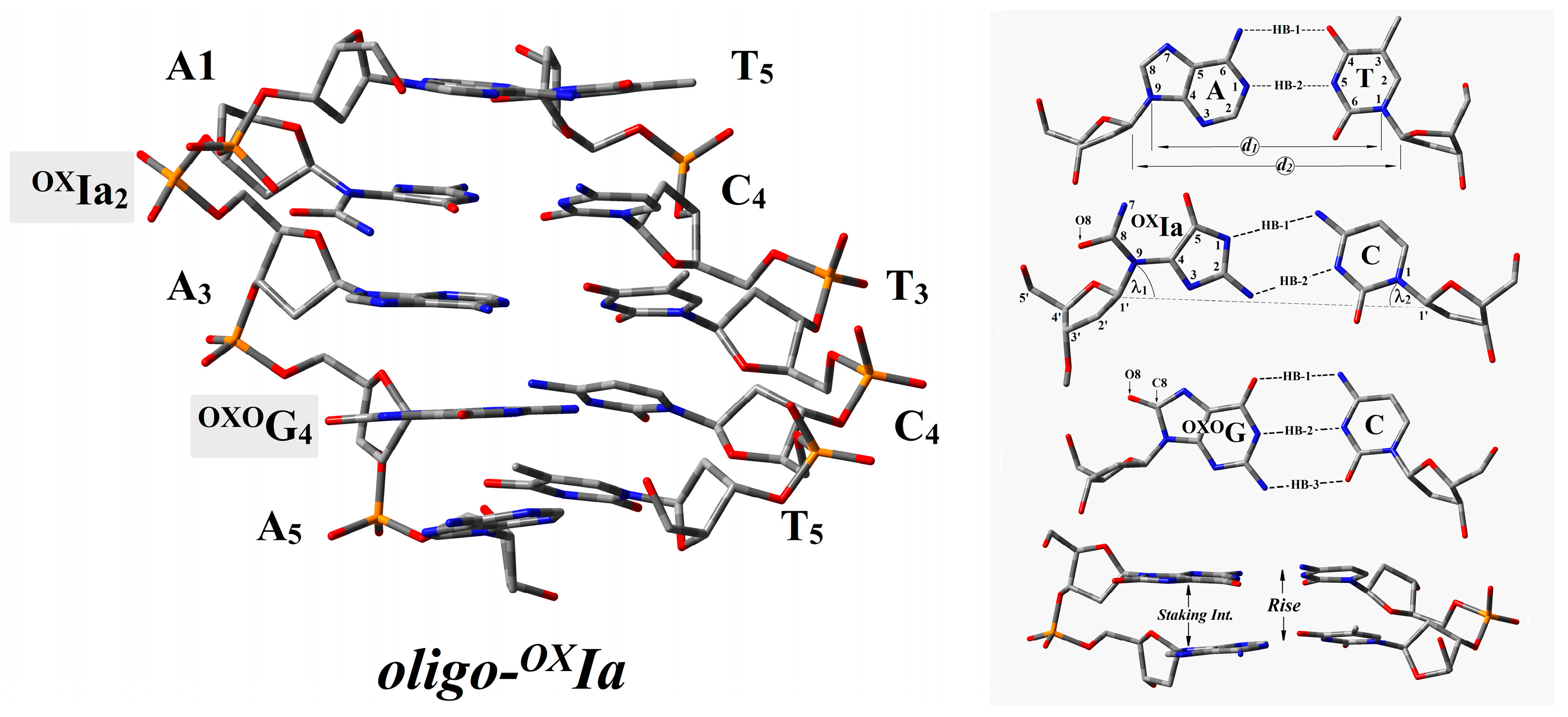
A clustered lesion is defined as two or more DNA lesions per one or two helix turns. This type of damage distribution in the genome poses a serious challenge for cell repair systems. The first step, not only in BER but also in all cases of DDR (DNA damage response), is target (lesion) recognition, which restricts and determines the rate of the whole process. With this in mind, the short double-stranded (ds) oligonucleotide containing OXIa and OXOdG in the purine strand, i.e., d[A1OXIa2A3OXOG4A5]*d[T5C4T3C2T1] (denoted as oligo-OXIa), was taken into consideration as a CDL model. The structures of oligo-OXIa in their neutral, anionic, and cationic forms were optimized at the M06-2x/D95**:M06-2x/sto-3G level of theory (Figure 2). The complexity of the analyzed system necessitated the application of the ONIOM (Our Own N-layered Integrated Molecular Orbital and Molecular Mechanics) strategy, thus reducing computational time costs. Since in living cells, all biochemical reactions, including charge transfer, take place in the aqueous phase, the polarizable continuum model (PCM) was applied. The double helix spatial geometry is stabilized by two factors: hydrogen bonds (HB) and staking interaction. The energy of both non-covalent interactions depends on the mutual position between complementary bases (HB) and the distance between base pairs within the formed dimer (Rise). Figure 2 presents a schematic representation of the discussed parameters. Structural analysis of the obtained geometry reveals that OXIa has negligible influence on the geometry of the double helix. Even though the purine ring was rearranged, converting to oxidized imino-allantoin in the base pair formed with cytidine (OXIa2::C4), the presence of two hydrogen bonds was still observed. Furthermore, the length and energy of the remaining HBs were noted at the same level as those obtained for the corresponding native ds-oligo (Table 1) [31,32]. Additionally, the distances d1 and d2, which describe the spatial arrangement of complementary bases, were observed at the levels of 9 Å and 10.8 Å, respectively. Only the value of angle λ1 was significantly lower than that obtained for other BPs, i.e., 39.2° versus 53° as the results of the imidazole ring opening in the OXIa moiety. The lack of significant spatial geometry distortion forced by OXIa is confirmed by the local base pair step parameter Rise [Å] and stacking interaction energy EST [kcal]. The distances between the two base pairs in the dimer (Rise) formed by OXIa2::C4 and A1::T5 or A3::T3 were as follows: 3.0 [Å] and 2.99 [Å], respectively. The above corresponds to 14.77 and 15.90 kcal of stacking interaction energy in the discussed system. It should be pointed out for reference that the native ds-oligo parameters were as follows: Rise: 3.21/2.96 [Å]and EST: 14.56/13.59 [kcal] for G2::C4 and A1::T5/A3::T3, respectively [33].

2.1. The Influence of OXIa as Part of a CDL on Double Helix Electronic Properties

Because of the (parallel) stacked nucleobase’s similarity to graphene, DNA has been considered a nanowire for the last two decades [34]. On the other hand, enzymes like glycosylases have utilized charge transfer to scan the double helix toward damage identification/location almost from the beginning of evolution [25]. The above indicates that a DNA lesion, especially a CDL, can modify the charge transfer. It has been recently postulated that electrons are able to migrate through ds-DNA at a distance of thousands of base pairs [13]. Therefore, it can be predicted that the appearance of additional electrons or the loss of electrons in the system should influence the double helix structure. Because of this, the presence of a CDL in oligo-OXIa can change its “global” electronic properties [35]. In these studies, computation power constraints only allowed consideration of the ds-pentamer in its adiabatic, cationic, neutral, and anionic forms in the aqueous phase as a native DNA environment.
Table 1. The structural local base pair parameters: Rise and d1(C1′…C1′) and d2(C9…C1) in [Å], λ1 and λ2 in [°] according to the standard DNA reference frame of oligo-OXIa, presented in Figure 2. The HB lengths are given in [Å] (HB-1: Ade(N1), Thy(N3) and Gua(O6), Cyt(N4); HB-2: Ade(N6), Thy(O4) and Gua(N1), Cyt(N3); HB-3: Gua(N2), Cyt(O2), for OXIa2C4: HB-1 (Cyt(N4), OXIa(N1), HB-2 Cyt(N3), OXIa(N2)). The HBs and stacking energies (EHB and EST) are given in [kcal].
Base PairHB-1HB-2HB-3EHBλ1λ2d1d2Base Pair
Dimer
RiseEST
A1T52.802.99 10.8451.552.010.78.83A1T5|OXIa2C43.0014.77
OXIa2C43.002.99 12.4042.555.511.49.37OXIa2C4|A3T32.9915.90
A3T32.842.89 10.7448.750.710.88.95A3T3|OXOG2C42.9716.70
OXOG2C42.862.882.8617.5452.155.510.89.00OXOG2C4|A5T12.9515.19
A5T12.822.99 10.5153.756.110.58.85
Parameters calculated for ideal mode [35]
AT [16]2.963.05 10.8154.554.510.78.58GC||AT [17]2.9614.56
GC [16]3.003.002.8717.2354.254.510.89.01AT||GC [17]3.3113.59
To clarify the above-mentioned hypotheses, a comparative analysis was conducted to compare neutral versus anion and neutral versus cation. The analysis revealed that there was a higher structural fluctuation following the acceptance of an extra electron in the system than following the loss of an electron. As the parameter for the above analysis, the RMSD was chosen in Å2. In both cases, a higher RMSD value was observed in the case of the sugar-phosphate backbone than for base pair (BP) leaders, as shown in Table 2. The above results suggest significant changes in electronic properties between positively and negatively charged molecules. Because all macromolecules existing in the cell were immersed in an aqueous environment, the non-equilibrated and equilibrated models of solvent-solute interaction were applied. The above, following the work of Sevilla et al., suitably described the influence of solvents on the electronic properties of macromolecules [36]. As expected, during the solvent equilibration process and structure relaxation, the calculated ionization potentials (IP) of oligo-OXIa decreased in the following order: VIPNE > VIPEQ > AI (Table 2). Moreover, the IP was found to be lower than that assigned for native ds-oligo in all cases, which indicates that the presence of OXOdG, as part of clustered lesions, plays a significant role. Contrary to the above, the adoption of an electron by oligo-OXOIa leads to anion formation, firstly vertical, then, after geometry relaxation, adiabatic. The results of the performed computation reveal that the appearance of OXIa in ds-oligo significantly increases electron affinity (EA) in comparison to oligo-N in all discussed cases, i.e., VEANE < VEAEQ < AEA.
However, the electron affinity at all points was noted to be almost two times higher than that assigned for unmodified double-stranded oligonucleotides (Table 2). The above indicates that oxidized imino-allantoin determines the spot of electron settling, even if the OXOdG is present in close proximity to the system. A systematic oligo-OXIa structural analysis revealed that there was a different place/location for the proton transfer (PT) process. A PT was observed between N1(OXOG4) and N2(C2) ofOXOdG4:::C2 (a shift of 0.11 [Å])when the ds-oligo lost an electron and formed the cation radical (Figure 3A). The appearance of an excess electron in the system caused radical anion formation and a proton transfer within the oxidized imino-allantoin moiety of OXIa2::C4 base pair between N7 and O5 atoms (a shift of 0.14 [Å]), as shown in Figure 3B.

2.2. Influence of OXIa as Part of a CDL on Charge Distribution and Electronic Properties of Basepairs Present in oligo-OXIa

It has been commonly accepted that within a double helix, the positive charge settles on the purine moiety, while the negative one is localized on pyrimidine nucleosides. The results of previous studies have shown that the presence of DNA damage, e.g., OXOdG, strongly influences the pattern of charge distribution. Much less is known, however, about the role of a CDL’s presence. Hence, the ds-pentamer oligo-OXIa with a local multi-damage site containing six- and two-electron oxidation dG products, i.e., oxidized imino-allantoin and 7,8-dihydro-8-oxo-guanosine, was taken into theoretical investigation. The non-equilibrated and equilibrated modes of aqueous phase oligo-OXIa interaction were used for the charge and spin distribution analysis according to Hirschfeld’s methodology. The formation of a one-electron oxidation spot in the predisposed part of the double helix triggers electron-hole migration through the base pair ladder. This process starts with (A) an interruption of the delicate equilibrium between the solvent and solute, resulting in the ds-DNA adopting a vertical cation non-equilibrated (NE) state (VCNE). Subsequently, (B) an equilibrium in the solvent-solute interaction takes place (EQ), and a VCEQ state is formed. This finally converts to (C) ds-oligo geometry rearmament and the appearance of an adiabatic cation (AC). This process was analyzed step by step (i.e., VCNE→VCEQ→AC) in light of the positive charge and spin distribution changes within oligo-OXIa. As expected, the presence of anOXOG4C2 base pair within the investigated ds-DNA, irrespective of the presence of OXIa, was identified as a radical cation sink with the lowest ionization potential (Figure 4). The above is in good agreement with previous results obtained during investigations into isolated DNA damage [37,38].
A different picture emerged when the appearance of an excess electron was investigated. Starting from the initial step, when the vertical anion in a non-equilibrated (VANE) state was generated through solvent-solute equilibration (VAEQ) culminating in the formation of an adiabatic anion (AA), the charge and spin were mainly located at theOXIa2C4 base pairs of oligo-OXIa. No additional electron was dispersed over other neighboring BPs, i.e., A1T5 and A3T3a, orOXOG4C2 (Figure 4A). This observation correlates well with the results of electron affinity calculations: the highest vertical and adiabatic values were invariably noted for OXIa2C4 as follows: 3.15 and 3.49 [eV], respectively (Figure 4B).

2.3. The Influence of OXIa as Part of a CDL on Charge Migration through oligo-OXIa

The results presented above, discussing spatial geometry, charge/spin distribution, and electronic properties of complete oligo-OXIa and isolated single base pairs, enable us to predict how the discussed CDL affects excess electron and electron-hole transfer through the double helix. These processes can be discussed in the context of (A) single-step tunneling, (B) random-walk multistep, and (C) polaron-like hopping [39]. According to Marcus’ theory of charge transfer (CT), the process charge moves through the stacked base pair ladder (in an oxidized or reductive state) is characterized by the kCT (rate constant) and the following energetic factors: ΔG (driving force), λ reorganization), Ea (activation), and Vda (electron coupling) [40]. In these studies, the values of all the above parameters were obtained at the M06-2x/6-31++G** level of theory in the condensed phase, as described previously. (For further details, please see reference [41].) Marcus theory was followed in order to investigate the influence of two different lesions derived from the source point of origin (dG), i.e., OXIa and OXOdG, in the CDL arrangement present in oligo-OXIa on the electron-hole/excess migration through the double helix.
As shown in Table 3, the highest Ea was observed in the case of the electron-hole transfer between OXIa2C4OXOG4C2, i.e., 0.76 [eV], while for the electron transfer between A1T5OXIa2C4, OXIa2C4→A3T3 and OXIa2C4←A5T1, the highest values were, in [eV], as follows: 2.19, 2.05 and 2.04, respectively. Surprisingly, when OXIa was settled between A1T5, A3T3, andA5T1, the negative values of Ea were noted, which results in kCT being undetectable in the case of radical cation migration. The above indicates that the hole-electron movement between AT pairs through OXIa pairs should be unaffected and make the transfer towardsOXOG4C2 privileged, as shown in Table 3. The migration of additional electrons within the oligo-OXIa structure was affected by the presence of OXIa on account of the electronic properties of OXIa2C4 (Figure 4). The kCT values were found at the level of 10−21 s−1 for A1T5OXIa2C4, OXIa2C4←A3T3, OXIa2C4OXOG4C2. For the above, a significantly high driving force value was identified (ΔG ~ 2 eV), which shifts the charge transfer process to the inverted region. Therefore, CT should be less privileged at these points because of the thermodynamic regime, which is in good agreement with Marcus theory [42].

3. Discussion

The ds-DNA structure contained a highly flexible phosphate-sugar skeleton (PS), and an internal rigid BP ladder. The PS can compensate for the fluctuations in geometry forced by the changes in complementary BP geometries. The above stabilized the energy of hydrogen bonds and stacking interactions [43]. It was found that the double helix structure, similar to graphene, is capable of transferring the excess charge over thousands of nucleosides, as shown by Barton et al. [44]. This observation sheds new light on how glycosylasesds-DNA highly effectively scans for lesions, e.g., MutY or Endo III [45]. Both of the above proteins, like other proteins active in the replication and repair of genetic material, contain [4Fe–4S]2+ as electron donors [46]. The activity of glycosylases is well known in the context of isolated DNA lesions. The situation becomes more complicated when clustered DNA lesions (CDLs) enter the equation. A CDL is defined as two or more lesions per one or two double-helix turns [47]. This type of damage is especially dangerous and challenging for cell repair systems, which, during lesion removal and repair, try to avoid a potentially lethal double-strand break (DSB) [48].
In the genome, during the oxidizing process, dG can be converted from OXOdG to OXIa via several “intermediate” products, as shown in Figure 1. These two lesions are formed in the cell in normoxia conditions, while in hypoxia, the FapydG formation is preferred [49]. Because of the differences in the structure and electronic properties, OXOdG and OXIa probably demonstrate a different influence on charge transfer through ds-oligo. In these studies, oligo-OXIa (d[AOXIaAOXOGA]*d[TCTCT]) was investigated at the M06-2x/6-31++G** level of theory in the aqueous phase. Both the non-equilibrated and equilibrated models of solvent-solute interaction and the Hirshfeld model of spin and charge distribution were considered. A comparative spatial geometry analysis revealed that a clustered damage site consisting of OXIa and OXOG in the oligo-OXIa structure had negligible influence according to the standard DNA reference frame parameters (Table 1). Similar to results obtained previously for a ds-DNA fragment containing two OXOG moieties [35], it was noted that the 3D structure was hardly affected at all, contrary to OXOGA mismatch [50].
It should be pointed out that most of the studies on electronic properties have discussed single lesions in the model of double-stranded trimers. Unfortunately, because of the structural regime (terminated base pairs), this approach is inadequate (it fails) in the case of CDL [51,52]. Given this, the role of d[OXIaAOXOG] in charge transfer through ds-DNA was deemed worth investigating. As commonly accepted, the excess electron or electron hole can migrate through a double helix at a distance greater than 200 [Å] ds-DNA in the form of a radical cation or anion [13].
The loss or adoption of an electron can trigger an electron-hole or electron migration through oligo-OXIa through π-stacked base pairs [53]. The results of the theoretical experiments presented in Table 2 show that during vertical cation/anion radical formation, the sugar-phosphate backbone may hardly be involved. This observation tallies with the data obtained for the corresponding native oligo-N [50]. Additionally, the recent research of Sevilla et al. shows that for this process, the solute-solvent interaction is crucial at the beginning point (non-equilibrated mode) [54]. The highest difference between the complete oligo-OXIa structure and base pair ladder was noted for VEANE, i.e., 2.42 [eV] and 1.82 [eV], respectively.
To shed light on this phenomenon, a Hirshfeld charge and spin calculation was performed. As shown in Figure 4A, the radical cation is mainly located on theOXOG4C2 base pair, regardless of its state—vertical or adiabatic. Moreover, no differences were noted when non-equilibrated and equilibrated solvent-solute interactions were considered. Similar observations were noted for negatively charged oligo-OXIa, i.e., the OXIa2C4 moiety was noted as the spot of a radical anion in all the discussed cases. Additionally, the above supports well the results of the electronic properties of base pairs isolated from oligo-OXIz (Figure 4B). The lowest vertical and adiabatic ionization potential was assigned for OXOG4C2, i.e., 5.94 and 5.52 [eV], respectively, while the highest absolute value of vertical and adiabatic electron affinity was found for OXIa2C4, i.e., 3.15 and 3.49 [eV], respectively. All calculations were performed at the M062x/6-31++G** level of theory in the aqueous phase.
The charge transfer process through ds-DNA, which strictly depends on mutual BP geometry, can be discussed as follows: (A) single-step tunneling, (B) a random-walk multistep, and (C) polaron-like hopping [39,55]. The charge migration over hundreds of angstroms can be discussed in an incoherent manner and depends on the bridge between the donor and acceptor [39]. Conversely, the charge tunneling path can be observed at a distance of only a few base pairs. Even though the above mechanisms are different, the geometry and electronic properties of purine/pyrimidine moieties are crucial. As shown in current and previous results, OXOdG becomes a radical cation sink when present in ds-oligo [56]. Here, it can be assumed that the electron-hole transfer through OXIa as a bridge should be unaffected (Table 3). Similar results were observed for single-lessoned ds-DNA [57,58]. However, in contrast to the above, it is possible to predict that the OXIaC moiety of the discussed ds-oligo will be the endpoint of the extra electron transfer. As shown in Figure 4, higher values of VEA and AEA as well as spin/charge concentration were obtained for OXIaC. However, the obtained values of driving force (ΔG) and activation energy (Ea) of A1T5OXIa2C4, OXIa2C4←A3T3, OXIa2C4OXOG4C2 settled this process in the inverted region (Table 3), which according to Marcus theory, makes the discussed processes unfavorable from a thermodynamic perspective [59]. The following values of kCT were found: 3.56 × 10⁻²², 6.32 × 10⁻²⁰, and 3.73 × 10⁻²¹ [s−1], respectively. Conversely, a charge transfer parameter analysis revealed that charge migration towards OXOGC within the discussed ds-pentamer should not be affected (Table 3). Therefore, it can be predicted that even if the OXIaC base pair becomes the sink for excess electrons (electronic properties), the charge transfer should be slowed down via a single-step tunneling process. At the same point, the electron transfer towards OXOGC should be privileged, the following kCT values were noted: 1.05 × 10¹⁴ and 3.12 × 10¹⁰ [s−1] for A3T3OXOG4C2 and OXOG4C2←A5T1, respectively. This can manifest as a slowing down of other processes involved in DNA lesion recognition and removal, which can increase the probability of mutagenesis and subsequent carcinogenesis or aging. However, with regard to anticancer therapy (radio/chemo), the presence of OXIa in the structure of clustered DNA damage can result in improved cancer treatment.

4. Materials and Methods

All theoretical experiments were performed as described previously [60]. The structure of the discussed oligo-OXIa(d[A1OXIa2A3OXOG4A5]*d[T5C4T3C2T1]) in neutral, positively (cation), and negatively (anion) charged forms was optimized using DFT (density functional theory) methodology in the condensed phase. The following ds-DNA sequence, containing all four nucleosides, has been previously investigated as a model for this set of studies. The above strategy enabled a comparative analysis of different components contained in the CDL to be completed. Moreover, the distribution of the proposed lesions enabled interference and mutual electronic interaction. The investigated ds-pentamer, which contains 10 bases and 10 deoxyriboses, is a complex system that pushes the limits of the computational resources used. Single geometry optimization to the ground state, at the below-mentioned level, took approximately 40 days (24 CPUs and 48 GB of RAM were dedicated to each process).

The oligo-OXIageometry Optimization in Condensed Phase

The chosen level of theory, as described below, has previously been carefully investigated and compared with other DFT functionals such as M-11, wB97-XD, and M06-L, for which the following basis set was utilized: 6-31++G*, 6-31++G** and D95** aug-cc-pVDZ Please refer to the supplementary materials for further details [61]. For all the above, Tomasi’s polarized continuum model for solvation was used. The geometry optimization was conducted using ONIOM (our Own N-layered Integrated Molecular Orbital and Molecular Mechanics) methodology [62]. Applying ONION strategy significantly reduces the time and cost of computation, especially when highly flexible units with a high degree of freedom, like deoxyribose, are present. In this study, the flat aromatic nucleobase moieties were described as a high layer and the flexible sugar-phosphate backbone as a low layer, and they were optimized at the M06-2X/D95** and M06-2X/sto-3G levels of theory in the aqueous phase, respectively [63]. It should be mentioned here that the sugar-phosphate backbone does not play a role in charge transfer and can be described as a low-level layer. The negative charges of all the phosphate groups were neutralized by the addition of a proton [64,65,66]. The obtained complete ds-oligo structures in their ground state were converted into a base pair skeleton and were further used for energy calculations. The necessary additional hydrogen atoms were optimized at the M06-2X/D95** level of theory in the aqueous phase.
All energy calculations were performed in the aqueous phase using the M06-2X functional with an augmented polarized valence double-ζ basis set 6-31++G** [63,67], as justified in previous studies [61].The transition dipole moment of excited states and the single point energy calculation were performed using the time-dependent DFT (TD-DFT) method at the M06-2X/6-31++G** level of theory [68,69]. The solvent effect (aqueous phase) was described by applying Tomasi’s polarized continuum model (PCM) [70]. In the PCM model, however, the dielectric constant was used instead of strict H2O distribution around ds-DNA. Moreover, the PCM model significantly reduces the required calculation time and accurately describes non-equilibrated solute-solvent interaction. Furthermore, the above enabled the avoidance of random water molecule post positioning, which might not match the native system. Due to the nature of charge transfer, two modes of solvent effect were investigated, i.e., non-equilibrium (NE) and equilibrated (EQ), as described previously [54]. For all optimized geometries, a charge and spin distribution analysis was achieved using the Hirshfeld theory [71]. Generalized Mulliken–Hush theory was used for the electron coupling calculation [72]. The electronic properties, i.e., adiabatic ionization potential (AIP), adiabatic electron affinity (AEA), vertical ionization potential (VIP), and vertical electron affinity (VEA), were calculated as described previously [73].
All calculations were performed on the Gaussian G16 (version C.01) suit [74]. The RMSD values were calculated using BioVia Discovery Studio v20.1.0.19295 software [75].The three-dimensional structural analyses of the investigated ds-DNAs, based on a standard reference frame, were obtained by a 3DNA software package using the web-based interface w3DNA (web 3DNA) [76].

5. Conclusions and Further Perspectives

The genome is continuously exposed to harmful factors such as ionization radiation and reactive oxygen/nitrogen species, among others. In the genome, guanine has the lowest ionization potential, which can be oxidized to 7,8-dihydro-8-oxo-2′-deoxyguanosine and subsequently to secondary lesions like OXIa. OXOdG was intensively studied, unlike Gh, Sp, and OXIa, on account of its stability and ease of determination using common HPLC methods. The mutagenic potential of OXOG was established at a level of 3% transversion. Under favorable conditions, with a pH above 8.2, 7,8-dihydro-8-oxo-2′-deoxyguanosine can be oxidized to Gh and subsequently converted to OXIa. This conversion increases the mutagenic potential up to 100%, with a G→C transversion observed.
In this study, the electronic properties and the influence on charge transfer through ds-DNA of the six-electron oxidation product OXIa were taken into consideration. Unlike its precursor, Gh, which exists in two diastereomers, OXIa is a non-chiral compound (Figure 1). With this in mind, the d[A1OXIa2A3OXOG4A5]*d[T5C4T3C2T1] ds-pentamer containing CDLs was investigated and compared with an unmodified analogue (oligo-N).
  • A structural analysis revealed that a clustered damage site consisting of OXIa and OXOG in the oligo-OXIa structure had negligible influence according to the standard DNA reference frame parameters.
  • Theoretical studies revealed that the radical cation prefers to settle on the OXOG moiety, irrespective of the presence of OXIa in a ds-oligo. The lowest vertical (5.94 [eV]) and adiabatic (5.52 [eV]) ionization potential values were found for the OXOG:::C base pair. Conversely, the highest vertical and adiabatic electron affinity was assigned for OXIaC as follows: 3.15 and 3.49 [eV], respectively.
  • The Hirshfeld charge and spin analysis indicated that the radical cation is mainly located on the OXOG4C2 base pair, regardless of its state—vertical or adiabatic. Moreover, no differences were noted when non-equilibrated and equilibrated solvent-solute interactions were considered. Similar observations were noted for negatively charged oligo-OXIa, i.e., the OXIa2C4 moiety was noted as the spot of a radical anion in all the discussed cases.
  • The calculated values of driving force (ΔG) and activation energy (Ea) of A1T5OXIa2C4, OXIa2C4←A3T3, and OXIa2C4OXOG4C2 settled this process in the inverted region, which is unfavorable from a thermodynamic perspective. The following values of kCT were found: 3.56 × 10⁻²², 6.32 × 10⁻²⁰, and 3.73 × 10⁻²¹ [s−1], respectively. At the same point, the electron transfer towards OXOGC should be privileged, and the following kCT values were noted: 1.05 × 10¹⁴ and 3.12 × 10¹⁰ [s−1] for A3T3OXOG4C2 and OXOG4C2←A5T1, respectively.
The above can lead to a slowing down of other processes involved in DNA lesion recognition and removal or genetic material replication, which can increase the probability of mutagenesis and subsequent carcinogenesis or aging. However, with regard to anticancer therapy (radio/chemo), the presence of OXIa in the structure of clustered DNA damage can result in improved cancer treatment.

Further Perspectives

DNA damage such as Gh, Sp, OXOG, and OXIa, when becoming part of clustered DNA damage, can affect the efficiency of the lesion recognition and repair process because it exerts a strong influence on the electron transfer process. Most current studies discuss the CDL in the context of native-type hydrogen bonds (Watson-Crick), while other types like Hoogsteen, reverse Hoogsteen, and Wobble have been investigated to a far lesser degree. Additionally, no studies have been performed on their influence on charge transfer, especially in terms of electron transfer. The same should be pointed out in the case of different ds-DNA forms A, B, and Z. In the context of Barton’s recent studies and the increase in the number of identified proteins involved in DNA repair and replication, an understanding of the influence of CDLs on protein-protein communication is of high scientific value. Investigating this communication process can lead to a greater understanding of the acceleration of harmful/undesirable processes like carcinogenesis or aging. Additionally, it may result in an increase in the efficiency of anticancer radiotherapy/chemotherapy or combined therapies. Therefore, the influence of clustered damage on charge transfer and its subsequent effect on single damage recognition by glycosylases warrants future investigation.

Supplementary Materials

The following supporting information can be downloaded at: https://www.mdpi.com/article/10.3390/ijms25115962/s1.

Funding

This study was supported by the low number of the above glycosylases by the Medical University of Lodz, 503/3-045-02/503-31-002, and in part by the PL-Grid infrastructure (Prometheus, ACC Cyfronet AGH, PLG/2021/014771).

Data Availability Statement

Data is contained within the article and Supplementary Materials.

Acknowledgments

The author would like to thank Idalgo van der Berg for fruitful discussion and mental support.

Conflicts of Interest

The author declares no conflict of interest.

References

  1. Minchin, S.; Lodge, J. Understanding biochemistry: Structure and function of nucleic acids. Essays Biochem. 2019, 63, 433–456. [Google Scholar] [CrossRef]
  2. Cohen, N.; Dagan, T.; Stone, L.; Graur, D. GC composition of the human genome: In search of isochores. Mol. Biol. Evol. 2005, 22, 1260–1272. [Google Scholar] [CrossRef] [PubMed]
  3. Huang, R.; Zhou, P. DNA Damage Repair: Historical Perspectives, Mechanistic Pathways and Clinical Translation for Targeted Cancer Therapy; Springer: Greer, SA, USA, 2021; ISBN 4139202100648. [Google Scholar]
  4. Sudhir Ambekar, S. DNA: Damage and Repair Mechanisms in Humans. Glob. J. Pharm. Pharm. Sci. 2017, 3, 555613. [Google Scholar] [CrossRef]
  5. Burrows, C.J.; Muller, J.G. Oxidative nucleobase modifications leading to strand scission. Chem. Rev. 1998, 98, 1109–1151. [Google Scholar] [CrossRef] [PubMed]
  6. Jiang, D.; Rusling, J.F. Oxidation Chemistry of DNA and p53 Tumor Suppressor Gene. ChemistryOpen 2019, 8, 252–265. [Google Scholar] [CrossRef]
  7. Kanvah, S.; Joseph, J.; Schuster, G.B.; Barnett, R.N.; Cleveland, C.L.; Landman, U.Z.I. Oxidation of DNA: Damage to Nucleobases. Acc. Chem. Res. 2010, 43, 280–287. [Google Scholar] [CrossRef] [PubMed]
  8. Luo, W.; Muller, J.G.; Rachlin, E.M.; Burrows, C.J. Characterization of Hydantoin Products from One-Electron Oxidation of 8-Oxo-7,8-dihydroguanosine in a Nucleoside Model. Chem. Res. Toxicol. 2001, 14, 927–938. [Google Scholar] [CrossRef]
  9. Cadet, J.; Richard Wagner, J. DNA base damage by reactive oxygen species, oxidizing agents, and UV radiation. Cold Spring Harb. Perspect. Biol. 2013, 5, a012559. [Google Scholar] [CrossRef]
  10. Giglia-mari, G.; Zotter, A.; Vermeulen, W. DNA Damage Response. Cold Spring Harb. Perspect. Biol. 2011, 3, a000745. [Google Scholar] [CrossRef]
  11. Hang, B. Base excision repair. In DNA Repair, Genetic Instability, and Cancer; World Scientific Publishing: Singapore, 2007; pp. 23–64. [Google Scholar] [CrossRef]
  12. Kino, K.; Sugiyama, H. UVR-induced G–C to C–G transversions from oxidative DNA damage. Mutat. Res./Fundam. Mol. Mech. Mutagen. 2005, 571, 33–42. [Google Scholar] [CrossRef]
  13. Elias, B.; Shao, F.; Barton, J.K. Charge Migration along the DNA Duplex: Hole versus Electron Transport. J. Am. Chem. Soc. 2008, 130, 1152–1153. [Google Scholar] [CrossRef] [PubMed]
  14. Zhu, J.; Fleming, A.M.; Orendt, A.M.; Burrows, C.J. Burrowsa pH-Dependent equilibrium between 5-guanidinohydantoin and iminoallantoin affects nucleotide insertion opposite the DNA lesion. J. Org. Chem. 2016, 176, 351–359. [Google Scholar] [CrossRef]
  15. Fleming, A.M.; Muller, J.G.; Ji, I.; Burrows, C.J. Characterization of 2′-deoxyguanosine oxidation products observed in the Fenton-like system Cu(ii)/H2O2/reductant in nucleoside and oligodeoxynucleotide contexts. Org. Biomol. Chem. 2011, 9, 3338–3348. [Google Scholar] [CrossRef]
  16. Alshykhly, O.R.; Fleming, A.M.; Burrows, C.J. 5-Carboxamido-5-formamido-2-iminohydantoin, in Addition to 8-oxo-7,8-Dihydroguanine, Is the Major Product of the Iron-Fenton or X-ray Radiation-Induced Oxidation of Guanine under Aerobic Reducing Conditions in Nucleoside and DNA Contexts. J. Org. Chem. 2015, 80, 6996–7007. [Google Scholar] [CrossRef] [PubMed]
  17. White, B.; Smyth, M.R.; Stuart, J.D.; Rusling, J.F. Oscillating formation of 8-oxoguanine during DNA oxidation. J. Am. Chem. Soc. 2003, 125, 6604–6605. [Google Scholar] [CrossRef]
  18. Kornyushyna, O.; Berges, A.M.; Muller, J.G.; Burrows, C.J. In vitro nucleotide misinsertion opposite the oxidized guanosine lesions spiroiminodihydantoin and guanidinohydantoin and DNA synthesis past the lesions using Escherichia coli DNA polymerase I (Klenow fragment). Biochemistry 2002, 41, 15304–15314. [Google Scholar] [CrossRef] [PubMed]
  19. Fleming, A.M.; Burrows, C.J. Formation and processing of DNA damage substrates for the hNEIL enzymes. Free. Radic. Biol. Med. 2017, 107, 35–52. [Google Scholar] [CrossRef] [PubMed]
  20. Henderson, P.T.; Delaney, J.C.; Muller, J.G.; Neeley, W.L.; Tannenbaum, S.R.; Burrows, C.J.; Essigmann, J.M. The hydantoin lesions formed from oxidation of 7,8-dihydro-8-oxoguanine are potent sources of replication errors in vivo. Biochemistry 2003, 42, 9257–9262. [Google Scholar] [CrossRef]
  21. Delaney, S.; Delaney, J.C.; Essigmann, J.M. Chemical-biological fingerprinting: Probing the properties of DNA lesions formed by peroxynitrite. Chem. Res. Toxicol. 2007, 20, 1718–1729. [Google Scholar] [CrossRef]
  22. Neeley, W.L.; Delaney, S.; Alekseyev, Y.O.; Jarosz, D.F.; Delaney, J.C.; Walker, G.C.; Essigmann, J.M. DNA polymerase V allows bypass of toxic guanine oxidation products in vivo. J. Biol. Chem. 2007, 282, 12741–12748. [Google Scholar] [CrossRef]
  23. Morland, I.; Rolseth, V.; Luna, L.; Rognes, T.; Bjørås, M.; Seeberg, E. Human DNA glycosylases of the bacterial Fpg/MutM superfamily: An alternative pathway for the repair of 8-oxoguanine and other oxidation products in DNA. Nucleic Acids Res. 2002, 30, 4926–4936. [Google Scholar] [CrossRef] [PubMed]
  24. Bandaru, V.; Sunkara, S.; Wallace, S.S.; Bond, J.P. A novel human DNA glycosylase that removes oxidative DNA damage and is homologous to Escherichia coli endonuclease VIII. DNA Repair 2002, 1, 517–529. [Google Scholar] [CrossRef] [PubMed]
  25. Syed, A.; Tainer, J.A. Charge Transport Communication through DNA by Protein Fe−S Clusters: How Far Is Not Too Far? ACS Cent. Sci. 2019, 5, 9–11. [Google Scholar] [CrossRef] [PubMed]
  26. de Oliveira, A.H.S.; da Silva, A.E.; de Oliveira, I.M.; Henriques, J.A.P.; Agnez-Lima, L.F. MutY-glycosylase: An overview on mutagenesis and activities beyond the GO system. Mutat. Res.-Fundam. Mol. Mech. Mutagen. 2014, 769, 119–131. [Google Scholar] [CrossRef] [PubMed]
  27. Kairupan, C.; Scott, R.J. Base excision repair and the role of MUTYH. Hered. Cancer Clin. Pract. 2007, 5, 199–209. [Google Scholar] [CrossRef] [PubMed]
  28. Eccles, L.J.; Neill, P.O.; Lomax, M.E. Mutation Research / Fundamental and Molecular Mechanisms of Mutagenesis Delayed repair of radiation induced clustered DNA damage: Friend or foe? Mutat. Res./Fundam. Mol. Mech. Mutagen. 2011, 711, 134–141. [Google Scholar] [CrossRef]
  29. Singleton, B.K.; Griffin, C.S.; Thacker, J. Clustered DNA damage leads to complex genetic changes in irradiated human cells. Cancer Res. 2002, 62, 6263–6269. [Google Scholar]
  30. France, C. Clustered DNA damages as dosemeters for ionising radiation exposure and biological responses. Radiat. Prot. Dosim. 2001, 97, 33–38. [Google Scholar]
  31. Olson, W.K.; Bansal, M.; Burley, S.K.; Dickerson, R.E.; Gerstein, M.; Harvey, S.C.; Heinemann, U.; Lu, X.J.; Neidle, S.; Shakked, Z.; et al. A standard reference frame for the description of nucleic acid base-pair geometry. J. Mol. Biol. 2001, 313, 229–237. [Google Scholar] [CrossRef]
  32. Karwowsk, B.T. The influence of single, tandem, and clustered DNA damage on the electronic properties of the double helix: A theoretical study. Molecules 2020, 25, 3126. [Google Scholar] [CrossRef]
  33. Karwowski, B.T. The Influence of (5′R)- and (5′S)-5′,8-Cyclo-2′-Deoxyadenosine on UDG and hAPE1 Activity. Tandem Lesions are the Base Excision Repair System’s Nightmare. Cells 2019, 8, 1303. [Google Scholar] [CrossRef]
  34. Nazari, Z.E.; Herrero, J.G.; Fojan, P.; Gurevich, L. Formation of Conductive DNA-Based Nanowires via Conjugation of dsDNA with Cationic Peptide. Nanomaterials 2017, 7, 128. [Google Scholar] [CrossRef]
  35. Karwowski, B. How Clustered DNA Damage Can Change the Electronic Properties of ds-DNA, Differences between GAG, GAOXOG, OXOGAOXOG. Biomolecules 2023, 13, 517. [Google Scholar] [CrossRef] [PubMed]
  36. Kumar, A.; Adhikary, A.; Sevilla, M.D.; Close, D.M. One-electron oxidation of ds(5′-GGG-3′) and ds(5′-G(8OG)G-3′) and the nature of hole distribution: A density functional theory (DFT) study. Phys. Chem. Chem. Phys. 2020, 22, 5078–5089. [Google Scholar] [CrossRef] [PubMed]
  37. Yang, X.; Wang, X.B.; Vorpagel, E.R.; Wang, L.S. Direct experimental observation of the low ionization potentials of guanine in free oligonucleotides by using photoelectron spectroscopy. Proc. Natl. Acad. Sci. USA 2004, 101, 17588–17592. [Google Scholar] [CrossRef]
  38. Diamantis, P.; Tavernelli, I.; Rothlisberger, U. Redox Properties of Native and Damaged DNA from Mixed Quantum Mechanical/Molecular Mechanics Molecular Dynamics Simulations. J. Chem. Theory Comput. 2020, 16, 6690–6701. [Google Scholar] [CrossRef]
  39. Lewis, F.D.; Liu, J.; Weigel, W.; Rettig, W.; Kurnikov, I.V.; Beratan, D.N. Donor-bridge-acceptor energetics determine the distance dependence of electron tunneling in DNA. Proc. Natl. Acad. Sci. USA 2002, 99, 12536–12541. [Google Scholar] [CrossRef] [PubMed]
  40. Marcus, R.A. Electron Transfer Reactions in Chemistry: Theory and Experiment (Nobel Lecture). Angew. Chem. Int. Ed. Engl. 1993, 32, 1111–1121. [Google Scholar] [CrossRef]
  41. Karwowski, B.T. The AT Interstrand Cross-Link: Structure, Electronic Properties, and Influence on Charge Transfer in dsDNA. Mol. Ther.-Nucleic Acids 2018, 13, 665–685. [Google Scholar] [CrossRef]
  42. Parada, G.A.; Goldsmith, Z.K.; Kolmar, S.; Rimgard, B.P.; Mercado, B.Q.; Hammarström, L.; Hammes-schiffer, S.; James, M. Concerted proton-electron transfer reactions in the Marcus inverted region. Science 2020, 364, 471–475. [Google Scholar] [CrossRef]
  43. Mignon, P.; Loverix, S.; Steyaert, J.; Geerlings, P. Influence of the π-π interaction on the hydrogen bonding capacity of stacked DNA/RNA bases. Nucleic Acids Res. 2005, 33, 1779–1789. [Google Scholar] [CrossRef] [PubMed]
  44. Arnold, A.R.; Grodick, M.A.; Barton, J.K. Review DNA Charge Transport: From Chemical Principles to the Cell. Cell Chem. Biol. 2016, 23, 183–197. [Google Scholar] [CrossRef] [PubMed]
  45. Boal, A.K.; Yavin, E.; Lukianova, O.A.; O’Shea, V.L.; David, S.S.; Barton, J.K. DNA-bound redox activity of DNA repair glycosylases containing [4Fe-4S] clusters. Biochemistry 2005, 44, 8397–8407. [Google Scholar] [CrossRef] [PubMed]
  46. Cammack, R. Iron-Sulfur Proteins, 2nd ed.; Elsevier Inc.: Amsterdam, The Netherlands, 2013; Volume 2, ISBN 9780123786319. [Google Scholar]
  47. Lomax, M.E.; Folkes, L.K.; Neill, P.O. Biological Consequences of Radiation-induced DNA Damage: Relevance to Radiotherapy Statement of Search Strategies Used and Sources of Information Why Radiation Damage is More Effective than Endogenous Damage at Killing Cells Ionising Radiation-induced Do. Clin. Oncol. 2013, 25, 578–585. [Google Scholar] [CrossRef] [PubMed]
  48. Sage, E.; Harrison, L. Mutation Research/Fundamental and Molecular Mechanisms of Mutagenesis Clustered DNA lesion repair in eukaryotes: Relevance to mutagenesis and cell survival. Mutat. Res.-Fundam. Mol. Mech. Mutagen. 2011, 711, 123–133. [Google Scholar] [CrossRef] [PubMed]
  49. Karwowski, B.T. Fapy dG in the Shadow of OXO dG—A Theoretical Study of Clustered DNA Lesions. Int. J. Mol. Sci. 2023, 24, 5361. [Google Scholar] [CrossRef] [PubMed]
  50. Karwowski, B.T. The influence of oxoG on the electronic properties of ds-DNA. Damageversus mismatch: A theoretical approach. Comput. Biol. Chem. 2021, 92, 107485. [Google Scholar] [CrossRef] [PubMed]
  51. Voityuk, A.A.; Jortner, J.; Bixon, M.; Rösch, N. Energetics of hole transfer in DNA. Chem. Phys. Lett. 2000, 324, 430–434. [Google Scholar] [CrossRef]
  52. Senthilkumar, K.; Grozema, F.C.; Guerra, C.F.; Bickelhaupt, F.M.; Siebbeles, L.D.A. Mapping the Sites for Selective Oxidation of Guanines in DNA. J. Am. Chem. Soc. 2003, 125, 13658–13659. [Google Scholar] [CrossRef]
  53. Kawai, K.; Majima, T. Hole transfer kinetics of DNA. Acc. Chem. Res. 2013, 46, 2616–2625. [Google Scholar] [CrossRef]
  54. Kumar, A.; Sevilla, M.D. DFT Studies of the Extent of Hole Delocalization in One-electron Oxidized Adenine and Guanine base Stacks. J. Phys. Chem. B 2011, 115, 4990–5000. [Google Scholar] [CrossRef] [PubMed]
  55. Suppan, P. The marcus inverted region. In Photoinduced Electron Transfer IV; Topics in Current Chemistry; Springer: Berlin/Heidelberg, Germany, 1992; Volume 163, pp. 95–130. [Google Scholar] [CrossRef]
  56. Aust, A.E.; Eveleigh, J.F. Mechanisms of DNA oxidation. Proc. Soc. Exp. Biol. Med. 1999, 222, 246–252. [Google Scholar] [CrossRef] [PubMed]
  57. Sanii, L.; Schuster, G.B. Long-Distance Charge Transport in DNA: Sequence-Dependent Radical Cation Injection Efficiency. J. Am. Chem. Soc. 2000, 122, 11545–11546. [Google Scholar] [CrossRef]
  58. Schuster, G.B.; Landman, U. The Mechanism of Long-Distance Radical Cation Transport in Duplex DNA: Ion-Gated Hopping of Polaron-Like Distortions. In Long-Range Charge Transfer in DNA I; Springer: Berlin/Heidelberg, Germany, 2012; pp. 139–161. [Google Scholar] [CrossRef]
  59. Wang, J.; Ding, T.; Gao, K.; Wang, L.; Zhou, P.; Wu, K. Marcus inverted region of charge transfer from low-dimensional semiconductor materials. Nat. Commun. 2021, 12, 6333. [Google Scholar] [CrossRef] [PubMed]
  60. Karwowski, B.T. Charge Transfer Depends on Its Diastereomeric Form: A Theoretical Study. Antioxidants 2023, 12, 881. [Google Scholar] [CrossRef] [PubMed]
  61. Karwowski, B.T. The 2Ih and OXOG Proximity Consequences on Charge Transfer through ds -DNA: Theoretical Studies of Clustered DNA Damage. Molecules 2023, 28, 2180. [Google Scholar] [CrossRef] [PubMed]
  62. Dapprich, S.; Komáromi, I.; Byun, K.S.; Morokuma, K.; Frisch, M.J. A new ONIOM implementation in Gaussian98. Part I. The calculation of energies, gradients, vibrational frequencies and electric field derivatives. J. Mol. Struct. THEOCHEM 1999, 461–462, 1–21. [Google Scholar] [CrossRef]
  63. Zhao, Y.; Pu, J.; Lynch, B.J.; Truhlar, D.G. Tests of second-generation and third-generation density functionals for thermochemical kineticsElectronic supplementary information (ESI) available: Mean errors for pure and hybrid DFT methods. Phys. Chem. Chem. Phys. 2004, 6, 673–676. [Google Scholar] [CrossRef]
  64. Gu, J.; Xie, Y.; Schaefer, H.F. Electron attachment to nucleotides in aqueous solution. Chemphyschem 2006, 7, 1885–1887. [Google Scholar] [CrossRef]
  65. Gu, J.; Wang, J.; Leszczynski, J. Electron attachment-induced DNA single-strand breaks at the pyrimidine sites. Nucleic Acids Res. 2010, 38, 5280–5290. [Google Scholar] [CrossRef]
  66. Gu, J.; Wang, J.; Leszczynski, J. Electron Attachment-Induced DNA Single Strand Breaks: C3′–O3′ σ-Bond Breaking of Pyrimidine Nucleotides Predominates. J. Am. Chem. Soc. 2006, 9322–9323. [Google Scholar] [CrossRef] [PubMed]
  67. Hehre, W.J.; Radom, L.; Schleyer, P.R.; Pople, J.; Wiley, J.; Wiberg, K.B.; Clark, T.; York, N. Ab Initio Molecular Orbital Theory by A Handbook of Computational Chem-istry: A Practical Guide to Chemical Structure and Energy Calculations. J. Comput. Chem. 1986, 7, 379–383. [Google Scholar]
  68. Lange, A.W.; Herbert, J.M. Both intra- and interstrand charge-transfer excited states in aqueous B-DNA are present at energies comparable to, or just above, the 1ππ* excitonic bright states. J. Am. Chem. Soc. 2009, 131, 3913–3922. [Google Scholar] [CrossRef] [PubMed]
  69. Li, T.C.; Tong, P.Q. Time-dependent density-functional theory for multicomponent systems. Phys. Rev. A 1986, 34, 529–532. [Google Scholar] [CrossRef] [PubMed]
  70. Miertus, S.; Tomasi, J. Approximate evaluations of the electrostatic free energy and internal energy changes in solution processes. Chem. Phys. 1982, 65, 239–245. [Google Scholar] [CrossRef]
  71. Hirshfeld, F.L. Bonded-atom fragments for describing molecular charge densities. Theor. Chim. Acta 1977, 44, 129–138. [Google Scholar] [CrossRef]
  72. Cave, R.J.; Newton, M.D. Generalization of the Mulliken-Hush treatment for the calculation of electron transfer matrix elements. Chem. Phys. Lett. 1996, 249, 15–19. [Google Scholar] [CrossRef]
  73. Karwowski, B.T. The influence of phosphorothioate on charge migration in single and double stranded DNA: A theoretical approach. Phys. Chem. Chem. Phys. 2015, 17, 21507–21516. [Google Scholar] [CrossRef]
  74. Frisch, M.J.; Trucks, G.W.; Schlegel, H.B.; Scuseria, G.E.; Robb, M.A.; Cheeseman, J.R.; Scalmani, G.; Barone, V.; Mennucci, B.; Petersson, G.A.; et al. Gaussian 16, Revision C.01; Gaussian Inc.: Wallingford, CT, USA, 2019. [Google Scholar]
  75. BIOVIA. Discovery Studio Visualizer; v16.1.0.15350; BIOVIA: San Diego, CA, USA, 2015. [Google Scholar]
  76. Li, S.; Olson, W.K.; Lu, X.J. Web 3DNA 2.0 for the analysis, visualization, and modeling of 3D nucleic acid structures. Nucleic Acids Res. 2019, 47, W26–W34. [Google Scholar] [CrossRef]
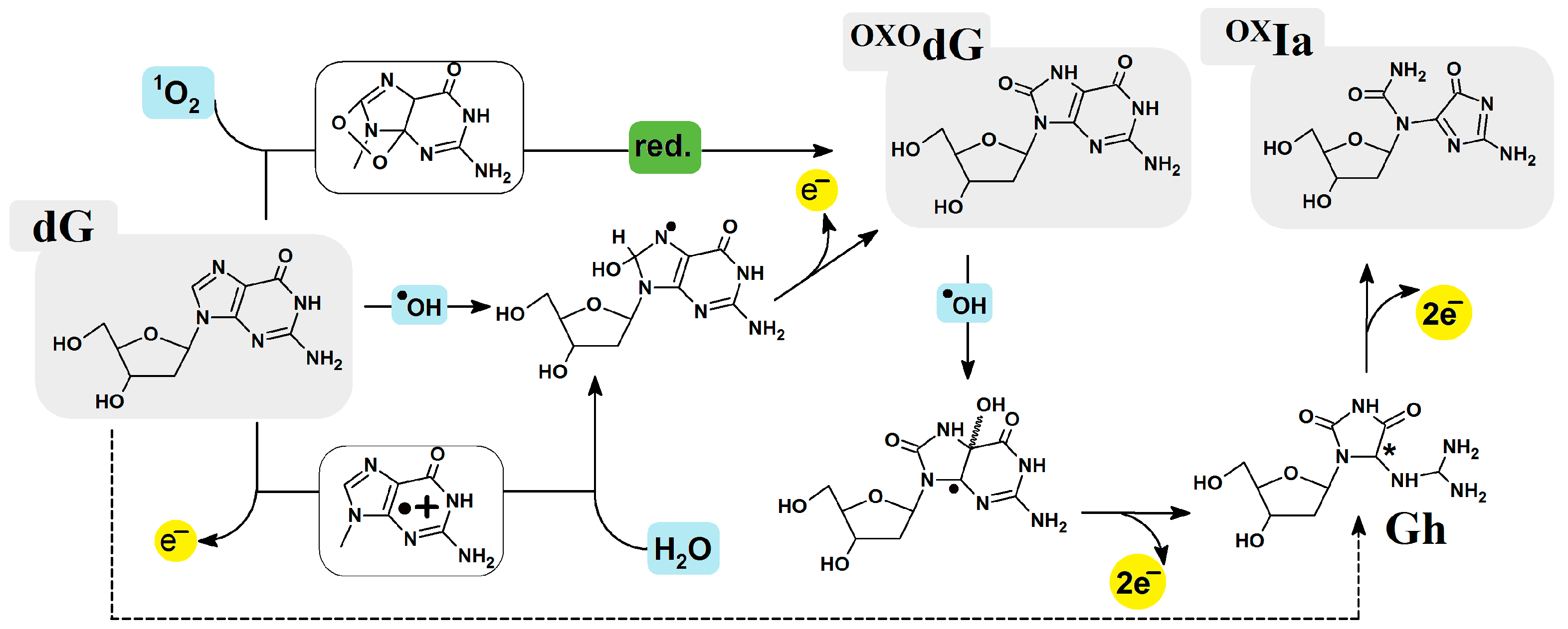
Figure 1. Graphical representation of oxidized imino-allantoin formation via 2′-deoxyGuanosine oxidation paths [9,10]. * chiral center 4R or 4S.
Ijms 25 05962 g001
Figure 2. The structure of double-stranded oligonucleotide (oligo-OXIa) containing cluster DNA damage site optimized on M06-2x/D95**:M06-2x/sto-3G level of theory in aqueous phase and graphical representation of geometrical parameters hydrogen bonds (HB), distances (d1 and d2), angels (λ), distance between base pairs within a dimmer (Rise), staking interaction, and suitable nucleoside atom numbering.
Ijms 25 05962 g002
Figure 3. Graphical representation of proton charge transfer after neutral oligo-OXIa conversion to: (A) adiabatic radical cation of OXOG4:::C2 and (B) to adiabatic radical anion of OXIa2::C4nucleoside pair of oligo-OXIa. The depicted distances have been given in [Å], R—2-deoxyribose.
Ijms 25 05962 g003
Figure 4. (A) A graphical visualization of Hirshfeld charge and spin distribution in [au and %] within neutral, cationic, and anionic forms of oligo-OXIa (Table S2 presents the raw data). VC—vertical cation VA—vertical anion, EQ—equilibrated solvent-solute mode, NE—non-equilibrated solvent-solute mode, AC—adiabatic cation, AA—adiabatic anion; (B) Electronic properties in [eV] of isolated base pairs from oligo-OXIz: vertical (VIP), adiabatic ionization potential (AIP), and vertical (VEA), adiabatic (AEA) electron affinity calculated at the M062x/6-31++G** level of theory in the aqueous phase.
Ijms 25 05962 g004
Table 2. Electronic properties, in [eV], of oligo-OXIa: Vertical (VIP), Adiabatic Ionization Potential (AIP) and Vertical (VEA), Adiabatic (AEA) Electron Affinity calculated at the M062x/6-31++G** level of theory in the aqueous phase. (a) complete double helix and (b) base pair skeletons, NE—non-equilibrated solvent-solute interaction, EQ—equilibrated solvent-solute interaction. Root-Mean-Square Deviation (RMSD) of atomic positions in [Å2], for Neutral, Anionic and Cationic forms of oligo-OXIa. Base pairs (BP), phospho-sugar (PS) frame. The row data have been given in Table S1.
VIPNEVIPEQAIPVEANEVEAEQAEA
oligo-OXIa(a) 6.505.885.492.423.083.59
(b) 6.305.805.391.862.873.24
oligo-N*(a) 6.726.085.650.841.582.09
(b) 6.485.985.580.601.341.90
RMSD: Anion versus NeutralRMSD: Cation versus Neutral
ds-DNABPPS-Frameds-DNABPPS-Frame
oligo-OXIa0.490.360.590.180.130.22
oligo-N [35]0.170.160.170.360.290.42
Table 3. Charge transfer parameters. The ΔG-driving force [eV], λ-reorganization energy [kcal], Ea-activation energy [eV], V12-electron coupling, and kCT-charge rate [s−1] constant of permissible transfer between base pairs of oligo-OXIa, calculated at the m062x/6-31++G** level of theory in the condensed phase. Arrows indicate the direction of charge migration. The raw data have been given in Tables S3a,b and S4.
Electron-Hole TransferExcess-Electron Transfer
SystemλΔGEaV12KCTSystemλΔGEaV12kCT
A1OXIa2A3OXOG4A5A1OXIa2A3OXOG4A5
A1T5OXIa2C4−0.01−0.39−5.510.380.00A1T5OXIa2C40.35−2.102.190.383.56 × 10−22
OXIa2C4→A3T3−0.01−0.44−6.270.090.00OXIa2C4←A3T30.36−2.072.050.326.32 × 10−20
A3T3OXOG4C20.42−1.100.270.431.13 × 1011A3T3OXOG4C20.003−0.080.580.021.05 × 1014
OXOG4C2←A5T10.35−1.160.460.365.59 × 107OXOG4C2←A5T10.008−0.080.160.013.12 × 1010
A1T5→A3T30.00−0.05−0.160.030.00A1T5→A3T30.01−0.030.010.075.61 × 1014
OXIa2C4OXOG4C20.41−1.540.760.057.04OXIa2C4OXOG4C20.33−1.982.040.063.73 × 10−21
A3T3←A5T10.00−0.06−0.240.040.00A3T3→A5T10.00−0.0030.010.055.16 × 1015
Disclaimer/Publisher’s Note: The statements, opinions and data contained in all publications are solely those of the individual author(s) and contributor(s) and not of MDPI and/or the editor(s). MDPI and/or the editor(s) disclaim responsibility for any injury to people or property resulting from any ideas, methods, instructions or products referred to in the content.