Article

Development of a Whole-Cell System Based on the Use of Genetically Modified Protoplasts to Detect Nickel Ions in Food Matrices

1
Department of Biological and Environmental Sciences and Technologies, University of Salento, Via Monteroni 165, 73100 Lecce, Italy
2
NBFC National Biodiversity Future Center, 90133 Palermo, Italy
*
Author to whom correspondence should be addressed.
Int. J. Mol. Sci. 2024, 25(11), 6090; https://doi.org/10.3390/ijms25116090
Submission received: 9 May 2024 / Revised: 29 May 2024 / Accepted: 30 May 2024 / Published: 31 May 2024
(This article belongs to the Special Issue Whole-Cell System and Synthetic Biology)

Abstract

:
Heavy metals are dangerous contaminants that constitute a threat to human health because they persist in soils and are easily transferred into the food chain, causing damage to human health. Among heavy metals, nickel appears to be one of the most dangerous, being responsible for different disorders. Public health protection requires nickel detection in the environment and food chains. Biosensors represent simple, rapid, and sensitive methods for detecting nickel contamination. In this paper, we report on the setting up a whole-cell-based system, in which protoplasts, obtained from Nicotiana tabacum leaves, were used as transducers to detect the presence of heavy metal ions and, in particular, nickel ions. Protoplasts were genetically modified with a plasmid containing the Green Fluorescent Protein reporter gene (GFP) under control of the promoter region of a sunflower gene coding for a small Heat Shock Protein (HSP). Using this device, the presence of heavy metal ions was detected. Thus, the possibility of using this whole-cell system as a novel tool to detect the presence of nickel ions in food matrices was assessed.

1. Introduction

Heavy metals are considered the most dangerous contaminants for the environment; they constitute a threat to human health, even when present in traces. The principal sources of heavy metal are the discharge of industrial effluents and fertilizers, which are responsible for soil contamination. The main threat to human health derives from the capability of metal ions to persist in soils, where they tend to accumulate and are easily transferred into the food chain [1,2], causing damage to human health, such as toxicity and carcinogenicity. The ingestion of heavy metal-contaminated vegetables can cause serious health problems, such as malnutrition, gastrointestinal cancer, the depletion of essential nutrients, the weakening of the immune defense, and the inhibition of mental growth (which also occurs during intrauterine development) [3,4]. Although it has been assessed that the presence of some heavy metals in trace amounts is nutritionally essential for healthy life, they can become toxic when they accumulate in human soft tissues because they are not metabolized. This causes a decrease in energy levels in vital organs, changes blood composition, and reduces mental and central nervous function [5].
Among heavy metals, nickel appears to be one of the most dangerous because it is naturally present in drinking water as well as in many food matrices, exposing the population ingesting it. Sensitivity to nickel prevalence varies in different countries, sitting in a range between 4 and 13.1% [6,7,8]. Nickel is extremely harmful for health because it can cause different disorders such as kidney, lung, and cardiovascular diseases, dermatitis and, sometimes, even some kind of cancer [9]. Therefore, public health protection certainly requires nickel detection in the environment and food chains. At present, the existing techniques used for trace analysis of heavy metals include chromatographic, voltametric, and spectroscopic methods. However, all these methods are quite expensive and are difficult to use for in situ analysis (an analysis conducted directly on the location where the sample is found). On the contrary, new tools such as biosensors can be used as simple, rapid, and sensitive methods to detect heavy metal contaminants, also performed using in situ analyses.
Classical biosensors are analytical devices characterized by three elements: a biological recognition element; a physical–chemical transducer converting the biological response into a detectable signal; a micro-electronic component able to amplify and convert the signal into a numeric record [10]. In particular, when a prokaryotic or eukaryotic cell represents a reporter system that incorporates both biological recognition and transducer elements, the device is called “whole-cell biosensor” or, in other words, it constitutes a whole-cell detection system [5,11,12]. This kind of biosensor responds to the presence of contaminants or to physiological stresses, producing a detectable cellular output signal. Generally, the cells used as biosensors are engineered to acquire the ability to behave as transducers or to amplify their sensitivity by introducing reporter genes, controlled by promoters that respond to environmental stimuli. Most of the cell types considered particularly useful for metal ion detection are genetically modified bacteria, although eukaryotic cells, such as yeast, algae, or protozoan, can also be used [5].
In this paper, we report setting up a whole-cell-based system, in which protoplasts obtained from Nicotiana tabacum leaves were used as transducers to detect the presence of heavy metal ions. The whole-cell detection system was based on the ability of plant cells to respond to environmental abiotic stresses such as the presence of metal ions, which elicits a molecular response. N. tabacum protoplasts were genetically modified with a plasmid containing the GFP reporter gene under the control of the promoter region of a sunflower gene coding a small heat shock protein (HSP). This device was used to test fro the presence of nickel ions in different food matrices known to possess high nickel contents, exploring the possibility of using this biosensor as a novel tool to detect the presence of nickel ions in food matrices.

2. Results

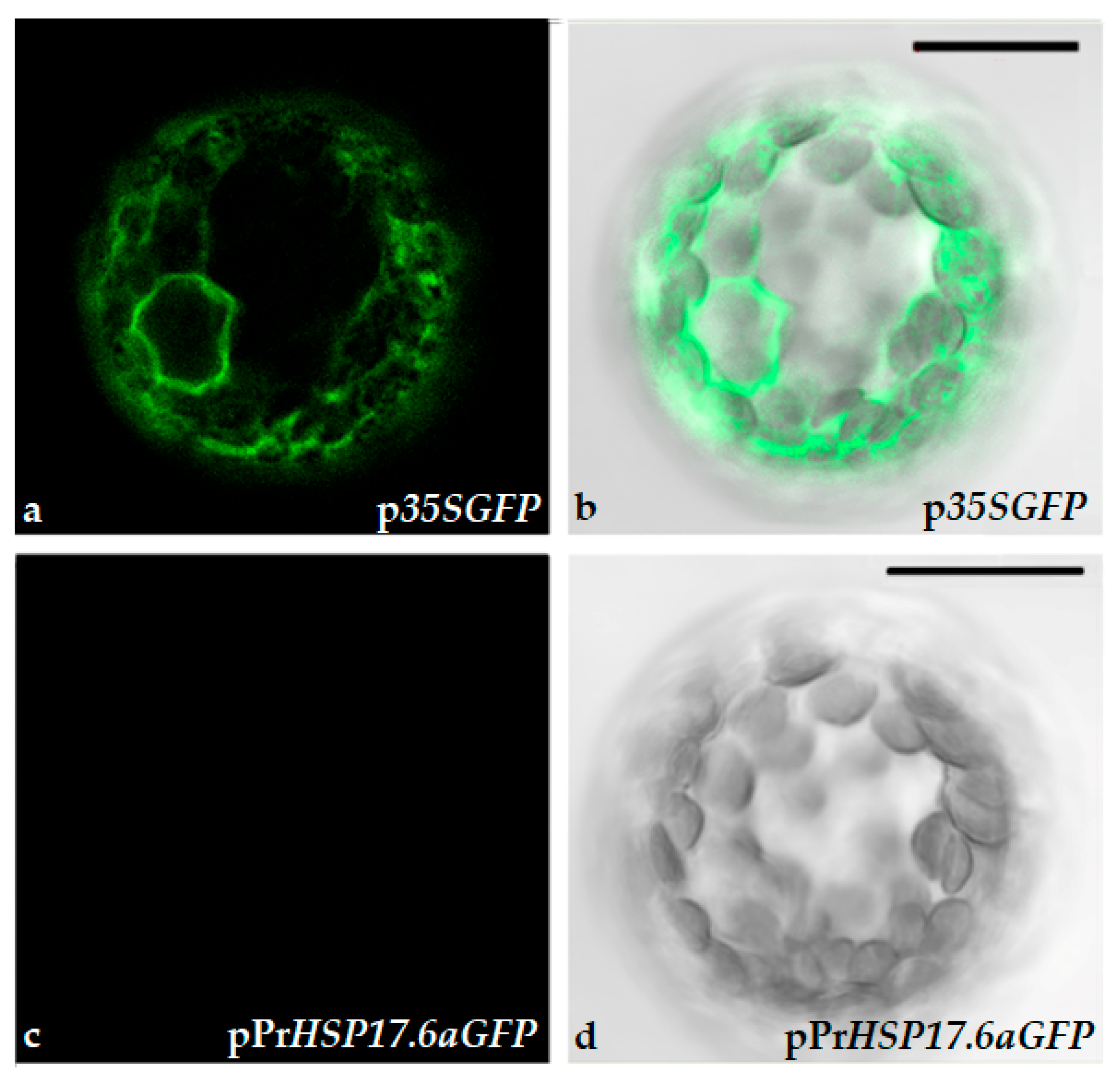
2.1. Protoplasts Transformation and Immobilization

Protoplasts obtained from leaves of N. tabacum were transformed with p35SGFP or pPrHSP17.6aGFP plasmids, containing GFP genes, under the control of the constitutive CAMV35S or the inducible sunflower HSP17.6a promoters, respectively. Protoplasts were in part maintained in a liquid K3 medium (floating) and in part immobilized in a K3 medium containing agarose (0.6%), being distributed into 96 multi-well plates. Untransformed (WT) and transformed protoplasts were tested for their viability by using a fluorescein diacetate (FDA) assay. FDA is a fluorophore that is able to penetrate living and dead cells, where it makes only the viable cells fluorescent; in fact, the fluorescent labelling is due to fluorescein cleavage by cellular esterases, which are only active in viable cells [13], promoting the emission of green fluorescence [14]. Untransformed (WT) and transformed protoplasts were observed using a confocal microscope. Protoplasts images, as seen in Figure 1, indicate that floating and immobilization do not alter the structure and functionality of protoplats, as deduced by the spherical form of fluorescent cells. Further, the number of viable protoplasts was determined. Table 1 reports that the percentage of viable protoplasts is almost the same for WT and transformed protoplasts, as well as for floating and immobilized protoplasts. Therefore, we deduced that our protoplast system was suitable for further analyses.
Immobilized protoplasts were analyzed to determine auto-fluorescence emissions. Fluorescence was measured in wild-type, as well as in engineered protoplasts, after 2, 3, 4, and 7 h from immobilization. A fluorescent signal was detected for all the protoplast samples. As reported in Figure 2, the level of the signal is almost the same during the time spent (after 2 to 7 h) on each group of protoplasts. Moreover, as expected, the fluorescence signal detected for p35SGFP-transformed protoplasts is always higher than the fluorescence signal detected for untransformed protoplasts (WT), as well as for pPrHSP17.6aGFP-transformed protoplasts. Statistical analysis indicated that there is no statistically significant difference in fluorescence signaling, measured at the various time points and expressed as fluorescence units (FU), within each protoplast group (WT, p35SGFP and pPrHSP17.6aGFP-transformed protoplasts); moreover, no significant difference was observed between the WT and pPrHSP17.6aGFP-transformed protoplast groups. On the contrary, a highly significant difference (p < 0.001) was observed between the p35SGFP-transformed protoplast groups and the WT group or the pPrHSP17.6aGFP-transformed protoplast groups (Figure 2).
To verify that differences in fluorescence were due to GFP gene expression, driven by the CAM35S constitutive promoter, protoplasts were observed via confocal microscopy. The data obtained indicated that differences in fluorescence, detected by the fluorometer, were due to the expression of GFP since only p35SGFP-transformed protoplasts exhibited green fluorescence (Figure 3).

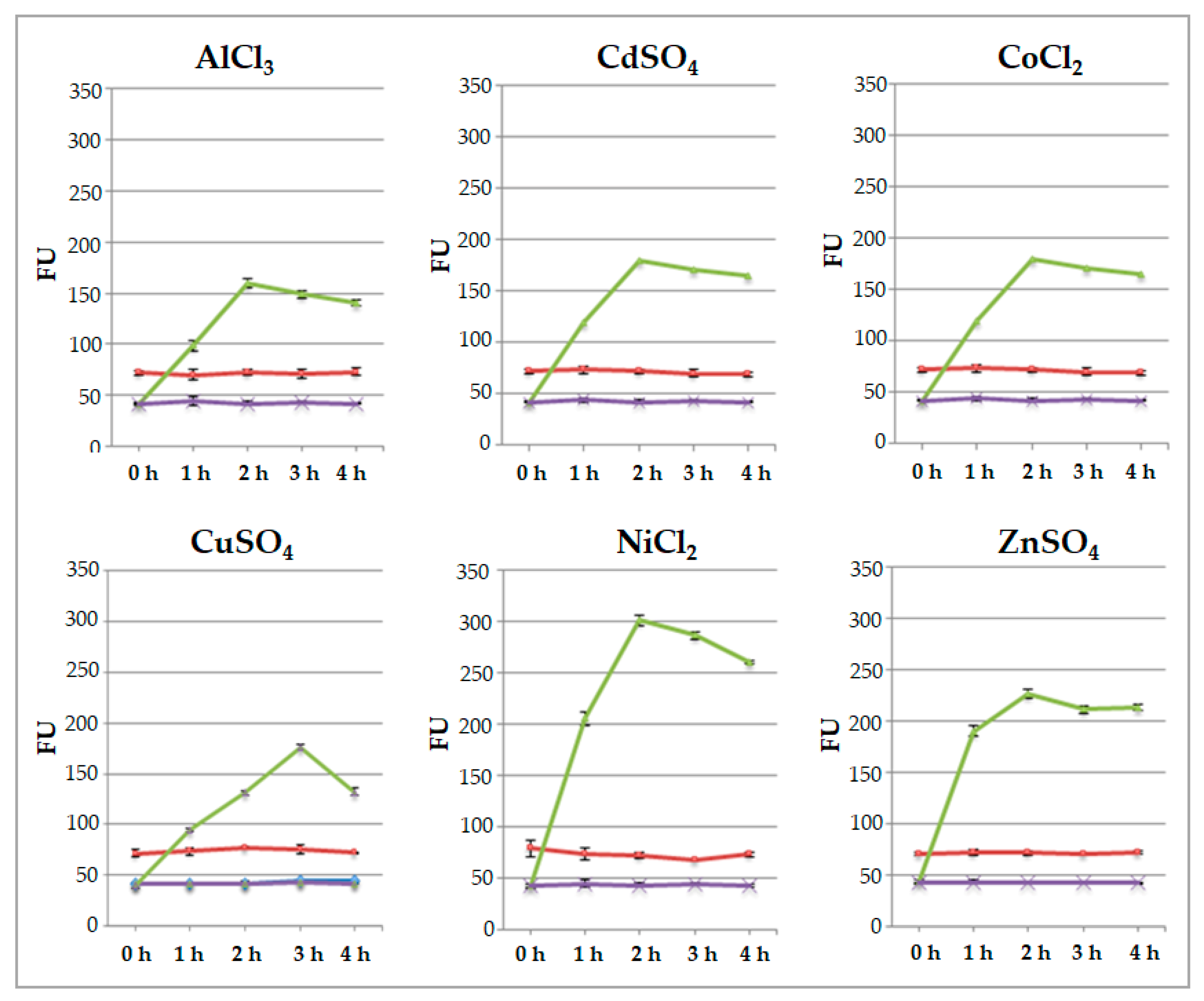
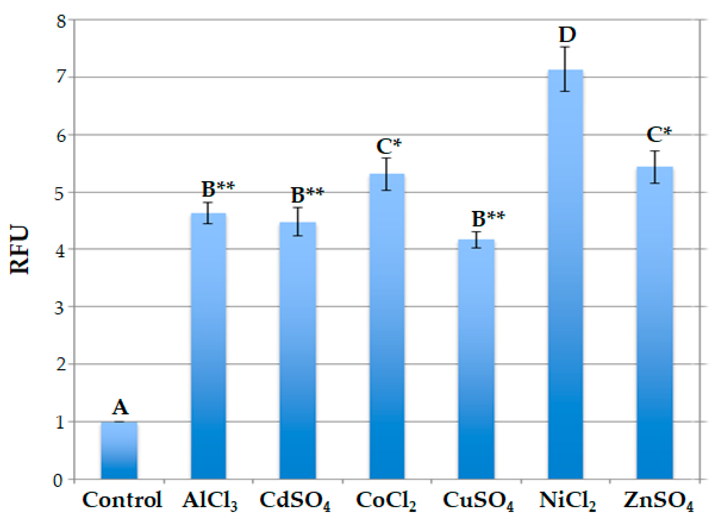
2.2. Responsiveness of Engineered Protoplast to Heavy Metal Ions

To test the ability of engineered protoplasts to sense the presence of metal ions, immobilized protoplasts were added with 50 μL of 20 μM each AlCl3, CdSO4, CoCl2, CuSO4, NiCl2, and ZnSO4 at room temperature. Fluorescence was measured by a fluorometer after 1, 2, 3, and 4 h. The values of fluorescence were measured in protoplasts transformed with p35SGFP plasmid, used as the control, as well as in protoplasts transformed with pPrHSP17.6aGFP plasmid. The values detected are shown in Figure 4 and indicate that the signals of p35SGFP-transformed protoplasts remain unchanged during the time course of all salt treatments. The fluorescence signals of pPrHSP17.6aGFP-transformed protoplasts increased during time course, reaching a maximum level after 2 h and remaining almost the same afterwards. When CuSO4 was used, and only then, the maximum induction was reached after 3 h and the fluorescence signal declined thereafter.
Moreover, the data obtained indicate that the signal’s intensity depends on the specific metal ion added. In particular, after 2 h, in the case of NiCl2 treatment, the signal was over 7-fold higher than that of the untreated protoplasts; it was more or less 5.5-fold higher after treatment with ZnSO4 and CoCl2; and it was 4.5-fold higher when using AlCl3 and CdSO4 salts. Following CuSO4 treatment, the maximum value was 4-fold higher (Figure 5).
Comparison among the relative fluorescence values displayed by engineered protoplasts indicated that these values were similar when engineered protoplasts were treated with AlCl3 and CdSO4 or with CuSO4; in fact, in these cases, no statistically significant difference was observed in terms of relative fluorescence values. The fluorescence signals were also almost the same in the presence of CoCl2 and ZnSO4, and no statistically significant difference was observed in this case. On the contrary, statistically significant differences were observed when the florescence signal values of protoplasts treated with Ni ions were compared with those of all the other value groups. In particular, highly significant differences (p < 0.001) were observed when the values reached by protoplasts treated with Ni ions were compared to the ones obtained after treatment with Al, Cd and Cu ions, while significant differences (p < 0.05) were observed when they were compared to the ones obtained after treatment with Co and Zn ions (Figure 5).
These data were confirmed via confocal observations and the fluorescence quantification of the protoplasts treated with the six different heavy metal ions (Figure 6). The protoplasts tested with all heavy metal ion treatments appeared to be fluorescent, although they showed different intensity and fluorescence patterns. The compartments of the secretion pathway (nuclear membrane in continuity with the endoplasmic reticulum and the Golgi complex) were clear in the protoplasts characterized by a higher level of GFP expression, i.e., protoplast treated with 50 μL of 20 μM CoCl2, NiCl2, and ZnSO4.
These data were also confirmed by fluorescence index values (Table 2). A comparison of the fluorescence index values obtained after NiCl2 treatment indicates significant differences with respect to all other salt treatments.
Since all the data indicated that nickel ions are the best inducers of fluorescence, to better characterize the response of the engineered protoplasts to this type of treatment, they were subjected for 2 h to various nickel ion concentrations, ranging from 2 μM to 40 μM. The results obtained indicate that, with rising Ni ion concentrations, the signal increased, reaching its maximum value at a concentration of 20 μM NiCl2; thereafter, the signal reached a plateau using higher Ni ion concentrations (Figure 7).

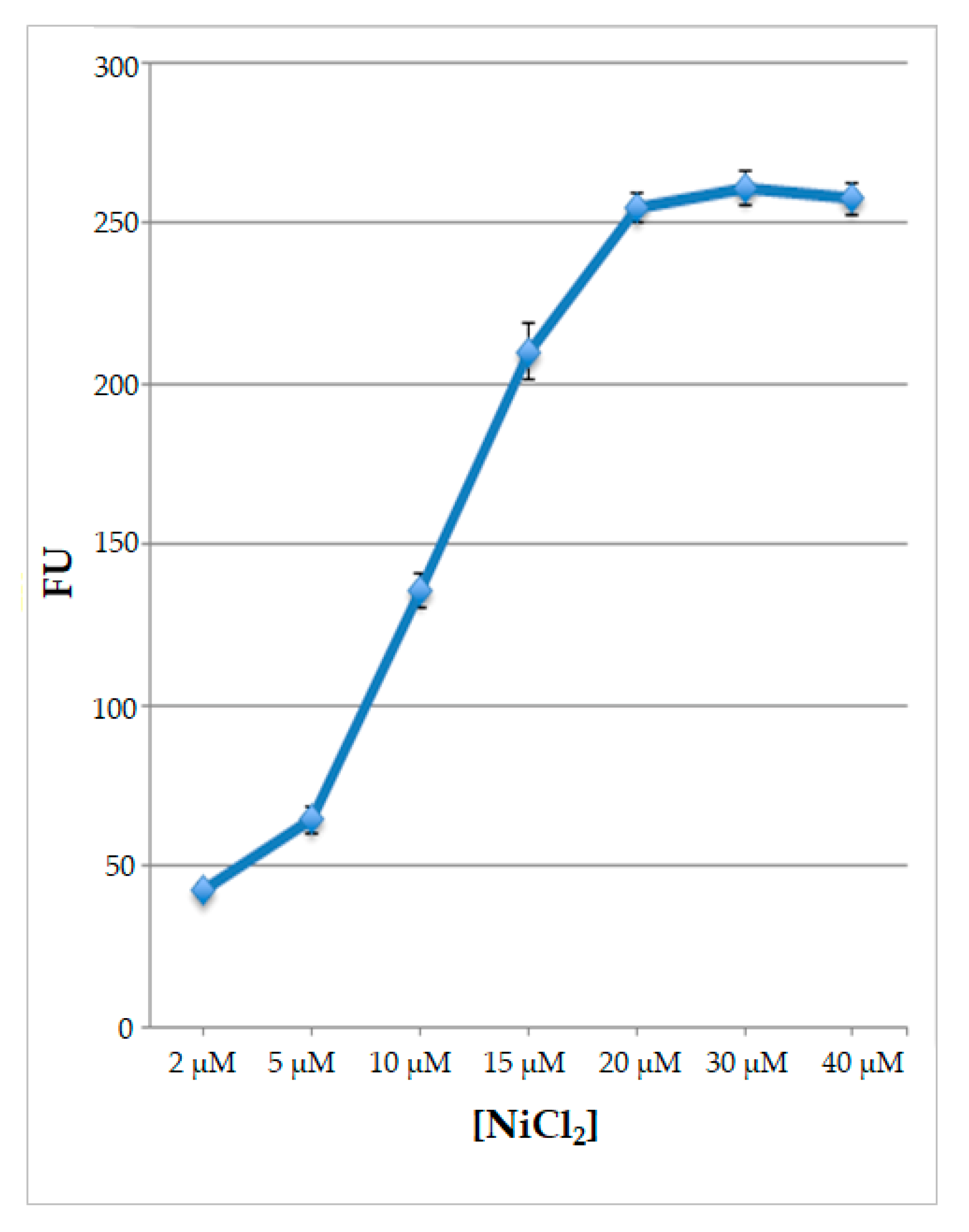
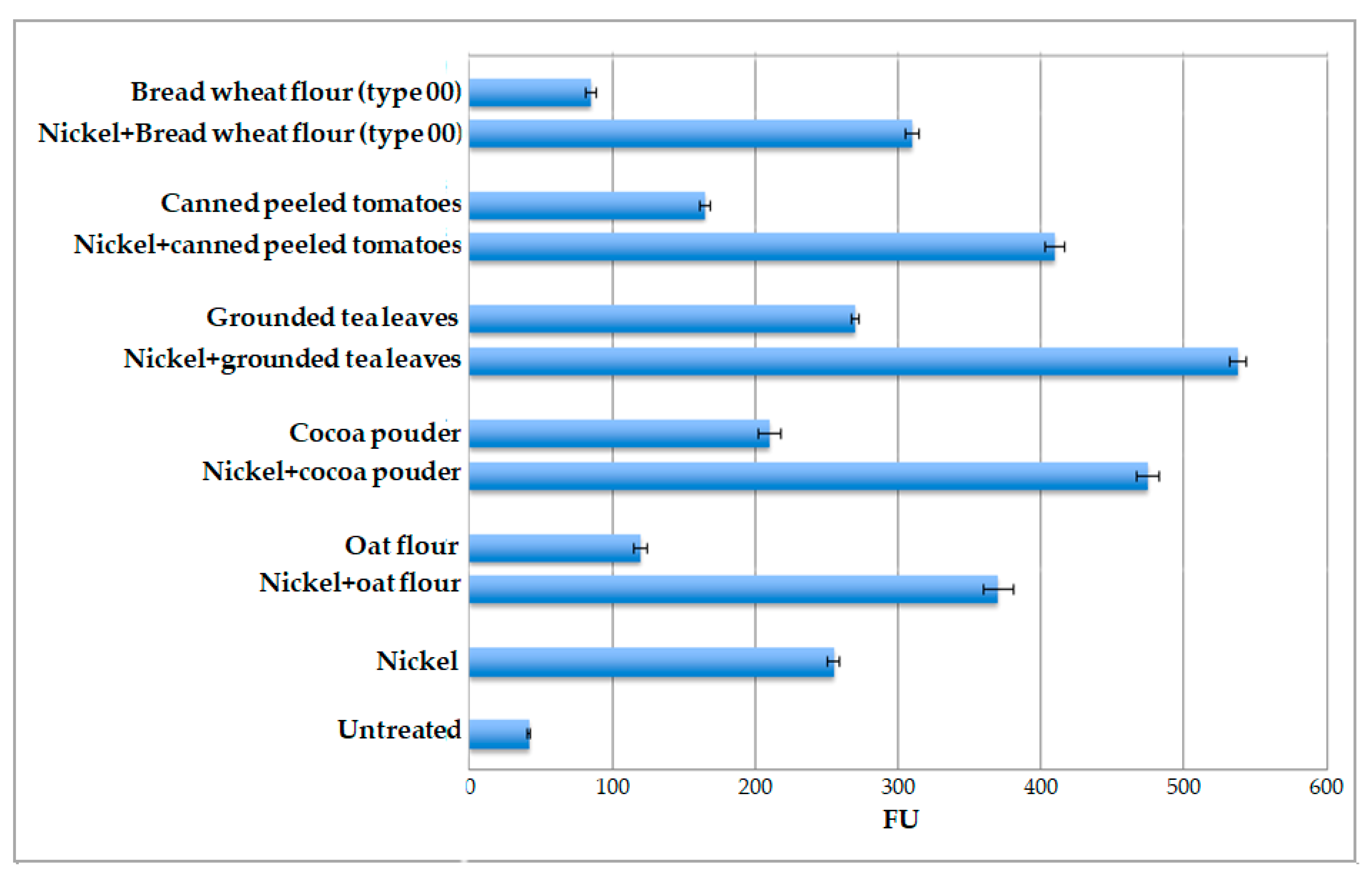
2.3. Nickel Ions Detection in Different Food Matrices

Considering that nickel ions appear to be the most efficient in terms of eliciting fluorescence signals and that they are naturally present in many food matrices, the ability of the whole-cell system to detect Ni ions in food was tested. For this, the pPrHSP17.6aGFP-engineered protoplasts were tested against various food matrices, known to be “high-nickel foods” (canned peeled tomatoes, cocoa powder, ground tea leaves, oat flour) or “low-nickel foods”, namely bread wheat flour type 00, which is the most refined bread wheat flour [15,16,17]. All the food matrices were tested after adding exogenous nickel ions. A NiCl2 solution was added to reach a final concentration of 20 μM. The results, reported in Figure 8, indicate that the level of fluorescence signal is higher than that measured after treatment with nickel ions only, depending on the matrix utilized. The highest signal was obtained with ground tea leaves (FU 538), while the lowest signal was obtained with bread wheat flour type 00 (FU 310).
Subsequently, protoplasts were tested against the different food matrices only. In general, all food matrices are able to induce an increase in GFP expression. The results obtained, reported in Figure 8, confirm that ground tea leaves are the most effective inducers (FU 270), while bread wheat flour (type 00) is the least effective inducer (FU 85). In particular, for ground tea leaves, the level of induction was 6-fold higher than that obtained in untreated protoplasts, for cocoa powder it was 5-fold, for canned peeled tomatoes it was 4-fold, for oat flour it was 3-fold, and for type 00 wheat flour it was only 2-fold with respect to the same control, which was represented by untreated transformed protoplasts.

3. Discussion

Technological processes and agrochemical treatments are the main forces responsible for the contamination of food products and for the reduction in food nutritional value. The development of safe and accurate analytical methodologies for water and food control is crucial for the detection, analysis, and diagnosis of a wide range of compounds affecting food quality and healthiness. Heavy metal ions are among the worst and most dangerous contaminants because they are widely present in the environment and also because they are easily transferred from soil and water to living organisms [3,18].
Plants exposed to adverse environmental conditions (biotic or abiotic stresses) have developed complex molecular mechanisms to protect cell homeostasis and minimize the potential damage caused by these stimuli [19]. In general, a plant’s stress response is based on the activation and/or inactivation of gene expression, rapidly triggered after the perception of the stress. The rapidity of the response is due to the presence of different stress-responsive cis elements in the promoter region of these genes [20]. The most important group of genes participating in stress responses is constituted by the heat shock gene family. These genes are not only activated by heat stress, but also by other stressful environmental conditions, such as the presence of heavy metal ions [21,22].
With the aim of testing the ability of the promoter region of a plant’s small HSP gene to sense the presence of heavy metal ions in different food matrices, N. tabacum protoplasts were transformed with a plasmid containing the GFP gene controlled by the promoter region of the sunflower HaHSP17.6a gene. This specific promoter was chosen on the basis of the characterization, in a previous study, of the HaHSP17.6a gene, reported to be inducible in sunflower seedlings by heat stress as well as by other stimuli, and in particular by the presence of heavy metal ions [22].
The data obtained in vitro using this transient expression system established that the promoter region of HaHSP17.6a gene is activated by all the metal ions tested, although at different levels, confirming its inducibility, already demonstrated in vivo in sunflower seedlings [22]; the data also indicated that the HaHSP17.6a promoter exhibits the best sensitivity to Ni ions.
Having assessed promoter inducibility, the engineered protoplast system was also tested for its ability to sense the presence of heavy metal ions, with particular regard to Ni, in different food matrices. Systemic nickel allergy syndrome affects a significant portion of the population. Developing a more sensitive and effective tool for the rapid and in situ detection of high concentrations of nickel ions in food could be crucial. Such an advancement would play an important role in improving future food analysis. The food matrices utilized in this work were chosen mostly on the basis of their already known nickel content, according to their classification or as “high-Ni foods” or as “low-Ni foods” [15,17,23,24,25].
The first step of this part of this study was aimed at verifying whether the molecular composition of food matrices can “quench” nickel ions present in a solution. In order to carry that out, the determination of fluorescence emissions was performed via the addition of a known quantity of NiCl2 to the different matrices. Subsequently, the same tests were repeated using the food matrix alone. In both cases, the induction of fluorescence was detectable, and it specifically varied according to the food matrix assayed, indicating that none of the food matrices utilized had a molecular structure that could interfere with the biological detection of nickel ions present in the test solution.
In conclusion, taken together, the data obtained indicate that the engineered immobilized protoplast system established is a useful tool for detecting the presence of nickel ions in food. To our knowledge, this is so far the first example of using biosensors to detect traces of heavy metal ions in food based on genetically engineered plant protoplasts. Starting from these data, broader use of the biosensor realized can be hypothesized. One application could be its use to examine and evaluate a wider number of food matrices for the presence of nickel ions, as well as the use of this device for the detection of other heavy metal (Al, Cd, Co, Cu, Zn) traces as contaminants along the food chain. On the basis of these considerations, whole-cell biosensors appear to be the most suitable detection tool, not only because they are chips and are portable, but also because they are specifically designed to have high sensitivity in detecting heavy metal ions at trace levels. In other words, whole-cell biosensors can be considered the best way to measure heavy metal ions in food, thus contributing to providing larger benefits to consumer health in relation to food production and safety.

4. Materials and Methods

4.1. Plasmids Construction

A DNA fragment, corresponding to the promoter region of the sunflower small HSP17.6a gene [22], was amplified using the primers For-tgcctcgaggtagtacacggtg and Rev-gtaaaattgttcaacgtgttctagaggat. The primers contained restriction sites for generating XhoI and BamHI ends. PCR was performed on genomic DNA obtained from sunflower seedlings (Heliantus annuus L., cv. Gloriasol) using “Pure Link Plant Total DNA purification” kit (Invitrogen, Carlsbad, CA, USA), according to supplier’s instructions. In order to verify an amplicon’s identity, the PCR product was purified and sequenced using standard procedures; the DNA sequence, 1769 bp long, was compared with the corresponding genomic clone (accession number AJ306557.2). The DNA fragment obtained was cloned into a vector containing the GFP (green fluorescent protein) gene, followed by nopaline synthase gene (Nos) terminator, previously digested with XhoI and BamHI; the plasmid obtained was named pPrHSP17.6aGFP. A plasmid containing the GFP gene under the control of CAMV35S promoter, named p35SGFP, was used as the control.

4.2. Protoplasts Preparation, Transformation and Immobilization

N. tabacum cultivar SR1 protoplasts were prepared from leaves of 7–8-week-old plants. The transformation was performed using PEG-mediated direct gene transfer, as previously described by Gullì and coworkers [26]. Ten micrograms of each constructed plasmid were used for the transformation of ~600,000 protoplasts. The protoplast suspension was gently mixed with a solution of 0.6% agarose in a K3 medium [27] at 40 °C; 150 μL of the mixture was distributed in a multiwall plate and solidification was completed after 30 min of treatment at room temperature. Immobilized protoplasts were kept at room temperature until stress treatments were performed.

4.3. Protoplasts Stress Treatments with Different Heavy Metal Ions

Transformed protoplasts were subjected to metal ion stress by adding 50 μL of 20 μM of AlCl3, CdSO4, CoCl2, and ZnSO4, or 2 μM, 5 μM, 10 μM, 15 μM, 20 μM, 30 μM, and 40 μM of NiCl2, at room temperature. They were observed after 1, 2, 3 and 4 h for fluorescence determination. The controls used were wild-type protoplasts (WT) and protoplasts transformed with the p35SGFP plasmid and then subjected to stress. We also used WT protoplasts and protoplasts transformed with the p35SGFP plasmid and pPrHSP17.6aGFP but not subjected to stress.

4.4. FDA Assay for Protoplast Viability Estimation

Fluorescein diacetate (FDA) staining was used to determine the viability of protoplasts. Fifty µL of the K3 medium, containing untransformed or transformed protoplasts, was transferred into a microtube; 1 µL of 0.2% FDA solution, dissolved in acetone, was added and incubated at room temperature for 2 min. All FDA-treated protoplasts were observed in a K3 culture medium and after agarose (0.6%) immobilization. Only viable protoplasts appeared to be green and fluorescent under the confocal laser microscope (LSM 710, Carl Zeiss, Oberkochen, Germany). For the viability measurements, three images were selected for each sample; the percentage of protoplast viability was expressed as the ratio between the number of the fluorescent protoplasts and the total number of protoplasts ×100. Each experiment was repeated three times.

4.5. Confocal Microscopy and Fluorescence Determination

For protoplast observation, a laser scanning confocal microscope (LSM 710, Zeiss) was used. To control protoplast viability after transformation and/or immobilization, protoplasts were observed in their culture medium and after agarose immobilization. To detect GFP or FDA fluorescence, a 488 nm argon ion laser line was used, and the emission was recorded with a 505–530 nm filter set. Chlorophyll epifluorescence was detected with the filter >650 nm and eliminated after He-Ne laser excitation at 543 nm, as previously reported [28]. The power of each laser line, the gain, and the offset were identical for each experiment so that the images were comparable. For fluorescence quantification, the Profile Tool of the ZEN2012 program of the LSM 710 confocal microscope was used. The mean of pixel intensities relative to the GFP channel was used for fluorescence quantification; 20 protoplasts were measured for each treatment to produce the quantification analysis and three independent experiments were performed. Images were processed using Adobe Photoshop 7.0 software (Mountain View, CA, USA). Protoplast fluorescence was also measured using and Infinite F200 fluorometer (TECAN, Männedorf, Switzerland) set as follows: excitation at 485 nm (±20) and emission at 510–560 nm. Fluorescence values are presented as arbitrary fluorescence units (FUs) or as relative fluorescence unit (RFUs), namely, the ratio of the fluorescence of the treated sample to that of the untreated control (response ratio). All data are the mean ± SD of three different measurements.

4.6. Food Matrices Utilized for Nickel Ions Detection

The food matrices utilized to assess the responsiveness of engineered protoplasts to the presence of metal ions in food were oat flour, cocoa powder, ground tea leaves, canned peeled tomatoes, and bread wheat flour (type 00). We added two grams of each food matrix to 8 mL of sterile distilled water and stirred the mixture for 2 h. One mL was centrifuged at 13,000 rpm for 2 min; 50 μL of supernatant, without or with NiCl2 at the final concentration of 20 μM, was added to immobilized protoplasts.

4.7. Statistical Analysis

Statistical analysis was performed using the SigmaStat version 3.11 software (Systat Software Inc., Chicago, IL, USA) as appropriate. The viability of protoplasts floating in K3 buffer or immobilized in 0.6% agarose were compared using Student’s t-test. The protoplast fluorescence values, measured fluorometrically or via confocal microscope tools, were analyzed via the pairwise multiple comparisons procedure using the Holm–Sidak method. All values were expressed as means ± standard deviation (SD) of at least three independent replicated experiments (n = 3). A p value ≤ 0.05 was considered to be statistically significant.

Author Contributions

Conceptualization, P.R. and C.P.; methodology, M.D.C. and P.R.; software, M.D.C.; validation, M.D.C., C.P. and P.R.; investigation, M.D.C. and P.R.; resources, P.R.; data curation, P.R.; writing—original draft preparation, M.D.C., C.P. and P.R.; writing—review and editing, C.P. and P.R.; supervision, P.R. All authors have read and agreed to the published version of the manuscript.

Funding

This research received no external funding.

Institutional Review Board Statement

Not applicable.

Informed Consent Statement

Not applicable.

Data Availability Statement

The original contributions presented in this study are included in the article.

Acknowledgments

The authors wish to thank G. Dalessandro for critical reading of the manuscript.

Conflicts of Interest

The authors declare no conflicts of interest.

References

  1. Kabata-Pendias, A.; Mukherjee, A.B. Soil. In Trace Elements from Soil to Human; Springer: Berlin/Heidelberg, Germany, 2007; pp. 9–38. [Google Scholar] [CrossRef]
  2. Taghavi, M.; Darvishiyan, M.; Momeni, M.; Eslami, H.; Fallahzadeh, R.A.; Zarei, A. Ecological risk assessment of trace elements (TEs) pollution and human health risk exposure in agricultural soils used for saffron cultivation. Sci. Rep. 2023, 13, 4556. [Google Scholar] [CrossRef] [PubMed]
  3. Rai, P.K.; Lee, S.S.; Zhang, M.; Tsang, Y.F.; Kim, K.-H. Heavy metals in food crops: Health risks, fate, mechanisms, and management. Environ. Int. 2019, 125, 365–385. [Google Scholar] [CrossRef] [PubMed]
  4. Munir, N.; Jahangeer, M.; Bouyahya, A.; Omari, N.E.; Ghchime, R.; Balahbib, A.; Aboulaghras, S.; Mahmood, Z.; Akram, M.; Shah, S.M.A.; et al. Heavy Metal Contamination of Natural Foods Is a Serious Health Issue: A Review. Sustainability 2022, 14, 161. [Google Scholar] [CrossRef]
  5. Gutiérrez, J.C.; Amaro, F.; Martin-Gonzáles, A. Heavy metal whole-cell biosensor using eukaryotic microorganism: An update critical review. Front. Microbiol. 2015, 6, 48. [Google Scholar] [CrossRef] [PubMed]
  6. Lusi, E.A.; Di Ciommo, V.M.; Patrissi, T.; Guarascio, P. High prevalence of nickel allergy in an overweight female population: A pilot observational analysis. PLoS ONE 2015, 10, e0123265. [Google Scholar] [CrossRef] [PubMed]
  7. Venter, C. Food Hypersensitivity: Diagnosing and Managing Food Allergies and Intolerances. J. Allergy 2012, 2012, 576017. [Google Scholar] [CrossRef] [PubMed]
  8. Anchidin-Norocel, L.; Savage, W.K.; Gutt, G.; Amariei, S. Development, Optimization, Characterization, and Application of Electrochemical Biosensors for Detecting Nickel Ions in Food. Biosensors 2021, 11, 519. [Google Scholar] [CrossRef] [PubMed]
  9. Genchi, G.; Carocci, A.; Lauria, G.; Sinicropi, M.S.; Catalano, A. Nickel: Human Health and Environmental Toxicology. Int. J. Environ. Res. Public Health 2020, 17, 679. [Google Scholar] [CrossRef] [PubMed]
  10. Belkin, S. Microbial whole-cell sensing systems of environmental pollutants. Curr. Opin. Microbiol. 2003, 6, 206–212. [Google Scholar] [CrossRef]
  11. Liu, C.; Yu, H.; Zhang, B.; Liu, S.; Liu, C.-g.; Li, F.; Song, H. Engineering whole-cell microbial biosensors: Design principles and applications in monitoring and treatment of heavy metals and organic pollutants. Biotechnol. Adv. 2022, 60, 108019. [Google Scholar] [CrossRef]
  12. Chen, S.; Chen, X.; Su, H.; Guo, M.; Liu, H. Advances in Synthetic-Biology-Based Whole-Cell Biosensors: Principles, Genetic Modules, and Applications in Food Safety. Int. J. Mol. Sci. 2023, 24, 7989. [Google Scholar] [CrossRef]
  13. Schoor, S.; Lung, S.C.; Sigurdson, D.; Chuong, S.D. Fluorescent Staining of Living Plant Cells. In Plant Microtechniques and Protocols; Yeung, E.C.T., Stasolla, C., Sumner, M.J., Huang, B.Q., Eds.; Springer: Cham, Switzerland, 2015; pp. 153–165. [Google Scholar] [CrossRef]
  14. Widholm, J.M. The use of fluorescein diacetate and phenosafranine for determining viability of cultured plant cells. Stain Technol. 1972, 47, 189–194. [Google Scholar] [CrossRef] [PubMed]
  15. Borghini, R.; De Amicis, N.; Bella, A.; Greco, N.; Donato, G.; Picarelli, A. Beneficial Effects of a Low-Nickel Diet on Relapsing IBS-Like and Extraintestinal Symptoms of Celiac Patients during a Proper Gluten-Free Diet: Nickel Allergic Contact Mucositis in Suspected Non-Responsive Celiac Disease. Nutrients 2020, 12, 2277. [Google Scholar] [CrossRef]
  16. Cappelli, A.; Bini, A.; Cini, E. The Effects of Storage Time and Environmental Storage Conditions on Flour Quality, Dough Rheology, and Biscuit Characteristics: The Case Study of a Traditional Italian Biscuit (Biscotto di Prato). Foods 2022, 11, 209. [Google Scholar] [CrossRef] [PubMed]
  17. Greco, M.; Pisano, A.; Mezzatesta, L.; Pettinelli, M.; Meacci, A.; Pignataro, M.G.; Giordano, C.; Picarelli, A. New Insights and Evidence on “Food Intolerances”: Non-Celiac Gluten Sensitivity and Nickel Allergic Contact Mucositis. Nutrients 2023, 15, 2353. [Google Scholar] [CrossRef] [PubMed]
  18. Scutarsu, E.C.; Trinca, L.C. Heavy Metals in Foods and Beverages: Global Situation, Health Risks and Reduction Methods. Foods 2023, 12, 3340. [Google Scholar] [CrossRef]
  19. Lamers, J.; van der Meer, T.; Testerink, C. How Plants Sense and Respond to Stressful Environments. Plant Physiol. 2022, 182, 1624–1635. [Google Scholar] [CrossRef] [PubMed]
  20. Wu, J.; Gao, T.; Hu, J.; Zhao, L.; Yu, C.; Ma, F. Research advances in function and regulation mechanisms of plant small heat shock proteins (sHSPs) under environmental stresses. Sci. Total Environ. 2022, 825, 154054. [Google Scholar] [CrossRef]
  21. Gullì, M.; Rampino, P.; Lupotto, E.; Marmiroli, N.; Perrotta, C. The effect of heat stress and cadmium ions on the expression of a small hsp gene in barley and maize. J. Cer. Sci. 2005, 42, 25–31. [Google Scholar] [CrossRef]
  22. Rampino, P.; Mita, G.; Assab, E.; De Pascali, M.; Giangrande, E.; Treglia, A.S.; Perrotta, C. Two sunflower 17.6HSP genes, arranged in tandem and highly homologous, are induced differently by various elicitors. Plant Biol. 2010, 12, 13–22. [Google Scholar] [CrossRef]
  23. Picarelli, A.; Di Tola, M.; Vallecoccia, A.; Libanori, V.; Magrelli, M.; Carlesimo, M.; Rossi, A. Oral mucosa patch test: A new tool to recognize and study the adverse effects of dietary nickel exposure. Biol. Trace Elem. Res. 2011, 139, 151–159. [Google Scholar] [CrossRef] [PubMed]
  24. Pizzutelli, S. Systemic nickel hypersensitivity and diet: Myth or reality? Eur. Ann. Allergy Clin. Immunol. 2011, 43, 5–18. [Google Scholar] [PubMed]
  25. Rizzi, A.; Nucera, E.; Laterza, L.; Gaetani, E.; Valenza, V.; Corbo, G.M.; Inchingolo, R.; Buonomo, A.; Schiavino, D.; Gasbarrini, A. Irritable Bowel Syndrome and Nickel Allergy: What Is the Role of the Low Nickel Diet? J. Neurogastroenterol. Motil. 2019, 23, 101–108. [Google Scholar] [CrossRef] [PubMed]
  26. Gullì, M.; De Pascali, M.; Perrotta, C.; Rampino, P. A stress-related transcription factor belonging to the YL-1 family is differently regulated in durum wheat cultivars differing in drought sensitivity. Plant Physiol. Biochem. 2022, 170, 307–315. [Google Scholar] [CrossRef] [PubMed]
  27. Freydl, E.; Meins, F., Jr.; Boller, T.; Neuhaus, J.M. Kinetics of prolyl hydroxylation, intracellular transport and C-terminal processing of the tobacco vacuolar chitinase. Planta 1995, 197, 250–256. [Google Scholar] [CrossRef]
  28. De Caroli, M.; Lenucci, M.S.; Manualdi, F.; Dalessandro, G.; De Lorenzo, G.; Piro, G. Molecular dissection of Phaseolus vulgaris polygalacturonase-inhibiting protein 2 reveals the presence of hold/release domains affecting protein trafficking toward the cell wall. Front. Plant Sci. 2015, 6, 660. [Google Scholar] [CrossRef]
Figure 1. Confocal microscope images of tobacco protoplasts after FDA staining. WT—untransformed protoplasts; p35SGFP—protoplast transformed with p35SGFP plasmid; pPrHSP17.6aGFP—protoplast transformed with pPrHSP17.6aGFP plasmid. (a,c,e) Floating protoplasts; (b,d,f) protoplasts immobilized in K3 medium containing 0.6% agarose. Scale bars: 50 µm. Objective: 10×; zoom: 0.6.
Ijms 25 06090 g001
Figure 2. Evaluation of fluorescence, measured by a fluorometer, after 2, 3, 4, and 7 h from immobilization of untransformed protoplasts (WT), protoplasts transformed with p35SGFP plasmid (p35SGFP), and protoplasts transformed with pPrHSP17.6aGFP plasmid (pPrHSP17.6aGFP). Each value represents the mean of three independent measurements ± SD. Different uppercase letters indicate significant differences among the FU values (p < 0.001).
Ijms 25 06090 g002
Figure 3. Confocal microscope images of immobilized tobacco protoplasts. (a,b) Protoplasts transformed with p35SGFP plasmid; (c,d) protoplasts transformed with pPrHSP17.6aGFP plasmid. (b,d) Bright field. Scale bars: 20 µm. Objective 40×, zoom 2.0.
Ijms 25 06090 g003
Figure 4. Evaluation of fluorescence by fluorometer after 1, 2, 3 and 4 h from protoplast immobilization. Red line: protoplasts transformed with p35SGFP plasmid; violet line: untreated protoplasts transformed with pPrHSP17.6aGFP plasmid; and green line: protoplasts transformed with pPrHSP17.6aGFP plasmid plus metal ion solutions (50 μL of 20 μM each). Each value represents the mean of three independent measurements ± SD.
Ijms 25 06090 g004
Figure 5. Relative fluorescence (expressed as relative fluorescence units, RFUs) at the maximum level of detection of protoplasts transformed with pPrHSP17.6aGFP and treated with different metal ions (50 μL of 20 μM each). Each value represents the mean of three independent measurements ± SD. Uppercase letters indicate statistically different values among the various protoplast groups. Asterisks correspond to statistically different values between the NiCl2-treated protoplasts and each other different group (*, significant difference, p < 0.05; **, highly significant difference, p < 0.001).
Ijms 25 06090 g005
Figure 6. Confocal microscope images of the immobilized protoplasts transformed with pPrHSP17.6aGFP plasmid in presence of 50 μL of 20 μM AlCl3 (a), CdSO4 (b), CoCl2 (c), CuSO4 (d), NiCl2 (e) and ZnSO4 (f). Scale bars: 20 µm. Objective: 40×; zoom: 2.0.
Ijms 25 06090 g006
Figure 7. Values of fluorescence, expressed as fluorescence units (FUs) of protoplasts transformed with pPrHSP17.6aGFP plasmids and treated with different NiCl2 concentrations. Each value represents the mean of three independent measurements ± SD.
Ijms 25 06090 g007
Figure 8. Values of fluorescence as expressed in fluorescence units (FUs) of protoplasts transformed with pPrHSP17.6aGFP plasmid plus the addition of different food matrices (in the presence or absence of 20 μM nickel ions) for 2 h. Protoplasts transformed with pPrHSP17.6aGFP plasmid with only nickel ions added (Nickel) and untreated protoplasts (Untreated) were used as controls. Each value represents the mean of three independent measurements ± SD.
Ijms 25 06090 g008
Table 1. Protoplast viability (%), determined by FDA assay, of untransformed and p35SGFP- and pPrHSP17.6aGFP-transformed protoplasts floating in a K3 medium or immobilized in K3 medium containing 0.6% agarose.
Floating Protoplasts (%)Immobilized Protoplasts (%)
WT98.77 ± 1.2397.70 ± 2.30
p35SGFP99.05 ± 0.9598.44 ± 1.56
pPrHSP17.6aGFP97.87 ± 2.1398.65 ± 1.35
Each value represents the mean of three independent measurements ± SD. WT, untrasformed protoplasts; p35SGFP, protoplasts transformed with p35SGFP plasmid; pPrHSP17.6aGFP, protoplasts transformed with pPrHSP17.6aGFP plasmid.
Table 2. Fluorescence index given by the mean of GFP pixels intensity per protoplast transformed with pPrHSP17.6aGFP and treated with the different metal ions (50 μL of 20 μM each). Differences between NiCl2-treated protoplasts and each different metal ion were significant (*, p < 0.05) or highly significant (**, p < 0.001).
Mean ± SD of GFP Pixel Intensity per Protoplast
AlCl3 (2 h)4.76 ± 0.43 **
CdSO4 (2 h)4.21 ± 0.46 **
CoCl2 (2 h)5.27 ± 0.66 **
CuSO4 (3 h)4.02 ± 0.51 **
NiCl2 (2 h)7.30 ± 0.46
ZnSO4 (2 h)5.18 ± 0.52 *
Disclaimer/Publisher’s Note: The statements, opinions and data contained in all publications are solely those of the individual author(s) and contributor(s) and not of MDPI and/or the editor(s). MDPI and/or the editor(s) disclaim responsibility for any injury to people or property resulting from any ideas, methods, instructions or products referred to in the content.

