Review

Host Cell Death and Modulation of Immune Response against Mycobacterium tuberculosis Infection

College of Osteopathic Medicine of the Pacific, Western University of Health Sciences, Pomona, CA 91766, USA
*
Author to whom correspondence should be addressed.
Int. J. Mol. Sci. 2024, 25(11), 6255; https://doi.org/10.3390/ijms25116255
Submission received: 1 May 2024 / Revised: 30 May 2024 / Accepted: 3 June 2024 / Published: 6 June 2024
(This article belongs to the Special Issue Programmed Cell Death and Oxidative Stress 2.0)

Abstract

:
Mycobacterium tuberculosis (Mtb) is the causative agent of tuberculosis (TB), a prevalent infectious disease affecting populations worldwide. A classic trait of TB pathology is the formation of granulomas, which wall off the pathogen, via the innate and adaptive immune systems. Some key players involved include tumor necrosis factor-alpha (TNF-α), foamy macrophages, type I interferons (IFNs), and reactive oxygen species, which may also show overlap with cell death pathways. Additionally, host cell death is a primary method for combating and controlling Mtb within the body, a process which is influenced by both host and bacterial factors. These cell death modalities have distinct molecular mechanisms and pathways. Programmed cell death (PCD), encompassing apoptosis and autophagy, typically confers a protective response against Mtb by containing the bacteria within dead macrophages, facilitating their phagocytosis by uninfected or neighboring cells, whereas necrotic cell death benefits the pathogen, leading to the release of bacteria extracellularly. Apoptosis is triggered via intrinsic and extrinsic caspase-dependent pathways as well as caspase-independent pathways. Necrosis is induced via various pathways, including necroptosis, pyroptosis, and ferroptosis. Given the pivotal role of host cell death pathways in host defense against Mtb, therapeutic agents targeting cell death signaling have been investigated for TB treatment. This review provides an overview of the diverse mechanisms underlying Mtb-induced host cell death, examining their implications for host immunity. Furthermore, it discusses the potential of targeting host cell death pathways as therapeutic and preventive strategies against Mtb infection.

1. Introduction

Tuberculosis (TB), caused by the bacillus Mycobacterium tuberculosis (Mtb), is a major public health concern today despite the prevalence of screening methods, vaccines, and the use of antibiotics. Although researchers have found that the number of deaths and those infected has continued to decrease over the past few decades, the immunocompromised and those in low-income countries continue to be the most at risk [1].
In 2022, per the latest data from the World Health Organization’s 2023 global tuberculosis report, approximately 10.6 million people fell ill with tuberculosis globally, which had increased from 10.3 million in 2021 and 10.0 million in 2020 [2]. TB caused around 1.6 million deaths in 2022, making it one of the top infectious disease killers worldwide [3]. The highest TB burden is in South East Asia, Africa, and the Western Pacific regions [2].
Acute TB typically presents slowly and nonspecifically with a persistent cough, chest pain, hemoptysis (coughing up blood), weight loss, fever, night sweats, and fatigue [4]. In immunocompromised individuals, such as those with HIV, acute TB can develop rapidly after initial infection [5]. Mtb is highly contagious in the acute phase, especially when symptoms are severe [6].
In chronic TB, also known as latent TB infection, individuals are often asymptomatic and are not contagious [7]. Mtb remains dormant for years or a lifetime without causing disease. Approximately 5–10% of latent infections progress to active TB disease, especially when the immune system is weakened. The reactivation of TB results in symptoms similar to those in acute TB, though they develop more gradually [8].
In terms of clinical disease burden, the complications of TB other than pulmonary symptoms include extrapulmonary dissemination and drug resistance. Pulmonary complications include cavitation, fibrosis, bronchiectasis, and lung impairment secondary to remodeling [9]. Extrapulmonary complications include miliary TB, TB meningitis, and TB pericarditis, which occur via the hematogenous spread of disseminated TB to multiple organs [10,11,12]. Additionally, multidrug-resistant TB (MDR-TB) can occur, where there is resistance to at least isoniazid and rifampin, the two most potent TB drugs [13].
Mtb infects alveolar macrophages and epithelial cells, which line the surface of the lungs and are transmitted by aerosol droplets when an infected individual coughs, sneezes, or talks [14]. Mtb’s entry into macrophages is accomplished through a lipid known as phthiocerol dimycocerosate (DIM), a major virulence factor [15]. After infection, Mtb is characteristically known to be “walled off” via the formation of granulomas [16,17]. However, this is not always successful in controlling infection, and Mtb has evolved multiple mechanisms to evade host immune responses [18].
Upon Mtb infection, the initial immune response involves important cytokines, such as tumor necrosis factor-alpha (TNF-α) and interferons (IFNs), specifically type II, which consists of a single member, IFN-γ. TNF is critical for activating macrophages, the primary cells involved in engulfing and attempting to kill M. tuberculosis [19]. Activated macrophages enhance their antimicrobial activity through the production of reactive nitrogen and oxygen intermediates, which can kill or inhibit the growth of the bacteria. TNF is essential for the formation and maintenance of granulomas, which are organized structures of immune cells that form around the bacteria to contain the infection, though they may also provide a niche where the bacteria can persist in a dormant state [19]. TNF promotes apoptosis and is a potent proinflammatory cytokine that recruits various immune cells, such as neutrophils and T cells, to the site of infection. While essential for containing the infection, excessive TNF production can lead to tissue damage and pathology associated with TB [20]. Furthermore, type II interferon IFN-γ, produced primarily by T cells and natural killer (NK) cells, is also crucial for the activation of macrophages, induction of antigen presentation by increasing the expression of major histocompatibility complex (MHC) molecules on macrophages, promoting the Th1 (T-helper 1) immune response, and promoting autophagy and enhancing the maturation of phagosomes, the vesicles in which macrophages contain ingested bacteria, leading to more effective fusion with lysosomes and destruction of the bacteria [21,22,23]. The coordinated action of TNF and IFN-γ is crucial for controlling TB infection; an insufficient response can lead to uncontrolled infection, while an excessive response can cause immunopathology.
Host cell death, a part of the innate immune response to microbial infection, is a significant mechanism in preventing the progression of Mtb infection via host–pathogen interactions [24]. Classically, the three types of cell death include apoptosis, autophagy, and necrosis [25]. New research has found new modes and pathways of cell death in recent years with the development of guidelines for categorizing cell death from morphological, biochemical, and functional perspectives [26,27]. Relevant non-apoptotic types of regulated cell death in Mtb infection include necroptosis, pyroptosis, and ferroptosis [27]. However, bacterial pathogens, including Mtb, have developed diverse strategies to manipulate host cell death and survival pathways, thereby enhancing their own replication and persistence [24]. Central to these strategies is the modulation of critical host cell pathways, including those governing mitochondrial pro-death signaling, nuclear factor-kappa B (NF-κB)-dependent pro-survival mechanisms, and inflammasome-dependent pathways [24].
Cell death of the Mtb-infected phagocyte most commonly occurs via apoptotic and non-apoptotic mechanisms. Apoptosis maintains an intact plasma membrane, whereas necrosis results in cell lysis, allowing Mtb to exit the phagocyte. As such, Mtb survival involves evading apoptosis and inducing necrosis [28]. Additionally, Mtb may take advantage of other innate and adaptive immune responses, such as autophagy and reactive oxygen species (ROS), to increase its virulence [29].
These findings are significant because they help to identify the factors correlated with the modulation of immune response by Mtb and also serve to identify targets for potential therapies. Although researchers have discovered specific factors contributing to Mtb virulence, confirming the mechanisms in which the human body responds to Mtb infections and Mtb’s evasion of the immune system will allow researchers to target specific organs and aspects of the immune system, leading to future treatments and potential vaccines. This article offers a general review on the modulation of immune responses and host cell death pathways involved in Mtb infections, as well as the ways in which Mtb subverts and evades these mechanisms, and discusses current therapies and future suggestions for host-directed therapies.

2. Immune System Modulation

Immune modulation refers to the dynamic process by which the host immune system regulates and adjusts its responses to pathogens. During Mtb infection, immune modulation involves the activation and regulation of various immune cells and signaling pathways to mount an effective defense against the pathogen while minimizing immunopathology. This includes the activation of macrophages, dendritic cells, T cells, and other immune cells, as well as the production of cytokines and chemokines that coordinate the immune response. Immune modulation aims to balance the need for pathogen clearance with the avoidance of excessive inflammation and tissue damage, thereby promoting host survival [30].

2.1. Innate and Adaptive Immune System

Host immune systems have a variety of responses to fight Mtb infections. To begin, the innate immune system has proven to be foundational against Mtb. In studies where the innate immune system has been knocked out, Mtb has proven to be fatal even with a significant adaptive immune response [31]. Thus, the innate immune system is key to understanding the pathogenesis of Mtb. The first defense against Mtb is alveolar macrophages. Mtb in its early stages is found primarily within alveolar macrophages and then is relocalized to the lung interstitium by IL-1 [32]. These alveolar macrophages phagocytose and degrade Mtb, containing it to prevent dissemination. However, Mtb has developed several protective measures to not only avoid degradation but proliferate within alveolar macrophages. Upon phagocytosis, Mtb inhibits the maturation of phagosome and inhibits phagolysosome formation [33]. Furthermore, if phagolysosome formation persists, Mtb is not degraded. The cellular endosome is unable to fuse with the phagolysosome, and this inhibits the acidification of Mtb [34]. In the phagolysosome, Mtb not only persists, but after several days, it is able to proliferate within the phagolysosome. One study by Houben et al. found that pathogenic Mtb can translocate to the cytosol to secrete 6 kDa early secretory antigenic target (ESAT-6) for cytosolic translocation via the ESAT-6 secretion system (ESX-1) [35].
Thymus-derived lymphocytes (T cells) bearing clusters of differentiation 4 glycoprotein (CD4+) are key to launching an effective adaptive immune response. Both alveolar macrophages and dendritic cells are antigen-presenting cells to activate CD4+ T cells after reaching the pulmonary lymph nodes [36]. In the same study, earlier dissemination of Mtb to the lymph nodes generated a faster T cell response. Furthermore, the faster the activation of CD4+ T cells, the greater the inhibition of Mtb [37]. However, Mtb has developed several mechanisms to inhibit T cell activation. During Mtb infection, the main antigen-presenting cells (alveolar macrophages and dendritic cells), once infected, have impaired recruitment to the lungs and migration to lymph nodes [38]. Furthermore, infected dendritic cells have been shown to have impaired CD4 T cell activation [39]. Infected macrophages similarly have decreased capacity for CD4+ T cell activation through decreased MHC II expression [40].
The importance of CD4+ T cells against Mtb infection is shown through Mtb and HIV coinfection. The depletion of appropriate CD4+ T cell responses in these studies led to an increased risk for active TB infection [41]. The direct activation of CD4 T cells rather than chemokine production leads to the control of intracellular Mtb [42]. Once activated, Th1 T cells produce interferon (IFN) gamma, which is key for resistance to Mtb infection [43]. CD4+ T cells have been shown to stimulate CD8 T cell IFN gamma production and cytotoxicity as well as the cytotoxicity of natural killer (NK) cells against Mtb [44].
There are various cytokines that are key to both the innate and adaptive immune response to Mtb. Granulocyte macrophage colony-stimulating factor (GM-CSF) controls macrophage survival and differentiation [45]. IL-12p40 is key for the migration of dendritic cells to the lymph to prime CD4 T cells [46]. CD4 T cells were found to produce multiple proinflammatory cytokines such as IFN gamma, TNF-α, and IL-2 that provide protective effects but also induce many of the damaging effects of Mtb during active infection [47].

2.2. TNF-α and Granuloma Formation

Of note, TNF-α is essential for the induction of programmed cell death (apoptosis) in infected cells, a process crucial for limiting the intracellular replication of Mtb and facilitating its clearance by the immune system.
After persistent Mtb infection, the host moves to limit the infection through the formation of granulomas. To wall off the infection, monocytes and macrophages develop into epithelial macrophages that intertwine [48]. The development and maintenance of the granuloma are highly dependent on TNF-α for the recruitment of macrophages [49]. CD4 T cells were found to be key to completely closing off lesions and inhibiting Mtb dissemination from macrophages [50]. The granuloma serves to wall off infection but also allows for the survival of Mtb through the expression of various metabolic genes [51]. However, Mtb is able to persist in the granuloma with the help of foamy macrophages to provide nutrients and ultimately spreads through the cavitation of these foamy macrophages [52]. Cavitation is an unsuccessful containment of Mtb that leads to the spread of Mtb. Successful granuloma formation or unsuccessful cavitation is dependent on the structural organization of the granuloma [53]. In the same study, vascularization and the continuous proliferation of the immune response were key to containing Mtb. Another study arrived at the same conclusion, wherein cavitation was preceded by a depletion of CD4 and clusters of differentiation 8 glycoprotein (CD8) T cells [54]. Therefore, the successful inhibition of Mtb spread and inhibition is dependent on both successful innate and adaptive immune responses.
Excessive or dysregulated production of TNF-α can also contribute to immunopathology in tuberculosis. High levels of TNF-α can lead to tissue damage and inflammation, contributing to the pathology associated with tuberculosis, including the formation of caseous necrosis and lung damage. Moreover, TNF-α blockade or deficiency has been linked with the reactivation of latent tuberculosis infection and increased susceptibility to disseminated disease [49].

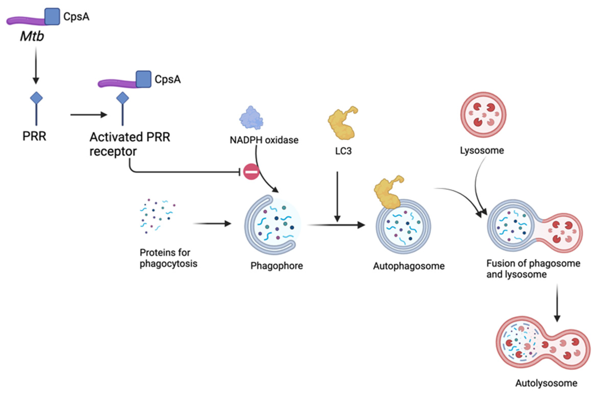
2.3. Role of Autophagy in Mtb Infection

Autophagy is a conserved cellular process crucial for maintaining cellular homeostasis by eliminating damaged organelles, misfolded proteins, and intracellular pathogens, which are engulfed by lysosomes for degradation [55]. Lysosomes break down these substrates and either recycle or create new sources of energy with the basic components [55]. Autophagy is triggered under stress conditions, such as caloric restriction, and as a defense against intracellular pathogens [56,57].
In the context of Mtb infection, autophagy plays a multifaceted role in innate and adaptive immune responses against the pathogen, including modulating proinflammatory cytokine production [58]. Host cells have pattern recognition receptors (PRRs) such as Toll-like receptors (TLRs) and nucleotide-binding oligomerization domain (NOD)-like receptors (NLRs) that recognize Mtb-associated molecular patterns (MAMPs) and trigger autophagy activation [59] as part of the innate immune response to control bacterial replication and promote antigen presentation [60]. Various signaling pathways, including the mammalian target of rapamycin (mTOR) and AMP-activated protein kinase (AMPK) pathways, are involved in the regulation of autophagy induction during Mtb infection [61].
Autophagy targets intracellular Mtb for degradation within autophagosomes, leading to bacterial killing and antigen presentation to activate adaptive immune responses [62]. Autophagic machinery components, such as autophagy-related proteins (ATGs) and LC3 (microtubule-associated protein 1A/1B-light chain 3), are recruited to Mtb-containing phagosomes to initiate autophagosome formation and engulf the bacteria [63,64]. Subsequently, autophagosomes fuse with lysosomes to form autolysosomes, where Mtb is degraded by lysosomal hydrolases [65].
Despite being targeted for autophagic clearance, Mtb has evolved various strategies to subvert or evade host autophagy mechanisms [29]. Virulent strains of Mtb can inhibit autophagy by interfering with signaling pathways or blocking autophagosome–lysosome fusion, allowing the bacteria to survive and replicate within host cells [66]. For example, LC3-associated phagocytosis (LAP) is a macrophage pathway which eliminates microbes by the recruitment of nicotinamide adenine dinucleotide phosphate (NADPH) oxidase to phagosomes, followed by phagosome association with LC3 and delivery of the bacteria to a degradative lysosome [67]. Koster et al. discovered that Mtb contains possible conserved protein A (CpsA), a member of the lytic repressor (LytR)-CpsA-PBP 5 synthesis repressor (Psr) (LCP) family involved in cell wall metabolism, which offers protection from LAP and ultimately prevents the successful clearance of Mtb, as seen in Figure 1 [68]. Furthermore, Mtb can actively inhibit autophagy by secreting virulence factors such as ESAT-6 (early secreted antigenic target 6) and CFP-10 (culture filtrate protein 10), which disrupt autophagic flux and impair bacterial clearance [69].
In addition to its direct antimicrobial effects, autophagy modulates host immune responses to Mtb infection by regulating inflammation, antigen presentation, and cytokine production. Autophagy-mediated degradation of Mtb-derived antigens enhances major histocompatibility complex (MHC) class II presentation to CD4+ T cells, promoting adaptive immunity against the pathogen [70]. Moreover, autophagy suppresses excessive inflammation by selectively targeting inflammasome components for degradation, thereby dampening cytokine release and mitigating immunopathology [71].

2.4. Foam Cell Macrophages and Mtb-Mediated Modulation

Macrophage-derived foam cells are filled with lipid droplets from the increased uptake of extracellular lipids from dead cells during Mtb infection [72,73]. These foam cells are observed in Mtb granulomas [72,73]. Histologically, Mtb foam cells are similar to those in atherosclerosis, though they distinctly have increased triglycerides relative to cholesterol [73]. Foam cells also produce eicosanoids, a lipid-based signaling molecule including prostaglandins, leukotrienes, and thromboxane [74,75]. An in vivo study in mice by Sorgi et al. further investigated the role of eicosanoids, in particular leukotriene and prostaglandins. While there were increased amounts of prostaglandin E2 (PGE2) detected during Mtb infection, the pharmacological inhibition of COX-2 induced a significant reduction in bacterial load via increased NO release and IFN-γ production and increased macrophage phagocytosis of Mtb. Additionally, a lack of the 5-lipoxygenase (5-LO) pathway necessary for producing leukotrienes was associated with Mtb resistance, and leukotriene B4 (LTB4) inoculation in mice without 5-LO increased susceptibility to Mtb infection [76]. This study implied that while PGE2 and LTB4 have proinflammatory effects necessary to combat Mtb, a fine balance is important to prevent severe inflammation that may shift the cell toward necrosis and ultimately Mtb dissemination [76]. Nore et al. further confirmed this with a study that showed the plasma levels of eicosanoids appearing to fluctuate across different clinical states of Mtb infection [77].

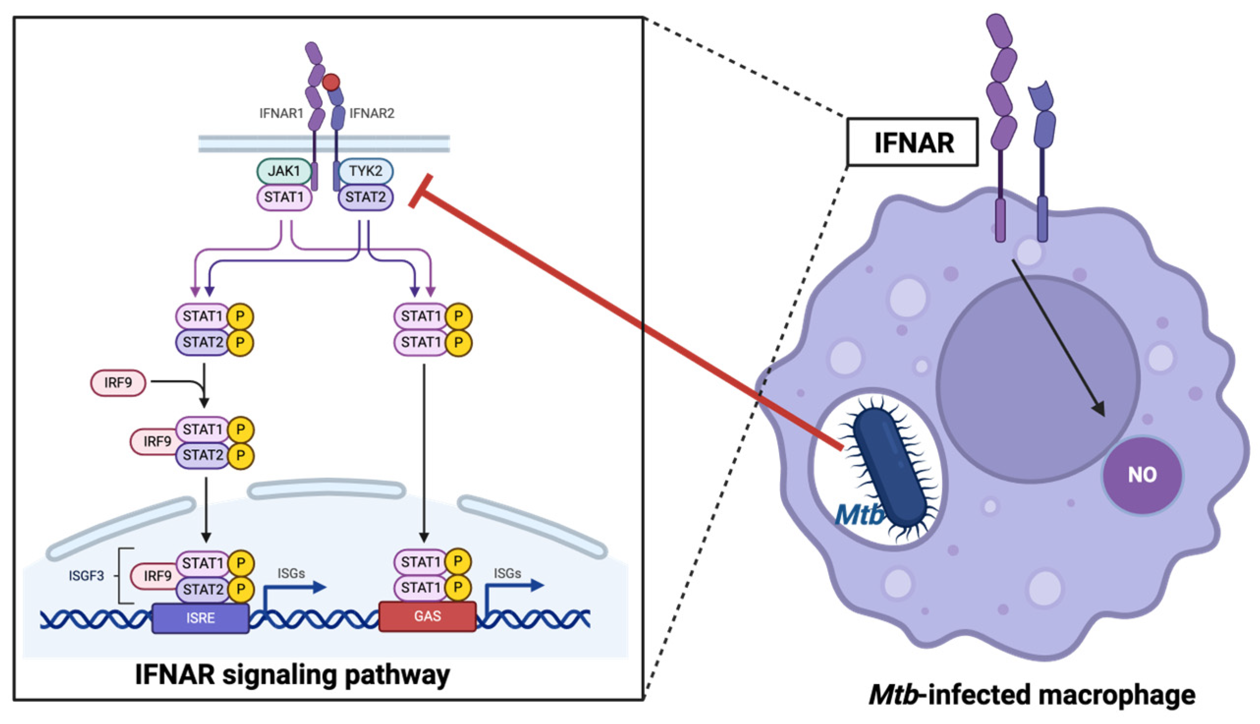
2.5. Role of Type I Interferons

Interferons (IFNs) are a group of signaling proteins that play a critical role in the immune response to infections, though not all types of interferons are equally involved in the response to Mtb infection due to their distinct mechanisms of action and the unique ways they modulate the immune system [22,78]. It is well known that IFN-γ, a type II interferon, is crucial for activating macrophages and is ultimately a protective function against Mtb [79]. In contrast, type I interferons (IFNs), including IFN-α and IFN-β, are pleiotropic cytokines that play a crucial role in the host response to viral infections [80,81]. However, emerging evidence suggests that type I IFNs may impair the immune response to Mtb infection and promote Mtb infection, albeit with complex and context-dependent effects [82,83].
A transcriptomic study in 2010 conducted among patients with active Mtb disease and individuals either latently infected or healthy in the UK and South Africa revealed type I IFN-inducible genes [84]. Specifically, there is an upregulation of IFN response genes, including STAT1 [82], IFITs [85], GBPs [86], MX1 [87], OAS1 [88], IRF1 [89], and others, in individuals progressing from tuberculosis exposure to active disease [82,90,91,92].
Sustained or dysregulated production of type I IFNs during chronic Mtb infection can have detrimental effects on the host immune response [93]. Excessive type I IFN signaling has been linked with immunopathology, including impaired macrophage function, dysregulated cytokine production, and altered T cell responses [93]. Specifically, the heightened production of type I IFN has been linked to the virulence of Mtb strains and heightened host vulnerability. Investigations into infections involving hypervirulent clinical isolates of Mtb, such as HN878 and BTB 02-171, in comparison to the less virulent laboratory strain (H37Rv), have demonstrated a direct association between elevated levels of type I IFN and heightened mycobacterial virulence [94,95,96]. This dysregulated immune response can contribute to the persistence of Mtb, exacerbating disease severity and promoting tissue damage [82].
A recent study showed that IFN-β signaling in mouse macrophages has a protective effect against Mtb by increasing the production of nitric oxide, which can inhibit bacterial growth. However, Mtb has evolved mechanisms to inhibit type I IFN signaling in infected cells, likely as a strategy to evade host defense mechanisms [97], highlighting the intricate interplay between the host immune response and Mtb evasion strategies during infection. This protection is mediated by direct signaling through the IFN-associated receptor (IFNAR), as blocking IFNAR1 prevents nitric oxide production [97]. Mtb inhibits IFN-α/β receptor-mediated signaling and the transcription of 309 IFN-β stimulated genes in a dose-dependent manner. This inhibition involves reduced phosphorylation of IFNAR-associated kinases JAK1 and TYK2, leading to decreased phosphorylation of downstream targets STAT1 and STAT2 [97], as seen in Figure 2. Mtb-mediated inhibition of type I IFN signaling occurs only in infected cells, suggesting an evasion of host defense mechanisms.

3. Host Cell Death Pathways in Mtb Infection

Once the bacteria have been phagocytosed and are able to survive the host immune response, a cascade of reactions can occur, leading to various outcomes. The process of phagocytosis is mediated by receptors on host macrophages’ surfaces, such as macrophage mannose receptors and complement receptors [98]. These receptors have been shown to be requirements necessary for the beginning of the intracellular pathway of Mtb [98].

3.1. Apoptosis: In Favor of the Host

Apoptosis, or programmed cell death, is a highly regulated process essential for maintaining tissue homeostasis and eliminating infected or damaged cells. Researchers have identified host macrophage apoptosis as a key protective factor in innate immune response, restricting the growth of Mtb through deprivation of the bacteria’s niche cell, eliminating an environment conducive for bacterial growth and replication, and facilitating the clearance of infected cells [99]. The apoptosis of infected macrophages is commonly considered to be most likely beneficial to the host cells and can reduce the viability of Mtb [100], though another hypothesis proposes that it is most beneficial in the early stages of Mtb infection [101]. In an in vitro study investigating Fas-mediated apoptosis, it was observed that the controlled apoptotic event of macrophages led to a significant reduction in bacterial viability [100]. Apoptotic cells are rapidly engulfed and cleared by phagocytes via a process called efferocytosis, preventing the release of intracellular Mtb and minimizing inflammation and tissue damage [102,103]. In particular, Mtb is only eradicated following efferocytosis, suggesting that apoptosis alone does not inherently eliminate the bacterium but necessitates subsequent phagocytic ingestion and lysosomal fusion of the apoptotic body containing the bacterium [103].

3.2. Caspase-Dependent Pathways

Mtb-induced apoptosis can be triggered through various signaling pathways, including intrinsic and extrinsic pathways, both of which converge at the activation of caspases, ultimately resulting in cell death via apoptosis [104]. The intrinsic pathway, also known as the mitochondrial pathway of apoptosis, is initiated by internal cellular stress signals such as DNA damage, oxidative stress, or growth factor withdrawal. Consequent cytotoxic stimuli and proapoptotic signal-transducing molecules, such as those of the Bcl family, induce permeability in the outer mitochondrial membrane [105,106]. Upon disruption of the outer mitochondrial membrane, various proteins found in the mitochondrial intermembrane space are released into the cytoplasm, most notably cytochrome c [107], triggering the formation of the apoptosome, a protein complex that activates caspase enzymes [108]. Another notable protein released is apoptosis-inducing factor (AIF), which provokes the release of caspase 9 [109]. This activation cascade ultimately leads to the cleavage of cellular components, DNA fragmentation, and cell death, as seen in Figure 3.
Snyder et al. showed that nitric oxide (NO) also activates the intrinsic pathway of apoptosis, specifically by activating Bcl-2 proteins BAX and BAK and releasing cytochrome c from the mitochondria [110,111]. Additionally, Herbst et al. showed that IFN-γ-activated macrophages upregulated the expression of nitric oxide synthase (NOS2) and exposes bacteria to nitric oxide (NO) via the caspase 3 and 7 pathways, allowing IFN-γ-induced apoptosis, as seen in Figure 3 [112].
In contrast, the extrinsic pathway, also known as the death receptor pathway of apoptosis, is initiated by external signals binding to death receptors on the cell surface, such as Fas ligand (FasL), tumor necrosis factor alpha (TNF-α), tumor necrosis factor-related apoptosis-inducing ligand RI (TRAIL-RI), and TRAIL-RII [100,104,113]. Binding of the ligand to the death receptor activates initiator caspase 8, which can directly cleave downstream effector caspases such as caspase 3 and, ultimately, cellular substrates and apoptosis, as seen in Figure 3 [114].
A study by Kelly et al. showed evidence of bystander macrophage apoptosis contributing to the innate immune response against Mtb, wherein infected macrophages induce the apoptosis of uninfected macrophages via a cell-to-cell contact-dependent mechanism [115]. There was no evidence of typical intrinsic or extrinsic pathway involvement of TNF- a, Fas, tumor necrosis factor-related apoptosis-inducing ligand, transforming growth factor β, Toll-like receptor 2, or MyD88 in contact-mediated bystander apoptosis [115]. Following cell death, antigen-presenting cells release extracellular vesicles that carry mycobacterial antigens to uninfected bystander cells to activate CD8 T cells that can recognize and lyse infected cells [116].
There are various studies showing that Mtb may both induce and inhibit apoptosis, which may be influenced by the viability of bacteria and host cells as well as the timing of infection [18]. Mtb’s ability to modulate apoptosis was seen in an in vitro study that showed different virulent strains inducing different levels of apoptosis. Specifically, virulent forms of Mtb modulated and reduced the occurrence of apoptosis, whereas more apoptosis occurred in avirulent or attenuated strains [117]. Mtb can secrete several proteins that inhibit apoptosis [18].
For example, Mtb may evade apoptosis by the release of TNF-R2 via an IL-10-dependent mechanism, resulting in the inactivation of TNF-α, as seen in Figure 3 [118,119,120]. TNF-α, a proinflammatory cytokine, has been identified as a factor that is associated with attenuated Mtb strains and apoptosis [118]. One piece of research showed that mice treated with a monoclonal antibody against TNF-α and those deficient in the TNF-α receptor developed lethal Mtb infections due to cell necrosis in comparison to controls [121]. Additionally, Mtb induces IL-10 production in macrophages, which further downregulates TNF-a production [119,120]. IL-10 is an anti-inflammatory cytokine associated with reduced CD4+ T cell response in Mtb infection [122,123]. IL-10 has also been found to decrease cytokine response in lymph nodes and lung granulomas, as well as reducing collagenization and fibrosis, affecting the structure of granulomas and demonstrating IL-10’s significant role in the modulation of the immune response and Mtb’s evasion of the adaptive immune system [124]. Furthermore, research shows that antigen-specific CD4+ T cells are activated once Mtb is in the mediastinal lymph nodes, despite the fact that there are a much larger number of bacteria in the lungs. These results allowed researchers to conclude that the delay in the activation of CD4+ T cells occurs due to a delay in the transport of Mtb from the lungs to the lymph nodes [125].
Mtb can also evade apoptosis by taking advantage of antiapoptotic mechanisms in the intrinsic apoptosis pathway [126,127,128]. Members of the B cell lymphoma 2 (BCL-2) family regulate apoptosis via the inhibition and induction of cell death [128,129]. They directly bind and regulate mitochondrial outer membrane permeability, leading to the release of proteins from the intermembrane space necessary for initiating the caspase cascade and ultimately apoptosis [130]. NF-κB has antiapoptotic activity and its activation inhibits death receptor-activated caspases [131]. As such, a study by Dhiman et al. found that the Mtb activation of NF-κB led to the upregulation of the BCL-2 antiapoptotic member bfl-1A1 [126]. Another study by Sly et al. found that virulent strains of Mtb upregulated and induced Mcl-1 expression [127,132].
Conversely, Mtb may facilitate host cell apoptosis via surface proteins or secreted proteins. For example, Mtb Rv3261 protein inhibits Mtb growth in macrophages and induces ROS production, which activates the caspase 3/9-dependent pathway and leads to macrophage apoptosis [133]. An in silico analysis study conducted by Medha et al. showed that Mtb PE6 (Rv0335c) had similar C-terminal and N-terminal sequences to Bcl2 proteins [134]. In vitro experiments further confirmed that Rv0335c’s molecular similarity to host Bcl2 proteins likely allows for host mitochondrial perturbations and apoptosis. A proposed hypothesis is that apoptosis during late-stage infection during granuloma formation might be a pathogenic process that increases Mtb’s persistence and future dissemination [101].

3.3. Caspase-Independent Pathways

Apoptosis during Mtb infection can occur through various caspase-independent pathways, including ROS induction, mitochondrial dysfunction, and endoplasmic reticulum (ER) stress [135].
ROS play a complex role in Mtb infection, acting as both a host defense mechanism and a target for bacterial evasion strategies [136]. ROS-induced oxidative stress can activate antimicrobial defense pathways within host cells, leading to the induction of autophagy and programmed cell death (apoptosis) to eliminate intracellular Mtb [137]. Furthermore, upon encountering Mtb, immune cells such as macrophages and neutrophils undergo a respiratory burst, resulting in the rapid production of ROS [138]. ROS exert direct antimicrobial effects by damaging essential macromolecules such as lipids, proteins, and DNA within Mtb. Furthermore, ROS can disrupt Mtb’s redox balance, impairing its metabolic activity and promoting bacterial killing [139].
Despite the bactericidal effects of ROS, Mtb has evolved various strategies to counteract oxidative stress and survive within the hostile intracellular environment. Firstly, Mtb possesses antioxidant defense mechanisms, including enzymes such as catalase-peroxidase (KatG) and superoxide dismutase (SOD) (Figure 4), which detoxify ROS and protect the bacterium from oxidative damage [140]. It has been reported that Mtb can evade host immunity by binding to triggering receptor expressed on myeloid cells (TREM2), a macrophage surface receptor that mediates anti-inflammatory immune signaling. This promotes the intracellular survival of Mtb by inducing type I IFN signaling and suppressing the production of ROS [45]. In another study, Afriyie et al. found that Mtb can inhibit ROS production and induce necrotic cell death by reducing the expression of focal adhesion kinase (FAK), which led to necrotic cell death and persistent lung damage [141]. The study used pharmacological interventions to inhibit FAK, which increased necrosis due to Mtb. In contrast, genetic overexpression of FAK prevented cell death during Mtb infection [141].
As previously mentioned above regarding the intrinsic pathway of apoptosis, mitochondrial damage releases apoptosis-inducing factor (AIF), which triggers chromatin condensation and DNA fragmentation [142]. Initial studies show the translocation of AIF from the mitochondria to the cytosol and ultimately the nucleus, where it facilitates chromatin condensation [135].
Stress on the endoplasmic reticulum (ER) can result in the accumulation and aggregation of unfolded proteins, which trigger apoptosis via the unfolded protein response [143,144,145]. Surprisingly, live Mtb H37Rv can induce an ER stress response, implying that the ER stress pathway also contributes to the persistence and pathogenesis of Mtb [146]. A recent study by Xu et al. discovered a novel Mtb protein CDP-diglyceride hydrolase (Cdh), which interacted with the endoplasmic reticulum and led to cell death [147].

4. Necrosis: In Favor of Mtb

Necrosis, in contrast to apoptosis, occurs due to cytotoxic signals that result in the swelling of cell organelles, plasma membrane rupture, and the eventual lysis of the cells, leading to the release of proteins that contribute to inflammasome activation and the secretion of proinflammatory cytokines [148]. Although traditionally considered a passive and unregulated form of cell death while apoptosis was considered the regulated form of cell death, emerging evidence suggests that necrosis can be induced by specific signaling pathways and molecular mechanisms, a process termed “regulated necrosis” or “necroptosis” [149].
During Mtb infection, necrosis can be induced through various pathways, including necroptosis, pyroptosis, and ferroptosis. Necroptosis is a form of regulated necrosis mediated by receptor-interacting protein kinases (RIPKs) and mixed-lineage kinase domain-like protein (MLKL), leading to plasma membrane rupture and the release of damage-associated molecular patterns (DAMPs) that exacerbate inflammation and tissue injury [149]. Pyroptosis, mediated by inflammasome activation and caspase 1 activation, leads to rapid cell lysis and the release of proinflammatory cytokines, promoting host defense but also contributing to immunopathology [150]. Ferroptosis, characterized by iron-dependent lipid peroxidation and oxidative stress, represents a novel form of regulated necrosis implicated in Mtb pathogenesis [151].
Virulent strains of Mtb interfere with the progression of apoptosis and instead facilitate necrosis to necrosis [152]. Investigation of the interaction between Mtb and macrophages finds that three distinct mechanisms contribute to macrophage necrosis. First, Mtb inhibits plasma membrane repair [153]. Second, virulent Mtb causes inner mitochondrial membrane damage [152]. Third, virulent Mtb inhibits the cross-linking of annexin-1, which impairs formation of the apoptotic envelope [28]. This proves to have significant outcomes as necrotic macrophages provide Mtb with an environment that is best suited for replication [154]. Ultimately, macrophage necrosis results in increased replication, dissemination, and virulence in Mtb.

5. Therapeutic Considerations Targeting Autophagy, Apoptosis, and Necrosis in Mtb Infection

Several pharmacologic agents have been identified for their ability to target autophagy to promote antimicrobial effects against Mtb infection. For example, the use of small molecules or compounds that activate autophagy pathways, such as rapamycin, an mTOR inhibitor, has been shown to induce autophagy and enhance the killing of Mtb in infected macrophages [155]. Metformin, an activator of AMP-activated protein kinase (AMPK) and regulator of the autophagy pathway, has been shown to induce autophagy, even in cases of Mtb drug resistance [61,156]. Transcription factors such as TFEB (transcription factor EB) regulate the expression of genes involved in autophagy and lysosomal biogenesis seen in Mtb. Other drugs investigated for their potential in anti-Tb therapy and inducing autophagy include histone deacetylase (HDAC) inhibitors [157], lysosomotropic agents [158], which accumulate in acidic compartments such as lysosomes and may enhance the autophagic clearance of intracellular Mtb, and the antiprotozoal drug nitazoxanide and its analogs, which activate autophagosome formation and mTORC1 inhibition [159]. Vitamin D has also demonstrated efficacy in reducing Mtb load through synthesis of the autophagic antimicrobial peptide LL-37, the active form of cathelecidin antimicrobial peptide (CAMP) [160].
Targeting apoptosis as a therapeutic strategy for Mtb infection is an active area of research, with several potential therapies under investigation. For example, compounds that can directly induce apoptosis in Mtb-infected cells may have therapeutic potential, such as BCL-2 homology domain 3 (BH3) mimetics, which mimic the action of proapoptotic proteins [161]. For Mtb strains which have evolved mechanisms to inhibit host cell apoptosis as a means of promoting their survival, targeting antiapoptotic pathways may enhance the clearance of Mtb-infected cells, as seen with cellular inhibitors of apoptosis proteins (cIAPs), which suppress apoptosis [162,163]. Host factors that regulate apoptosis can be targeted for therapeutic intervention. p53, a tumor suppressor protein that plays a role in apoptosis, has been implicated in the host response to Mtb infection. Lim et al. found that nutlin-3, a p53 activator, significantly suppressed the intracellular survival of Mtb in hosts, suggesting another potential therapeutic target for Mtb therapy [164]. Therapies aimed at modulating the host immune response to Mtb infection may indirectly influence apoptosis and promote bacterial clearance. For example, cytokine-based therapies or immune checkpoint inhibitors could enhance the ability of the immune system to eliminate Mtb-infected cells through apoptosis, though there have been several reported cases of severe or reactivated Mtb in cancer patients [165,166].
Targeting necrosis as a therapeutic strategy for Mtb infection is also undergoing exploration, although specific therapies directly targeting necrosis are limited. Since necrosis is often associated with excessive inflammation, anti-inflammatory agents may help mitigate necrosis-associated tissue damage and inflammation during Mtb infection. For example, corticosteroids or non-steroidal anti-inflammatory drugs (NSAIDs) may be used as adjunctive therapy to reduce inflammation and tissue damage caused by necrosis [167]. NSAIDs and corticosteroids, among other therapies modulating eicosanoids such as the 5-lipoxygenase inhibitor zileuton and amine tricyclic antidepressant desipramine, have also demonstrated an ability to reduce necroptosis by impeding the arachidonic acid pathway [168]. This pathway is utilized by virulent strains of Mtb through the production of lipoxin A4, which favors necrosis by inhibiting prostaglandin E2, an essential cellular membrane maintenance eicosanoid. The arachidonic acid pathway also leads to increased TNF, a necroptotic cytokine favoring high inflammation states such as Mtb infection, which can be targeted by phosphodiesterase (PDE) inhibitors [169], specifically the PDE4 inhibitor CC-11050, which has demonstrated an enhancement of isoniazid antimycobacterial activity in animal models [169].
While necrosis itself is not directly targeted by antimicrobial agents, the effective treatment of Mtb infection with antibiotics may help prevent or reduce necrosis by controlling bacterial replication and limiting tissue damage. First-line anti-tuberculosis drugs, such as isoniazid, rifampicin, and pyrazinamide, are essential components of standard TB treatment regimens and play a crucial role in controlling Mtb growth and spread [170]. Unfortunately, standard antibiotic therapy involves expensive long-term treatment and side effects and has led to the development of multi-drug-resistant TB [171]. Alternative antibiotics and nitroimidazoles, such as delamanid and pretomanid, have been investigated; however, concerns exist over their resistance and side effects [172]. Experimentation with alternative therapies, such as the utilization of bacteriophages to deliver lytic enzymes, has been proposed as adjunctive therapy for current treatment regimens, but such approaches remain experimental [173]. Host-directed therapies that aim to modulate host factors involved in necrosis may hold promise for improving treatment outcomes in Mtb infection. For example, agents targeting cell death pathways, such as necroptosis inhibitors or modulators of mitochondrial function, could potentially reduce necrosis and its detrimental effects on host tissues [168]. Potential adjunctive therapies which target autophagy, apoptosis, and necrosis, outlined in Table 1, may improve the current TB treatment landscape in reducing antimicrobial resistance.

6. Conclusions

In conclusion, the intricate balance between Mtb infection, immunomodulation, and host cell death via apoptosis underscores the critical role of programmed cell death in controlling tuberculosis pathogenesis. Through apoptosis, infected cells sacrifice themselves, curtailing the replication and dissemination of Mtb within the host. This not only limits the spread of the pathogen but also facilitates the removal of infected cells by the immune system, thus impeding disease progression. Without apoptosis-mediated host cell death, Mtb would exploit host resources unchecked, leading to an uncontrolled proliferation and exacerbation of infection, preferably through necrosis. Furthermore, apoptosis plays a pivotal role in shaping the immune response, facilitating antigen presentation, and orchestrating the clearance of Mtb-infected cells. Therefore, understanding the mechanisms governing apoptosis during Mtb infection is paramount for developing effective therapeutic strategies to combat tuberculosis. A better understanding of host cell death pathways may offer promising avenues for the development of novel interventions aimed at bolstering the host’s defense mechanisms against Mtb, ultimately advancing efforts towards tuberculosis control and eradication.
While apoptosis-mediated host cell death is a crucial defense mechanism against Mtb infection, it has several potential side effects that can impact the human body. While apoptosis is generally a non-inflammatory form of cell death, excessive or dysregulated apoptosis [174] can lead to significant side effects, including tissue damage, inflammation, and the disruption of granuloma integrity, leading to the dissemination of Mtb [175]. Additionally, virulent strains of Mtb have evolved mechanisms to subvert apoptosis, leading to macrophage necrosis [28,176] and, thus, the release of cellular contents and inflammation. Persistent inflammation can exacerbate tissue damage and contribute to disease progression [151,177]. Lastly, apoptotic cells can sometimes be less effective in presenting antigens to T cells compared to cells that undergo other forms of cell death, which can impair the activation of specific immune responses necessary for controlling Mtb infection [178]. Understanding these side effects is important for developing therapeutic strategies that can modulate apoptosis and improve outcomes in tuberculosis.

Author Contributions

Conceptualization, V.V., A.V. and I.G.; methodology, V.V., A.V. and I.G.; resources, A.V.; writing—original draft preparation, V.V., A.V., I.G., G.C., S.Y., J.N. and A.S.; writing—review and editing, V.V., A.V. and I.G.; visualization, A.V.; supervision, V.V., A.V. and I.G.; project administration, V.V.; funding acquisition, V.V. All authors have read and agreed to the published version of the manuscript.

Funding

We appreciate the funding support of NIH-NHLBI (2R15HL143545-02).

Data Availability Statement

No new data were created or analyzed in this study. Data sharing is not applicable to this article.

Conflicts of Interest

The authors declare no conflicts of interest.

References

  1. Zhang, T.; Zhang, J.; Wei, L.; Liang, H.; Zhang, J.; Shi, D.; Wang, Z. The global, regional, and national burden of tuberculosis in 204 countries and territories, 1990–2019. J. Infect. Public Health 2023, 16, 368–375. [Google Scholar] [CrossRef] [PubMed]
  2. World Health Organization. Global Tuberculosis Report 2023. 2023. Available online: https://www.who.int/publications/i/item/9789240083851 (accessed on 12 February 2024).
  3. World Health Organization. Global Tuberculosis Report 2021. 2021. Available online: https://www.who.int/publications/i/item/9789240037021 (accessed on 12 February 2024).
  4. Luies, L.; du Preez, I. The Echo of Pulmonary Tuberculosis: Mechanisms of Clinical Symptoms and Other Disease-Induced Systemic Complications. Clin. Microbiol. Rev. 2020, 33, 10-1128. [Google Scholar] [CrossRef] [PubMed]
  5. Bruchfeld, J.; Correia-Neves, M.; Källenius, G. Tuberculosis and HIV Coinfection. Cold Spring Harb. Perspect. Med. 2015, 5, a017871. [Google Scholar] [CrossRef] [PubMed]
  6. Menzies, D. Effect of treatment on contagiousness of patients with active pulmonary tuberculosis. Infect. Control Hosp. Epidemiol. 1997, 18, 582–586. [Google Scholar] [CrossRef]
  7. Gideon, H.P.; Flynn, J.L. Latent tuberculosis: What the host “sees”? Immunol. Res. 2011, 50, 202–212. [Google Scholar] [CrossRef] [PubMed]
  8. Flynn, J.L.; Chan, J. Tuberculosis: Latency and reactivation. Infect. Immun. 2001, 69, 4195–4201. [Google Scholar] [CrossRef] [PubMed]
  9. Ravimohan, S.; Kornfeld, H.; Weissman, D.; Bisson, G.P. Tuberculosis and lung damage: From epidemiology to pathophysiology. Eur. Respir. Rev. 2018, 27, 170077. [Google Scholar] [CrossRef] [PubMed]
  10. Vohra, S.; Dhaliwal, H.S. Miliary Tuberculosis. In StatPearls; StatPearls Publishing LLC.: Treasure Island, FL, USA, 2024. [Google Scholar]
  11. Slane, V.H.; Unakal, C.G. Tuberculous Meningitis. In StatPearls; StatPearls Publishing LLC.: Treasure Island, FL, USA, 2024. [Google Scholar]
  12. Naicker, K.; Ntsekhe, M. Tuberculous pericardial disease: A focused update on diagnosis, therapy and prevention of complications. Cardiovasc. Diagn. Ther. 2020, 10, 289–295. [Google Scholar] [CrossRef] [PubMed]
  13. Seung, K.J.; Keshavjee, S.; Rich, M.L. Multidrug-Resistant Tuberculosis and Extensively Drug-Resistant Tuberculosis. Cold Spring Harb. Perspect. Med. 2015, 5, a017863. [Google Scholar] [CrossRef]
  14. Bussi, C.; Gutierrez, M.G. Mycobacterium tuberculosis infection of host cells in space and time. FEMS Microbiol. Rev. 2019, 43, 341–361. [Google Scholar] [CrossRef]
  15. Augenstreich, J.; Haanappel, E.; Ferre, G.; Czaplicki, G.; Jolibois, F.; Destainville, N.; Guilhot, C.; Milon, A.; Astarie-Dequeker, C.; Chavent, M. The conical shape of DIM lipids promotes Mycobacterium tuberculosis infection of macrophages. Proc. Natl. Acad. Sci. USA 2019, 116, 25649–25658. [Google Scholar] [CrossRef] [PubMed]
  16. Ramakrishnan, L. Revisiting the role of the granuloma in tuberculosis. Nat. Rev. Immunol. 2012, 12, 352–366. [Google Scholar] [CrossRef] [PubMed]
  17. Russell, D.G. Who puts the tubercle in tuberculosis? Nat. Rev. Microbiol. 2007, 5, 39–47. [Google Scholar] [CrossRef] [PubMed]
  18. Nisa, A.; Kipper, F.C.; Panigrahy, D.; Tiwari, S.; Kupz, A.; Subbian, S. Different modalities of host cell death and their impact on Mycobacterium tuberculosis infection. Am. J. Physiol. Cell Physiol. 2022, 323, C1444–C1474. [Google Scholar] [CrossRef] [PubMed]
  19. Lin, P.L.; Plessner, H.L.; Voitenok, N.N.; Flynn, J.L. Tumor necrosis factor and tuberculosis. J. Investig. Dermatol. Symp. Proc. 2007, 12, 22–25. [Google Scholar] [CrossRef] [PubMed]
  20. Yuk, J.M.; Kim, J.K.; Kim, I.S.; Jo, E.K. TNF in Human Tuberculosis: A Double-Edged Sword. Immune Netw. 2024, 24, e4. [Google Scholar] [CrossRef] [PubMed]
  21. Alspach, E.; Lussier, D.M.; Schreiber, R.D. Interferon γ and Its Important Roles in Promoting and Inhibiting Spontaneous and Therapeutic Cancer Immunity. Cold Spring Harb. Perspect. Biol. 2019, 11, a028480. [Google Scholar] [CrossRef] [PubMed]
  22. Shanmuganathan, G.; Orujyan, D.; Narinyan, W.; Poladian, N.; Dhama, S.; Parthasarathy, A.; Ha, A.; Tran, D.; Velpuri, P.; Nguyen, K.H.; et al. Role of Interferons in Mycobacterium tuberculosis Infection. Clin. Pract. 2022, 12, 788–796. [Google Scholar] [CrossRef] [PubMed]
  23. Bradley, L.M.; Dalton, D.K.; Croft, M. A direct role for IFN-gamma in regulation of Th1 cell development. J. Immunol. 1996, 157, 1350–1358. [Google Scholar] [CrossRef]
  24. Ashida, H.; Mimuro, H.; Ogawa, M.; Kobayashi, T.; Sanada, T.; Kim, M.; Sasakawa, C. Cell death and infection: A double-edged sword for host and pathogen survival. J. Cell Biol. 2011, 195, 931–942. [Google Scholar] [CrossRef]
  25. Green, D.R.; Llambi, F. Cell Death Signaling. Cold Spring Harb. Perspect. Biol. 2015, 7, a006080. [Google Scholar] [CrossRef] [PubMed]
  26. Galluzzi, L.; Vitale, I.; Aaronson, S.A.; Abrams, J.M.; Adam, D.; Agostinis, P.; Alnemri, E.S.; Altucci, L.; Amelio, I.; Andrews, D.W.; et al. Molecular mechanisms of cell death: Recommendations of the Nomenclature Committee on Cell Death 2018. Cell Death Differ. 2018, 25, 486–541. [Google Scholar] [CrossRef]
  27. Tang, D.; Kang, R.; Berghe, T.V.; Vandenabeele, P.; Kroemer, G. The molecular machinery of regulated cell death. Cell Res. 2019, 29, 347–364. [Google Scholar] [CrossRef]
  28. Behar, S.M.; Martin, C.J.; Booty, M.G.; Nishimura, T.; Zhao, X.; Gan, H.X.; Divangahi, M.; Remold, H.G. Apoptosis is an innate defense function of macrophages against Mycobacterium tuberculosis. Mucosal Immunol. 2011, 4, 279–287. [Google Scholar] [CrossRef]
  29. Chai, Q.; Wang, L.; Liu, C.H.; Ge, B. New insights into the evasion of host innate immunity by Mycobacterium tuberculosis. Cell Mol. Immunol. 2020, 17, 901–913. [Google Scholar] [CrossRef] [PubMed]
  30. Eberhardt, N.; Bergero, G.; Mazzocco Mariotta, Y.L.; Aoki, M.P. Purinergic modulation of the immune response to infections. Purinergic Signal. 2022, 18, 93–113. [Google Scholar] [CrossRef]
  31. Fremond, C.M.; Yeremeev, V.; Nicolle, D.M.; Jacobs, M.; Quesniaux, V.F.; Ryffel, B. Fatal Mycobacterium tuberculosis infection despite adaptive immune response in the absence of MyD88. J. Clin. Investig. 2004, 114, 1790–1799. [Google Scholar] [CrossRef] [PubMed]
  32. Cohen, S.B.; Gern, B.H.; Delahaye, J.L.; Adams, K.N.; Plumlee, C.R.; Winkler, J.K.; Sherman, D.R.; Gerner, M.Y.; Urdahl, K.B. Alveolar Macrophages Provide an Early Mycobacterium tuberculosis Niche and Initiate Dissemination. Cell Host Microbe 2018, 24, 439–446.e4. [Google Scholar] [CrossRef]
  33. Iantomasi, R.; Sali, M.; Cascioferro, A.; Palucci, I.; Zumbo, A.; Soldini, S.; Rocca, S.; Greco, E.; Maulucci, G.; De Spirito, M.; et al. PE_PGRS30 is required for the full virulence of Mycobacterium tuberculosis. Cell Microbiol. 2012, 14, 356–367. [Google Scholar] [CrossRef]
  34. van der Wel, N.; Hava, D.; Houben, D.; Fluitsma, D.; van Zon, M.; Pierson, J.; Brenner, M.; Peters, P.J. M. tuberculosis and M. leprae translocate from the phagolysosome to the cytosol in myeloid cells. Cell 2007, 129, 1287–1298. [Google Scholar] [CrossRef]
  35. Houben, D.; Demangel, C.; van Ingen, J.; Perez, J.; Baldeon, L.; Abdallah, A.M.; Caleechurn, L.; Bottai, D.; van Zon, M.; de Punder, K.; et al. ESX-1-mediated translocation to the cytosol controls virulence of mycobacteria. Cell Microbiol. 2012, 14, 1287–1298. [Google Scholar] [CrossRef] [PubMed]
  36. Chackerian, A.A.; Alt, J.M.; Perera, T.V.; Dascher, C.C.; Behar, S.M. Dissemination of Mycobacterium tuberculosis is influenced by host factors and precedes the initiation of T-cell immunity. Infect. Immun. 2002, 70, 4501–4509. [Google Scholar] [CrossRef] [PubMed]
  37. Jung, Y.J.; Ryan, L.; LaCourse, R.; North, R.J. Properties and protective value of the secondary versus primary T helper type 1 response to airborne Mycobacterium tuberculosis infection in mice. J. Exp. Med. 2005, 201, 1915–1924. [Google Scholar] [CrossRef]
  38. Garcia-Romo, G.S.; Pedroza-Gonzalez, A.; Lambrecht, B.N.; Aguilar-Leon, D.; Estrada-Garcia, I.; Hernandez-Pando, R.; Flores-Romo, L. Mycobacterium tuberculosis manipulates pulmonary APCs subverting early immune responses. Immunobiology 2013, 218, 393–401. [Google Scholar] [CrossRef] [PubMed]
  39. Srivastava, S.; Grace, P.S.; Ernst, J.D. Antigen Export Reduces Antigen Presentation and Limits T Cell Control of M. tuberculosis. Cell Host Microbe 2016, 19, 44–54. [Google Scholar] [CrossRef] [PubMed]
  40. Pai, R.K.; Pennini, M.E.; Tobian, A.A.; Canaday, D.H.; Boom, W.H.; Harding, C.V. Prolonged toll-like receptor signaling by Mycobacterium tuberculosis and its 19-kilodalton lipoprotein inhibits gamma interferon-induced regulation of selected genes in macrophages. Infect. Immun. 2004, 72, 6603–6614. [Google Scholar] [CrossRef] [PubMed]
  41. Geldmacher, C.; Schuetz, A.; Ngwenyama, N.; Casazza, J.P.; Sanga, E.; Saathoff, E.; Boehme, C.; Geis, S.; Maboko, L.; Singh, M.; et al. Early depletion of Mycobacterium tuberculosis-specific T helper 1 cell responses after HIV-1 infection. J. Infect. Dis. 2008, 198, 1590–1598. [Google Scholar] [CrossRef] [PubMed]
  42. Srivastava, S.; Ernst, J.D. Cutting edge: Direct recognition of infected cells by CD4 T cells is required for control of intracellular Mycobacterium tuberculosis in vivo. J. Immunol. 2013, 191, 1016–1020. [Google Scholar] [CrossRef] [PubMed]
  43. Green, A.M.; Difazio, R.; Flynn, J.L. IFN-gamma from CD4 T cells is essential for host survival and enhances CD8 T cell function during Mycobacterium tuberculosis infection. J. Immunol. 2013, 190, 270–277. [Google Scholar] [CrossRef]
  44. Lu, Y.J.; Barreira-Silva, P.; Boyce, S.; Powers, J.; Cavallo, K.; Behar, S.M. CD4 T cell help prevents CD8 T cell exhaustion and promotes control of Mycobacterium tuberculosis infection. Cell Rep. 2021, 36, 109696. [Google Scholar] [CrossRef]
  45. Mishra, A.; Singh, V.K.; Jagannath, C.; Subbian, S.; Restrepo, B.I.; Gauduin, M.C.; Khan, A. Human Macrophages Exhibit GM-CSF Dependent Restriction of Mycobacterium tuberculosis Infection via Regulating Their Self-Survival, Differentiation and Metabolism. Front. Immunol. 2022, 13, 859116. [Google Scholar] [CrossRef] [PubMed]
  46. Khader, S.A.; Partida-Sanchez, S.; Bell, G.; Jelley-Gibbs, D.M.; Swain, S.; Pearl, J.E.; Ghilardi, N.; Desauvage, F.J.; Lund, F.E.; Cooper, A.M. Interleukin 12p40 is required for dendritic cell migration and T cell priming after Mycobacterium tuberculosis infection. J. Exp. Med. 2006, 203, 1805–1815. [Google Scholar] [CrossRef] [PubMed]
  47. De Rosa, S.C.; Lu, F.X.; Yu, J.; Perfetto, S.P.; Falloon, J.; Moser, S.; Evans, T.G.; Koup, R.; Miller, C.J.; Roederer, M. Vaccination in humans generates broad T cell cytokine responses. J. Immunol. 2004, 173, 5372–5380. [Google Scholar] [CrossRef] [PubMed]
  48. Adams, D.O. The structure of mononuclear phagocytes differentiating in vivo. II. The effect of Mycobacterium tuberculosis. Am. J. Pathol. 1975, 80, 101–116. [Google Scholar] [PubMed]
  49. Algood, H.M.; Lin, P.L.; Flynn, J.L. Tumor necrosis factor and chemokine interactions in the formation and maintenance of granulomas in tuberculosis. Clin. Infect. Dis. 2005, 41 (Suppl. S3), S189–S193. [Google Scholar] [CrossRef] [PubMed]
  50. Sandor, M.; Weinstock, J.V.; Wynn, T.A. Granulomas in schistosome and mycobacterial infections: A model of local immune responses. Trends Immunol. 2003, 24, 44–52. [Google Scholar] [CrossRef] [PubMed]
  51. Chan, K.; Knaak, T.; Satkamp, L.; Humbert, O.; Falkow, S.; Ramakrishnan, L. Complex pattern of Mycobacterium marinum gene expression during long-term granulomatous infection. Proc. Natl. Acad. Sci. USA 2002, 99, 3920–3925. [Google Scholar] [CrossRef] [PubMed]
  52. Russell, D.G.; Cardona, P.J.; Kim, M.J.; Allain, S.; Altare, F. Foamy macrophages and the progression of the human tuberculosis granuloma. Nat. Immunol. 2009, 10, 943–948. [Google Scholar] [CrossRef] [PubMed]
  53. Ulrichs, T.; Kosmiadi, G.A.; Jorg, S.; Pradl, L.; Titukhina, M.; Mishenko, V.; Gushina, N.; Kaufmann, S.H. Differential organization of the local immune response in patients with active cavitary tuberculosis or with nonprogressive tuberculoma. J. Infect. Dis. 2005, 192, 89–97. [Google Scholar] [CrossRef]
  54. Kaplan, G.; Post, F.A.; Moreira, A.L.; Wainwright, H.; Kreiswirth, B.N.; Tanverdi, M.; Mathema, B.; Ramaswamy, S.V.; Walther, G.; Steyn, L.M.; et al. Mycobacterium tuberculosis growth at the cavity surface: A microenvironment with failed immunity. Infect. Immun. 2003, 71, 7099–7108. [Google Scholar] [CrossRef]
  55. D’Arcy, M.S. Cell death: A review of the major forms of apoptosis, necrosis and autophagy. Cell Biol. Int. 2019, 43, 582–592. [Google Scholar] [CrossRef] [PubMed]
  56. Gómez-Virgilio, L.; Silva-Lucero, M.D.; Flores-Morelos, D.S.; Gallardo-Nieto, J.; Lopez-Toledo, G.; Abarca-Fernandez, A.M.; Zacapala-Gómez, A.E.; Luna-Muñoz, J.; Montiel-Sosa, F.; Soto-Rojas, L.O.; et al. Autophagy: A Key Regulator of Homeostasis and Disease: An Overview of Molecular Mechanisms and Modulators. Cells 2022, 11, 2262. [Google Scholar] [CrossRef] [PubMed]
  57. Wileman, T. Autophagy as a defence against intracellular pathogens. Essays Biochem. 2013, 55, 153–163. [Google Scholar] [CrossRef] [PubMed]
  58. Kleinnijenhuis, J.; Oosting, M.; Plantinga, T.S.; van der Meer, J.W.; Joosten, L.A.; Crevel, R.V.; Netea, M.G. Autophagy modulates the Mycobacterium tuberculosis-induced cytokine response. Immunology 2011, 134, 341–348. [Google Scholar] [CrossRef] [PubMed]
  59. Zihad, S.; Sifat, N.; Islam, M.A.; Monjur-Al-Hossain, A.S.M.; Sikdar, K.; Sarker, M.M.R.; Shilpi, J.A.; Uddin, S.J. Role of pattern recognition receptors in sensing Mycobacterium tuberculosis. Heliyon 2023, 9, e20636. [Google Scholar] [CrossRef] [PubMed]
  60. Liu, C.H.; Liu, H.; Ge, B. Innate immunity in tuberculosis: Host defense vs pathogen evasion. Cell Mol. Immunol. 2017, 14, 963–975. [Google Scholar] [CrossRef] [PubMed]
  61. Jo, E.K.; Silwal, P.; Yuk, J.M. AMPK-Targeted Effector Networks in Mycobacterial Infection. Front. Microbiol. 2019, 10, 520. [Google Scholar] [CrossRef] [PubMed]
  62. Kim, Y.S.; Silwal, P.; Kim, S.Y.; Yoshimori, T.; Jo, E.K. Autophagy-activating strategies to promote innate defense against mycobacteria. Exp. Mol. Med. 2019, 51, 1–10. [Google Scholar] [CrossRef] [PubMed]
  63. Periyasamy-Thandavan, S.; Jiang, M.; Schoenlein, P.; Dong, Z. Autophagy: Molecular machinery, regulation, and implications for renal pathophysiology. Am. J. Physiol. Renal Physiol. 2009, 297, F244–F256. [Google Scholar] [CrossRef]
  64. Kabeya, Y.; Mizushima, N.; Ueno, T.; Yamamoto, A.; Kirisako, T.; Noda, T.; Kominami, E.; Ohsumi, Y.; Yoshimori, T. LC3, a mammalian homologue of yeast Apg8p, is localized in autophagosome membranes after processing. EMBO J. 2000, 19, 5720–5728. [Google Scholar] [CrossRef]
  65. Ganley, I.G. Autophagosome maturation and lysosomal fusion. Essays Biochem. 2013, 55, 65–78. [Google Scholar] [CrossRef] [PubMed]
  66. Zhai, W.; Wu, F.; Zhang, Y.; Fu, Y.; Liu, Z. The Immune Escape Mechanisms of Mycobacterium Tuberculosis. Int. J. Mol. Sci. 2019, 20, 340. [Google Scholar] [CrossRef] [PubMed]
  67. Mehta, P.; Henault, J.; Kolbeck, R.; Sanjuan, M.A. Noncanonical autophagy: One small step for LC3, one giant leap for immunity. Curr. Opin. Immunol. 2014, 26, 69–75. [Google Scholar] [CrossRef] [PubMed]
  68. Köster, S.; Upadhyay, S.; Chandra, P.; Papavinasasundaram, K.; Yang, G.; Hassan, A.; Grigsby, S.J.; Mittal, E.; Park, H.S.; Jones, V.; et al. Mycobacterium tuberculosis is protected from NADPH oxidase and LC3-associated phagocytosis by the LCP protein CpsA. Proc. Natl. Acad. Sci. USA 2017, 114, E8711–E8720. [Google Scholar] [CrossRef] [PubMed]
  69. Jung, B.G.; Wang, X.; Yi, N.; Ma, J.; Turner, J.; Samten, B. Early Secreted Antigenic Target of 6-kDa of Mycobacterium tuberculosis Stimulates IL-6 Production by Macrophages through Activation of STAT3. Sci. Rep. 2017, 7, 40984. [Google Scholar] [CrossRef] [PubMed]
  70. Crotzer, V.L.; Blum, J.S. Autophagy and its role in MHC-mediated antigen presentation. J. Immunol. 2009, 182, 3335–3341. [Google Scholar] [CrossRef]
  71. Deretic, V. Autophagy in inflammation, infection, and immunometabolism. Immunity 2021, 54, 437–453. [Google Scholar] [CrossRef] [PubMed]
  72. Kim, M.J.; Wainwright, H.C.; Locketz, M.; Bekker, L.G.; Walther, G.B.; Dittrich, C.; Visser, A.; Wang, W.; Hsu, F.F.; Wiehart, U.; et al. Caseation of human tuberculosis granulomas correlates with elevated host lipid metabolism. EMBO Mol. Med. 2010, 2, 258–274. [Google Scholar] [CrossRef] [PubMed]
  73. Agarwal, P.; Gordon, S.; Martinez, F.O. Foam Cell Macrophages in Tuberculosis. Front. Immunol. 2021, 12, 775326. [Google Scholar] [CrossRef]
  74. Almeida, P.E.; Roque, N.R.; Magalhães, K.G.; Mattos, K.A.; Teixeira, L.; Maya-Monteiro, C.; Almeida, C.J.; Castro-Faria-Neto, H.C.; Ryffel, B.; Quesniaux, V.F.; et al. Differential TLR2 downstream signaling regulates lipid metabolism and cytokine production triggered by Mycobacterium bovis BCG infection. Biochim. Biophys. Acta 2014, 1841, 97–107. [Google Scholar] [CrossRef]
  75. Knight, M.; Braverman, J.; Asfaha, K.; Gronert, K.; Stanley, S. Lipid droplet formation in Mycobacterium tuberculosis infected macrophages requires IFN-γ/HIF-1α signaling and supports host defense. PLoS Pathog. 2018, 14, e1006874. [Google Scholar] [CrossRef] [PubMed]
  76. Sorgi, C.A.; Soares, E.M.; Rosada, R.S.; Bitencourt, C.S.; Zoccal, K.F.; Pereira, P.A.T.; Fontanari, C.; Brandão, I.; Masson, A.P.; Ramos, S.G.; et al. Eicosanoid pathway on host resistance and inflammation during Mycobacterium tuberculosis infection is comprised by LTB(4) reduction but not PGE(2) increment. Biochim. Biophys. Acta Mol. Basis Dis. 2020, 1866, 165574. [Google Scholar] [CrossRef] [PubMed]
  77. Nore, K.G.; Jørgensen, M.J.; Dyrhol-Riise, A.M.; Jenum, S.; Tonby, K. Elevated Levels of Anti-Inflammatory Eicosanoids and Monocyte Heterogeneity in Mycobacterium tuberculosis Infection and Disease. Front. Immunol. 2020, 11, 579849. [Google Scholar] [CrossRef] [PubMed]
  78. Fenton, S.E.; Saleiro, D.; Platanias, L.C. Type I and II Interferons in the Anti-Tumor Immune Response. Cancers 2021, 13, 1037. [Google Scholar] [CrossRef] [PubMed]
  79. Travar, M.; Petkovic, M.; Verhaz, A. Type I, II, and III Interferons: Regulating Immunity to Mycobacterium tuberculosis Infection. Arch. Immunol. Ther. Exp. (Warsz) 2016, 64, 19–31. [Google Scholar] [CrossRef] [PubMed]
  80. Pestka, S.; Langer, J.A.; Zoon, K.C.; Samuel, C.E. Interferons and their actions. Annu. Rev. Biochem. 1987, 56, 727–777. [Google Scholar] [CrossRef] [PubMed]
  81. Goubau, D.; Deddouche, S.; Reis e Sousa, C. Cytosolic sensing of viruses. Immunity 2013, 38, 855–869. [Google Scholar] [CrossRef]
  82. Moreira-Teixeira, L.; Mayer-Barber, K.; Sher, A.; O’Garra, A. Type I interferons in tuberculosis: Foe and occasionally friend. J. Exp. Med. 2018, 215, 1273–1285. [Google Scholar] [CrossRef]
  83. Ji, D.X.; Yamashiro, L.H.; Chen, K.J.; Mukaida, N.; Kramnik, I.; Darwin, K.H.; Vance, R.E. Type I interferon-driven susceptibility to Mycobacterium tuberculosis is mediated by IL-1Ra. Nat. Microbiol. 2019, 4, 2128–2135. [Google Scholar] [CrossRef]
  84. Berry, M.P.; Graham, C.M.; McNab, F.W.; Xu, Z.; Bloch, S.A.; Oni, T.; Wilkinson, K.A.; Banchereau, R.; Skinner, J.; Wilkinson, R.J.; et al. An interferon-inducible neutrophil-driven blood transcriptional signature in human tuberculosis. Nature 2010, 466, 973–977. [Google Scholar] [CrossRef]
  85. Madhvi, A.; Mishra, H.; Chegou, N.N.; Baker, B. Increased Interferon-Induced Protein with Tetracopeptides (IFITs) Reduces Mycobacterial Growth. Front. Cell Infect. Microbiol. 2022, 12, 828439. [Google Scholar] [CrossRef] [PubMed]
  86. Khan, A.; Zhang, K.; Singh, V.K.; Mishra, A.; Kachroo, P.; Bing, T.; Won, J.H.; Mani, A.; Papanna, R.; Mann, L.K.; et al. Human M1 macrophages express unique innate immune response genes after mycobacterial infection to defend against tuberculosis. Commun. Biol. 2022, 5, 480. [Google Scholar] [CrossRef] [PubMed]
  87. Cilloniz, C.; Pantin-Jackwood, M.J.; Ni, C.; Carter, V.S.; Korth, M.J.; Swayne, D.E.; Tumpey, T.M.; Katze, M.G. Molecular signatures associated with Mx1-mediated resistance to highly pathogenic influenza virus infection: Mechanisms of survival. J. Virol. 2012, 86, 2437–2446. [Google Scholar] [CrossRef] [PubMed]
  88. Leisching, G.; Wiid, I.; Baker, B. OAS1, 2, and 3: Significance During Active Tuberculosis? J. Infect. Dis. 2018, 217, 1517–1521. [Google Scholar] [CrossRef] [PubMed]
  89. Wu, L.; Cheng, Q.; Wen, Z.; Song, Y.; Zhu, Y.; Wang, L. IRF1 as a potential biomarker in Mycobacterium tuberculosis infection. J. Cell Mol. Med. 2021, 25, 7270–7279. [Google Scholar] [CrossRef] [PubMed]
  90. Zak, D.E.; Penn-Nicholson, A.; Scriba, T.J.; Thompson, E.; Suliman, S.; Amon, L.M.; Mahomed, H.; Erasmus, M.; Whatney, W.; Hussey, G.D.; et al. A blood RNA signature for tuberculosis disease risk: A prospective cohort study. Lancet 2016, 387, 2312–2322. [Google Scholar] [CrossRef] [PubMed]
  91. Singhania, A.; Verma, R.; Graham, C.M.; Lee, J.; Tran, T.; Richardson, M.; Lecine, P.; Leissner, P.; Berry, M.P.R.; Wilkinson, R.J.; et al. A modular transcriptional signature identifies phenotypic heterogeneity of human tuberculosis infection. Nat. Commun. 2018, 9, 2308. [Google Scholar] [CrossRef]
  92. Esmail, H.; Lai, R.P.; Lesosky, M.; Wilkinson, K.A.; Graham, C.M.; Horswell, S.; Coussens, A.K.; Barry, C.E., 3rd; O’Garra, A.; Wilkinson, R.J. Complement pathway gene activation and rising circulating immune complexes characterize early disease in HIV-associated tuberculosis. Proc. Natl. Acad. Sci. USA 2018, 115, E964–E973. [Google Scholar] [CrossRef] [PubMed]
  93. Dorhoi, A.; Yeremeev, V.; Nouailles, G.; Weiner, J., 3rd; Jörg, S.; Heinemann, E.; Oberbeck-Müller, D.; Knaul, J.K.; Vogelzang, A.; Reece, S.T.; et al. Type I IFN signaling triggers immunopathology in tuberculosis-susceptible mice by modulating lung phagocyte dynamics. Eur. J. Immunol. 2014, 44, 2380–2393. [Google Scholar] [CrossRef]
  94. Carmona, J.; Cruz, A.; Moreira-Teixeira, L.; Sousa, C.; Sousa, J.; Osorio, N.S.; Saraiva, A.L.; Svenson, S.; Kallenius, G.; Pedrosa, J.; et al. Mycobacterium tuberculosis Strains Are Differentially Recognized by TLRs with an Impact on the Immune Response. PLoS ONE 2013, 8, e67277. [Google Scholar] [CrossRef]
  95. Ordway, D.; Henao-Tamayo, M.; Harton, M.; Palanisamy, G.; Troudt, J.; Shanley, C.; Basaraba, R.J.; Orme, I.M. The hypervirulent Mycobacterium tuberculosis strain HN878 induces a potent TH1 response followed by rapid down-regulation. J. Immunol. 2007, 179, 522–531. [Google Scholar] [CrossRef] [PubMed]
  96. Manca, C.; Tsenova, L.; Freeman, S.; Barczak, A.K.; Tovey, M.; Murray, P.J.; Barry, C.; Kaplan, G. Hypervirulent M. tuberculosis W/Beijing strains upregulate type I IFNs and increase expression of negative regulators of the Jak-Stat pathway. J. Interferon Cytokine Res. 2005, 25, 694–701. [Google Scholar] [CrossRef] [PubMed]
  97. Banks, D.A.; Ahlbrand, S.E.; Hughitt, V.K.; Shah, S.; Mayer-Barber, K.D.; Vogel, S.N.; El-Sayed, N.M.; Briken, V. Mycobacterium tuberculosis Inhibits Autocrine Type I IFN Signaling to Increase Intracellular Survival. J. Immunol. 2019, 202, 2348–2359. [Google Scholar] [CrossRef] [PubMed]
  98. Kang, B.K.; Schlesinger, L.S. Characterization of mannose receptor-dependent phagocytosis mediated by Mycobacterium tuberculosis lipoarabinomannan. Infect. Immun. 1998, 66, 2769–2777. [Google Scholar] [CrossRef] [PubMed]
  99. Elmore, S. Apoptosis: A review of programmed cell death. Toxicol. Pathol. 2007, 35, 495–516. [Google Scholar] [CrossRef] [PubMed]
  100. Oddo, M.; Renno, T.; Attinger, A.; Bakker, T.; MacDonald, H.R.; Meylan, P.R. Fas ligand-induced apoptosis of infected human macrophages reduces the viability of intracellular Mycobacterium tuberculosis. J. Immunol. 1998, 160, 5448–5454. [Google Scholar] [CrossRef] [PubMed]
  101. Mohareer, K.; Medikonda, J.; Vadankula, G.R.; Banerjee, S. Mycobacterial Control of Host Mitochondria: Bioenergetic and Metabolic Changes Shaping Cell Fate and Infection Outcome. Front. Cell Infect. Microbiol. 2020, 10, 457. [Google Scholar] [CrossRef] [PubMed]
  102. Lam, A.; Prabhu, R.; Gross, C.M.; Riesenberg, L.A.; Singh, V.; Aggarwal, S. Role of apoptosis and autophagy in tuberculosis. Am. J. Physiol. Lung Cell Mol. Physiol. 2017, 313, L218–L229. [Google Scholar] [CrossRef] [PubMed]
  103. Martin, C.J.; Booty, M.G.; Rosebrock, T.R.; Nunes-Alves, C.; Desjardins, D.M.; Keren, I.; Fortune, S.M.; Remold, H.G.; Behar, S.M. Efferocytosis is an innate antibacterial mechanism. Cell Host Microbe 2012, 12, 289–300. [Google Scholar] [CrossRef]
  104. Fulda, S.; Debatin, K.M. Extrinsic versus intrinsic apoptosis pathways in anticancer chemotherapy. Oncogene 2006, 25, 4798–4811. [Google Scholar] [CrossRef]
  105. Costantini, P.; Jacotot, E.; Decaudin, D.; Kroemer, G. Mitochondrion as a novel target of anticancer chemotherapy. J. Natl. Cancer Inst. 2000, 92, 1042–1053. [Google Scholar] [CrossRef] [PubMed]
  106. Green, D.R.; Kroemer, G. The pathophysiology of mitochondrial cell death. Science 2004, 305, 626–629. [Google Scholar] [CrossRef] [PubMed]
  107. Saelens, X.; Festjens, N.; Vande Walle, L.; van Gurp, M.; van Loo, G.; Vandenabeele, P. Toxic proteins released from mitochondria in cell death. Oncogene 2004, 23, 2861–2874. [Google Scholar] [CrossRef] [PubMed]
  108. Cain, K.; Bratton, S.B.; Langlais, C.; Walker, G.; Brown, D.G.; Sun, X.M.; Cohen, G.M. Apaf-1 oligomerizes into biologically active approximately 700-kDa and inactive approximately 1.4-MDa apoptosome complexes. J. Biol. Chem. 2000, 275, 6067–6070. [Google Scholar] [CrossRef] [PubMed]
  109. Susin, S.A.; Lorenzo, H.K.; Zamzami, N.; Marzo, I.; Snow, B.E.; Brothers, G.M.; Mangion, J.; Jacotot, E.; Costantini, P.; Loeffler, M.; et al. Molecular characterization of mitochondrial apoptosis-inducing factor. Nature 1999, 397, 441–446. [Google Scholar] [CrossRef] [PubMed]
  110. Snyder, C.M.; Shroff, E.H.; Liu, J.; Chandel, N.S. Nitric oxide induces cell death by regulating anti-apoptotic BCL-2 family members. PLoS ONE 2009, 4, e7059. [Google Scholar] [CrossRef] [PubMed]
  111. Peña-Blanco, A.; García-Sáez, A.J. Bax, Bak and beyond-mitochondrial performance in apoptosis. FEBS J. 2018, 285, 416–431. [Google Scholar] [CrossRef] [PubMed]
  112. Herbst, S.; Schaible, U.E.; Schneider, B.E. Interferon gamma activated macrophages kill mycobacteria by nitric oxide induced apoptosis. PLoS ONE 2011, 6, e19105. [Google Scholar] [CrossRef] [PubMed]
  113. Ashkenazi, A.; Dixit, V.M. Death receptors: Signaling and modulation. Science 1998, 281, 1305–1308. [Google Scholar] [CrossRef]
  114. Walczak, H.; Krammer, P.H. The CD95 (APO-1/Fas) and the TRAIL (APO-2L) apoptosis systems. Exp. Cell Res. 2000, 256, 58–66. [Google Scholar] [CrossRef]
  115. Kelly, D.M.; ten Bokum, A.M.; O’Leary, S.M.; O’Sullivan, M.P.; Keane, J. Bystander macrophage apoptosis after Mycobacterium tuberculosis H37Ra infection. Infect. Immun. 2008, 76, 351–360. [Google Scholar] [CrossRef] [PubMed]
  116. Schaible, U.E.; Winau, F.; Sieling, P.A.; Fischer, K.; Collins, H.L.; Hagens, K.; Modlin, R.L.; Brinkmann, V.; Kaufmann, S.H. Apoptosis facilitates antigen presentation to T lymphocytes through MHC-I and CD1 in tuberculosis. Nat. Med. 2003, 9, 1039–1046. [Google Scholar] [CrossRef] [PubMed]
  117. Keane, J.; Remold, H.G.; Kornfeld, H. Virulent Mycobacterium tuberculosis strains evade apoptosis of infected alveolar macrophages. J. Immunol. 2000, 164, 2016–2020. [Google Scholar] [CrossRef] [PubMed]
  118. Keane, J.; Balcewicz-Sablinska, M.K.; Remold, H.G.; Chupp, G.L.; Meek, B.B.; Fenton, M.J.; Kornfeld, H. Infection by Mycobacterium tuberculosis promotes human alveolar macrophage apoptosis. Infect. Immun. 1997, 65, 298–304. [Google Scholar] [CrossRef] [PubMed]
  119. Balcewicz-Sablinska, M.K.; Keane, J.; Kornfeld, H.; Remold, H.G. Pathogenic Mycobacterium tuberculosis evades apoptosis of host macrophages by release of TNF-R2, resulting in inactivation of TNF-alpha. J. Immunol. 1998, 161, 2636–2641. [Google Scholar] [CrossRef] [PubMed]
  120. Balcewicz-Sablinska, M.K.; Gan, H.; Remold, H.G. Interleukin 10 produced by macrophages inoculated with Mycobacterium avium attenuates mycobacteria-induced apoptosis by reduction of TNF-alpha activity. J. Infect. Dis. 1999, 180, 1230–1237. [Google Scholar] [CrossRef] [PubMed]
  121. Flynn, J.L.; Goldstein, M.M.; Chan, J.; Triebold, K.J.; Pfeffer, K.; Lowenstein, C.J.; Schreiber, R.; Mak, T.W.; Bloom, B.R. Tumor necrosis factor-alpha is required in the protective immune response against Mycobacterium tuberculosis in mice. Immunity 1995, 2, 561–572. [Google Scholar] [CrossRef] [PubMed]
  122. Ferreira, C.M.; Barbosa, A.M.; Barreira-Silva, P.; Silvestre, R.; Cunha, C.; Carvalho, A.; Rodrigues, F.; Correia-Neves, M.; Castro, A.G.; Torrado, E. Early IL-10 promotes vasculature-associated CD4+ T cells unable to control Mycobacterium tuberculosis infection. JCI Insight 2021, 6, e150060. [Google Scholar] [CrossRef] [PubMed]
  123. Iyer, S.S.; Cheng, G. Role of interleukin 10 transcriptional regulation in inflammation and autoimmune disease. Crit. Rev. Immunol. 2012, 32, 23–63. [Google Scholar] [CrossRef]
  124. Wong, E.A.; Evans, S.; Kraus, C.R.; Engelman, K.D.; Maiello, P.; Flores, W.J.; Cadena, A.M.; Klein, E.; Thomas, K.; White, A.G.; et al. IL-10 Impairs Local Immune Response in Lung Granulomas and Lymph Nodes during Early Mycobacterium tuberculosis Infection. J. Immunol. 2020, 204, 644–659. [Google Scholar] [CrossRef]
  125. Wolf, A.J.; Desvignes, L.; Linas, B.; Banaiee, N.; Tamura, T.; Takatsu, K.; Ernst, J.D. Initiation of the adaptive immune response to Mycobacterium tuberculosis depends on antigen production in the local lymph node, not the lungs. J. Exp. Med. 2008, 205, 105–115. [Google Scholar] [CrossRef] [PubMed]
  126. Dhiman, R.; Raje, M.; Majumdar, S. Differential expression of NF-kappaB in mycobacteria infected THP-1 affects apoptosis. Biochim. Biophys. Acta 2007, 1770, 649–658. [Google Scholar] [CrossRef] [PubMed]
  127. Sly, L.M.; Hingley-Wilson, S.M.; Reiner, N.E.; McMaster, W.R. Survival of Mycobacterium tuberculosis in host macrophages involves resistance to apoptosis dependent upon induction of antiapoptotic Bcl-2 family member Mcl-1. J. Immunol. 2003, 170, 430–437. [Google Scholar] [CrossRef] [PubMed]
  128. Hardwick, J.M.; Soane, L. Multiple functions of BCL-2 family proteins. Cold Spring Harb. Perspect. Biol. 2013, 5, a008722. [Google Scholar] [CrossRef] [PubMed]
  129. Qian, S.; Wei, Z.; Yang, W.; Huang, J.; Yang, Y.; Wang, J. The role of BCL-2 family proteins in regulating apoptosis and cancer therapy. Front. Oncol. 2022, 12, 985363. [Google Scholar] [CrossRef] [PubMed]
  130. Kale, J.; Osterlund, E.J.; Andrews, D.W. BCL-2 family proteins: Changing partners in the dance towards death. Cell Death Differ. 2018, 25, 65–80. [Google Scholar] [CrossRef] [PubMed]
  131. Aggarwal, B.B. Apoptosis and nuclear factor-kappa B: A tale of association and dissociation. Biochem. Pharmacol. 2000, 60, 1033–1039. [Google Scholar] [CrossRef] [PubMed]
  132. Han, L.; Lu, Y.; Wang, X.; Zhang, S.; Wang, Y.; Wu, F.; Zhang, W.; Wang, X.; Zhang, L. Regulatory role and mechanism of the inhibition of the Mcl-1 pathway during apoptosis and polarization of H37Rv-infected macrophages. Medicine 2020, 99, e22438. [Google Scholar] [CrossRef] [PubMed]
  133. Lee, K.I.; Choi, S.; Choi, H.G.; Kebede, S.G.; Dang, T.B.; Back, Y.W.; Park, H.S.; Kim, H.J. Recombinant Rv3261 protein of Mycobacterium tuberculosis induces apoptosis through a mitochondrion-dependent pathway in macrophages and inhibits intracellular bacterial growth. Cell Immunol. 2020, 354, 104145. [Google Scholar] [CrossRef]
  134. Medha; Priyanka; Bhatt, P.; Sharma, S.; Sharma, M. Role of C-terminal domain of Mycobacterium tuberculosis PE6 (Rv0335c) protein in host mitochondrial stress and macrophage apoptosis. Apoptosis 2023, 28, 136–165. [Google Scholar] [CrossRef]
  135. Lorenzo, H.K.; Susin, S.A. Mitochondrial effectors in caspase-independent cell death. FEBS Lett. 2004, 557, 14–20. [Google Scholar] [CrossRef] [PubMed]
  136. Hmama, Z.; Peña-Díaz, S.; Joseph, S.; Av-Gay, Y. Immunoevasion and immunosuppression of the macrophage by Mycobacterium tuberculosis. Immunol. Rev. 2015, 264, 220–232. [Google Scholar] [CrossRef] [PubMed]
  137. Shin, D.M.; Jeon, B.Y.; Lee, H.M.; Jin, H.S.; Yuk, J.M.; Song, C.H.; Lee, S.H.; Lee, Z.W.; Cho, S.N.; Kim, J.M.; et al. Mycobacterium tuberculosis eis regulates autophagy, inflammation, and cell death through redox-dependent signaling. PLoS Pathog. 2010, 6, e1001230. [Google Scholar] [CrossRef] [PubMed]
  138. Hussain Bhat, K.; Mukhopadhyay, S. Macrophage takeover and the host-bacilli interplay during tuberculosis. Future Microbiol. 2015, 10, 853–872. [Google Scholar] [CrossRef] [PubMed]
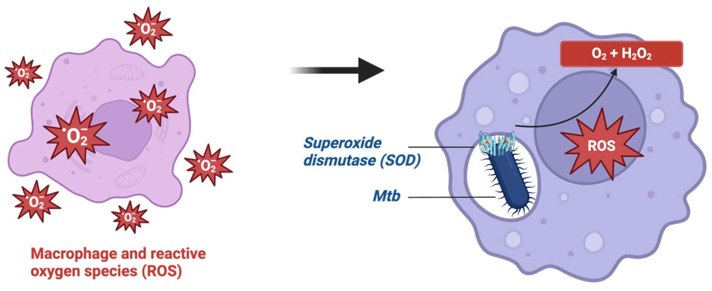
  139. Tyagi, P.; Dharmaraja, A.T.; Bhaskar, A.; Chakrapani, H.; Singh, A. Mycobacterium tuberculosis has diminished capacity to counteract redox stress induced by elevated levels of endogenous superoxide. Free Radic. Biol. Med. 2015, 84, 344–354. [Google Scholar] [CrossRef] [PubMed]
  140. Nambi, S.; Long, J.E.; Mishra, B.B.; Baker, R.; Murphy, K.C.; Olive, A.J.; Nguyen, H.P.; Shaffer, S.A.; Sassetti, C.M. The Oxidative Stress Network of Mycobacterium tuberculosis Reveals Coordination between Radical Detoxification Systems. Cell Host Microbe 2015, 17, 829–837. [Google Scholar] [CrossRef] [PubMed]
  141. Afriyie-Asante, A.; Dabla, A.; Dagenais, A.; Berton, S.; Smyth, R.; Sun, J. Mycobacterium tuberculosis Exploits Focal Adhesion Kinase to Induce Necrotic Cell Death and Inhibit Reactive Oxygen Species Production. Front. Immunol. 2021, 12, 742370. [Google Scholar] [CrossRef] [PubMed]
  142. Lorenzo, H.K.; Susin, S.A.; Penninger, J.; Kroemer, G. Apoptosis inducing factor (AIF): A phylogenetically old, caspase-independent effector of cell death. Cell Death Differ. 1999, 6, 516–524. [Google Scholar] [CrossRef] [PubMed]
  143. Szegezdi, E.; Logue, S.E.; Gorman, A.M.; Samali, A. Mediators of endoplasmic reticulum stress-induced apoptosis. EMBO Rep. 2006, 7, 880–885. [Google Scholar] [CrossRef]
  144. Tabas, I.; Ron, D. Integrating the mechanisms of apoptosis induced by endoplasmic reticulum stress. Nat. Cell Biol. 2011, 13, 184–190. [Google Scholar] [CrossRef]
  145. Read, A.; Schröder, M. The Unfolded Protein Response: An Overview. Biology 2021, 10, 384. [Google Scholar] [CrossRef] [PubMed]
  146. Lim, Y.J.; Choi, J.A.; Choi, H.H.; Cho, S.N.; Kim, H.J.; Jo, E.K.; Park, J.K.; Song, C.H. Endoplasmic reticulum stress pathway-mediated apoptosis in macrophages contributes to the survival of Mycobacterium tuberculosis. PLoS ONE 2011, 6, e28531. [Google Scholar] [CrossRef] [PubMed]
  147. Xu, P.; Tang, J.; He, Z.G. Induction of Endoplasmic Reticulum Stress by CdhM Mediates Apoptosis of Macrophage During Mycobacterium tuberculosis Infection. Front. Cell Infect. Microbiol. 2022, 12, 877265. [Google Scholar] [CrossRef] [PubMed]
  148. Khalid, N.; Azimpouran, M. Necrosis. In StatPearls; StatPearls Publishing LLC.: Treasure Island, FL, USA, 2024. [Google Scholar]
  149. Liu, Z.G.; Jiao, D. Necroptosis, tumor necrosis and tumorigenesis. Cell Stress. 2019, 4, 1–8. [Google Scholar] [CrossRef] [PubMed]
  150. Man, S.M.; Karki, R.; Kanneganti, T.D. Molecular mechanisms and functions of pyroptosis, inflammatory caspases and inflammasomes in infectious diseases. Immunol. Rev. 2017, 277, 61–75. [Google Scholar] [CrossRef] [PubMed]
  151. Amaral, E.P.; Costa, D.L.; Namasivayam, S.; Riteau, N.; Kamenyeva, O.; Mittereder, L.; Mayer-Barber, K.D.; Andrade, B.B.; Sher, A. A major role for ferroptosis in Mycobacterium tuberculosis-induced cell death and tissue necrosis. J. Exp. Med. 2019, 216, 556–570. [Google Scholar] [CrossRef] [PubMed]
  152. Chen, M.; Gan, H.; Remold, H.G. A mechanism of virulence: Virulent Mycobacterium tuberculosis strain H37Rv, but not attenuated H37Ra, causes significant mitochondrial inner membrane disruption in macrophages leading to necrosis. J. Immunol. 2006, 176, 3707–3716. [Google Scholar] [CrossRef] [PubMed]
  153. Divangahi, M.; Chen, M.; Gan, H.; Desjardins, D.; Hickman, T.T.; Lee, D.M.; Fortune, S.; Behar, S.M.; Remold, H.G. Mycobacterium tuberculosis evades macrophage defenses by inhibiting plasma membrane repair. Nat. Immunol. 2009, 10, 899–906. [Google Scholar] [CrossRef] [PubMed]
  154. Lerner, T.R.; Borel, S.; Greenwood, D.J.; Repnik, U.; Russell, M.R.; Herbst, S.; Jones, M.L.; Collinson, L.M.; Griffiths, G.; Gutierrez, M.G. Mycobacterium tuberculosis replicates within necrotic human macrophages. J. Cell Biol. 2017, 216, 583–594. [Google Scholar] [CrossRef]
  155. Andersson, A.M.; Andersson, B.; Lorell, C.; Raffetseder, J.; Larsson, M.; Blomgran, R. Autophagy induction targeting mTORC1 enhances Mycobacterium tuberculosis replication in HIV co-infected human macrophages. Sci. Rep. 2016, 6, 28171. [Google Scholar] [CrossRef]
  156. Singhal, A.; Jie, L.; Kumar, P.; Hong, G.S.; Leow, M.K.; Paleja, B.; Tsenova, L.; Kurepina, N.; Chen, J.; Zolezzi, F.; et al. Metformin as adjunct antituberculosis therapy. Sci. Transl. Med. 2014, 6, 263ra159. [Google Scholar] [CrossRef]
  157. Moreira, J.D.; Koch, B.E.V.; van Veen, S.; Walburg, K.V.; Vrieling, F.; Mara Pinto Dabés Guimarães, T.; Meijer, A.H.; Spaink, H.P.; Ottenhoff, T.H.M.; Haks, M.C.; et al. Functional Inhibition of Host Histone Deacetylases (HDACs) Enhances in vitro and in vivo Anti-mycobacterial Activity in Human Macrophages and in Zebrafish. Front. Immunol. 2020, 11, 36. [Google Scholar] [CrossRef] [PubMed]
  158. Adikesavalu, H.; Gopalaswamy, R.; Kumar, A.; Ranganathan, U.D.; Shanmugam, S. Autophagy Induction as a Host-Directed Therapeutic Strategy against Mycobacterium tuberculosis Infection. Medicina 2021, 57, 522. [Google Scholar] [CrossRef]
  159. Lam, K.K.; Zheng, X.; Forestieri, R.; Balgi, A.D.; Nodwell, M.; Vollett, S.; Anderson, H.J.; Andersen, R.J.; Av-Gay, Y.; Roberge, M. Nitazoxanide stimulates autophagy and inhibits mTORC1 signaling and intracellular proliferation of Mycobacterium tuberculosis. PLoS Pathog. 2012, 8, e1002691. [Google Scholar] [CrossRef] [PubMed]
  160. Periyasamy, K.M.; Ranganathan, U.D.; Tripathy, S.P.; Bethunaickan, R. Vitamin D—A host directed autophagy mediated therapy for tuberculosis. Mol. Immunol. 2020, 127, 238–244. [Google Scholar] [CrossRef]
  161. Singh, R.; Letai, A.; Sarosiek, K. Regulation of apoptosis in health and disease: The balancing act of BCL-2 family proteins. Nat. Rev. Mol. Cell Biol. 2019, 20, 175–193. [Google Scholar] [CrossRef]
  162. Falkenhorst, J.; Grunewald, S.; Mühlenberg, T.; Marino-Enriquez, A.; Reis, A.C.; Corless, C.; Heinrich, M.; Treckmann, J.; Podleska, L.E.; Schuler, M.; et al. Inhibitor of Apoptosis Proteins (IAPs) are commonly dysregulated in GIST and can be pharmacologically targeted to enhance the pro-apoptotic activity of imatinib. Oncotarget 2016, 7, 41390–41403. [Google Scholar] [CrossRef] [PubMed]
  163. Stutz, M.D.; Allison, C.C.; Ojaimi, S.; Preston, S.P.; Doerflinger, M.; Arandjelovic, P.; Whitehead, L.; Bader, S.M.; Batey, D.; Asselin-Labat, M.L.; et al. Macrophage and neutrophil death programs differentially confer resistance to tuberculosis. Immunity 2021, 54, 1758–1771.e7. [Google Scholar] [CrossRef]
  164. Lim, Y.J.; Lee, J.; Choi, J.A.; Cho, S.N.; Son, S.H.; Kwon, S.J.; Son, J.W.; Song, C.H. M1 macrophage dependent-p53 regulates the intracellular survival of mycobacteria. Apoptosis 2020, 25, 42–55. [Google Scholar] [CrossRef]
  165. Zhuang, L.; Yang, L.; Li, L.; Ye, Z.; Gong, W. Mycobacterium tuberculosis: Immune response, biomarkers, and therapeutic intervention. MedComm (2020) 2024, 5, e419. [Google Scholar] [CrossRef]
  166. Jeong, E.K.; Lee, H.J.; Jung, Y.J. Host-Directed Therapies for Tuberculosis. Pathogens 2022, 11, 1291. [Google Scholar] [CrossRef] [PubMed]
  167. Kroesen, V.M.; Gröschel, M.I.; Martinson, N.; Zumla, A.; Maeurer, M.; van der Werf, T.S.; Vilaplana, C. Non-Steroidal Anti-inflammatory Drugs As Host-Directed Therapy for Tuberculosis: A Systematic Review. Front. Immunol. 2017, 8, 772. [Google Scholar] [CrossRef] [PubMed]
  168. Tobin, D.M. Host-Directed Therapies for Tuberculosis. Cold Spring Harb. Perspect. Med. 2015, 5, a021196. [Google Scholar] [CrossRef] [PubMed]
  169. Subbian, S.; Tsenova, L.; Holloway, J.; Peixoto, B.; O’Brien, P.; Dartois, V.; Khetani, V.; Zeldis, J.B.; Kaplan, G. Adjunctive Phosphodiesterase-4 Inhibitor Therapy Improves Antibiotic Response to Pulmonary Tuberculosis in a Rabbit Model. eBioMedicine 2016, 4, 104–114. [Google Scholar] [CrossRef] [PubMed]
  170. Alsayed, S.S.R.; Gunosewoyo, H. Tuberculosis: Pathogenesis, Current Treatment Regimens and New Drug Targets. Int. J. Mol. Sci. 2023, 24, 5202. [Google Scholar] [CrossRef] [PubMed]
  171. Xiong, X.S.; Zhang, X.D.; Yan, J.W.; Huang, T.T.; Liu, Z.Z.; Li, Z.K.; Wang, L.; Li, F. Identification of Mycobacterium tuberculosis Resistance to Common Antibiotics: An Overview of Current Methods and Techniques. Infect. Drug Resist. 2024, 17, 1491–1506. [Google Scholar] [CrossRef]
  172. Negi, A.; Perveen, S.; Gupta, R.; Singh, P.P.; Sharma, R. Unraveling Dilemmas and Lacunae in the Escalating Drug Resistance of Mycobacterium tuberculosis to Bedaquiline, Delamanid, and Pretomanid. J. Med. Chem. 2024, 67, 2264–2286. [Google Scholar] [CrossRef] [PubMed]
  173. Mtimka, S.; Pillay, P.; Kwezi, L.; Pooe, O.J.; Tsekoa, T.L. An Exploratory Review of the Potential of Lytic Proteins and Bacteriophages for the Treatment of Tuberculosis. Microorganisms 2024, 12, 570. [Google Scholar] [CrossRef] [PubMed]
  174. Hirsch, C.S.; Johnson, J.L.; Okwera, A.; Kanost, R.A.; Wu, M.; Peters, P.; Muhumuza, M.; Mayanja-Kizza, H.; Mugerwa, R.D.; Mugyenyi, P.; et al. Mechanisms of apoptosis of T-cells in human tuberculosis. J. Clin. Immunol. 2005, 25, 353–364. [Google Scholar] [CrossRef]
  175. Pagán, A.J.; Ramakrishnan, L. Immunity and Immunopathology in the Tuberculous Granuloma. Cold Spring Harb. Perspect. Med. 2014, 5, a018499. [Google Scholar] [CrossRef]
  176. Dallenga, T.; Repnik, U.; Corleis, B.; Eich, J.; Reimer, R.; Griffiths, G.W.; Schaible, U.E. M. tuberculosis-Induced Necrosis of Infected Neutrophils Promotes Bacterial Growth Following Phagocytosis by Macrophages. Cell Host Microbe 2017, 22, 519–530.e3. [Google Scholar] [CrossRef] [PubMed]
  177. Toossi, Z. The inflammatory response in Mycobacterium tuberculosis infection. Arch. Immunol. Ther. Exp. 2000, 48, 513–519. [Google Scholar] [PubMed]
  178. Su, H.; Zhu, S.; Zhu, L.; Huang, W.; Wang, H.; Zhang, Z.; Xu, Y. Recombinant Lipoprotein Rv1016c Derived from Mycobacterium tuberculosis Is a TLR-2 Ligand that Induces Macrophages Apoptosis and Inhibits MHC II Antigen Processing. Front. Cell Infect. Microbiol. 2016, 6, 147. [Google Scholar] [CrossRef] [PubMed]
Figure 1. CpsA protein in LC3-associated phagocytosis. In LC3-associated phagocytosis, NADPH oxidase is recruited to the phagosome. There is a subsequent recruitment of LC3 and maturation of the phagophore, followed by a fusion of the subsequent autophagosome with a lysosome containing degradative enzymes, resulting in an autolysosome. The CpsA protein of Mtb may activate PRR but will inhibit the recruitment of NADPH oxidase.
Ijms 25 06255 g001
Figure 2. Mtb inhibition of IFN-associated receptor (IFNAR) signaling pathway, preventing nitric oxide production.
Ijms 25 06255 g002
Figure 3. Caspase-dependent apoptosis may occur via the intrinsic or extrinsic pathway. The intrinsic pathway is triggered by internal cellular stress or by proapoptotic Bcl-2 proteins, which cause the release of factors such as cytochrome c and apoptosis-inducing factor (AIF) from the mitochondrial intermembrane space. Such factors activate caspase 9, which activates the apoptosome, which further activates caspase 3 or 7. Of note, nitric oxide (NO) is found to upregulate Bcl-2 proteins. IFN-γ-activated macrophages further upregulate nitric oxide synthase, thereby increasing NO and thus proapoptotic Bcl-2 proteins. In contrast, the extrinsic pathway is activated via death receptors, such as Fas or TNF receptor. This activates caspase 8, which further activates caspase 3 or 7. Of note, Mtb may secrete TNF-R2, which can inhibit the TNF receptor.
Ijms 25 06255 g003
Figure 4. Antioxidant defense in Mtb via superoxide dismutase.
Ijms 25 06255 g004
Table 1. List of potential and existing therapies with their associated pathway and targeted mechanism.
TherapyPathwayMechanism
Rapamycin, histone deacetylase inhibitorsAutophagyInhibition of mTOR
MetforminAutophagyActivation of AMPK
Transcription factor EBAutophagyRegulates autophagy gene expression
Lysosomotropic agentsAutophagyEnhance lysosomal acidity
Nitazoxanide AutophagyActivation of mTORC1 inhibition
Vitamin DAutophagySynthesis of CAMP
BH3 mimeticsApoptosisBLC-2-mediated apoptosis
Nutlin-3ApoptosisActivation of p53
NSAIDs, corticosteroid, zileuton, desipramineNecrosisEicosanoid modulation
Phosphodiesterase inhibitorsNecrosisTNF reduction
Disclaimer/Publisher’s Note: The statements, opinions and data contained in all publications are solely those of the individual author(s) and contributor(s) and not of MDPI and/or the editor(s). MDPI and/or the editor(s) disclaim responsibility for any injury to people or property resulting from any ideas, methods, instructions or products referred to in the content.

