Article

Precision Medicine for Blood Glutamate Grabbing in Ischemic Stroke

by
Pablo Hervella
1,
Ana Sampedro-Viana
1,
Sabela Fernández-Rodicio
1,
Manuel Rodríguez-Yáñez
2,
Iria López-Dequidt
3,
José M. Pumar
1,4,
Antonio J. Mosqueira
1,4,
Marcos Bazarra-Barreiros
1,
María Teresa Abengoza-Bello
1,
Sara Ortega-Espina
1,
Alberto Ouro
5,6,
María Pérez-Mato
7,
Francisco Campos
7,
Tomás Sobrino
5,6,
José Castillo
1,
Maria Luz Alonso-Alonso
1,* and
Ramón Iglesias-Rey
1,*
1
Neuroimaging and Biotechnology Laboratory (NOBEL), Clinical Neurosciences Research Laboratory (LINC), Health Research Institute of Santiago de Compostela (IDIS), 15706 Santiago de Compostela, Spain
2
Stroke Unit, Department of Neurology, Hospital Clínico Universitario, 15706 Santiago de Compostela, Spain
3
Department of Neurology, Hospital Clínico Universitario de Ferrol, 15405 Ferrol, Spain
4
Department of Neuroradiology, Hospital Clínico Universitario, Health Research Institute of Santiago de Compostela (IDIS), 15706 Santiago de Compostela, Spain
5
NeuroAging Group (NEURAL), Clinical Neurosciences Research Laboratory (LINC), Health Research Institute of Santiago de Compostela (IDIS), 15706 Santiago de Compostela, Spain
6
Centro de Investigación Biomédica en Red de Enfermedades Neurodegenerativas, Instituto de Salud Carlos III, 28029 Madrid, Spain
7
Translational Stroke Laboratory (TREAT), Clinical Neurosciences Research Laboratory (LINC), Health Research Institute of Santiago de Compostela (IDIS), 15706 Santiago de Compostela, Spain
*
Authors to whom correspondence should be addressed.
Int. J. Mol. Sci. 2024, 25(12), 6554; https://doi.org/10.3390/ijms25126554
Submission received: 13 May 2024 / Revised: 1 June 2024 / Accepted: 9 June 2024 / Published: 14 June 2024
(This article belongs to the Special Issue New Trends in Research on Cerebral Ischemia)

Abstract

:
Glutamate grabbers, such as glutamate oxaloacetate transaminase (GOT), have been proposed to prevent excitotoxicity secondary to high glutamate levels in stroke patients. However, the efficacy of blood glutamate grabbing by GOT could be dependent on the extent and severity of the disruption of the blood–brain barrier (BBB). Our purpose was to analyze the relationship between GOT and glutamate concentration with the patient’s functional status differentially according to BBB serum markers (soluble tumor necrosis factor-like weak inducer of apoptosis (sTWEAK) and leukoaraiosis based on neuroimaging). This retrospective observational study includes 906 ischemic stroke patients. We studied the presence of leukoaraiosis and the serum levels of glutamate, GOT, and sTWEAK in blood samples. Functional outcome was assessed using the modified Rankin Scale (mRS) at 3 months. A significant negative correlation between GOT and glutamate levels at admission was shown in those patients with sTWEAK levels > 2900 pg/mL (Pearson’s correlation coefficient: −0.249; p < 0.0001). This correlation was also observed in patients with and without leukoaraiosis (Pearson’s correlation coefficients: −0.299; p < 0.001 vs. −0.116; p = 0.024). The logistic regression model confirmed the association of higher levels of GOT with lower odds of poor outcome at 3 months when sTWEAK levels were >2900 pg/mL (OR: 0.41; CI 95%: 0.28–0.68; p < 0.0001) or with leukoaraiosis (OR: 0.75; CI 95%: 0.69–0.82; p < 0.0001). GOT levels are associated with glutamate levels and functional outcomes at 3 months, but only in those patients with leukoaraiosis and elevated sTWEAK levels. Consequently, therapies targeting glutamate grabbing might be more effective in patients with BBB dysfunction.

1. Introduction

The aging of the population in developed countries, along with relevant advances in medical care, has increased the number of people who overcome serious illnesses, such as stroke. However, this involves an increase in morbidity [1]. Although stroke remains the second-leading cause of death worldwide and the third-leading cause of death and disability combined [2,3], the increase in survival rate with severe disabilities represents a remarkable cost, not only in terms of health burden but also in the quality of life of patients and their caregivers [4,5]. It should be highlighted that the development of stroke units in which patients receive specialized stroke care has played a significant role in this improvement of outcomes [6]. The current therapeutic options for acute ischemic stroke, the main contributor to the global incidence of stroke [2], are mechanical thrombectomy, thrombolytic therapy, or a combination of both [7]. However, these therapeutical options still present notable limitations, including the number of eligible patients and the time lapse from stroke onset to specialized assistance [8]. Additionally, recanalization success is not always correlated with clinical improvement [9,10,11,12,13]. Neuroprotection was proposed as a potential option to improve the poor outcomes of patients. Several targets have been studied; unfortunately, despite the promising results obtained in preclinical assays, numerous neuroprotective drugs have failed to be transferred to clinical practice [14,15].
Several factors have been described to be involved in this failure, such as the differences in the time window selected in preclinical and clinical studies and the heterogeneity of patients included in clinical trials, among others [16]. All this evidence reinforces the increasing interest in precision medicine in stroke, which integrates blood biomarkers and clinical and neuroimaging data [17,18].
Among others, it was demonstrated that high serum glutamate concentration could be considered a blood biomarker of poor outcomes in stroke patients [19,20,21,22,23]. Glutamate plays an important role in neural injury during ischemia-induced excitotoxicity [24]. Recently, glutamate grabbers or scavengers have been proposed to prevent a cascade of events triggered by excessive extracellular glutamate [25,26,27].
In physiological conditions, the excess of extracellular glutamate is transported into endothelial cells, belonging to the blood–brain barrier (BBB), from where it diffuses to blood due to the difference in concentration [28]. However, in ischemic conditions, structural integrity of the BBB is affected, promoting an increase in the permeability [29]. Based on these data, we hypothesize that glutamate grabbing through a normal endothelium would not be enough to decrease excitotoxicity in the penumbra zones. Therefore, the efficacy of blood glutamate grabbing could depend on the extent and severity of the disruption of the BBB. Soluble tumor necrosis factor-like weak inducer of apoptosis (sTWEAK) plays a role in triggering neuroinflammation and BBB breakdown during ischemic stroke [30]. This makes it a potential serum marker of BBB disruption. Additionally, leukoaraiosis, defined as an abnormality in the white matter substance detected on computed tomography (CT) or magnetic resonance image (MRI) [31], has been previously associated with sTWEAK levels by our group [32].
On the other hand, it is well known that glutamate oxaloacetate transaminase (GOT) is a glutamate grabber. This enzyme catalyzes the reversible transformation of glutamate and oxaloacetate to α–ketoglutarate and aspartate [25,33]. It has been shown that high blood levels of GOT are associated with better outcomes both in animal models [34,35] and ischemic stroke patients [36,37].
The main purpose of this study was to analyze the relationship between GOT and glutamate concentration, determined in plasma at admission, and the patient’s functional status at 3 months. This analysis was performed differentially in patients with serum (sTWEAK) and neuroimaging (leukoaraiosis) markers of BBB disruption determined at admission.

2. Results

2.1. Sample Description

After applying the inclusion and exclusion criteria established, 906 patients were finally enrolled for this analysis. The flowchart for patient screening from Biobanco Ictus del Complejo Hospitalario Universitario de Santiago (BICHUS) is shown in Figure S1. The sex distribution of the sample was 51% males vs. 49% females, and the mean age was 72.2 ± 12.1 years. These patients were classified according to the TOAST (Trial of Org 10,172 in Acute Stroke Treatment) criteria as atherothrombotic (20.5%), cardioembolic (37%), lacunar (9.4%), and indeterminate (31.1%). 5.4% of patients have undergone thrombectomy and 14.5% have intravenous fibrinolysis.

2.2. Demographic and Clinical Features Associated with Poor Functional Outcome at 3 Months

The bivariate analysis of demographic and clinical variables collected from the ischemic stroke patients showed that the time lapse between the onset of symptoms and the emergency assistance is an essential variable (277.8 ± 214.5 min vs. 269.9 ± 180.7 min; p = 0.002) for clinical progress. Similarly, the presence of leukoaraiosis in the neuroimaging studies was associated with poor outcomes (p < 0.0001). Concerning potential biomarkers measured in blood samples, results showed that the levels of GOT at admission were significantly lower in those patients with poor functional outcomes at 3 months (39.7 ± 13.3 U/L vs. 22.6 ± 10.3 U/L; p < 0.0001). Contrary to this, the levels of glutamate (134.2 ± 112.5 μM/mL vs. 227.1 ± 123.8 μM/mL; p = 0.036) and sTWEAK (3143.2 ± 2677.1 pg/mL vs. 5828.6 ± 3248.9 pg/mL; p = 0.002) were significantly higher in these patients. There were also significant differences between those with good and poor outcomes in terms of age (p = 0.003), alcohol consumption (p = 0.004), sex, smoking, and atrial fibrillation (all p < 0.0001). Additionally, those patients who showed higher temperature (p = 0.029), blood glucose, and leukocyte count at admission (both p < 0.0001) showed worse outcomes at 3 months. TOAST criteria and higher National Institute of Health Stroke Scale (NIHSS) scores at admission (both p < 0.0001) were also significantly associated with poorer long-term outcomes (Table 1).

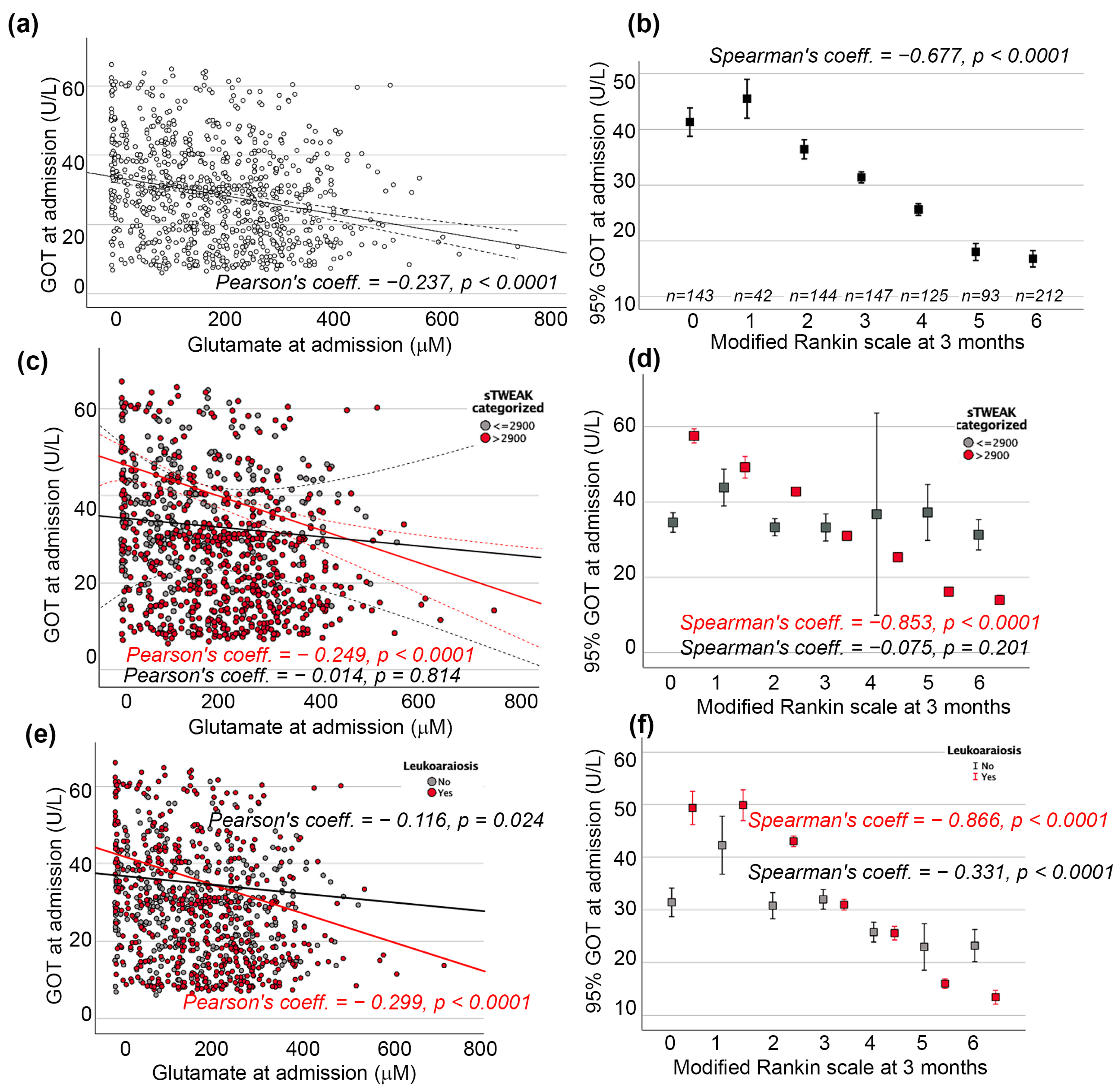
2.3. GOT and sTWEAK Association with Poor Functional Outcome at 3 Months

A significant negative correlation between GOT and glutamate levels was found at admission (Pearson’s correlation coefficient: −0.237; p < 0.0001; Figure 1a). It was also observed that lower levels of GOT were correlated with higher scores on the modified Rankin Scale (mRS) and, therefore, with higher rates of morbidity and mortality at 3 months (Figure 1b).
Regarding the other blood biomarkers, sTWEAK, according to ROC curve analysis, can discriminate between patients with poor outcomes and those with good outcomes at 3 months with a sensitivity of 88% and a specificity of 70% (Figure S2a). The cut-off for it was established at 2900 pg/mL (Figure S2a). Results showed a higher percentage of patients with sTWEAK levels higher than 2900 pg/mL for all the ischemic stroke types classified following the TOAST criteria, except for lacunar strokes (Figure S2b).
When the correlation between GOT and glutamate levels at admission was reanalyzed according to the sTWEAK serum level, the significant negative correlation between them was confirmed in those patients with sTWEAK > 2900 pg/mL (Pearson’s correlation coefficient: −0.249; p < 0.0001) but not for patients with lower levels (Figure 1c). Similarly, the significant negative correlation between GOT and mRS was retained only in patients with high levels of sTWEAK (Figure 1d). The logistic regression model confirmed this independent association of higher levels of GOT with lower odds of poor functional outcome at 3 months when sTWEAK was >2900 pg/mL (OR: 0.41; CI 95%: 0.28–0.68; p < 0.0001; Table 2).

2.4. GOT and Leukoaraiosis Association with Poor Functional Outcome at 3 Months

As shown in Figure S3, GOT and sTWEAK showed a significant association (p < 0.001) with the degree of leukoaraiosis observed in the neuroimaging studies. Meanwhile, the relationship with sTWEAK was positive (Figure S3a), and a negative correlation with GOT was found (Figure S3b).
Additionally, the correlation between GOT and glutamate levels at admission was re-analyzed according to the leukoaraiosis degree; the significant negative correlation between them was confirmed in both groups with (Pearson’s correlation coefficient: −0.299; p < 0.0001) and without leukoaraiosis (Pearson’s correlation coefficient: −0.116; p = 0.024) (Figure 1e). Consequently, the significant negative correlation between GOT and mRS was maintained regardless of the presence or absence of leukoaraiosis (Figure 1f). However, the logistic regression model only confirmed this independent association of higher levels of GOT with lower odds of poor functional outcome at 3 months in the presence of this neuroimaging biomarker (OR: 0.75; CI 95%: 0.69–0.82; p < 0.0001; Table 3).

3. Discussion

In this study, the association between the enzyme GOT with glutamate at admission and with the patient’s functional status at 3 months was evaluated by classifying patients according to two biomarkers of BBB disruption, sTWEAK and leukoaraiosis. The inverse correlations between GOT with glutamate and with functional status previously described were supported by present results [36,37].
Precision medicine is defined by the National Institutes of Health as “an innovative approach that takes into account individual differences in patients’ genes, environments, and lifestyles” [38]. The interest in this field has increasingly grown in the last decades for a wide range of diseases [39], including cerebrovascular diseases [18]. The tools used for this purpose are very varied [40]. Different blood biomarkers have been reported as useful tools for diagnosis, treatment decisions, and outcome prediction in stroke [41]. This type of biomarker has the advantage that it can be incorporated into daily clinical practice more easily than others since blood collection is a routine activity. Blood is the most common sample evaluated in recent biomarker trials for stroke [42].
On the other hand, currently there is only one clinical trial based on glutamate grabbers, riboflavin (vitamin B2) (NCT02446977; completed), which was developed by our group. Although the results of this clinical trial showed a demonstration of the efficacy of this blood glutamate grabber in the treatment of patients with stroke [27], a precision medicine approach was not followed, which is a point to be improved in the future. Taking in mind the relevance of maintaining BBB integrity for brain health and cerebrovascular diseases, we hypothesized that biomarkers of its disruption would improve not only the clinical approach of current treatments but also will be an advantageous tool considered for the selection of patients in future clinical trials. In the present study, we selected a blood biomarker, i.e., sTWEAK, and a neuroimaging marker, leukoaraiosis. Both would be easily transferred into clinical practice since blood sample analysis and neuroimaging studies are usual in stroke patients approaches.
The sTWEAK, a cytokine involved in different cellular activities such as proliferation, migration, differentiation, apoptosis, angiogenesis, and inflammation [43], has been identified as a potential biomarker of BBB disruption based on its implication in disruption of the structure of BBB and the increment of its permeability. Thus, in 2005 Polavarapu et al. [44] showed that intracerebral injection of TWEAK in wild-type mice led to disruption in the structure of the neurovascular unit and to an increase of BBB permeability. In this study, the inhibition of its activity resulted in a significant preservation of the integrity of the neurovascular unit and a diminution of the BBB permeability. The results of Zhang et al. in 2007 [45] by the inhibition of the TWEAK-fibroblast growth factor-inducible 14 (Fn14) axis in an animal model of middle cerebral artery occlusion also support the role of TWEAK in the disruption of the BBB. Finally, in 2013, Stephan et al. [46] showed that TWEAK increased permeability in an in vitro model of BBB with endothelial cells.
High levels in sTWEAK serum have been associated with poor outcomes not only in cerebrovascular diseases, such as ischemic stroke [13,47], intracerebral hemorrhage [48,49], but also in traumatic brain injury [50]. Similarly, we found poor outcomes associated with higher serum levels of sTWEAK. Our results have allowed us to establish a cut-off value of 2900 pg/mL at admission to differentiate good and bad outcomes at 3 months with a sensitivity of 88% and a specificity of 70%. Although the cut-off value is an essential fact for biomarker research, it has been reported that different cut-off values of sTWEAK according to the main variable studied. Thus, Hervella et al. [13] reported a cut-off value of 7000 pg/mL for stroke recurrence, and da Silva-Candal et al. [47,49] determined a cut-off value of 5600 pg/mL for hematoma growth and 6700 pg/mL for poor outcome in reperfused ischemic stroke patients, whereas Comertpay et al. [51] stated a cut-off value of barely 995.5 pg/mL for the diagnosis of acute ischemic stroke. Our cut-off value of 2900 pg/mL was taken into consideration when analyzing the relationship of GOT and glutamate; this was only significant in those patients whose serum levels of sTWEAK were higher than the cut-off established. Therefore, sTWEAK could play a key role in the association of high levels of GOT described in stroke patients with good outcomes [36,37].
Similar results were obtained when the presence of leukoaraiosis was taken into consideration. Leukoaraiosis is defined as an abnormality in the white matter substance detected on CT or MRI [31]. The implication of the dysfunction of BBB in the pathogenies of white matter lesions is suggested by several studies [52,53,54]. Additionally, a previous study of our group showed an association between sTWEAK levels and leukoaraiosis degree [32]. Based on these facts, leukoaraiosis could also be considered a biomarker of BBB dysfunction. Although leukoaraiosis did not initially appear to affect the negative correlation between GOT and glutamate, adjusted regression analysis did show that leukoaraiosis, like sTWEAK, could play a role in this enzyme-amino acid relationship. This supports the leukoaraiosis useless as a differential criterion between poor and good outcomes previously described in stroke patients [13,47,55,56].
Therefore, our results showed that BBB disruption could be a key factor in the effectiveness of glutamate grabber therapy. The breakdown of BBB during stroke is due to the triggering of cascades that lead to an increase in permeability and loss of integrity as a result of the changes in endothelial cells [29,57]. It is well established that endothelial cells are involved in the elimination of glutamate from the brain to blood by facilitating the BBB transport [58], where glutamate is metabolized by a glutamate grabber. The changes suffered by endothelial cells and the subsequent BBB disruption could promote the brain-to-blood glutamate efflux, promoting the effectivity of glutamate grabbers. To determine the specific mechanisms involved in this process, further in vitro and in vivo studies would be necessary.
This study has been focused on GOT as a glutamate grabber. However, it should be noted that there are other strategies for lowering blood glutamate, including another enzyme, glutamate-pyruvate transaminase (GPT), and hemodialysis and peritoneal dialysis [25]. Similarly to GOT, high levels of GPT have been associated with good outcomes both in animal models [59] and ischemic stroke patients in whom this association was weaker than for GOT [37]. Regarding hemodialysis and peritoneal dialysis, studies in patients with renal diseases have shown that both methods effectively reduced levels of glutamate in the blood, which makes them a potential tool for preventing excitotoxicity in stroke [60,61]. There are two clinical trials (NCT04297345 and NCT03454867) testing this hypothesis of blood filtration for clearance of glutamate. Finally, peritoneal dialysis. Therefore, future studies about the association of these glutamate-lowering strategies with BBB disruption biomarkers could also be relevant for advancing in precision medicine in ischemic stroke.
Finally, our study has several limitations. First, this is a single-center study. These results should be validated by patients from other databases from different centers. Secondly, those related to its retrospective design. Thirdly, the serum levels of the biomarkers were only measured at admission, except for sTWEAK, which was also analyzed at 3 months. On the other hand, the strengths of this work include the unbiased selection of patients, the large sample size included, and the blind analysis.

4. Materials and Methods

4.1. Standard Protocol Approval and Patient Consent

This research was conducted following the principles of the Declaration of Helsinki of the World Medical Association and approved by the Research Ethics Committee of Santiago (project identification code 2019/616). Informed consent was obtained from each patient or their relatives after fully explaining the procedures.

4.2. Study Design

This is a retrospective observational study of patients with stroke admitted to the Stroke Unit of the Hospital Clínico Universitario of Santiago de Compostela (Spain) between January 2008 and December 2018. They were consecutively and prospectively registered in an approved data bank, BICHUS. All patients admitted to the Stroke Unit were treated by expert neurologists according to Spanish Neurological Society protocol.
Upon admission to the Stroke Unit, the intensity of the neurological deficit was determined by NIHSS [62]. Regarding functional outcome, it was assessed using mRS [63] at 3 months ± 15 days (face-to-face in 80.8% of the sample). A mRS > 2 was classified as a poor outcome. Internationally certified neurologists evaluated both scales. Stroke etiology was classified according to TOAST criteria [64].
MRI or CT studies were performed at admission. Leukoaraiosis was defined as low density on CT and hyperintensity on T2-weighted or FLAIR MRI. The presence and severity of leukoaraiosis were assessed using the Fazekas scale with a total score from 0 to 6 (grade I, 1–2; grade II, 3–4; and grade III, 5–6) [65]. These neuroimaging studies were performed using the same protocol and supervised by the same neuroradiologists.

4.3. Inclusion and Exclusion Criteria

Inclusion criteria for this analysis were as follows: (1) authorization for the anonymous use of individuals’ data for research purposes; (2) MRI or CT study at admission. Exclusion criteria were: (1) patients without a minimum follow-up (face-to-face or telephone) of 3 months; (2) institutionalized patients; (3) comorbidity and life expectancy of less than 1 year; (4) lack of subsequent diagnostic confirmation; (5) absence of blood tests on the admission of the patient with the determination of GOT; and (6) unavailability of frozen plasma sample for analysis of glutamate and sTWEAK. The exclusion of patients did not imply bias in the main clinical variables in relation to the selection of patients valid for this study (Table S1).
Demographic and clinical features of patients analyzed in the present study were obtained from the anamnesis and the medical history.

4.4. Biochemical and Hematological Analysis

Biochemistry, hematology, and coagulation assays from blood samples collected during the first 6 h after admission were performed in the hospital’s central laboratory. In addition, we evaluated the sTWEAK in the patients’ serum at admission and after 3 months. Although these analyses were not performed simultaneously, they used the same standardized methods, and the same researchers supervised them.
Venous blood samples were collected in vacutainer tubes (Becton Dickinson, San Jose, CA, USA) in the first 6 h after symptom onset (always after the administration of the thrombolytic bolus). After allowing the samples to coagulate for 60 min, the blood samples were centrifuged at 3000× g for 10 min at 4 °C, and the serum was immediately aliquoted, frozen, and stored at −80 °C until analysis.
The determination of sTWEAK serum concentration was conducted without knowledge of the clinical data, using the enzyme-linked immunosorbent assay (ELISA) technique as per the manufacturer’s instructions (ThermoFisher Scientific, Waltham, MA, USA). The minimum assay sensitivity was 40 pg/mL, and both intra- and inter-assay coefficients of variation were kept below 10% and 12%, respectively.
Serum glutamate concentration was analyzed through high-performance liquid chromatography (1260 Infinity II, Agilent Technologies, Santa Clara, CA, USA) employing the AccQ-Tag™ Precolumn derivatization method for amino acid analysis (Waters, Milford, MA, USA), following a previously outlined method [66].
GOT levels were measured using an automated auto-analyzer (ADVIA 2400, Bayer Diagnostics, Leverkusen, Germany).
All the samples were anonymized using a code so that the researcher who developed the analysis had no information about the clinical status through the follow-up period, avoiding biases in the analysis.

4.5. Statistical Analysis

Categorical variables were described with frequency and percentage. For this descriptive study, the quantitative variables were described with the mean ± standard deviation, or the median and interquartile range, according to the type of distribution determined by the Kolmogorov–Smirnov test for a sample with the significance correction of Lilliefors.
Analysis of variance (ANOVA) was used to compare differences between more than two groups. According to the nature of the contrast variable and its adjustment to normality, the significance of the differences between the two groups was estimated using the chi-square test or Wilcoxon test. Logistic regression analyses were performed to identify those variables independently associated with poor outcomes at 3 months. The results were expressed as odds ratios (ORs) with 95% confidence intervals (95% CI). The analysis of correlations was performed using Pearson’s coefficient and Spearman’s coefficient. The sensitivity and specificity of the biomarkers selected were represented graphically by receiver operating characteristic (ROC) curves. The statistical significance was set at a p-value < 0.05. All statistical analyses were performed with SPSS V 29.0.1.0 (171) (IBM, New York, NY, USA).

5. Conclusions

The conducted analyses lead to the conclusion that plasma concentrations of GOT are correlated with both glutamate levels and the functional status of patients at the 3-month mark. However, this correlation is observed only in patients with elevated sTWEAK concentrations or those exhibiting leukoaraiosis, indicating BBB disruption. Conversely, the time elapsed since symptom onset and the examined inflammatory biomarkers do not demonstrate a significant relationship with these variables. Consequently, it is plausible to propose that therapies targeting the entrapment of glutamate and reduction of its plasma levels may be more effective in patients experiencing increased BBB permeability and dysfunction. This aspect should be considered for the selection of patients in future clinical trials.

Supplementary Materials

The following supporting information can be downloaded at: https://www.mdpi.com/article/10.3390/ijms25126554/s1.

Author Contributions

Conceptualization: P.H., J.C. and R.I.-R.; Formal analysis: J.C. and R.I.-R.; Investigation: M.R.-Y., I.L.-D., J.M.P. and A.J.M.; Methodology: M.R.-Y., I.L.-D., J.M.P. and A.J.M.; Supervision: A.O., M.P.-M., F.C. and T.S.; Writing—original draft: P.H., A.S.-V., S.F.-R., J.C., M.L.A.-A. and R.I.-R.; Writing—review and editing: M.B.-B., M.T.A.-B., S.O.-E., A.O., M.P.-M., F.C., T.S. and M.L.A.-A. All authors have read and agreed to the published version of the manuscript.

Funding

This research was funded by Spanish Ministry of Science and Innovation (SAF2017-84267-R), PDC2021-121455-I00, Xunta de Galicia (Axencia Galega de Innovación: IN607A2022-03, IN607A2022/07), Instituto de Salud Carlos III (ISCIII) (PI17/01103, PI22/00938, ISCIII/PI21/01256/Co-financed by the European Union DTS23/00103), Spanish Research Network on Cerebrovascular Diseases RETICS-INVICTUS PLUS (RD16/0019/0001), RICORS-ICTUS (Cerebrovascular diseases) RD21/0006/0003 and CIBER, Consorcio Centro de Investigación Biomédica en Red, Enfermedades Neurodegnerativas (CIBERNED) (CB22/05/00067). Furthermore, this study was also supported by grants from the INTERREG Atlantic Area (EAPA_791/2018_ NEUROATLANTIC project), INTER-REG V A España Portugal (POCTEP) (0624_2IQBIONEURO_6_E), and the European Regional Development Fund (ERDF). T. Sobrino (CPII17/00027) and F. Campos (CPII19/00020) from the Miguel Servet Program of Instituto de Salud Carlos III. R. Iglesias-Rey (CP22/00061) from the Miguel Servet Program of Instituto de Salud Carlos III and co-financed by the EU. M. Bazarra-Barreiros (FI22/00200), PFIS Researcher of Instituto de Salud Carlos III. Sponsors did not participate in the study design, collection, analysis, or interpretation of the data, or in writing the report.

Institutional Review Board Statement

This research was conducted following the principles of the Declaration of Helsinki of the World Medical Association and approved by the Research Ethics Committee of Santiago (project identification code 2019/616).

Informed Consent Statement

Informed consent was obtained from each patient or their relatives after fully explaining the procedures.

Data Availability Statement

The statistical analysis plan is available upon request. The data bank is not available for legal and ethical reasons.

Conflicts of Interest

The authors declare no conflicts of interest.

References

  1. Christensen, K.; Doblhammer, G.; Rau, R.; Vaupel, J.W. Ageing Populations: The Challenges Ahead. Lancet 2009, 374, 1196–1208. [Google Scholar] [CrossRef]
  2. Feigin, V.L.; Stark, B.A.; Johnson, C.O.; Roth, G.A.; Bisignano, C.; Abady, G.G.; Abbasifard, M.; Abbasi-Kangevari, M.; Abd-Allah, F.; Abedi, V.; et al. Global, Regional, and National Burden of Stroke and Its Risk Factors, 1990–2019: A Systematic Analysis for the Global Burden of Disease Study 2019. Lancet Neurol. 2021, 20, 795–820. [Google Scholar] [CrossRef]
  3. World Health Organization Global Health Estimates: Life Expectancy and Leading Causes of Death and Disability. Available online: https://www.who.int/data/gho/data/themes/mortality-and-global-health-estimates (accessed on 22 January 2024).
  4. Rochmah, T.N.; Rahmawati, I.T.; Dahlui, M.; Budiarto, W.; Bilqis, N. Economic Burden of Stroke Disease: A Systematic Review. Int. J. Environ. Res. Public Health 2021, 18, 7552. [Google Scholar] [CrossRef]
  5. Rudberg, A.-S.; Berge, E.; Gustavsson, A.; Näsman, P.; Lundström, E. Long-Term Health-Related Quality of Life, Survival and Costs by Different Levels of Functional Outcome Six Months after Stroke. Eur. Stroke J. 2018, 3, 157–164. [Google Scholar] [CrossRef]
  6. Langhorne, P.; Ramachandra, S.; Stroke Unit Trialists’ Collaboration. Organised Inpatient (Stroke Unit) Care for Stroke: Network Meta-Analysis. Cochrane Database Syst. Rev. 2020, 4, CD000197. [Google Scholar] [CrossRef] [PubMed]
  7. Powers, W.J.; Rabinstein, A.A.; Ackerson, T.; Adeoye, O.M.; Bambakidis, N.C.; Becker, K.; Biller, J.; Brown, M.; Demaerschalk, B.M.; Hoh, B.; et al. Guidelines for the Early Management of Patients with Acute Ischemic Stroke: 2019 Update to the 2018 Guidelines for the Early Management of Acute Ischemic Stroke: A Guideline for Healthcare Professionals From the American Heart Association/American Stroke. Stroke 2019, 50, e344–e418. [Google Scholar] [CrossRef]
  8. Campbell, B. Thrombolysis and Thrombectomy for Acute Ischemic Stroke: Strengths and Synergies. Semin. Thromb. Hemost. 2016, 43, 185–190. [Google Scholar] [CrossRef] [PubMed]
  9. Murphy, A.; Symons, S.P.; Hopyan, J.; Aviv, R.I. Factors Influencing Clinically Meaningful Recanalization after IV-RtPA in Acute Ischemic Stroke. Am. J. Neuroradiol. 2013, 34, 146–152. [Google Scholar] [CrossRef]
  10. El Amki, M.; Wegener, S. Improving Cerebral Blood Flow after Arterial Recanalization: A Novel Therapeutic Strategy in Stroke. Int. J. Mol. Sci. 2017, 18, 2669. [Google Scholar] [CrossRef] [PubMed]
  11. Iglesias-Rey, R.; Rodríguez-Yáñez, M.; Rodríguez-Castro, E.; Pumar, J.M.; Arias, S.; Santamaría, M.; López-Dequidt, I.; Hervella, P.; Correa-Paz, C.; Sobrino, T.; et al. Worse Outcome in Stroke Patients Treated with Rt-PA without Early Reperfusion: Associated Factors. Transl. Stroke Res. 2018, 9, 347–355. [Google Scholar] [CrossRef] [PubMed]
  12. Hervella, P.; Rodríguez-Castro, E.; Rodríguez-Yáñez, M.; Arias, S.; Santamaría-Cadavid, M.; López-Dequidt, I.; Estany-Gestal, A.; Maqueda, E.; López-Loureiro, I.; Sobrino, T.; et al. Intra- and Extra-Hospital Improvement in Ischemic Stroke Patients: Influence of Reperfusion Therapy and Molecular Mechanisms. Sci. Rep. 2020, 10, 3513. [Google Scholar] [CrossRef] [PubMed]
  13. Hervella, P.; Pérez-Mato, M.; Rodríguez-Yáñez, M.; López-Dequidt, I.; Pumar, J.M.; Sobrino, T.; Campos, F.; Castillo, J.; da Silva-Candal, A.; Iglesias-Rey, R. STWEAK as Predictor of Stroke Recurrence in Ischemic Stroke Patients Treated with Reperfusion Therapies. Front. Neurol. 2021, 12, 652867. [Google Scholar] [CrossRef] [PubMed]
  14. Ghozy, S.; Reda, A.; Varney, J.; Elhawary, A.S.; Shah, J.; Murry, K.; Sobeeh, M.G.; Nayak, S.S.; Azzam, A.Y.; Brinjikji, W.; et al. Neuroprotection in Acute Ischemic Stroke: A Battle Against the Biology of Nature. Front. Neurol. 2022, 13, 870141. [Google Scholar] [CrossRef] [PubMed]
  15. Savitz, S.I.; Fisher, M. Future of Neuroprotection for Acute Stroke: In the Aftermath of the SAINT Trials. Ann. Neurol. 2007, 61, 396–402. [Google Scholar] [CrossRef] [PubMed]
  16. Gladstone, D.J.; Black, S.E.; Hakim, A.M. Toward Wisdom From Failure. Stroke 2002, 33, 2123–2136. [Google Scholar] [CrossRef] [PubMed]
  17. Petersen, N.H.; Sheth, K.N.; Jha, R.M. Precision Medicine in Neurocritical Care for Cerebrovascular Disease Cases. Stroke 2023, 54, 1392–1402. [Google Scholar] [CrossRef]
  18. Hinman, J.D.; Rost, N.S.; Leung, T.W.; Montaner, J.; Muir, K.W.; Brown, S.; Arenillas, J.F.; Feldmann, E.; Liebeskind, D.S. Principles of Precision Medicine in Stroke. J. Neurol. Neurosurg. Psychiatry 2017, 88, 54–61. [Google Scholar] [CrossRef]
  19. Castellanos, M.; Sobrino, T.; Pedraza, S.; Moldes, O.; Pumar, J.M.; Silva, Y.; Serena, J.; García-Gil, M.; Castillo, J.; Dávalos, A.; et al. High Plasma Glutamate Concentrations Are Associated with Infarct Growth in Acute Ischemic Stroke. Neurology 2008, 71, 1862–1868. [Google Scholar] [CrossRef]
  20. Meng, X.; Li, N.; Guo, D.-Z.; Pan, S.-Y.; Li, H.; Yang, C. High Plasma Glutamate Levels Are Associated with Poor Functional Outcome in Acute Ischemic Stroke. Cell Mol. Neurobiol. 2015, 35, 159–165. [Google Scholar] [CrossRef]
  21. Castillo, J.; Dávalos, A.; Naveiro, J.; Noya, M. Neuroexcitatory Amino Acids and Their Relation to Infarct Size and Neurological Deficit in Ischemic Stroke. Stroke 1996, 27, 1060–1065. [Google Scholar] [CrossRef]
  22. Castillo, J.; Dávalos, A.; Noya, M. Progression of Ischaemic Stroke and Excitotoxic Aminoacids. Lancet 1997, 349, 79–83. [Google Scholar] [CrossRef]
  23. Dávalos, A.; Castillo, J.; Serena, J.; Noya, M.; Dávalos, A.; Castillo, J.; Serena, J.; Noya, M. Duration of Glutamate Release after Acute Ischemic Stroke. Stroke 1997, 28, 708–710. [Google Scholar] [CrossRef] [PubMed]
  24. Belov Kirdajova, D.; Kriska, J.; Tureckova, J.; Anderova, M. Ischemia-Triggered Glutamate Excitotoxicity From the Perspective of Glial Cells. Front. Cell. Neurosci. 2020, 14, 51. [Google Scholar] [CrossRef]
  25. Castillo, J.; Loza, M.I.; Mirelman, D.; Brea, J.; Blanco, M.; Sobrino, T.; Campos, F. A Novel Mechanism of Neuroprotection: Blood Glutamate Grabber. J. Cereb. Blood Flow Metab. 2016, 36, 292–301. [Google Scholar] [CrossRef]
  26. Zlotnik, A.; Sinelnikov, I.; Gruenbaum, B.F.; Gruenbaum, S.E.; Dubilet, M.; Dubilet, E.; Leibowitz, A.; Ohayon, S.; Regev, A.; Boyko, M.; et al. Effect of Glutamate and Blood Glutamate Scavengers Oxaloacetate and Pyruvate on Neurological Outcome and Pathohistology of the Hippocampus after Traumatic Brain Injury in Rats. Anesthesiology 2012, 116, 73–83. [Google Scholar] [CrossRef]
  27. da Silva-Candal, A.; Pérez-Díaz, A.; Santamaría, M.; Correa-Paz, C.; Rodríguez-Yáñez, M.; Ardá, A.; Pérez-Mato, M.; Iglesias-Rey, R.; Brea, J.; Azuaje, J.; et al. Clinical Validation of Blood/Brain Glutamate Grabbing in Acute Ischemic Stroke. Ann. Neurol. 2018, 84, 260–273. [Google Scholar] [CrossRef] [PubMed]
  28. Teichberg, V.I.; Cohen-Kashi-Malina, K.; Cooper, I.; Zlotnik, A. Homeostasis of Glutamate in Brain Fluids: An Accelerated Brain-to-Blood Efflux of Excess Glutamate Is Produced by Blood Glutamate Scavenging and Offers Protection from Neuropathologies. Neuroscience 2009, 158, 301–308. [Google Scholar] [CrossRef]
  29. Rajeev, V.; Fann, D.Y.; Dinh, Q.N.; Kim, H.A.; De Silva, T.M.; Lai, M.K.P.; Chen, C.L.-H.; Drummond, G.R.; Sobey, C.G.; Arumugam, T.V. Pathophysiology of Blood Brain Barrier Dysfunction during Chronic Cerebral Hypoperfusion in Vascular Cognitive Impairment. Theranostics 2022, 12, 1639–1658. [Google Scholar] [CrossRef]
  30. Yepes, M. TWEAK and Fn14 in the Neurovascular Unit. Front. Immunol. 2013, 4, 367. [Google Scholar] [CrossRef]
  31. O’Sullivan, M. Leukoaraiosis. Pract. Neurol. 2008, 8, 26–38. [Google Scholar] [CrossRef]
  32. da Silva-Candal, A.; Custodia, A.; López-Dequidt, I.; Rodríguez-Yáñez, M.; Alonso-Alonso, M.L.; Ávila-Gómez, P.; Pumar, J.M.; Castillo, J.; Sobrino, T.; Campos, F.; et al. STWEAK Is a Leukoaraiosis Biomarker Associated with Neurovascular Angiopathy. Ann. Clin. Transl. Neurol. 2022, 9, 171–180. [Google Scholar] [CrossRef]
  33. Campos, F.; Sobrino, T.; Ramos-Cabrer, P.; Castillo, J. Oxaloacetate: A Novel Neuroprotective for Acute Ischemic Stroke. Int. J. Biochem. Cell Biol. 2012, 44, 262–265. [Google Scholar] [CrossRef]
  34. Dopico-López, A.; Pérez-Mato, M.; da Silva-Candal, A.; Iglesias-Rey, R.; Rabinkov, A.; Bugallo-Casal, A.; Sobrino, T.; Mirelman, D.; Castillo, J.; Campos, F. Inhibition of Endogenous Blood Glutamate Oxaloacetate Transaminase Enhances the Ischemic Damage. Transl. Res. 2021, 230, 68–81. [Google Scholar] [CrossRef] [PubMed]
  35. Campos, F.; Sobrino, T.; Ramos-Cabrer, P.; Argibay, B.; Agulla, J.; Pérez-Mato, M.; Rodríguez-González, R.; Brea, D.; Castillo, J. Neuroprotection by Glutamate Oxaloacetate Transaminase in Ischemic Stroke: An Experimental Study. J. Cereb. Blood Flow Metab. 2011, 31, 1378–1386. [Google Scholar] [CrossRef] [PubMed]
  36. Campos, F.; Sobrino, T.; Ramos-Cabrer, P.; Castellanos, M.; Blanco, M.; Rodríguez-Yáñez, M.; Serena, J.; Leira, R.; Castillo, J. High Blood Glutamate Oxaloacetate Transaminase Levels Are Associated with Good Functional Outcome in Acute Ischemic Stroke. J. Cereb. Blood Flow Metab. 2011, 31, 1387–1393. [Google Scholar] [CrossRef] [PubMed]
  37. Campos, F.; Rodríguez-Yáñez, M.; Castellanos, M.; Arias, S.; Pérez-Mato, M.; Sobrino, T.; Blanco, M.; Serena, J.; Castillo, J. Blood Levels of Glutamate Oxaloacetate Transaminase Are More Strongly Associated with Good Outcome in Acute Ischaemic Stroke than Glutamate Pyruvate Transaminase Levels. Clin. Sci. 2011, 121, 11–17. [Google Scholar] [CrossRef]
  38. The Promise of Precision Medicine. Available online: https://www.nih.gov/about-nih/what-we-do/nih-turning-discovery-into-health/promise-precision-medicine (accessed on 29 May 2024).
  39. Akhoon, N. Precision Medicine: A New Paradigm in Therapeutics. Int. J. Prev. Med. 2021, 12, 12. [Google Scholar] [CrossRef]
  40. Naithani, N.; Sinha, S.; Misra, P.; Vasudevan, B.; Sahu, R. Precision Medicine: Concept and Tools. Med. J. Armed Forces India 2021, 77, 249–257. [Google Scholar] [CrossRef] [PubMed]
  41. Kamtchum-Tatuene, J.; Jickling, G.C. Blood Biomarkers for Stroke Diagnosis and Management. Neuromol. Med. 2019, 21, 344–368. [Google Scholar] [CrossRef]
  42. Simpkins, A.N.; Janowski, M.; Oz, H.S.; Roberts, J.; Bix, G.; Doré, S.; Stowe, A.M. Biomarker Application for Precision Medicine in Stroke. Transl. Stroke Res. 2020, 11, 615–627. [Google Scholar] [CrossRef] [PubMed]
  43. Winkles, J.A. The TWEAK–Fn14 Cytokine–Receptor Axis: Discovery, Biology and Therapeutic Targeting. Nat. Rev. Drug Discov. 2008, 7, 411–425. [Google Scholar] [CrossRef] [PubMed]
  44. Polavarapu, R.; Gongora, M.C.; Winkles, J.A.; Yepes, M. Tumor Necrosis Factor-Like Weak Inducer of Apoptosis Increases the Permeability of the Neurovascular Unit through Nuclear Factor-ΚB Pathway Activation. J. Neurosci. 2005, 25, 10094–10100. [Google Scholar] [CrossRef] [PubMed]
  45. Zhang, X.; Winkles, J.A.; Gongora, M.C.; Polavarapu, R.; Michaelson, J.S.; Hahm, K.; Burkly, L.; Friedman, M.; Li, X.-J.; Yepes, M. TWEAK—Fn14 Pathway Inhibition Protects the Integrity of the Neurovascular Unit during Cerebral Ischemia. J. Cereb. Blood Flow Metab. 2007, 27, 534–544. [Google Scholar] [CrossRef] [PubMed]
  46. Stephan, D.; Sbai, O.; Wen, J.; Couraud, P.-O.; Putterman, C.; Khrestchatisky, M.; Desplat-Jégo, S. TWEAK/Fn14 Pathway Modulates Properties of a Human Microvascular Endothelial Cell Model of Blood Brain Barrier. J. Neuroinflamm. 2013, 10, 9. [Google Scholar] [CrossRef] [PubMed]
  47. da Silva-Candal, A.; Pérez-Mato, M.; Rodríguez-Yáñez, M.; López-Dequidt, I.; Pumar, J.M.; Ávila-Gómez, P.; Sobrino, T.; Campos, F.; Castillo, J.; Hervella, P.; et al. The Presence of Leukoaraiosis Enhances the Association between STWEAK and Hemorrhagic Transformation. Ann. Clin. Transl. Neurol. 2020, 7, 2103–2114. [Google Scholar] [CrossRef] [PubMed]
  48. Dai, J.-X.; Cai, J.-Y.; Sun, J.; Lin, Q.; Yu, Z.-Q. Serum Soluble Tumor Necrosis Factor-like Weak Inducer of Apoptosis Is a Potential Biomarker for Outcome Prediction of Patients with Aneurysmal Subarachnoid Hemorrhage. Clin. Chim. Acta 2020, 510, 354–359. [Google Scholar] [CrossRef]
  49. da Silva-Candal, A.; López-Dequidt, I.; Rodriguez-Yañez, M.; Ávila-Gómez, P.; Pumar, J.M.; Castillo, J.; Sobrino, T.; Campos, F.; Iglesias-Rey, R.; Hervella, P. STWEAK Is a Marker of Early Haematoma Growth and Leukoaraiosis in Intracerebral Haemorrhage. Stroke Vasc. Neurol. 2021, 6, 528–535. [Google Scholar] [CrossRef] [PubMed]
  50. Tang, B.; Zhong, Z.; Qiu, Z.; Wu, H.-P.; Hu, J.-Y.; Ma, J.-P.; Wu, J.-P. Serum Soluble TWEAK Levels in Severe Traumatic Brain Injury and Its Prognostic Significance. Clin. Chim. Acta 2019, 495, 227–232. [Google Scholar] [CrossRef] [PubMed]
  51. Comertpay, E.; Vural, S.; Eroğlu, O.; Dindar Badem, N.; Bilgili, Y.; Coskun, F. The Diagnostic Value of STWEAK in Acute Ischemic Stroke. Balkan Med. J. 2020, 37, 336–340. [Google Scholar] [CrossRef]
  52. Huang, J.; Li, J.; Feng, C.; Huang, X.; Wong, L.; Liu, X.; Nie, Z.; Xi, G. Blood-Brain Barrier Damage as the Starting Point of Leukoaraiosis Caused by Cerebral Chronic Hypoperfusion and Its Involved Mechanisms: Effect of Agrin and Aquaporin-4. Biomed Res. Int. 2018, 2018, 2321797. [Google Scholar] [CrossRef]
  53. Lin, J.; Wang, D.; Lan, L.; Fan, Y. Multiple Factors Involved in the Pathogenesis of White Matter Lesions. Biomed Res. Int. 2017, 2017, 9372050. [Google Scholar] [CrossRef]
  54. Simpson, J.E.; Wharton, S.B.; Cooper, J.; Gelsthorpe, C.; Baxter, L.; Forster, G.; Shaw, P.J.; Savva, G.; Matthews, F.E.; Brayne, C.; et al. Alterations of the Blood–Brain Barrier in Cerebral White Matter Lesions in the Ageing Brain. Neurosci. Lett. 2010, 486, 246–251. [Google Scholar] [CrossRef]
  55. Zhang, J.; Puri, A.S.; Khan, M.A.; Goddeau, R.P.; Henninger, N. Leukoaraiosis Predicts a Poor 90-Day Outcome after Endovascular Stroke Therapy. Am. J. Neuroradiol. 2014, 35, 2070–2075. [Google Scholar] [CrossRef] [PubMed]
  56. Ryu, W.-S.; Woo, S.-H.; Schellingerhout, D.; Jang, M.U.; Park, K.-J.; Hong, K.-S.; Jeong, S.-W.; Na, J.-Y.; Cho, K.-H.; Kim, J.-T.; et al. Stroke Outcomes Are Worse with Larger Leukoaraiosis Volumes. Brain 2017, 140, 158–170. [Google Scholar] [CrossRef]
  57. Bernardo-Castro, S.; Sousa, J.A.; Brás, A.; Cecília, C.; Rodrigues, B.; Almendra, L.; Machado, C.; Santo, G.; Silva, F.; Ferreira, L.; et al. Pathophysiology of Blood–Brain Barrier Permeability Throughout the Different Stages of Ischemic Stroke and Its Implication on Hemorrhagic Transformation and Recovery. Front. Neurol. 2020, 11, 594672. [Google Scholar] [CrossRef]
  58. Hawkins, R.; Viña, J. How Glutamate Is Managed by the Blood–Brain Barrier. Biology 2016, 5, 37. [Google Scholar] [CrossRef]
  59. Boyko, M.; Zlotnik, A.; Gruenbaum, B.F.; Gruenbaum, S.E.; Ohayon, S.; Kuts, R.; Melamed, I.; Regev, A.; Shapira, Y.; Teichberg, V.I. Pyruvate’s Blood Glutamate Scavenging Activity Contributes to the Spectrum of Its Neuroprotective Mechanisms in a Rat Model of Stroke. Eur. J. Neurosci. 2011, 34, 1432–1441. [Google Scholar] [CrossRef] [PubMed]
  60. Martínez-Miguel, P.; Albalate, M.; Durán-Laforet, V.; Peña-Martínez, C.; de Sequera, P.; Bouarich, H.; Peña-Esparragoza, K.; López-Ongil, S.; Lizasoain, I.; Sánchez-Prieto, J.; et al. Effective Glutamate Clearance from the Systemic Circulation by Hemodialysis: Potential Relevance for Cerebral Ischemia Management. Artif. Organs 2021, 45, 1183–1188. [Google Scholar] [CrossRef] [PubMed]
  61. Rogachev, B.; Tsesis, S.; Gruenbaum, B.F.; Gruenbaum, S.E.; Boyko, M.; Klein, M.; Shapira, Y.; Vorobiev, M.; Zlotnik, A. The Effects of Peritoneal Dialysis on Blood Glutamate Levels. J. Neurosurg. Anesthesiol. 2013, 25, 262–266. [Google Scholar] [CrossRef]
  62. Montaner, J.; Alvarez-Sabín, J. NIH Stroke Scale and Its Adaptation to Spanish. Neurologia 2006, 21, 192–202. [Google Scholar]
  63. van Swieten, J.C.; Koudstaal, P.J.; Visser, M.C.; Schouten, H.J.; van Gijn, J. Interobserver Agreement for the Assessment of Handicap in Stroke Patients. Stroke 1988, 19, 604–607. [Google Scholar] [CrossRef] [PubMed]
  64. Adams, H.P.; Bendixen, B.H.; Kappelle, L.J.; Biller, J.; Love, B.B.; Gordon, D.L.; Marsh, E.E. Classification of Subtype of Acute Ischemic Stroke. Definitions for Use in a Multicenter Clinical Trial. TOAST. Trial of Org 10172 in Acute Stroke Treatment. Stroke 1993, 24, 35–41. [Google Scholar] [CrossRef] [PubMed]
  65. Ferguson, K.J.; Cvoro, V.; MacLullich, A.M.J.; Shenkin, S.D.; Sandercock, P.A.G.; Sakka, E.; Wardlaw, J.M. Visual Rating Scales of White Matter Hyperintensities and Atrophy: Comparison of Computed Tomography and Magnetic Resonance Imaging. J. Stroke Cerebrovasc. Dis. 2018, 27, 1815–1821. [Google Scholar] [CrossRef] [PubMed]
  66. White, J.A.; Hart, R.J.; Fry, J.C. An Evaluation of the Waters Pico-Tag System for the Amino-Acid Analysis of Food Materials. J. Automat. Chem. 1986, 8, 170–177. [Google Scholar] [CrossRef] [PubMed]
Figure 1. Correlation coefficient analysis between glutamic-oxaloacetic transaminase (GOT) and glutamate levels at admission (a) without patient categorization, (c) by soluble tumor necrosis factor-like weak inducer of apoptosis (sTWEAK) level, and (e) by leukoaraiosis. Correlation coefficient analysis between GOT and modified Rankin scale at 3 months (b) without patient categorization, (d) by soluble tumor necrosis factor-like weak inducer of apoptosis (sTWEAK) level, and (f) by leukoaraiosis.
Ijms 25 06554 g001
Table 1. Bivariate analysis of demographic and clinical features according to functional outcome at 3 months.
Good Outcome
n = 329
Poor Outcome
n = 577
p
Latency time277.8 ± 214.5269.9 ± 180.70.002
Age, years68.2 ± 13.276.1 ± 12.10.003
Female gender, %38.954.2<0.0001
Wake-up stroke, %12.514.60.423
Pre-morbid mRS [IQR]0 [0, 0]0 [0, 1]0.055
Arterial hypertension, %60.863.80.392
Diabetes mellitus, %30.424.40.060
Smoking, %20.412.0<0.0001
Alcohol consumption, %9.716.60.004
Hyperlipidemia, %30.432.90.459
Ischemic heart disease, %10.013.30.169
Atrial fibrillation, %12.531.9<0.0001
Heart failure, %2.75.40.066
Carotid disease, %0.90.90.604
Temperature at admission, °C37.1 ± 0.737.3 ± 0.80.029
Glucose at admission, mg/dL130.7 ± 54.0154.6 ± 77.7<0.0001
Leukocytes at admission, ×103/mL8.9 ± 2.810.3 ± 3.7<0.0001
Fibrinogen at admission, mg/dL443.9 ± 103.0478.2 ± 104.80.459
LDL cholesterol, mg/dL119.9 ± 53.2113.3 ± 47.60.203
Sedimentation rate, mm/1st hr25.5 ± 23.833.0 ± 25.10.310
GOT at admission, U/L39.7 ± 13.322.6 ± 10.3<0.0001
Intravenous fibrinolysis, %14.914.20.769
Thrombectomy, %5.85.20.761
Leukoaraiosis <0.0001
  No, %50.236.2
  Grade I, %31.026.5
  Grade II, %14.918.2
  Grade III, %4.019.1
TOAST <0.0001
  Atherothrombotic, %20.120.8
  Cardioembolic, %15.549.4
  Lacunar, %23.41.4
  Indeterminate, %38.626.9
  Other, %2.41.6
NIHSS at admission5 [3, 11]17 [11, 22]<0.0001
mRs at 3 months1 [0, 2]5 [3, 6]<0.0001
Glutamate at admission, μM134.2 ± 112.5227.1 ± 123.80.036
sTWEAK at admission, pg/mL3143.2 ± 2677.15828.6 ± 3248.90.002
IL-6, pg/mL20.9 ± 17.830.9 ± 18.30.743
TOAST, Trial of Org 10,172 in Acute Stroke Treatment; NIHSS, National Institute of Health Stroke Scale; GOT, glutamic-oxaloacetic transaminase; mRs, Mod. Rankin scale; sTWEAK, soluble tumor necrosis factor-like weak inducer of apoptosis; L-6, interleukin-6.
Table 2. Logistic regression model for demographic and clinical features. Dependent variable: poor outcome at 3 months.
sTWEAK ≤ 2900 pg/mL
Not AdjustedAdjusted
ORCI 95%pORCI 95%p
Latency time 1.000.99–1.010.9731.001.00–1.050.061
Age1.061.03–1.08<0.00011.010.97–1.060.601
Women1.861.08–3.190.0251.710.26–2.910.494
Smoking0.360.16–0.840.0180.860.38–9.020.443
Alcoholism1.120.46–2.710.8031.150.21–6.390.870
Atrial fibrillation4.362.32–8.18<0.00011.120.34–3.610.854
Temperature at admission1.031.00–1.440.0471.240.16–5.610.433
Glucose at admission 1.000.99–1.010.1781.000.99–1.050.146
Leukocytes at admission1.121.02–1.220.0131.211.00–1.470.042
Cardioembolic5.223.05–6.18<0.00011.371.11–4.160.035
Lacunar0.640.34–0.93<0.00010.850.47–1.030.126
GOT at admission0.980.96–1.010.1700.980.94–1.030.514
NIHSS at admission1.181.13–1.24<0.00011.311.12–1.44<0.0001
sTWEAK > 2900 pg/mL
Not AdjustedAdjusted
ORCI 95%pORCI 95%p
Latency time1.000.99–1.000.6881.010.99–1.010.387
Age1.041.02–1.06<0.00010.950.87–1.040.292
Women1.601.39–2.920.0192.350.32–17.350.401
Smoking0.890.48–1.650.7030.830.03–21.340.908
Alcoholism1.341.15–1.760.0093.390.17–68.080.425
Atrial fibrillation2.881.59–5.21<0.00011.510.04–6.290.600
Temperature at admission3.182.31–4.38<0.00011.720.11–4.720.735
Glucose at admission 1.001.00–1.010.0100.980.97–1.000.093
Leukocytes at admission1.141.06–1.22<0.00011.460.99–2.140.055
Cardioembolic3.182.17–5.39<0.00012.571.19–5.630.048
Lacunar0.810.63–0.99<0.00010.890.70–1.090.084
GOT at admission0.640.56–0.71<0.00010.410.28– 0.68<0.0001
NIHSS at admission1.271.21–1.33<0.00011.141.00–1.310.006
sTWEAK, soluble tumor necrosis factor-like weak inducer of apoptosis; NIHSS, National Institute of Health Stroke Scale; GOT, glutamic-oxaloacetic transaminase.
Table 3. Logistic regression model for demographic and clinical features. Dependent variable: poor outcome at 3 months.
No Leukoaraiosis
Not AdjustedAdjusted
ORCI 95%pORCI 95%p
Latency time1.000.99–1.000.8991.001.00–1.000.061
Age1.041.02–1.06<0.00011.020.98–1.040.295
Women1.661.43–1.990.0451.620.32–2.200.154
Smoking0.460.27–0.790.0050.550.22–1.350.189
Alcoholism1.680.87–3.230.1231.600.58–4.310.362
Atrial fibrillation1.691.23–2.69<0.00011.550.69–3.500.290
Temperature at admission1.060.81–1.400.6541.680.43–3.070.097
Glucose at admission 1.001.00–1.010.0231.000.99–1.000.316
Leukocytes at admission1.101.02–1.180.0061.050.95–1.160.335
Cardioembolic4.722.71–7.23<0.00013.771.62–5.230.019
Lacunar0.710.50–0.97<0.00010.770.52–0.940.048
GOT at admission0.950.93–0.97<0.00010.950.92–1.020.105
NIHSS at admission1.151.11–1.19<0.00011.171.10–1.23<0.0001
Leukoaraiosis
Not AdjustedAdjusted
ORCI 95%pORCI 95%p
Latency time1.000.99–1.000.4070.990.99–1.000.556
Age1.051.04–1.07<0.00011.061.00–1.120.036
Women1.461.31–1.67<0.00011.230.36–4.220.075
Smoking0.650.39–1.100.1110.320.06–1.780.193
Alcoholism1.891.07–3.340.0281.160.21–6.350.864
Atrial fibrillation3.972.35–6.71<0.00014.420.85–6.370.270
Temperature at admission2.842.17–3.75<0.00012.490.92–6.720.071
Glucose at admission 1.001.00–1.01<0.00011.000.99–1.000.711
Leukocytes at admission1.181.11–1.26<0.00011.261.01–1.560.039
Cardioembolic3.291.58–6.03<0.00013.581.23–6.190.038
Lacunar0.580.39–0.91<0.00010.690.43–0.900.024
GOT at admission0.770.74–0.82<0.00010.750.69–0.82<0.0001
NIHSS at admission1.271.21–1.32<0.00011.141.03–1.250.008
sTWEAK, soluble tumor necrosis factor-like weak inducer of apoptosis; NIHSS, National Institute of Health Stroke Scale; GOT, glutamic-oxaloacetic transaminase.
Disclaimer/Publisher’s Note: The statements, opinions and data contained in all publications are solely those of the individual author(s) and contributor(s) and not of MDPI and/or the editor(s). MDPI and/or the editor(s) disclaim responsibility for any injury to people or property resulting from any ideas, methods, instructions or products referred to in the content.

