
LINC00662 Promotes Aggressive Traits by Modulating OCT4 Expression through miR-335-5p in Gallbladder Cancer Cells

 , , and
Abstract
:1. Introduction
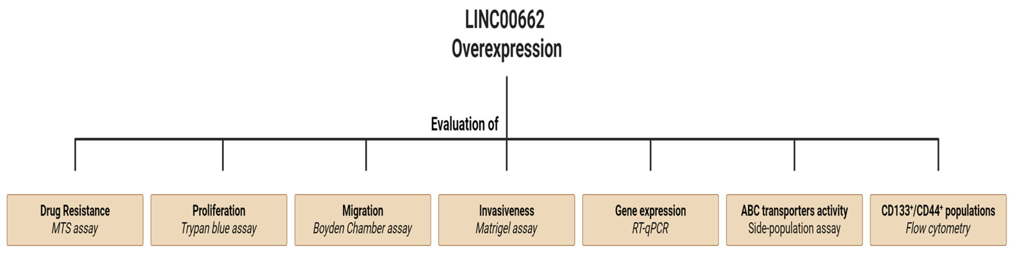
2. Results
2.1. LINC00662 Is Overexpressed in GBC Tissues and Shows an Uneven Expression in GBC Cells
2.2. Association between LINC00662 Expression and Clinicopathological Features and GBC
2.3. LINC00662 Promotes CSC-like Features in GBC Cells
2.4. LINC00662 Promotes Drug Resistance in GBC Cell Lines
2.5. LINC00662 Promotes Epithelial–Mesenchymal Transition (EMT)-Associated Invasiveness in GBC Lines
2.6. Aggressive Traits in GBC Cells Are Induced by LINC00662/miR-335-5p/OCT4 Axis
3. Discussion
4. Materials and Methods
4.1. Cell Culture and Sample Tissues
4.2. Lentiviral Transduction
4.3. RT-qPCR
4.4. CD133+/44+ Populations by Flow Cytometry
4.5. Side-Population Assay by Flow Cytometry
4.6. Cell Migration
4.7. Cell Invasion
4.8. Viability Assay for Drug Resistance
4.9. Statistical Analysis
5. Conclusions
Supplementary Materials
Author Contributions
Funding
Institutional Review Board Statement
Informed Consent Statement
Data Availability Statement
Conflicts of Interest
References
- Sung, H.; Ferlay, J.; Siegel, R.L.; Laversanne, M.; Soerjomataram, I.; Jemal, A.; Bray, F. Global Cancer Statistics 2020: GLOBOCAN Estimates of Incidence and Mortality Worldwide for 36 Cancers in 185 Countries. CA Cancer J. Clin. 2021, 71, 209–249. [Google Scholar] [CrossRef]
- Hundal, R.; Shaffer, E.A. Gallbladder cancer: Epidemiology and outcome. Clin. Epidemiol. 2014, 6, 99–109. [Google Scholar] [CrossRef]
- Slack, F.J.; Chinnaiyan, A.M. The Role of Non-coding RNAs in Oncology. Cell 2019, 179, 1033–1055. [Google Scholar] [CrossRef]
- Wang, K.C.; Chang, H.Y. Molecular mechanisms of long noncoding RNAs. Mol. Cell 2011, 43, 904–914. [Google Scholar] [CrossRef]
- Gao, N.; Li, Y.; Li, J.; Gao, Z.; Yang, Z.; Liu, H.; Fan, T. Long Non-Coding RNAs: The Regulatory Mechanisms, Research Strategies, and Future Directions in Cancers. Front. Oncol. 2020, 10, 598817. [Google Scholar] [CrossRef]
- Yang, N.; Liu, K.; Yang, M.; Gao, X. ceRNAs in Cancer: Mechanism and Functions in a Comprehensive Regulatory Network. J. Oncol. 2021, 2021, 4279039. [Google Scholar] [CrossRef]
- Wang, S.H.; Yang, Y.; Wu, X.C.; Zhang, M.D.; Weng, M.Z.; Zhou, D.; Wang, J.D.; Quan, Z.W. Long non-coding RNA MINCR promotes gallbladder cancer progression through stimulating EZH2 expression. Cancer Lett. 2016, 380, 122–133. [Google Scholar] [CrossRef]
- Ma, M.Z.; Chu, B.F.; Zhang, Y.; Weng, M.Z.; Qin, Y.Y.; Gong, W.; Quan, Z.W. Long non-coding RNA CCAT1 promotes gallbladder cancer development via negative modulation of miRNA-218-5p. Cell Death Dis. 2015, 6, e1583. [Google Scholar] [CrossRef]
- Liang, C.; Yang, P.; Han, T.; Wang, R.Y.; Xing, X.L.; Si, A.F.; Ma, Q.Y.; Chen, Z.; Li, H.Y.; Zhang, B. Long non-coding RNA DILC promotes the progression of gallbladder carcinoma. Gene 2019, 694, 102–110. [Google Scholar] [CrossRef]
- He, Y.; Xu, Y.; Yu, X.; Sun, Z.; Guo, W. The Vital Roles of LINC00662 in Human Cancers. Front. Cell Dev. Biol. 2021, 9, 711352. [Google Scholar] [CrossRef]
- Nguyen, L.V.; Vanner, R.; Dirks, P.; Eaves, C.J. Cancer stem cells: An evolving concept. Nat. Rev. Cancer 2012, 12, 133–143. [Google Scholar] [CrossRef]
- Hadjimichael, C.; Chanoumidou, K.; Papadopoulou, N.; Arampatzi, P.; Papamatheakis, J.; Kretsovali, A. Common stemness regulators of embryonic and cancer stem cells. World J. Stem Cells 2015, 7, 1150–1184. [Google Scholar] [CrossRef]
- Begicevic, R.R.; Falasca, M. ABC Transporters in Cancer Stem Cells: Beyond Chemoresistance. Int. J. Mol. Sci. 2017, 18, 2362. [Google Scholar] [CrossRef]
- Kalluri, R.; Weinberg, R.A. The basics of epithelial-mesenchymal transition. J. Clin. Investig. 2009, 119, 1420–1428. [Google Scholar] [CrossRef]
- Ye, L.; Wang, F.; Wu, H.; Yang, H.; Yang, Y.; Ma, Y.; Xue, A.; Zhu, J.; Chen, M.; Wang, J.; et al. Functions and Targets of miR-335 in Cancer. Onco Targets Ther. 2021, 14, 3335–3349. [Google Scholar] [CrossRef]
- Chen, J.; Yu, Y.; Li, H.; Hu, Q.; Chen, X.; He, Y.; Xue, C.; Ren, F.; Ren, Z.; Li, J.; et al. Long non-coding RNA PVT1 promotes tumor progression by regulating the miR-143/HK2 axis in gallbladder cancer. Mol. Cancer 2019, 18, 33. [Google Scholar] [CrossRef]
- Beylerli, O.; Gareev, I.; Sufianov, A.; Ilyasova, T.; Guang, Y. Long noncoding RNAs as promising biomarkers in cancer. Noncoding RNA Res. 2022, 7, 66–70. [Google Scholar] [CrossRef]
- Xu, Z.Y.; Peng, J.; Shi, Z.Z.; Chen, X.L.; Cheng, H.Z.; Wang, H.; Wang, Y.; Wang, G.P.; Jiang, W.; Peng, H. Silencing linc00662 inhibits cell proliferation and colony formation of lung cancer cells via regulating the miR-145-5p-PAFAH1B2 axis. Biochem. Cell Biol. 2021, 99, 330–338. [Google Scholar] [CrossRef]
- Li, N.; Zhang, L.Y.; Qiao, Y.H.; Song, R.J. Long noncoding RNA LINC00662 functions as miRNA sponge to promote the prostate cancer tumorigenesis through targeting miR-34a. Eur. Rev. Med. Pharmacol. Sci. 2019, 23, 3688–3698. [Google Scholar] [CrossRef]
- Cheng, L.; Xing, Z.; Zhang, P.; Xu, W. Long non-coding RNA LINC00662 promotes proliferation and migration of breast cancer cells via regulating the miR-497-5p/EglN2 axis. Acta Biochim. Pol. 2020, 67, 229–237. [Google Scholar] [CrossRef]
- Pérez-Moreno, P.; Riquelme, I.; García, P.; Brebi, P.; Roa, J.C. Environmental and Lifestyle Risk Factors in the Carcinogenesis of Gallbladder Cancer. J. Pers. Med. 2022, 12, 234. [Google Scholar] [CrossRef]
- Batlle, E.; Clevers, H. Cancer stem cells revisited. Nat. Med. 2017, 23, 1124–1134. [Google Scholar] [CrossRef]
- Jordan, C.T.; Guzman, M.L.; Noble, M. Cancer stem cells. N. Engl. J. Med. 2006, 355, 1253–1261. [Google Scholar] [CrossRef]
- An, C.; Hu, Z.; Li, Y.; Zhao, P.; Liu, R.; Zhang, Q.; Zhu, P.; Wang, Y. LINC00662 enhances cell progression and stemness in breast cancer by MiR-144-3p/SOX2 axis. Cancer Cell Int. 2022, 22, 184. [Google Scholar] [CrossRef]
- Nunes, T.; Hamdan, D.; Leboeuf, C.; El Bouchtaoui, M.; Gapihan, G.; Nguyen, T.T.; Meles, S.; Angeli, E.; Ratajczak, P.; Lu, H.; et al. Targeting Cancer Stem Cells to Overcome Chemoresistance. Int. J. Mol. Sci. 2018, 19, 4036. [Google Scholar] [CrossRef]
- Vasan, N.; Baselga, J.; Hyman, D.M. A view on drug resistance in cancer. Nature 2019, 575, 299–309. [Google Scholar] [CrossRef]
- Moserle, L.; Ghisi, M.; Amadori, A.; Indraccolo, S. Side population and cancer stem cells: Therapeutic implications. Cancer Lett. 2010, 288, 1–9. [Google Scholar] [CrossRef]
- Hirschmann-Jax, C.; Foster, A.E.; Wulf, G.G.; Nuchtern, J.G.; Jax, T.W.; Gobel, U.; Goodell, M.A.; Brenner, M.K. A distinct “side population” of cells with high drug efflux capacity in human tumor cells. Proc. Natl. Acad. Sci. USA 2004, 101, 14228–14233. [Google Scholar] [CrossRef]
- Sznurkowska, M.K.; Aceto, N. The gate to metastasis: Key players in cancer cell intravasation. FEBS J. 2022, 289, 4336–4354. [Google Scholar] [CrossRef]
- Gong, W.; Su, Y.; Liu, Y.; Sun, P.; Wang, X. Long non-coding RNA LINC00662 promotes cell invasion and contributes to cancer stem cell-like phenotypes in lung cancer cells. J. Biochem. 2018, 164, 461–469. [Google Scholar] [CrossRef]
- Yao, Y.; Liu, Y.; Jin, F.; Meng, Z. LINC00662 Promotes Oral Squamous Cell Carcinoma Cell Growth and Metastasis through miR-144-3p/EZH2 Axis. Yonsei Med. J. 2021, 62, 640–649. [Google Scholar] [CrossRef]
- Saitoh, M. Transcriptional regulation of EMT transcription factors in cancer. Semin. Cancer Biol. 2023, 97, 21–29. [Google Scholar] [CrossRef]
- Weidenfeld, K.; Barkan, D. EMT and Stemness in Tumor Dormancy and Outgrowth: Are They Intertwined Processes? Front. Oncol. 2018, 8, 381. [Google Scholar] [CrossRef]
- Najafi, M.; Farhood, B.; Mortezaee, K. Cancer stem cells (CSCs) in cancer progression and therapy. J. Cell Physiol. 2019, 234, 8381–8395. [Google Scholar] [CrossRef]
- He, F.; Song, Z.; Chen, H.; Chen, Z.; Yang, P.; Li, W.; Yang, Z.; Zhang, T.; Wang, F.; Wei, J.; et al. Long noncoding RNA PVT1-214 promotes proliferation and invasion of colorectal cancer by stabilizing Lin28 and interacting with miR-128. Oncogene 2019, 38, 164–179. [Google Scholar] [CrossRef]
- Yang, Y.; Wang, F.; Huang, H.; Zhang, Y.; Xie, H.; Men, T. lncRNA SLCO4A1-AS1 promotes growth and invasion of bladder cancer through sponging miR-335-5p to upregulate OCT4. Onco Targets Ther. 2019, 12, 1351–1358. [Google Scholar] [CrossRef]
- Ji, Y.Y.; Song, Y.; Wang, A.N. MiR-335-5p inhibits proliferation of Huh-7 liver cancer cells via targeting the OCT4/Akt pathway. Eur. Rev. Med. Pharmacol. Sci. 2021, 25, 1853–1860. [Google Scholar] [CrossRef]
- Guo, X.; Yu, L.; Zhang, Z.; Dai, G.; Gao, T.; Guo, W. miR-335 negatively regulates osteosarcoma stem cell-like properties by targeting POU5F1. Cancer Cell Int. 2017, 17, 29. [Google Scholar] [CrossRef]
- Gao, L.; Yang, Y.; Xu, H.; Liu, R.; Li, D.; Hong, H.; Qin, M.; Wang, Y. MiR-335 functions as a tumor suppressor in pancreatic cancer by targeting OCT4. Tumour Biol. 2014, 35, 8309–8318. [Google Scholar] [CrossRef]
- Zhang, X.; Wang, W.; Zhu, W.; Dong, J.; Cheng, Y.; Yin, Z.; Shen, F. Mechanisms and Functions of Long Non-Coding RNAs at Multiple Regulatory Levels. Int. J. Mol. Sci. 2019, 20, 5573. [Google Scholar] [CrossRef]
- Untergasser, A.; Cutcutache, I.; Koressaar, T.; Ye, J.; Faircloth, B.C.; Remm, M.; Rozen, S.G. Primer3–new capabilities and interfaces. Nucleic Acids Res. 2012, 40, e115. [Google Scholar] [CrossRef]


| Clinicopathological Features | % Patients | LINC00662 Expression (Mean ± SD) | p Value |
|---|---|---|---|
| Age | 0.832 | ||
| ≥63 | 61.5 | 1.87 ± 1.47 | |
| <63 | 38.5 | 1.62 ± 0.86 | |
| Gender | |||
| Male | 26.6 | 1.94 ± 0.83 | 0.825 |
| Female | 73.4 | 1.4 ± 1.39 | |
| Tumor size | |||
| ≥4.5 cm2 | 62.5 | 2.72 ± 1.47 | 0.049 * |
| <4.5 cm2 | 37.5 | 0.99 ± 0.16 | |
| Lymph node metastasis | |||
| Positive | 42.8 | 2.36 ± 0.82 | 0.028 * |
| Negative | 57.2 | 1.025 ± 0.15 | |
| TNM stage | |||
| I and II | 28.6 | 1.025 ± 0.01 | 0.186 |
| III and IV | 71.4 | 2.36 ± 0.97 | |
| Histological grade | |||
| Grade 1 | 8.3 | 1.2 ± 0.14 | |
| Grade 2 | 66.7 | 2.08 ± 1.43 | |
| Grade 3 | 25 | 1.62 ± 0.76 | |
| Grade 1 vs. Grade 2 | 0.768 | ||
| Grade 2 vs. Grade 3 | 0.867 | ||
| Grade 1 vs. Grade 3 | 0.937 |
| Gene | Forward Primer | Reverse Primer |
|---|---|---|
| NANOG | 5′-AA CAAAGGATGAAGTGCAAGCGG | 5′-TCCAAGTTGGGTTGGTCCAAGTCT |
| OCT4 | 5′-AAT TTG TTC CTG CAG TGC CC | 5′-GCA GCC TCA AAA TCC TCT CG |
| GAPDH | 5′-GAGTCAACGGATTTGGTCGT | 5′-GACAAGCTTCCCGTTCTCAG |
| ABCC1 | 5′-CCG TGT ACT CCA ACG CTG ACA T | 5′-ATG CTG TGC GTG ACC AAG ATC C |
| ABCG2 | 5′-GTT CTC AGC AGC TCT TCG GCT T | 5′-TCC TCC AGA CAC ACC ACG GAT A |
| ABCB1 | 5′-GCT GTC AAG GAA GCC AAT GCC T | 5′-TGC AAT GGC GAT CCT CTG CTT C |
| N-cadherin | 5′-CCT CCA GAG TTT ACT GCC ATG AC | 5′-GTA GGA TCT CCG CCA CTG ATT C |
| E-cadherin | Fw 5′-TGG AGG AAT TCT TGC TTT GC | 5′-CGT ACA TGT CAG CCA GCT TC |
| SNAI1 | 5′-TGC CCT CAA GAT GCA CAT CCG A | 5′-GGG ACA GGA GAA GGG CTT CTC |
| TWIST | 5′-GCC AGG TAC ATC GAC TTC CTC T | 5′-TCC ATC CTC CAG ACC GAG AAG G |
| SLUG | 5′-ATC TGC GGC AAG GCG TTT TCC A | 5′-GAG CCC TCA GAT TTG ACC TGT C |
| LINC00662 | 5′-GCT TCA TGA CTT GTG CCC AA | 5′-CAC CAG TTT CAG AAG CGT GT |
Disclaimer/Publisher’s Note: The statements, opinions and data contained in all publications are solely those of the individual author(s) and contributor(s) and not of MDPI and/or the editor(s). MDPI and/or the editor(s) disclaim responsibility for any injury to people or property resulting from any ideas, methods, instructions or products referred to in the content. |
© 2024 by the authors. Licensee MDPI, Basel, Switzerland. This article is an open access article distributed under the terms and conditions of the Creative Commons Attribution (CC BY) license (https://creativecommons.org/licenses/by/4.0/).
Share and Cite
Pérez-Moreno, P.; Riquelme, I.; Bizama, C.; Vergara-Gómez, L.; Tapia, J.C.; Brebi, P.; García, P.; Roa, J.C. LINC00662 Promotes Aggressive Traits by Modulating OCT4 Expression through miR-335-5p in Gallbladder Cancer Cells. Int. J. Mol. Sci. 2024, 25, 6740. https://doi.org/10.3390/ijms25126740
Pérez-Moreno P, Riquelme I, Bizama C, Vergara-Gómez L, Tapia JC, Brebi P, García P, Roa JC. LINC00662 Promotes Aggressive Traits by Modulating OCT4 Expression through miR-335-5p in Gallbladder Cancer Cells. International Journal of Molecular Sciences. 2024; 25(12):6740. https://doi.org/10.3390/ijms25126740
Chicago/Turabian StylePérez-Moreno, Pablo, Ismael Riquelme, Carolina Bizama, Luis Vergara-Gómez, Julio C. Tapia, Priscilla Brebi, Patricia García, and Juan Carlos Roa. 2024. "LINC00662 Promotes Aggressive Traits by Modulating OCT4 Expression through miR-335-5p in Gallbladder Cancer Cells" International Journal of Molecular Sciences 25, no. 12: 6740. https://doi.org/10.3390/ijms25126740