Article

Association between Circulating T Cells and the Gut Microbiome in Healthy Individuals: Findings from a Pilot Study

1
Department of Laboratory Medicine and Pathology, University of Minnesota, MMC 609, 420 Delaware Street, Minneapolis, MN 55455, USA
2
Division of Biostatistics, School of Public Health, University of Minnesota, Minneapolis, MN 55455, USA
3
Hennepin Healthcare Research Institute (HHRI), Minneapolis, MN 55404, USA
4
Department of Internal Medicine, Abbott Northwestern Hospital, Minneapolis, MN 55407, USA
5
Department of Surgery, University of Minnesota, Minneapolis, MN 55455, USA
*
Author to whom correspondence should be addressed.
These authors contributed equally to this work.
Int. J. Mol. Sci. 2024, 25(13), 6831; https://doi.org/10.3390/ijms25136831
Submission received: 15 May 2024 / Revised: 10 June 2024 / Accepted: 12 June 2024 / Published: 21 June 2024
(This article belongs to the Special Issue Microbiome and Metabolome in the Gastrointestinal Tract, 2nd Edition)

Abstract

:
Though the microbiome’s impact on immune system homeostasis is well documented, the effect of circulating T cells on the gut microbiome remains unexamined. We analyzed data from 50 healthy volunteers in a pilot trial of aspirin, using immunophenotyping and 16S rRNA sequencing to evaluate the effect of baseline T cells on microbiome changes over 6 weeks. We employed an unsupervised sparse canonical correlation analysis (sCCA) and used multivariable linear regression models to evaluate the association between selected T cell subsets and selected bacterial genera after adjusting for covariates. In the cross-sectional analysis, percentages of naïve CD4+ T cells were positively associated with a relative abundance of Intestinimonas, and the percentage of activated CD8+ T cells was inversely associated with Cellulosibacter. In the longitudinal analysis, the baseline percentages of naïve CD4+ T cells and activated CD4+ T cells were inversely associated with a 6-week change in the relative abundance of Clostridium_XlVb and Anaerovorax, respectively. The baseline percentage of terminal effector CD4+ T cells was positively associated with the change in Flavonifractor. Notably, the microbiome taxa associated with T cell subsets exclusively belonged to the Bacillota phylum. These findings can guide future experimental studies focusing on the role of T cells in impacting gut microbiome homeostasis.

1. Introduction

The gut microbiome, which is the collection of a diverse community of microorganisms, including bacteria, viruses, and fungi that live in the digestive tract, plays an important role in the development and function of the immune system. The gut microbiome can influence the differentiation and activation of immune cells, such as T cells and B cells [1,2,3], and also the production of immune molecules, such as antibodies and cytokines [4,5]. Different bacterial genera may have different functions and effects on the immune system, depending on the context and other factors such as diet or disease conditions. Bacillota and Bacteroidota are the two major phyla found in the gut. Certain bacterial genera within the Bacillota phylum affect T cell immunity in the local environment. For instance, Lactobacillus promotes the production of Th1 cells that play a key role in the host defense against intracellular pathogens [6]. Additionally, Bifidobacterium, which belongs to the Actinomyceota phylum, downregulates the production of Th2 and Th17 cells, upregulates the production of interferon beta, and increases the number and suppressive function of regulatory T cells (Tregs) [7,8,9,10]. In contrast, Bacteroides, one of the most abundant genera under phylum Bacteroidota, is found to promote Th17 cell differentiation and excessive Th17 cell activity, which play a critical role in the host defense against extracellular bacteria and fungi [11,12]. Thus, a healthy gut microbiome can help to promote a well-functioning immune system by strengthening the barrier function of the gut [13], producing anti-inflammatory molecules, and training the immune system to recognize and respond to pathogens.
Though the critical role of the microbiome in shaping the immune system is well recognized, there is now substantial evidence that there is a bidirectional relationship between the microbiome and both the innate and adaptive immune systems [14]. Prior studies have documented the role of tissue resident innate and adaptive immune cells in the intestinal mucosa in shaping the intestinal microbiome composition [15,16]. Specifically, the intestinal innate immune system secretes antimicrobial peptides and pathogen recognition receptors (e.g., Toll-like receptors) that play important roles in shaping the intestinal microbiome [17]. In addition, the intestinal adaptive immune system consisting of both B cells and T cells shapes the intestinal microbiome by producing IgA-specific responses against commensal bacteria via both T cell-dependent and T cell-independent mechanisms [18]. While the role of the local intestinal immune system in shaping the gut microbiome has been studied in previous studies, the role of circulating T cell subsets in peripheral blood in shaping the gut microbiome has not been previously evaluated.
In this study, we collected blood and repeated stools samples in a clinical trial of healthy participants and measured immune cells at baseline to comprehensively characterize the associations between the percentages of peripheral blood T cell subsets and the relative abundance of gut microbiomes. We hypothesized that a higher proportion of T cell subsets such as naïve CD4+ T cells that have been previously associated with lower multimorbidity and mortality will be associated with bacterial taxa that reduce inflammation and are beneficial for human health, while a higher proportion of circulating T cells subsets such as effector and activated T cells that indicate a recent or ongoing immune response will be associated with bacterial taxa that are detrimental to human health.

2. Results

The study participants had a mean age of 61.3 ± 5.1 years and mean BMI of 27.7 ± 4.5 kg/m2. Females represented 64% (N = 32) of the participants, and 60% (N = 30) of the participants received aspirin during the clinical trial. Aspirin intake was not associated with any of the T cell subsets studied. The sCCA was performed separately for the cross-sectional and longitudinal (6-week microbiome change) analyses between 10 T cell subsets and 50 gut bacterial genera and computed 10 canonical variates. The canonical correlation coefficients between different T cell subsets and bacterial genera ranged from 0.47 to 0.69 in the cross-sectional analysis (Supplementary Table S1) and from 0.26 to 0.73 in the longitudinal analysis (Supplementary Table S2). In the cross-sectional analysis, we found three significant canonical variates (Pearson correlation coefficient: K1 [r = 0.69, p = 0.007]; K3 [r = 0.64, p = 0.003]; and K5 [r = 0.60, p = 0.045] Table S1) and one in the 6-week change analysis (K1 [r = 0.73, p = 0.006] Table S2).

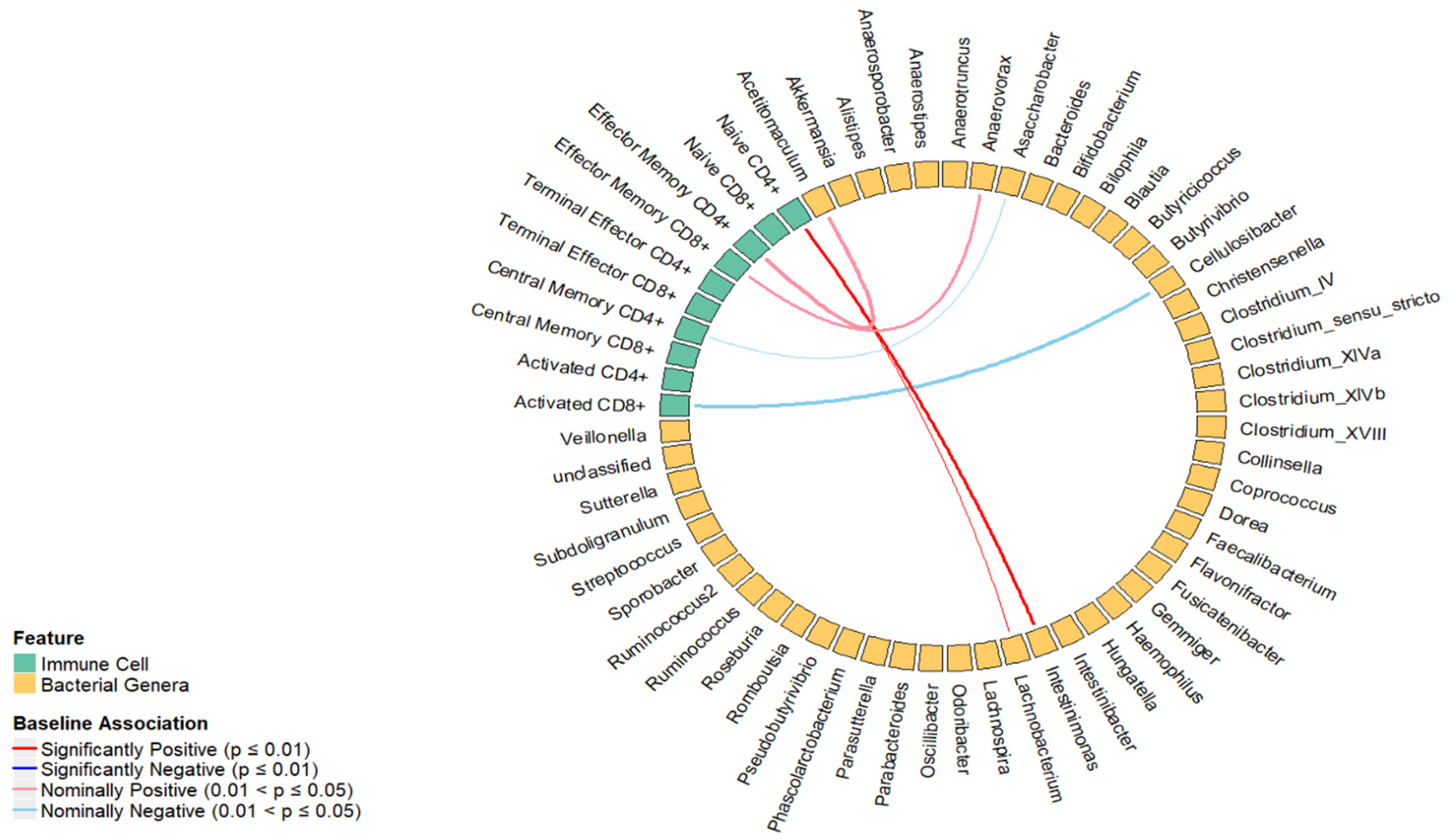
2.1. Cross-Sectional Association between the Percentage of T Cell Subsets and Relative Abundance of Bacterial Genera

In the cross-sectional analysis, canonical correlation analysis between T cell subsets and bacterial genera from three significant canonical variates identified 98 pairs of bacterial genera and T cell subsets for further linear regression analysis (Supplementary Table S1). As shown in Figure 1, we found that the percentage of naive CD4+ T cells was positively associated with Intestinimonas (β = 0.03, p = 0.01). Though not statistically significant at the pre-determined significance level of 0.01, we found a nominal association between the percentage of naive CD4+ T cells and Lachnobacterium (β = 0.02, p = 0.01), central memory CD4+ T cells and Asaccharobacter (β = −0.02, p = 0.05), effector memory CD4+ T cells and Acetitomaculum (β = 0.05, p = 0.04), effector memory CD8+ T cells and Anaerovorax (β = 0.03, p = 0.05), and activated CD8+ T cells and Cellulosibacter (β = −0.11, p = 0.02). The univariate and multivariable regression results are shown in Figure 1 and Table 1, respectively.

2.2. Longitudinal Analysis Percentage of T Cell Subsets at the Baseline and Fold Change of Bacterial Genera over 6 Weeks

In the longitudinal analysis, 108 pairs of bacterial genera and T cell subsets were identified from significant canonical variates (Supplementary Table S2). We found that terminal effector CD4+ T cells were positively associated with Flavonifractor (β = 0.06, p < 0.01). Though not statistically significant at the 0.01 significance level, the terminal effector CD8+ T cells were nominally associated with Flavonifractor (β = 0.02, p = 0.03). We found inverse associations between the baseline percentages of central memory CD8+ and Alistipes (β = −0.05, p = 0.01) and naive CD4+ T cells and Clostridium XlVb (β = −0.02, p = 0.03) and activated CD4+ T cells and Anaerovorax (β = −0.06, p = 0.04). The results of the univariate regression analysis are shown in Figure 2, and the multiple linear regression analysis is shown in Table 2.
In both cross-sectional and longitudinal analyses, we found that the percentages of naive CD4+ T cells were positively associated with the relative abundance of Intestinimonas and nominally associated with Clostridium XlVb (Table 3). Of note, the majority of bacterial genera that displayed significant associations with T cell subsets were predominantly found within the Bacillota phylum.

3. Discussion

Though the link between the composition of the gut microbiome and immune cells with human health and disease has been previously investigated in multiple studies [14,16,19], the connection between immune subsets in the peripheral blood and gut microbiome in healthy individuals remains largely unexplored. In this study, we showed that baseline percentages of circulating T cell subsets in peripheral blood were associated with changes in the relative abundance of bacterial genera over 6 weeks of follow-up in healthy participants enrolled in a clinical trial. To the best of our knowledge, this is the first study that has evaluated the association between T cell subsets in peripheral blood and change in the relative abundance of gut bacteria in healthy participants. We utilized a sparse canonical correlation analysis (sCCA) to evaluate the relationship between high dimensional gut microbiome data and the comprehensive adaptive immune T cell phenotype by preserving the main facets that explain the correlation between two feature sets. sCCA is commonly used in genomics [20,21] or neuroscience [21,22,23] research areas to analyze high-dimensional data. This method allowed us to evaluate a selected set of T cell subsets and bacterial genera that had the highest correlation in the canonical component.
In our cross-sectional analysis, we found that a higher proportion of naïve CD4+ T cells was associated with an increased relative abundance of bacterial genera Intestinimonas. Intestinimonas is a genus within the Oscillospiraceae family, and previous studies have shown that certain species of Intestinimonas may play a role in alleviating inflammatory and metabolic diseases [24,25]. A previous study showed that a higher relative abundance of Intestinimonas genera is associated with elevated levels of IL-4, suggesting that alterations in the composition of the gut microbiota may play a role in contributing to systemic inflammation in patients with Huntington’s disease [24]. This indicates Bacillota-derived molecular patterns like lipoteichoic acids, lipoproteins, and peptidoglycan are recognized by TLR2 heterodimers (TLR2/TLR1 or TLR2/TLR6), which allow the innate immune system to sense and respond to infections by these Gram-positive bacteria [26]. The TLR2-mediated signaling upon binding these ligands results in the production of inflammatory cytokines and the activation of immune responses against Bacillota pathogens [27]. Another study identified that treatment with Chinese caterpillar fungus polysaccharides was associated with an increase in short-chain fatty acid (SCFA)-producing bacteria within the gut microbiome, including Intestinimonas butyriciproducens, leading to a higher production of SCFAs, which, in turn, played a role in reducing elevated glucose levels [25]. Thus, a higher relative abundance of Intestinimonas appears to be beneficial for human health. We have previously shown that lower naïve CD4+ T cells are a biomarker for an age-related immune phenotype (ARIP) and are associated with an increased risk of age-related morbidity and mortality [28]. Hence, the positive correlation between Intestinimonas and the percentage of naive CD4+ T cells in the cross-sectional analysis suggests that ARIP markers and intestinal microbial abundance may contribute to future human health. In addition, we found that the proportion of activated CD8+ T cell subsets was inversely associated with the relative abundance of Cellulosibacter. The dysregulation or excessive activation of CD8+ T cells can lead to various immune-mediated diseases, including autoimmune disorders and chronic inflammatory conditions. For instance, the dysregulation of CD8+ T cell activation can contribute to the development of autoimmune diseases such as autoinflammatory diseases and chronic autoimmune conditions, leading to increased morbidity and mortality [29,30]. The balance of T cell activation is crucial, as it can result in either tolerogenic or proinflammatory outcomes, impacting the immune response and the development of immune-mediated diseases.
In our longitudinal analysis, we observed that a reduced proportion of activated CD4+ T cells were inversely associated with the relative abundance of Anaerovorax. Limited research has investigated the impact of Anaerovorax on host health status [31,32,33]. A study on children showed that moderate-to-vigorous physical activity was associated with a higher relative abundance of Anaerovorax [32], indicating that a higher relative abundance of Anaerovorax may confer potential health benefits. Conversely, elevated levels of activated CD4+ T cells, which indicate a negative prognosis, were observed during the acute phase of HIV-1 infection [34]. In general, it has been hypothesized that the higher frequencies of activated T cells of various phenotypes could serve as biomarkers of poorer outcomes in several conditions, including vaccine-induced protection (at defined time point(s) post-vaccination), disease activity (for T cell-mediated autoimmune or infectious diseases), and chronic inflammation (as has been shown in HIV infection) [35,36]. Consequently, the observed negative association between activated CD4+ T cells and Anaerovorax in our longitudinal analysis is consistent with the health-promoting behaviors associated with Anerovorax and the poor health outcomes associated with activated CD4+ cells. Hence, this finding warrants further investigation into their associations with health and disease states. The longitudinal analysis also showed that reduced levels of CD4+ naive T cells are associated with increased levels of Clostridium XIVb. Clostridium XlVb has demonstrated both positive and detrimental effects on human health and disease conditions. In two studies on ulcerative colitis, Clostridium XlVb was beneficial to the immune response or could reduce the risk of developing Clostridioides difficile infection [37,38]. Conversely, studies on cognitive function revealed harmful effects of Clostridium XlVb in individuals with type 2 diabetes mellitus and Parkinson’s disease [39,40]. While a reduced proportion of naïve CD4+ T cells, an ARIP marker, has been associated with an increased risk of multimorbidity and short-term mortality, the biological significance of the observed association with an increase in Clostridium XIVb should be further evaluated. In addition, we observed an elevated proportion of terminal effector CD4+ cells positively associated with Flavonifractor in the 6-week follow-up. Flavonifractor has proinflammatory properties that were found in higher abundance in patients with various brain-related diseases [41]. Our previous study showed that CD4+ effector memory T cells are associated with lower immune function in aging and are associated with type 2 diabetes, which is itself proinflammatory. Another study found that Flavonifractor plautii was found to increase the expression of mRNAs encoding IFN-γ and IL-10 in vitro, and IFN-γ has been linked to proinflammatory effects [42]. Some studies have suggested that infections caused by pathogens like human cytomegalovirus (CMV) and dengue virus (DENV) lead to an increase in terminal effector CD4+ cells [43,44]. Thus, a higher relative abundance of Flavonifractor appears to be harmful to human health. The impact of immune cells on overall health, as well as the increased proinflammatory state, could potentially be modulated through the gut microbiome. However, further research is necessary to identify specific microbial taxa responsible for mediating these effects.
Of interest, we found that all the bacteria found to be associated with the distribution of circulating T cell subsets in the peripheral blood belonged to the Bacillota phylum. Bacillota bacteria in the gut are known to produce short-chain fatty acids (SCFAs), which have been linked to improvements in immune function [45]. Resistant starch can be broken down into SCFAs by bacteria from the Bacillota phylum. The colon predominantly contains three types of SCFAs: acetate, propionate, and butyrate, which serve as a source of energy for epithelial cells and possess properties that reduce inflammation and facilitate immune signaling [45,46]. Our study demonstrated an association between T cell subsets and Bacillota bacteria, which suggests the involvement of common biological processes between the regulation of T cells and gut microbiome, although the exact mechanism remains unknown.

Strengths and Limitations

This is the first study to evaluate the association between circulating T cell subsets in peripheral blood and gut microbial taxa obtained from healthy individuals. By analyzing healthy participants, we had a broad understanding of the connection between T cell subsets and bacterial genera. This approach will facilitate future comparisons across studies. Incorporating bacterial genera at both baseline and after 6 weeks enabled us to detect alterations in the bacterial genera over the course of time. The major limitations of this study are the small sample size and short follow-up time (6 weeks). Since our primary interest was not to evaluate the effect of aspirin on T cell distribution or the gut microbiome but rather to evaluate the association between circulating T cells and the gut microbiome, we analyzed the data by combining both study arms together despite the randomized clinical trial study design to maximize the sample size. Since aspirin intake was not associated with T cell subset distribution at baseline and aspirin intake did not significantly impact the bacterial taxa that were associated with the T cell subsets, combining the two study arms into a single analysis did not introduce any apparent bias in the study results. Furthermore, we adjusted for the study treatment arm (aspirin/placebo) in all regression analyses to minimize any bias in the study results. Another limitation was that 16S rRNA sequencing only provided information on the relative abundance of bacterial taxa and did not provide information on their function or activity. This means that changes in the abundance of specific bacterial taxa over time may not necessarily reflect absolute changes in microbial genera or changes in their metabolic activity or contribution to host health. While 16s sequencing can be informative about the gut microbiome, additional methods, such as metagenomic sequencing, may provide a more comprehensive understanding of longitudinal changes in the gut microbiome, as they can provide more detailed insights into alterations in bacterial genera and their function.

4. Material and Methods

4.1. Study Design

The study participants were healthy individuals from the greater Minneapolis–St. Paul, Minnesota area, aged 50–75, who were recruited as part of our double-blinded randomized placebo-controlled trial clinical trial—the Effect of Aspirin on Gut Microbiome (ASMIC) study (NCT02761486) [47]. The ASMIC study hypothesized that aspirin intake reduced CRC risk by decreasing the abundance of proinflammatory gut bacteria and increasing anti-inflammatory gut bacteria. The study objective was to compare gut microbiome changes linked to aspirin intake using a double-blinded, placebo-controlled clinical trial design of aspirin intake with 50 healthy volunteers over 6 weeks. Among the 50 study participants, 30 participants received a daily dose of 325 mg of aspirin and 20 received a placebo. The ASMIC study design has been described previously in more detail [47]. We collected blood samples at the baseline visit and stool samples at the baseline and at the 6-week follow-up visit from all participants. We profiled 10 T cell subsets in the blood and gut microbiome from the stool samples.

4.2. Collection and Processing of the Stool Samples

Participants collected their stool samples at home using stool collection kits. All stool samples collected at each visit were processed in one batch [47]. DNA was extracted from the stool, and the V4 hypervariable region on the 16S rRNA gene was amplified and sequenced using MiSeq (2 × 300 paired-end; Illumina, San Diego, CA, USA) at the University of Minnesota Genomics Center (UMGC) using protocols published previously [47].

4.3. Data Preprocessing

Mothur ver. 1.35.1 was used to process and analyze the sequenced data, and the high-quality sequences were aligned with SILVA database ver. 123.33. UCHIME package (Edgar, 2010) was utilized to remove chimeric sequences. The clustering of OTUs was performed to 97% identify, and the taxonomic classification was completed using version 16 data from the Ribosomal Database Project [47]. Bacterial genera were included for further analyses if they had a non-zero relative abundance among at least 60% of participants at the baseline and at the follow-up visit (high-frequency genera). We included 50 high-frequency bacterial genera in the downstream analyses. Participants with missing microbiome data at baseline (n = 1) and at the 6-week visit (n = 3) were excluded from the analysis, leaving 47 participants with complete data at two visits.

4.4. Collection of Blood Samples and Measurement of T Cell Subsets

Two milliliters (mL) of blood collected in EDTA tubes was incubated for 10 min in 10 milliliters of 1X Red Blood Cell (RBC) lysis buffer (BioLegend, San Diego, CA, USA), centrifuged, and immunophenotyped on the same day using the standardized protocol [35]. The immunophenotyping and differential white blood cell test were conducted by the Advanced Research and Diagnostic Laboratory (ARDL) at the University of Minnesota (Minneapolis, MN, USA). Briefly, the T cells were measured after thawing using standard procedures and stained using the following antibody cocktail fixable viability stain for identifying live/dead cells: FVS570, CD3-APC, CD19-PE-Cy7, IgD-BUV737, HLA-DR-PE-CF594, CD4-APC-Cy7, CD8-BUV395, CD27-FITC, CD127-PE-CF594, CD45RA-BV711 CCR7-BV421, CD95-BV605, and CD28-BV510. We used FlowJo and OpenCyto for the gating and data analysis of all 10 T cell subsets. The major T cell subsets measured were CD4+ T cells (CD3+ CD19− CD4+ CD8−) and CD8+ T cells (CD3+ CD19− CD4− CD8+). The subsets of both CD4+ and CD8+ T cells were defined as follows: activated T cells (HLA− DR+), naïve (CD45RA+ CCR7+ CD28+), central memory (CM: CD45RA− CCR7+ CD28+), effector memory (EM: CD45RA− CCR7− CD28−), and terminal effector (CD45RA+ CCR7− CD28−).

4.5. Covariates

Participant’s age, sex, weight (lbs.), and height (inches) were collected by a trained technician at the baseline visit. We estimated the body mass index (BMI) in kg/m2 based on the equation weight (lbs.)/[height (inches)]2 × 703 if available. A brief baseline questionnaire queried about the intake of meat, yogurt, and fruits and vegetables (servings per week).

4.6. Statistical Analysis

The statistical analysis included a cross-sectional analysis, which examined associations between T cell subsets and bacterial genera at baseline, and a longitudinal analysis of the 6-week changes in the microbiome, which examined associations between the baseline percentages of T cell subsets and the fold changes in the relative abundances of the bacterial genera between 6 weeks and at baseline. All 10 T cell subsets had a normal distribution. We evaluated the association between T cell subsets and aspirin intake at baseline. To reduce the impact of outliers due to the skewed data and to approximate normal distribution, log transformation and standardization were applied to all bacterial genera.

4.7. Sparse Canonical Correlation Analysis (sCCA)

We used sparse canonical correlation analysis (sCCA) to identify linear projections of group-level correlations between T cell subsets and the gut microbiome. sCCA has been used in several studies for multi-omics data integration and analysis [48]. Sparse CCA is an adapted version of CCA for feature selection after applying L1 or lasso penalty in CCA in the high-dimensional feature space. The sCCA analysis was conducted using the Penalized Multivariate Analysis (PMA) package in R [49], which resulted in sparsity through penalized matrix decomposition with Lasso penalty (L1). The L1 parameter was tuned using a permutation approach in the CCA.permute function in R, where a grid search ranging from 0.1 to 0.7 was used to find the optimal L1 value that maximized the correlation between the microbiome and T cell subsets at a significance level of 0.05. We applied the L1 value of 0.367 on the T cell subsets and 0.433 on the bacterial taxa for the cross-sectional analysis and 0.5 on the T cell subset and 0.7 on the bacterial taxa for the longitudinal analysis. The objective function of sCCA is shown below (Equation (1)) [49]. We performed a permutation test (number of permutations = 1000) at the 0.05 significance level.
m a x m i z e u , v u T X T Y v   s u b j e c t   t o u 2 2 1 , v 2 2 1 , P 1 u c 1 , P 2 v c 2 ,
N o t e : P 1   a n d   P 2   a r e   c o n v e x   f u n c t i o n s .

4.8. Linear Regression Analysis

We selected significant pairs of canonical variates to select pairs of bacterial genera and T cell subsets for further association analysis. The multivariable linear regression models were adjusted for age, sex, BMI, and fruit and vegetable servings per week in the cross-sectional analysis and for the treatment arm (aspirin/placebo). In addition, we adjusted for the baseline bacterial genera measurements in the longitudinal analysis. We considered a p-value of ≤0.01 for an association between a T cell subset and bacterial genera to be statistically significant in the regression analysis.

5. Conclusions

In conclusion, our study provided preliminary evidence that multiple T cell subsets are associated with relative change in bacterial genera over 6 weeks of follow-up time. Future experimental studies with longer longitudinal follow-up are needed to focus on the role of circulating T cells in determining the gut microbiome composition and thereby maintaining gut homeostasis. The bacterial genera in the Bacillota phylum were predominantly associated with various T cell subsets in both the cross-sectional and longitudinal change analyses after adjusting for covariates. This might suggest that the Bacillota being major producers of short-chain fatty acids (SCFAs) could potentially play a crucial role in the homeostasis of the gut microbiome. These study findings are particularly important in settings where a mechanistic understanding of the role of adaptive immune T cell subsets is critical, such as in the prevention and treatment of immune-related diseases, including cancer, inflammatory bowel disease, allergies, and autoimmune disorders.

Supplementary Materials

The following supporting information can be downloaded at: https://www.mdpi.com/article/10.3390/ijms25136831/s1.

Author Contributions

S.V. and B.T. conceptualized the study. Y.S.S. and S.V. performed the data analysis. S.V., Y.S.S. and B.T. drafted and revised the manuscript. A.E.P., W.G., A.B.K., G.O., M.O. and C.S. critically reviewed the manuscript. All authors have read and agreed to the published version of the manuscript.

Funding

This research was supported by the University of Minnesota Grant-in-Aid of Research, Artistry, and Scholarship program and the National Institutes of Health’s National Center for Advancing Translational Sciences, grant UL1TR002494. The content is solely the responsibility of the authors and does not necessarily represent the official views of the National Institutes of Health’s National Center for Advancing Translational Sciences.

Institutional Review Board Statement

Not applicable.

Informed Consent Statement

The trial was registered at www.clinicaltrials.gov, NCT02761486. The study protocol was approved by the Institutional Review Board at the University of Minnesota, and all participants provided written informed consent. The study was conducted in accordance with the Declaration of Helsinki.

Data Availability Statement

The data that support the findings of this study are available upon request from the corresponding author (B.T.). The data are not publicly available due to the regulations on clinical trial data.

Acknowledgments

We are thankful to the study coordinators, Jennifer Stromberg and Allison Iwan, and other staff (Jeremiah Menk, Lori Strayer, and Frank Strahan) who helped with the study. We thank the University of Minnesota Genomics Center for conducting the genetic analyses (sequencing data were processed and analyzed using the resources of the Minnesota Supercomputing Institute); Eicosanoid Core Laboratory, University of Vanderbilt (Ginger Milne) for measuring the urine biomarkers; Fairview Investigational Drug Services (Luke Darlette) for preparing the treatment capsules; and the Epidemiology Clinical Research Center (Margaret Krieser) for providing space for the recruitment of the study subjects. We are also grateful to Michael Franklin, a medical writer, for reviewing the paper. The study was registered using Clinical Trials Registration NCT02761486.

Conflicts of Interest

There are no competing financial or non-financial interests in relation to this manuscript. The authors declare no conflicts of interest. A.B.K. received speaker honoraria from Siemens Healthcare Diagnostics, the American Society of Nephrology, and Yale University Department of Laboratory Medicine, all unrelated to this manuscript.

References

  1. Rosser, E.C.; Piper, C.J.; Matei, D.E.; Blair, P.A.; Rendeiro, A.F.; Orford, M.; Alber, D.G.; Krausgruber, T.; Catalan, D.; Klein, N.; et al. Microbiota-Derived Metabolites Suppress Arthritis by Amplifying Aryl-Hydrocarbon Receptor Activation in Regulatory B Cells. Cell Metab. 2020, 31, 837–851.e10. [Google Scholar] [CrossRef] [PubMed]
  2. Yasuda, K.; Takeuchi, Y.; Hirota, K. The pathogenicity of Th17 cells in autoimmune diseases. Semin. Immunopathol. 2019, 41, 283–297, Erratum in Semin. Immunopathol. 2019, 41, 299. [Google Scholar] [CrossRef] [PubMed]
  3. Yao, Y.; Cai, X.; Zheng, Y.; Zhang, M.; Fei, W.; Sun, D.; Zhao, M.; Ye, Y.; Zheng, C. Short-chain fatty acids regulate B cells differentiation via the FFA2 receptor to alleviate rheumatoid arthritis. Br. J. Pharmacol. 2022, 179, 4315–4329. [Google Scholar] [CrossRef] [PubMed]
  4. Brown, E.L.; Essigmann, H.T.; Hoffman, K.L.; Palm, N.W.; Gunter, S.M.; Sederstrom, J.M.; Petrosino, J.F.; Jun, G.; Aguilar, D.; Perkison, W.B.; et al. Impact of Diabetes on the Gut and Salivary IgA Microbiomes. Infect. Immun. 2020, 88, e00301-20. [Google Scholar] [CrossRef] [PubMed]
  5. Schirmer, M.; Smeekens, S.P.; Vlamakis, H.; Jaeger, M.; Oosting, M.; Franzosa, E.A.; Ter Horst, R.; Jansen, T.; Jacobs, L.; Bonder, M.J.; et al. Linking the Human Gut Microbiome to Inflammatory Cytokine Production Capacity. Cell 2016, 167, 1125–1136.e8. [Google Scholar] [CrossRef]
  6. Jung, Y.-J.; Kim, K.-H.; Ko, E.-J.; Lee, Y.; Kim, M.-C.; Lee, Y.-T.; Kim, C.-H.; Jeeva, S.; Park, B.R.; Kang, S.-M. Adjuvant effects of killed Lactobacillus casei DK128 on enhancing T helper type 1 immune responses and the efficacy of influenza vaccination in normal and CD4-deficient mice. Vaccine 2020, 38, 5783–5792. [Google Scholar] [CrossRef] [PubMed]
  7. Henrick, B.M.; Rodriguez, L.; Lakshmikanth, T.; Pou, C.; Henckel, E.; Arzoomand, A.; Olin, A.; Wang, J.; Mikes, J.; Tan, Z.; et al. Bifidobacteria-mediated immune system imprinting early in life. Cell 2021, 184, 3884–3898.e11. [Google Scholar] [CrossRef] [PubMed]
  8. Hidalgo-Cantabrana, C.; Delgado, S.; Ruiz, L.; Ruas-Madiedo, P.; Sánchez, B.; Margolles, A. Bifidobacteria and Their Health-Promoting Effects. Microbiol. Spectr. 2017, 5, 73–98. [Google Scholar] [CrossRef]
  9. Avershina, E.; Storrø, O.; Øien, T.; Johnsen, R.; Wilson, R.; Egeland, T.; Rudi, K. Bifidobacterial Succession and Correlation Networks in a Large Unselected Cohort of Mothers and Their Children. Appl. Environ. Microbiol. 2013, 79, 497–507. [Google Scholar] [CrossRef]
  10. Sun, S.; Luo, L.; Liang, W.; Yin, Q.; Guo, J.; Rush, A.M.; Lv, Z.; Liang, Q.; Fischbach, M.A.; Sonnenburg, J.L.; et al. Bifidobacterium alters the gut microbiota and modulates the functional metabolism of T regulatory cells in the context of immune checkpoint blockade. Proc. Natl. Acad. Sci. USA 2020, 117, 27509–27515. [Google Scholar] [CrossRef]
  11. Brennan, C.A.; Clay, S.L.; Lavoie, S.L.; Bae, S.; Lang, J.K.; Fonseca-Pereira, D.; Rosinski, K.G.; Ou, N.; Glickman, J.N.; Garrett, W.S. Fusobacterium nucleatum drives a pro-inflammatory intestinal microenvironment through metabolite receptor-dependent modulation of IL-17 expression. Gut Microbes 2021, 13, 1987780. [Google Scholar] [CrossRef] [PubMed]
  12. Chen, L.; Ruan, G.; Cheng, Y.; Yi, A.; Chen, D.; Wei, Y. The role of Th17 cells in inflammatory bowel disease and the research progress. Front. Immunol. 2023, 13, 1055914. [Google Scholar] [CrossRef] [PubMed]
  13. Hiippala, K.; Jouhten, H.; Ronkainen, A.; Hartikainen, A.; Kainulainen, V.; Jalanka, J.; Satokari, R. The Potential of Gut Commensals in Reinforcing Intestinal Barrier Function and Alleviating Inflammation. Nutrients 2018, 10, 988. [Google Scholar] [CrossRef] [PubMed]
  14. Zheng, D.; Liwinski, T.; Elinav, E. Interaction between microbiota and immunity in health and disease. Cell Res. 2020, 30, 492–506. [Google Scholar] [CrossRef] [PubMed]
  15. Hegazy, A.N.; West, N.R.; Stubbington, M.J.; Wendt, E.; Suijker, K.I.; Datsi, A.; This, S.; Danne, C.; Campion, S.; Duncan, S.H.; et al. Circulating and Tissue-Resident CD4+ T Cells With Reactivity to Intestinal Microbiota Are Abundant in Healthy Individuals and Function Is Altered During Inflammation. Gastroenterology 2017, 153, 1320–1337.e16. [Google Scholar] [CrossRef] [PubMed]
  16. Zhang, H.; Sparks, J.B.; Karyala, S.V.; Settlage, R.; Luo, X.M. Host adaptive immunity alters gut microbiota. ISME J. 2014, 9, 770–781. [Google Scholar] [CrossRef] [PubMed]
  17. Pagliari, D.; Piccirillo, C.A.; Larbi, A.; Cianci, R. The Interactions between Innate Immunity and Microbiota in Gastrointestinal Diseases. J. Immunol. Res. 2015, 2015, 1–3. [Google Scholar] [CrossRef]
  18. Yang, Y.; Palm, N.W. Immunoglobulin A and the microbiome. Curr. Opin. Microbiol. 2020, 56, 89–96. [Google Scholar] [CrossRef] [PubMed]
  19. Wu, H.J.; Wu, E. The role of gut microbiota in immune homeostasis and autoimmunity. Gut Microbes 2012, 3, 4–14. [Google Scholar] [CrossRef]
  20. Dutta, D.; Sen, A.; Satagopan, J. Sparse canonical correlation to identify breast cancer related genes regulated by copy number aberrations. PLoS ONE 2022, 17, e0276886. [Google Scholar] [CrossRef]
  21. Du, L.; Liu, K.; Yao, X.; Risacher, S.L.; Han, J.; Saykin, A.J.; Guo, L.; Shen, L. Multi-Task Sparse Canonical Correlation Analysis with Application to Multi-Modal Brain Imaging Genetics. IEEE/ACM Trans. Comput. Biol. Bioinform. 2019, 18, 227–239. [Google Scholar] [CrossRef] [PubMed]
  22. Minnier, J.; Emmett, M.R.; Perez, R.; Ding, L.-H.; Barnette, B.L.; Larios, R.E.; Hong, C.; Hwang, T.H.; Yu, Y.; Fallgren, C.M.; et al. Associations between lipids in selected brain regions, plasma miRNA, and behavioral and cognitive measures following 28Si ion irradiation. Sci. Rep. 2021, 11, 14899. [Google Scholar] [CrossRef] [PubMed]
  23. Wang, S.; Qian, Y.; Wei, K.; Kong, W. Identifying Biomarkers of Alzheimer’s Disease via a Novel Structured Sparse Canonical Correlation Analysis Approach. J. Mol. Neurosci. 2021, 72, 323–335. [Google Scholar] [CrossRef] [PubMed]
  24. Du, G.; Dong, W.; Yang, Q.; Yu, X.; Ma, J.; Gu, W.; Huang, Y. Altered Gut Microbiota Related to Inflammatory Responses in Patients With Huntington’s Disease. Front. Immunol. 2021, 11, 603594. [Google Scholar] [CrossRef] [PubMed]
  25. Yao, Y.; Yan, L.; Chen, H.; Wu, N.; Wang, W.; Wang, D. Cyclocarya paliurus polysaccharides alleviate type 2 diabetic symptoms by modulating gut microbiota and short-chain fatty acids. Phytomedicine 2020, 77, 153268. [Google Scholar] [CrossRef] [PubMed]
  26. Irvine, K.L.; Hopkins, L.J.; Gangloff, M.; Bryant, C.E. The molecular basis for recognition of bacterial ligands at equine TLR2, TLR1 and TLR6. Vet. Res. 2013, 44, 50. [Google Scholar] [CrossRef] [PubMed]
  27. Janssens, S.; Beyaert, R. Role of Toll-Like Receptors in Pathogen Recognition. Clin. Microbiol. Rev. 2003, 16, 637–646. [Google Scholar] [CrossRef] [PubMed]
  28. Ramasubramanian, R.; Meier, H.C.S.; Vivek, S.; Klopack, E.; Crimmins, E.M.; Faul, J.; Nikolich-Žugich, J.; Thyagarajan, B. Evaluation of T-cell aging-related immune phenotypes in the context of biological aging and multimorbidity in the Health and Retirement Study. Immun. Ageing 2022, 19, 33. [Google Scholar] [CrossRef] [PubMed]
  29. Klöß, S.; Dehmel, S.; Braun, A.; Parnham, M.J.; Köhl, U.; Schiffmann, S. From Cancer to Immune-Mediated Diseases and Tolerance Induction: Lessons Learned From Immune Oncology and Classical Anti-cancer Treatment. Front. Immunol. 2020, 11, 1423. [Google Scholar] [CrossRef]
  30. Bourque, J.; Hawiger, D. Variegated Outcomes of T Cell Activation by Dendritic Cells in the Steady State. J. Immunol. 2022, 208, 539–547. [Google Scholar] [CrossRef]
  31. Tian, T.; Zhang, X.; Luo, T.; Wang, D.; Sun, Y.; Dai, J. Effects of Short-Term Dietary Fiber Intervention on Gut Microbiota in Young Healthy People. Diabetes Metab. Syndr. Obes. 2020, 14, 3507–3516. [Google Scholar] [CrossRef] [PubMed]
  32. Santarossa, S.; Sitarik, A.R.; Johnson, C.C.; Li, J.; Lynch, S.V.; Ownby, D.R.; Ramirez, A.; Yong, G.L.; Cassidy-Bushrow, A.E. Associations of physical activity with gut microbiota in pre-adolescent children. Phys. Act. Nutr. 2021, 25, 24–37. [Google Scholar] [CrossRef]
  33. Wei, M.; Li, C.; Dai, Y.; Zhou, H.; Cui, Y.; Zeng, Y.; Huang, Q.; Wang, Q. High-Throughput Absolute Quantification Sequencing Revealed Osteoporosis-Related Gut Microbiota Alterations in Han Chinese Elderly. Front. Cell. Infect. Microbiol. 2021, 11, 630372. [Google Scholar] [CrossRef] [PubMed]
  34. Xia, H.; Jiang, W.; Zhang, X.; Qin, L.; Su, B.; Li, Z.; Sun, J.; Zhang, Y.; Zhang, T.; Lu, X.; et al. Elevated Level of CD4+ T Cell Immune Activation in Acutely HIV-1-Infected Stage Associates With Increased IL-2 Production and Cycling Expression, and Subsequent CD4+ T Cell Preservation. Front. Immunol. 2018, 9, 616. [Google Scholar] [CrossRef]
  35. Maecker, H.T.; McCoy, J.P.; Nussenblatt, R. Standardizing immunophenotyping for the Human Immunology Project. Nat. Rev. Immunol. 2012, 12, 191–200. [Google Scholar] [CrossRef]
  36. Kestens, L.; Vanham, G.; Gigase, P.; Young, G.; Hannet, I.; Vanlangendonck, F.; Hulstaert, F.; Bach, B.A. Expression of activation antigens, HLA-DR and CD38, on CD8 lymphocytes during HIV-1 infection. AIDS 1992, 6, 793–798. [Google Scholar] [CrossRef]
  37. Yang, Z.-H.; Liu, F.; Zhu, X.-R.; Suo, F.-Y.; Jia, Z.-J.; Yao, S.-K. Altered profiles of fecal bile acids correlate with gut microbiota and inflammatory responses in patients with ulcerative colitis. World J. Gastroenterol. 2021, 27, 3609–3629. [Google Scholar] [CrossRef] [PubMed]
  38. Wan, J.; Zhang, Y.; He, W.; Tian, Z.; Lin, J.; Liu, Z.; Li, Y.; Chen, M.; Han, S.; Liang, J.; et al. Gut Microbiota and Metabolite Changes in Patients With Ulcerative Colitis and Clostridioides difficile Infection. Front. Microbiol. 2022, 13, 802823. [Google Scholar] [CrossRef]
  39. Du, Y.; Li, X.; An, Y.; Song, Y.; Lu, Y. Association of gut microbiota with sort-chain fatty acids and inflammatory cytokines in diabetic patients with cognitive impairment: A cross-sectional, non-controlled study. Front. Nutr. 2022, 9, 930626. [Google Scholar] [CrossRef]
  40. Qian, Y.; Yang, X.; Xu, S.; Wu, C.; Song, Y.; Qin, N.; Chen, S.-D.; Xiao, Q. Alteration of the fecal microbiota in Chinese patients with Parkinson’s disease. Brain Behav. Immun. 2018, 70, 194–202. [Google Scholar] [CrossRef]
  41. Eicher, T.P.; Mohajeri, M.H. Overlapping Mechanisms of Action of Brain-Active Bacteria and Bacterial Metabolites in the Pathogenesis of Common Brain Diseases. Nutrients 2022, 14, 2661. [Google Scholar] [CrossRef] [PubMed]
  42. Ogita, T.; Yamamoto, Y.; Mikami, A.; Shigemori, S.; Sato, T.; Shimosato, T. Oral Administration of Flavonifractor plautii Strongly Suppresses Th2 Immune Responses in Mice. Front. Immunol. 2020, 11, 379. [Google Scholar] [CrossRef] [PubMed]
  43. Weiskopf, D.; Bangs, D.J.; Sidney, J.; Kolla, R.V.; De Silva, A.D.; de Silva, A.M.; Crotty, S.; Peters, B.; Sette, A. Dengue virus infection elicits highly polarized CX3CR1+ cytotoxic CD4+ T cells associated with protective immunity. Proc. Natl. Acad. Sci. USA 2015, 112, E4256–E4263. [Google Scholar] [CrossRef] [PubMed]
  44. Tian, Y.; Babor, M.; Lane, J.; Schulten, V.; Patil, V.S.; Seumois, G.; Rosales, S.L.; Fu, Z.; Picarda, G.; Burel, J.; et al. Unique phenotypes and clonal expansions of human CD4 effector memory T cells re-expressing CD45RA. Nat. Commun. 2017, 8, 1473. [Google Scholar] [CrossRef] [PubMed]
  45. Noriega, B.S.; Sanchez-Gonzalez, M.A.; Salyakina, D.; Coffman, J. Understanding the Impact of Omega-3 Rich Diet on the Gut Microbiota. Case Rep. Med. 2016, 2016, 3089303. [Google Scholar] [CrossRef] [PubMed]
  46. Lin, H.; An, Y.; Hao, F.; Wang, Y.; Tang, H. Correlations of Fecal Metabonomic and Microbiomic Changes Induced by High-fat Diet in the Pre-Obesity State. Sci. Rep. 2016, 6, 21618. [Google Scholar] [CrossRef] [PubMed]
  47. Prizment, A.E.; Staley, C.; Onyeaghala, G.C.; Vivek, S.; Thyagarajan, B.; Straka, R.J.; Demmer, R.T.; Knights, D.; Meyer, K.A.; Shaukat, A.; et al. Randomised clinical study: Oral aspirin 325 mg daily vs placebo alters gut microbial composition and bacterial taxa associated with colorectal cancer risk. Aliment. Pharmacol. Ther. 2020, 52, 976–987. [Google Scholar] [CrossRef] [PubMed]
  48. Lee-Sarwar, K.A.; Kelly, R.S.; Lasky-Su, J.; Zeiger, R.S.; O‘Connor, G.T.; Sandel, M.T.; Bacharier, L.B.; Beigelman, A.; Laranjo, N.; Gold, D.R.; et al. Integrative analysis of the intestinal metabolome of childhood asthma. J. Allergy Clin. Immunol. 2019, 144, 442–454. [Google Scholar] [CrossRef]
  49. Witten, D.M.; Tibshirani, R.; Hastie, T. A penalized matrix decomposition, with applications to sparse principal components and canonical correlation analysis. Biostatistics 2009, 10, 515–534. [Google Scholar] [CrossRef]
Figure 1. Results of the cross-sectional association between T cell subsets and bacterial genera from significant canonical variates after adjusting for covariates. The blue line indicates an inverse association, and the red line indicates a positive association between a T cell subset with bacterial genera.
Ijms 25 06831 g001
Figure 2. Results of the longitudinal association between T cell subsets and bacterial genera from significant canonical variates after adjusting for covariates. The blue line indicates an inverse association, and the red line indicates a positive association between a T cell subset with bacterial genera.
Ijms 25 06831 g002
Table 1. Association between the T cell subsets and bacterial genera identified from significant canonical variates in the cross-sectional analysis.
Immune CellEstimateStandard Errorp-ValueBacterial Genera (Phylum)
Naïve CD4+0.030.01<0.01Intestinimonas (Bacillota)
Activated CD8+−0.110.050.03Cellulosibacter (Bacillota)
Table 2. Association between the T cell subsets and microbiome identified from significant canonical variates in the longitudinal analysis.
Immune CellEstimateStandard Errorp-ValueBacterial Genera (Phylum)
Activated CD4+−0.070.030.03Anaerovorax (Bacillota)
Terminal Effector CD4+0.050.020.01Flavonifractor (Bacillota)
Naïve CD4+−0.030.030.02Clostridium_XlVb (Bacillota)
Table 3. Common significant and nominal combinations of bacterial genera and immune cells between two analyses.
MicrobiomeImmune Cells and Time Point Estimate (p-Value)
IntestinimonasNaïve CD4+Baseline0.03 (<0.01)
Clostridium_XlVb6-weeks change−0.03 (0.02)
Disclaimer/Publisher’s Note: The statements, opinions and data contained in all publications are solely those of the individual author(s) and contributor(s) and not of MDPI and/or the editor(s). MDPI and/or the editor(s) disclaim responsibility for any injury to people or property resulting from any ideas, methods, instructions or products referred to in the content.

