Case Report

Long-Term Remission with Novel Combined Immune-Targeted Treatment for Histiocytic Sarcoma Accompanied by Follicular Lymphoma: Case Report and Literature Review

1
Department of Hematology, Ren Ji Hospital, School of Medicine, Shanghai Jiao Tong University, Shanghai 200127, China
2
Department of Pathology, Ren Ji Hospital, School of Medicine, Shanghai Jiao Tong University, Shanghai 200127, China
*
Authors to whom correspondence should be addressed.
These authors contributed equally to this work.
Int. J. Mol. Sci. 2024, 25(13), 7293; https://doi.org/10.3390/ijms25137293 (registering DOI)
Submission received: 6 May 2024 / Revised: 18 June 2024 / Accepted: 26 June 2024 / Published: 2 July 2024
(This article belongs to the Special Issue New Advances in B-cell Lymphoma Biology)

Abstract

:
Histiocytic sarcoma (HS) is an extremely rare but aggressive hematopoietic malignancy, and the prognosis has been reported to be rather unfavorable with a median overall survival of merely 6 months. We presented a 58-year-old female patient complaining of abdominal pain and fever, who was admitted to our institution in September 2021. Fluorine-18-fluorodeoxyglucose (FDG) positron emission tomography–computed tomography (PET/CT) scan showed enlargement of generalized multiple lymph nodes. Subsequently, laparoscopic retroperitoneal lesion biopsy and bone marrow aspiration were performed. The pathological findings indicated the diagnosis of HS concurrent with follicular lymphoma. The immunohistochemistry (IHC) staining of the tumor lesion revealed a high expression of CD38 and PD-L1 proteins. Furthermore, KRAS gene mutation was identified by means of next-generation sequencing. The patient exhibited poor treatment response to both first- and second-line cytotoxic chemotherapies. Therefore, she underwent six cycles of Daratumumab (anti-CD38 monoclonal antibody), Pazopanib (multi-target receptor tyrosine kinases inhibitor) combined with third-line chemotherapy, followed by involved-site radiotherapy and maintenance therapy with the PD-1 inhibitor Tislelizumab. Long-term partial remission was finally achieved after multi-modality treatment. Duration of remission and overall survival reached 22 and 32 months, respectively. Our case indicated that immuno-targeted treatment coupled with chemotherapy and radiotherapy might constitute a potential therapeutic option for HS.

1. Introduction

Histiocytic sarcoma (HS) is an extremely rare lymphohematopoietic tumor. The tumor cells derive from the monocyte–macrophage lineage and are highly aggressive [1]. It is categorized as an independent disease under the broad of histiocytic and dendritic cell tumors in the fifth edition of the WHO Classification of Tumors of Hematopoietic and Lymphoid Tissues [1]. The etiology of HS remains unknown, and it is rare in clinical practice. As such, the diagnosis and differential diagnosis of HS can be challenging, and it is frequently misdiagnosed as other malignant tumors. A small number of patients with HS were also accompanied by lymphoma [2,3,4]. Patients with HS had poor prognoses due to lack of effective treatments [5]. Herein, we presented a case of HS concurrent with follicular lymphoma achieving objective disease response and favorable prognosis with a combination of frontline immune-targeted therapy.

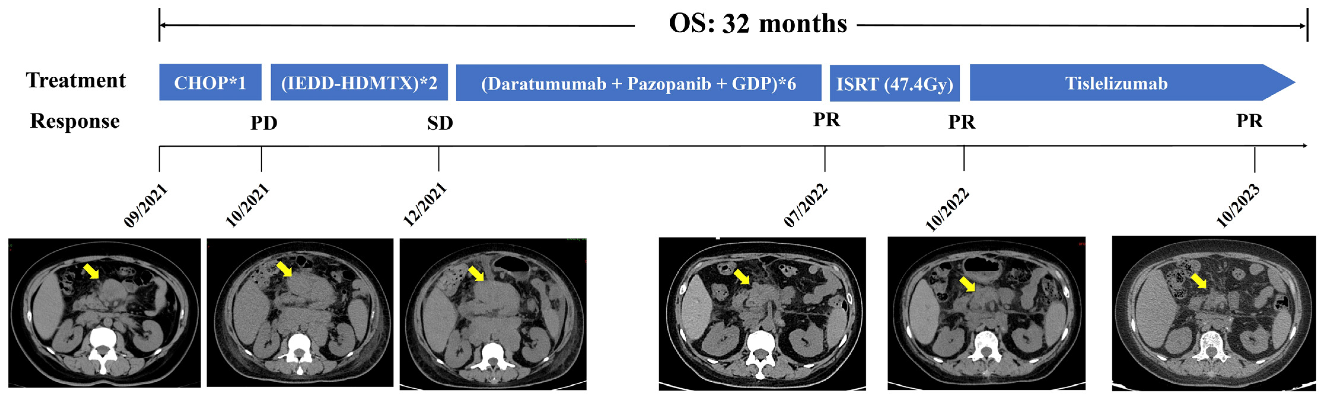
2. Case Presentation

A 58-year-old female patient presented with asymptomatic retroperitoneal lymphadenectasis by ultrasonography during her physical examination one year before admission. In September 2021, due to upper abdominal pain and recurrent fever with a maximal body temperature of 38 °C for two weeks, she was admitted to our department. Physical examination showed that a round mass (about 3*3cm in size) which was hard and slightly tender was palpated below the xiphoid process. Fluorine-18-fluorodeoxyglucose (18FDG) positron emission tomography–computed tomography (PET/CT) scan was performed and demonstrated generalized enlargement of the lymph nodes with increased FDG metabolism (SUVmax = 5.6–25.8) in the supraclavicular region, axillary region, celiac region, and the inguinal area (Figure 1). The largest one was found in the mesenteric area under the body of the pancreas, approximately 4 × 4 cm in size. Then the patient underwent laparoscopic resection of the retroperitoneal lesion. Postoperative pathology indicated histiocytic sarcoma based on Hematoxylin and Eosin (H&E) staining (Figure 2A) and immunohistochemistry (IHC) staining, revealing that the tumor cells were positive for CD163, Lysosome, CD4, and CD68 (Figure 2B–E), whereas they were negative for myeloid (MPO), T cell (CD3 and CD8), B cell (CD19), and dendritic cell (CD1a and CD21) lineage markers. The Ki-67 proliferation index was 70%. Additionally, follicular lymphoma (FL) was diagnosed, as detected by small number of small lymphocytes diffusely infiltrating surrounding adipose tissue (Figure 2F), which were positive for CD20 (Figure 2G), CD10 (Figure 2H), and BCL-2 (Figure 2I) by IHC staining and were associated with BCL2 gene-related translocation t(18q21) by fluorescence in situ hybridization (FISH) (Figure 2J). Moreover, the bone marrow examination also suggested the infiltration of BM by FL, supported by the abnormal small lymphocytes expressing CD19, CD20, CD22, and CD10 and a restrictive expression of membranous immunoglobulin light chain Kappa by flow cytometry (Figure 3). The patient was ultimately diagnosed with HS accompanied by FL.
The patient started to receive cytotoxic chemotherapy in September 2021, including one cycle of the CHOP regimen (cyclophosphamide, vincristine, doxorubicin, and prednisone) and two cycles of the IEDD-HDMTX regimen (ifosfamide, etoposide, liposomal doxorubicin, dexamethasone, and high-dose methotrexate). However, lymphadenopathy had no improvement in CT scans after chemotherapy. To explore potential therapeutic targets, further IHC staining was performed and demonstrated that the tumor cells were positive for PD-L1 (Figure 2K) and CD38 (Figure 2L). Moreover, the tumor biopsy specimen was sequenced by capture-based next-generation DNA/mRNA sequencing with a panel containing 495 hematological malignancy-related genes (Geneseeq Technology Inc., Toronto, ON, Canada). In addition to the BCL2-IgH fusion gene, a total of 22 somatic mutations were identified, including KRAS (c.183A > C, p.Q61H) and KMT2D (c.12374del, p. S4125Lfs*17) mutations (Supplementary Table S1). Thereafter, a combination of immune-targeted therapy with cytotoxic chemotherapy consisting of anti-CD38 monoclonal antibody Daratumumab, multi-target receptor tyrosine kinases inhibitor Pazopanib, plus the GDP regimen (gemcitabine, cisplatin, and dexamethasone) was administered in December 2021. Following six cycles of treatment, the CT scan showed that the sum of the perpendicular diameters (SPDs) of the enlarged lymph nodes was reduced by about 50% in comparison to that before treatment (Figure 4), indicating partial remission (PR). Subsequently, the patient received involved-site radiotherapy (ISRT) to the left side of the neck and abdomen at a total dose of 47.4Gy, followed by PD-1 inhibitor Tislelizumab maintenance therapy every 3–4 weeks. An efficacy evaluation with CT scan imaging demonstrated that the SPD of the enlarged lymph nodes was decreased by approximately 70% (Figure 4). The patient remains alive up to now; the duration of remission (DOR) and overall survival (OS) reached 22 and 32 months, respectively.

3. Discussion

HS is an aggressive and rare hematopoietic malignancy, which is derived from the monocyte–macrophage system. We reported a Chinese female patient with HS and co-occurring FL was successfully treated with frontline combined immune-targeted therapy. A series of interesting observations were obtained from this case.
Currently, there have been few large-scale studies on HS until now. Most have been clinical case reports from different research centers [2,3,4,6,7,8,9,10,11]. Kommalapati et al. retrospectively analyzed the clinical features of 159 patients diagnosed with HS in the Surveillance, Epidemiology, and End Results (SEER) database from 2000 to 2014 [5]. The age-adjusted incidence rate of HS was 0.17/1,000,000 individuals. The median age of onset for HS was 63 years old. The most common sites of tumor involvement were skin and connective tissue (35.8%), followed by the lymph nodes (17%). Furthermore, a small minority of patients with HS were reported to co-exist with or be secondary to lymphoid neoplasms, which are summarized in Supplementary Table S2 [2,3,4,10,12,13,14,15,16,17,18,19,20,21,22,23,24,25,26]. Some studies indicated the transdifferentiation of HS from lymphoid neoplasms, as evidenced by a clonal relationship between these two diseases through identical clonal IG gene rearrangements or an IGH/BCL2 sequence. In the current case, although the patient had a one-year history of an asymptomatic enlarged retroperitoneal lymph node before admission, no convincing evidence showed a common clonal origin between HS and FL. Therefore, it is difficult to infer whether HS was transdifferentiated from FL for the current patient.
The diagnosis primarily depends on histopathology with H&E staining and IHC staining [27,28]. IHC of HS suggested that the tumor cells expressed at least two mature histiocytic markers, including CD68, CD163, CD4, and Lysozyme [27,28]. Additionally, there were reports in the literature of tumor cells over-expressing PD-L1 [29,30,31,32] and CD38 [33], which is consistent with our patient and regarded as the potential therapeutic targets. Recently, genetic alterations of HS have been investigated in a series of reports [10,26,27,28,34,35,36,37]. Egan C, et al. identified genetic alterations within the RAS/RAF/MAPK pathway by whole-exome sequencing and RNA sequencing in HS patients, including NF1, MAP2K1, PTPN11, BRAF, KRAS, NRAS, and LZTR1 [36]. In addition, other studies reported that patients with HS harbored a recurrent KMT2D gene mutation [10,26,37], which is an epigenetic regulator. In the current study, this patient was also found to have mutations in the KMT2D and KRAS genes by NGS. Overall, these molecular features of HS indicated that alterations in the RAS/RAF/MAPK pathway and chromatin regulation may be considered as future therapeutic targets.
To date, there is no standard treatment regimen for HS. Lymphoma-type chemotherapy protocols, such as CHOP, ICE, and bendamustine, have been widely used in clinical practice [18,38,39,40]. Application of allogeneic or autologous hematopoietic stem cell transplantation in relapsed or refractory disease was also reported in a series of HS cases [40,41]. However, the outcomes were inconsistent. Furthermore, with in-depth study on the molecular features of HS, some novel targeted drugs have been increasingly applied in the treatment of refractory recurrent HS, with a kinase inhibitor (imatinib and sorafenib), anti-vascular endothelial growth factor (VEGF) antibodies (bevacizumab), an MEK inhibitor (trametinib), a BRAF inhibitor (vemurafenib), and immune checkpoint inhibitors (PD-1 inhibitors), leading to favorable clinical responses [30,32,42,43,44,45,46,47]. In this case, combined immune-targeted therapies were implemented based on the results of genetic molecular testing of tumor tissues. Daratumumab, a humanized, IgG1-κ-type anti-CD38 monoclonal antibody, has been approved for the treatment of multiple myeloma. Pazopanib is a multi-target tyrosine kinase inhibitor, which can suppress the RAS/RAF/MAPK signaling pathway [48,49]. It has been widely used in the treatment of renal carcinoma [48,49]. As CD38 and PD-L1 were highly expressed by IHC staining and a KRAS mutation was detected by NGS in this patient, multi-modality therapy with Daratumumab, Tislelizumab, and Pazopanib was successively implemented and achieved a favorable response. To the best of our knowledge, this is the first report of Pazopanib and Daratumumab being applied in the treatment of HS.
HS is a highly aggressive malignant hematological tumor with poor prognosis [5]. The median OS of patients with HS was reported to be approximately 6 months from the National Cancer Database (NCDB) [50]. Our patient was still alive and maintained disease remission with an OS of 32 months after receiving immune-targeted treatment combined with chemotherapy and radiotherapy, which was far beyond that of HS patients registered in the NCDB.

4. Conclusions

HS is an extremely rare hematological malignancy with poor prognosis. We presented the first case report of HS co-existing with FL benefitting from novel combined immune-targeted therapies and obtaining favorable therapeutic effects. We recognize the clinical importance of genomic and molecular studies for the establishment of individualized treatment in such patients with an otherwise dismal outcome.

Supplementary Materials

The supporting information can be downloaded at: https://www.mdpi.com/article/10.3390/ijms25137293/s1.

Author Contributions

H.H. and J.H. contributed to the study conception and design. Material preparation, data collection, and analysis were performed by M.Z., F.X., Y.Z., J.F., Y.S., Z.L. and D.Z. The first draft of the manuscript was written by M.Z. H.H. contributed to the revision of the manuscript. All authors reviewed the manuscript. All authors have read and agreed to the published version of the manuscript.

Funding

This research received no external funding.

Institutional Review Board Statement

The studies involving humans were approved by the Ren Ji Hospital, School of Medicine, Shanghai Jiao Tong University. The studies were conducted in accordance with the local legislation and institutional requirements. Written informed consent for participation in this study was provided by the participants’ legal guardians/next of kin. Written informed consent was obtained from the individual(s) for the publication of any potentially identifiable images or data included in this article.

Informed Consent Statement

Written informed consent was obtained from the patient for publication of the details of his medical case and any accompanying images.

Data Availability Statement

The original contributions presented in this study are included in this article/Supplementary Materials. Further inquiries can be directed to the corresponding author.

Conflicts of Interest

The authors declare that the research was conducted in the absence of any commercial or financial relationships that could be construed as a potential conflict of interest. The authors declare that this case report in Chinese has been collected in the Chinese medical case repository before submission.

References

  1. Khoury, J.D.; Solary, E.; Abla, O.; Akkari, Y.; Alaggio, R.; Apperley, J.F.; Bejar, R.; Berti, E.; Busque, L.; Chan, J.K.C.; et al. The 5th edition of the World Health Organization Classification of Haematolymphoid Tumours: Myeloid and Histiocytic/Dendritic Neoplasms. Leukemia 2022, 36, 1703–1719. [Google Scholar] [CrossRef]
  2. Feldman, A.L.; Arber, D.A.; Pittaluga, S.; Martinez, A.; Burke, J.S.; Raffeld, M.; Camos, M.; Warnke, R.; Jaffe, E.S. Clonally related follicular lymphomas and histiocytic/dendritic cell sarcomas: Evidence for transdifferentiation of the follicular lymphoma clone. Blood 2008, 111, 5433–5439. [Google Scholar] [CrossRef]
  3. Wang, E.; Hutchinson, C.B.; Huang, Q.; Sebastian, S.; Rehder, C.; Kanaly, A.; Moore, J.; Datto, M. Histiocytic sarcoma arising in indolent small B-cell lymphoma: Report of two cases with molecular/genetic evidence suggestive of a ‘transdifferentiation’ during the clonal evolution. Leuk. Lymphoma 2010, 51, 802–812. [Google Scholar] [CrossRef]
  4. Hure, M.C.; Elco, C.P.; Ward, D.; Hutchinson, L.; Meng, X.; Dorfman, D.M.; Yu, H. Histiocytic sarcoma arising from clonally related mantle cell lymphoma. J. Clin. Oncol. 2012, 30, e49–e53. [Google Scholar] [CrossRef]
  5. Kommalapati, A.; Tella, S.H.; Durkin, M.; Go, R.S.; Goyal, G. Histiocytic sarcoma: A population-based analysis of incidence, demographic disparities, and long-term outcomes. Blood 2018, 131, 265–268. [Google Scholar] [CrossRef]
  6. Gupta, D.; Gupta, A.; Nalwa, A.; Yadav, T.; Chaudhary, R.; Rao, M. Cytopathologic findings in histiocytic sarcoma of thyroid mimicking as anaplastic carcinoma: A report of a rare case with review of literature. Diagn. Cytopathol. 2021, 49, E218–E221. [Google Scholar] [CrossRef] [PubMed]
  7. Krishnamurthy, K.; Delgado, R.; Kochiyil, J.; Medina, A.M. Primary Histiocytic Sarcoma in Adult Polycystic Kidney Disease: Case Report and Review of Literature. Int. J. Surg. Pathol. 2021, 29, 321–326. [Google Scholar] [CrossRef] [PubMed]
  8. Matsunaga, R.; Kanazawa, Y.; Matsuno, K.; Kakinuma, D.; Tokura, T.; Marumo, A.; Yui, S.; Ando, F.; Masuda, Y.; Hagiwara, N.; et al. An advanced case of gastric histiocytic sarcoma treated with chemotherapy and gastrectomy: A case report and review of literature. Clin. J. Gastroenterol. 2021, 14, 1053–1059. [Google Scholar] [CrossRef] [PubMed]
  9. Montalvo, N.; Lara-Endara, J.; Redrobán, L.; Leiva, M.; Armijos, C.; Russo, L. Primary splenic histiocytic sarcoma associated with hemophagocytic lymphohistiocytosis: A case report and review of literature of next-generation sequencing involving FLT3, NOTCH2, and KMT2A mutations. Cancer Rep. 2022, 5, e1496. [Google Scholar] [CrossRef]
  10. Péricart, S.; Waysse, C.; Siegfried, A.; Struski, S.; Delabesse, E.; Laurent, C.; Evrard, S. Subsequent development of histiocytic sarcoma and follicular lymphoma: Cytogenetics and next-generation sequencing analyses provide evidence for transdifferentiation of early common lymphoid precursor—A case report and review of literature. Virchows Arch. 2020, 476, 609–614. [Google Scholar] [CrossRef]
  11. Wang, W.; Zhang, Y.; Dong, Y.; Li, S.; Qin, H. Primary central nervous system histiocytic sarcoma with somatic NF2 mutation: Case report and review of literature. Clin. Neuropathol. 2022, 41, 253–262. [Google Scholar] [CrossRef] [PubMed]
  12. Fernandez-Pol, S.; Bangs, C.D.; Cherry, A.; Arber, D.A.; Gratzinger, D. Two cases of histiocytic sarcoma with BCL2 translocations and occult or subsequent follicular lymphoma. Hum. Pathol. 2016, 55, 39–43. [Google Scholar] [CrossRef]
  13. Sabatini, P.J.B.; Tremblay-Lemay, R.; Ahmadi Moghaddam, P.; Delabie, J.M.A.; Sakhdari, A. Marginal zone lymphoma transdifferentiated to histiocytic sarcoma. Br. J. Haematol. 2021, 194, 1090–1094. [Google Scholar] [CrossRef] [PubMed]
  14. Bassarova, A.; Trøen, G.; Fosså, A.; Ikonomou, I.M.; Beiske, K.; Nesland, J.M.; Delabie, J. Transformation of B cell lymphoma to histiocytic sarcoma: Somatic mutations of PAX-5 gene with loss of expression cannot explain transdifferentiation. J. Hematop. 2009, 2, 135–141. [Google Scholar] [CrossRef] [PubMed]
  15. Brunner, P.; Rufle, A.; Dirnhofer, S.; Lohri, A.; Willi, N.; Cathomas, G.; Tzankov, A.; Juskevicius, D. Follicular lymphoma transformation into histiocytic sarcoma: Indications for a common neoplastic progenitor. Leukemia 2014, 28, 1937–1940. [Google Scholar] [CrossRef] [PubMed]
  16. Etancelin, P.; Boussaid, I. Two sides of the same coin: Transdifferentiation from Burkitt lymphoma to histiocytic sarcoma. Blood 2023, 142, 1576. [Google Scholar] [CrossRef] [PubMed]
  17. Cheon, M.; Yoo, J.; Kim, H.S.; Lee, M. Enhanced Computed Tomography and (18)F-fluorodeoxyglucose Positron Emission Tomography/Computed Tomography in the Uncommon Histiocytic Sarcoma of Small Intestine Arising after Gastric Large B-Cell Lymphoma. Diagnostics 2023, 13, 3189. [Google Scholar] [CrossRef] [PubMed]
  18. Farris, M.; Hughes, R.T.; Lamar, Z.; Soike, M.H.; Menke, J.R.; Ohgami, R.S.; Winkfield, K. Histiocytic Sarcoma Associated with Follicular Lymphoma: Evidence for Dramatic Response with Rituximab and Bendamustine Alone and a Review of the Literature. Clin. Lymphoma Myeloma Leuk. 2019, 19, e1–e8. [Google Scholar] [CrossRef] [PubMed]
  19. Kumar, J.; Al-Kawaaz, M.; Martin, B.A.; Hegazi, M.M.; Tan, B.; Gratzinger, D. Histiocytic Sarcoma with CCND1 Gene Rearrangement Clonally Related and Transdifferentiated from Mantle Cell Lymphoma. Am. J. Clin. Pathol. 2022, 158, 449–455. [Google Scholar] [CrossRef]
  20. Shao, H.; Xi, L.; Raffeld, M.; Feldman, A.L.; Ketterling, R.P.; Knudson, R.; Rodriguez-Canales, J.; Hanson, J.; Pittaluga, S.; Jaffe, E.S. Clonally related histiocytic/dendritic cell sarcoma and chronic lymphocytic leukemia/small lymphocytic lymphoma: A study of seven cases. Mod. Pathol. 2011, 24, 1421–1432. [Google Scholar] [CrossRef]
  21. Cheng, F.; Yu, F.; Wang, X.; Huang, K.; Lu, H.; Wang, Z. A Pedigree Analysis and Clonal Correlations of the Coexistence of B-Cell Lymphoma and Histiocytic/Dendritic Cell Tumor. Int. J. Surg. Pathol. 2021, 29, 906–914. [Google Scholar] [CrossRef]
  22. D’amore, E.S.; Mainardi, C.; Mussolin, L.; Carraro, E.; Alaggio, R.; Lazzari, E.; Fusetti, S.; Ghirotto, C.; Marzollo, A.; Biddeci, G.; et al. Histiocytic sarcoma arising in a child affected by Burkitt lymphoma, with t(8;14)(q24;q32) positivity in both tumors. Pediatr. Hematol. Oncol. 2021, 38, 1–7. [Google Scholar] [CrossRef]
  23. Zhang, D.; McGuirk, J.; Ganguly, S.; Persons, D.L. Histiocytic/dendritic cell sarcoma arising from follicular lymphoma involving the bone: A case report and review of literature. Int. J. Hematol. 2009, 89, 529–532. [Google Scholar] [CrossRef]
  24. Schünemann, C.; Göhring, G.; Behrens, Y.L.; Kreipe, H.-H.; Ganser, A.; Thol, F. Histiocytic sarcoma progressing from follicular lymphoma and mimicking acquired hemophagocytic lymphohistiocytosis. Ann. Hematol. 2020, 99, 2441–2443. [Google Scholar] [CrossRef]
  25. Shi, C.M.; Lytle, A.M.; Milman, T.; Penne, R.; Bagg, A. Composite Histiocytic Sarcoma and Chronic Lymphocytic Leukemia/Small Lymphocytic Lymphoma of the Ocular Adnexa. Ophthalmic Plast. Reconstr. Surg. 2024, 10, 1097. [Google Scholar] [CrossRef] [PubMed]
  26. Egan, C.; Lack, J.; Skarshaug, S.; Pham, T.A.; Abdullaev, Z.; Xi, L.; Pack, S.; Pittaluga, S.; Jaffe, E.S.; Raffeld, M. The mutational landscape of histiocytic sarcoma associated with lymphoid malignancy. Mod. Pathol. 2020, 34, 336–347. [Google Scholar] [CrossRef]
  27. Hung, Y.P.; Qian, X. Histiocytic Sarcoma. Arch. Pathol. Lab. Med. 2020, 144, 650–654. [Google Scholar] [CrossRef] [PubMed]
  28. Tocut, M.; Vaknine, H.; Potachenko, P.; Elias, S.; Zandman-Goddard, G. Histiocytic Sarcoma. Isr. Med. Assoc. J. 2020, 22, 645–647. [Google Scholar] [PubMed]
  29. Campedel, L.; Kharroubi, D.; Vozy, A.; Spano, J.P.; Emile, J.F.; Haroche, J. Malignant Histiocytosis With PD-L1 Expression-Dramatic Response to Nivolumab. Mayo Clin. Proc. 2022, 97, 1401–1403. [Google Scholar] [CrossRef]
  30. Imataki, O.; Uemura, M.; Fujita, H.; Kadowaki, N. Application of PD-L1 blockade in refractory histiocytic sarcoma: A case report. Mol. Clin. Oncol. 2022, 17, 1–6. [Google Scholar] [CrossRef]
  31. May, J.M.; Waddle, M.R.; Miller, D.H.; Stross, W.C.; Kaleem, T.A.; May, B.C.; Miller, R.C.; Jiang, L.; Strong, G.W.; Trifiletti, D.M.; et al. Primary histiocytic sarcoma of the central nervous system: A case report with platelet derived growth factor receptor mutation and PD-L1/PD-L2 expression and literature review. Radiat. Oncol. 2018, 13, 167. [Google Scholar] [CrossRef]
  32. Zhao, Y.; Deng, Y.; Jiang, Y.; Zheng, W.; Tan, Y.; Yang, Z.; Wang, Z.; Xu, F.; Cheng, Z.; Yuan, L.; et al. Case report: Targeting the PD-1 receptor and genetic mutations validated in primary histiocytic sarcoma with hemophagocytic lymphohistiocytosis. Front. Immunol. 2023, 14, 1127599. [Google Scholar] [CrossRef]
  33. Zhao, J.; Niu, X.; Wang, Z.; Lu, H.; Lin, X.; Lu, Q. Histiocytic sarcoma combined with acute monocytic leukemia: A case report. Diagn. Pathol. 2015, 10, 1–6. [Google Scholar] [CrossRef]
  34. Michonneau, D.; Kaltenbach, S.; Derrieux, C.; Trinquand, A.; Brouzes, C.; Gibault, L.; North, M.O.; Delarue, R.; Varet, B.; Emile, J.F.; et al. BRAF(V600E) mutation in a histiocytic sarcoma arising from hairy cell leukemia. J. Clin. Oncol. 2014, 32, e117–e121. [Google Scholar] [CrossRef]
  35. Shanmugam, V.; Griffin, G.K.; Jacobsen, E.D.; Fletcher, C.D.M.; Sholl, L.M.; Hornick, J.L. Identification of diverse activating mutations of the RAS-MAPK pathway in histiocytic sarcoma. Mod. Pathol. 2019, 32, 830–843. [Google Scholar] [CrossRef]
  36. Egan, C.; Nicolae, A.; Lack, J.; Chung, H.-J.; Skarshaug, S.; Pham, T.A.; Navarro, W.; Abdullaev, Z.; Aguilera, N.S.; Xi, L.; et al. Genomic profiling of primary histiocytic sarcoma reveals two molecular subgroups. Haematologica 2020, 105, 951–960. [Google Scholar] [CrossRef]
  37. Hung, Y.P.; Lovitch, S.B.; Qian, X. Histiocytic sarcoma: New insights into FNA cytomorphology and molecular characteristics. Cancer Cytopathol. 2017, 125, 604–614. [Google Scholar] [CrossRef]
  38. Narita, K.; Noro, R.; Seike, M.; Matsumoto, M.; Fujita, K.; Matsumura, J.; Takahashi, M.; Kawamoto, M.; Gemma, A. Successful treatment of histiocytic sarcoma and concurrent HIV infection using a combination of CHOP and antiretroviral therapy. Intern. Med. 2013, 52, 2805–2809. [Google Scholar] [CrossRef]
  39. Philip, D.S.J.; Sherief, A.; Narayanan, G.; Nair, S.G.; Av, J. Histiocytic Sarcoma: Clinical Features and Outcomes of Patients Treated at a Tertiary Cancer Care Center. Cureus 2022, 14, e25814. [Google Scholar] [CrossRef]
  40. Tsujimura, H.; Miyaki, T.; Yamada, S.; Sugawara, T.; Ise, M.; Iwata, S.; Yonemoto, T.; Ikebe, D.; Itami, M.; Kumagai, K. Successful treatment of histiocytic sarcoma with induction chemotherapy consisting of dose-escalated CHOP plus etoposide and upfront consolidation auto-transplantation. Int. J. Hematol. 2014, 100, 507–510. [Google Scholar] [CrossRef]
  41. Zeidan, A.; Bolaños-Meade, J.; Kasamon, Y.; Aoki, J.; Borowitz, M.; Swinnen, L.; Symons, H.; Luznik, L.; Fuchs, E.; Jones, R.; et al. Human leukocyte antigen-haploidentical hematopoietic stem cell transplant for a patient with histiocytic sarcoma. Leuk. Lymphoma 2012, 54, 655–657. [Google Scholar] [CrossRef] [PubMed]
  42. Schlick, K.; Aigelsreiter, A.; Pichler, M.; Reitter, S.; Neumeister, P.; Hoefler, G.; Beham-Schmid, C.; Linkesch, W. Histiocytic sarcoma—Targeted therapy: Novel therapeutic options? A series of 4 cases. Onkologie 2012, 35, 447–450. [Google Scholar] [CrossRef] [PubMed]
  43. Gounder, M.M.; Solit, D.B.; Tap, W.D. Trametinib in Histiocytic Sarcoma with an Activating MAP2K1 (MEK1) Mutation. N. Engl. J. Med. 2018, 378, 1945–1947. [Google Scholar] [CrossRef] [PubMed]
  44. Hu, B.; Patel, J.L.; Tao, R.; Cannon, R.B.; Monroe, M.; Goyal, G. Near Complete Response to Trametinib Treatment in Histiocytic Sarcoma Harboring a Somatic KRAS Mutation. J. Natl. Compr. Cancer Netw. 2022, 20, 618–621. [Google Scholar] [CrossRef] [PubMed]
  45. Idbaih, A.; Mokhtari, K.; Emile, J.-F.; Galanaud, D.; Belaid, H.; de Bernard, S.; Benameur, N.; Barlog, V.-C.; Psimaras, D.; Donadieu, J.; et al. Dramatic response of a BRAF V600E-mutated primary CNS histiocytic sarcoma to vemurafenib. Neurology 2014, 83, 1478–1480. [Google Scholar] [CrossRef] [PubMed]
  46. Vaughn, J.L.; Freitag, C.E.; Hemminger, J.A.; Jones, J.A. BRAF V600E expression in histiocytic sarcoma associated with splenic marginal zone lymphoma: A case report. J. Med. Case Rep. 2017, 11, 92. [Google Scholar] [CrossRef] [PubMed]
  47. Furui, Y.; Kurata, T.; Komori, K.; Uchida, E.; Miyairi, Y.; Chiba, A.; Ogiso, Y.; Sakashita, K. A case of recurrent refractory cervical primary histiocytic sarcoma treated with pembrolizumab. Int. Cancer Conf. J. 2022, 11, 280–285. [Google Scholar] [CrossRef]
  48. Frampton, J.E. Pazopanib: A Review in Advanced Renal Cell Carcinoma. Target. Oncol. 2017, 12, 543–554. [Google Scholar] [CrossRef]
  49. Ward, J.E.; Stadler, W.M. Pazopanib in renal cell carcinoma. Clin. Cancer Res. 2010, 16, 5923–5927. [Google Scholar] [CrossRef]
  50. Kommalapati, A.; Tella, S.H.; Go, R.S.; Goyal, G. Predictors of survival, treatment patterns, and outcomes in histiocytic sarcoma. Leuk. Lymphoma 2018, 60, 553–555. [Google Scholar] [CrossRef]
Figure 1. The positron emission tomography–computed tomography (PET/CT) images demonstrated extensive enlargement of the lymph nodes with increased fluorine-18-fluorodeoxyglucose (FDG) metabolism at initial diagnosis.
Ijms 25 07293 g001
Figure 2. Pathological analysis of retroperitoneal lesion. Images of H&E staining (100× oil) of histiocytic sarcoma cells (A) and immunohistochemistry (IHC) staining (400×) of CD163 (B), lysosome (C), CD4 (D), CD68 (E), PD-L1 (K), and CD38 (L); Images of H&E staining (100× oil) of follicular lymphoma cells (F), IHC staining (400×) of CD20 (G), CD10 (H), and BCL-2 (I), as well as BCL2 gene translocation (J) by fluorescence in situ hybridization (FISH). The red arrows in Figure 2A and Figure 2F represented histiocytic sarcoma cells and follicular lymphoma cells, respectively.
Ijms 25 07293 g002
Figure 3. Representative images of multiparameter flow cytometry analysis of bone marrow at initial diagnosis indicating the involvement of bone marrow by follicular lymphoma cells. The tumor cells (green dots circled) were small in size (A) with positive for CD 19 (B), CD 20 (C), CD10 (C), and CD22 (E) while negative for CD5 or CD23 (F). The tumor cell had a restrictive expression of membranous immunoglobulin light chain Kappa (D).
Ijms 25 07293 g003
Figure 4. Representative computed tomography (CT) images, treatment, and clinical courses of this patient. CHOP, cyclophosphamide, vincristine, doxorubicin, and prednisone; GDP, gemcitabine, cisplatin, and dexamethasone; IEDD-HDMTX, ifosfamide, etoposide, liposomal doxorubicin, dexamethasone, and high-dose methotrexate; PD, progressive disease; PR, partial remission; SD, stable disease. The yellow arrow indicated enlarged the lymph nodes in mesenteric area.
Ijms 25 07293 g004
Disclaimer/Publisher’s Note: The statements, opinions and data contained in all publications are solely those of the individual author(s) and contributor(s) and not of MDPI and/or the editor(s). MDPI and/or the editor(s) disclaim responsibility for any injury to people or property resulting from any ideas, methods, instructions or products referred to in the content.

