
Can Isoflavone-Rich Legume Plants Be Useful in the Chemoprevention of Hormone-Dependent Cancers?—A Systematic Review

Abstract
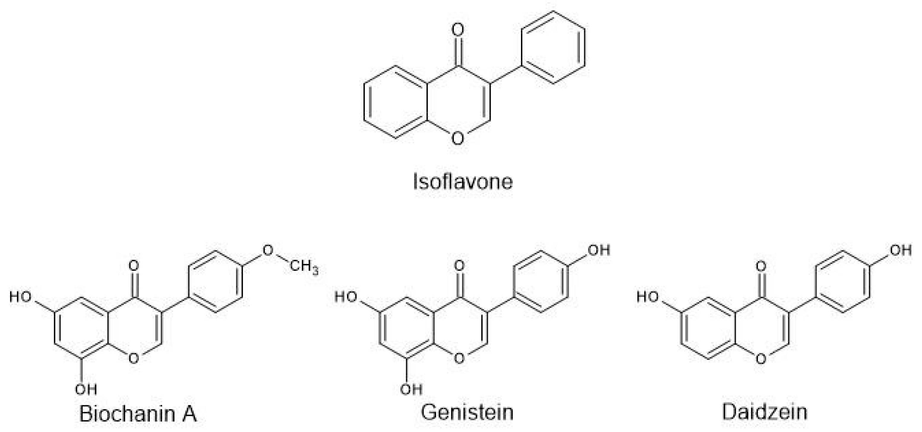
:1. Introduction
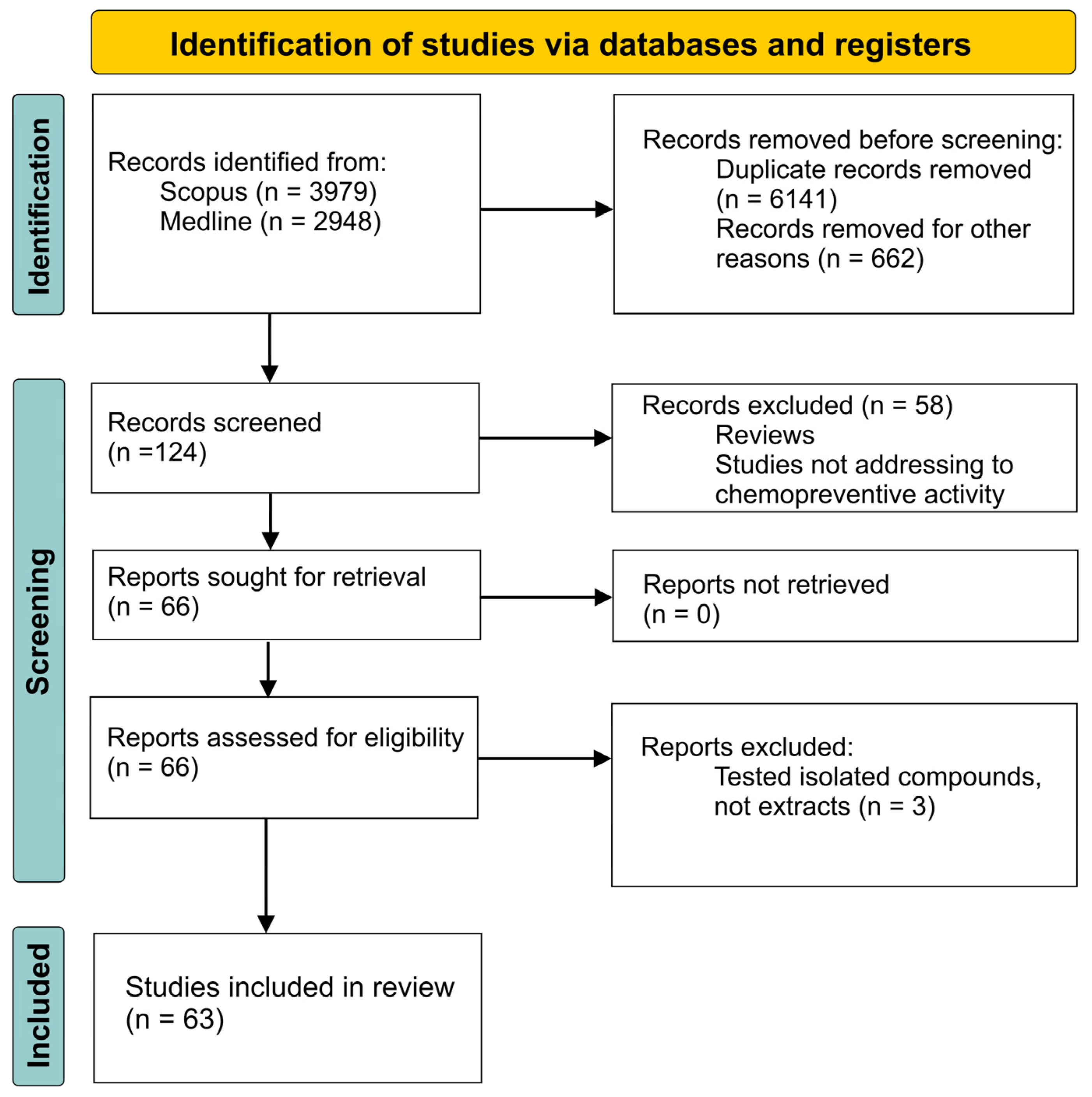
2. Materials and Methods
3. Results
3.1. Astragalus sp.
3.1.1. Cytotoxic Activity
3.1.2. Antioxidant Potential
3.2. Cytisus sp.
3.2.1. Cytotoxic Activity
3.2.2. Anti-Inflammatory Activity
3.2.3. Antioxidant Potential
3.3. Dorycnium sp.
Cytotoxic Activity
3.4. Genista sp.
3.4.1. Cytotoxic and Anti-Inflammatory Activity
3.4.2. In Vivo Studies
3.4.3. Antioxidant Potential
3.5. Lupinus sp.
3.5.1. Cytotoxic Activity
3.5.2. Antioxidant Potential
3.6. Medicago sp.
3.6.1. Cytotoxic Activity
3.6.2. Antioxidant Potential
3.7. Melilotus sp.
3.7.1. Cytotoxic Activity
3.7.2. Antioxidant and Anti-Inflammatory Potential
3.8. Ononis sp.
3.8.1. Cytotoxic Activity
3.8.2. In Vivo Studies
3.8.3. Antioxidant Potential
3.9. Trifolium sp.
3.9.1. Cytotoxic Activity
3.9.2. In Vivo Studies
3.9.3. Antioxidant Potential
3.10. Trigonella sp.
3.10.1. Cytotoxic Activity
3.10.2. Antioxidant Potential
3.11. Vicia sp.
Antioxidant Potential
3.12. Other Species
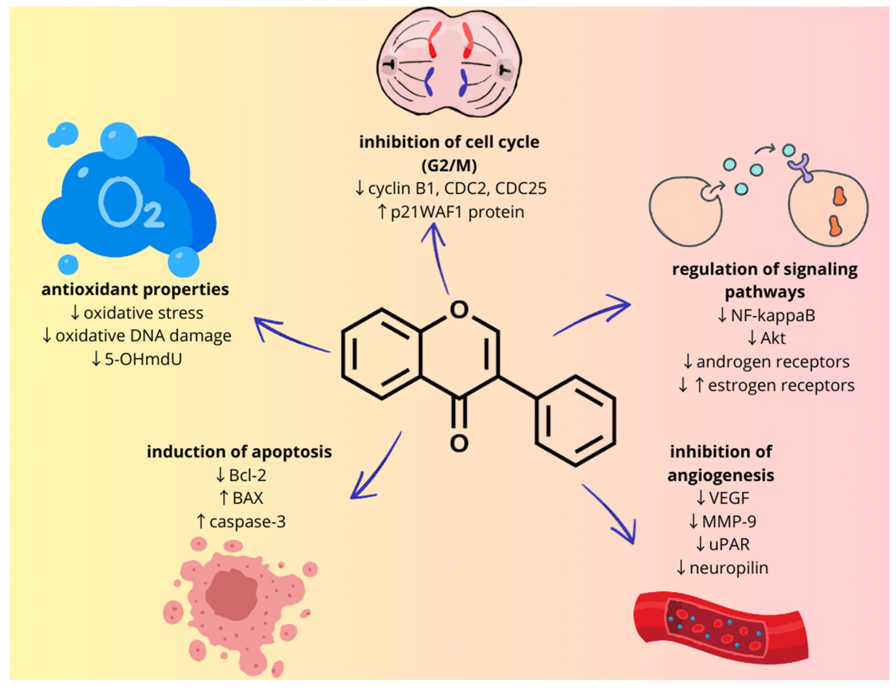
4. Discussion
5. Conclusions
Author Contributions
Funding
Conflicts of Interest
References
- Greenwald, P.; Kelloff, G.; Burch-Whitman, C.; Kramer, B.S. Chemoprevention. CA Cancer J. Clin. 1995, 45, 31–49. [Google Scholar] [CrossRef]
- El-Sherbeni, A.A.; El-Kadi, A.O.S. The role of epoxide hydrolases in health and disease. Arch. Toxicol. 2014, 88, 2013–2032. [Google Scholar] [CrossRef]
- Biswas, D.K.; Dai, S.C.; Cruz, A.; Weiser, B.; Graner, E.; Pardee, A.B. The nuclear factor kappa B (NF-kappaB): A potential therapeutic target for estrogen receptornegative breast cancers. Proc. Natl. Acad. Sci. USA 2001, 98, 10386–10391. [Google Scholar] [CrossRef]
- Bharti, A.C.; Aggarwal, B.B. Nuclear factor-kappaB and cancer: Its role in prevention and therapy. Biochem. Pharmacol. 2002, 64, 883–888. [Google Scholar] [CrossRef]
- Sarkar, F.H.; Li, Y. The role of isoflavones in cancer chemoprevention. Front. Biosci. 2004, 9, 2714–2724. [Google Scholar] [CrossRef]
- Pana, M.H.; Ho, C.T. Chemopreventive effects of natural dietary compounds on cancer development. Chem. Soc. Rev. 2008, 37, 2558–2574. [Google Scholar] [CrossRef]
- Bijak, M.; Połać, I.; Borowiecka, M.; Nowak, P.; Stetkiewicz, T.; Pertyński, T. Izoflawony jako alternatywa dla terapii hormonalnej wieku menopauzalnego. Przegląd Menopauzalny 2010, 6, 402–406. [Google Scholar]
- Czerpak, R.; Pietryczuk, A.; Jabłońska-Trypuć, A.; Obrębska, K. Aktywność biologiczna izoflawonoidów i ich znaczenie terapeutyczne i kosmetyczne. Przegląd Menopauzalny 2009, 2, 113–121. [Google Scholar]
- Yu, J.; Bi, X.; Yu, B.; Che, D. Isoflavones: Anti-Inflammatory Benefit and Possible Caveats. Nutrients 2016, 8, 361. [Google Scholar] [CrossRef]
- Gottstein, N.; Ewins, B.A.; Eccleston, C.; Hubbard, G.P.; Kavanagh, I.C.; Minihane, A.M.; Weinberg, P.D.; Rimbach, G. Effect of genistein and daidzein on platelet aggregation and monocyte and endothelial function. Br. J. Nutr. 2003, 89, 607–615. [Google Scholar] [CrossRef]
- Mirza, S.; Sharma, G.; Parshad, R.; Gupta, S.D.; Pandya, P.; Ralhan, R. Expression of DNA Methyltransferases in Breast Cancer Patients and to Analyze the Effect of Natural Compounds on DNA Methyltransferases and Associated Proteins. J. Breast Cancer 2013, 16, 23–31. [Google Scholar] [CrossRef]
- Adjakly, M.; Bosviel, R.; Rabiau, N.; Boiteux, J.P.; Bignon, Y.J.; Guy, L.; Bernard-Gallon, D. DNA methylation and soy phytoestrogens: Quantitative study in DU-145 and PC-3 human prostate cancer cell lines. Epigenomics 2011, 3, 795–803. [Google Scholar] [CrossRef]
- Carvalho, A.A.; dos Santos, L.R.; de Freitas, J.S.; Chaves, M.H. Isoflavonoids of the tribe dalbergieae: A chemosystematic contribution to the subfamily papilionoideae. Quimica Nova 2020, 43, 1294–1311. [Google Scholar]
- Watanabe, S.; Uehara, M. Health Effects and Safety of Soy and Isoflavones. In The Role of Functional Food Security in Global Health, 1st ed.; Watson, R.R., Singh, R.B., Takahashi, T., Eds.; Elsevier: Amsterdam, The Netherlands, 2018; Volume 1, pp. 379–394. [Google Scholar]
- Drenin, A.A.; Botirov, E.K. Flavonoids and isoflavones of plants of the genus Trifolium L. Structural diversity and biological activity. Rast. Syr. 2017, 3, 39–53. [Google Scholar]
- Nasi, A.; Picariello, G.; Ferranti, P. Proteomic approaches to study structure, functions and toxicity of legume seeds lectins. Perspectives for the assessment of food quality and safety. J. Proteom. 2009, 72, 527–538. [Google Scholar] [CrossRef]
- Sharma, A. A review on traditional technology and safety challenges with regard to antinutrients in legume food. J. Food Sci. Technol. 2021, 58, 2863–2883. [Google Scholar] [CrossRef]
- Martínez-Montemayor, M.M.; Otero-Franqui, E.; Martinez, J.; De La Mota-Peynado, A.; Cubano, L.A.; Dharmawardhane, S. Individual and combined soy isoflavones exert differential effects on metastatic cancer progression. Clin. Exp. Metastasis 2010, 27, 465–480. [Google Scholar] [CrossRef]
- Castillón, E.E.; Quintanilla, J.A.V.; Delgado-Salinas, A.; Rebman, J.P. The genus Astragalus (Leguminosae: Papilionoideae: Galegeae) in Mexico. Phytotaxa 2023, 586, 3–5. [Google Scholar]
- Butkute, B.; Dagilyte, A.; Benetis, R.; Padarauskas, A.; Ceseviciene, J.; Olsauskaite, V.; Lemeziene, N. Mineral and Phytochemical Profiles and Antioxidant Activity of Herbal Material from Two Temperate Astragalus Species. Nat. Prod. Res. 2018, 2018, 318630. [Google Scholar]
- Liu, Q.; Li, J.; Gu, M.; Kong, W.; Lin, Z.; Mao, J.; Zhang, M.; Jiang, L.; Liu, C.; Wang, Y.; et al. High-Throughput Phytochemical Unscrambling of Flowers Originating from Astragalus membranaceus (Fisch.) Bge. var. mongholicus (Bge.) P. K. Hsiao and Astragalus membranaceus (Fisch.) Bug. by Applying the Intagretive Plant Metabolomics Method Using UHPLC−Q−TOF−MS/MS. Molecules 2023, 28, 6115. [Google Scholar] [CrossRef] [PubMed]
- Abu-Dahab, R.; Afifi, F. Antiproliferative activity of selected medicinal plants of Jordan against a breast adenocarcinoma cell line (MCF7). Sci. Pharm. 2007, 75, 121–136. [Google Scholar] [CrossRef]
- Hanganu, D.; Vlase, L.; Olah, N. Phytochemical Analysis of Isoflavons from some Fabaceae Species Extracts. Not. Bot. Horti Agrobot. Cluj 2010, 38, 57–60. [Google Scholar]
- Hanganu, D.; Vlase, L.; Olah, N. LC/MS analysis of isoflavones from Fabaceae species extracts. FARMACIA 2010, 58, 177–183. [Google Scholar]
- Cunha, S.C.; Faria, M.A.; Sousa, T.; Nunes, E. Isoflavone determination in spontaneous legumes identified by DNA barcodes. Food Chem. 2012, 134, 2262–2627. [Google Scholar] [CrossRef]
- Bouziane, A.; Bakchiche, B.; Dias, M.I.; Barros, L.; Ferreira, I.C.F.R.; Al Salamat, H.A.; Bardaweel, S.K. Phenolic Compounds and Bioactivity of Cytisus villosus Pourr. Molecules 2018, 23, 1994. [Google Scholar] [CrossRef]
- Garcia-Oliveira, P.; Carreira-Casais, A.; Pereira, E.; Dias, M.I.; Pereira, C.; Calhelha, R.C.; Stojkovic, D.; Sokovic, M.; Simal-Gandara, J.; Prieto, M.A.; et al. From Tradition to Health: Chemical and Bioactive Characterization of Five Traditional Plants. Molecules 2022, 27, 6495. [Google Scholar] [CrossRef]
- Zouhri, A.; El Menyiy, N.; El-mernissi, Y.; Bouddine, T.; El-mernissi, R.; Amhamdi, H.; Elharrak, A.; Salamatullah, A.M.; Nafidi, H.A.; Khallouki, F.; et al. Mineral composition, principal polyphenolic components, and evaluation of the anti-inflammatory, analgesic, and antioxidant properties of Cytisus villosus Pourr leaf extracts. Open Chem. 2023, 21, 20220338. [Google Scholar] [CrossRef]
- Sahnin, B.; Karaman, S.; Ates, M.A.; Aytac, Z. Dorycnium vuralii (Fabaceae), a new species from Turkiye. Turk. J. Bot. 2024, 48, 120–134. [Google Scholar] [CrossRef]
- Koygun, G.; Arslan, E.; Zengin, G.; Orlando, G.; Ferrante, C. Comparison of Anticancer Activity of Dorycnium pentaphyllum Extract on MCF-7 and MCF-12A Cell Line: Correlation with Invasion and Adhesion. Biomolecules 2021, 11, 671. [Google Scholar] [CrossRef] [PubMed]
- Rigano, D.; Cardile, V.; Formisano, C.; Maldini, M.T.; Piacente, S.; Bevelacqua, Y.; Russo, A.; Senatore, F. Genista sessilifolia DC. and Genista tinctoria L. inhibit UV light and nitric oxide-induced DNA damage and human melanoma cell growth. Chem. Biol. Interact. 2009, 180, 211–219. [Google Scholar] [CrossRef] [PubMed]
- Kiss, B.; Popa, D.S.; Paltinean, R.; Loghin, F. A high-throughput UPLC-MS/MS for the simultaneous analysis of six phytoestrogens from Genista tinctoria extracts. J. Liq. Chromatogr. Relat. Technol. 2012, 35, 2735–2752. [Google Scholar] [CrossRef]
- Cheilari, A.; Vontzalidou, A.; Makropoulou, M.; Meligova, A.K.; Fokialakis, N.; Mitakou, S.; Alexis, M.N.; Aligiannis, N. Isoflavonoid Profiling and Estrogen-Like Activity of Four Genista Species from the Greek Flora. Molecules 2020, 25, 5507. [Google Scholar] [CrossRef]
- Bontempo, P.; Rigano, D.; Doto, A.; Formisano, C.; Conte, M.; Nebbioso, A.; Carafa, V.; Caserta, G.; Sica, V.; Molinari, A.M.; et al. Genista sessilifolia DC. extracts induce apoptosis across a range of cancer cell lines. Cell Prolif. 2013, 46, 183–192. [Google Scholar] [CrossRef] [PubMed]
- Diaz, L.; Cely-Veloza, W.; Coy-Barrera, E. Identification of Anti-Proliferative Compounds from Genista monspessulana Seeds through Covariate-Based Integration of Chemical Fingerprints and Bioactivity Datasets. Molecules 2022, 27, 3996. [Google Scholar] [CrossRef] [PubMed]
- Simoes, M.A.M.; Pinto, D.C.G.A.; Neves, B.M.R.; Silva, A.M.S. Flavonoid Profile of the Genista tridentata L., a Species Used Traditionally to Treat Inflammatory Processes. Molecules 2020, 25, 812. [Google Scholar] [CrossRef] [PubMed]
- Popa, D.S.; Bolfa, P.; Kiss, B.; Vlase, L.; Paltinean, R.; Pop, A.; Catoi, C.; Crisan, G.; Loghin, F. Influence of Genista Tinctoria L. or Methylparaben on Subchronic Toxicity of Bisphenol A in Rats. Biomed. Environ. Sci. 2014, 27, 85–96. [Google Scholar] [PubMed]
- Orhan, I.E.; Tosun, F.; Tamer, U.; Duran, A.; Alan, B.; Kok, A.F. Quantification of genistein and daidzein in two endemic Genista species and their antioxidant activity. J. Serb. Chem. Soc. 2011, 76, 35–42. [Google Scholar] [CrossRef]
- Kerkatou, M.; Menad, A.; Sarri, D.; León, F.; Brouard, I.; Bouldjedj, R.; Chalard, P.; Ameddah, S.; Benayache, S.; Benayache, F. Secondary metabolites from Genista aspalathoides Lamk ssp. aspalathoides. M. Pharm. Lett. 2013, 5, 285–289. [Google Scholar]
- Serrilli, A.M.; Graziosi, V.; Ballero, M.; Foddis, C.; Serafini, M.; Poli, F.; Scartezzini, P.; Bianco, A. Endemic Sardinian plants: The case of Genista cadasonensis Valsecchi. Nat. Prod. Res. 2010, 24, 942–947. [Google Scholar] [CrossRef]
- Rauter, A.P.; Martins, A.; Lopes, R.; Ferreira, J.; Serralheiro, L.S.; Araújo, M.E.; Borges, C.; Justino, J.; Silva, F.V.; Goulart, M.; et al. Bioactivity studies and chemical profile of the antidiabetic plant Genista tenera. J. Ethnopharmacol. 2009, 122, 384–393. [Google Scholar] [CrossRef]
- Batista, D.; Falé, P.L.; Serralheiro, M.L.; Araújo, M.E.; Madeira, P.J.A.; Borges, C.; Torgal, I.; Goulart, M.; Justino, J.; Martins, A.; et al. New In Vitro Studies on the Bioprofile of Genista tenera Antihyperglycemic Extract. Nat. Prod. Bioprospect. 2015, 5, 277–285. [Google Scholar] [CrossRef] [PubMed]
- Bermúdez-Torres, K.; Ferval, M.; Legal, L. Lupinus Species in Central Mexico in the Era of Climate Change: Adaptation, Migration, or Extinction? In Climate Change Impacts on High-Altitude Ecosystems, 1st ed.; Öztürk, M., Hakeem, K., Faridah-Hanum, I., Efe, R., Eds.; Springer: Berlin/Heidelberg, Germany, 2015; Volume 1, pp. 215–228. [Google Scholar]
- Kaufman, P.; Duke, J.; Brielmann, H.; Boik, J.C.; Hoyt, J.E. A comparative survey of leguminous plants as sources of the isoflavones, genistein and daidzein: Implications for human nutrition and health. J. Altern. Complement. Med. 1997, 3, 7–12. [Google Scholar] [CrossRef] [PubMed]
- Andor, B.; Danciu, C.; Alexa, E.; Zupko, I.; Hogea, E.; Cioca, A.; Coricovac, D.; Pinzaru, I.; Pătrașcu, J.M.; Mioc, M.; et al. Germinated and Ungerminated Seeds Extract from Two Lupinus Species: Biological Compounds Characterization and In Vitro and In Vivo Evaluations. J. Evid. Based Complement. Altern. Med. 2016, 2016, 7638542. [Google Scholar]
- Ranilla, L.G.; Genovese, M.I.; Lajolo, F.M. Isoflavones and antioxidant capacity of Peruvian and Brazilian lupin cultivars. J. Food Compos. Anal. 2009, 22, 397–404. [Google Scholar] [CrossRef]
- Stapel, J.; Oppermann, C.; Richter, D.U.; Ruth, W.; Briese, V. Anti-carcinogenic effects of ethanolic extracts from root and shoot of Lupinus angustifolius on breast carcinoma cell lines MCF-7 and BT20. J. Med. Plant Res. 2015, 9, 561–568. [Google Scholar] [CrossRef]
- Dueñas, M.; Hernández, T.; Estrella, I.; Fernández, D. Germination as a Process to increase the polypherol content and antioxidant activity of lupin seeds (Lupines angustifolius L.). Food Chem. 2009, 117, 599–607. [Google Scholar] [CrossRef]
- Urrego-Pava, F.; Coy-Barrera, E. Isoflavone Content and Nutritional-Related Properties of Debittered Seeds from Two Andean Lupin (Lupinus mutabilis Sweet) Ecotypes Propagated in Two Soils. Foods 2023, 12, 1841. [Google Scholar] [CrossRef] [PubMed]
- Wyse, J.M.; Latif, S.; Gurusinghe, S.; Berntsen, E.D.; Weston, L.A.; Stephen, C.P. Characterization of Phytoestrogens in Medicago sativa L. and Grazing Beef Cattle. Metabolites 2021, 11, 550. [Google Scholar] [CrossRef]
- Butkute, B.; Benetis, R.; Padarauskas, A.; Cesevičiene, J.; Dagilytė, A.; Taujenis, L.; Rodovičius, H.; Lemežienė, N. Young herbaceous legumes—A natural reserve of bioactive compounds and antioxidants for healthy food and supplements. J. Appl. Bot. Food Qual. 2017, 90, 346–353. [Google Scholar]
- Barreira, J.C.M.; Visnevschi-Necrasov, T.; Nunes, E.; Cunha, S.C.; Pereira, G.; Beatriz, M.; Oliveira, P.P. Medicago spp. as potential sources of bioactive isoflavones: Characterization according to phylogenetic and phenologic factors. Phytochemistry 2015, 116, 230–238. [Google Scholar] [CrossRef]
- Rodrigues, F.; Almeida, I.; Sarmento, B.; Amaral, M.H.; Beatriz, M.; Oliveira, P.P. Study of the isoflavone content of different extracts of Medicago spp. as potential active ingredient. Ind. Crops Prod. 2014, 57, 110–115. [Google Scholar] [CrossRef]
- Asadi-Samani, M.; Rafieian-Kopaei, M.; Lorigooini, Z.; Shirzad, H. A screening of growth inhibitory activity of Iranian medicinal plants on prostate cancer cell lines. Biomedicine 2018, 8, 8. [Google Scholar] [CrossRef]
- Asadi-Samani, M.; Rafieian-Kopaei, M.; Lorigooini, Z.; Shirzad, H. A screening of anti-breast cancer effects and antioxidant activity of twenty medicinal plants gathered from Chaharmahal va Bakhtyari province, Iran. J. Pharm. Pharmacogn. Res. 2019, 7, 213–222. [Google Scholar] [CrossRef]
- Di, H.; Duan, Z.; Luo, K.; Zhang, D.; Wu, F.; Zhang, J.; Liu, W.; Wang, Y. Interspecific phylogenic relationships within genus Melilotus based on nuclear and chloroplast DNA. PLoS ONE 2015, 10, e0132596. [Google Scholar] [CrossRef] [PubMed]
- Paun, G.; Neagu, E.; Albu, C.; Savin, S.; Radu, G.L. In Vitro Evaluation of Antidiabetic and Anti-Inflammatory Activities of Polyphenolic-Rich Extracts from Anchusa officinalis and Melilotus officinalis. ACS Omega 2020, 5, 13014–13022. [Google Scholar] [CrossRef] [PubMed]
- Leuner, O.; Havlik, J.; Hummelova, J.; Prokudina, E.; Novy, P.; Kokoska, L. Distribution of isoflavones and coumestrol in neglected tropical and subtropical legumes. J. Sci. Food Agric. 2013, 93, 575–579. [Google Scholar] [CrossRef]
- Fayed, A.A.A.; El-Hadidy, A.H.; Faried, A.M.; Olwey, A.O. Taxonomic revision of the genus Ononis (Trifolieae, Fabaceae) in Egypt, with the first record of Ononis viscosa subsp. breviflora. Phytotaxa 2019, 408, 1–29. [Google Scholar] [CrossRef]
- Spiegler, V.; Gierlikowska, B.; Saenger, T.; Addotey, J.N.; Sendker, J.; Jose, J.; Kiss, A.K.; Hensel, A. Root Extracts from Ononis spinosa Inhibit IL-8 Release via Interactions With Toll-Like Receptor 4 and Lipopolysaccharide. Front. Pharmacol. 2020, 11, 889. [Google Scholar] [CrossRef] [PubMed]
- Gampe, N.; Nagy, E.; Kursinszki, L.; Bén, S. Quantitative determination of isoflavonoids in Ononis species by UPLC-UV-DAD. Phytochem. Anal. 2021, 32, 474–481. [Google Scholar] [CrossRef]
- Benedec, D.; Vlase, L.; Oniga, I.; Toiu, A. Isoflavonoids from Glycyrrhiza sp. and Ononis spinosa. FARMACIA 2012, 60, 615–620. [Google Scholar]
- Gampe, N.; Darcsi, A.; Nagyné Nedves, A.; Boldizsár, I.; Kursinszki, L.; Béni, S. Phytochemical analysis of Ononis arvensis L. by liquid chromatography coupled with mass spectrometry. J. Mass. Spectrom. 2019, 54, 121–133. [Google Scholar] [CrossRef]
- Stojković, D.; Drakulić, D.; Gašić, U.; Zengin, G.; Stevanović, M.; Rajčevićd, N.; Soković, M. Ononis spinosa L., an edible and medicinal plant: UHPLC-LTQ-Orbitrap/MS chemical profiling and biological activities of the herbal extract. Food Funct. 2020, 11, 7138–7151. [Google Scholar] [CrossRef] [PubMed]
- Al-Zereini, W.A. Ononis natrix and Salvia verbenaca: Two Jordanian Medicinal Plants with Cytotoxic and Antibacterial Activities. J. Herbs Spices Med. Plants 2012, 8, 417–423. [Google Scholar] [CrossRef]
- Talib, W.H.; Mahasneh, A.M. Combination of Ononis hirta and Bifidobacterium longum decreases syngeneic mouse mammary tumor burden and enhances immune response. J. Cancer Res. Ther. 1995, 45, 31–49. [Google Scholar] [CrossRef]
- Talib, W.H.; Mahasneh, A.M. Antiproliferative Activity of Plant Extracts Used against Cancer in Traditional Medicine. Sci. Pharm. 2010, 78, 33–46. [Google Scholar] [CrossRef] [PubMed]
- Öz, B.E.; İşcan, G.S.; Akkol, E.K.; Süntar, I.; Acıkara, O.B. Isoflavonoids as wound healing agents from Ononidis Radix. J. Ethnopharmacol. 2018, 211, 384–393. [Google Scholar]
- Ghribi, L.; Waffo-Téguo, P.; Cluzet, S.; Marchal, A.; Marques, J.; Mérillon, J.M.; Jannet, H.B. Isolation and structure elucidation of bioactive compounds from the roots of the Tunisian Ononis angustissima L. Bioorg. Med. Chem. Lett. 2015, 18, 3825–3830. [Google Scholar] [CrossRef] [PubMed]
- Burton, J.C. Rhizobium relationships. In Clover Science and Technology, 1st ed.; Norman, R., Ed.; Wiley: New York, NY, USA, 2015; Volume 1, pp. 161–184. [Google Scholar]
- Sayed, A.H.I.; Badr, A.; El-Shazly, H.H.; Watson, L.; Fuoad, A.S.; Ellmouni, F.Y. Molecular phylogeny of Trifolium L. Section Trifolium with reference to chromosome number and subsections delimitation. Plants 2021, 10, 1985. [Google Scholar] [CrossRef] [PubMed]
- Gligor, O.; Clichici, S.; Moldovan, R.; Decea, N.; Vlase, A.M.; Fizesan, I.; Pop, A.; Virag, P.; Filip, G.A.; Vlase, L.; et al. An In Vitro and In Vivo Assessment of Antitumor Activity of Extracts Derived from Three Well-Known Plant Species. Plants 2023, 12, 1840. [Google Scholar] [CrossRef]
- Kazlauskaite, J.K.; Matulyte, I.; Marksa, M.; Lelesius, R.; Pavilonis, A.; Bernatoniene, J. Application of Antiviral, Antioxidant and Antibacterial Glycyrrhiza glabra L., Trifolium pratense L. Extracts and Myristica fragrans Houtt. Essential Oil in Microcapsules. Pharmaceutics 2023, 15, 464. [Google Scholar] [CrossRef]
- Rumball, W.; Keogh, R.G.; Sparks, G.A. ‘Grasslands HF1’ red clover (Trifolium pratense L.)—A cultivar bred for isoflavone content. N. Z. J. Agric. Res. 2005, 48, 345–347. [Google Scholar] [CrossRef]
- Vetter, J. Isoflavones in Different Parts of Common Trifolium Species. J. Agric. Food Chem. 1995, 43, 106–108. [Google Scholar] [CrossRef]
- Zgórka, G.; Maciejewska-Turska, M.; Makuch-Kocka, A.; Plech, T. In Vitro Evaluation of the Antioxidant Activity and Chemopreventive Potential in Human Breast Cancer Cell Lines of the Standardized Extract Obtained from the Aerial Parts of Zigzag Clover (Trifolium medium L.). Pharmaceuticals 2022, 15, 699. [Google Scholar] [CrossRef] [PubMed]
- Cosentino, M.; Marinoa, F.; Rasini, E.; Legnaro, M.; Bombellia, R.; Luinia, A.; Pacchetti, B. Improved solubility and increased biological activity of NeoSol™RCL40, a novel Red Clover Isoflavone Aglycones extract preparation. Biomed. Pharmacother. 2019, 111, 91–98. [Google Scholar] [CrossRef] [PubMed]
- Dunlap, T.L.; Howell, C.E.; Mukand, N.; Chen, S.N.; Pauli, G.F.; Dietz, B.M.; Bolton, J.L. Red Clover Aryl Hydrocarbon Receptor (AhR) and Estrogen Receptor (ER) Agonists Enhance Genotoxic Estrogen Metabolism. Chem. Res. Toxicol. 2017, 30, 2084–2092. [Google Scholar] [CrossRef]
- Booth, N.L.; Overk, C.R.; Yao, P.; Totura, S.; Deng, Y.; Hedayat, A.S.; Bolton, J.L.; Pauli, G.F.; Farnsworth, N.R. Seasonal variation of red clover (Trifolium pratense L., Fabaceae) isoflavones and estrogenic activity. J. Agric. Food Chem. 2006, 54, 1277–1282. [Google Scholar] [CrossRef] [PubMed]
- Pedrazza, G.P.R.; Morais, C.B.; Dettenborn, G.R.; Ceolato, P.C.; Apel, M.A.; Schapoval, E.E.S.; Dall’Agnol, M.; Zuanazzi, J.A.S. Anti-inflammatory activity and chemical analysis of extracts from Trifolium riograndense. Rev. Bras. Farmacogn. 2017, 27, 334–338. [Google Scholar] [CrossRef]
- Gościniak, A.; Szulc, P.; Zielewicz, W.; Walkowiak, J.; Cielecka-Piontek, J. Multidirectional Effects of Red Clover (Trifolium pratense L.) in Support of Menopause Therapy. Molecules 2023, 28, 5178. [Google Scholar] [CrossRef] [PubMed]
- Aboushanab, S.; Shevyrin, V.; Kamel, M.; Kambele, J.; Kovaleva, E. Phytochemical screening and properties of botanical crude extracts and ethyl acetate fractions isolated by deep eutectic solvent. Chim. Techno Acta 2022, 9. [Google Scholar] [CrossRef]
- Tava, A.; Pecio, Ł.; Lo Scalzo, R.; Stochmal, A.; Pecetti, L. Phenolic Content and Antioxidant Activity in Trifolium Germplasm from Different Environments. Molecules 2019, 24, 298. [Google Scholar] [CrossRef]
- Küçükboyacı, N.; Güvenç, A.; Dinç, E.; Adıgüzel, N.; Bani, B. New HPLC-chemometric approaches to the analysis of isoflavones in Trifolium lucanicum. Gasp. J. Sep. Sci. 2010, 33, 2558–2567. [Google Scholar] [CrossRef] [PubMed]
- Jain, S.C.; Agrawal, M.; Sharma, R.A. The genus Trigonella—Phytochemistry and biology. Anc. Sci. Life 1996, 16, 108–117. [Google Scholar] [PubMed]
- Salam, S.G.A.; Rashed, M.M.; Ibrahim, N.A.; Rahim, E.A.A.; Alsufiani, H.M.; Mansouri, R.A.; Afifi, M.; Al-Farga, A. Cell Growth Inhibition, DNA Fragmentation and Apoptosis-Inducing Properties of Household-Processed Leaves and Seeds of Fenugreek (Trigonella Foenum-Graecum Linn.) against HepG2, HCT-116, and MCF-7 Cancerous Cell Lines. Curr. Issues Mol. Biol. 2023, 45, 936–953. [Google Scholar] [CrossRef]
- Khoja, K.K.; Howes, M.J.R.; Hider, R.; Sharp, P.A.; Farrell, I.W.; Latunde-Dada, G.O. Cytotoxicity of Fenugreek Sprout and Seed Extracts and Their Bioactive Constituents on MCF-7 Breast Cancer Cells. Nutrients 1995, 14, 784. [Google Scholar] [CrossRef]
- Gungor, S.S.U.; Ozay, S.G.; Ilcim, A.; Kokdil, G. Composition and content of total phenolics and flavonoids, and antioxidant activity of Trigonella isthmocarpa. Eur. J. Chem. 2013, 4, 7–9. [Google Scholar] [CrossRef]
- Raveendar, S.; Lee, G.A.; Jeon, Y.A.; Lee, Y.J.; Lee, J.R.; Cho, G.T.; Cho, J.H.; Park, J.H.; Ma, K.H.; Chung, J.W. Cross-amplification of Vicia sativa subsp. sativa microsatellites across 22 other Vicia species. Molecules 2015, 20, 1543–1550. [Google Scholar] [CrossRef]
- Jaihyunk, R.; Dong-Gun, K.; Min-Kyu, L.; Jung, M.K.; Min, J.H.; Kyung-Yun, K.; Seok, H.E.; Si-Yong, K.; Jin-Baek, K.; Soon-Jae, K. Fatty Acid Composition, Isoflavone and L-3,4-dihydroxyphenylalanine (L-dopa) Contents in Different Parts of Faba Bean (Vicia faba) Genotypes. Plant Breed. Biotechnol. 2017, 5, 314–324. [Google Scholar]
- Sibul, F.; Orčić, D.; Vasić, M.; Anačkov, G.; Nadpal, J.; Savić, A.; Mimica-Dukić, N. Phenolic profile, antioxidant and anti-inflammatory potential of herb and root extracts of seven selected legumes. Ind. Crops Prod. 2016, 83, 641–653. [Google Scholar] [CrossRef]
- Han, X.; Akhov, L.; Ashe, P.; Lewis, C.; Deibert, L.; Zaharia, L.I.; Forseille, L.; Xiang, D.; Datla, R.; Nosworthy, M.; et al. Comprehensive compositional assessment of bioactive compounds in diverse pea accessions. Int. Food Res. 2023, 165, 112455. [Google Scholar] [CrossRef]

| Extract | Effects | References |
|---|---|---|
| Cytisus multiflorus (flowers) | Oxidative hemolysis inhibition assay: IC50 no activity for EtOH extract vs. 109 ± 9 (µg/mL) for infusion vs. 85 ± 2 for Trolox. Thiobarbituric acid reactive substance assay: IC50 3.7 ± 0.1 (µg/mL) for EtOH extract vs. 5.3 ± 0.1 for infusion vs. 23 ± 2 for Trolox. | [27] |
| Cytisus villosus (aerial parts) | DPPH: IC50 59 ± 2 (μg/mL) for H2O extract vs. 31 ± 2 for EtOAc extract vs. 3.1 ± 0.1 for ascorbic acid. ABTS: IC50 468 ± 34 (μg/mL) for H2O extract vs. 232 ± 2 for EtOAc extract vs. 101 ± 3 for ascorbic acid. | [26] |
| Cytisus villosus Pourr. (leaves) | DPPH: IC50 3.94 ± 0.09 (μg/mL) for H2O extract vs. 4.81 ± 0.061 for EtOH extract vs. 4.15 ± 0.19 for BHT. ABTS: IC50 2.88 ± 0.07 (μg/mL) for H2O extract vs. 3.32 ± 0.12 for EtOH extract vs. 2.14 ± 0.07 for ascorbic acid. RP: IC50 1.94 ± 0.10 (μg/mL) for H2O extract vs. 2.69 ± 0.06 for EtOH extract. | [28] |
| Species | Experiment | Effects | References |
|---|---|---|---|
| cytotoxic properties | |||
| Genista acanthoclada, Genista hassertiana, Genista depressa and Genista millii (aerial parts) | Cell lines: MCF-7 and Ishikawa Extract: EtOAc and MeOH at concentrations of 0.01, 0.1 and 1 μg/mL on Ishikawa cells and 1 μg/mL on MCF-7 cells Reference: estradiol (0.1 nM) Different groups: none Methods: estrogen agonist activity |
| [33] |
| Genista sessilifolia (aerial parts) | Cell lines: MCF-7, MDA-MB-231, HeLa, LNCaP Extract: MeOH ar concentrations of 0.5, 0.75 and 1.5 mg/mL, for 24 and 48 h Reference: none Different groups: untreated cells Methods: morphological analysis, Western blot |
| [34] |
| Genista tridentata (flowers) | Cell lines: MCF-7, HeLa Extract: hydroethanolic and infusion (6.25–400 μg/mL) Reference: ellipticine (0.91–3.2 µg/mL) Different groups: untreated cells Methods: sulforhodamine B assay |
| [28] |
| Genista monspessulana (seeds) | Cell lines: PC-3, SiHa Extract: EtOH (0.8–500 µg/mL) Reference: curcumin (0.16–100 µg/mL) Different groups: untreated cells Methods: MTT assay |
| [35] |
| anti-inflammatory properties | |||
| Genista tridentata (stems, leaves and roots) | Cell line: macrophages RAW 264.7 stimulated by LPS Extract: EtOH at a concentration of 100 μg/mL Reference: none Different groups: none Methods: biochemical analysis, Western blot |
| [36] |
| Genista tridentata (flowers) | Cell line: macrophages RAW 264.7 stimulated by LPS Extract: hydroethanolic extract and infusion (6.25–400 μg/mL) Reference: dexamethasone Different groups: none Methods: biochemical analysis NO |
| [27] |
| Extract | Effects | References |
|---|---|---|
| Genista aspalathoides Lamk ssp. aspalathoides (aerial parts) | DPPH: IC50 14.49 ± 0.94 (μg/mL) for n-BuOH extract vs. 6.87 ± 0.15 for Trolox vs. 2.19 ± 0.26 for ascorbic acid. | [39] |
| Genista cadasonensis Valsecchi (aerial parts) | DPPH: IC50 10.36 ± 0.84 (mg/mL) for DCM extract vs. 2.98 ± 0.81 for Ace extract vs. 2.42 ± 0.63 for MeOH extract vs. 0.18 ± 0.32 for BHA. ABTS: IC50 2.83 (mg/mL) for DE vs. 0.88 ± 0.62 for (AE) vs. 1.1 ± 0.73 for MeOH vs. 0.17 for BHA. | [40] |
| Genista sandrasica (aerial parts) | DPPH: inhibition % 15.90 ± 0.42, 24.77 ± 3.43 and 46.16 ± 1.09 for extracts before acid hydrolysis vs. 87.65 ± 0.28, 91.61 ± 0.06 and 92.57 ± 0.10 for gallic acid at concentrations of 0.25, 0.5 and 1 mg/mL, respectively. DPPH: inhibition % 69.51 ± 2.85, 85.64 ± 0.59 and 87.18 ± 0.08 for extracts after acid hydrolysis vs. 87.65 ± 0.28, 91.61 ± 0.06 and 92.57 ± 0.10 for gallic acid at concentrations of 0.25, 0.5 and 1 mg/mL, respectively. Ferrous ion-chelating capacity: inhibition % 6.89 ± 0.88, 8.29 ± 0.71 and 8.21 ± 0.42 for extracts before acid hydrolysis vs. n.d., 21.71 ± 1.10 and 26.94 ± 1.48 for butylated hydroxyanisol at concentrations of 0.25, 0.5 and 1 mg/mL, respectively. | [38] |
| Genista tenera (aerial parts) | DPPH: bleaching percent (139.1 µg/mL) 39.1 (%) for diethyl ether extract vs. 48.7 for EtOAc extract vs. 24 for n-BuOH extract vs. 96 for quercetin. | [41] |
| Genista tridentata (flowers) | Oxidative hemolysis inhibition assay: IC50 76 ± 5 (µg/mL) for EtOH extract vs. 78 ± 6 for infusion (I) vs. 85 ± 2 for Trolox. Thiobarbituric acid reactive substance assay: IC50 3.19 ± 0.02 (µg/mL) for EtOH vs. 5.3 ± 0.1 for I vs. 23 ± 2 for Trolox. | [27] |
| Genista vuralii (aerial parts) | DPPH: inhibition % 16.85 ± 1.25, 29.91 ± 0.01 and 50.70 ± 3.87 for extracts before acid hydrolysis vs. 87.65 ± 0.28, 91.61 ± 0.06 and 92.57 ± 0.10 for gallic acid at concentrations of 0.25, 0.5 and 1 mg/mL, respectively. DPPH: inhibition % 69.80 ± 3.27, 85.05 ± 0.25 and 86.12 ± 0.42 for extracts after acid hydrolysis vs. 87.65 ± 0.28, 91.61 ± 0.06 and 92.57 ± 0.10 for gallic acid at concentrations of 0.25, 0.5 and 1 mg/mL, respectively. Ferrous ion-chelating capacity: inhibition % 3.28 ± 1.24, 7.47 ± 0.11 and 10.21 ± 1.7 for extracts before acid hydrolysis vs. n.d., 21.71 ± 1.10 and 26.94 ± 1.48 for butylated hydroxyanisol at concentrations of 0.25, 0.5 and 1 mg/mL, respectively. Ferrous ion-chelating capacity: inhibition % 8.83 ± 0.89, 8.95 ± 0.65 and 13.90 ± 0.91 for extracts after acid hydrolysis vs. n.d., 21.71 ± 1.10 and 26.94 ± 1.48 for butylated hydroxyanisol at concentrations of 0.25, 0.5 and 1 mg/mL, respectively. | [38] |
| Extract | Effects | References |
|---|---|---|
| Lupinus albus L. (hypocotyl) | DPPH: IC50 202 ± 6 (µM Trolox equivalents/100 g fresh weight) for methanolic hypocotyl extract. | [46] |
| Lupinus angustifolius L. (hypocotyl) | DPPH: IC50 250 ± 10 (µM Trolox equivalents/100 g fresh weight) for methanolic hypocotyl extract. | [46] |
| Lupinus mutabilis Sweet (seeds) | DPPH: IC50 720 ± 20 (µM Trolox equivalents/100 g fresh weight) for methanolic seed coat extract. | [46] |
| Lupinus angustifolius L. (seeds) | DPPH: IC50 19.1 ± 0.2 (mg Trolox/g sample) for methanolic seed extract 9 days after germination. | [48] |
| Lupinus mutabilis L. (seeds) | DPPH: IC50 63.2 (µM Troxol (TE) per 100 g dry seed powder) for ethanolic ecotype extract grown on silty loam soil and extracted with citric acid. | [49] |
| Species | Experiment | Effects | References |
|---|---|---|---|
| in vitro studies | |||
| Ononis spinosa L. (aerial parts) | Cell lines: MCF-7 and SiHa. Extract: MeOH. Concentration range: 50–250 μg/mL. Reference: none. Different groups: control (DMSO). Methods: crystal violet assay. |
| [64] |
| Ononis spinosa L. (shoots) | Cell line: MDA-MB-231. Extract: EtAOc. Concentration range: 10–100 μg/mL. Reference: tamoxifen. Different groups: control (DMSO). Methods: cytotoxicity assay. |
| [65] |
| Ononis hirta L. (aerial parts) | Cell line: 66 cl-4-GFP. Extracts: EtOH, CHCl3, H2O, n-Hex, MeOH, BuOH. Concentration range: 4.56–13.73%. Reference: vincristine sulfate 0.05–100 nM. Different groups: control (DMSO), untreated control. Methods: MTT assay. |
| [66] |
| Ononis hirta L. and Ononis siculata L. (aerial parts) | Cell line: MCF-7. Extracts: H2O, n-Hex, MeOH, BuOH. Concentrations range: 5–200 μg/mL. Reference: vincristine sulfate (0.05–100 nM). Different groups: control (DMSO), untreated control. Methods: MTT assay. |
| [67] |
| Ononis natrix L. (aerial parts) | Cell line: MCF-7. Extract: EtOH. Concentrations range: 0.1–100 μg/mL. Reference: vincristine sulfate (0.05–100 nM). Different groups: none. Methods: Trypan blue assay. |
| [22] |
| in vivo studies | |||
| Ononis hirta (aerial parts) | Animal model: breast-tumor-bearing female mice Balb/C mice (n = 10). Extract: methanolic: 28.5 mg/mL intraperitoneally (i.p.) used daily for 14 days, 9 days after tumor cells’ inoculation. Different groups: control (5% Tween 20 w PBS), intratumoral injection (100 μL, 1.5 × 10⁷ bacterial cells) of Bifidobacterium longum, combined group. Methods: tumor size measured every 2 days for 14 days. Histological assessment. |
| [66] |
| Species | Experiment | Effects | References |
|---|---|---|---|
| Ononis spinosa L. subsp. Liosperma (roots) | Experiment 1 (E1): Animal model: carrageenan-induced paw edema in male Swiss albino mice and Sprague–Dawley rats (n = 18). Extract: EtOAc (6 fractions: Fr-E1–Fr-E6) 100 mg/kg orally administered 60 min before carrageenan injection. Reference: indometacin 10 mg/kg b.w. Different groups: negative control. Methods: paw volume measured at 1.5, 3, 4.5 and 6 h after injection. Experiment 2 (E2): Animal model: acetic-acid-induced increase in capillary permeability in male Swiss albino mice and Sprague–Dawley rats (n = 18) Extract: EtOAc (6 fractions: Fr-E1–Fr-E6) 100 mg/kg orally administered 40 min before acetic acid (i.p.) injection. Reference: indomethacin 10 mg/kg b.w. Different groups: negative control. Methods: spectrophotometric measure of Evans blue concentration (i.v. injection 10 min before acetic acid). Experiment 3 (E3): Animal model: TPA-induced male Swiss albino mice and Sprague–Dawley rats with ear edema (n = 18). Extract: EtOAc (6 fractions: Fr-E1–Fr-E6) 0.5 mg/ear on surface of ear after TPA application. Reference: indomethacin 0.5 mg/ear. Different groups: negative control. Methods: weight edema and swelling thickness measured 4 h after applications. |
| [60] |
| Extract | Effects | References |
|---|---|---|
| Ononis angustissima L. (roots) | DPPH: IC50 66.87 ± 1.97 (μg/mL) for CHCl3 extract vs. 24.48 ± 0.55 for EtOAc extract vs. 189.08 ± 1.99 for BuOH extract vs. 4.89 ± 0.18 for quercetin vs. 9.23 ± 0.38 for BHT. ABTS: IC50 56.08 ± 0.63 (μg/mL) for CHCl3 extract vs. 49.25 ± 1.18 for EtOAc extract vs. 112.91 ± 1.43 for BuOH extract vs. 6.94 ± 0.73 for quercetin. Fer(III): IC50 112.54 ± 2.04 (μg/mL) for CHCl3 extract vs. 63.42 ± 0.78 for EtOAc extract vs. 141.37 ± 2.52 for BuOH extract vs. 41.64 ± 0.83 for BHT. | [69] |
| Ononis spinosa L. (aerial parts) | DPPH: IC50 96 ± 0.97 (mg Trolox (TE) per g extract) for MeOH extract. ABTS: IC50 61.29 ± 0.82 (mg TE per g extract) for MeOH extract. Phosphomolybdenum method: 1.50 ± 0.11 (mmol TE per g extract) for MeOH extract. Cupric-ion-reducing activity: 101.44 ± 1.05 (mg TE per g extract) for MeOH extract. Ferric-reducing antioxidant power: 59.61 ± 0.87 (mg TE per g extract) for MeOH extract. Metal-chelating activity: 7.55 ± 0.72 (mg EDTAE per g extract) for MeOH extract. | [56] |
| Ononis spinosa L. (roots) | DPPH: IC50 48.17 (μg/mL) for EtOAc extract (Fr-E5) vs. 4.21 for ascorbic acid. ABTS: IC50 62.82 (μg/mL) for EtOAc extract (Fr-E5) vs. 8.62 for ascorbic acid. Reducing power assay: ↓ 31.08% for EtOAc extract (Fr-E5) vs. 67.81% for ascorbic acid. OH radical inhibition: IC50 44.36 (μg/mL) for ethyl EtOAc (Fr-E5) vs. 5.57 for ascorbic acid. | [60] |
| Species | Experiment | Effects | References |
|---|---|---|---|
| Trifolium pratense L. | Cell line: T47D-KBluc. Extract: EtOH (50–1000 µg/mL) for 24 or 48 h. Reference: none. Different groups: untreated cells. Methods: Alamar Blue assay. |
| [72] |
| Trifolium pratense L. | Cell lines: MCF-7, MDA-MB-231. Extract: EtOH (1–1000 µg/mL) for 24 or 48 h. Reference: none. Different groups: untreated cells. Methods: MTT assay. |
| [76] |
| Trifolium pratense L. | Cell line: MCF-7. Extract: EtOH (100 mg), NeoSolTM TMRCL 500 (500 mg, 20%), NeoSolTM TMRCL 250 (250 mg, 20%), NeoSolTM TMRCL 100 (100 mg, 20%). Reference: none. Different groups: none. Methods: estrogenic assay. |
| [77] |
| Trifolium pratense L. | Cell lines: MCF-7, MCF-10A. Extract: T. pratense (30% of isoflavones—biochanin A (14.47%), formononetin (14.26%), genistein (0.41%) and daidzein (0.23%)) at a dose of 10 µg/mL. Reference: none. Different groups: none. Methods: LC-MS/MS assay of estrogen metabolism, RT-qPCR assay of gene expression. |
| [78] |
| Trifolium pratense L. | Cell lines: Ishikawa, MCF-7. Extract: 16 EtOH extracts from aboveground parts and flower heads in dose of 20 µg/mL. Reference: tamoxifen citrate (10 and 30 μM). Different groups: negative control (0.25% DMSO), positive control (1 μM 17β-estradiol). Methods: XTT assay (MCF-7), estrogenic assay (Ishikawa). |
| [79] |
| Extract | Effects | References |
|---|---|---|
| Trifolium pratense L. var. Tenia (leaves and flowers) | DPPH: IC50 6.352 ± 0.262 (mg/mL) for ethanolic leaf extracts vs. 3.033 ± 0.129 (mg/mL) for ethanolic flower extracts vs. 0.068 ± 0.005 for ascorbic acid CUPRAC: IC50 2.049 ± 0.108 (mg/mL) for ethanolic leaf extracts vs. 1.206 ± 0.012 for ethanolic flower extracts vs. 0.073 ± 0.003 for ascorbic acid | [81] |
| Trifolium pratense L. var. Atlantis (leaves and flowers) | DPPH: IC50 6.469 ± 0.106 (mg/mL) for ethanolic leaf extracts vs. 2.280 ± 0.039 (mg/mL) for ethanolic flower extracts vs. 0.068 ± 0.005 for ascorbic acid CUPRAC: IC50 1.856 ± 0.062 (mg/mL) for ethanolic leaf extracts vs. 0.814 ± 0.0012 for ethanolic flower extracts vs. 0.073 ± 0.003 for ascorbic acid | [81] |
| Trifolium pratense L. var. Lucrum (leaves and flowers) | DPPH: IC50 4.854 ± 0.273 (mg/mL) for ethanolic leaf extracts vs. 1.393 ± 0.096 (mg/mL) for ethanolic flower extracts vs. 0.068 ± 0.005 for ascorbic acid CUPRAC: IC50 1.710 ± 0.029 (mg/mL) for ethanolic leaf extracts vs. 0.644 ± 0.004 for ethanolic flower extracts vs. 0.073 ± 0.003 for ascorbic acid | [81] |
| Trifolium pratense L. var. Magellan (leaves and flowers) | DPPH: IC50 7.924 ± 0.082 (mg/mL) for ethanolic leaf extracts vs. 2.375 ± 0.060 (mg/mL) for ethanolic flower extracts vs. 0.068 ± 0.005 for ascorbic acid CUPRAC: IC50 3.363 ± 0.067 (mg/mL) for ethanolic leaf extracts vs. 0.955 ± 0.019 for ethanolic flower extracts vs. 0.073 ± 0.003 for ascorbic acid | [81] |
| Trifolium pratense L. var. Lemmon (leaves and flowers) | DPPH: IC50 2.583 ± 0.089 (mg/mL) for ethanolic leaf extracts vs. 2.197 ± 0.057 (mg/mL) for ethanolic flower extracts vs. 0.068 ± 0.005 for ascorbic acid CUPRAC: IC50 0.683 ± 0.0037 (mg/mL) for ethanolic leaf extracts vs. 0.832 ± 0.0017 for ethanolic flower extracts vs. 0.073 ± 0.003 for ascorbic acid | [81] |
| Trifolium pratense L. var. Milena (leaves and flowers) | DPPH: IC50 4.090 ± 0.029 (mg/mL) for ethanolic leaf extracts vs. 2.125 ± 0.044 (mg/mL) for ethanolic flower extracts vs. 0.068 ± 0.005 for ascorbic acid CUPRAC: IC50 1.266 ± 0.045 (mg/mL) for ethanolic leaf extracts vs. 0.723 ± 0.029 for ethanolic flower extracts vs. 0.073 ± 0.003 for ascorbic acid | [81] |
| Trifolium pratense L. (flowers) | DPPH: IC50 26.27 ± 0.31 (µg TE/ g d.w.) for ethanolic flower extract ABTS: IC50 638.55 ± 9.14 (µg TE/ g d.w.) for ethanolic flower extract FRAP: IC50 526.86 ± 3.21 (mg FS/g d.w.) for ethanolic flower extract | [73] |
| Trifolium pratense L. (flowers) | DPPH: inhibition (%) 90.4 ± 0.06 | [82] |
| Trifolium pratense L. (flowers) | Superoxide Anion Scavenging: 4.07 μmol GAE/100 g freeze-dried mass Peroxyl Radical Scavenging: 88.25 mmol TE/100 g freeze-dried mass Fremy’s Salt Scavenging: 15.40 μmol GAE/100 g freeze-dried mass | [83] |
| Trifolium pratense L. (flowers) | Superoxide Anion Scavenging: 1.92 μmol/100 g freeze-dried mass Peroxyl Radical Scavenging: 130.00 mmol TE/100 g freeze-dried mass Fremy’s Salt Scavenging: 15.40 μmol GAE/100 g freeze-dried mass | [83] |
| Trifolium pratense L. subsp. Nivale (flowers) | Superoxide Anion Scavenging: 2.19 μmol GAE/100 g freeze-dried mass Peroxyl Radical Scavenging: 21.60 mmol TE/100 g freeze-dried mass Fremy’s Salt Scavenging: 9.40 μmol GAE/100 g freeze-dried mass | [83] |
| Trifolium pratense L. subsp. nivale (flowers) | Superoxide Anion Scavenging: 1.58 μmol GAE/100 g freeze-dried mass Peroxyl Radical Scavenging: 110.85 mmol TE/100 g freeze-dried mass Fremy’s Salt Scavenging: 5.54 μmol GAE/100 g freeze-dried mass | [83] |
| Trifolium medium L. | DPPH: IC50 30.18 ± 0.37 (µg/mL) for ethanolic extracts vs. 3.00 ± 0.90 (µg/mL) TE vs. 0.89 ± 1.80 for GAE ABTS: IC50 30.30 ± 0.18 (µg/mL) for ethanolic extracts vs. 2.75 ± 0.07 (µg/mL) for TE vs. 3.00 ± 0.90 for GAE CUPRAC: 48.00 ± 1.87 (mg GAE/g dm) for ethanolic extracts Folin–Ciocalteu: 107.50 ± 0.26 (mg GAE/g dm) for ethanolic extracts | [76] |
| Trifolium alexandrinum (flowers) | Superoxide Anion Scavenging: 2.12 μmol GAE/100 g freeze-dried mass Peroxyl Radical Scavenging: 28.00 TE/100 g freeze-dried mass Fremy’s Salt Scavenging: 5.37 μmol GAE/100 g freeze-dried mass | [83] |
| Trifolium subterraneum (flowers) | Superoxide Anion Scavenging: 1.82 μmol GAE/100 g freeze-dried mass Peroxyl Radical Scavenging: 137.50 mmol TE/100 g freeze-dried mass Fremy’s Salt Scavenging: 1.47 μmol GAE/100 g freeze-dried mass | [83] |
| Trifolium subterraneum (flowers) | Superoxide Anion Scavenging: 0.61 μmol GAE/100 g freeze-dried mass Peroxyl Radical Scavenging: 8.85 mmol TE/100 g freeze-dried mass Fremy’s Salt Scavenging: 2.15 μmol GAE/100 g freeze-dried mass | [83] |
| Trifolium thalii (flowers) | Superoxide Anion Scavenging: 1.43 μmol GAE/100 g freeze-dried mass Peroxyl Radical Scavenging: 22.45 mmol TE/100 g freeze-dried mass Fremy’s Salt Scavenging: 2.59 μmol GAE/100 g freeze-dried mass | [83] |
| Trifolium longidentatum (aerial parts) | DPPH: IC50 1.71 ± 0.09 (mg/mL) for methanolic extract vs. 0.006 ± 1.10 for ascorbic acid vs. 0.016 ± 1.00 for BHT Lipid peroxidation inhibition activity: IC50 41.58 ± 4.33 (μg/mL) for methanolic extract vs. 2.38 ± 0.01 for propyl gallate Trolox equivalent antioxidant capacity: 1.07 ± 0.12 mmol/l Trolox/g for methanolic extract | [84] |
Disclaimer/Publisher’s Note: The statements, opinions and data contained in all publications are solely those of the individual author(s) and contributor(s) and not of MDPI and/or the editor(s). MDPI and/or the editor(s) disclaim responsibility for any injury to people or property resulting from any ideas, methods, instructions or products referred to in the content. |
© 2024 by the authors. Licensee MDPI, Basel, Switzerland. This article is an open access article distributed under the terms and conditions of the Creative Commons Attribution (CC BY) license (https://creativecommons.org/licenses/by/4.0/).
Share and Cite
Paździora, W.; Paśko, P.; Grabowska, K.; Galanty, A. Can Isoflavone-Rich Legume Plants Be Useful in the Chemoprevention of Hormone-Dependent Cancers?—A Systematic Review. Int. J. Mol. Sci. 2024, 25, 7389. https://doi.org/10.3390/ijms25137389
Paździora W, Paśko P, Grabowska K, Galanty A. Can Isoflavone-Rich Legume Plants Be Useful in the Chemoprevention of Hormone-Dependent Cancers?—A Systematic Review. International Journal of Molecular Sciences. 2024; 25(13):7389. https://doi.org/10.3390/ijms25137389
Chicago/Turabian StylePaździora, Wojciech, Paweł Paśko, Karolina Grabowska, and Agnieszka Galanty. 2024. "Can Isoflavone-Rich Legume Plants Be Useful in the Chemoprevention of Hormone-Dependent Cancers?—A Systematic Review" International Journal of Molecular Sciences 25, no. 13: 7389. https://doi.org/10.3390/ijms25137389