Review

Therapeutic Potential of Mesenchymal Stem Cell-Derived Exosomes as Nanomedicine for Peripheral Nerve Injury

1
Department of Trauma and Orthopedics, Peking University People’s Hospital, Beijing 100044, China
2
Key Laboratory of Trauma and Neural Regeneration, Peking University, Beijing 100044, China
3
National Center for Trauma Medicine, Beijing 100044, China
*
Author to whom correspondence should be addressed.
Int. J. Mol. Sci. 2024, 25(14), 7882; https://doi.org/10.3390/ijms25147882
Submission received: 5 May 2024 / Revised: 2 July 2024 / Accepted: 7 July 2024 / Published: 18 July 2024
(This article belongs to the Special Issue Peripheral Neuropathies: Molecular Research and Novel Therapy)

Abstract

:
Peripheral nerve injury (PNI) is a complex and protracted process, and existing therapeutic approaches struggle to achieve effective nerve regeneration. Recent studies have shown that mesenchymal stem cells (MSCs) may be a pivotal choice for treating peripheral nerve injury. MSCs possess robust paracrine capabilities, and exosomes, as the primary secretome of MSCs, are considered crucial regulatory mediators involved in peripheral nerve regeneration. Exosomes, as nanocarriers, can transport various endogenous or exogenous bioactive substances to recipient cells, thereby promoting vascular and axonal regeneration while suppressing inflammation and pain. In this review, we summarize the mechanistic roles of exosomes derived from MSCs in peripheral nerve regeneration, discuss the engineering strategies for MSC-derived exosomes to improve therapeutic potential, and explore the combined effects of MSC-derived exosomes with biomaterials (nerve conduits, hydrogels) in peripheral nerve regeneration.

1. Introduction

The challenge of addressing limb paralysis and functional impairments resulting from peripheral nerve injury is a global clinical concern [1]. In the United States, the average care cost for a patient with upper limb peripheral nerve injury reaches USD 45,000, exhibiting an annual growth rate of 9.59%. Consequently, the annual healthcare expenditure for treating diverse conditions arising from peripheral nerve injury amounts to USD 150 billion [2]. The current therapeutic approaches for peripheral nerve injury depend on the extent of nerve damage, with autologous nerve transplantation representing the gold standard for the restoration of lost neural tissue associated with peripheral nerve injury [3]. However, limitations in the source of donor nerves and the potential loss of donor site nerve function restrict the application of nerve transplantation [4]. Currently, researchers focus on developing novel methods to promote functional and phenotypic changes in Schwann cells (SCs) at the nerve injury site, restore the neurovascular barrier, facilitate rapid clearance of myelin debris, and reduce inflammatory factor levels.
Mesenchymal stem cells (MSCs) have garnered considerable attention in the field of nerve injury repair [5,6]. Previous reports have indicated that transplantation of MSCs, in conjunction with nerve conduit, into the site of sciatic nerve lesion stimulated axonal growth, promoted myelin sheath formation, and facilitated recovery from denervation-induced muscle atrophy [7,8]. Although MSCs can differentiate into Schwann cell-like cells in vitro under specific culture conditions for neural tissue repair, cell transplantation does not lead to Schwann cell differentiation within the injury microenvironment in vivo [9,10]. These results suggested that the therapeutic efficacy of transplanted MSCs depends on the indirect regeneration of endogenous SCs via paracrine mechanisms rather than transdifferentiation. Furthermore, studies have documented a decrease in cell viability and uncontrolled differentiation of MSCs during transplantation [11,12], which prompted researchers to explore innovative strategies for cell-free therapies in the context of peripheral nerve injury.
Exosomes, ranging in diameter from 30 to 150 nm, represent a subset of cell-derived vesicles actively secreted into surrounding bodily fluids via the paracrine pathway [13,14]. Recent investigations have underscored the significance of exosomes in the diagnosis and treatment of diverse diseases, such as neurodegenerative disorders [15], nervous system tumors [16], and nerve injuries [17]. These nanovesicles carry a variety of cargo from parent cells, encompassing proteins, nucleic acids, and lipids [18]. Engineering approaches have been explored to enhance exosomes yield and optimize therapeutic potential, thereby ensuring a sustained, precise, and effective impact on recipient cells. Accumulating evidence indicates that exosomes derived from mesenchymal stem cells (MSCs) exhibit remarkable functions in regenerative medicine, such as regulating inflammation [19], facilitating cell proliferation [20], enhancing vascular regeneration [21], and inhibiting cell apoptosis [22]. Therefore, this review delineates the reparative advancement of MSC-derived exosomes in peripheral nerve injury. Additionally, we explore engineering strategies to enhance the therapeutic potential of MSC-derived exosomes and their combined application with nerve conduits, shedding light on potential synergies in neural tissue repair.

2. Overview of Peripheral Nerve Injury

Peripheral nerve injury is a complex process involving multiple factors, and ongoing efforts are directed toward continually clarifying its pathophysiological mechanisms. Peripheral nerve injuries are divided into three categories based on severity: neurapraxia, axonotmesis, and neurotmesis [23]. Neurotmesis represents the most severe type of peripheral nerve injury, involving the complete transection of the peripheral nerve connection, presenting substantial challenges to nerve regeneration and functional recovery [24]. Following peripheral nerve injury, pathological alterations manifest in the injured site, nerve stumps, neurons, and target organs [25]. Neuronal bodies exhibit swelling, nuclear deviation, and chromatolysis, while distal segments of nerve fibers undergo Wallerian degeneration (WD) [26,27]. Nerve terminal organs, such as motor endplates and sensory corpuscles, undergo degeneration and denervation [28]. Concomitantly, mature Schwann cells dedifferentiated into repair Schwann cells, participating in the recruitment of macrophages, engulfment of myelin debris, and the formation of Büngner bands [29,30].
While mild peripheral nerve injury can be alleviated through self-repair mechanisms, severe peripheral nerve discontinuity injury and mixed nerve injury require external intervention. Clinically, autologous nerve transplantation is the preferred strategy for the reconstruction of long peripheral nerve defects, but it is constrained by size mismatch, low graft availability, and risk of neuroma formation [31]. In recent years, significant progress has been made in repairing peripheral nerve injury through tissue engineering techniques. Mesenchymal stem cells and their secretions have emerged as promising candidates for peripheral nerve regeneration due to their outstanding functional contributions to tissue regeneration [5,32].

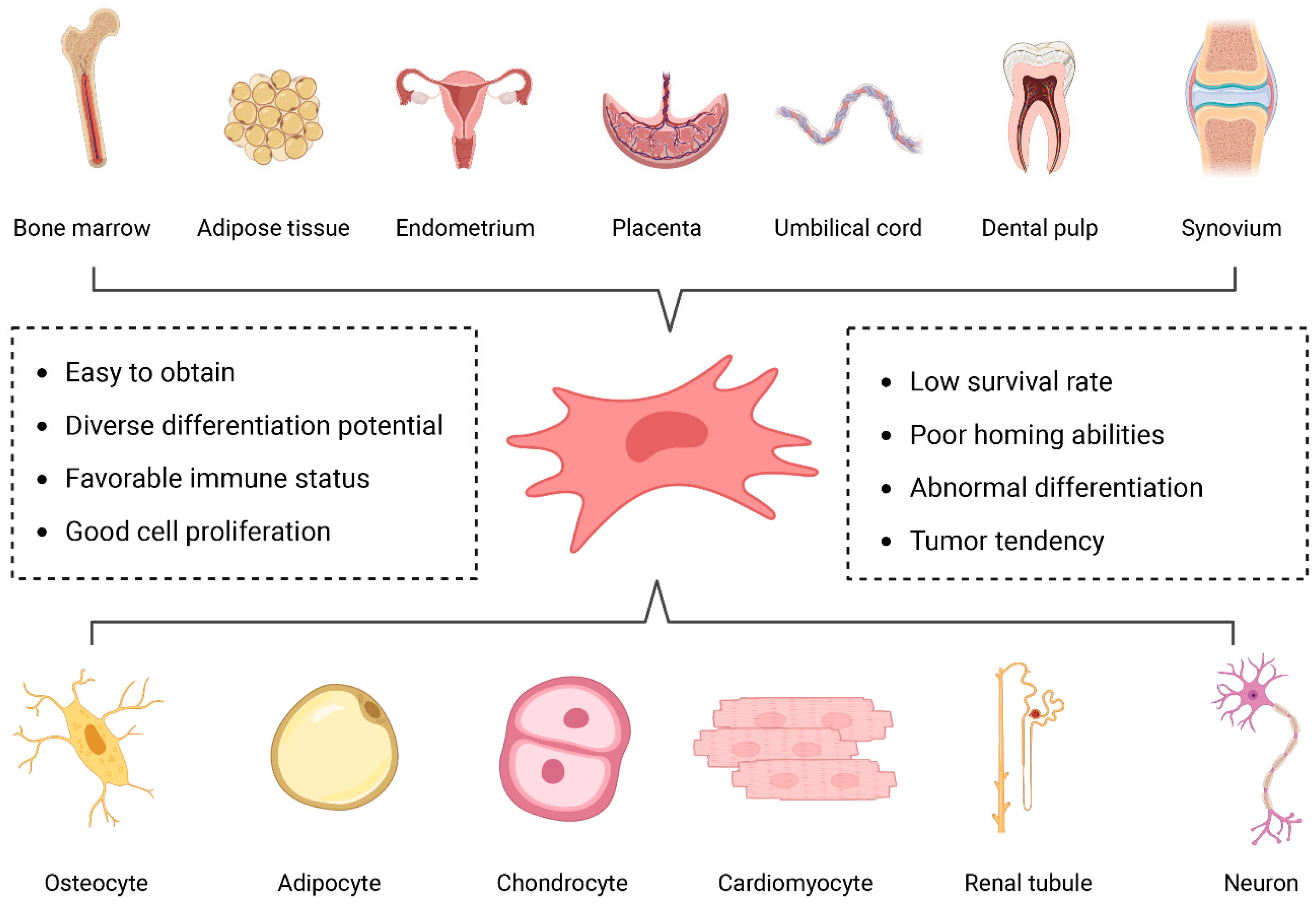
3. Mesenchymal Stem Cells

Mesenchymal stem cells (MSCs) are a subtype of adult stem cells derived from the mesodermal layer during early embryonic development [33], exhibiting self-renewal capacity, multipotent differentiation potential, and the ability to maintain their biological characteristics after in vitro scale-up expansion [34]. MSCs are found in various tissues, such as bone marrow, adipose tissue, dental pulp, placenta, and umbilical cord [35] (Figure 1). Under the influence of different induction culture media in vitro, MSCs can differentiate into cell-specific lineages, including chondrocytes, osteoblasts, adipocytes, neurons, and glial cells [36] (Figure 1). For instance, when cultured with media pre-incubated with cerebrospinal fluid to provide appropriate neurotrophic factors, MSCs can differentiate into neurons [37]. Furthermore, Wang et al. [38] reported that bone mesenchymal stem cells (BMSCs) can differentiate into the Schwann cell phenotype in vitro after being cocultured with peripheral nerve extracts from the distal segment of degenerated rat sciatic nerve. However, the application of MSCs is accompanied by certain disadvantages, such as low survival rate of transplantation, poor migration and homing abilities, abnormal differentiation, and tumor tendency [39].

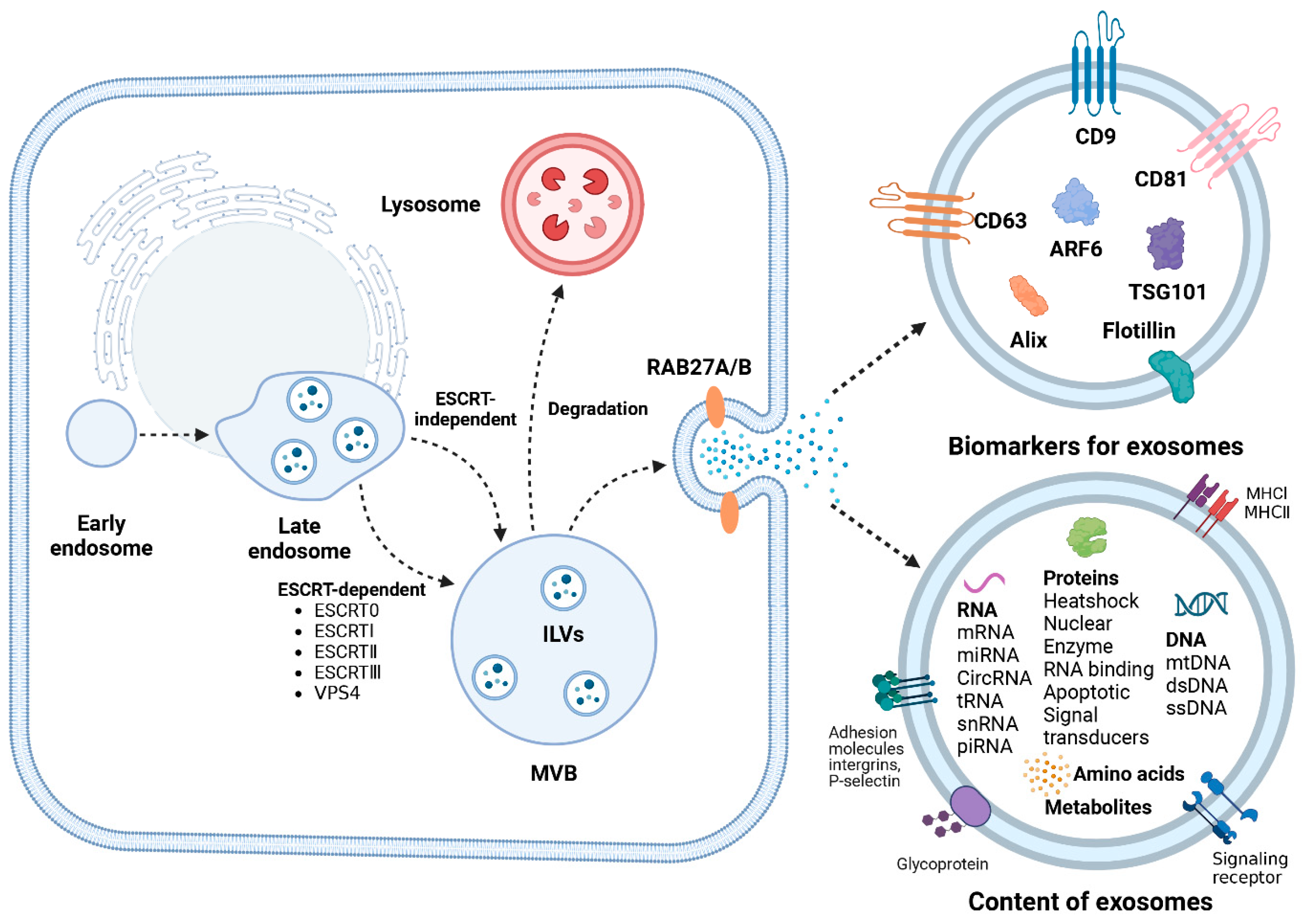
4. Biogenesis of Exosomes

In 1987, Johnstone et al. [40] first described extracellular “exosomes” as a new extracellular vesicle (EV) subtype. Since then, the origin and functions of exosomes have garnered widespread attention. Studies have revealed that exosomes’ biogenesis begins in the endosomal pathway, where continuous invagination of the limiting membrane during early endosome maturation results in the formation of intraluminal vesicles (ILVs) [41] (Figure 2). During this process, proteins, nucleic acids, and lipids are selectively sorted into ILVs. Subsequently, ILVs further mature into multivesicular bodies (MVBs), with the endosomal sorting complex required for transport (ESCRT) playing a pivotal role in this highly intricate process [42]. The ESCRT machinery comprises five complexes, ESCRT-0, ESCRT-I, ESCRT-II, ESCRT-III, and vacuolar protein sorting 4 (VPS4), along with the Bro1 protein family (ALIX, HDPTPT) [43]. ESCRT facilitates the budding of ILVs and their release into the lumen of MVBs. ESCRT-0, consisting of HRS and STAM proteins, each containing two ubiquitin-binding domains (UBD), facilitates the capture of ubiquitinated cargo proteins for aggregation [44]. Additionally, ESCRT-I recruits ESCRT-II to form a stable vesicle neck and regulates the release of vesicles; ESCRT-II mediates ESCRT-III assembly and regulates cytoskeleton separation [45]. ESCRT-III lacks similar structural motifs; instead, it assembles into a filamentous structure associated with the membrane and catalyzes membrane scission and factor recycling through the VPS4 complex, ultimately forming MVBs [46]. The biogenesis of exosomes is not exclusively mediated by the ESCRT pathway, as complete disruption of ESCRT function does not eliminate ILV formation. Non-ESCRT-dependent mechanisms for MVB formation, particularly the neutral sphingomyelinase (nSMase) pathway, have been extensively reported [47].
The ultimately formed multivesicular body (MVB) undergoes regulation by ISGylation, with a fraction directed towards lysosomal degradation and another fraction transported to the plasma membrane mediated by Rab GTPase [48,49]. Subsequently, late endosomes fuse with specific regions of the plasma membrane, facilitating the release of exosomes from the MVB into the extracellular microenvironment through either SNARE-dependent or independent pathways.

5. Isolation of Exosomes

Generally, exosomes are widely present in a variety of body fluids, such as amniotic fluid, blood, breast milk, cerebrospinal fluid, saliva, and urine [50], which can be extracted and purified based on their different characteristics and research objectives for applying in diagnosis and treatment [51,52] (Table 1). Ultracentrifugation is a widely used protocol for exosome isolation, which is considered the gold standard method. In one study, three commercial kits were used to isolate culture supernatants compared to ultracentrifugation as a control [53]. Although ultracentrifugation possessed potential advantages of easy operation and high purity of products, the disadvantages of low yield, time consumption and protein aggregation still limited its large-scale application. Moreover, precipitation-based methods were used to tie up water molecules and force less-soluble components out of samples. Polyethylene glycol is a kind of hydrophilic polymer, which can surround exosomes to allow exosomes’ precipitation at low-speed centrifugation. This method is simple and increases exosome yield, but the purity of the exosomes is hampered by other proteins precipitated together with exosomes [54]. To improve the efficiency of exosome isolation, some studies have sought alternative innovative separation techniques. For instance, Shi et al. [55] developed an insulator-based dielectrophoresis (DEP) method for the non-specific and rapid capture of exosomes. Based on the dielectric properties of vesicles, exosomes were captured and pre-concentrated from healthy human plasma in 2 min using a DEP device with a charged nanopipette. The results showed that the size of the nanopipette pore did not affect separation efficiency, but different geometry of the same pore size produced inconsistent results. This indicated that there are still potential factors affecting the extraction of exosomes using charge-based methods. Additionally, microfluidic-based techniques are novel isolation approaches, which utilize the physical and biochemical characteristics of exosomes to offer refined microcarriers for exosome preparation. This technology integrates advanced techniques, such as acoustics, electrophoresis and electromagnetic operations with conventional methods (size, density and immunoaffinity). It increases the production of high-purity exosomes and has greater efficiency of exosome isolation, while being unavailable for large-scale production of exosomes [56]. Currently, these strategies for exosome isolation are restricted to laboratory research because of their potential shortcomings. Therefore, the development of more efficient and scaled-up separation techniques for exosomes are driven forward for the clinical treatment of peripheral nerve injury.

6. Effects of MSC-Derived Exosomes on Regeneration of PNI

Exosomes serve as the principal paracrine effectors of mesenchymal stem cells, facilitating intercellular communication, and maintaining the homeostatic and dynamic microenvironment [57,58]. When introduced into the site of peripheral nerve injury, exosomes derived from MSCs employ cellular internalization, receptor-ligand recognition, and membrane fusion mechanisms to enter recipient cells [59]. The process promotes angiogenesis and axonal regeneration at the site of peripheral nerve injury, modulates neuroinflammation, and alleviates neuropathic pain (Figure 3). The realization of these functions involves a diverse array of molecular mechanisms and signaling pathways (Table 2).

6.1. MSC Exosomes Promote Angiogenesis

After peripheral nerve injury, the disruption of the blood–nerve barrier is a crucial factor resulting in secondary damage to peripheral nerves [60,61]. The reconstruction of the vascular network provides a regenerative microenvironment for axonal growth, making the maintenance of vascular integrity a key therapeutic target for peripheral nerve regeneration [62,63]. Endothelial cells constitute a critical component of the vascular wall, with the ability to secrete various biologically active substances favorable to axonal growth. MSC-derived exosomes were rich in proteins associated with angiogenesis, such as vascular endothelial growth factor (VEGF) and platelet-derived growth factor-D (PDGF-D), which acted on endothelial cells, exerting a pro-angiogenic effect [64,65]. Moreover, MSC-derived exosomes transferred angiogenic miRNAs to human umbilical vein endothelial cells (HUVECs) to modulate the expression of miRNA target genes. For example, BMSC-derived exosomes can carry miRNA-1260a, miRNA-21-5p, and miRNA-29b-3p to enhance vascular regeneration [66,67,68]. These exosomal miRNAs entered endothelial cells and regulated multiple signaling pathways, such as AKT/eNOS and PI3K/Akt [21,66]. In peripheral nerve injury, exosomes derived from adipose-derived mesenchymal stem cells (ADMSC) towards the Schwann cell phenotype (dExo) can enhance antioxidative, angiogenic, anti-inflammatory, and axon growth properties, which are related to upregulated miRNAs in dExo, such as miRNA-132-3p and miRNA-199b-5p [69]. All these studies collectively suggest that exosome-mediated angiogenesis is beneficial for the repair of peripheral nerve injury.

6.2. MSC Exosomes Mediate Axonal Regeneration

Exosomes derived from neural or non-neuronal cells have been demonstrated to facilitate neurite outgrowth or enhance axonal regeneration in damaged neurons [70,71]. Currently, the mechanisms by which exosomes promote axonal regeneration remain unclear but are possibly attributable to the cargo carried by these vesicles, including miRNA, peptides, and various signaling molecules [72]. Exosomes from different sources of MSCs, such as BMSC and ADMSC, have been shown to promote the growth of dorsal root ganglion neuron axons [17,73]. Xie et al. [74] reported that exosomes derived from ADMSCs stimulate proliferation and migration of PC12 cells (rat pheochromocytoma cell line, model for the study of neuronal differentiation and neuroregeneration) by activating the PI3K/AKT pathway. In a recent study, exosomes from gingival mesenchymal stem cells (GMSCs) were found to enhance Schwann cell dedifferentiation, thereby promoting damaged axonal regeneration and functional recovery through activating gene expression of c-JUN [75].
Furthermore, MSC-derived exosomes carrying specific miRNAs promoted axonal regeneration compared to naturally secreted exosomes from MSCs. Previous investigations have demonstrated that exosomes derived from BMSCs can facilitate the survival and axonal regeneration of retinal ganglion cells following optic nerve crush injury in rats [76]. This pivotal effect relied on miRNA, as evidenced by the diminished therapeutic effects upon downregulation of the miRNA effector molecule Argonaute-2. Multifaceted studies have confirmed that exosomes derived from genetically engineered mesenchymal stem cells enriched with miR-26a, miR-133b, and miR-17-92 cluster increased axonal growth [77,78,79]. Therefore, the presence of bioactive molecules in MSC-derived exosomes associated with neural repair offers the potential for regulating the recovery of denervation atrophy, neuronal survival, and axonal growth.

6.3. MSC Exosomes Modulate Neuroinflammation

Neuroinflammation is one of the key factors in peripheral nerve injury and regeneration. When peripheral nerve injury occurs, the distal nerve myelinated SCs are dedifferentiated into repaired SCs, releasing a variety of chemokines and proinflammatory cytokines to recruit peripheral immune cells (such as circulating macrophages) to accumulate in the injured site [80]. Macrophages removed myelin fragments and avoided the inhibition of axon growth by residual axon fragments in the later stage of Wallerian degeneration. However, excessive inflammation hinders peripheral nerve regeneration. MSC-derived exosomes can communicate with the inflammatory microenvironment to exert neuroprotective effects and regulate complementary systems to promote nerve function recovery. Ti et al. [81] demonstrated that lipopolysaccharide (LPS) pre-treated MSCs can enhance M2 macrophage activation, upregulate anti-inflammatory factor expression, reduce inflammation infiltration, and effectively alleviate chronic inflammation. Further analysis revealed that LPS pre-treated exosomes exert their function by carrying miRNA, specifically let-7b, which participated in regulating the TLR4/NF-κB/STAT3/AKT signaling pathway. Moreover, Sun et al. [82] found the intravenous injection of exosomes secreted by human umbilical cord MSCs (hUCMSCs) can convert M1 macrophages into M2 macrophages. The conversion led to a downregulation of inflammatory cytokines, thereby alleviating inflammation in the damaged area, promoting tissue healing, and expediting the recovery of functional impairments. Collectively, these findings suggest exosome-mediated neuroinflammatory modulation for the repair of peripheral nerve injury.

6.4. MSC Exosomes Alleviate Neuropathic Pain

Neuropathic pain, arising from lesions or diseases affecting the central or peripheral nervous system, constitutes a refractory chronic condition [83]. Current therapeutic strategies for neuropathic pain, encompassing physical, pharmacological, and interventional approaches, exhibit limited efficacy [84]. MSC-derived exosomes present a promising pathway for alleviating chronic pain by mitigating inflammatory responses. Fan et al. [85] administered peripheral intravenous injections of BMSC-derived exosomes into diabetic model mice, significantly reducing mechanical and thermal pain thresholds. Bioinformatics analysis revealed an abundance of miRNAs targeting the TLR4/NF-κB signaling pathway within BMSC-derived exosomes, suggesting their potential role in suppressing inflammation and reducing neuropathic pain. Shiue et al. [86] demonstrated that intrathecal injection of exosomes extracted from MSCs significantly alleviated spinal nerve ligation (SNL)-induced neuropathic pain. In SNL rats, ipsilateral L5 spinal dorsal horn ganglia exhibited a marked increase in IL-10, brain-derived, and glial cell-derived neurotrophic factors compared to the control group. The expression of TNF-α and IL-1β decreased due to exosome-mediated inhibition of microglial and astrocytic cell activation. Similarly, Hsu et al. [87] observed significant relief of neuropathic pain induced by the SNL rat model following the injection of UCMSC-derived exosomes at the injury site. This intervention led to a reduction in TNF-α and IL-1β levels in the L5-6 dorsal root ganglia on the injured side, accompanied by an elevation in IL-10 and brain-derived neurotrophic factor levels, underscoring the role of exosomes in alleviating pain through the inhibition of inflammatory progression. In other studies, MSC-derived exosomal miRNAs, such as miR-181c-5p and miR-146a-5p, have also demonstrated potential in alleviating neuropathic pain [84,88].
Table 2. Mechanisms of MSC exosomes promoting peripheral nerve regeneration.
Source CellExosomal CargoSignaling PathwayEffectsReferences
ADMSCsmiRN-132-3p, miRNA-199b-5pN/AProtected SCs from oxidative stress, enhanced angiogenesis, promoted axonal growth[69]
ADMSCsN/API3K/AKT signaling pathwayPromoted PC12 cell proliferation and migration, inhibited apoptosis[74]
GMSCsN/AExpression of c-JUN key transcription factorPromoted axonal regeneration and functional recovery, activated repair phenotype of SCs[75]
MenSCsN/API3K/AKT signaling pathwayAgainst neuron injury induced by glutamate[89]
BMSCsmiR-17-92 clusterPI3K/protein kinase B/mechanistic target of rapamycin/glycogen synthase kinase 3βIncreased neural plasticity and functional recovery[79]
BMSCsLet-7a, miR-23a and miR-125bTLR/NF-κB signaling pathwayIncreased angiogenesis and nerve regeneration, suppressed proinflammatory cytokines[85]

7. Strategies to Improve Therapeutic Function of MSC Exosomes

7.1. Pretreatment Approaches of Parental Cells

The quantity and function of natural exosomes produced by different MSCs are limited. Interestingly, source cells can regulate the production and bioactivity of exosomes with the change in microenvironment. The previous studies have shown that low gas (low oxygen, low carbon dioxide) in culture environment significantly influenced cellular physiological functions [90,91]. For example, ADMSCs cultured in a low carbon dioxide environment exhibited remarkable upregulation of miR-218 in the engineered exosomes, thereby promoting the activity of PC12 cells [91]. In vivo, hypocapnia-stimulated exosomes delivering miR-218 combined with bioscaffold facilitated the regeneration of motor and nerve fibers (Figure 4A). Additionally, cells cultured in a 3D system demonstrated a closer resemblance to in vivo conditions compared to traditional 2D culture methods, enhancing the paracrine effects of MSCs and optimizing exosome function [92]. For instance, hBMSCs cultured in 3D Synthemax II microcarriers in the PBS mini 0.1 L Vertical-Wheel bioreactor system resulted in a 2.5-fold increase in exosome secretion compared to 2D culture. Furthermore, the expression of exosomes biogenesis markers, glycolysis genes, and miRNAs (miR-10, 19a, 19b, 21,132, and 377) were upregulated in the exosomes from 3D bioreactor culture compared to static 2D culture [93].
Additionally, the secretory function of MSCs undergoes alteration under the influence of biochemical reagents, consequently imparting specific biological functions to exosomes. Li et al. [94] treated MSCs with lipopolysaccharide (LPS) to generate exosomes with immunomodulatory and anti-inflammatory functions. When exosomes derived from LPS-pre-MSC (LPS pre-Exos) were applied in the sciatic nerve injury model and inflammatory model, LPS pre-Exos promoted functional recovery, axonal regeneration, remyelination, and M2 macrophage polarization. The potential mechanism involved high enrichment of TNF stimulated gene-6 (TSG-6) in LPS pre-Exos, which exerted inhibitory effects on NF-κB and NOD-like receptor protein 3 (NLRP3) during macrophage polarization (Figure 4B). The immunosuppressive drug tacrolimus (FK506), in conjunction with ADMSCs), was employed in cell therapy for organ transplantation and vascularized composite allotransplantation [95,96]. Kuo et al. [97] locally administered exosomes derived from FK506-treated ADMSCs (ADMSC-F-exo) in the sciatic nerve crush injury model, revealing a significant reduction in the autophagy of macrophages in the spinal cord segment (Figure 4C). The phenomenon may be associated with the involvement of heat shock protein family A member 8 (HSPA8) and eukaryotic translation elongation factor 1 alpha 1 (EEF1A1) in exosome-mediated autophagy.
Figure 4. Exosomes produced from preconditioned MSCs enhanced peripheral nerve regeneration. (A) Exosomes isolated from the hypocapnia environment cultured ADMSCs upregulated miR−218 expression (I), promoted PC12 migration (II), and restored the regeneration of motor and nerve fibers (III,IV). Adapted with permission from [91], copyright 2022, Frontiers. (B) Exosomes derived from LPS-preconditioned MSCs accelerated the M2 macrophage polarization (I) and delivered TSG6 protein into macrophages to inhibit the expression of NF−κB and NLRP3 (II). Adapted with permission from [94], copyright 2022, Elsevier. (C) Exosomes secreted by ADMSC following FK506 stimulation reduced the expression of autophagy−related markers (LC3, P62, or LAMP-1) in the spinal segment after nerve crush injury. Adapted with permission from [97], copyright 2021, MDPI.
Ijms 25 07882 g004

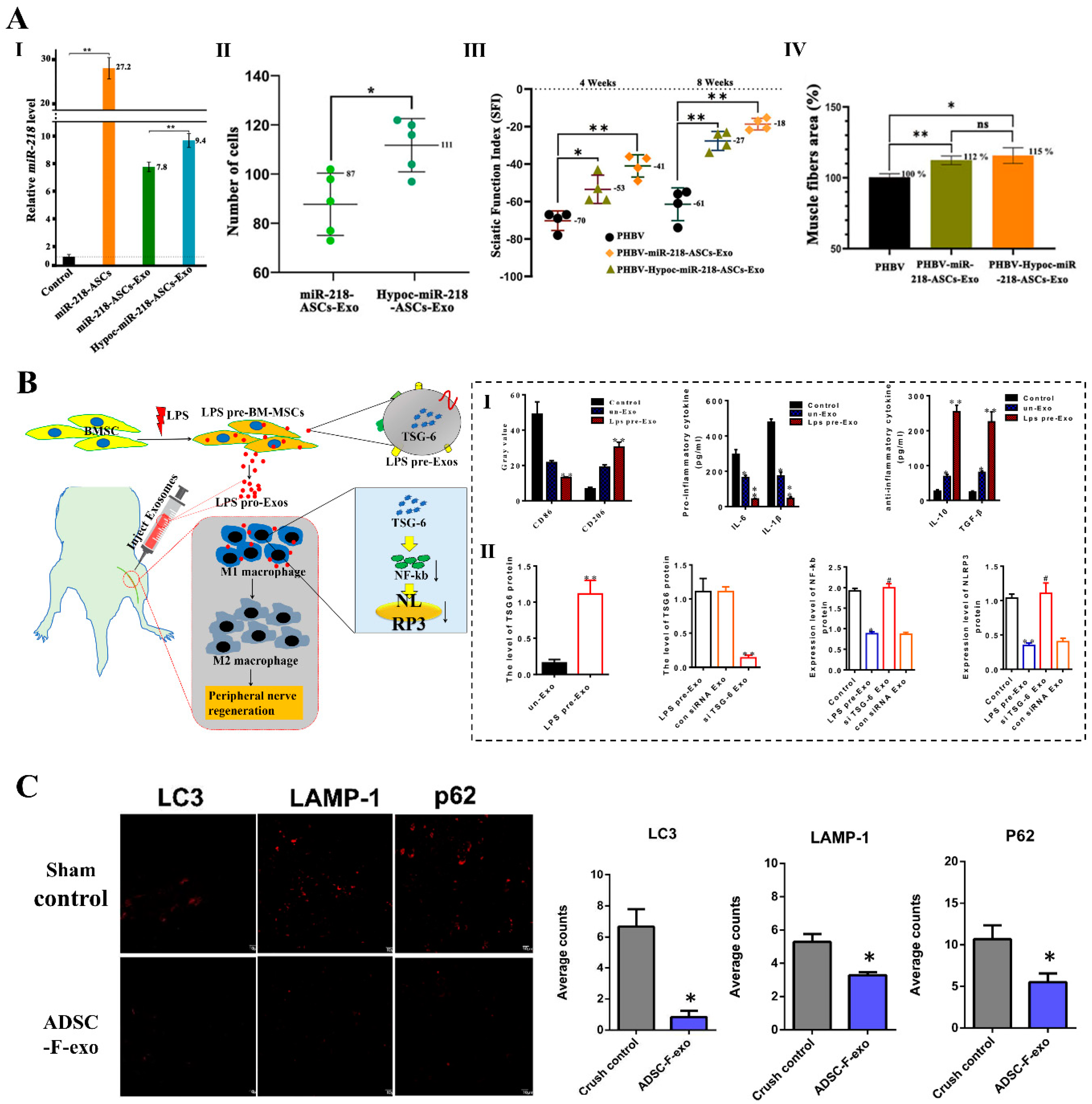
7.2. Direct Engineering Approaches of Exosomes

Although exosomes can be engineered at the cellular level through culture condition and biochemical reagents, hydrophobic and hydrophilic drugs, fluorescent probes, and siRNA face challenges in being loaded into exosomes due to changes in cellular uptake. To overcome the limitations of cell engineering strategies on the drug delivery, fluorescence imaging, and gene therapy potential of natural exosomes, some studies have explored direct engineering approaches for exosomes. These engineered approaches include incubation with cargos and membrane permeabilizer, extrusion, electroporation, sonication, freeze and thaw cycles, liposome-based transfection, and click chemistry [98] (Figure 5A).
In brain injury and spinal cord injury, the application of directly engineered exosomes in nerve regeneration has been extensively documented. However, limited research has been conducted on the direct engineering of exosomes in peripheral nerve regeneration. In the diabetic peripheral neuropathy, Singh et al. [99] devised a combinatorial approach involving the fusion of exosomes derived from BMSCs with liposome-encapsulated polypyrrole nanoparticles (PpyNPs) to provide biochemical and electrical signals. The combination was achieved by subjecting exosomes derived from BMSCs and liposomes loaded with PpyNPs to ten cycles of freeze–thaw (Figure 5B). The composite system offered biochemical and electrical signal stimulation for nerve regeneration, as exosomes contained biochemical components, while PpyNPs can induce electrical conduction for nerve regeneration. Intramuscular injection of the delivery system contributed to the normalization of nerve conduction velocity and compound muscle action potentials, along with the restoration of gastrocnemius muscle morphology, muscle mass, and integrity. Remarkably, engineered exosomes exhibited paracrine effects in controlling hyperglycemia and weight loss.

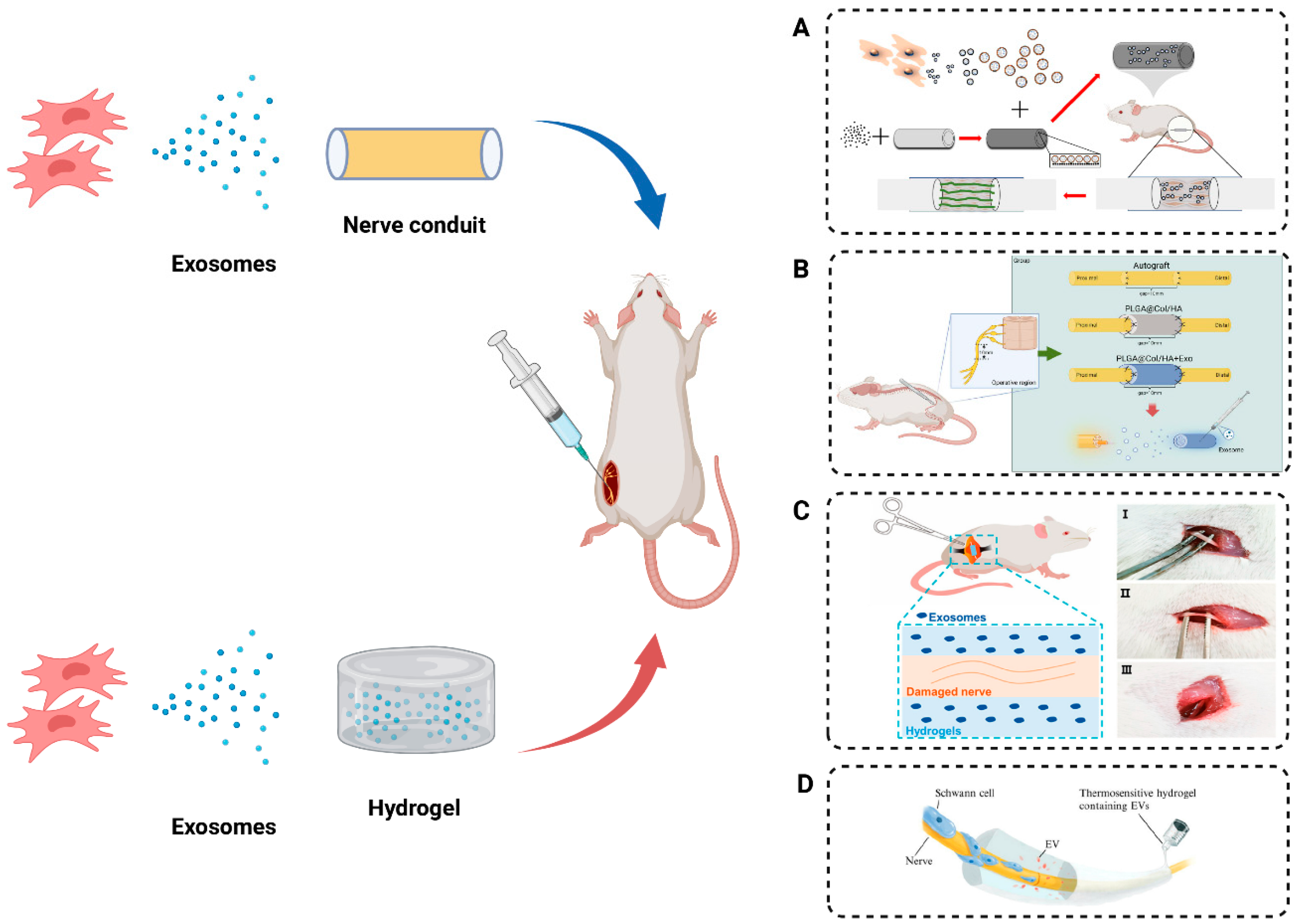
8. MSC Exosomes Combined with Biomaterials Promote Peripheral Nerve Regeneration

Recent studies indicated that the local application of nerve conduits and hydrogels plays a facilitative role in promoting peripheral nerve regeneration. Applying exosomes derived from mesenchymal stem cells directly to the site of peripheral nerve injury bypasses metabolic clearance challenges linked to systemic delivery. The integration of exosomes into nerve conduits and hydrogels facilitates localized and sustained release, mitigating clearance by the host organism.

8.1. Nerve Conduit Incorporating Exosomes

The nerve conduit utilizes a tubular support structure to complement autologous nerve transplantation as a therapeutic technique and has extensive applications in peripheral nerve regeneration. Nerve conduits can prevent the misdirection of nerve fibers and inhibit inward tissue growth, thereby reducing the occurrence of neuromas. However, the reparative capacity of a single nerve conduit transplantation for peripheral nerve injury is limited. Local injection of MSC-derived exosomes at the residual end of nerve injuries has been proven effective in promoting nerve repair and yielding favorable outcomes [100,101,102,103]. In this context, our research group previously developed a biodegradable chitosan nerve conduit. Subsequently, Rao et al. [104]. demonstrated that local injection of dental pulp mesenchymal stem cell (DPMSC)-derived exosomes into the nerve conduit significantly increased the quantity and diameter of sciatic nerve fibers. The promotion of myelin sheath formation consequently facilitated the restoration of muscle function, nerve conduction, and motor function. Similarly, Zhang et al. [105] locally injected exosomes derived from BMSCs building on the earlier development of rGO-GelMA-PCL nerve conduits for repairing peripheral nerve injury. The results indicated superior outcomes, including increased numbers of newly formed blood vessels and axons at the injury site, as well as the recovery of nerve function compared to using nerve conduit alone. Nevertheless, the injected exosomes may leak from the conduit interstice due to limb movement. To better preserve and sustain the release of exosomes, Li et al. [106] modified chitosan conduits with dopamine and subsequently loaded exosomes to repair a 2 mm sciatic nerve defect in rats, resulting in accelerated nerve healing and improved nerve function (Figure 6A). Furthermore, researchers successfully repaired a 10 mm peripheral nerve injury using a 3D composite nerve conduit composed of synthetically derived polymers (electrospun hollow poly (lactic-co-glycolic acid) tube with collagen/hyaluronic acid inner sponge) in conjunction with exosomes from UCMSCs [107] (Figure 6B). The studies on the combined application of nerve conduit and MSC-derived exosomes in peripheral nerve injury were listed in Table 3.

8.2. Hydrogels Incorporating Exosomes

Hydrogels, characterized by highly hydrophilic three-dimensional network structures, have attracted considerable attention owing to their tailorable release properties, transparency, biocompatibility, and exceptional mechanical flexibility [108]. Research indicated that the hardness, conductivity, and composition of hydrogels are pivotal factors influencing the delivery of drugs or molecules and tissue repair. For instance, the hardness of photoresponsive hyaluronic acid methacrylate hydrogels plays a vital role in nerve injury repair [109] (Figure 6C). Compared to rigid hydrogels loaded with exosomes, soft hydrogels enable the rapid release of exosomes. These inhibited the infiltration of macrophages into damaged nerves and promoted the expression of pro-inflammatory factors IL-1β and TNF-α, thereby enhancing peripheral nerve repair. Conductive hydrogels, possessing mechanical and bioelectrical characteristics similar to neural tissue, are found to have extensive applications in neural regeneration and motor function recovery following peripheral nerve injuries. Yang et al. [110] applied electroconductive hydrogels (ECH) loaded with exosomes derived from BMSCs to treat diabetic peripheral nerve damage. ECH-Exos promoted Schwann cell adhesion and migration, while the exosomes in the delivery system regulated M2 macrophage polarization through the NF-κB pathway, alleviating ammation and pain associated with diabetic peripheral nerve damage. Additionally, ECH-Exos enhanced myelinated axon regeneration through the MEK/ERK pathway in vitro and in vivo, ameliorating muscle denervation and contributing to enhanced functional recovery. Temperature-sensitive hydrogels undergo phase changes or swelling/collapse in response to temperature variations, offering improved local drug permeation, optimal spatial and temporal control for drug delivery. After microsurgery, nerve regeneration is slow. To expedite peripheral nerve regeneration, researchers applied ADMSC exosomes in thermosensitive hydrogels. These hydrogels rapidly solidify, maintaining a high concentration of exosomes around the nerves [111] (Figure 6D). Similarly, thermosensitive hydrogel composed of hydroxyethyl chitosan/β-glycerophosphate exhibited excellent injectability, structural stability, and temperature sensitivity. In vivo experiments demonstrated that hydrogels loaded with exosomes from ADMSCs enhanced exosome retention and slow release [112].
Figure 6. Approaches for MSC exosomes’ delivery to the peripheral nerve injury. (A) The polydopamine-modified chitosan nerve conduit was loaded with BMSC-derived exosomes to bridge 2 mm defect of sciatic nerve in rat. Adapted with permission from [106], copyright 2022, Wolters Kluwer Medknow Publications. (B) hUCMSC-derived exosomes combined with 3D composite conduit (PLGA@Col/HA) repaired long-gap peripheral nerve injury, promoting motor function recovery and reducing muscle atrophy. Adapted with permission from [107], copyright 2023, Wolters Kluwer Medknow Publications. (C) Different concentrations of HAMA solution mixed with hUCMSC-derived exosomes were injected into the injured nerve, and after curing with ultraviolet light, the hydrogels with different mechanical properties were formed at the sciatic nerve crush injury site (I, uninjured rat sciatic nerve; II, injured sciatic nerve; and III, injured nerve embedded in hydrogel). Adapted with permission from [109], copyright 2022, Frontiers. (D) Exosomes released from the PALDE hydrogel promoted peripheral nerve regeneration by directly stimulating neurite outgrowth and indirectly promoting Schwann cell migration and proliferation. Adapted with permission from [111], copyright 2022, AIP Publishing.
Ijms 25 07882 g006

9. Conclusions and Future Perspectives

Mesenchymal stem cell (MSC)-derived exosomes have shown significant potential as a nanomedicine in supporting peripheral nerve regeneration. The remarkable therapeutic effects attributed to these nanovesicles are due to the abundant bioactive components encapsulated within them, such as growth factors, anti-inflammatory molecules, and miRNAs. The synergistic actions of these constituents contribute to vascular regeneration at the site of nerve injury, axonal regeneration, regulation of inflammatory responses, promotion of neuronal survival, and stimulation and guidance of the regeneration and repair processes in the surrounding nerves. Physiological stimuli or small molecule modulators have been demonstrated to enhance the secretion of MSC exosomes. However, a comprehensive investigation into the physicochemical properties, composition, and functions of MSC-derived exosomes obtained through these approaches is imperative. Due to the limitations associated with the sole application of MSC exosomes, several studies have combined MSC exosomes with biomaterials or utilized them as carriers for genes or drug delivery. Therefore, it is crucial to further elucidate the collaborative actions and targeted delivery mechanisms of MSC exosomes.
With an increasingly profound understanding of the therapeutic mechanisms associated with MSC-derived exosomes, researchers are continuously striving to optimize the exosome preparation process to enhance their therapeutic efficacy, stability, and controllability. This endeavor involves exploring more efficient extraction methods, refining techniques for exosome loading, and designing personalized treatment regimens. Concurrently, with the advancement of clinical investigations, there is a growing opportunity to validate the efficacy of exosomes in diverse types of nerve injuries, thereby providing robust support for their broader application in clinical practice (NCT03384433, NCT06138210, NCT05035134, NCT05370105, Source: ClinicalTrials.gov). For example, Civelek et al. [113] conducted a preliminary clinical study of MSC-derived exosomes in the treatment of a patient with total radial nerve. Consequently, MSC-derived exosomes, being an innovative and promising therapeutic modality, bring new treatment prospects and a more sustainable trajectory for the recovery of patients with peripheral nerve injuries in the future.

Author Contributions

Q.L. designed the study. F.Z. and X.F. collected the literature. Q.L. wrote the manuscript and contributed to the figures’ editing. N.H. revised the manuscript and made final changes to the final version before submission. All authors have read and agreed to the published version of the manuscript.

Funding

This work was supported by the Natural Foundation of Beijing Municipality [No. 7222198 to Na Han]; Key Research and Development Program of China [No. 2023YFC2509901 to Na Han].

Conflicts of Interest

The authors declare no conflicts of interest.

References

  1. Liu, J.H.; Tang, Q.; Liu, X.X.; Qi, J.; Zeng, R.X.; Zhu, Z.W.; He, B.; Xu, Y.B. Analysis of transcriptome sequencing of sciatic nerves in Sprague-Dawley rats of different ages. Neural Regen. Res. 2018, 13, 2182–2190. [Google Scholar] [CrossRef]
  2. Sullivan, T.B.; Robert, L.C.; Teebagy, P.A.; Morgan, S.E.; Beatty, E.W.; Cicuto, B.J.; Nowd, P.K.; Rieger-Christ, K.M.; Bryan, D.J. Spatiotemporal microRNA profile in peripheral nerve regeneration: miR-138 targets vimentin and inhibits Schwann cell migration and proliferation. Neural Regen. Res. 2018, 13, 1253–1262. [Google Scholar] [CrossRef]
  3. Hopf, A.; Al-Bayati, L.; Schaefer, D.J.; Kalbermatten, D.F.; Guzman, R.; Madduri, S. Optimized Decellularization Protocol for Large Peripheral Nerve Segments: Towards Personalized Nerve Bioengineering. Bioengineering 2022, 9, 412. [Google Scholar] [CrossRef]
  4. Aigner, T.B.; Haynl, C.; Salehi, S.; O’Connor, A.; Scheibel, T. Nerve guidance conduit design based on self-rolling tubes. Mater. Today Bio 2020, 5, 100042. [Google Scholar] [CrossRef]
  5. Babu, S.; Krishnan, M.; Panneerselvam, A.; Chinnaiyan, M. A comprehensive review on therapeutic application of mesenchymal stem cells in neuroregeneration. Life Sci. 2023, 327, 121785. [Google Scholar] [CrossRef]
  6. Yousefi, F.; Lavi Arab, F.; Nikkhah, K.; Amiri, H.; Mahmoudi, M. Novel approaches using mesenchymal stem cells for curing peripheral nerve injuries. Life Sci. 2019, 221, 99–108. [Google Scholar] [CrossRef]
  7. Rodríguez-Sánchez, D.N.; Pinto, G.B.A.; Cartarozzi, L.P.; de Oliveira, A.L.R.; Bovolato, A.L.C.; de Carvalho, M.; da Silva, J.V.L.; Dernowsek, J.A.; Golim, M.; Barraviera, B.; et al. 3D-printed nerve guidance conduits multi-functionalized with canine multipotent mesenchymal stromal cells promote neuroregeneration after sciatic nerve injury in rats. Stem Cell Res. Ther. 2021, 12, 303. [Google Scholar] [CrossRef]
  8. Zhang, Q.; Wu, P.; Chen, F.; Zhao, Y.; Li, Y.; He, X.; Huselstein, C.; Ye, Q.; Tong, Z.; Chen, Y. Brain Derived Neurotrophic Factor and Glial Cell Line-Derived Neurotrophic Factor-Transfected Bone Mesenchymal Stem Cells for the Repair of Periphery Nerve Injury. Front. Bioeng. Biotechnol. 2020, 8, 874. [Google Scholar] [CrossRef]
  9. Das, S.R.; Uz, M.; Ding, S.; Lentner, M.T.; Hondred, J.A.; Cargill, A.A.; Sakaguchi, D.S.; Mallapragada, S.; Claussen, J.C. Electrical Differentiation of Mesenchymal Stem Cells into Schwann-Cell-Like Phenotypes Using Inkjet-Printed Graphene Circuits. Adv. Healthc. Mater. 2017, 6, 1601087. [Google Scholar] [CrossRef]
  10. Sowa, Y.; Kishida, T.; Imura, T.; Numajiri, T.; Nishino, K.; Tabata, Y.; Mazda, O. Adipose-Derived Stem Cells Promote Peripheral Nerve Regeneration In Vivo without Differentiation into Schwann-Like Lineage. Plast. Reconstr. Surg. 2016, 137, 318e–330e. [Google Scholar] [CrossRef]
  11. Xie, N.; Li, Z.; Adesanya, T.M.; Guo, W.; Liu, Y.; Fu, M.; Kilic, A.; Tan, T.; Zhu, H.; Xie, X. Transplantation of placenta-derived mesenchymal stem cells enhances angiogenesis after ischemic limb injury in mice. J. Cell. Mol. Med. 2016, 20, 29–37. [Google Scholar] [CrossRef]
  12. Li, K.L.; Li, J.Y.; Xie, G.L.; Ma, X.Y. Exosomes Released From Human Bone Marrow-Derived Mesenchymal Stem Cell Attenuate Acute Graft-Versus-Host Disease After Allogeneic Hematopoietic Stem Cell Transplantation in Mice. Front. Cell Dev. Biol. 2021, 9, 617589. [Google Scholar] [CrossRef]
  13. Xiao, Q.; Lin, C.; Peng, M.; Ren, J.; Jing, Y.; Lei, L.; Tao, Y.; Huang, J.; Yang, J.; Sun, M.; et al. Circulating plasma exosomal long non-coding RNAs LINC00265, LINC00467, UCA1, and SNHG1 as biomarkers for diagnosis and treatment monitoring of acute myeloid leukemia. Front. Oncol. 2022, 12, 1033143. [Google Scholar] [CrossRef]
  14. Wu, H.; Zhou, J.; Mei, S.; Wu, D.; Mu, Z.; Chen, B.; Xie, Y.; Ye, Y.; Liu, J. Circulating exosomal microRNA-96 promotes cell proliferation, migration and drug resistance by targeting LMO7. J. Cell. Mol. Med. 2017, 21, 1228–1236. [Google Scholar] [CrossRef]
  15. Kojima, R.; Bojar, D.; Rizzi, G.; Hamri, G.C.; El-Baba, M.D.; Saxena, P.; Ausländer, S.; Tan, K.R.; Fussenegger, M. Designer exosomes produced by implanted cells intracerebrally deliver therapeutic cargo for Parkinson’s disease treatment. Nat. Commun. 2018, 9, 1305. [Google Scholar] [CrossRef]
  16. Cheng, J.; Meng, J.; Zhu, L.; Peng, Y. Exosomal noncoding RNAs in Glioma: Biological functions and potential clinical applications. Mol. Cancer 2020, 19, 66. [Google Scholar] [CrossRef]
  17. Zhao, J.; Ding, Y.; He, R.; Huang, K.; Liu, L.; Jiang, C.; Liu, Z.; Wang, Y.; Yan, X.; Cao, F.; et al. Dose-effect relationship and molecular mechanism by which BMSC-derived exosomes promote peripheral nerve regeneration after crush injury. Stem Cell Res. Ther. 2020, 11, 360. [Google Scholar] [CrossRef]
  18. Soares Martins, T.; Marçalo, R.; da Cruz, E.S.C.B.; Trindade, D.; Catita, J.; Amado, F.; Melo, T.; Rosa, I.M.; Vogelgsang, J.; Wiltfang, J.; et al. Novel Exosome Biomarker Candidates for Alzheimer’s Disease Unravelled through Mass Spectrometry Analysis. Mol. Neurobiol. 2022, 59, 2838–2854. [Google Scholar] [CrossRef]
  19. Zhang, S.; Teo, K.Y.W.; Chuah, S.J.; Lai, R.C.; Lim, S.K.; Toh, W.S. MSC exosomes alleviate temporomandibular joint osteoarthritis by attenuating inflammation and restoring matrix homeostasis. Biomaterials 2019, 200, 35–47. [Google Scholar] [CrossRef]
  20. Zhang, S.; Chuah, S.J.; Lai, R.C.; Hui, J.H.P.; Lim, S.K.; Toh, W.S. MSC exosomes mediate cartilage repair by enhancing proliferation, attenuating apoptosis and modulating immune reactivity. Biomaterials 2018, 156, 16–27. [Google Scholar] [CrossRef]
  21. Yu, M.; Liu, W.; Li, J.; Lu, J.; Lu, H.; Jia, W.; Liu, F. Exosomes derived from atorvastatin-pretreated MSC accelerate diabetic wound repair by enhancing angiogenesis via AKT/eNOS pathway. Stem Cell Res. Ther. 2020, 11, 350. [Google Scholar] [CrossRef]
  22. Wei, H.; Xu, Y.; Chen, Q.; Chen, H.; Zhu, X.; Li, Y. Mesenchymal stem cell-derived exosomal miR-223 regulates neuronal cell apoptosis. Cell Death Dis. 2020, 11, 290. [Google Scholar] [CrossRef]
  23. Burnett, M.G.; Zager, E.L. Pathophysiology of peripheral nerve injury: A brief review. Neurosurg. Focus 2004, 16, E1. [Google Scholar] [CrossRef]
  24. Matos Cruz, A.J.; De Jesus, O. Neurotmesis. In StatPearls; StatPearls Publishing LLC.: Treasure Island, FL, USA, 2023. [Google Scholar]
  25. Wang, Y.; Shan, Q.; Meng, Y.; Pan, J.; Yi, S. Mrpl10 and Tbp Are Suitable Reference Genes for Peripheral Nerve Crush Injury. Int. J. Mol. Sci. 2017, 18, 263. [Google Scholar] [CrossRef]
  26. Wang, W.; Gao, J.; Na, L.; Jiang, H.; Xue, J.; Yang, Z.; Wang, P. Craniocerebral injury promotes the repair of peripheral nerve injury. Neural Regen. Res. 2014, 9, 1703–1708. [Google Scholar] [CrossRef]
  27. Chen, J.; Zhou, X.J.; Sun, R.B. Effect of the combination of high-frequency repetitive magnetic stimulation and neurotropin on injured sciatic nerve regeneration in rats. Neural Regen. Res. 2020, 15, 145–151. [Google Scholar] [CrossRef]
  28. Fu, C.; Yin, Z.; Yu, D.; Yang, Z. Substance P and calcitonin gene-related peptide expression in dorsal root ganglia in sciatic nerve injury rats. Neural Regen. Res. 2013, 8, 3124–3130. [Google Scholar] [CrossRef]
  29. Xue, J.; Yang, J.; O’Connor, D.M.; Zhu, C.; Huo, D.; Boulis, N.M.; Xia, Y. Differentiation of Bone Marrow Stem Cells into Schwann Cells for the Promotion of Neurite Outgrowth on Electrospun Fibers. ACS Appl. Mater. Interfaces 2017, 9, 12299–12310. [Google Scholar] [CrossRef]
  30. Man, L.L.; Liu, F.; Wang, Y.J.; Song, H.H.; Xu, H.B.; Zhu, Z.W.; Zhang, Q.; Wang, Y.J. The HMGB1 signaling pathway activates the inflammatory response in Schwann cells. Neural Regen. Res. 2015, 10, 1706–1712. [Google Scholar] [CrossRef]
  31. Lee, J.H.; Kim, K.H.; Kwon, O.H.; Kwon, O.K.; Uyama, H.; Kim, Y.J. Photodynamic Activity of Protoporphyrin IX-Immobilized Cellulose Monolith for Nerve Tissue Regeneration. Int. J. Mol. Sci. 2022, 23, 1035. [Google Scholar] [CrossRef]
  32. Zhang, P.X.; Han, N.; Kou, Y.H.; Zhu, Q.T.; Liu, X.L.; Quan, D.P.; Chen, J.G.; Jiang, B.G. Tissue engineering for the repair of peripheral nerve injury. Neural Regen. Res. 2019, 14, 51–58. [Google Scholar] [CrossRef]
  33. Bahrehbar, K.; Rezazadeh Valojerdi, M.; Esfandiari, F.; Fathi, R.; Hassani, S.N.; Baharvand, H. Human embryonic stem cell-derived mesenchymal stem cells improved premature ovarian failure. World J. Stem Cells 2020, 12, 857–878. [Google Scholar] [CrossRef]
  34. Tian, J.; Zhu, Q.; Zhang, Y.; Bian, Q.; Hong, Y.; Shen, Z.; Xu, H.; Rui, K.; Yin, K.; Wang, S. Olfactory Ecto-Mesenchymal Stem Cell-Derived Exosomes Ameliorate Experimental Colitis via Modulating Th1/Th17 and Treg Cell Responses. Front. Immunol. 2020, 11, 598322. [Google Scholar] [CrossRef]
  35. Zhou, S.; Lei, Y.; Wang, P.; Chen, J.; Zeng, L.; Qu, T.; Maldonado, M.; Huang, J.; Han, T.; Wen, Z.; et al. Human Umbilical Cord Mesenchymal Stem Cells Encapsulated with Pluronic F-127 Enhance the Regeneration and Angiogenesis of Thin Endometrium in Rat via Local IL-1β Stimulation. Stem Cells Int. 2022, 2022, 7819234. [Google Scholar] [CrossRef]
  36. Liu, X.; Zhang, G.; Wei, P.; Hao, L.; Zhong, L.; Zhong, K.; Liu, C.; Liu, P.; Feng, Q.; Wang, S.; et al. 3D-printed collagen/silk fibroin/secretome derived from bFGF-pretreated HUCMSCs scaffolds enhanced therapeutic ability in canines traumatic brain injury model. Front. Bioeng. Biotechnol. 2022, 10, 995099. [Google Scholar] [CrossRef]
  37. Ren, C.; Yin, P.; Ren, N.; Wang, Z.; Wang, J.; Zhang, C.; Ge, W.; Geng, D.; Wang, X. Cerebrospinal fluid-stem cell interactions may pave the path for cell-based therapy in neurological diseases. Stem Cell Res. Ther. 2018, 9, 66. [Google Scholar] [CrossRef]
  38. Wang, H.; Zhang, H.; Liu, M.; Wang, N. Distal segment extracts of the degenerated rat sciatic nerve induce bone marrow stromal cells to express Schwann cell markers in vitro. Neurosci. Lett. 2013, 544, 89–93. [Google Scholar] [CrossRef]
  39. Ren, Z.; Qi, Y.; Sun, S.; Tao, Y.; Shi, R. Mesenchymal Stem Cell-Derived Exosomes: Hope for Spinal Cord Injury Repair. Stem Cells Dev. 2020, 29, 1467–1478. [Google Scholar] [CrossRef]
  40. Keysberg, C.; Hertel, O.; Schelletter, L.; Busche, T.; Sochart, C.; Kalinowski, J.; Hoffrogge, R.; Otte, K.; Noll, T. Exploring the molecular content of CHO exosomes during bioprocessing. Appl. Microbiol. Biotechnol. 2021, 105, 3673–3689. [Google Scholar] [CrossRef]
  41. Qing, L.; Chen, H.; Tang, J.; Jia, X. Exosomes and Their MicroRNA Cargo: New Players in Peripheral Nerve Regeneration. Neurorehabil. Neural Repair 2018, 32, 765–776. [Google Scholar] [CrossRef]
  42. Li, Q.C.; Li, C.; Zhang, W.; Pi, W.; Han, N. Potential Effects of Exosomes and their MicroRNA Carrier on Osteoporosis. Curr. Pharm. Des. 2022, 28, 899–909. [Google Scholar] [CrossRef]
  43. Nepal, B.; Sepehri, A.; Lazaridis, T. Mechanisms of negative membrane curvature sensing and generation by ESCRT III subunit Snf7. Protein Sci. 2020, 29, 1473–1485. [Google Scholar] [CrossRef] [PubMed]
  44. Raiborg, C.; Bache, K.G.; Gillooly, D.J.; Madshus, I.H.; Stang, E.; Stenmark, H. Hrs sorts ubiquitinated proteins into clathrin-coated microdomains of early endosomes. Nat. Cell Biol. 2002, 4, 394–398. [Google Scholar] [CrossRef] [PubMed]
  45. Henne, W.M.; Stenmark, H.; Emr, S.D. Molecular mechanisms of the membrane sculpting ESCRT pathway. Cold Spring Harb. Perspect. Biol. 2013, 5, a016766. [Google Scholar] [CrossRef]
  46. Sun, S.; Zhou, X.; Corvera, J.; Gallick, G.E.; Lin, S.H.; Kuang, J. ALG-2 activates the MVB sorting function of ALIX through relieving its intramolecular interaction. Cell Discov. 2015, 1, 15018. [Google Scholar] [CrossRef]
  47. Trajkovic, K.; Hsu, C.; Chiantia, S.; Rajendran, L.; Wenzel, D.; Wieland, F.; Schwille, P.; Brügger, B.; Simons, M. Ceramide triggers budding of exosome vesicles into multivesicular endosomes. Science 2008, 319, 1244–1247. [Google Scholar] [CrossRef] [PubMed]
  48. Villarroya-Beltri, C.; Baixauli, F.; Mittelbrunn, M.; Fernández-Delgado, I.; Torralba, D.; Moreno-Gonzalo, O.; Baldanta, S.; Enrich, C.; Guerra, S.; Sánchez-Madrid, F. ISGylation controls exosome secretion by promoting lysosomal degradation of MVB proteins. Nat. Commun. 2016, 7, 13588. [Google Scholar] [CrossRef]
  49. Yuan, X.; Sun, L.; Jeske, R.; Nkosi, D.; York, S.B.; Liu, Y.; Grant, S.C.; Meckes, D.G., Jr.; Li, Y. Engineering extracellular vesicles by three-dimensional dynamic culture of human mesenchymal stem cells. J. Extracell. Vesicles 2022, 11, e12235. [Google Scholar] [CrossRef]
  50. Sun, B.; Peng, J.; Wang, S.; Liu, X.; Zhang, K.; Zhang, Z.; Wang, C.; Jing, X.; Zhou, C.; Wang, Y. Applications of stem cell-derived exosomes in tissue engineering and neurological diseases. Rev. Neurosci. 2018, 29, 531–546. [Google Scholar] [CrossRef]
  51. Kimiz-Gebologlu, I.; Oncel, S.S. Exosomes: Large-scale production, isolation, drug loading efficiency, and biodistribution and uptake. J. Control. Release 2022, 347, 533–543. [Google Scholar] [CrossRef]
  52. Yang, D.; Zhang, W.; Zhang, H.; Zhang, F.; Chen, L.; Ma, L.; Larcher, L.M.; Chen, S.; Liu, N.; Zhao, Q.; et al. Progress, opportunity, and perspective on exosome isolation—Efforts for efficient exosome-based theranostics. Theranostics 2020, 10, 3684–3707. [Google Scholar] [CrossRef] [PubMed]
  53. Patel, G.K.; Khan, M.A.; Zubair, H.; Srivastava, S.K.; Khushman, M.; Singh, S.; Singh, A.P. Comparative analysis of exosome isolation methods using culture supernatant for optimum yield, purity and downstream applications. Sci. Rep. 2019, 9, 5335. [Google Scholar] [CrossRef]
  54. Dash, M.; Palaniyandi, K.; Ramalingam, S.; Sahabudeen, S.; Raja, N.S. Exosomes isolated from two different cell lines using three different isolation techniques show variation in physical and molecular characteristics. Biochim. Biophys. Acta Biomembr. 2021, 1863, 183490. [Google Scholar] [CrossRef]
  55. Shi, L.; Rana, A.; Esfandiari, L. A low voltage nanopipette dielectrophoretic device for rapid entrapment of nanoparticles and exosomes extracted from plasma of healthy donors. Sci. Rep. 2018, 8, 6751. [Google Scholar] [CrossRef]
  56. Hassanpour Tamrin, S.; Sanati Nezhad, A.; Sen, A. Label-Free Isolation of Exosomes Using Microfluidic Technologies. ACS Nano 2021, 15, 17047–17079. [Google Scholar] [CrossRef] [PubMed]
  57. Yin, S.; Lin, S.; Xu, J.; Yang, G.; Chen, H.; Jiang, X. Dominoes with interlocking consequences triggered by zinc: Involvement of microelement-stimulated MSC-derived exosomes in senile osteogenesis and osteoclast dialogue. J. Nanobiotechnol. 2023, 21, 346. [Google Scholar] [CrossRef]
  58. Tian, S.; Zhou, X.; Zhang, M.; Cui, L.; Li, B.; Liu, Y.; Su, R.; Sun, K.; Hu, Y.; Yang, F.; et al. Mesenchymal stem cell-derived exosomes protect against liver fibrosis via delivering miR-148a to target KLF6/STAT3 pathway in macrophages. Stem Cell Res. Ther. 2022, 13, 330. [Google Scholar] [CrossRef] [PubMed]
  59. Gurung, S.; Perocheau, D.; Touramanidou, L.; Baruteau, J. The exosome journey: From biogenesis to uptake and intracellular signalling. Cell Commun. Signal. CCS 2021, 19, 47. [Google Scholar] [CrossRef] [PubMed]
  60. Moreau, N.; Mauborgne, A.; Bourgoin, S.; Couraud, P.O.; Romero, I.A.; Weksler, B.B.; Villanueva, L.; Pohl, M.; Boucher, Y. Early alterations of Hedgehog signaling pathway in vascular endothelial cells after peripheral nerve injury elicit blood-nerve barrier disruption, nerve inflammation, and neuropathic pain development. Pain 2016, 157, 827–839. [Google Scholar] [CrossRef]
  61. Lim, T.K.Y.; Shi, X.Q.; Martin, H.C.; Huang, H.; Luheshi, G.; Rivest, S.; Zhang, J. Blood-nerve barrier dysfunction contributes to the generation of neuropathic pain and allows targeting of injured nerves for pain relief. Pain 2014, 155, 954–967. [Google Scholar] [CrossRef]
  62. Thibodeau, A.; Galbraith, T.; Fauvel, C.M.; Khuong, H.T.; Berthod, F. Repair of peripheral nerve injuries using a prevascularized cell-based tissue-engineered nerve conduit. Biomaterials 2022, 280, 121269. [Google Scholar] [CrossRef]
  63. Wang, H.; Zhang, P.; Lu, P.; Cai, X.; Wang, G.; Xu, X.; Liu, Y.; Huang, T.; Li, M.; Qian, T.; et al. Neural tissue-engineered prevascularization in vivo enhances peripheral neuroregeneration via rapid vascular inosculation. Mater. Today. Bio 2023, 21, 100718. [Google Scholar] [CrossRef]
  64. Du, W.; Zhang, K.; Zhang, S.; Wang, R.; Nie, Y.; Tao, H.; Han, Z.; Liang, L.; Wang, D.; Liu, J.; et al. Enhanced proangiogenic potential of mesenchymal stem cell-derived exosomes stimulated by a nitric oxide releasing polymer. Biomaterials 2017, 133, 70–81. [Google Scholar] [CrossRef]
  65. Ma, J.; Zhao, Y.; Sun, L.; Sun, X.; Zhao, X.; Sun, X.; Qian, H.; Xu, W.; Zhu, W. Exosomes Derived from Akt-Modified Human Umbilical Cord Mesenchymal Stem Cells Improve Cardiac Regeneration and Promote Angiogenesis via Activating Platelet-Derived Growth Factor D. Stem Cells Transl. Med. 2017, 6, 51–59. [Google Scholar] [CrossRef]
  66. Zhang, L.; Ouyang, P.; He, G.; Wang, X.; Song, D.; Yang, Y.; He, X. Exosomes from microRNA-126 overexpressing mesenchymal stem cells promote angiogenesis by targeting the PIK3R2-mediated PI3K/Akt signalling pathway. J. Cell. Mol. Med. 2021, 25, 2148–2162. [Google Scholar] [CrossRef]
  67. Hu, H.; Hu, X.; Li, L.; Fang, Y.; Yang, Y.; Gu, J.; Xu, J.; Chu, L. Exosomes Derived from Bone Marrow Mesenchymal Stem Cells Promote Angiogenesis in Ischemic Stroke Mice via Upregulation of MiR-21-5p. Biomolecules 2022, 12, 883. [Google Scholar] [CrossRef]
  68. Zheng, J.; Zhang, X.; Cai, W.; Yang, Y.; Guo, T.; Li, J.; Dai, H. Bone Marrow Mesenchymal Stem Cell-Derived Exosomal microRNA-29b-3p Promotes Angiogenesis and Ventricular Remodeling in Rats with Myocardial Infarction by Targeting ADAMTS16. Cardiovasc. Toxicol. 2022, 22, 689–700. [Google Scholar] [CrossRef]
  69. Liu, B.; Kong, Y.; Shi, W.; Kuss, M.; Liao, K.; Hu, G.; Xiao, P.; Sankarasubramanian, J.; Guda, C.; Wang, X.; et al. Exosomes derived from differentiated human ADMSC with the Schwann cell phenotype modulate peripheral nerve-related cellular functions. Bioact. Mater. 2022, 14, 61–75. [Google Scholar] [CrossRef]
  70. López-Leal, R.; Díaz-Viraqué, F.; Catalán, R.J.; Saquel, C.; Enright, A.; Iraola, G.; Court, F.A. Schwann cell reprogramming into repair cells increases miRNA-21 expression in exosomes promoting axonal growth. J. Cell Sci. 2020, 133, jcs239004. [Google Scholar] [CrossRef]
  71. Zhao, Y.; Liu, J.; Liu, S.; Yang, P.; Liang, Y.; Ma, J.; Mao, S.; Sun, C.; Yang, Y. Fibroblast exosomal TFAP2C induced by chitosan oligosaccharides promotes peripheral axon regeneration via the miR-132-5p/CAMKK1 axis. Bioact. Mater. 2023, 26, 249–263. [Google Scholar] [CrossRef]
  72. Tang, B.L. Promoting axonal regeneration through exosomes: An update of recent findings on exosomal PTEN and mTOR modifiers. Brain Res. Bull. 2018, 143, 123–131. [Google Scholar] [CrossRef]
  73. Bucan, V.; Vaslaitis, D.; Peck, C.T.; Strauß, S.; Vogt, P.M.; Radtke, C. Effect of Exosomes from Rat Adipose-Derived Mesenchymal Stem Cells on Neurite Outgrowth and Sciatic Nerve Regeneration After Crush Injury. Mol. Neurobiol. 2019, 56, 1812–1824. [Google Scholar] [CrossRef]
  74. Xie, Y.; Chen, Y.; Zhu, Y.; Chen, X.; Lin, T.; Zhou, D. Adipose Mesenchymal Stem Cell-Derived Exosomes Enhance PC12 Cell Function through the Activation of the PI3K/AKT Pathway. Stem Cells Int. 2021, 2021, 2229477. [Google Scholar] [CrossRef]
  75. Mao, Q.; Nguyen, P.D.; Shanti, R.M.; Shi, S.; Shakoori, P.; Zhang, Q.; Le, A.D. Gingiva-Derived Mesenchymal Stem Cell-Extracellular Vesicles Activate Schwann Cell Repair Phenotype and Promote Nerve Regeneration. Tissue Eng. Part A 2019, 25, 887–900. [Google Scholar] [CrossRef]
  76. Mead, B.; Tomarev, S. Bone Marrow-Derived Mesenchymal Stem Cells-Derived Exosomes Promote Survival of Retinal Ganglion Cells Through miRNA-Dependent Mechanisms. Stem Cells Transl. Med. 2017, 6, 1273–1285. [Google Scholar] [CrossRef]
  77. Chen, Y.; Tian, Z.; He, L.; Liu, C.; Wang, N.; Rong, L.; Liu, B. Exosomes derived from miR-26a-modified MSCs promote axonal regeneration via the PTEN/AKT/mTOR pathway following spinal cord injury. Stem Cell Res. Ther. 2021, 12, 224. [Google Scholar] [CrossRef]
  78. Li, D.; Zhang, P.; Yao, X.; Li, H.; Shen, H.; Li, X.; Wu, J.; Lu, X. Exosomes Derived From miR-133b-Modified Mesenchymal Stem Cells Promote Recovery After Spinal Cord Injury. Front. Neurosci. 2018, 12, 845. [Google Scholar] [CrossRef]
  79. Xin, H.; Katakowski, M.; Wang, F.; Qian, J.Y.; Liu, X.S.; Ali, M.M.; Buller, B.; Zhang, Z.G.; Chopp, M. MicroRNA cluster miR-17-92 Cluster in Exosomes Enhance Neuroplasticity and Functional Recovery after Stroke in Rats. Stroke 2017, 48, 747–753. [Google Scholar] [CrossRef]
  80. Qu, W.R.; Zhu, Z.; Liu, J.; Song, D.B.; Tian, H.; Chen, B.P.; Li, R.; Deng, L.X. Interaction between Schwann cells and other cells during repair of peripheral nerve injury. Neural Regen. Res. 2021, 16, 93–98. [Google Scholar] [CrossRef]
  81. Ti, D.; Hao, H.; Tong, C.; Liu, J.; Dong, L.; Zheng, J.; Zhao, Y.; Liu, H.; Fu, X.; Han, W. LPS-preconditioned mesenchymal stromal cells modify macrophage polarization for resolution of chronic inflammation via exosome-shuttled let-7b. J. Transl. Med. 2015, 13, 308. [Google Scholar] [CrossRef]
  82. Sun, G.; Li, G.; Li, D.; Huang, W.; Zhang, R.; Zhang, H.; Duan, Y.; Wang, B. hucMSC derived exosomes promote functional recovery in spinal cord injury mice via attenuating inflammation. Mater. Sci. Eng. C Mater. Biol. Appl. 2018, 89, 194–204. [Google Scholar] [CrossRef]
  83. Yang, H.K.; Son, W.S.; Lim, K.S.; Kim, G.H.; Lim, E.J.; Gadhe, C.G.; Lee, J.Y.; Jeong, K.S.; Lim, S.M.; Pae, A.N. Synthesis and biological evaluation of pyrrolidine-based T-type calcium channel inhibitors for the treatment of neuropathic pain. J. Enzym. Inhib. Med. Chem. 2018, 33, 1460–1471. [Google Scholar] [CrossRef]
  84. Zhang, Y.U.; Ye, G.; Zhao, J.; Chen, Y.; Kong, L.; Sheng, C.; Yuan, L. Exosomes carried miR-181c-5p alleviates neuropathic pain in CCI rat models. An. Acad. Bras. Cienc. 2022, 94, e20210564. [Google Scholar] [CrossRef]
  85. Fan, B.; Li, C.; Szalad, A.; Wang, L.; Pan, W.; Zhang, R.; Chopp, M.; Zhang, Z.G.; Liu, X.S. Mesenchymal stromal cell-derived exosomes ameliorate peripheral neuropathy in a mouse model of diabetes. Diabetologia 2020, 63, 431–443. [Google Scholar] [CrossRef]
  86. Shiue, S.J.; Rau, R.H.; Shiue, H.S.; Hung, Y.W.; Li, Z.X.; Yang, K.D.; Cheng, J.K. Mesenchymal stem cell exosomes as a cell-free therapy for nerve injury-induced pain in rats. Pain 2019, 160, 210–223. [Google Scholar] [CrossRef]
  87. Hsu, J.M.; Shiue, S.J.; Yang, K.D.; Shiue, H.S.; Hung, Y.W.; Pannuru, P.; Poongodi, R.; Lin, H.Y.; Cheng, J.K. Locally Applied Stem Cell Exosome-Scaffold Attenuates Nerve Injury-Induced Pain in Rats. J. Pain Res. 2020, 13, 3257–3268. [Google Scholar] [CrossRef]
  88. Hua, T.; Yang, M.; Song, H.; Kong, E.; Deng, M.; Li, Y.; Li, J.; Liu, Z.; Fu, H.; Wang, Y.; et al. Huc-MSCs-derived exosomes attenuate inflammatory pain by regulating microglia pyroptosis and autophagy via the miR-146a-5p/TRAF6 axis. J. Nanobiotechnol. 2022, 20, 324. [Google Scholar] [CrossRef]
  89. Lopez-Verrilli, M.A.; Caviedes, A.; Cabrera, A.; Sandoval, S.; Wyneken, U.; Khoury, M. Mesenchymal stem cell-derived exosomes from different sources selectively promote neuritic outgrowth. Neuroscience 2016, 320, 129–139. [Google Scholar] [CrossRef]
  90. Tian, H.; Yang, X.; Zhao, J.; Liu, X.; Liu, X.; Cai, Y.; Wehbe, A.; Ding, Y.; Yu, S.; Wei, L.; et al. Hypoxia-Preconditioned Bone Marrow Mesenchymal Stem Cells Improved Cerebral Collateral Circulation and Stroke Outcome in Mice. Arterioscler. Thromb. Vasc. Biol. 2023, 43, 1281–1294. [Google Scholar] [CrossRef]
  91. Wang, Y.; Yu, T.; Hu, F. Hypocapnia Stimuli-Responsive Engineered Exosomes Delivering miR-218 Facilitate Sciatic Nerve Regeneration. Front. Bioeng. Biotechnol. 2022, 10, 825146. [Google Scholar] [CrossRef]
  92. Abdollahi, S. Extracellular vesicles from organoids and 3D culture systems. Biotechnol. Bioeng. 2021, 118, 1029–1049. [Google Scholar] [CrossRef]
  93. Jeske, R.; Liu, C.; Duke, L.; Canonicco Castro, M.L.; Muok, L.; Arthur, P.; Singh, M.; Jung, S.; Sun, L.; Li, Y. Upscaling human mesenchymal stromal cell production in a novel vertical-wheel bioreactor enhances extracellular vesicle secretion and cargo profile. Bioact. Mater. 2023, 25, 732–747. [Google Scholar] [CrossRef]
  94. Li, C.; Li, X.; Shi, Z.; Wu, P.; Fu, J.; Tang, J.; Qing, L. Exosomes from LPS-preconditioned bone marrow MSCs accelerated peripheral nerve regeneration via M2 macrophage polarization: Involvement of TSG-6/NF-κB/NLRP3 signaling pathway. Exp. Neurol. 2022, 356, 114139. [Google Scholar] [CrossRef]
  95. Watanabe, H.; Tsuchiya, T.; Shimoyama, K.; Shimizu, A.; Akita, S.; Yukawa, H.; Baba, Y.; Nagayasu, T. Adipose-derived mesenchymal stem cells attenuate rejection in a rat lung transplantation model. J. Surg. Res. 2018, 227, 17–27. [Google Scholar] [CrossRef]
  96. Schweizer, R.; Taddeo, A.; Waldner, M.; Klein, H.J.; Fuchs, N.; Kamat, P.; Targosinski, S.; Barth, A.A.; Drach, M.C.; Gorantla, V.S.; et al. Adipose-derived stromal cell therapy combined with a short course nonmyeloablative conditioning promotes long-term graft tolerance in vascularized composite allotransplantation. Am. J. Transplant. 2020, 20, 1272–1284. [Google Scholar] [CrossRef]
  97. Kuo, P.J.; Rau, C.S.; Wu, S.C.; Lin, C.W.; Huang, L.H.; Lu, T.H.; Wu, Y.C.; Wu, C.J.; Tsai, C.W.; Hsieh, C.H. Exosomes Secreted by Adipose-Derived Stem Cells Following FK506 Stimulation Reduce Autophagy of Macrophages in Spine after Nerve Crush Injury. Int. J. Mol. Sci. 2021, 22, 9628. [Google Scholar] [CrossRef]
  98. Peng, X.; Zhang, T.; Liu, R.; Jin, X. Potential in exosome-based targeted nano-drugs and delivery vehicles for posterior ocular disease treatment: From barriers to therapeutic application. Mol. Cell. Biochem. 2023, 479, 1319–1333. [Google Scholar] [CrossRef]
  99. Singh, A.; Raghav, A.; Shiekh, P.A.; Kumar, A. Transplantation of engineered exosomes derived from bone marrow mesenchymal stromal cells ameliorate diabetic peripheral neuropathy under electrical stimulation. Bioact. Mater. 2021, 6, 2231–2249. [Google Scholar] [CrossRef]
  100. Yang, Z.; Yang, Y.; Xu, Y.; Jiang, W.; Shao, Y.; Xing, J.; Chen, Y.; Han, Y. Biomimetic nerve guidance conduit containing engineered exosomes of adipose-derived stem cells promotes peripheral nerve regeneration. Stem Cell Res. Ther. 2021, 12, 442. [Google Scholar] [CrossRef]
  101. Chen, J.; Ren, S.; Duscher, D.; Kang, Y.; Liu, Y.; Wang, C.; Yuan, M.; Guo, G.; Xiong, H.; Zhan, P.; et al. Exosomes from human adipose-derived stem cells promote sciatic nerve regeneration via optimizing Schwann cell function. J. Cell. Physiol. 2019, 234, 23097–23110. [Google Scholar] [CrossRef]
  102. Namini, M.S.; Ebrahimi-Barough, S.; Ai, J.; Jahromi, H.K.; Mikaeiliagah, E.; Azami, M.; Bahrami, N.; Lotfibakhshaiesh, N.; Saremi, J.; Shirian, S. Tissue-Engineered Core-Shell Silk-Fibroin/Poly-l-Lactic Acid Nerve Guidance Conduit Containing Encapsulated Exosomes of Human Endometrial Stem Cells Promotes Peripheral Nerve Regeneration. ACS Biomater. Sci. Eng. 2023, 9, 3496–3511. [Google Scholar] [CrossRef]
  103. Cui, T.W.; Lu, L.F.; Cao, X.D.; Zhang, Q.P.; He, Y.B.; Wang, Y.R.; Ren, R.; Ben, X.Y.; Ni, P.L.; Ma, Z.J.; et al. Exosomes combined with biosynthesized cellulose conduits improve peripheral nerve regeneration. IBRO Neurosci. Rep. 2023, 15, 262–269. [Google Scholar] [CrossRef]
  104. Rao, F.; Zhang, D.; Fang, T.; Lu, C.; Wang, B.; Ding, X.; Wei, S.; Zhang, Y.; Pi, W.; Xu, H.; et al. Exosomes from Human Gingiva-Derived Mesenchymal Stem Cells Combined with Biodegradable Chitin Conduits Promote Rat Sciatic Nerve Regeneration. Stem Cells Int. 2019, 2019, 2546367. [Google Scholar] [CrossRef]
  105. Zhang, W.; Fang, X.X.; Li, Q.C.; Pi, W.; Han, N. Reduced graphene oxide-embedded nerve conduits loaded with bone marrow mesenchymal stem cell-derived extracellular vesicles promote peripheral nerve regeneration. Neural Regen. Res. 2023, 18, 200–206. [Google Scholar] [CrossRef]
  106. Li, C.; Liu, S.Y.; Zhang, M.; Pi, W.; Wang, B.; Li, Q.C.; Lu, C.F.; Zhang, P.X. Sustained release of exosomes loaded into polydopamine-modified chitin conduits promotes peripheral nerve regeneration in rats. Neural Regen. Res. 2022, 17, 2050–2057. [Google Scholar] [CrossRef]
  107. Tang, H.; Li, J.; Wang, H.; Ren, J.; Ding, H.; Shang, J.; Wang, M.; Wei, Z.; Feng, S. Human umbilical cord mesenchymal stem cell-derived exosomes loaded into a composite conduit promote functional recovery after peripheral nerve injury in rats. Neural Regen. Res. 2024, 19, 900–907. [Google Scholar] [CrossRef]
  108. Ilochonwu, B.C.; Mihajlovic, M.; Maas-Bakker, R.F.; Rousou, C.; Tang, M.; Chen, M.; Hennink, W.E.; Vermonden, T. Hyaluronic Acid-PEG-Based Diels-Alder In Situ Forming Hydrogels for Sustained Intraocular Delivery of Bevacizumab. Biomacromolecules 2022, 23, 2914–2929. [Google Scholar] [CrossRef]
  109. Liu, Z.; Tong, H.; Li, J.; Wang, L.; Fan, X.; Song, H.; Yang, M.; Wang, H.; Jiang, X.; Zhou, X.; et al. Low-Stiffness Hydrogels Promote Peripheral Nerve Regeneration through the Rapid Release of Exosomes. Front. Bioeng. Biotechnol. 2022, 10, 922570. [Google Scholar] [CrossRef]
  110. Yang, Q.; Su, S.; Liu, S.; Yang, S.; Xu, J.; Zhong, Y.; Yang, Y.; Tian, L.; Tan, Z.; Wang, J.; et al. Exosomes-loaded electroconductive nerve dressing for nerve regeneration and pain relief against diabetic peripheral nerve injury. Bioact. Mater. 2023, 26, 194–215. [Google Scholar] [CrossRef]
  111. Chen, S.H.; Kao, H.K.; Wun, J.R.; Chou, P.Y.; Chen, Z.Y.; Chen, S.H.; Hsieh, S.T.; Fang, H.W.; Lin, F.H. Thermosensitive hydrogel carrying extracellular vesicles from adipose-derived stem cells promotes peripheral nerve regeneration after microsurgical repair. APL Bioeng. 2022, 6, 046103. [Google Scholar] [CrossRef]
  112. Liu, S.; Li, R.; Dou, K.; Li, K.; Zhou, Q.; Fu, Q. Injectable thermo-sensitive hydrogel containing ADSC-derived exosomes for the treatment of cavernous nerve injury. Carbohydr. Polym. 2023, 300, 120226. [Google Scholar] [CrossRef] [PubMed]
  113. Civelek, E.; Kabatas, S.; Savrunlu, E.C.; Diren, F.; Kaplan, N.; Ofluoğlu, D.; Karaöz, E. Effects of exosomes from mesenchymal stem cells on functional recovery of a patient with total radial nerve injury: A pilot study. World J. Stem Cells 2024, 16, 19–32. [Google Scholar] [CrossRef] [PubMed]
Figure 1. Source and differentiation potential of mesenchymal stem cells. MSCs are derived from a variety of tissues, including bone marrow, adipose tissue, endometrium, placenta, umbilical cord, dental pulp, and synovium, which can differentiate into osteocytes, adipocyte, chondrocytes, cardiomyocytes, renal tubules, neurons, and Schwann cells through special induction. Figures created with Biorender.com.
Ijms 25 07882 g001
Figure 2. Biogenesis and hallmarks of exosomes. (1) Biogenesis of living cell-derived exosomes originates from the endocytosis pathway. (2) Exosomes contain specific proteins, such as CD9, CD63, CD81, and TSG101. (3) Exosomes carry a variety of contents, including RNA, proteins, DNA, amino acids, and metabolites. Figures created with Biorender.com.
Ijms 25 07882 g002
Figure 3. Exosomes derived from MSCs act on peripheral nerve injury, promoting angiogenesis, enhancing axonal growth, modulating neuroinflammation, and alleviating neuropathic pain. Figures created with Biorender.com.
Ijms 25 07882 g003
Figure 5. Engineering approach for exosome application in peripheral nerve injury. (A) Engineering exosomes to achieve cargo engineering and surface engineering through co-incubation, extrusion, electroporation, sonication, freeze and thaw cycles, liposome-based transfection, and click chemistry. Adapted with permission from [98], copyright 2023, Springer Nature. (B) Membrane fusion of BMSC-derived exosomes and PpyNps containing liposomes by the freeze−thawing mechanism. Adapted with permission from [99], copyright 2021, Elsevier.
Ijms 25 07882 g005
Table 1. Comparison of exosome isolation techniques.
Isolation TechniqueIsolation PrinciplePotential AdvantagePotential Disadvantage
UltracentrifugationDensity, Size
  • Gold standard method
  • Low cost and high purity
  • Large sample preparation
  • Minimal expertise requirement
  • Expensive equipment requirement
  • Labor intensive
  • Low yield and biological activity
  • Protein aggregation
  • Time consuming
UltrafiltrationMolecular weight,
Size difference
  • Fast operation
  • Good portability
  • Low equipment cost
  • Exosomes loss due to clogging and membrane trapping
  • Moderate purity
Immunoaffinity captureAffinity
  • Easy operation
  • High specificity
  • Hight purity
  • Expensive antibodies
  • Extra steps for exosomes elution after binding to antibodies
  • Low sample capacity and yield
Polymer precipitationSolubility or dispersibility,
Surface charge
  • Easy operation
  • High yield
  • Large sample preparation
  • Ordinary equipment requirement
  • Affecting downstream analysis
  • Low purity
  • Long processing time
  • Require complicated clean-up
Size exclusion chromatographyMolecular weight,
Size difference
  • Fast operation
  • High purity
  • High yield
  • Keep native function and structure
  • Contamination of similarly sized proteins
  • Require equipment for exosomes enrichment
  • Special device requirement
Ion exchange chromatographyCharge
  • Easy to use
  • High purity
  • Specific isolation unavailable
Microfluidics-based techniquesAcoustic, density, electrophoretic, electromagnetic immunoaffinity, size
  • Easy automation and integration
  • Fast operation
  • High portability
  • High purity
  • Low sample capacity
  • Lack of method validation and standardization
Membrane-based isolationSurface properties
  • Fast operation
  • High affinity
  • Successful with greater exosomes
  • Contamination of similar surface property molecules
  • Low purity
Table 3. Nerve conduit combined with MSC exosomes repair PNI.
Nerve ConduitSource CellExosome Isolation MethodsExosome ConcentrationEffectsReferences
Chitin conduithGMSCUltracentrifugation100 μg/mL (in vitro), 10 μg exosomes in 10 μL PBS (in vivo)Increased the number and diameter of nerve fibers and promoted myelin formation[104]
Polydopamine-modified chitin conduitBMSCsUltracentrifugationN/APromoted the functional recovery of injured peripheral nerves[106]
rGO-GelMA-PCL nerve conduitBMSCsUltracentrifugation20 μg exosomes in 20 μL PBS (in vitro and in vivo)Increased the number of newly formed vessels and axonal sprouts[105]
Silicone tubeADMSCsUltracentrifugation20 μg/mL (in vitro), 1 mg/mL in alginate solution (in vivo)Improved the function recovery of gastrocenemius muscles and promoted nerve regeneration[100]
Silicone tubeADMSCsUltracentrifugation5, 10, 20 μg/mL (in vitro), 20 μg/mL (in vivo)Improved axon regeneration, myelination, and restoration of denervation muscle atrophy.[101]
Electrospun hollow poly (lactic-co-glycolic acid) tube with collagen/hyaluronic acidhUCMSCsUltracentrifugationPBS containing 10 μL of exosomes (10 mg/mL in PBS)Promoted peripheral nerve regeneration and restoration of motor function.[107]
Core-shell silk-fibroin/poly-l-Lactic acid nerve conduitHuman endometrial stem cellsUltracentrifugationN/AEnhanced the regeneration process of axons and improved the functional recovery of rat sciatic nerve defects[102]
Biosynthetic cellulose conduitADMSCsUltracentrifugation4 μL exosome solution (1.9 × 10⁸ exosomes/μL)Produced comprehensive and durable repair of peripheral nerve defects[103]
PHBV nanofibrous scaffoldADMSCUltracentrifugation5, 10, 20 μg/mL (in vitro), 2 mg for each rat (in vivo)Facilitated the regeneration of injured sciatic nerves[91]
Disclaimer/Publisher’s Note: The statements, opinions and data contained in all publications are solely those of the individual author(s) and contributor(s) and not of MDPI and/or the editor(s). MDPI and/or the editor(s) disclaim responsibility for any injury to people or property resulting from any ideas, methods, instructions or products referred to in the content.

