Article

Vitamin D Receptor Regulates the Expression of the Grainyhead-Like 1 Gene

by
Agnieszka Taracha-Wisniewska
1,
Emma G. C. Parks
2,
Michal Miller
3,
Lidia Lipinska-Zubrycka
1,
Sebastian Dworkin
2 and
Tomasz Wilanowski
1,*
1
Faculty of Biology, Institute of Genetics and Biotechnology, University of Warsaw, 02-096 Warsaw, Poland
2
Department of Physiology, Anatomy and Microbiology, La Trobe University, Melbourne, VIC 3086, Australia
3
Nencki Institute of Experimental Biology of Polish Academy of Sciences, 02-093 Warsaw, Poland
*
Author to whom correspondence should be addressed.
Int. J. Mol. Sci. 2024, 25(14), 7913; https://doi.org/10.3390/ijms25147913
Submission received: 23 May 2024 / Revised: 28 June 2024 / Accepted: 11 July 2024 / Published: 19 July 2024

Abstract

:
Vitamin D plays an important pleiotropic role in maintaining global homeostasis of the human body. Its functions go far beyond skeletal health, playing a crucial role in a plethora of cellular functions, as well as in extraskeletal health, ensuring the proper functioning of multiple human organs, including the skin. Genes from the Grainyhead-like (GRHL) family code for transcription factors necessary for the development and maintenance of various epithelia. Even though they are involved in many processes regulated by vitamin D, a direct link between vitamin D-mediated cellular pathways and GRHL genes has never been described. We employed various bioinformatic methods, quantitative real-time PCR, chromatin immunoprecipitation, reporter gene assays, and calcitriol treatments to investigate this issue. We report that the vitamin D receptor (VDR) binds to a regulatory region of the Grainyhead-like 1 (GRHL1) gene and regulates its expression. Ectopic expression of VDR and treatment with calcitriol alters the expression of the GRHL1 gene. The evidence presented here indicates a role of VDR in the regulation of expression of GRHL1 and correspondingly a role of GRHL1 in mediating the actions of vitamin D.

1. Introduction

Vitamin D, traditionally heralded for its role in enhancing calcium absorption and maintaining serum calcium and phosphate levels for bone health, is crucial for preventing skeletal disorders such as rickets in children and osteomalacia in adults [1]. Its significance is further underscored by its role, in tandem with calcium, in staving off osteoporosis in the elderly [2]. However, the discovery of vitamin D receptors in non-skeletal tissues has shed light on its expansive role beyond bone health and calcium metabolism [3]. These findings suggest that vitamin D’s influence is far-reaching, impacting various extraskeletal functions and contributing to overall human health [4].
Vitamin D encompasses two precursor molecules: vitamin D3, or cholecalciferol, and vitamin D2, or ergocalciferol. Cholecalciferol, the primary natural source of vitamin D, is synthesized in the skin’s lower epidermis through a photochemical reaction with sunlight’s ultraviolet B rays. It can also be obtained through dietary sources like fish and egg yolks, or dietary supplements. Ergocalciferol, on the other hand, is structurally different from cholecalciferol, which influences its catabolism and results in a lower affinity for vitamin D-binding protein (VDBP). Despite these differences, both D2 and D3 undergo similar metabolic pathways and are collectively known as vitamin D. In its initial form, vitamin D is biologically inactive and requires activation through enzymatic and non-enzymatic processes. The deficiency of vitamin D has been linked to the development of autoimmune diseases such as multiple sclerosis, rheumatoid arthritis, and type 1 diabetes, underscoring its immunomodulatory role beyond bone formation and maintenance.
Skin keratinocytes, able to produce vitamin D, constitute the main source of this essential compound and possess the enzymatic machinery necessary for its conversion to the most biologically active metabolite, calcitriol (1alpha,25-dihydroxyvitamin D3), the actions of which are mediated by the nuclear receptor vitamin D receptor (VDR) [5]. VDR is a specific nuclear protein mediating the pleiotropic biological actions of calcitriol through its ability to modulate the expression of its target genes [6]. Either bound to its ligand, or in a ligand-free form, VDR regulates two central processes in the skin, basal cell proliferation and hair follicle cycling [7]. Its involvement has also been described in the context of more diverse cellular functions, such as innate immunity and formation of the permeability barrier in the upper layers of the epidermis [8]. Moreover, vitamin D synthesized in the skin upon sunlight exposure not only maintains skin barrier function but also facilitates wound healing and protects against certain dermatological conditions such as psoriasis, eczema, atopic dermatitis, and acne [9,10].
The discovery of VDR in tissues beyond bone has revolutionized our understanding of vitamin D’s effects, indicating a broader spectrum of physiological functions. VDR acts as a transcription factor, influencing gene expression by binding to vitamin D response elements (VDREs) in DNA, thereby regulating target genes [4]. To date, hundreds of genes, including dozens coding for transcription factors, have been characterized as direct VDR targets outside of the skeletal system. These include cell cycle regulatory factors such as SP1, CTCF, and FOXO4, genes within the cardiovascular system that regulate development, blood pressure, mitigate inflammation and enhance endothelial function, such as GATA4 and NF-kB, genes that drive neurulation and nervous system function such as POU4F2, and within the immune and hemopoietic system, such as BCL6, NFE2, ELF4 and STAT3 [11,12,13]. However, the full spectrum of transcription factors regulated by VDR signaling remains to be elucidated, and targeted approaches to uncover novel synergistic pathways are necessary to further our understanding of the signaling role played by VDR during development, homeostasis and disease.
Our team has long been interested in the function of a highly conserved group of transcription factors, namely, the Grainyhead-like (GRHL) family of transcription factors. Originally identified in Drosophila, the vertebrate orthologues of this family comprise GRHL1, GRHL2, and GRHL3 (with the role of GRHL2 subfunctionalized into grhl2a and grhl2b in zebrafish [14]), and are highly expressed in the epidermis and epithelial layers, where they play indispensable roles in ensuring the correct epithelial structure and regeneration [15]. Beyond their involvement in skin barrier formation, GRHL1–3 factors are crucial for epithelial maintenance and regeneration of the epidermis after wounding and have been associated with skin malignancies [15]. Playing key roles in the regulation of various physiological epithelial processes, GRHL1 participates in the acquisition of the skin barrier [15], promoting epithelial integrity [16], and maintaining hair follicle homeostasis [17]. Despite these similarities that associate them with the VDR transcription factor, a direct connection between cellular pathways involving vitamin D and genes from the GRHL1–3 family has never been reported.
Our results indicate that VDR, acting as a transcription factor, can bind to a regulatory sequence within the GRHL1 gene promoter and regulate its expression, which implies a direct link between these two proteins.

2. Results

2.1. Bioinformatic Analyses of Potential Binding Sites for VDR in the Regulatory Regions of the GRHL1 Gene

To identify binding sites for the VDR transcription factor within the GRHL1 gene locus, we utilized a variety of algorithms provided by the Gene Transcription Regulation Database (GTRD), including meta clusters, motifs, and clusters derived from GEM (Genome-Wide Event Finding and Motif Discovery), PICS (Probabilistic Inference for ChIP-seq), MACS2 (Model-Based Analysis of ChIP-seq 2), and SISSRs (Site Identification from Short-Sequence Reads). Leveraging the GTRD, which houses an extensive array of uniformly processed chromatin immunoprecipitation (ChIP)-seq experimental data, facilitated the detection of VDR transcription factor binding sites. These findings, obtained through the application of diverse algorithms, showed partial overlap, and these identified VDR binding sites also matched regulatory regions within the GRHL1 gene locus as delineated in the Ensembl database (Figure 1A,B).
Moreover, these identified sites were found to align with CpG islands and markers indicative of open chromatin states (H3K4Me1 and H3K4Me3), as documented in UCSC Genome Browser data (Figure 1C). This alignment enables a more nuanced and thorough understanding of the regulatory mechanisms that influence GRHL1 gene expression. In addition, the comparison of the identified VDR transcription factor binding sites within the promoter region of the GRHL1 gene across different species highlighted a significant evolutionary conservation of these regions, as depicted in Figure 1D. Such a high degree of conservation indicates the likely critical role of these regulatory elements in preserving the fundamental functions of the GRHL1 gene across diverse organisms. The existence of these conserved VDR binding sites implies that the regulatory mechanisms governing GRHL1 expression could be integral to its involvement in various epithelial processes.
Using the GTRD, we also identified potential VDR binding sites in the regulatory regions of the GRHL2 and GRHL3 genes, which are listed in Supplementary Table S1.
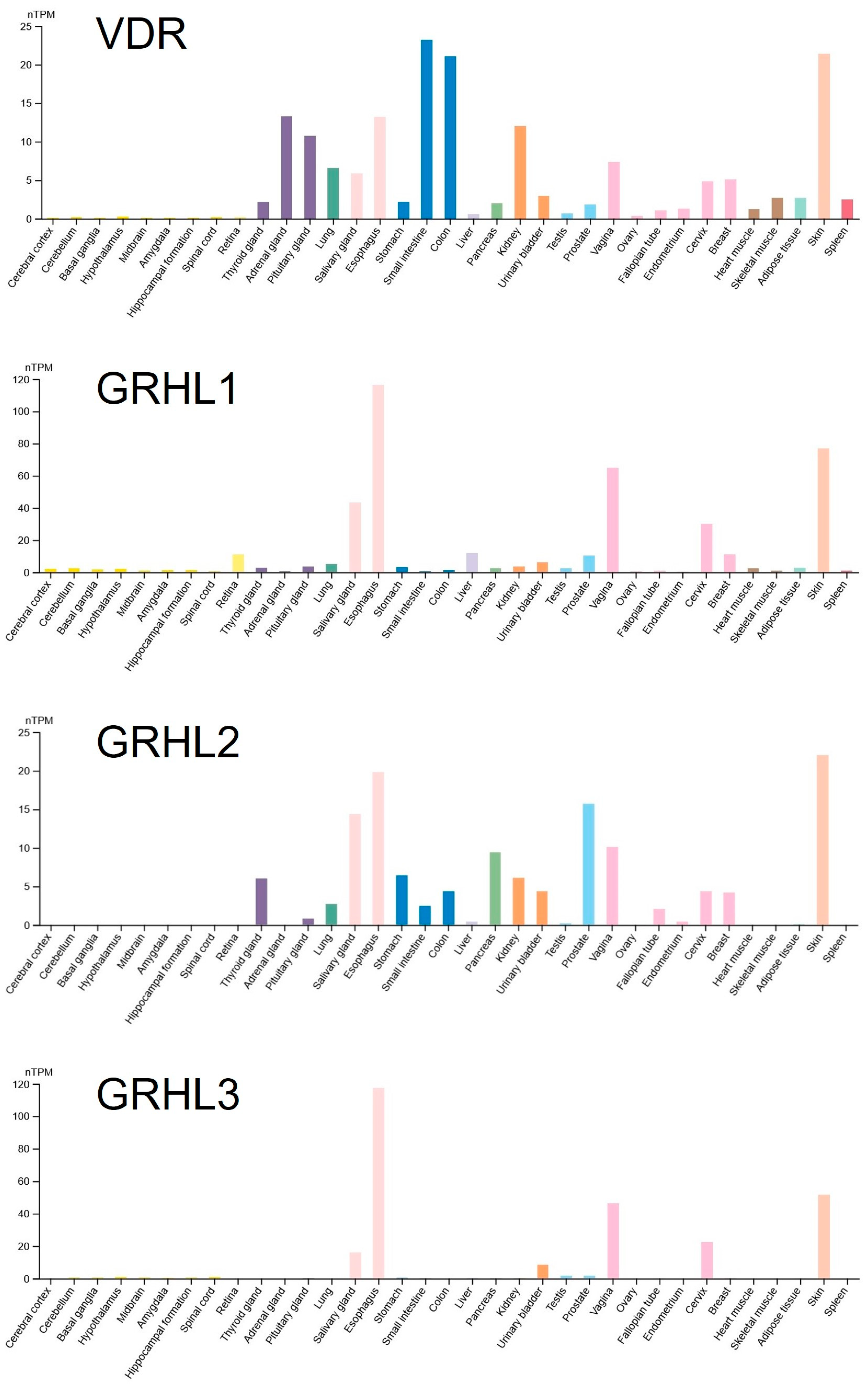
In parallel, we investigated in which organs VDR and the GRHL1–3 genes are co-expressed. We discovered that some of their highest expression levels are found in the skin (Figure 2). For this reason, we selected for our experimental analyses the HaCaT keratinocyte cell line derived from human adult skin [18].

2.2. VDR and Calcitriol Regulate the Expression of the GRHL1 Gene

In order to check whether calcitriol influences the expression of GRHL1–3 genes, HaCaT cells were transiently transfected with VDR-expressing plasmid and a control empty vector under different calcitriol treatment durations. However, in these preliminary experiments, we did not subject the cells to serum starvation, and none of the observed differences was statistically significant (Supplementary Figure S1).
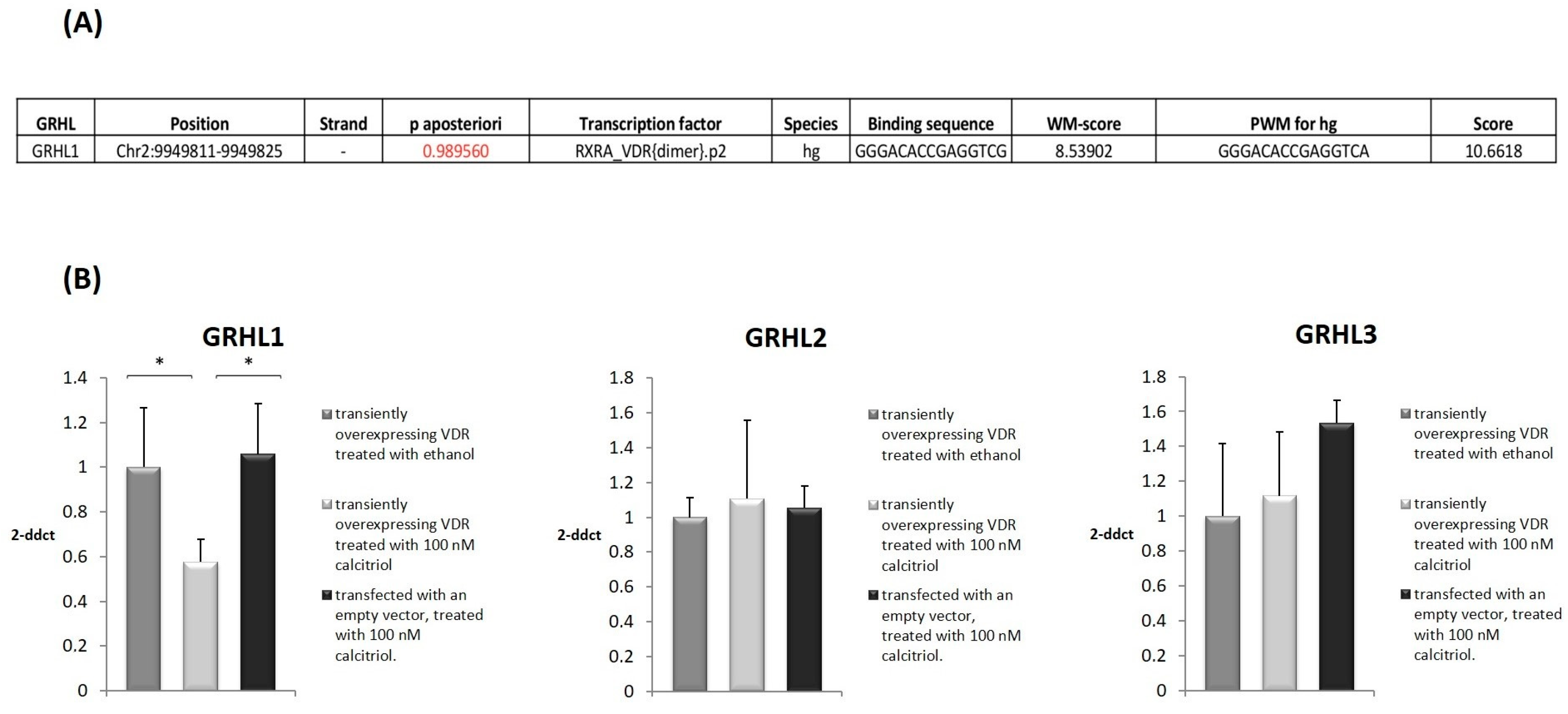
In the subsequent experiment, prior to the 2 h calcitriol treatment, cells were exposed to serum starvation in serum-free DMEM overnight. Three experimental protocols were employed: (i) VDR-overexpressing cells treated with calcitriol, (ii) VDR-overexpressing cells treated with ethanol as negative control, and (iii) control vector-transfected cells treated with calcitriol. Next, using quantitative real-time PCR (qRT-PCR), we quantified the levels of expression of GRHL1–3 genes. In VDR-overexpressing HaCaT cells treated with calcitriol, the expression of the GRHL1 gene was downregulated by 42% when compared to the respective control cells (Figure 3B, p < 0.05), i.e., VDR-overexpressing cells treated with alcohol and control vector-transfected cells treated with calcitriol. We did not observe any statistically significant changes in the expression of GRHL2 or GRHL3 (Figure 3B). For this reason, in subsequent experiments, we focused exclusively on examining the possible regulation of expression of the GRHL1 gene by the VDR transcription factor.

2.3. VDR Binds to a Regulatory Region of the GRHL1 Gene

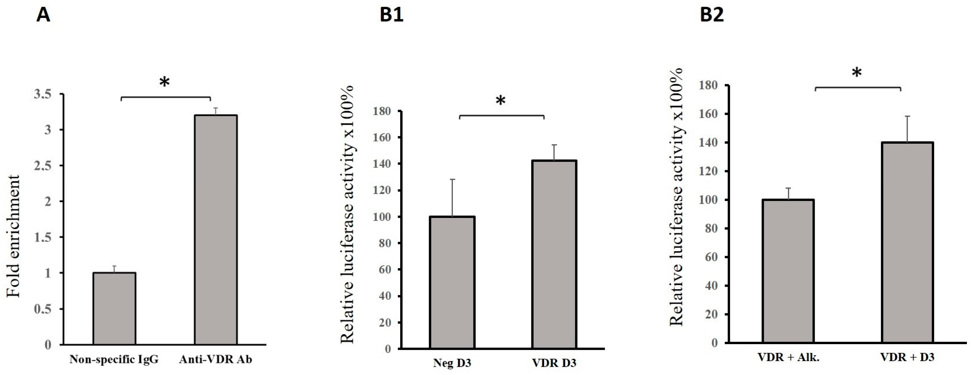
Only one potential VDR binding site in the regulatory region of the GRHL1 gene was identified using the MotEvo algorithm (Figure 3A). To determine whether this site is bound by VDR in living cells, ChIP analyses were carried out. Chromatin from HaCaT cells transfected with VDR-expressing vector was immunoprecipitated with 12.5 μg of anti-DYK (FLAG) antibody (ab1162, Abcam, Cambridge, UK) or 1 μg of normal rabbit IgG as negative control (10500C, Thermo Fisher Scientific, Waltham, MA, USA) and then examined by qRT-PCR using the primers listed in Table 1. As a result, we found that the investigated DNA fragments were significantly enriched in a pool precipitated with anti-DYK antibody in comparison with nonspecific IgG, indicating VDR binding to this region of the GRHL1 promoter (Figure 4A).
We performed co-transfection studies using a fragment of the regulatory region of the GRHL1 gene containing the putative VDR binding site fused to the reporter gene–firefly luciferase. We transiently transfected HaCaT cells with reporter constructs containing the Luc gene under the control of a DNA fragment with the VDR binding site from the GRHL1 promoter and (i) VDR-overexpressing vector or (ii) a control empty vector. The cells were then treated with calcitriol for 2 h. The results obtained indicate that in cells overexpressing VDR, the expression of the Luc gene fused to the GRHL1 promoter fragment was increased (Figure 4B1). In another experiment, the HaCaT cells were simultaneously transfected with the same reporter construct and a VDR-overexpressing vector and treated with either calcitriol or ethanol vehicle as negative control. The expression of the Luc gene fused to the GRHL1 promoter fragment was increased in cells subjected to calcitriol treatment (Figure 4B2).

3. Discussion

Before the present study, there were no reports in the literature linking vitamin D signaling to the regulation of expression of the GRHL1 gene. The novelty of our results lies in the fact that they provide the first experimental evidence of such links, and they indicate a molecular mechanism explaining these links: that the VDR transcription factor binds to the promoter of the GRHL1 gene and regulates its expression.
VDR and GRHL1 are both transcription factors involved in the regulation of gene expression, where they play essential roles in a multitude of well-conserved cellular processes. The expression of the GRHL1 gene is predominantly observed in various types of epithelial tissues, with the GRHL1 transcription factor being essential for structure and regeneration of various epithelia [15,16,17]. Similarly, VDR, expressed in epithelial tissues, plays a key role in the regulation of various physiological epithelial processes [19]. It has been associated with stimulation of differentiation, including permeability barrier formation, by regulating the expression levels of the tight-junction proteins claudin 2, 5, 12, and 15, strengthening the epithelial barrier function [20,21]. GRHL1 is also necessary for the functioning of skin barrier, as it regulates the expression of desmosomal cadherin desmoglein 1 (DSG1) and transglutaminase 5 (TGM5) in suprabasal layers of the epidermis and has been associated with disruption of the epidermal barrier [16,17]. These findings indicate that VDR and GRHL1 perform very similar roles in the context of skin barrier function. Despite that, there are no reports in the literature linking the functioning of VDR and GRHL1 in this or any other context. The regulation of expression of the GRHL1 gene by the VDR transcription factor, which we describe here, may thus be relevant in the context of skin barrier function.
The interaction between these transcription factors can be crucial for maintaining epithelial integrity. The influence of VDR on gene expression profiles related to cellular junctions and barrier function can complement the role of GRHL1 in regulating genes crucial for epithelial repair and structural integrity. For example, VDR-mediated regulation of proteins such as claudin 2 and 12 enhances epithelial barrier functions, which could synergistically interact with GRHL1’s regulation of DSG1, crucial for intercellular adhesion. This presents a novel hypothesis for functional interrogation for future analyses with co-immunoprecipitation and ChIP techniques from primary mouse skin during both embryogenesis and in adults to determine potential co-regulation of these target genes by the VDR–GRHL1 pathway.
VDR is necessary to maintain hair follicle homeostasis, and its deficiency leads to alopecia, as has been shown in mouse and rat models [22,23,24]. Similarly, Grhl1-null mice exhibit delayed hair growth and poor hair shaft anchoring [17], which alongside the key role of DSG1 may also co-involve pathways such as Wnt/β-catenin and SHH signaling, which are crucial for hair follicle development and cycling [25,26]. Therefore, hair follicles provide yet another context in which VDR and GRHL1 perform very similar functions. The novelty of our report is that it proposes a hitherto unknown molecular mechanism of regulation of expression of the GRHL1 gene by the VDR transcription factor, which may be relevant in the context of the functioning of hair follicles.
Both VDR and GRHL1 have been studied in the context of skin cancers. GRHL1 acts as a tumor suppressor in non-melanoma skin cancer (NMSC). Grhl1-null mice exposed to chemically induced skin carcinogenesis develop more squamous cell carcinomas with an earlier onset than their control wild-type littermates [27]. This phenotype is associated with aberrant terminal differentiation of keratinocytes and mild chronic skin inflammation in Grhl1−/− mice [27]. In human NMSC, the expression of GRHL1 is reduced, as it is negatively regulated by the oncogenic microRNA miR-21 [28]. The relationship between VDR and NMSC is more complex and not yet fully understood. Extensive evidence supports its tumor-suppressive role in this context [29]; however, the role of vitamin D in NMSC in human patients remains controversial [30,31]. Due to the contradictory evidence regarding the involvement of VDR in NMSC, it is difficult to speculate whether the regulation of expression of the GRHL1 gene by the VDR transcription factor is relevant in this context. Nevertheless, it is possible that our findings provide a novel, hitherto unknown molecular mechanism through which VDR may act in NMSC.
The regulatory complexity of VDR and GRHL1 involves multiple layers of control, including post-translational modifications and interactions with other transcription factors and co-factors, and some studies suggest a potential synergistic or parallel regulation by VDR and GRHL1 in these processes [32]. The interactions between VDR and its co-regulators can significantly influence their transcriptional activity and the cellular response to environmental and physiological stimuli [33]. Furthermore, the modulation of VDR activity through phosphorylation by protein kinases has been noted to alter its binding efficiency and regulatory impact on target genes [34]. These interactions highlight the nuanced regulation of VDR activity, which could influence how GRHL1 functions are modulated in epithelial cells.
We are cognizant that the direction of changes in the expression of GRHL1 and the reporter luciferase gene is not consistent: Figure 3B indicates that VDR and calcitriol negatively regulate GRHL1 expression, while the results presented in Figure 4B suggest the opposite. We have discussed and explained such contradictions in our earlier publication [35]. Briefly, overexpression of a transcriptional transactivator can sometimes repress the transcription of its target genes: in fact, this mechanism has previously been described for the GRHL family, where GRHL3 has been shown to both induce [36] and repress [37] the expression of direct target gene E-cadherin. Also, simple rearrangements of TFBSs, such as those caused by subcloning of a promoter fragment into a reporter plasmid, can trigger qualitatively different responses to a single transcription factor. None of these contradicts our conclusion that VDR regulates the expression of the GRHL1 gene.
The contradictions observed in the regulation of GRHL1 by VDR, as indicated in the results of current and previous publications, suggest complex regulatory interactions that may depend heavily on the cellular context and experimental conditions [38]. These findings underline the need for further studies that employ physiologically relevant models and advanced genetic tools to dissect the intricate relationships between these transcription factors and their broader implications in epithelial biology and pathology.
Our analyses did not detect any influence of VDR on the expression of GRHL2 or GRHL3 in HaCaT cells, even though we identified binding sites for the VDR transcription factor in the regulatory regions of all three GRHL1–3 genes in the GTRD (Supplementary Table S1). A possible explanation may lie in the fact that these sites were identified in different cell sets and may not be functional in keratinocytes. This hypothesis is further supported when one considers the incredible functional heterogeneity of GRHL-dependent transcriptional mechanisms. Our previously published meta-analysis of GRHL1–3 target genes highlights that the GRHL-dependent transcriptome is extremely context-specific [39], dependent on cell type, tissue of origin, developmental stage, organism and whether or not the cells were transformed, and hence it stands to reason that the upstream signaling factors that regulate GRHL1–3 function are also tightly regulated with a similar spatiotemporal profile.
Future work will focus on exploring the interaction of VDR with all three members of the GRHL1–3 family outside of keratinocyte cells, particularly during early embryogenesis in mouse and zebrafish embryos, where both VDR and GRHL1–3 factors are known to function. In particular, we aim to investigate the relationship between grhl3 and the zebrafish orthologues of VDR (vdra and vdrb) in craniofacial development. Knockdown experiments in zebrafish revealed distinct roles for these paralogues: while loss of vdra has minimal effects on cartilage elements, loss of vdrb leads to reduced and malformed craniofacial cartilages. Simultaneous disruption of both genes results in more severe defects or complete loss of cartilage. Moreover, knockdown of vdrb leads to elevated expression of follistatin a (fsta), a bone morphogenetic protein (BMP) antagonist, in the adjacent pharyngeal endoderm [40]. As grhl3 is expressed within the pharyngeal endoderm during zebrafish craniofacial growth [41], this spatiotemporal overlap suggests linked functions in the formation and growth of craniofacial cartilage during the establishment of the lower jaw. Additionally, we have recently shown that the murine orthologue of Grhl2 regulates another BMP antagonist, namely, Noggin, during craniofacial and neural tube tissue fusion [42], leading to a further hypothesis of potential conserved functional mechanistic overlap between VDR and GRHL1–3 signaling in embryogenesis.
Many proteins that interact with VDR are already known (Table 2). However, literally nothing is known about protein partners of GRHL1 and very little about protein partners of the closely related transcription factors GRHL2 and GRHL3 [43]. Therefore, here we identify another new avenue of research to pursue. It is worth exploring whether GRHL1 interacts with any of the proteins listed in Table 2, particularly those that VDR interacts with in organs in which GRHL1 plays important functions, such as the epidermis and the kidneys [17,27,28,44,45,46]. It is even tempting to speculate that VDR, GRHL1, and some of the proteins listed in Table 2 form functional multimeric complexes. This would add yet another level of regulation of activity of the GRHL1 transcription factor and place it firmly in the context of known signaling pathways.
Studying signaling pathways involving VDR will certainly be insufficient to account for all the effects of vitamin D. It is well established that the metabolism of vitamin D is very complex: many enzymes from the cytochrome P450 superfamily are involved and many derivatives of vitamin D are produced, which perform biological functions [59,60]. Some of these derivatives act through alternative receptors, such as the aryl hydrocarbon receptor (AhR), liver X receptors (LXRs) and retinoic acid receptor-related orphan receptors (RORs) [60,61]. In future research, all these will have to be taken into account, not only in investigations aimed at deciphering the relationship between vitamin D and GRHL1–3 transcription factors but also in the studies of other functions of vitamin D.
These future investigations may take advantage of invertebrate models, in addition to the vertebrate models discussed above, as vitamin D performs important functions in all plants, animals, and even fungi [62]. In particular, insect models, such as Drosophila melanogaster, are proving very useful in such research [62].

4. Materials and Methods

4.1. In Silico Prediction of VDR Binding Sites in the Regulatory Elements of the GRHL1 Gene

In our previous research, we identified regulatory regions within the GRHL1–3 genes using MotEvo, as outlined by Arnold et al. [35,63]. We then examined the predicted VDR binding sites within these regulatory regions by employing the Gene Transcription Regulation Database (GTRD) version 21.12, following standard protocols [64]. Our approach involved several techniques within the GTRD to pinpoint the VDR transcription factor binding sites in the GRHL1 locus. These included the use of meta clusters, Genome-Wide Event Finding and Motif Discovery (GEM), Probabilistic Inference for ChIP-Seq (PICS), Model-Based Analysis of ChIP-Seq (MACS2), and Site Identification from Short-Sequence Reads (SISSRs). After identifying these binding sites, we compared them to known regulatory sequences within the GRHL1 gene, utilizing data from the Ensembl database version 111 (https://www.ensembl.org/, last accessed 7 February 2024). This comprehensive approach allowed us to better understand the regulatory mechanisms affecting the GRHL1 gene, particularly in relation to VDR binding. We also utilized the UCSC Genome Browser (https://genome.ucsc.edu/, last accessed 7 February 2024) to confirm whether the identified VDR binding sites in the GRHL1 locus align with known regulatory regions, characterized by markers of open chromatin and other transcription factor binding sites.
For every GRHL1 region pinpointed by the meta-cluster analysis as a potential VDR binding site, we retrieved its genomic alignment with corresponding sequences from six mammalian species. These species, selected from Ensembl, include the chimpanzee (Pan troglodytes), mouse (Mus musculus), rat (Rattus norvegicus), polar bear (Ursus maritimus), dog (Canis lupus), and cow (Bos taurus). We conducted these alignments through the “Comparative genomics” feature in the Ensembl Genome Browser, focusing on the human genomic coordinates for these regions. The alignments were executed using the BLASTz/LASTz algorithms, with the results saved in the Fasta file format for further analysis. Multiple alignments were carried out with T-Coffee (ver. 11.0) [65] to determine if the VDR binding sites are conserved in other organisms. We used Pro-Coffee mode. The command line used to execute the alignment was: t_coffee -in=data_411e99a0.in -mode=procoffee -output=score_html clustalw_aln fasta_aln score_ascii phylip -maxnseq=150 -maxlen=10000 -case=upper -seqnos=off -outorder=input -run_name=result -multi_core=4 -quiet=stdout.

4.2. Cell Culture

Human immortalized keratinocyte HaCaT cells [18] were cultured in DMEM GlutaMAX medium supplemented with 10% fetal bovine serum and 100 IU/mL penicillin–streptomycin at 37 °C in a humidified incubator under 5% CO2. Cell culture reagents were purchased from Thermo Fisher Scientific.

4.3. Plasmids

The pcDNA3.1-K-DYK-VDR plasmid was purchased from GenScript and the control pcDNA3.1 plasmid was purchased from Invitrogen. Primers flanking a regulatory element of the GRHL1 gene including a VDR binding site were used to PCR amplify this element and clone it into the firefly luciferase vector with SV40 promoter (pGL3-promoter) (Promega, Madison, WA, USA) using In-Fusion® HD Cloning Kit (Takara Bio, Kusatsu, Shiga Prefecture, Japan), according to a protocol provided by the manufacturer. The construct was verified by sequencing. The control vector with the Renilla luciferase gene (pRL-CMV) was also purchased from Promega. Primers used for cloning are listed in Table 3.

4.4. Total RNA Extraction, Reverse Transcription, and qRT-PCR Assays

We grew HaCaT cells in 6-well plates with a well diameter of 34.8 mm. In each of the experimental procedures described in Section 4.4, Section 4.5 and Section 4.6, we followed the protocols supplied by the manufacturers. For cell transfections, we used Lipofectamine 3000 (Thermo Fisher Scientific). In each transfection, we used 1.0 μg of pcDNA3.1-K-DYK-VDR or pcDNA3.1-empty plasmids (negative control). After transfections, we cultured the cells for 24 h. Subsequently, we extracted and purified total RNA using the Total RNA Mini Plus Kit (A&A Biotechnology). We quantified the yield of RNA by measuring the absorbance at 260 nm, and we evaluated RNA purity according to the A260/A280 and A260/A230 ratio (NanoDrop ND-1000, Thermo Fisher Scientific). We then examined our samples by electrophoresis on 1.5% agarose, where the presence of sharp bands corresponding to 18S and 28S rRNA confirmed the integrity of total RNA. Subsequently we reverse-transcribed 1 μg of total RNA into first-strand cDNA with ReadyScript cDNA Synthesis Mix (Sigma-Aldrich). We carried out qRT-PCR in triplicate on a StepOnePlus Real Time PCR system (Applied Biosystems), using TaqMan Fast Universal PCR Master Mix No AmpErase UNG (Thermo Fisher Scientific) and TaqMan probes (Thermo Fisher Scientific). We normalized gene expression levels to the hypoxanthine phosphoribosyltransferase 1 housekeeping gene (HPRT1) (the highest stability based on the literature [66]) and calculated using the 2−ΔΔCt method [67]. In our experiments, we used the following TaqMan probes: Hs01119372_m1 for GRHL1, Hs00227745_m1 for GRHL2, Hs00297962_m1 for GRHL3, and Hs02800695_m1 for HPRT1. We determined statistical differences for relative expression levels using Student’s t-test. We considered p ≤ 0.05 to be statistically significant.

4.5. Chromatin Immunoprecipitation Assays (ChIP)

We cultured and transfected HaCaT cells as described in Section 4.4 (above). For transfections, we used 1.0 μg of pcDNA3.1-K-DYK-VDR plasmid. After transfections, we cultured the cells for 24 h. Subsequently, we cross-linked the cells for 10 min by adding formaldehyde to the final concentration of 1%. We sonicated the cross-linked material for 25 min (30 s on/30 s off) (Bioruptor, Diagenode) to generate ~500 bp DNA fragments. We isolated chromatin using an Imprint Chromatin Immunoprecipitation Kit (Sigma-Aldrich). We immunoprecipitated DNA fragments with 12.5 μg of anti-DYK (FLAG) antibody (ab1162, Abcam) and 1 μg of normal rabbit IgG as negative control (10500C, Thermo Fisher Scientific), and analyzed them in triplicate using qRT-PCR. We calculated fold changes related to 10% input delta Ct as 2−ΔΔCt [67]. Primers used for ChIP are listed in Table 1. We determined statistical differences for relative expression levels using Student’s t-test. We considered p ≤ 0.05 to be statistically significant.

4.6. Reporter Gene Assays

We cultured and transfected HaCaT cells as described in Section 4.4 (above). For transfections, we used 500 ng of pcDNA3.1-DYK-VDR or pcDNA3.1-empty plasmid, 25 ng pRL-CMV and 500 ng of the firefly luciferase vector with VDR binding site (described in Section 4.3—Plasmids). After transfections, we cultured the cells for 24 h. Subsequently, we lysed the cells using a lysis buffer provided in the Dual-Luciferase Reporter Assay System (Promega), and we measured the luciferase activity using materials supplied in this system and a Tecan Infinite M1000 PRO luminometer. We calculated and normalized relative reporter activity based on Renilla luciferase activity. We carried out all assays in triplicate twice. We performed statistical evaluations using Student’s t-test. We considered p ≤ 0.05 to be statistically significant.

4.7. Calcitriol Treatment

The cells were exposed to serum starvation in serum-free DMEM for 12 h and treated with calcitriol dissolved in ethanol (100 nM final culture concentration) (Sigma) or ethanol vehicle as negative control for 2 h prior to the experiments.

5. Conclusions

In this study, we identified a potential VDR binding site in the promoter region of the GRHL1 gene. We experimentally confirmed that VDR binds to this site in a human keratinocyte cell line. We demonstrated that VDR and calcitriol regulate the expression of the GRHL1 gene. Despite previously known numerous parallels between vitamin D signaling and signaling involving the GRHL1–3 factors, a direct connection between these two cellular pathways has not been reported before. Our findings indicate such a connection. Thus, we describe a hitherto unknown potential molecular mechanism through which vitamin D can act, as well as a novel mechanism of regulation of expression of the GRHL1 gene.

Supplementary Materials

The following supporting information can be downloaded at https://www.mdpi.com/article/10.3390/ijms25147913/s1.

Author Contributions

Conceptualization, A.T.-W., E.G.C.P., S.D. and T.W.; methodology, A.T.-W., M.M., L.L.-Z. and T.W.; validation, A.T.-W. and L.L.-Z.; formal analysis, A.T.-W., M.M. and L.L.-Z.; investigation, A.T.-W., M.M. and L.L.-Z.; resources, T.W.; writing—original draft preparation, A.T.-W., E.G.C.P. and L.L.-Z.; writing—review and editing, S.D. and T.W.; visualization, A.T.-W., M.M. and L.L.-Z.; supervision, S.D. and T.W.; project administration, T.W.; funding acquisition, T.W. All authors have read and agreed to the published version of the manuscript.

Funding

This research was funded by the National Science Centre (Poland), grant 2016/21/B/NZ1/00279.

Institutional Review Board Statement

Not applicable.

Informed Consent Statement

Not applicable.

Data Availability Statement

All data generated or analyzed during this study are included in the manuscript and/or the Supplementary Materials.

Conflicts of Interest

The authors declare no conflicts of interest.

References

  1. Pitt, M.J. Rickets and osteomalacia are still around. Radiol. Clin. N. Am. 1991, 29, 97–118. [Google Scholar] [CrossRef] [PubMed]
  2. Sahota, O. Osteoporosis and the role of vitamin D and calcium-vitamin D deficiency, vitamin D insufficiency and vitamin D sufficiency. Age Ageing 2000, 29, 301–304. [Google Scholar] [CrossRef] [PubMed]
  3. Wharton, B.; Bishop, N. Rickets. Lancet 2003, 362, 1389–1400. [Google Scholar] [CrossRef] [PubMed]
  4. Kechichian, E.; Ezzedine, K. Vitamin D and the Skin: An Update for Dermatologists. Am. J. Clin. Dermatol. 2018, 19, 223–235. [Google Scholar] [CrossRef] [PubMed]
  5. Bikle, D.D.; Chang, S.; Crumrine, D.; Elalieh, H.; Man, M.Q.; Choi, E.H.; Dardenne, O.; Xie, Z.; Arnaud, R.S.; Feingold, K.; et al. 25 Hydroxyvitamin D 1 α-Hydroxylase Is Required for Optimal Epidermal Differentiation and Permeability Barrier Homeostasis. J. Investig. Dermatol. 2004, 122, 984–992. [Google Scholar] [CrossRef] [PubMed]
  6. Nezbedova, P.; Brtko, J. 1alpha,25-dihydroxyvitamin D3 inducible transcription factor and its role in the vitamin D action. Endocr. Regul. 2004, 38, 29–38. [Google Scholar] [PubMed]
  7. Bikle, D.D.; Oda, Y.; Tu, C.L.; Jiang, Y. Novel mechanisms for the vitamin D receptor (VDR) in the skin and in skin cancer. J. Steroid Biochem. Mol. Biol. 2015, 148, 47–51. [Google Scholar] [CrossRef] [PubMed]
  8. Oda, Y.; Sihlbom, C.; Chalkley, R.J.; Huang, L.; Rachez, C.; Chang, C.P.; Burlingame, A.L.; Freedman, L.P.; Bikle, D.D. Two distinct coactivators, DRIP/mediator and SRC/p160, are differentially involved in vitamin D receptor transactivation during keratinocyte differentiation. Mol. Endocrinol. 2003, 17, 2329–2339. [Google Scholar] [CrossRef] [PubMed]
  9. Palmer, D.J. Vitamin D and the Development of Atopic Eczema. J. Clin. Med. 2015, 4, 1036–1050. [Google Scholar] [CrossRef]
  10. Lim, S.K.; Ha, J.M.; Lee, Y.H.; Lee, Y.; Seo, Y.J.; Kim, C.D.; Lee, J.H.; Im, M. Comparison of Vitamin D Levels in Patients with and without Acne: A Case-Control Study Combined with a Randomized Controlled Trial. PLoS ONE 2016, 11, e0161162. [Google Scholar] [CrossRef]
  11. Carlberg, C. Vitamin D and Its Target Genes. Nutrients 2022, 14, 1354. [Google Scholar] [CrossRef] [PubMed]
  12. Singh, P.K.; van den Berg, P.R.; Long, M.D.; Vreugdenhil, A.; Grieshober, L.; Ochs-Balcom, H.M.; Wang, J.; Delcambre, S.; Heikkinen, S.; Carlberg, C.; et al. Integration of VDR genome wide binding and GWAS genetic variation data reveals co-occurrence of VDR and NF-kappaB binding that is linked to immune phenotypes. BMC Genom. 2017, 18, 132. [Google Scholar] [CrossRef] [PubMed]
  13. von Essen, M.R.; Geisler, C. VDR, the Vitamin D Receptor. In Encyclopedia of Signaling Molecules; Choi, S., Ed.; Springer: New York, NY, USA, 2016; pp. 1–8. [Google Scholar]
  14. Dworkin, S.; Darido, C.; Georgy, S.R.; Wilanowski, T.; Srivastava, S.; Ellett, F.; Pase, L.; Han, Y.; Meng, A.; Heath, J.K.; et al. Midbrain-hindbrain boundary patterning and morphogenesis are regulated by diverse grainy head-like 2-dependent pathways. Development 2012, 139, 525–536. [Google Scholar] [CrossRef]
  15. Deng, Z.; Cangkrama, M.; Butt, T.; Jane, S.M.; Carpinelli, M.R. Grainyhead-like transcription factors: Guardians of the skin barrier. Vet. Dermatol. 2021, 32, 553-e152. [Google Scholar] [CrossRef] [PubMed]
  16. Cangkrama, M.; Darido, C.; Georgy, S.R.; Partridge, D.; Auden, A.; Srivastava, S.; Wilanowski, T.; Jane, S.M. Two Ancient Gene Families Are Critical for Maintenance of the Mammalian Skin Barrier in Postnatal Life. J. Investg. Dermatol. 2016, 136, 1438–1448. [Google Scholar] [CrossRef]
  17. Wilanowski, T.; Caddy, J.; Ting, S.B.; Hislop, N.R.; Cerruti, L.; Auden, A.; Zhao, L.L.; Asquith, S.; Ellis, S.; Sinclair, R.; et al. Perturbed desmosomal cadherin expression in grainy head-like 1-null mice. EMBO J. 2008, 27, 886–897. [Google Scholar] [CrossRef] [PubMed]
  18. Boukamp, P.; Petrussevska, R.T.; Breitkreutz, D.; Hornung, J.; Markham, A.; Fusenig, N.E. Normal keratinization in a spontaneously immortalized aneuploid human keratinocyte cell line. J. Cell Biol. 1988, 106, 761–771. [Google Scholar] [CrossRef] [PubMed]
  19. Bikle, D.D. Vitamin D and the skin: Physiology and pathophysiology. Rev. Endocr. Metab. Disord. 2012, 13, 3–19. [Google Scholar] [CrossRef] [PubMed]
  20. Oda, Y.; Uchida, Y.; Moradian, S.; Crumrine, D.; Elias, P.M.; Bikle, D.D. Vitamin D receptor and coactivators SRC2 and 3 regulate epidermis-specific sphingolipid production and permeability barrier formation. J. Investg. Dermatol. 2009, 129, 1367–1378. [Google Scholar] [CrossRef]
  21. Sun, J.; Zhang, Y.G. Vitamin D Receptor Influences Intestinal Barriers in Health and Disease. Cells 2022, 11, 1129. [Google Scholar] [CrossRef]
  22. Chen, C.H.; Sakai, Y.; Demay, M.B. Targeting expression of the human vitamin D receptor to the keratinocytes of vitamin D receptor null mice prevents alopecia. Endocrinology 2001, 142, 5386–5389. [Google Scholar] [CrossRef] [PubMed]
  23. Joko, Y.; Yamamoto, Y.; Kato, S.; Takemoto, T.; Abe, M.; Matsumoto, T.; Fukumoto, S.; Sawatsubashi, S. VDR is an essential regulator of hair follicle regression through the progression of cell death. Life Sci. Alliance 2023, 6, e202302014. [Google Scholar] [CrossRef] [PubMed]
  24. Kise, S.; Iijima, A.; Nagao, C.; Okada, T.; Mano, H.; Nishikawa, M.; Ikushiro, S.; Kanemoto, Y.; Kato, S.; Nakanishi, T.; et al. Functional analysis of vitamin D receptor (VDR) using adenovirus vector. J. Steroid Biochem. Mol. Biol. 2023, 230, 106275. [Google Scholar] [CrossRef] [PubMed]
  25. Rishikaysh, P.; Dev, K.; Diaz, D.; Qureshi, W.M.; Filip, S.; Mokry, J. Signaling involved in hair follicle morphogenesis and development. Int. J. Mol. Sci. 2014, 15, 1647–1670. [Google Scholar] [CrossRef] [PubMed]
  26. Narhi, K.; Jarvinen, E.; Birchmeier, W.; Taketo, M.M.; Mikkola, M.L.; Thesleff, I. Sustained epithelial beta-catenin activity induces precocious hair development but disrupts hair follicle down-growth and hair shaft formation. Development 2008, 135, 1019–1028. [Google Scholar] [CrossRef] [PubMed]
  27. Mlacki, M.; Darido, C.; Jane, S.M.; Wilanowski, T. Loss of Grainy head-like 1 is associated with disruption of the epidermal barrier and squamous cell carcinoma of the skin. PLoS ONE 2014, 9, e89247. [Google Scholar] [CrossRef] [PubMed]
  28. Kikulska, A.; Rausch, T.; Krzywinska, E.; Pawlak, M.; Wilczynski, B.; Benes, V.; Rutkowski, P.; Wilanowski, T. Coordinated expression and genetic polymorphisms in Grainyhead-like genes in human non-melanoma skin cancers. BMC Cancer 2018, 18, 23. [Google Scholar] [CrossRef] [PubMed]
  29. Reichrath, J.; Saternus, R.; Vogt, T. Endocrine actions of vitamin D in skin: Relevance for photocarcinogenesis of non-melanoma skin cancer, and beyond. Mol. Cell. Endocrinol. 2017, 453, 96–102. [Google Scholar] [CrossRef] [PubMed]
  30. Seretis, K.; Bounas, N.; Sioka, C. The Association of Vitamin D with Non-Melanoma Skin Cancer Risk: An Umbrella Review of Systematic Reviews and Meta-Analyses. Medicina 2023, 59, 2130. [Google Scholar] [CrossRef]
  31. Caini, S.; Gnagnarella, P.; Stanganelli, I.; Bellerba, F.; Cocorocchio, E.; Queirolo, P.; Bendinelli, B.; Saieva, C.; Raimondi, S.; Gandini, S. Vitamin D and the Risk of Non-Melanoma Skin Cancer: A Systematic Literature Review and Meta-Analysis on Behalf of the Italian Melanoma Intergroup. Cancers 2021, 13, 4815. [Google Scholar] [CrossRef]
  32. Liu, M.; Freedman, L.P. Transcriptional synergism between the vitamin D3 receptor and other nonreceptor transcription factors. Mol. Endocrinol. 1994, 8, 1593–1604. [Google Scholar] [PubMed]
  33. Zenata, O.; Vrzal, R. Fine tuning of vitamin D receptor (VDR) activity by post-transcriptional and post-translational modifications. Oncotarget 2017, 8, 35390–35402. [Google Scholar] [CrossRef] [PubMed]
  34. Arriagada, G.; Paredes, R.; Olate, J.; van Wijnen, A.; Lian, J.B.; Stein, G.S.; Stein, J.L.; Onate, S.; Montecino, M. Phosphorylation at serine 208 of the 1α, 25-dihydroxy Vitamin D3 receptor modulates the interaction with transcriptional coactivators. J. Steroid Biochem. Mol. Biol. 2007, 103, 425–429. [Google Scholar] [CrossRef] [PubMed]
  35. Kotarba, G.; Taracha-Wisniewska, A.; Miller, M.; Dabrowski, M.; Wilanowski, T. Transcription factors Kruppel-like factor 4 and paired box 5 regulate the expression of the Grainyhead-like genes. PLoS ONE 2021, 16, e0257977. [Google Scholar] [CrossRef] [PubMed]
  36. Phatak, M.; Kulkarni, S.; Miles, L.B.; Anjum, N.; Dworkin, S.; Sonawane, M. Grhl3 promotes retention of epidermal cells under endocytic stress to maintain epidermal architecture in zebrafish. PLoS Genet. 2021, 17, e1009823. [Google Scholar] [CrossRef] [PubMed]
  37. Zhao, P.; Guo, S.; Tu, Z.; Di, L.; Zha, X.; Zhou, H.; Zhang, X. Grhl3 induces human epithelial tumor cell migration and invasion via downregulation of E-cadherin. Acta Biochim. Biophys. Sin. 2016, 48, 266–274. [Google Scholar] [CrossRef] [PubMed]
  38. Dunlop, M.J.; Cox, R.S., III; Levine, J.H.; Murray, R.M.; Elowitz, M.B. Regulatory activity revealed by dynamic correlations in gene expression noise. Nat. Genet. 2008, 40, 1493–1498. [Google Scholar] [CrossRef] [PubMed]
  39. Mathiyalagan, N.; Miles, L.B.; Anderson, P.J.; Wilanowski, T.; Grills, B.L.; McDonald, S.J.; Keightley, M.C.; Charzynska, A.; Dabrowski, M.; Dworkin, S. Meta-Analysis of Grainyhead-Like Dependent Transcriptional Networks: A Roadmap for Identifying Novel Conserved Genetic Pathways. Genes 2019, 10, 876. [Google Scholar] [CrossRef] [PubMed]
  40. Kwon, H.J. Vitamin D Receptor Signaling Regulates Craniofacial Cartilage Development in Zebrafish. J. Dev. Biol. 2019, 7, 13. [Google Scholar] [CrossRef]
  41. Dworkin, S.; Simkin, J.; Darido, C.; Partridge, D.D.; Georgy, S.R.; Caddy, J.; Wilanowski, T.; Lieschke, G.J.; Doggett, K.; Heath, J.K.; et al. Grainyhead-like 3 regulation of endothelin-1 in the pharyngeal endoderm is critical for growth and development of the craniofacial skeleton. Mech. Dev. 2014, 133, 77–90. [Google Scholar] [CrossRef]
  42. de Vries, M.E.; Carpinelli, M.R.; Fuller, J.N.; Sutton, Y.; Partridge, D.D.; Auden, A.; Anderson, P.J.; Jane, S.M.; Dworkin, S. Grainyhead-like 2 interacts with noggin to regulate tissue fusion in mouse. Development 2024, 151, dev202420. [Google Scholar] [CrossRef] [PubMed]
  43. Gasperoni, J.G.; Fuller, J.N.; Darido, C.; Wilanowski, T.; Dworkin, S. Grainyhead-like (Grhl) Target Genes in Development and Cancer. Int. J. Mol. Sci. 2022, 23, 2735. [Google Scholar] [CrossRef] [PubMed]
  44. Pawlak, M.; Kikulska, A.; Wrzesinski, T.; Rausch, T.; Kwias, Z.; Wilczynski, B.; Benes, V.; Wesoly, J.; Wilanowski, T. Potential protective role of Grainyhead-like genes in the development of clear cell renal cell carcinoma. Mol. Carcinog. 2017, 56, 2414–2423. [Google Scholar] [CrossRef] [PubMed]
  45. Walkowska, A.; Pawlak, M.; Jane, S.M.; Kompanowska-Jezierska, E.; Wilanowski, T. Effects of high and low sodium diet on blood pressure and heart rate in mice lacking the functional grainyhead-like 1 gene. Physiol. Res. 2017, 66, 163–165. [Google Scholar] [CrossRef] [PubMed]
  46. Pawlak, M.; Walkowska, A.; Mlacki, M.; Pistolic, J.; Wrzesinski, T.; Benes, V.; Jane, S.M.; Wesoly, J.; Kompanowska-Jezierska, E.; Wilanowski, T. Consequences of the loss of the Grainyhead-like 1 gene for renal gene expression, regulation of blood pressure and heart rate in a mouse model. Acta Biochim. Pol. 2015, 62, 287–296. [Google Scholar] [CrossRef] [PubMed]
  47. Blanco, J.C.; Wang, I.M.; Tsai, S.Y.; Tsai, M.J.; O’Malley, B.W.; Jurutka, P.W.; Haussler, M.R.; Ozato, K. Transcription factor TFIIB and the vitamin D receptor cooperatively activate ligand-dependent transcription. Proc. Natl. Acad. Sci. USA 1995, 92, 1535–1539. [Google Scholar] [CrossRef]
  48. Herdick, M.; Bury, Y.; Quack, M.; Uskokovic, M.R.; Polly, P.; Carlberg, C. Response element and coactivator-mediated conformational change of the vitamin D(3) receptor permits sensitive interaction with agonists. Mol. Pharmacol. 2000, 57, 1206–1217. [Google Scholar] [PubMed]
  49. Tagami, T.; Lutz, W.H.; Kumar, R.; Jameson, J.L. The Interaction of the Vitamin D Receptor with Nuclear Receptor Corepressors and Coactivators. Biochem. Biophys. Res. Commun. 1998, 253, 358–363. [Google Scholar] [CrossRef]
  50. Takeshita, A.; Ozawa, Y.; Chin, W.W. Nuclear receptor coactivators facilitate vitamin D receptor homodimer action on direct repeat hormone response elements. Endocrinology 2000, 141, 1281–1284. [Google Scholar] [CrossRef]
  51. Takeyama, K.; Masuhiro, Y.; Fuse, H.; Endoh, H.; Murayama, A.; Kitanaka, S.; Suzawa, M.; Yanagisawa, J.; Kato, S. Selective interaction of vitamin D receptor with transcriptional coactivators by a vitamin D analog. Mol. Cell. Biol. 1999, 19, 1049–1055. [Google Scholar] [CrossRef]
  52. Pike, J.W.; Meyer, M.B. The vitamin D receptor: New paradigms for the regulation of gene expression by 1,25-dihydroxyvitamin D(3). Endocrinol. Metab. Clin. N. Am. 2010, 39, 255–269. [Google Scholar] [CrossRef] [PubMed]
  53. Polly, P.; Herdick, M.; Moehren, U.; Baniahmad, A.; Heinzel, T.; Carlberg, C. VDR-Alien: A novel, DNA-selective vitamin D(3) receptor-corepressor partnership. FASEB J. 2000, 14, 1455–1463. [Google Scholar] [CrossRef] [PubMed]
  54. Rachez, C.; Suldan, Z.; Ward, J.; Chang, C.P.; Burakov, D.; Erdjument-Bromage, H.; Tempst, P.; Freedman, L.P. A novel protein complex that interacts with the vitamin D3 receptor in a ligand-dependent manner and enhances VDR transactivation in a cell-free system. Genes Dev. 1998, 12, 1787–1800. [Google Scholar] [CrossRef] [PubMed]
  55. Warwick, T.; Schulz, M.H.; Günther, S.; Gilsbach, R.; Neme, A.; Carlberg, C.; Brandes, R.P.; Seuter, S. A hierarchical regulatory network analysis of the vitamin D induced transcriptome reveals novel regulators and complete VDR dependency in monocytes. Sci. Rep. 2021, 11, 6518. [Google Scholar] [CrossRef] [PubMed]
  56. Zhang, C.; Baudino, T.A.; Dowd, D.R.; Tokumaru, H.; Wang, W.; MacDonald, P.N. Ternary complexes and cooperative interplay between NCoA-62/Ski-interacting protein and steroid receptor coactivators in vitamin D receptor-mediated transcription. J. Biol. Chem. 2001, 276, 40614–40620. [Google Scholar] [CrossRef] [PubMed]
  57. Hsieh, J.C.; Whitfield, G.K.; Oza, A.K.; Dang, H.T.; Price, J.N.; Galligan, M.A.; Jurutka, P.W.; Thompson, P.D.; Haussler, C.A.; Haussler, M.R. Characterization of unique DNA-binding and transcriptional-activation functions in the carboxyl-terminal extension of the zinc finger region in the human vitamin D receptor. Biochemistry 1999, 38, 16347–16358. [Google Scholar] [CrossRef] [PubMed]
  58. Haussler, M.R.; Jurutka, P.W.; Mizwicki, M.; Norman, A.W. Vitamin D receptor (VDR)-mediated actions of 1alpha,25(OH)2vitamin D3: Genomic and non-genomic mechanisms. Best Pr. Res. Clin. Endocrinol. Metab. 2011, 25, 543–559. [Google Scholar] [CrossRef] [PubMed]
  59. Slominski, A.T.; Tuckey, R.C.; Jetten, A.M.; Holick, M.F. Recent Advances in Vitamin D Biology: Something New under the Sun. J. Investg. Dermatol. 2023, 143, 2340–2342. [Google Scholar] [CrossRef] [PubMed]
  60. Slominski, A.T.; Tuckey, R.C.; Jenkinson, C.; Li, W.; Jetten, A.M. Alternative pathways for vitamin D metabolism. In Feldman and Pike’s Vitamin D; Elsevier: Amsterdam, The Netherlands, 2024; pp. 85–109. [Google Scholar]
  61. Slominski, A.T.; Brozyna, A.A.; Zmijewski, M.A.; Janjetovic, Z.; Kim, T.K.; Slominski, R.M.; Tuckey, R.C.; Mason, R.S.; Jetten, A.M.; Guroji, P.; et al. The Role of Classical and Novel Forms of Vitamin D in the Pathogenesis and Progression of Nonmelanoma Skin Cancers. Adv. Exp. Med. Biol. 2020, 1268, 257–283. [Google Scholar]
  62. Kim, T.K.; Slominski, R.M.; Pyza, E.; Kleszczynski, K.; Tuckey, R.C.; Reiter, R.J.; Holick, M.F.; Slominski, A.T. Evolutionary formation of melatonin and vitamin D in early life forms: Insects take centre stage. Biol. Rev. 2024. [Google Scholar] [CrossRef]
  63. Arnold, P.; Erb, I.; Pachkov, M.; Molina, N.; van Nimwegen, E. MotEvo: Integrated Bayesian probabilistic methods for inferring regulatory sites and motifs on multiple alignments of DNA sequences. Bioinformatics 2011, 28, 487–494. [Google Scholar] [CrossRef] [PubMed]
  64. Kolmykov, S.; Yevshin, I.; Kulyashov, M.; Sharipov, R.; Kondrakhin, Y.; Makeev, V.J.; Kulakovskiy, I.V.; Kel, A.; Kolpakov, F. GTRD: An integrated view of transcription regulation. Nucleic Acids Res. 2021, 49, D104–D111. [Google Scholar] [CrossRef] [PubMed]
  65. Notredame, C.; Higgins, D.G.; Heringa, J. T-Coffee: A novel method for fast and accurate multiple sequence alignment. J. Mol. Biol. 2000, 302, 205–217. [Google Scholar] [CrossRef] [PubMed]
  66. Radonić, A.; Thulke, S.; Mackay, I.M.; Landt, O.; Siegert, W.; Nitsche, A. Guideline to reference gene selection for quantitative real-time PCR. Biochem. Biophys. Res. Commun. 2004, 313, 856–862. [Google Scholar] [CrossRef]
  67. Livak, K.J.; Schmittgen, T.D. Analysis of Relative Gene Expression Data Using Real-Time Quantitative PCR and the 2−ΔΔCT Method. Methods 2001, 25, 402–408. [Google Scholar] [CrossRef]
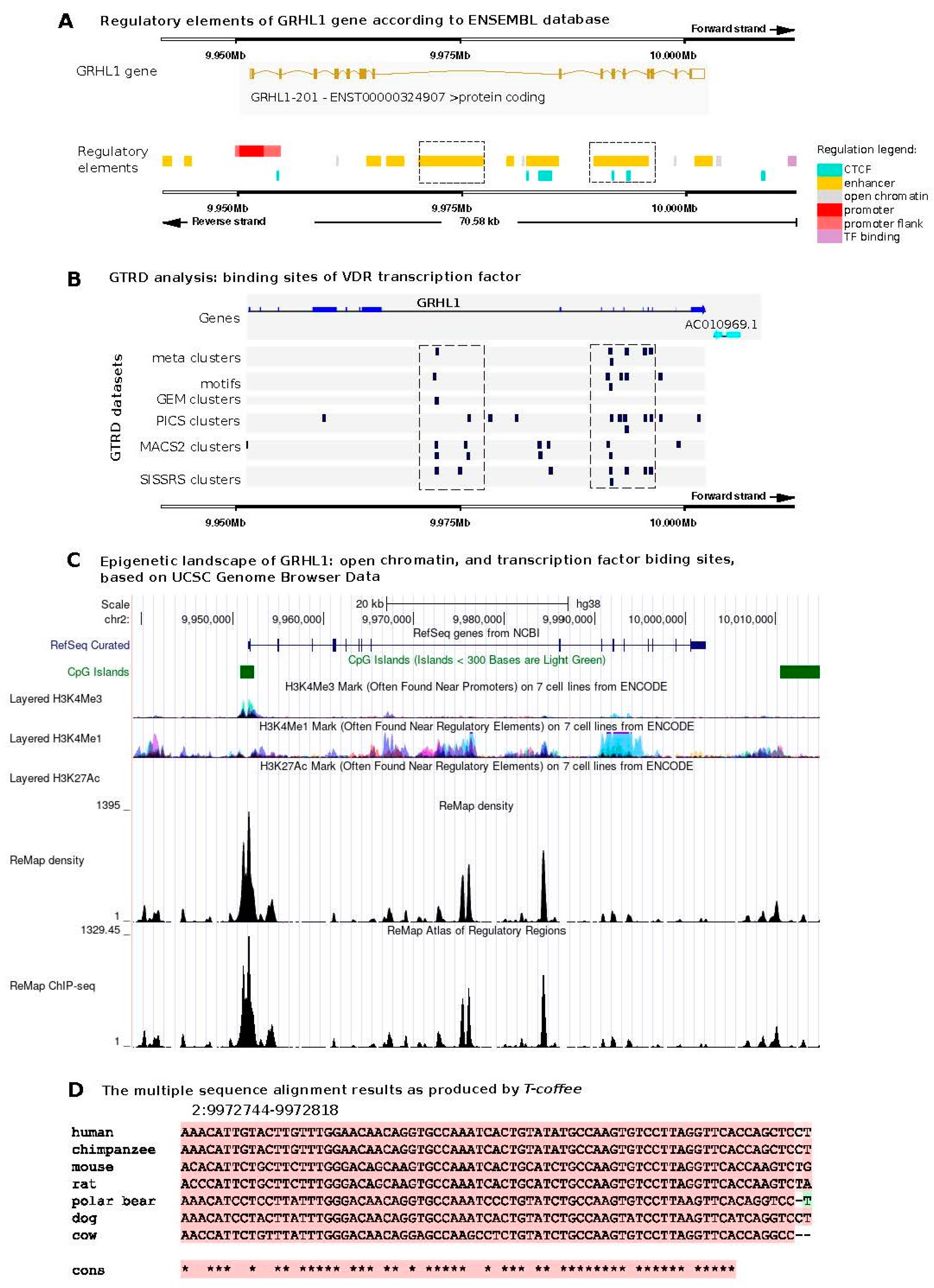
Figure 1. Bioinformatic predictions of transcription factor binding site (TFBS) motifs for VDR in the human GRHL1 gene. (A) Genomic location of regulatory regions of the GRHL1 gene according to the Ensembl database. (B) VDR binding sites identified in the GRHL1 gene using GTRD modules: meta-clusters, motifs, and GEM–PICS–MACS2–SISSRs clusters. (C) Genomic location of CpG Islands, markers of open chromatin (H3K4Me1, H3K4Me3, H3K27Ac), and transcription factor binding sites (ChIP-seq). (D) Multiple sequence alignment of a TFBS example across different species using T-Coffee (Pro-Coffee mode). Asterisks indicate conserved nucleotides.
Ijms 25 07913 g001
Figure 2. Baseline expression of VDR and GRHL1–3 genes in various tissues. Based on https://www.proteinatlas.org/, last accessed 11 June 2024. TPM, transcripts per million. Database version: Genotype-Tissue Expression (GTEx) RNA-seq data v8, available from the above portal.
Ijms 25 07913 g002
Figure 3. Overexpression of VDR alters mRNA level of GRHL1 gene. (A) Genomic coordinates of the binding site for VDR in the promoter region of the GRHL1 gene, obtained from the MotEvo database. (B) The mRNA expression levels of the GRHL1–3 genes in HaCaT cells (1) transiently overexpressing VDR treated with ethanol, (2) transiently overexpressing VDR treated with 100 nM calcitriol, or (3) transfected with an empty vector treated with 100 nM calcitriol. The results represent relative expression of the respective target gene vs. HPRT genes. Data are shown as means ± SEM of experiments independently performed in triplicate, * significantly different at p ≤ 0.05.
Ijms 25 07913 g003
Figure 4. (A) Quantitative ChIP-PCR analysis of VDR occupancy of the GRHL1 regulatory region was performed in HaCaT cells transfected with pcDNA3.1-K-DYK-VDR. Chromatin was immunoprecipitated with anti-DYK (FLAG) antibody or nonspecific antibody. The amount of DNA amplified from immunoprecipitated DNA was normalized to that amplified from input DNA. Data are shown as means ± SEM from experiments independently performed in triplicate, * significantly different at p≤ 0.05. (B1,B2) HaCaT cells were transfected with (B1) pcDNA3.1-K-DYK-VDR or pcDNA3.1-empty plasmid, 500 ng of the firefly luciferase vector with VDR binding site derived from the regulatory region of the GRHL1 gene, and 25 ng pRL-CMV Renilla luciferase control reporter vector and treated with 100 nM calcitriol or (B2) pcDNA3.1-K-DYK-VDR, 500 ng of the firefly luciferase vector with VDR binding site derived from the regulatory region of the GRHL1 gene, and 25 ng pRL-CMV Renilla luciferase control reporter vector and treated with 100 nM calcitriol or ethanol vehicle. Data are shown as means ± SEM of experiments independently performed in triplicate, * significantly different at p≤ 0.05.
Ijms 25 07913 g004
Table 1. List of ChIP qRT-PCR primers.
Binding SitePrimerSequence (5′→3′)
VDR in GRHL1 promoterForwardGGGCACAGAGGAGGGACT
ReverseGAGACAGAAGACGGGGACAC
Table 2. Vitamin D receptor-mediated transcription factor regulation across organ systems.
Transcription FactorOrganismTissue/Organ/Cell TypeDetails of InteractionSource
Liver
RXRHuman, mouse, ratLiverForms heterodimers with VDR to regulate gene transcription.[47]
TFIIBHuman, mouseLiverInteracts directly with VDR to facilitate transcription initiation.[47]
p300/CBPHuman, mouse, ratLiverActs as a histone acetyltransferase, enhancing transcription by relaxing DNA.[48]
HNF-4αHuman, mouse, rat LiverInteracts with VDR to regulate metabolic processes in liver cells.[48]
Kidney
RXRHuman, mouse, ratKidneyForms heterodimers with VDR to regulate gene transcription.[47]
TFIIBHuman, mouse KidneyInteracts directly with VDR to facilitate transcription initiation.[47]
p300/CBPHuman, mouse, ratKidneyActs as a histone acetyltransferase, enhancing transcription by relaxing DNA.[48]
HNF-4αHuman, mouse, ratKidneyInteracts with VDR to regulate metabolic processes in kidney cells.[48]
Intestine
RXRHuman, mouse, rat IntestineForms heterodimers with VDR to regulate gene transcription.[47]
TFIIBHuman, mouseIntestineInteracts directly with VDR to facilitate transcription initiation.[47]
p300/CBPHuman, rat, mouse IntestineActs as a histone acetyltransferase, enhancing transcription by relaxing DNA.[48]
HNF-4αHuman, mouse, ratIntestineInteracts with VDR to regulate metabolic processes in intestinal cells.[48]
Bone
RXRHuman, mouse, ratOsteoblasts, osteoclastsForms heterodimers with VDR to regulate gene transcription.[47]
NCoRHuman, mouse Osteoblasts, osteoclastsActs as a corepressor that interacts with VDR to suppress transcription.[49]
SMRTHuman, mouseOsteoblasts, osteoclastsAnother corepressor that interacts with VDR to regulate gene expression.[49]
SRC-1Human, mouseOsteoblasts, osteoclastsCoactivator that enhances VDR-mediated transcription.[50]
Muscle Cells
MyoDHuman, mouseMyoblastsInteracts with VDR to influence muscle cell differentiation and growth.[49]
Skin Cells
TIF2Human, mouseKeratinocytesCoactivator that interacts with VDR to enhance transcription in skin cells.[51]
Nervous System
NF-1Human, mouseNeurons, glial cellsInteracts with VDR to regulate genes involved in nervous system development and function.[32]
FOXO3Human, mouseNeurons, glial cellsInteracts with VDR to regulate genes involved in stress resistance, metabolism, and neuronal function.[32]
Cancer Cells
c-MycHuman, mouse Various cancer cells (colon, breast, prostate)Oncogene that interacts with VDR, influencing cell proliferation and cancer progression.[32]
HOXB13Human, mouse Prostate cancer cellsInteracts with VDR in prostate cancer cells to regulate gene expression related to cancer progression.[52]
Immune System
NF-κBHuman, mouse, ratImmune cells (T cells, B cells)Modulates immune responses and inflammation through interaction with VDR.[32]
AP-1Human, mouse, rat Immune cells (T cells, B cells)Regulates gene expression in response to a variety of stimuli, interacting with VDR to modulate immune functions.[32]
AlienHuman, mouseImmune cells (T cells, B cells)Acts as a corepressor that interacts with VDR to modulate transcriptional repression.[53]
BCL6Human, mouse B cells, T cellsRegulates differentiation and function of T cells and B cells, interacting with VDR.[32]
STAT3Human, mouseVarious immune cells (T cells, NK cells, macrophages)Involved in the signaling pathways that mediate immune responses and interacts with VDR.[32]
GATA3Human, mouseT cells, Th2 cellsInteracts with VDR to regulate immune responses, particularly in T-helper 2 (Th2) cells.[32]
NFATHuman, mouseImmune cells (T cells, B cells)Interacts with VDR to modulate the expression of immune-related genes.[32]
IRF1Human, mouseImmune cells (T cells, B cells)Interacts with VDR to regulate the transcription of interferon-responsive genes.[32]
c-FosHuman, mouse Immune cells (T cells, B cells)Component of the AP-1 transcription factor, interacts with VDR to modulate immune responses.[32]
Development
Oct4Human, mouseEmbryonic stem cellsInteracts with VDR to regulate gene expression during development.[54]
Wnt Signaling
TCF/LEFHuman, mouse, ratVarious cells (liver, kidney, intestine, bone)Mediates gene expression changes in response to Wnt signaling, interacts with VDR.[48]
TCF7L2HumanVarious tissues (liver, kidney, intestine, bone)Interacts with VDR in the Wnt signaling pathway to regulate gene expression.[55]
Transcription Regulation
GRIP1Human, mouseVarious cells (liver, kidney, intestine, bone)Coactivator that interacts with VDR to enhance transcriptional activation.[54]
RAC3Human, mouseVarious cells (liver, kidney, intestine, bone)Coactivator that works with VDR to enhance transcription.[54]
SKIPHuman, mouse Various cells (liver, kidney, intestine, bone)Involved in the assembly of the transcriptional complex with VDR.[56]
VDRE Binding ProteinsHumanVarious cells (liver, kidney, intestine, bone)Proteins that bind to Vitamin D Response Elements (VDREs) to regulate gene expression in coordination with VDR.[57]
General
TRAM-1Human, mouse, ratVarious tissues (liver, kidney, intestine, bone)Coactivator that enhances VDR-mediated transcription.[50]
TIF1Human, mouseVarious tissues (liver, kidney, intestine, bone)Interacts with VDR to modulate transcription.[51]
TRAP220Human, mouse, ratVarious tissues (liver, kidney, intestine, bone)Part of the mediator complex, linking transcriptional regulators to the RNA polymerase II initiation complex.[54]
SUG1Human, mouseVarious tissues (liver, kidney, intestine, bone)Component of the 26S proteasome, involved in non-proteolytic roles like nuclear receptor-mediated transcription.[54]
BAF60aHuman, mouseVarious tissues (liver, kidney, intestine, bone)Part of the SWI/SNF chromatin remodeling complex, facilitates transcriptional activation with VDR.[54]
NCoA1Human, mouseVarious tissues (liver, kidney, intestine, bone)Enhances the transcriptional activities of steroid hormone receptors like VDR.[51]
c-JunHuman, mouse, ratVarious tissues (liver, kidney, intestine, bone)Component of the AP-1 transcription factor, interacts with VDR to respond to stress signals and inflammation.[32]
CREBHuman, mouseVarious tissues (liver, kidney, intestine, bone)Interacts with VDR to mediate cAMP response element-binding protein transcriptional activities.[48]
COUP-TFHuman, mouseVarious tissues (liver, kidney, intestine, bone)Interacts with VDR to regulate gene expression across various tissues.[52]
USF1Human, mouseVarious tissues (liver, kidney, intestine, bone)Interacts with VDR to regulate gene expression across various tissues.[52]
GFI1Human, mouseVarious tissues (liver, kidney, intestine, bone)Interacts with VDR to regulate gene expression across various tissues.[52]
SUG2Human, mouseVarious tissues (liver, kidney, intestine, bone)Component of the 26S proteasome, involved in nuclear receptor-mediated transcription with VDR.[54]
VDR-AP (alternative pocket)Human, mouseVarious tissues (liver, kidney, intestine, bone)Represents a different binding conformation within VDR that might interact uniquely with ligands or coregulators.[58]
Table 3. List of oligonucleotides used for cloning.
DirectionSequences of Oligonucleotides (5′→3′)
ForwardactGAGCTCcaaacaacaggctgcatgga
ReverseactgCTCGAGcgagagtctgttttggacgt
Disclaimer/Publisher’s Note: The statements, opinions and data contained in all publications are solely those of the individual author(s) and contributor(s) and not of MDPI and/or the editor(s). MDPI and/or the editor(s) disclaim responsibility for any injury to people or property resulting from any ideas, methods, instructions or products referred to in the content.