Article

Transcriptome-Wide Identification of m6A Writers, Erasers and Readers and Their Expression Profiles under Various Biotic and Abiotic Stresses in Pinus massoniana Lamb.

1
State Key Laboratory of Tree Genetics and Breeding, Nanjing Forestry University, Nanjing 210037, China
2
Key Open Laboratory of Forest Genetics and Gene Engineering of National Forestry & Grassland Administration, Nanjing 210037, China
3
Co-Innovation Center for Sustainable Forestry in Southern China, Nanjing Forestry University, Nanjing 210037, China
4
Beijing National Laboratory for Molecular Sciences, Peking University, Beijing 100871, China
*
Authors to whom correspondence should be addressed.
Int. J. Mol. Sci. 2024, 25(14), 7987; https://doi.org/10.3390/ijms25147987 (registering DOI)
Submission received: 29 June 2024 / Revised: 17 July 2024 / Accepted: 18 July 2024 / Published: 22 July 2024
(This article belongs to the Section Molecular Plant Sciences)

Abstract

:
N6-methyladenosine (m6A) RNA modification is the most prevalent form of RNA methylation and plays a crucial role in plant development. However, our understanding of m6A modification in Masson pine (Pinus massoniana Lamb.) remains limited. In this study, a complete analysis of m6A writers, erasers, and readers in Masson pine was performed, and 22 m6A regulatory genes were identified in total, including 7 m6A writers, 7 m6A erases, and 8 readers. Phylogenetic analysis revealed that all m6A regulators involved in Masson pine could be classified into three distinct groups based on their domains and motifs. The tissue expression analysis revealed that the m6A regulatory gene may exert a significant influence on the development of reproductive organs and leaves in Masson pine. Moreover, the results from stress and hormone expression analysis indicated that the m6A regulatory gene in Masson pine might be involved in drought stress response, ABA-signaling-pathway activation, as well as resistance to Monochamus alternatus. This study provided valuable and anticipated insights into the regulatory genes of m6A modification and their potential epigenetic regulatory mechanisms in Masson pine.

1. Introduction

Over 160 chemical modifications have been identified in RNA, predominantly within transfer RNA (tRNA) and ribosomal RNA (rRNA), where they play pivotal roles in governing RNA functionality [1]. Messenger RNA (mRNA), acting as an intermediary between DNA and proteins, also undergoes diverse chemical modifications, including N7-methylguanosine (m7G), N6-methyladenosine (m6A), 5-methylcytosine (m5C), N1-methyladenosine (m1A), pseudouridine (ψ), inosine (I), and N6, 2′-O-dimethyladenosine (m6Am), to fulfill its biological functions [2]. Among these modifications, m6A is the most prevalent internal modification observed in eukaryotic mRNA and is also present across various bacteria and RNA viruses [3]. M6A is a dynamic and reversible mechanism that undergoes regulation by diverse proteins, encompassing writers (methyltransferases), erasers (demethylases), and readers (m6A-binding proteins), which act synergistically to regulate the abundance of m6A [4,5,6]. Writers and erasers can bind the conserved consensus sequence RRACH (R=A or G; H=A, U, or C) to add and remove m6A modifications, and these modified RNAs eventually perform various functions by binding readers to the m6A sites [7,8,9]. The previous focus of m6A studies primarily revolved around animals. In recent years, with the rapid advancement of plant research, an increasing number of reports on m6A in plants have emerged.
In plants, m6A writers include MTA (homolog of human METTL3), MTB (homolog of human METTL14), FIP37 (homolog of human WTAP), VIRILIZER (VIR) (homolog of human VIRMA), and HAKAI [10,11,12,13,14]. By inhibiting the expression or knocking out of these genes, there is a resultant decrease in m6A levels, which subsequently affects the plant’s normal development and response to adversity. It is worth noting that disruption of MTA, FIP37 or VIR function leads to an approximate 80–90% reduction in m6A methylation levels [15,16,17,18]. However, it does not completely abolish m6A methylation. These findings suggest the involvement of unidentified writers in Arabidopsis thaliana for mRNA m6A methylation. The identification of m6A erasers have revealed the reversible nature of m6A modification and facilitated the advancement of functional studies on m6A. FTO [19] and ALKBH5, proteins involved in alkylated DNA repair [20], are recognized as mammalian m6A demethylases belonging to the Fe(II)/a-kg-dependent dioxygenase superfamily. However, no homologs of FTO have been identified in plants [21]. A. thaliana possesses 13 homologous proteins related to ALKBH, with 5 proteins (ALKBH9A/9B/9C/10A/10B) exhibiting similarities to ALKBH5 [22]. The demethylase activity of ALKBH9B and ALKBH10B has been confirmed in A. thaliana. However, LC-MS analysis does not detect any significant alteration in m6A content in ALKBH9B mutants [23]. Notably, while both FTO and ALKBH5 are localized within the nucleus, ALKBH9B and SlALKBH2 are cytoplasmic proteins, and ALKBH10B exhibits nucleo-cytoplasmic distribution. These findings suggest that plants may possess different types of erasers with distinct functions [22,24,25]. The functionality of the m6A is primarily dependent on m6A readers. The investigation of m6A readers in plants is currently limited and primarily focused on YTH proteins. In Arabidopsis, there are a total of 13 proteins containing the YTH domain, predominantly from the evolutionarily conserved C-terminal region (ECT) family [26]. The YTH domain sequences in Arabidopsis and animals exhibit high conservation, with continuous tryptophan residues serving as crucial sites for m6A binding. Therefore, these YTH-containing proteins in Arabidopsis possess the potential to bind m6A [27,28]. ECT2 regulates the development of Arabidopsis epidermis by binding to m6A and regulating the stability of substrate mRNA [29,30]. Further experiments have demonstrated that the function of ECT2 is redundant in comparison to that of ECT3 and ECT4, suggesting that ECT3 and ECT4 are also m6A readers [31]. CPSF30-L is an m6A reader, homologous to YTHDC1, localized in the nucleus, and involved in selective polyadenylation (APA) regulation [32]. Additionally, recent studies have revealed an enhanced affinity of ECT8 towards m6A under conditions of salt stress [33].
Masson pine, an economically valuable industrial tree in China, plays a crucial role in enhancing its economic benefits. Therefore, it is essential to study the molecular mechanisms underlying the growth, development, and stress response of Masson pine. Additionally, investigating the physiological and molecular characteristics of Masson pine as a conifer species holds significant biological value due to its distinct differences from broad-leaved trees. Although previous studies have demonstrated the close association between m6A modification and plant growth, development, and stress response [34], investigations on m6A in conifers such as Masson pine are lacking, and the identification of m6A-related proteins remains elusive. In this study, we utilized the transcriptome of Masson pine to identify regulators of m6A and comprehensively analyzed their physicochemical properties, phylogenetic relationships, and tissue expression patterns in response to hormonal and stress stimuli to predict the molecular function of these m6A regulators. This investigation provides a pivotal theoretical foundation for further elucidating the role and molecular mechanism of genes associated with the m6A regulatory pathway in coniferous species.

2. Results

2.1. Identification and Characterization Analysis of m6A Pathway Genes in Masson Pine

We have identified a total of seven writers, seven erasers, and eight reader proteins (a total of 22) in Masson pine. The length of transcripts, coding DNA sequence (CDS), polypeptide length, molecular weight, isoelectric point, and subcellular localization of each member of the newly identified writer, eraser, and reader families were further analyzed (Table S1). The CDS lengths of the writers range from 840 bp to 5649 bp. Therefore, PmVIR, with its polypeptide length of 1882 aa and molecular weight of 206.3 kDa, is the largest member of the writing family, whereas PmMTC, with its polypeptide length of 279 aa and molecular weight of 51.1 kDa, is the smallest member. Writers in the rice family had pI values ranging from 5.40 (PmVIR) to 7.10 (PmHAKAI2). Six of the seven writers had an acidic pI (below 7.0), whereas the PmHAKAI2 was alkaline. Six of the seven writers were predicted to be nuclear-localized, suggesting that they contribute to the methylation of the transcriptome. Seven erasers were found to have CDS lengths ranging from 807 bp to 2001 bp, respectively. PmALKBH7 is 666 aa in length and has a molecular weight of 72.7 kDa, making it the longest and largest eraser protein. PmALKBH6 is the smallest eraser protein, with a molecular weight of 30.0 kDa. The pI values for PmALKBH1, PmALKBH2, PmALKBH5 and PmALKBH6 erasers are acidic, and the rest of the erasers are basic. Similarly, a wide range in CDS length, from 1872 bp to 2376 bp, was observed among eight reader genes. Accordingly, PmYTHDF5 is the shortest reader protein (623 aa long, with a weight of 69.5 kDa), whereas PmYTHDF2 is the longest (791 aa and 86.9 kDa). The subcellular localization of all m6A readers is predicted to be in the nucleus (Table S1).

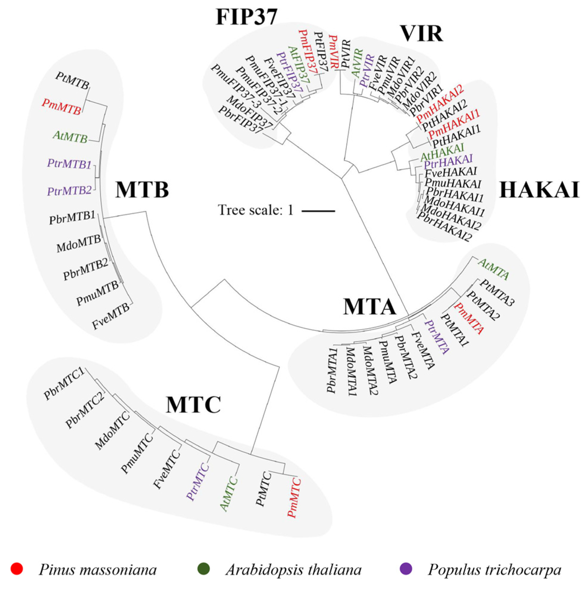
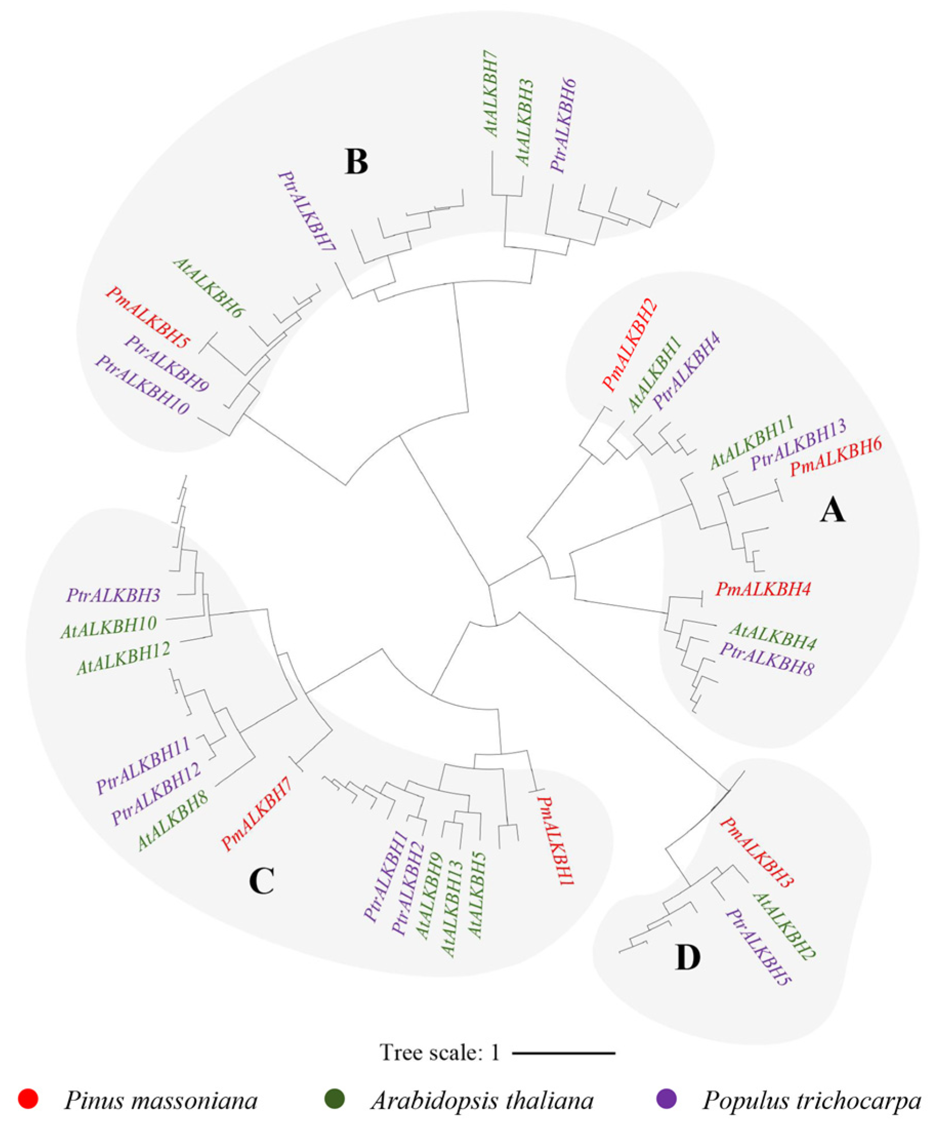
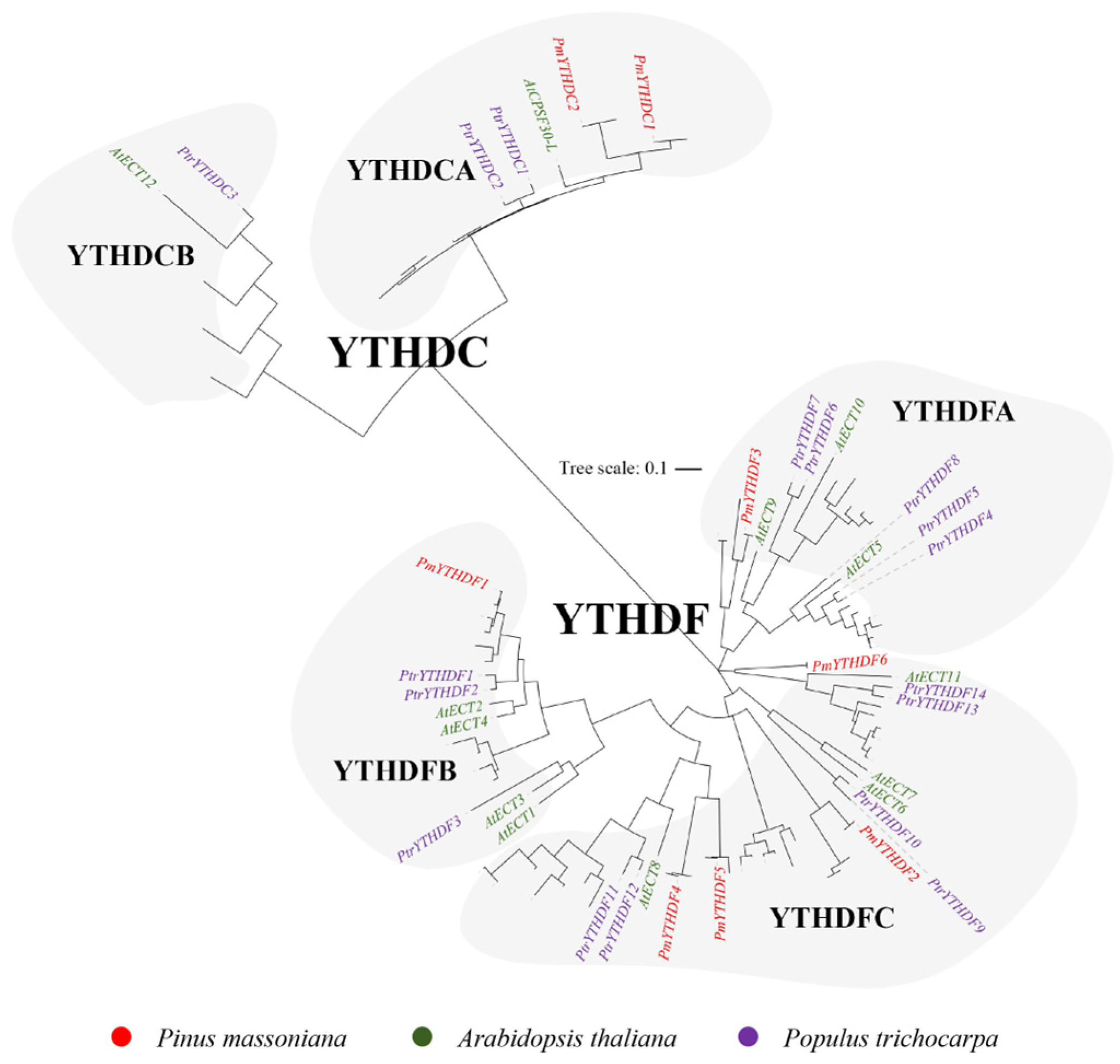
2.2. Phylogenetic and Gene Characterization Analyses of m6A Writers, Erasers, and Readers

In order to explore the phylogenetic and evolutionary relationships among these m6A regulators, we generated a comprehensive phylogenetic tree utilizing 32 A. thaliana m6A regulators, 37 in Populus trichocarpa and 276 proteins in the nine Rosaceae species. The findings revealed that the m6A-associated genes could be categorized into three distinct clusters based on their phylogenetic relationships, namely the writers, readers, and erasers.
The readers in Masson pine can be classified into four subgroups: MT, FIP37, VIR, and HAKAI proteins, respectively. Within the MT family, there are three distinct subfamilies: MTA, MTB, and MTC (Figure 1). The eraser family in Masson pine exclusively comprises ALKBH proteins. Furthermore, the ALKBH proteins can be categorized into four branches, A, B, C, and D, and each branch includes ALKBH members of Masson pine (Figure 2). The readers in Masson pine could be further classified into two subfamilies, namely DF and DC groups. Additionally, the DF subfamily can be subdivided into three groups, DFA, DFB, and DFC, whereas the DC subfamily can be divided into two groups, DCA and DCB. It is worth noting that no member of DCB has been identified by Masson pine (Figure 3).

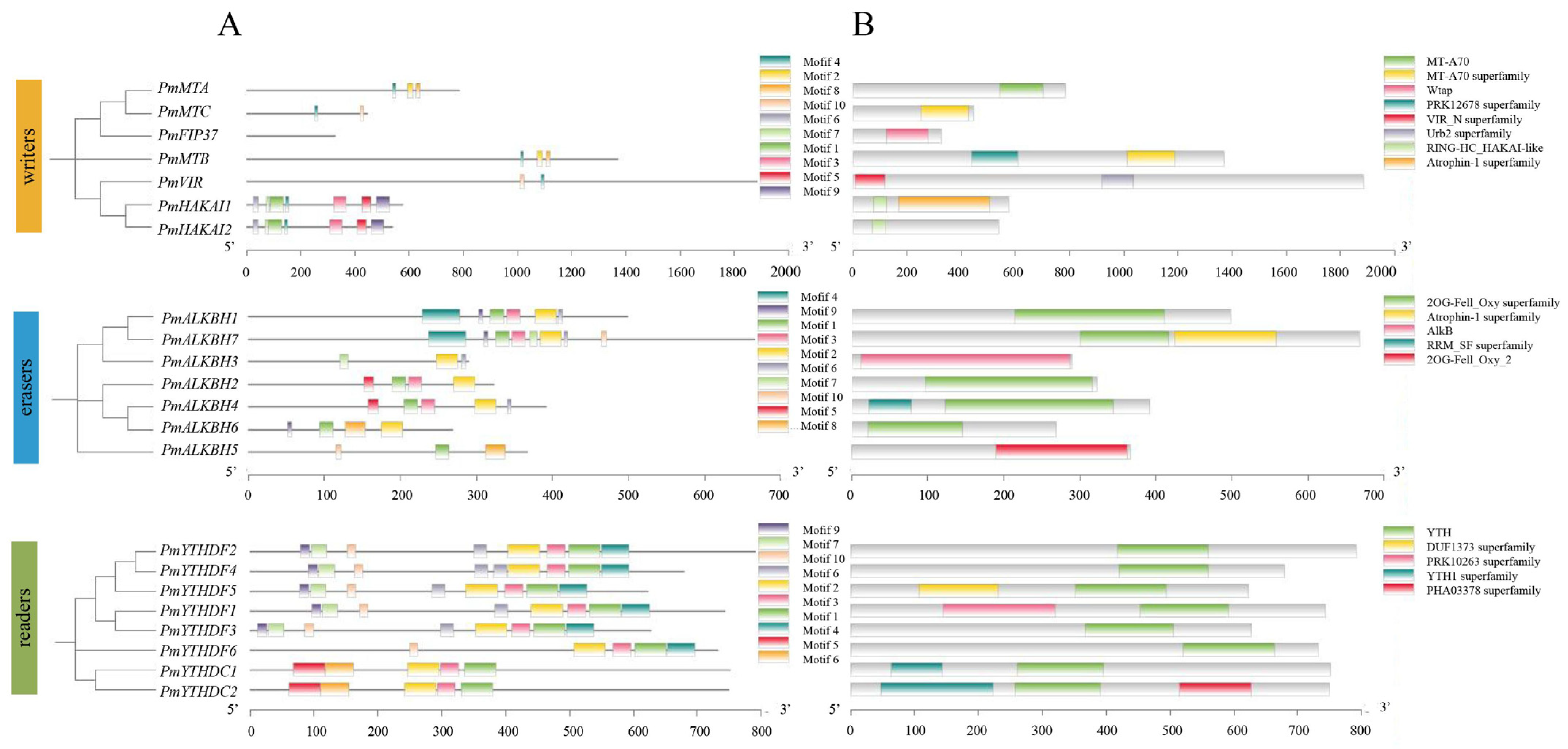
2.3. Gene Structure and Conserved Motif Analysis

The conserved motifs and gene structures of the m6A regulators were identified to elucidate the sequential characteristics in Masson pine (Figure 4, Table S2). In writers, the MT-A70 domain, comprising motifs 2, 4, and 8, was distributed on the MTA. In contrast, both MTB and MTC contained the MT-A70 superfamily domain consisting of motifs 4 and 10. Furthermore, HAKAI1/2 exhibit distributed RING-HC_HAKAI-like domains formed by motifs 1 and 7. PmFIP37 featured a Wtap domain, while PmVIR contained a VIR_N superfamily domain (Figure 4A,B). In erasers, motifs 11 and 12 were observed within the 20G-Fell Oxy domain, which was ubiquitously distributed across nearly all PmALKBH members. Conversely, motif 4 exclusively manifested in group C, suggesting potential distinctive functionalities among its constituents (Figure 4A,B). In readers, the YTH domain in the PmYTHs comprised three conserved motifs (21, 22, and 23) (Figure 4A,B). Notably, the aromatic cage within the YTH domain of PmYTHs was constituted by tryptophan residues (WWW) (Figure S1).

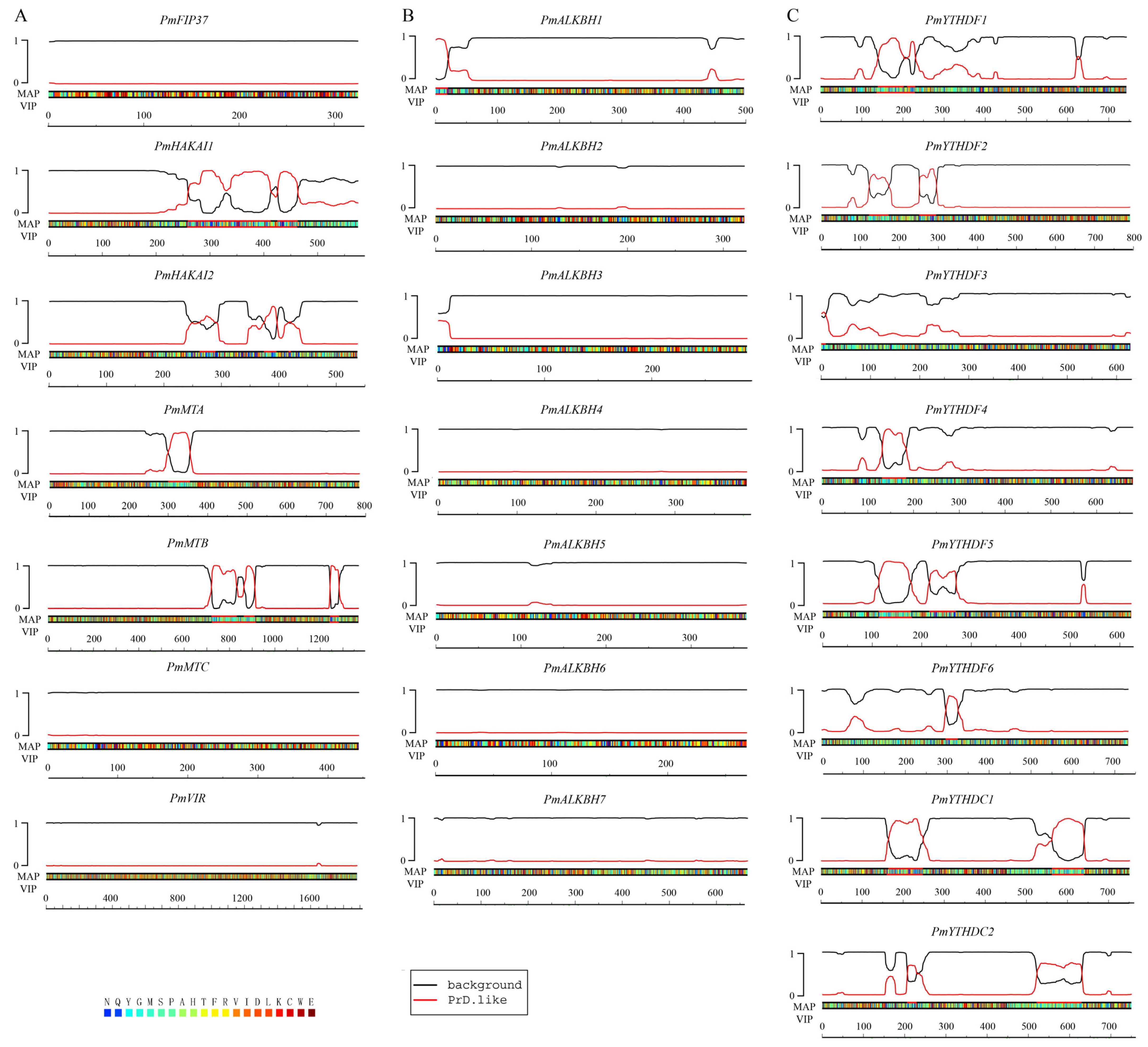
2.4. Certain Authors and Readers May Participate in the Process of Phase Separation

Previous studies have provided evidence for the regulatory role of m6A site quantity and distribution in cellular mRNAs in transcriptome composition during liquid–liquid phase separation (LLPS). The analysis identified the potential participation of four writers (PmHAKAI1, PmHAKAI2, PmMTA, PmMTB) and seven readers (PmYTHDF1, PmYTHDF2, PmYTHDF4, PmYTHDF5, PmYTHDF6, PmYTHDC1, PmYTHDC2) in the LLPS process (Figure 5).

2.5. Expression Levels of m6A Regulators in Different Tissues

Previous studies have demonstrated that the expression levels of key m6A regulators in meristems are comparatively higher than those in differentiated and mature tissues [35]. In this study, we analyzed the expression level of m6A regulators across various tissues of Masson pine, including roots, needles, semi-lignified stems, lignified stems, male cones, female cones, and cones (Figure 6A). Remarkably elevated expression levels of m6A regulators were observed in male cones, female cones, and cones, as well as in needles (Figure 6B). These findings strongly indicate a pivotal role for m6A in the reproductive development of Masson pine. The expression level of PmYTHDF1 was the highest among all seven tissues (Figure S2).

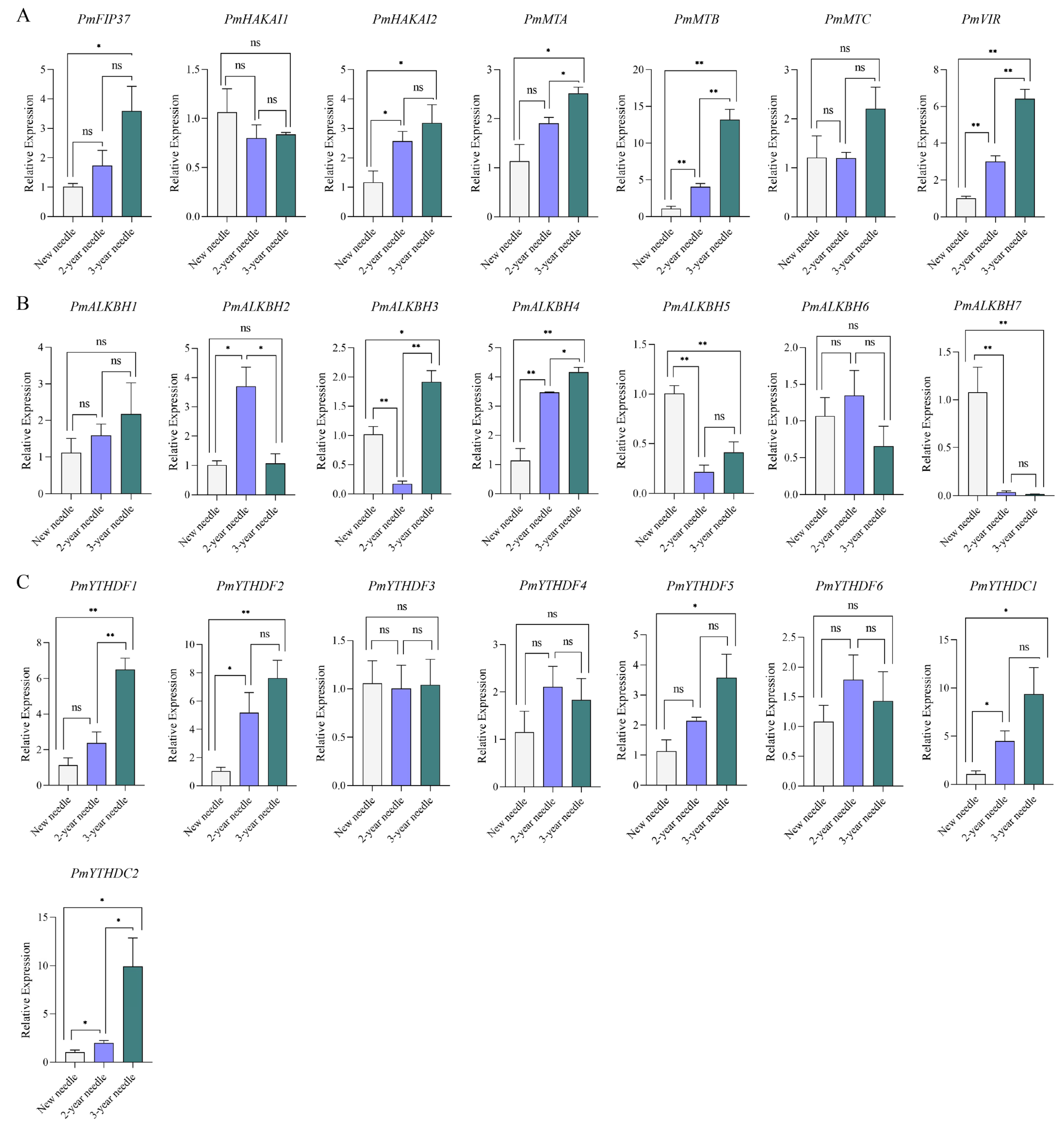
2.6. Expression Levels of m6A Regulators during Conifer Development

Considering the distinctive characteristics of pine needles, we investigated the differential expression patterns of m6A regulators in needles at various developmental stages. The expression levels of PmFip37, PmHAKAI2, PmMTA, PmMTB, PmVIR, PmALKBH4, PmYTHDF1, PmYTHDF2, PmYTHDF5, PmYTHDC1, and PmYTHDC2 were generally up-regulated with increasing needle age. These genes primarily belonged to writers (5 out of 11) and readers (5 out of 11). Conversely, the expression levels of demethylated proteins such as PmALKBH5 and PmALKBH7 decreased with advancing needle age. Notably, the expression pattern of PmALKBH3 exhibited a decrease followed by an increase with increasing needle age, while that of PmALKBH2 showed an initial increase followed by a subsequent decrease (Figure 7).

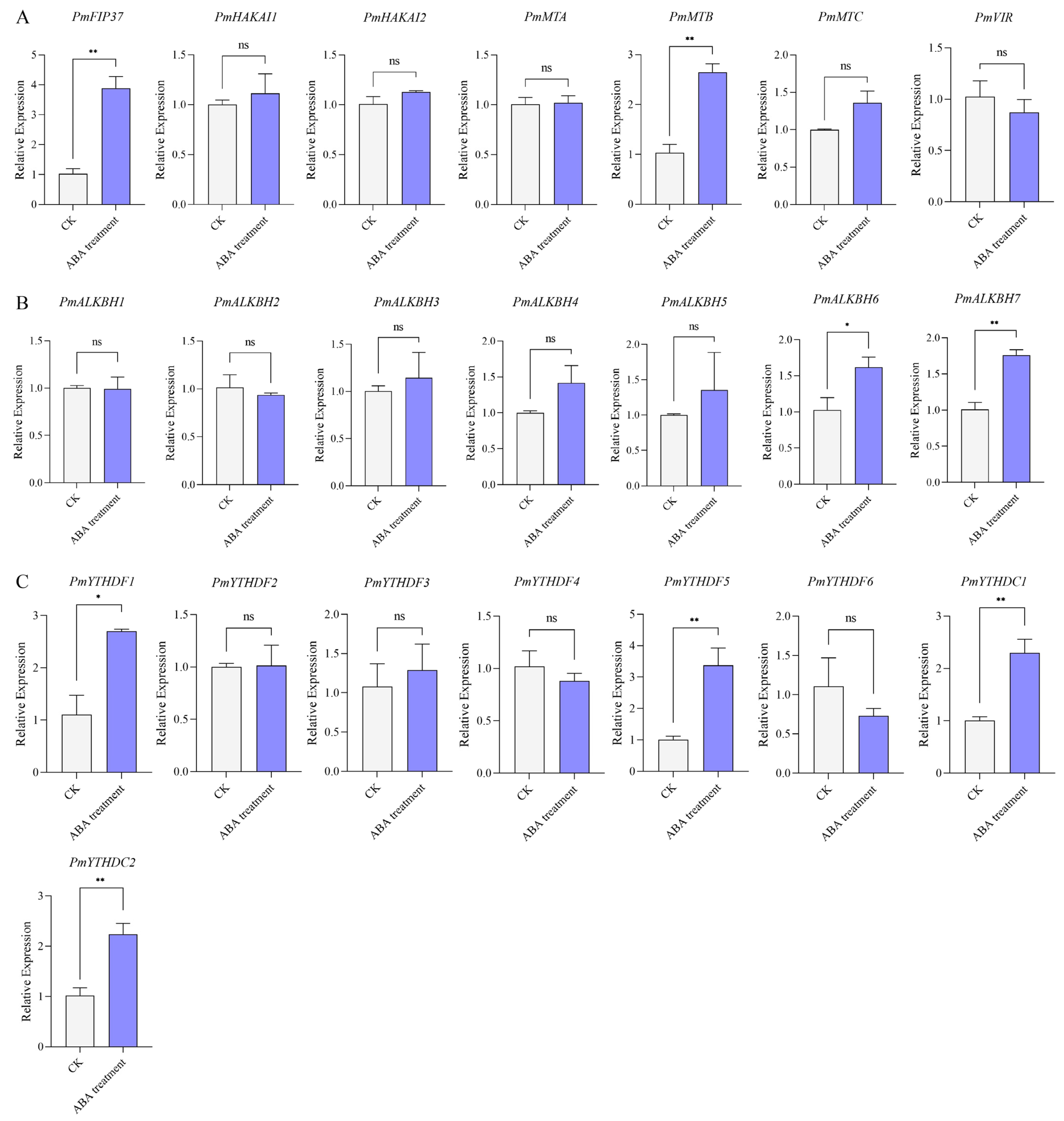
2.7. Expression Levels of m6A Regulators under ABA Treatment

Previous studies have demonstrated the involvement of m6A regulatory proteins in the modulation of ABA signaling through multiple pathways. Upon exposure to ABA treatment, a total of eight Masson pine m6A regulators, comprising two writers, two erasers, and four readers, exhibited significant alterations in their expression levels. Moreover, these genes displayed an up-regulated trend following exposure to ABA (Figure 8).

2.8. Expression Levels of m6A Regulators under Different Stresses

Numerous studies have unequivocally demonstrated the pivotal role of RNA m6A modification in mediating plant responses to both abiotic and biotic stresses. To comprehensively elucidate the role of m6A regulatory proteins in the response of Masson pine to drought stress, we employed high-throughput RNA-seq data to investigate the expression patterns of these proteins under varying degrees of drought stress (Figure 9). Remarkably, the expression levels of four key m6A regulators (PmALKBH4, PmALKBH6, PmYTHDF1, and PmYTHDF3) exhibited a consistent upward trend with increasing severity of drought stress. Conversely, sixteen other m6A regulators (PmALKBH1, PmALKBH3, PmYTHDF2, PmYTHDF4, PmYTHDF5, PmYTHDF6, PmYTHDC1, PmYTHDC2, and all writers) displayed a notable overall downward trend as drought intensity escalated. The expression levels of PmALKBH5 and PmALKBH7 exhibited a peak at drought degree B, followed by a subsequent decline with increasing severity of drought. This observed trend is noteworthy to mention; this observation strongly suggests that these particular m6A regulators play a crucial role in modulating the level of m6A modifications in plants to effectively cope with drought-induced stress (Figure 9A).
The differential expression of m6A regulatory proteins was analyzed between high resistance and susceptibility clones of Masson pine against Monochamus alternatus. The results revealed that the expression levels of three genes (PmALKBH3, PmYTHDF5, PmHAKAI1) were comparatively lower in the sensitive clones compared to those in the high-resistant clones, whereas the expression levels of eleven genes (PmMTA, PmMTB, PmMTC, PmHAKAI2, PmVIR, PmALKBH4, PmALKBH6, PmYTHDF1, PmYTHDF2, PmYTHDF3, PmYTHDC2) were significantly higher in the high-resistant clones. These findings suggest a potential role for m6A modification in influencing resistance to M. alternatus in Masson pine. These findings suggest that these m6A regulators have strong correlations with stress regulation (Figure 9B).

3. Discussion

N6-Methyladenosine, as a burgeoning research area, provides a novel perspective for biological investigations. The m6A modification is a reversible chemical modification. It is deposited by methyltransferases (writers), removed by demethylases (erasers), and recognized by m6A-binding proteins (readers) [34]. However, the m6A regulators have not yet been discovered in conifer species. In this study, we identified the m6A regulators and characterized their expression pattern in Masson pine, thus establishing a fundamental basis for further elucidating the biological function of m6A regulators in the growth, development, and stress response of coniferous species.

3.1. The m6A Regulators of Masson Pine May Have a Similar But Unique Molecular Mechanism to Them in Other Plants

In this study, we identified 22 m6A-related genes in Masson pine, including 7 writers, 7 erasers and 8 readers (Figure 1, Figure 2 and Figure 3, Table S1). The domains of these proteins exhibited a high degree of conservation (Figure 4). Phylogenetic relationships analysis showed that all of the Masson pine m6A writers, erasers, and readers could be divided into three groups separately. The phylogenetic analysis revealed the presence of m6A regulators in Masson pine across almost all subgroups (Figure 1, Figure 2 and Figure 3). It is worth noting that, similar to monocotyledonous plants, an absence of members belonging to the YTHDCB subfamily is observed in Masson pine (Figure 3). This phenomenon has also been observed in Pinus tabuliformis (Table S2). The findings imply that the m6A in Masson pine may possess a distinct molecular mechanism compared to dicotyledonous plants. However, more data are needed to support this.

3.2. The m6A Regulators May Participate in LLPS Process through PrLDs Domain

Prion-like domains can induce phase transitions, and further hardening of the droplets leads to pathological fibrous aggregation [36,37]. In mammals, prion-like domains are found in the structure of m6A regulatory proteins, indicating that m6A modified RNA is usually associated with phase separation [38,39,40,41]. This phenomenon was also found in the study of Arabidopsis m6A regulators. In this study, prion-like domains were found in most of the readers and writers, suggesting that these proteins have the potential to undergo phase separation (Figure 5). It is noteworthy that, among the erasers, only PmALKBH1 possesses a PrLD-like motif (Figure 5). Therefore, it can be speculated that the impact of erasers on LLPS in Masson Pine is relatively minor compared to that of writers and readers.

3.3. The m6A Regulators of Masson Pine Potentially Have a Crucial Impact on Both the Development of Reproductive Organs and the Senescence Process of Pine Needles

Understanding gene expression patterns in tissues is crucial for mining functional genes. The development of reproductive organs in plants has been demonstrated by several studies to be influenced by m6A regulators. AtALKBH10B regulates flowering by promoting demethylation of FT, SPL3, and SPL9 [23]. The knockout of MTA results in a reduction in m6A methylation, thereby impeding the transition of the developing embryo beyond the globular stage [34]. AtCPSF30-L affects the flowering of A. thaliana through the binding ability of m6A [32]. In our study, we found that m6A regulators were expressed in almost all tissues. However, the majority of m6A regulators displayed a predilection for expression in male cones, female cones, and cones (Figure 6). This tissue-specific expression pattern implied that m6A regulators in Masson pine may play a pivotal role in the reproductive growth of this species. It is worth mentioning that the expression level of PmYTHDF1 in these seven tissues was significantly higher than that of other m6A regulators (Figure S2), and a similar phenomenon was found in Arabidopsis [30]. This suggests that PmYTHDF1 may have a unique role in RNA modification.
Recently, multiple studies have demonstrated the involvement of ECT2/3/4 in regulating Arabidopsis leaf development [31,35]. To investigate the expression pattern of m6A regulators during coniferous leaf development, we examined Masson pine and found that PmYTHDF1 and PmYTHDF2, homologous genes to ECT2/3/4, exhibited significantly up-regulated expression levels during needle leaf development, suggesting their potential role in this process (Figure 7C). Furthermore, PmYTHDF5 also displayed an increasing trend throughout needle development, indicating that PmYTHDF5 is involved in needle development (Figure 7C). Notably, among all expressed genes outside of PmYTHDF1, PmYTHDF5 showed the highest level of expression, which has not been observed in studies involving other species (Table S2). Additionally, some writers (PmFIP37, PmHAKAI2, PmMTA, PmMTB, PmVIR) and erasers (PmALKBH2, PmALKBH3, PmALKBH4, PmALKBH5, PmALKBH7) exhibited differential expression patterns during needle development (Figure 7A,B). This observation warrants attention for future investigations into m6A regulation of needle development.

3.4. The m6A Regulators of Masson Pine Potentially Play a Pivotal Role in Drought and ABA Response and Confers Resistance Against the M. alternatus

In Arabidopsis, transcripts encoding osmotic stress response proteins tended to gain m6A, improving their stability under drought stress. Deficiency in ALKBH10B was associated with drought-hypersensitive phenotypes. AtALKBH10B-mediated m6A modification modulates the mRNA stability of several negative regulators of drought stress, including P5CS1, HVAD22D, ERD10, RD21A, RD22, CORI3, COR15B, and LOX2, by affecting alternative polyadenylation [36]. Another study demonstrated that the GhALKBH10B-mediated m6A modification modulates the mRNA stability of several regulators of drought stress by affecting alternative polyadenylation [37]. ECT8, acting as an ABA receptor in Arabidopsis, plays a pivotal role in the regulation of the ABA signaling pathway and response to drought stress [38]. Masson pine exhibits high tolerance to drought conditions. Surprisingly, the expression levels of all m6A regulators in Masson pine exhibit significant variations under different drought conditions, highlighting the potential pivotal role of m6A regulators in augmenting drought tolerance in Masson pine (Figure 9A).
M6A is also implicated in ABA signaling. For instance, AtCPSF30-L regulates the selective polyadenylation of RPN10 and FYVE1 in response to ABA [32]. ECT2/3/4 synergistically enhances target gene stability by binding PAB protein, thereby regulating the molecular mechanism of ABA response [35]. Furthermore, ALKBH10B responds to salt stress and osmotic stress and is induced by two stress-responsive hormones, ABA and JA. Similar expression patterns have been observed in tomatoes and apples [39]. ALKBH9B also modulates ABA response by controlling the mRNA m6A level of two negative regulators of ABA signaling, ABI1 and BES1 [40]. The m6A reader ECT8 is induced by ABA. Recent studies have demonstrated that ECT8 mutants exhibit hypersensitivity to ABA, highlighting the crucial role of ECT8 in abiotic stress responses [33]. Therefore, we hypothesize that m6A regulators in Masson pine may participate in pathways associated with ABA signaling. In our study, the expression of PmYTHDF1 (homolog of AtECT2), PmYTHDF5 (homolog of AtECT8), PmYTHDC1, PmYTHDC2 (homolog of AtCPSF30-L), and PmALKBH7 (homolog of AtALKBH10) were significantly enhanced upon induction with ABA (Figure 8). This suggests that m6A is relatively conserved in the ABA regulatory pathway.
It has been discovered that m6A plays a significant role in the interaction between plants and viruses, as well as pathogenic fungi. Recent advancements have also shed light on the involvement of m6A in insect resistance mechanisms. PxMETTL3 and PxMETTL14, repress the expression of JHE to induce an increased JH titer, mitigating the fitness costs associated with a robust defense against the Bt pathogen [41]. However, the enhancement of insect resistance in plants through m6A modification has not been documented in existing studies. In our study, we identified 14 m6A regulators that exhibited significantly disparate expression levels between two phenotypes of high-resistance and susceptibility clones against M. alternatus (Figure 9B). Nonetheless, our findings strongly imply a significant correlation between these m6A regulators and the resistance of Masson pine to M. alternatus.
In conclusion, the m6A regulators play a pivotal role in plant growth, development, and stress response. The identification and expression pattern analysis of m6A regulators in Masson pine establish a fundamental basis for further investigations into the functional roles of these genes. Furthermore, this identification provides a theoretical framework and direction for the application of coniferous tree RNA epigenetic molecular breeding.

4. Materials and Methods

4.1. Plant Materials

The seven tissue samples and coniferous specimens representing the three distinct age cohorts of Masson pine were specifically obtained from a 10-year-old tree situated within the Washan state-owned forest farm, Quanjiao, Anhui Province, China (32°10′ N, 118°27′ E). One-year-old Masson pine seedlings, obtained from the seed orchard of a Baisha state-owned forest farm, Shanghang, Fujian Province, China (25°150′ N, 116°620′ E), were used in this study. Individuals of the same clones with similar heights, uniform growth, and strong growth potential were selected as the test materials and subsequently moved into a growth chamber to recover for 15 d. The growth conditions were 10 h light/14 h dark cycles at 25 °C in the chamber. The expression patterns of m6A regulators were investigated following treatment with ABA. A 100 mL solution of ABA (200 mg·L1) was applied as a foliar spray on the experimental seedlings every morning from 9:00 to 10:00. On the 8th day, the middle and upper needles of the seedlings were uniformly collected.

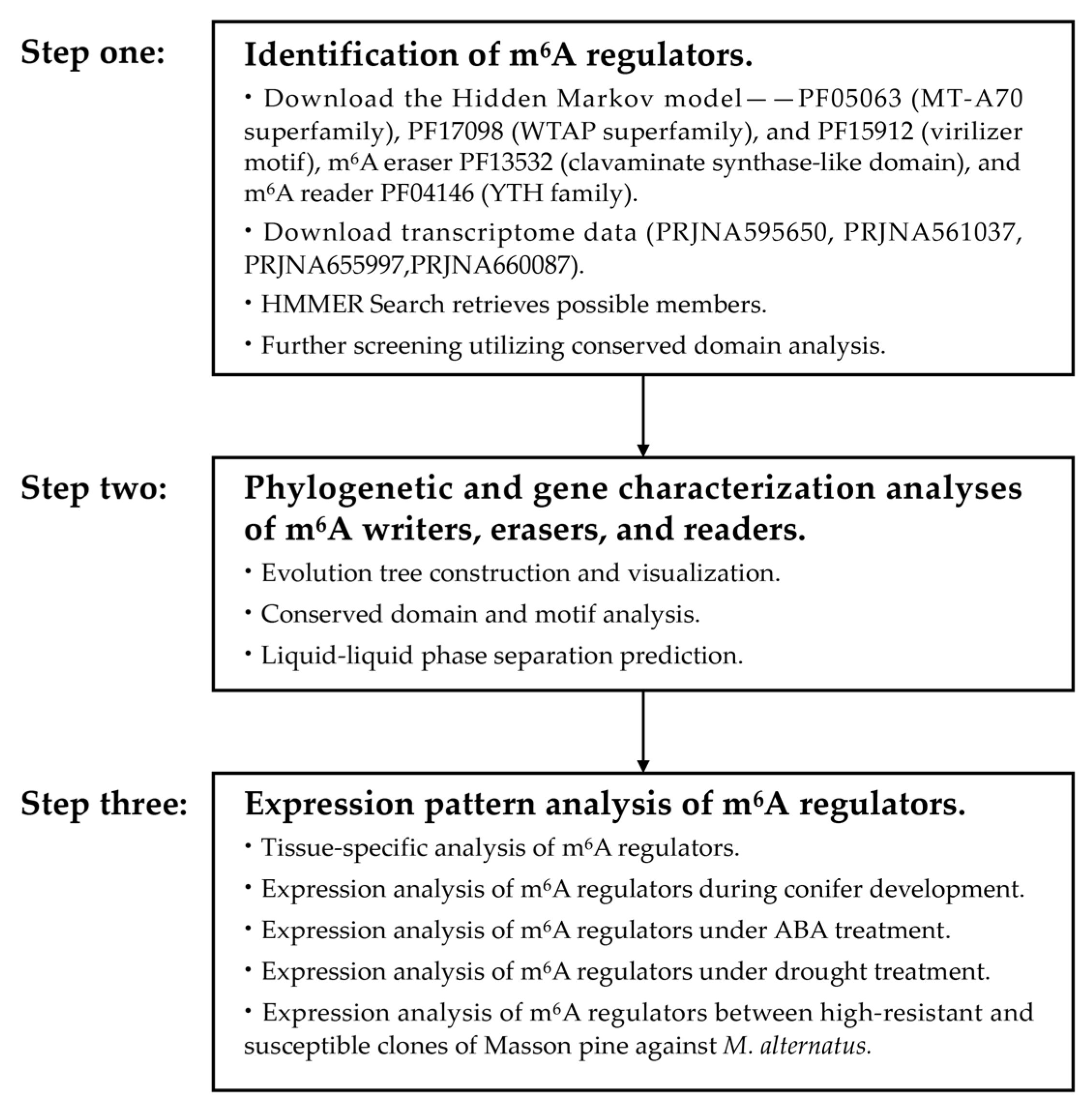
4.2. Identification of m6A Regulators: Writers, Erasers, and Readers in Masson Pine

The transcriptome data for Masson pine were obtained from the previously identified drought stress transcriptome (PRJNA595650), CO2 stress transcriptome (PRJNA561037), young shoots transcriptome (PRJNA655997), Masson pine inoculated with the pine wood nematode transcriptome (PRJNA660087). Hidden Markov Model (HMM) profiles of m6A writers PF05063 (MT-A70 superfamily), PF17098 (WTAP superfamily), PF15912 (virilizer motif), m6A eraser PF13532 (clavaminate synthase-like domain), and m6A reader PF04146 (YTH family) were employed for HMM searches in Masson pine. To ensure gene integrity, sequences with overlapping regions were excluded, while the CDD tool was utilized to verify conserved domains (Figure 10).

4.3. Sequence Analysis

The molecular weights and isoelectric points of the identified m6A regulators were determined using tools available on the ExPASy website. The molecular weights and isoelectric points of the identified m6A regulators were determined utilizing computational tools provided by the ExPASy website. The subcellular localization of m6A regulators was predicted and analyzed using PSORT (accessed on 14 March 2024). Previously published articles were used to identify m6A regulators of A. Thaliana and 9 Roseaceae plants [42], while the sequence data for P. trichocarpa m6A regulators were downloaded from NCBI. Multiple sequence alignments of the MT-A70 superfamily, ALKB family, and YTH family in nine Rosaceae species and Arabidopsis were performed using MUSCLE v5. The resultant comparative dataset was employed to generate maximum likelihood phylogenetic trees comprising 1000 bootstrap replicates via IQ-TREE (accessed on 14 March 2024).

4.4. Conserved Motifs and Functional Domains Analyses

The conserved motifs of m6A regulators were analyzed using Multiple Expectation Maximization for Motif Elicitation (MEME), with the parameters being set to a minimum and maximum motif width of 6 and 50, respectively. Additionally, a maximum number of motifs at 10 was considered. Functional domain annotation was performed utilizing the NCBI CDD (Figure 10).

4.5. LLPS Prediction

The PrLDs and disordered regions are predicted using the PLAAC (accessed on 15 March 2024). The visualization of the predicted outcomes is facilitated by an AI-based approach.

4.6. RNA Extraction and Quantitative Real-Time Reverse Transcription PCR

The total RNA was extracted using the RNAprep Pure Kit (DP441, Tiangen Biotech, Beijing, China). The concentration and purity of RNA were measured using a NanoDrop 2000 spectrophotometer (Thermo Fisher Scientific, Waltham, MA, USA), while the integrity of RNA was assessed through 1.2% agarose gel electrophoresis [43]. First-strand cDNA synthesis was performed using the One-step gDNA Removal and cDNA Synthesis Kit (AT311, TransGen Biotech, Beijing, China). Primers for quantitative real-time reverse transcription PCR (qRT-PCR) were designed with Primer 5.0 software (Table S3). SYBR Green reagents were utilized for target sequence detection. Each PCR mixture (10 µL) consisted of 1 µL of diluted cDNA (20× dilution), 5 µL of SYBR Green Real-time PCR Master Mix, 0.4 µL of each primer (10 µM), and 3.2 µL of ddH2O. The PCR program comprised six stages: preincubation at 95 °C for 60 s; amplification with denaturation at 95 °C for 15 s, annealing at 60 °C for 15 s and extension at 72 °C for 10s repeated 40 times; melting curve analysis with denaturation at 95 °C for 0.5 s; and annealing at 60 °C for 1 min. The quality of PCR products was evaluated based on melting curves. TUA (α-tubulin) served as an internal control. Three independent biological replicates with three technical replicates per biological replicate were examined. Quantification was achieved by comparing cycle threshold (Ct) values, and gene expression levels were calculated using the 2−∆∆Ct method. Significance was determined by t-test analysis conducted in SPSS 26.0 (IBM, New York, NY, USA) (* p < 0.05,** p < 0.01).

4.7. RNA-Seq Data Analysis of m6A Regulators

The RNA-seq data of Masson pine under drought treatment were acquired from the NCBI database (PRJNA595650). The RNA-seq data comprised high-resistant and susceptible clones of Masson pine against M. alternatus transcriptome (Table S4) in our group. Fragments per kilobase of the exon model per million reads mapped (FPKM) values were calculated to estimate the abundance of m6A transcripts. TBtools (Toolbox for Biologists) software (v2.034) was used to create heat maps of partial genes based on the values of log2(FPKM+1), and analyses were performed at the row scale.

4.8. Statistical Analysis

The statistical analysis was performed using GraphPad Prism v8.0.2 software. One-way ANOVA was employed to compare mean differences, and statistical significance was considered at * p < 0.05, ** p < 0.01 levels. Untreated samples were utilized as a control for significant gene expression analysis.

5. Conclusions

In this study, we initially identified 22 m6A-related genes in Masson pine, including 7 writers, 7 erasers, and 8 readers. The domains of these proteins exhibited a high degree of conservation. PrLDs were found in the readers and writers, suggesting that these genes may be involved in the process of liquid–liquid phase separation. The tissue expression analysis suggests that the m6A-related genes of Masson pine may potentially contribute to the development of reproductive organs and the senescence process of needles. The expression analysis of stress treatment revealed the sensitivity of m6A-related genes to ABA and drought treatments. Moreover, we found that m6A-related genes exhibited significantly disparate expression levels between two phenotypes of high-resistance and susceptibility clones against M. alternatus, suggesting that m6A may confer resistance against the M. alternatus. These findings provide a foundation for future functional analysis of m6A-related genes.

Supplementary Materials

The supporting information can be downloaded at: https://www.mdpi.com/article/10.3390/ijms25147987/s1.

Author Contributions

Conceived and designed the experiments: K.J. and Q.Y.; performed the experiments: S.Y., Y.S. and X.C.; analyzed the data: S.Y., D.W., J.Z., Q.L. and Q.C.; wrote the paper: S.Y., K.J. and Q.Y.; All authors have read and agreed to the published version of the manuscript.

Funding

This research was supported by the National Key R&D Program of China (2022YFD2200202), the Beijing National Laboratory for Molecular Sciences (BNLMS202202), and the project was funded by the Priority Academic Program Development of Jiangsu Higher Education Institutions (PAPD).

Institutional Review Board Statement

Not applicable.

Informed Consent Statement

Not applicable.

Data Availability Statement

Data are contained within the article and supplementary materials.

Conflicts of Interest

The authors declare no conflicts of interest.

References

  1. Boccaletto, P.; Stefaniak, F.; Ray, A.; Cappannini, A.; Mukherjee, S.; Purta, E.; Kurkowska, M.; Shirvanizadeh, N.; Destefanis, E.; Groza, P.; et al. MODOMICS: A database of RNA modification pathways. Nucleic Acids Res. 2022, 50, 231–235. [Google Scholar] [CrossRef] [PubMed]
  2. Li, X.; Xiong, X.; Yi, C. Epitranscriptome sequencing technologies: Decoding RNA modifications. Nat. Methods 2016, 14, 23–31. [Google Scholar] [CrossRef]
  3. Yue, Y.; Liu, J.; He, C. RNA N6-methyladenosine methylation in post-transcriptional gene expression regulation. Genes Dev. 2015, 29, 1343–1355. [Google Scholar] [CrossRef] [PubMed]
  4. Dominissini, D.; Moshitch-Moshkovitz, S.; Schwartz, S.; SalmonDivon, M.; Ungar, L.; Osenberg, S.; Cesarkas, K.; Jacob-Hirsch, J.; Amariglio, N.; Kupiec, M.; et al. Topology of the human and mouse m6A RNA methylomes revealed by m6A-seq. Nature 2012, 485, 201–206. [Google Scholar] [CrossRef] [PubMed]
  5. Arribas-Hernández, L.; Bressendorff, S.; Hansen, M.H.; Poulsen, C.; Erdmann, S.; Brodersen, P. An m6A-YTH module controls developmental timing and morphogenesis in Arabidopsis. Plant Cell 2018, 30, 952–967. [Google Scholar] [CrossRef] [PubMed]
  6. Roundtree, I.; Evans, M.; Pan, T.; He, C. Dynamic RNA modifications in gene expression regulation. Cell 2017, 169, 1187–1200. [Google Scholar] [CrossRef]
  7. Schwartz, S.; Agarwala, S.D.; Mumbach, M.R.; Jovanovic, M.; Mertins, P.; Shishkin, A.; Tabach, Y.; Mikkelsen, T.S.; Satija, R.; Ruvkun, G.; et al. High-resolution mapping reveals a conserved, widespread, dynamic mRNA methylation program in yeast meiosis. Cell 2013, 155, 1409–1421. [Google Scholar] [CrossRef] [PubMed]
  8. Li, Y.; Wang, X.; Li, C.; Hu, S.; Yu, J.; Song, S. Transcriptomewide N6-methyladenosine profiling of rice callus and leaf reveals the presence of tissue-specific competitors involved in selective mRNA modification. RNA Biol. 2014, 11, 1180–1188. [Google Scholar] [CrossRef]
  9. Parker, M.T.; Knop, K.; Sherwood, A.V.; Schurch, N.J.; Mackinnon, K.; Gould, P.D.; Hall, A.J.; Barton, G.J.; Simpson, G.G. Nanopore direct RNA sequencing maps the complexity of Arabidopsis mRNA processing and m6A modification. Elife 2020, 9, 49658. [Google Scholar] [CrossRef]
  10. Zhong, S.; Li, H.; Bodi, Z.; Button, J.; Vespa, L.; Herzog, M.; Fray, R.G. MTA is an Arabidopsis messenger RNA adenosine methylase and interacts with a homolog of a sex-specific splicing factor. Plant Cell 2008, 20, 1278–1288. [Google Scholar] [CrossRef]
  11. Yang, J.; Li, L.; Li, X.; Zhong, M.; Li, X.; Qu, L.; Zhang, H.; Tang, D.; Liu, X.; He, C.; et al. The blue light receptor CRY1 interacts with FIP37 to promote N6-methyladenosine RNA modification and photomorphogenesis in Arabidopsis. New Phytol. 2022, 237, 840–854. [Google Scholar] [CrossRef] [PubMed]
  12. Ruzicka, K.; Zhang, M.; Campilho, A.; Bodi, Z.; Kashif, M.; Saleh, M.; Eeckhout, D.; El-Showk, S.; Li, H.; Zhong, S.; et al. Identification of factors required for m6A mRNA methylation in Arabidopsis reveals a role for the conserved E3 ubiquitin ligase HAKAI. New Phytol. 2017, 215, 157–172. [Google Scholar] [CrossRef] [PubMed]
  13. Parker, M.T.; Knop, K.; Zacharaki, V.; Sherwood, A.V.; Tomé, D.; Yu, X.; Martin, P.G.; Beynon, J.; Michaels, S.D.; Barton, G.J.; et al. Widespread premature transcription termination of Arabidopsis thaliana NLR genes by the spen protein FPA. Elife 2021, 10, 65537. [Google Scholar] [CrossRef] [PubMed]
  14. Zhang, M.; Bodi, Z.; Mackinnon, K.; Zhong, S.; Archer, N.; Mongan, N.P.; Simpson, G.G.; Fray, R.G. Two zinc finger proteins with functions in m6A writing interact with HAKAI. Nat. Commun. 2022, 13, 1127. [Google Scholar] [CrossRef]
  15. Bhat, S.S.; Bielewicz, D.; Gulanicz, T.; Bodi, Z.; Xiang, Y.; Anderson, S.J.; Szewc, L.; Bajczyk, M.; Dolata, J.; Grzelak, N.; et al. mRNA adenosine methylase (MTA) deposits m6A on pri-miRNAs to modulate miRNA biogenesis in Arabidopsis thaliana. Proc. Natl. Acad. Sci. USA 2020, 117, 21785–21795. [Google Scholar] [CrossRef] [PubMed]
  16. Shen, L.; Liang, Z.; Yu, H. N6-methyladenosine RNA modification regulates shoot stem cell fate in Arabidopsis. Mech. Dev. 2017, 145, S171. [Google Scholar] [CrossRef]
  17. Zhang, M.; Zeng, Y.; Peng, R.; Dong, J.; Lan, Y.; Duan, S.; Chang, Z.; Ren, J.; Luo, G.; Liu, B.; et al. N6-methyladenosine RNA modification regulates photosynthesis during photodamage in plants. Nat. Commun. 2022, 13, 7441. [Google Scholar] [CrossRef]
  18. Hu, J.; Cai, J.; Park, S.J.; Lee, K.; Li, Y.; Chen, Y.; Yun, J.Y.; Xu, T.; Kang, H. N6-methyladenosine mRNA methylation is important for salt stress tolerance in Arabidopsis. Plant J. 2021, 106, 1759–1775. [Google Scholar] [CrossRef]
  19. Jia, G.; Fu, Y.; Zhao, X.; Dai, Q.; Zheng, G.; Yang, Y.; Yi, C.; Lindahl, T.; Pan, T.; Yang, Y.G.; et al. N6-methyladenosine in nuclear RNA is a major substrate of the obesity-associated FTO. Nat. Chem. Biol. 2011, 7, 885–887. [Google Scholar] [CrossRef]
  20. Zheng, G.; Dahl, J.A.; Niu, Y.; Fedorcsak, P.; Huang, C.M.; Li, C.J.; Shi, Y.; Wang, W.L.; Song, S.H.; Lu, Z.; et al. ALKBH5 is a mammalian RNA demethylase that impacts RNA metabolism and mouse fertility. Mol. Cell 2013, 49, 18–29. [Google Scholar] [CrossRef]
  21. Hofmann, N.R. Epitranscriptomics and flowering: mRNA methylation/demethylation regulates flowering time. Plant Cell 2017, 29, 2949–2950. [Google Scholar] [CrossRef] [PubMed]
  22. Mielecki, D.; Zugaj, D.Ł.; Muszewska, A.; Piwowarski, J.; Chojnacka, A.; Mielecki, M.; Nieminuszczy, J.; Grynberg, M.; Grzesiuk, E. Novel AlkB dioxygenases-alternative models for in silico and in vivo studies. PLoS ONE 2012, 7, 30588. [Google Scholar] [CrossRef] [PubMed]
  23. Duan, H.C.; Wei, L.H.; Zhang, C.; Wang, Y.; Chen, L.; Lu, Z.; Chen, P.R.; He, C.; Jia, G. ALKBH10B is an RNA N6-methyladenosine demethylase affecting Arabidopsis floral transition. Plant Cell 2017, 29, 2995–3011. [Google Scholar] [CrossRef]
  24. Martínez-Pérez, M.; Aparicio, F.; López-Gresa, M.P.; Bellés, J.M.; Sánchez-Navarro, J.A.; Pallás, V. Arabidopsis m6A demethylase activity modulates viral infection of a plant virus and the m6A abundance in its genomic RNAs. Proc. Natl. Acad. Sci. USA 2017, 114, 10755–10760. [Google Scholar] [CrossRef] [PubMed]
  25. Zhou, L.; Tian, S.; Qin, G. RNA methylomes reveal the m6A-mediated regulation of DNA demethylase gene SlDML2 in tomato fruit ripening. Genome Biol. 2019, 20, 156. [Google Scholar] [CrossRef] [PubMed]
  26. Li, D.; Zhang, H.; Hong, Y.; Huang, L.; Li, X.; Zhang, Y.; Ouyang, Z.; Song, F. Genome-wide identification, biochemical characterization, and expression analyses of the YTH domain-containing RNA-binding protein family in Arabidopsis and Rice. Plant Mol. Biol. Rep. 2014, 32, 1169–1186. [Google Scholar] [CrossRef]
  27. Wang, X.; Zhao, B.S.; Roundtree, I.A.; Lu, Z.; Han, D.; Ma, H.; Weng, X.; Chen, K.; Shi, H.; He, C. N6-methyladenosine modulates messenger RNA translation efficiency. Cell 2015, 161, 1388–1399. [Google Scholar] [CrossRef] [PubMed]
  28. Wang, X.; Lu, Z.; Gomez, A.; Hon, G.C.; Yue, Y.; Han, D.; Fu, Y.; Parisien, M.; Dai, Q.; Jia, G.; et al. N6-methyladenosinedependent regulation of messenger RNA stability. Nature 2014, 505, 117–120. [Google Scholar] [CrossRef] [PubMed]
  29. Scutenaire, J.; Deragon, J.; Jean, V.; Benhamed, M.; Raynaud, C.; Favory, J.; Merret, R.; Bousquet-Antonelli, C. The YTH domain protein ECT2 is an m6A reader required for normal trichome branching in Arabidopsis. Plant Cell 2018, 30, 986–1005. [Google Scholar] [CrossRef]
  30. Wei, L.H.; Song, P.Z.; Wang, Y.; Lu, Z.K.; Tang, Q.; Yu, Q.; Xiao, Y.; Zhang, X.; Duan, H.C.; Jia, G.F. The m6A reader ECT2 controls trichome morphology by affecting mRNA stability in Arabidopsis. Plant Cell 2018, 30, 968–985. [Google Scholar] [CrossRef]
  31. Arribas-Hernández, L.; Simonini, S.; Hansen, M.H.; Paredes, E.B.; Bressendorff, S.; Dong, Y.; Østergaard, L.; Brodersen, P. Recurrent requirement for the m6A-ECT2/ECT3/ECT4 axis in the control of cell proliferation during plant organogenesis. Development 2020, 147, 189134. [Google Scholar] [CrossRef]
  32. Song, P.; Yang, J.; Wang, C.; Lu, Q.; Shi, L.; Tayier, S.; Jia, G. Arabidopsis N6-methyladenosine reader CPSF30-L recognizes FUE signals to control polyadenylation site choice in liquid-like nuclear bodies. Mol. Plant 2021, 14, 571–587. [Google Scholar] [CrossRef]
  33. Cai, Z.; Tang, Q.; Song, P.; Tian, E.; Yang, J.; Jia, G. The m6A reader ECT8 is an abiotic stress sensor that accelerates mRNA decay in Arabidopsis. Plant Cell 2024, koae149. [Google Scholar] [CrossRef]
  34. Zhang, F.; Zhang, Y.; Liao, J.; Yu, Y.; Zhou, Y.; Feng, Y.; Yang, Y.; Lei, M.; Bai, M.; Wu, H.; et al. The subunit of RNA N6-methyladenosine methyltransferase OsFIP regulates early degeneration of microspores in rice. PLoS Genet. 2019, 15, e1008120. [Google Scholar] [CrossRef]
  35. Song, P.; Wei, L.; Chen, Z.; Cai, Z.; Lu, Q.; Wang, C.; Tian, E.; Jia, G. m6A readers ECT2/ECT3/ECT4 enhance mRNA stability through direct recruitment of the poly(A) binding proteins in Arabidopsis. Genome Biol. 2023, 24, 103. [Google Scholar] [CrossRef]
  36. Han, R.; Shoaib, Y.; Cai, J.; Kang, H. ALKBH10B-mediated m6A demethylation is crucial for drought tolerance by affecting mRNA stability in Arabidopsis. Environ. Exp. Bot. 2023, 209, 105306. [Google Scholar] [CrossRef]
  37. Cui, C.; Ma, Z.; Wan, H.; Gao, J.; Zhou, B. GhALKBH10 negatively regulates salt tolerance in cotton. Plant Physiol. Biochem. 2022, 192, 87–100. [Google Scholar] [CrossRef]
  38. Wu, X.; Su, T.; Zhang, S.; Zhang, Y.; Wong, C.; Ma, J.; Shao, Y.; Hua, C.; Shen, L.; Yu, H. N6-methyladenosine-mediated feedback regulation of abscisic acid perception via phase-separated ECT8 condensates in Arabidopsis. Nat. Plants 2024, 10, 469–482. [Google Scholar] [CrossRef]
  39. Tang, J.; Yang, J.; Duan, H.; Jia, G. ALKBH10B, an mRNA m6A demethylase, modulates ABA response during seed germination in Arabidopsis. Front. Plant Sci. 2021, 12, 712713. [Google Scholar] [CrossRef] [PubMed]
  40. Tang, J.; Yang, J.; Lu, Q.; Tang, Q.; Chen, S.; Jia, G. The RNA N6-methyladenosine demethylase ALKBH9B modulates ABA responses in Arabidopsis. J. Integr. Plant Biol. 2022, 64, 2361–2373. [Google Scholar] [CrossRef]
  41. Guo, Z.; Bai, Y.; Zhang, X.; Guo, L.; Zhu, L.; Sun, D.; Sun, K.; Xu, X.; Yang, X.; Xie, W.; et al. RNA m6A methylation suppresses insect juvenile hormone degradation to minimize fitness costs in response to a pathogenic attack. Adv. Sci. 2024, 11, 2307650. [Google Scholar] [CrossRef]
  42. Han, C.; Dong, H.; Qiao, Q.; Dai, Y.; Huang, X.; Zhang, S. Comparative genomic analysis of N6-methyladenosine regulators in nine rosaceae species and functional characterization in response to drought stress in pear. Hortic. Plant J. 2023, 9, 693–704. [Google Scholar] [CrossRef]
  43. Zhu, P.H.; Ma, Y.Y.; Zhu, L.Z.; Chen, Y.; Ji, K.S. Selection of suitable reference genes in Pinus massoniana Lamb. under different abiotic stresses for qPCR normalization. Forests 2019, 10, 632. [Google Scholar] [CrossRef]
Figure 1. Phylogenetic analysis of m6A writers from A. thaliana, Populus trichocarpa, nine Rosaceae plants and P. massoniana.
Ijms 25 07987 g001
Figure 2. Phylogenetic analysis of ALKB family from A. thaliana, P. trichocarpa, nine Rosaceae plants, and P. massoniana. A, B, C, and D represent different subgroups.
Ijms 25 07987 g002
Figure 3. Phylogenetic analysis of YTH family from A. thaliana, P. trichocarpa, nine Rosaceae plants, and P. massoniana.
Ijms 25 07987 g003
Figure 4. Schematics of the conserved motifs (A) and functional domains (B) of m6A regulators in Masson pine.
Ijms 25 07987 g004
Figure 5. Predictions of PrLDs and disordered regions made by the PLAAC (accessed on 15 March 2024). (A): m6A writers, (B): m6A erasers, (C): m6A readers. The black line represents the background and the red line is the prediction of the prion structure region. If the red line is in the non-baseline region, it indicates that the prion structure region is at that location and the phase transition is highly likely.
Ijms 25 07987 g005
Figure 6. Heat map of RNA-Seq expression of m6A regulators in different tissues of Masson pine. (A): The organization diagram of Masson pine. (B): Heat map for tissue specific analysis.
Ijms 25 07987 g006
Figure 7. Relative expression levels of m6A genes during conifer development based on qRT-PCR analysis. (A): m6A writers, (B): m6A erasers, (C): m6A readers. The relative expression level was measured with the expression level of “New needle” as the control. Different numbers of “*” indicate significant differences (* p < 0.05, ** p < 0.01), ns indicate no significant difference. Data are shown as mean ± SE, with three biological replicates and three technical replicates.
Ijms 25 07987 g007
Figure 8. Relative expression levels of m6A genes under ABA treatment based on qRT-PCR analysis. (A): m6A writers, (B): m6A erasers, (C): m6A readers. Different numbers of “*” indicate significant differences (* p < 0.05, ** p < 0.01), ns indicate no significant difference. Data are shown as mean ± SE, with three biological replicates and three technical replicates.
Ijms 25 07987 g008
Figure 9. Expression pattern of m6A regulatory genes under biotic and abiotic stress conditions. Note: (A): Heat map of expression of Masson pine under drought stress. The soil moisture content for the growth of Masson pine was set to four gradients: A (80 ± 5)%, B (65 ± 5)%, C (50 ± 5)%, and D (35 ± 5)%, respectively. They were placed at 75% humidity for 60 d and subsequently sequenced. Fragments per kilobase of exon model per million mapped fragments (FPKM) values were computed to assess the expression level of m6A regulators. (B): The Masson pine clones MK27-1 and MK30-1 exhibited relatively low resistance to Monochamus alternatus, whereas the MK94-1 clones demonstrated comparatively high resistance against M. alternatus. Transcripts Per Kilobase of exonmodel per Million mapped reads (TPM) values were computed to assess the expression level of m6A regulators.
Ijms 25 07987 g009
Figure 10. Block diagram of the research stepwise procedure.
Ijms 25 07987 g010
Disclaimer/Publisher’s Note: The statements, opinions and data contained in all publications are solely those of the individual author(s) and contributor(s) and not of MDPI and/or the editor(s). MDPI and/or the editor(s) disclaim responsibility for any injury to people or property resulting from any ideas, methods, instructions or products referred to in the content.

