Article

Discordant ALK Status in Non-Small Cell Lung Carcinoma: A Detailed Reevaluation Comparing IHC, FISH, and NGS Analyses

by
Katarína Tobiášová
1,
Martina Barthová
1,
Ľuboslava Janáková
1,
Katarína Lešková
1,
Anna Farkašová
2,
Dušan Loderer
3,
Marián Grendár
3 and
Lukáš Plank
1,2,*
1
Department of Pathological Anatomy, University Hospital Martin, Jessenius Faculty of Medicine, Comenius University, 036 01 Martin, Slovakia
2
Martin’s Biopsy Center, Ltd., 036 01 Martin, Slovakia
3
Biomedical Centre Martin—BioMed Martin, Jessenius Faculty of Medicine, Comenius University, 036 01 Martin, Slovakia
*
Author to whom correspondence should be addressed.
Int. J. Mol. Sci. 2024, 25(15), 8168; https://doi.org/10.3390/ijms25158168 (registering DOI)
Submission received: 14 June 2024 / Revised: 19 July 2024 / Accepted: 23 July 2024 / Published: 26 July 2024
(This article belongs to the Special Issue Novel Molecular Pathways in Oncology 2.0)

Abstract

:
ALK detection was performed on 2813 EGFR-unmutated NSCLC cases by simultaneous use of immunohistochemistry (VENTANA® anti-ALK D5F3, Roche Molecular Systems, Inc., Rotkreuz, Switzerland) and fluorescence in situ hybridization with the ALK break apart and the ALK/EML4 fusion probe (ZytoVision, Bremerhaven, Germany). A total of 33 cases were positive discordant (FISH-positive, IHC-negative) and 17 cases were negative discordant (FISH-negative, IHC-positive). This study’s aim was to reevaluate the methods used and compare discordant samples to positive concordant samples in order to ellucidate the differences. FISH signal variants were examined and compared. Positive discordant cases featured one pattern of ALK rearrangement in 41.4%, two patterns in 48.3%, and three patterns in 10.3% of analysed samples, with a higher variability of detected patterns and a higher number of ALK copy gains. Positive concordant cases displayed one pattern of rearrangement in 82%, two patterns in 17.8%, and three patterns in 0.6% of analysed samples. The association between number of patterns and concordance/discordance was statistically significant (p < 0.05). Eleven positive discordant and two negative concordant cases underwent NGS analysis, which resulted in identification of ALK fusion in one positive discordant and two negative discordant cases. Positive protein expression regardless of FISH result correlated more with a positive NGS result compared to samples with a positive FISH result with negative protein expression. FISH analysis was able to detect atypical or heterogenous patterns of rearrangement in a proportion of cases with negative protein expression, which may be associated with more extensive genetic alterations rather than true ALK rearrangement.

1. Introduction

Treatment of non-small cell lung carcinoma (NSCLC) has gone through significant changes in the span of last two decades. Based on presence of oncogenic mutations, two categories of NSCLC have been established—the “oncogene-addicted” and “non-oncogene-addicted” NSCLC [1], with separate guidelines for treatment.
One of the most frequent targetable genetic alterations in NSCLC is the rearrangement of the ALK gene, found in approximately 3–5% of all NSCLC cases [2]. ALK protein is a transmembrane protein with intracytoplasmic tyrosine-kinase domain, which upon activation triggers the downstream cell signaling cascades regulating proliferation, differentiation, and migration of cells. Oncogenic alteration of the ALK gene is usually caused by a chromosomal split in intron 19 of the ALK gene and the subsequent fusion of the tyrosine-kinase domain corresponding part to another partner gene, causing production of large quantities of the chimeric intracytoplasmic ALK protein [3] with aberrant activation independent of external stimuli. The second breaking point is usually localized within the EML4 gene, leading to an intrachromosomal ALK::EML4 inversion with currently more than 21 identified variants [4]; other fusions, such as ALK::KIF5B, ALK::KLC1, or ALK::TPR, occur less often, in approximately 5% of cases [5].
ALK-rearranged NSCLC responds well to small molecule inhibitors of receptors with tyrosine-kinase activity (TKIs). First-generation TKI crizotinib has a superior response rate compared to cytotoxic chemotherapy and displays significantly better overall survival (OS) as first-line therapy [6,7]. Second-generation CNS-penetrant ALK-TKIs, including ceritinib, alectinib, and brigatinib, show even longer progression-free survival (PFS) compared to crizotinib and therefore are currently preferred in first-line treatment [8,9,10,11].
ALK rearrangement detection has been performed mainly by immunohistochemistry (IHC) and in situ hybridization, nowadays with increasing demand for multiple gene analysis available through next-generation sequencing (NGS). Fluorescence in situ hybridization (FISH) allows detection of a split in the ALK gene on the chromosomal level; however, it is expensive, requires adequate quality of tissue, and expertise in interpretation of signal variants. ALK::EML4 fusions tend to display a so-called short break pattern (less than two signal diameter), which can be interpreted as a false negative [12], though using the ALK/EML4 fusion probe mitigates this pitfall.
IHC analysis performed with more sensitive rabbit monoclonal antibodies (e.g., D5F3, Ventana) has become a valuable counterpart to FISH and is currently approved as a screening method for ALK rearrangement, even being acceptable as an equivalent alternative in the case of FISH unavailability or failure [1]. IHC is easier to interpret and cheaper; however, it cannot fully replace FISH as the method of choice [13,14,15], mainly due to the rare occurrence of discrepancies: some cases are either positive discordant (FISH+, IHC−) or negative discordant (FISH−, IHC+). This can be partially attributed to preanalytical steps [16], as well as post-translational changes and miscellaneous properties of the ALK protein produced by different rearrangement variants. A part of FISH negative cases can still be confirmed as ALK positive using another method [17]. IHC seems to correlate with NGS more and potentially is even more sensitive [18], although this can be attributed to technical challenges of FISH analysis. The simultaneous use of IHC and FISH is therefore still recommended [19,20], and whenever possible, discrepant cases should undergo analysis using NGS.
Discordance of ALK status has been a widely discussed topic, mainly due to clinical implications. Several case reports [21,22] and larger patient cohorts [23] conclude that ALK expression even with negative FISH result was associated with the desirable response to TKI treatment. Some published studies prove that patients with a positive discordant result may respond inadequately compared to negative discordant patients (62% response rate compared to 100% response rate) [24] or TKI treatment in positive discordant patients who are not confirmed as positive by NGS may be completely ineffective [25]. However, negative discordance (FISH−, IHC+) can predict worse overall survival [26] compared to concordant patients.
Our department performs the analysis of ALK status using IHC and FISH simultaneously, which over time naturally led to the accumulation of discordant cases. Our main concern was the reevaluation of methods used and finding possible explanations for the discrepancies. FISH analysis occasionally produced some unusual variants reports, leading us to the question whether the ALK discrepancies can be partially elucidated by comparing the FISH signal variants found in discordant cases to positive concordant samples. Furthermore, we aimed to validate these observations by comparing with NGS performed on a proportion of our cohort as a part of a pilot study.

2. Results

2.1. Concordance and Discordance between FISH and IHC

ALK detection using IHC and FISH simultaneously was successfully performed on 2683 samples. In 130 samples, one or both methods failed due to material limitations and were not further included into our study.
A total of 2444 samples were negative using both FISH and IHC, so-called negative concordant. A total of 184 cases were positive concordant; 38 cases were positive for ALK rearrangement using FISH, but negative for ALK protein expression and were labeled as positive discordant. In 9 of them, material quantity and quality were sufficient to repeat the IHC analysis. 5 cases displayed the so-called “dot-like” positivity and were reclassified as positive concordant. In 4 cases, the repeated IHC was still negative. The final number of positive concordant cases was 189, and the number of positive discordant cases was 33. A total of 17 cases were negative for ALK rearrangement with positive ALK protein expression and were labeled as negative discordant. Reevaluation of FISH did not reveal any false negatives.
Discordant cases altogether comprised 1.86% of all cases, with 1.23% being positive discordant and 0.63% being negative discordant. Out of all FISH-positive cases, 14.9% were positive discordant. Out of all FISH-negative cases, 0.7% were negative discordant. Sensitivity of FISH was calculated to be 91.7%, and specificity of FISH was 98.6%.

2.2. Demographic Distribution

The population group consisted of 1716 male patients and 967 female patients (Table 1), with age ranging from 23 to 91, with the mean of 65 years and median 66 years. NSCLC diagnosis occurred most frequently in patients in the sixth decade.
The overall frequency of ALK rearrangement was 5.5% in the male population, 13.1% in the female population, and 8.3% regardless of gender. ALK rearrangement was most prevalent in population of young patients, with peak incidence in female patients under 40 (7 out of 18 cases) and under 50 (15 out of 58 analyzed cases), as well as male patients under 50 years (22 out of 91 cases). With increasing age, the prevalence of ALK rearrangement became progressively lower but was more frequent in the female population in every age group. Negative discordant cases were found almost exclusively in the female population (14 out of 17 cases); positive discordant cases showed a slight male predominance (20 out of 33 cases).

2.3. Histopathology

In the positive discordant cohort, material limitations did not allow further specification than the “adenocarcinoma, NOS” category in 11 out of 33 cases (33.33%). The most frequently occurring types were solid predominant adenocarcinoma in four cases (12.12%), then G3 adenocarcinoma and metastatic adenocarcinoma in three cases (9.09%). Mucin production was especially noted in the report in 2 cases (6.06%), was not detected in 3 cases (9.09%), and was not specified in 28 cases (84.85%).
In the negative discordant cohort, the category “adenocarcinoma, NOS” was used in four cases (23.53%), equally in number as solid predominant adenocarcinoma (23.53%). Other subtypes did not occur particularly frequently. Mucin production was specially noted in the report in 1 case (5.88%), was not detected in 2 cases (11.76%) and was not specified in 14 cases (82.35%).
In the positive concordant cohort, “adenocarcinoma, NOS” was used in 57 cases (30.16%), followed equally by acinar predominant, solid predominant, and metastatic adenocarcinoma, each comprising 21 cases (11.11%). “Adenocarcinoma, NOS, mucin producing”, and mucinous adenocarcinoma accounted for eight cases each (4.23%), followed by cases classified as micropapillary predominant adenocarcinoma and G3 adenocarcinoma, with six cases for each category (3.17%), and signet ring cell adenocarcinoma with four cases (2.12%). Mucin production was noted in at least one component in 38 cases (20.10%), not detected in 6 cases (3.17%), and not specified in 145 cases (76.72%) (Table 2).

2.4. Comparison of ALK Copy Number Gains in ALK Concordant and Discordant Samples

The positive discordant cohort had 3 or less ALK copies in 12 out of 33 cases (36.36%), 4 to 6 ALK copies in 8 cases (24.24%), 7 to 9 ALK copies in 6 cases (18.18%), and 10+ ALK copies in 7 cases (21.21%). The negative discordant cohort featured three or less ALK copies in 11 cases (64.7%), 4 to 6 ALK copies in 2 cases (11.76%), 7 to 9 ALK copies in 1 case (5.88%), and 10+ ALK copies in 3 cases (17.65%). The positive concordant cohort displayed 3 or less ALK copies in 129 cases (68.25%), 4 to 6 ALK copies in 40 cases (21.16%), 7 to 9 ALK copies in 19 cases (10.05%), and 10+ ALK copies in 1 case (0.53%) (Table 3).

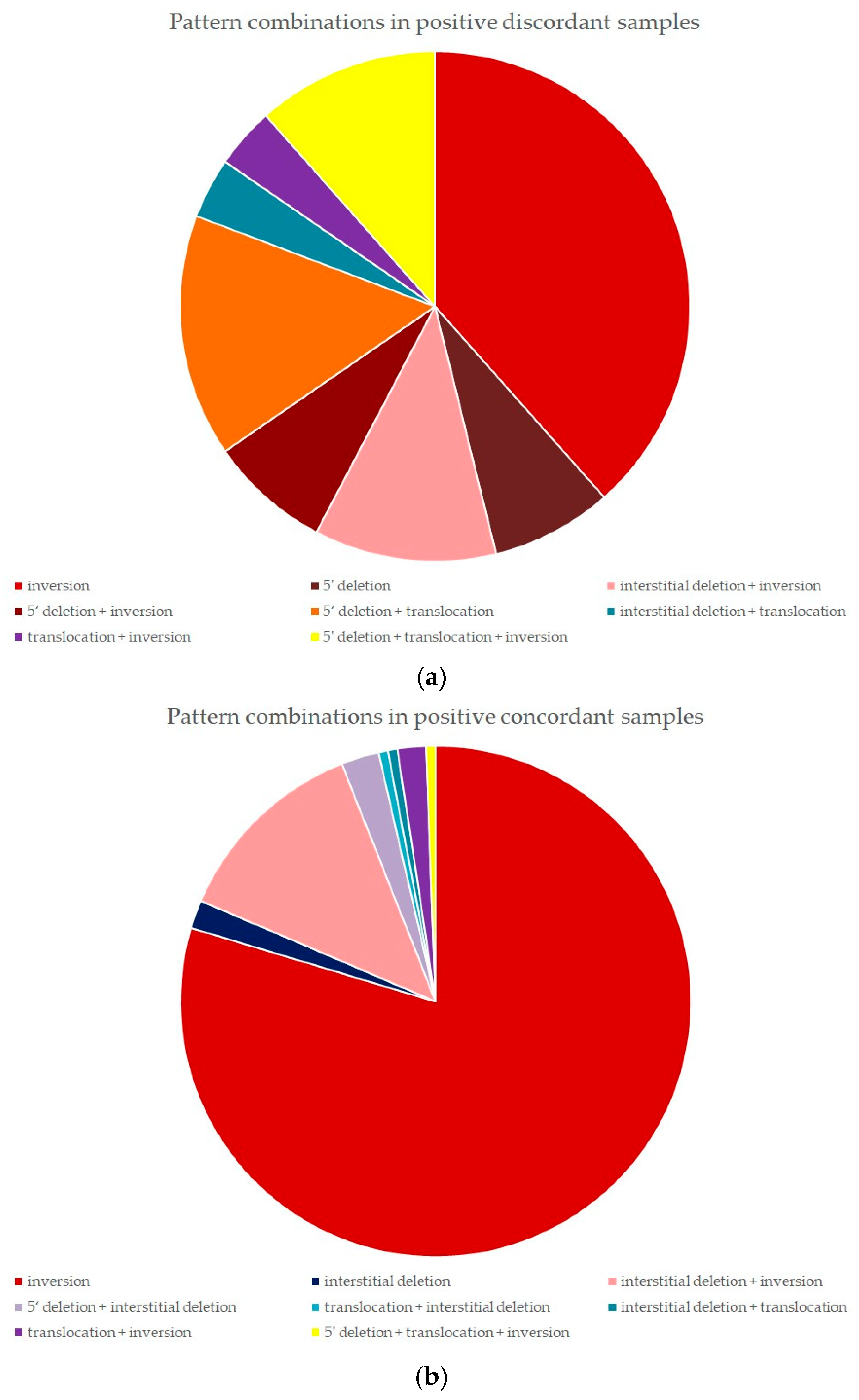
2.5. FISH Pattern Distribution in Positive Discordant and Positive Concordant Samples

A total of 197 samples previously tested with ALK break apart probe were analyzed with ALK/EML4 fusion probe and classified according to their predominant pattern of rearrangement as described in Section 4. The cohort consisted of 29 positive discordant samples and 168 positive concordant samples.

2.5.1. Positive Discordant Samples

In 13 samples (44.8%), the positive result was a combination of several signal variants each below 15%. In five of them, it was possible after elimination of sectioning artifacts to identify one pattern of rearrangement (all inversion). In seven samples, two patterns were identified, and in one sample, three patterns were found. In 16 samples (51.8%), at least one pattern of rearrangement reached the cut off. Seven of them were evaluated as containing one rearrangement pattern (five cases of inversion, two cases of 5′ deletion); in seven samples, two patterns of rearrangement were identified, and in two samples, three patterns of rearrangement were identified (Table 4).
One pattern was overall identified in 12 out of 29 samples (41.4%), two patterns were found in 14 samples (48.3%) and three patterns in 3 samples (10.3%). Inversion was identified in 10 out of 12 samples with single rearrangement pattern (83.3%). Another single rearrangement pattern was 5′ deletion, in 2 cases (16.7%). Interstitial deletion and translocation did not occur as single identified patterns, only in combinations.
Out of two pattern combinations, variants featuring mainly 5′ deletion were present in 6 out of 14 samples (42.8%), interstitial deletion as the predominant pattern was found in 4 samples (28.6%), inversion in 3 samples (21.4%), and translocation in 1 sample (7.1%). Three samples with three rearrangement patterns all contained 5′ deletion combined with translocation and inversion (in that order of percentage) (Figure 1).

2.5.2. Positive Concordant Samples

In two samples (1.2%), the positive result was a combination of several signal variants each below 15%, which were, however, identified as resulting from inversion. In 166 cases (98.8%), at least one signal variant met or exceeded the 15% cut-off. A single pattern of rearrangement was overall found in 137 out of 168 samples (81.5%). A total of 30 samples (17.8%) were classified as containing two patterns. One sample featured three rearrangement patterns (0.6%) (Table 4). Samples with one pattern identified featured almost exclusively inversion in 133 samples. Four samples (3%) displayed the interstitial deletion pattern. Translocation and 5′ end deletion did not occur as a single pattern, only in combinations.
A total of 30 samples had a combination of two patterns, with the most frequent one being interstitial deletion and inversion in 21 samples (70%), followed by 5′ deletion coupled with interstitial deletion in four samples (13.3%), as well as translocation combined with inversion in three samples (10%). Interstitial deletion combined with translocation, and the reverse order variant translocation combined with interstitial deletion, were found in one case (3.3%) each. One sample with three identified patterns featured 5′ deletion, translocation, and inversion (Figure 1).

2.5.3. Statistical Analysis of Pattern Distribution

There was a statistically significant association (p-value 0.0000055) between the type of sample (concordant/discordant) and variant of FISH signal (three patterns). Using the standard calibration of Cramer’s V for 2 degrees of freedom, the association may be judged strong (Cramer’s V is 0.37, with 95% confidence interval (0.24, 0.51)). Pattern frequency did not show a statistically significant association between the type of sample (concordant/discordant) (Supplementary Materials File S1).

2.6. NGS Analysis

NGS analysis was successfully performed on 75 samples: 58 negative concordant, 4 positive concordant, 11 positive discordant, and 2 negative discordant cases. NGS did not identify ALK fusion in any of the negative discordant cases (other alterations, not listed in this publication, were found). NGS identified ALK::EML4 fusion in three positive cases and cases and HIP::ALK fusion in one positive concordant case. In 10 positive discordant cases, ALK fusion was not detected, and one case resulted in identification of ALK::EML4 fusion transcript. In two negative discordant samples, NGS detected ALK fusion transcripts (ALK::EML4 and ALK::CSFT3) (Table 5).

3. Discussion

Our main concern was to reevaluate the results of the applied tests of samples with discordant results and reevaluate correct interpretations of all the analyses. In the second step, comparison between methods was performed. This process revealed striking differences between positive discordant and positive concordant cases.
FISH patterns of rearrangement in the positive concordant cohort almost always (98.8%) reached levels above the cut-off value and consisted mainly of one common pattern of rearrangement (81.5%) identified in all positive nuclei. The positive discordant cohort partially (44.8%) consisted of cases where the positive result was reached by a combination of signal variants below the 15% cut-off. Higher variability in number of rearrangement patterns was also recorded (41.4% cases had one rearrangement pattern, two patterns were found in 48.3% samples and three patterns in 10.3% of samples), which was proven to be statistically significant. Heterogeneity of ALK rearrangement in positive discordant samples might be therefore responsible for the lack of ALK protein expression, and the ALK status should be evaluated by another method.
The positive concordant group showed high predominance of tumor samples featuring the classic ALK::EML4 inversion, either as the single pattern (97% of samples with one pattern identified and 79.1% of all positive concordant samples) or very frequently coupled with interstitial deletion (70% of samples with two patterns identified). In contrast, inversion was the most frequently occurring single pattern in positive discordant samples as well (83.3% of cases with one pattern identified), but overall, it comprised only approximately over a third of all positive discordant cases.
5′ deletion never occurred on its own in the positive discordant group, but it represented the main rearrangement pattern in 5 out of 31 samples with two or three identified patterns (16.1%). In the positive discordant group, it was found as the only pattern in two cases, and as the main pattern in 9 out of 17 (52.9%) samples with two or more identified patterns of rearrangement.
The FISH result featuring the isolated 5‘ orange signal with negative ALK protein expression may signify a false positive case, as was examined in a study by Gao et al. [27], in which IHC-negative patients with this FISH signal variant did not fit into the typical profile of ALK-positive patients, with older age at diagnosis and more extensive smoking history. The FISH result was estimated to be caused by structural variants affecting the 5′ binding site, associated with higher genomic instability of the tumor typical for prolonged smoking; therefore, it likely did not represent true ALK rearrangement. Clavé et al. [28] describe two IHC-negative cases with a predominant 5′ deletion signal that tested negative for ALK fusion using NGS, with the recommendation to always retest this particular rearrangement variant. A similar finding was reported by Dacic et al. [29], where NGS confirmed ALK fusion only in 4 out of 10 cases with 5′ end deletion pattern only, while all showed IHC negativity.
IHC-positive cases, regardless of FISH status, tended to have fewer ALK CNG, as seen in 129 (68.25%) positive concordant cases, compared to 12 (36.36%) positive discordant cases. Multiple copies of ALK can be caused by ALK amplification or polysomy, but neither variant shows any clinical significance for indication of TKI treatment [30]. Increased CNG likely comes from early genetic events triggered by cigarette smoking, independent of true ALK rearrangement, and is associated with genomic instability and more aggressive biological behavior, as well as potential higher sensitivity to chemotherapy [31,32], and also including one of the mechanisms of crizotinib resistance both in vitro and in patients progressing on TKI treatment [33,34]. Lower CNG in IHC-positive cases may indicate a more stable genomic profile compared to higher heterogeneity of positive discordant cases.
Cases with a notable presence of multiple components were described as such in the FISH report, and in our cohort were found predominantly in the positive discordant group, including 4 out of 13 positive discordant samples re-tested with NGS (cases 6, 9, 10, and 11). In these cases, either the ALK rearranged part represented only a small component of the overall tumor mass (cases 9 and 11), or these tumors contained two different components (adenocarcinoma and squamous cell carcinoma for case 6, well-differentiated and poorly differentiated component in case 10). The pattern of rearrangement included 5‘ end deletion (case 9), inversion (case 6, only adenocarcinoma component) and translocation with inversion (poorly differentiated area of case 10). A small part of the tumor cells may display alterations of the ALK locus, but its clinical significance and targetability by TKIs remains questionable. The patients’ age in this subgroup of four patients (two male patients aged 63 and 78 and two female patients aged 70 and 79) also fell outside of the typical profile of ALK-positive patients; however, this parameter cannot be reliably used to determine ALK status. It is worth noting, though, that NGS analysis did not detect ALK fusion in any of these cases.
Out of 11 positive discordant cases tested with NGS, only one case (Case 3, Table 5) revealed by NGS the ALK::EML4 fusion transcript, and in all the other the NGS did not demonstrate ALK fusion. In comparison, ALK fusion was identified in two out of two negative discordant cases tested by NGS. One case (Case 12, Table 4) featured predominantly an unusual 3′ deletion pattern in 74% of evaluated nuclei. The 3′ end of the hybridization probe binds to the tyrosine-kinase domain of the ALK gene necessary for activation of signal pathways; therefore, this alteration has uncertain clinical implications. Although the ALK locus was obviously altered in this sample and ALK protein expression was detected, it could not be classified as ALK positive under valid guidelines. However, NGS analysis revealed ALK::EML4 fusion transcript and KLF7::EML4 fusion as an additional finding.
NGS analysis in our cohort corresponded more with positive protein IHC expression than with FISH results, and similar results were also reported by Lin et al. [18]. We might agree with the authors that it does not imply FISH to be less reliable, but that it rather points out the technical challenges of interpreting FISH and higher requirements for tissue quality to be eligible for testing. Our own experience in detailed analysis of all occurring signal variants offers a plausible explanation: FISH might be able to detect alterations in ALK locus associated with more extensive mutational profile. It is not entirely correct to consider these FISH results as false positives, if the method is performed adequately and interpreted correctly, but notable heterogeneity in detected rearrangement patterns, along with increased ALK CNGs in cases with negative ALK protein expression, can indicate more severe genomic alterations in the analyzed sample. All such cases should be therefore verified using NGS.

4. Materials and Methods

A team composed of the Pathology Department of JFM CU and UH Martin and Martin’s Biopsy Center, Ltd., performed the detection of ALK protein expression using D5F3 antibody in 2813 NSCLC specimens, with all EGFR unmutated as examined by RT-PCR, as a part of the standard diagnosis algorithm. FFPE samples of either primary metastatic NSCLC were sent from other departments to undergo further molecular analysis to detect relevant predictive biomarkers of either targeted treatment and/or immunotherapy, including ALK, ROS-1, MET, RET, and NTRK1/2/3 alterations and PDL-1 status. ALK rearrangement was tested by two standard methods: on the level of protein expression by IHC, and on the cytogenetic level using FISH. Discordant cases were carefully evaluated, and a small fraction of cases was additionally analyzed using NGS as a part of a pilot study.

4.1. Immunohistochemistry

Immunohistochemical analysis of ALK protein expression was performed on 2813 of 3–4 µm thick FFPE sections using monoclonal antibody VENTANA anti-ALK (clone D5F3, Roche Molecular Systems, Inc., Rotkreuz, Switzerland) on the BenchMark GX platform (Roche Molecular Systems, Inc., Rotkreuz, Switzerland). The analysis was realized according to the protocol recommended by the manufacturer. Sections were kept in the thermostat at 37 °C all night and then were placed into BenchMark GX. After staining, sections were rinsed in running water for 10 min, dehydrated with graded alcohol and xylene, and coverslipped.
IHC quality control was performed through QuiP (Qualitatssicherungs-Initiative Pathologie GmbH, Berlin, Germany). Internal control was a part of every analyzed slide, using tissue from another ALK-positive case, which was exchanged regularly when the block aged. The whole slide was stained, but only the tumor area was evaluated for ALK protein expression. The outcomes were evaluated semiquantitatively according to the following criteria: the presence of strong diffuse granular cytoplasmic staining (2+, 3+). In the reevaluation stage, the so-called dot-like positivity was also labelled as positive. No staining, weak cytoplasmic staining (1+), or so-called stippled pattern were all interpreted as negative.

4.2. Fluorescence In Situ Hybridization

FISH Analysis

FISH analysis was performed on 3–4 µm thick formalin-fixed paraffin-embedded (FFPE) tissue sections using ZytoLight®SPEC ALK Dual Color Break Apart Probe (ZytoVision, GmbH, Bremerhaven, Germany). FFPE specimens were processed manually using a ZytoLight®FISH-Tissue Implementation Kit according to the manufacturer’s instructions. Results were analyzed using a fluorescence BX61 microscope (Olympus, Tokyo, Japan) with the support of the software Lucia FISH v.3.1 (Laboratory Imaging, Prague, Czech Republic).
ALK rearrangement was scored according to the valid guidelines [35]. In all cases with sufficient tumor cell count, 100 nuclei were enumerated. If material was critically limited, then the evaluated tumor cell count was lower than 100 but had to include minimally 50 cells. Cases with less than 50 cells were excluded from our study.
The presence of two isolated signals (3′ orange signal and 5′ green signal) at least two-signal diameter apart and/or a single 3′-isolated orange signal were marked as positive, demonstrating a split in the structure of ALK gene. An orange/green fusion signal indicated an intact ALK locus, andtherefore it was interpreted as negative. Specimens with ≥15% of positive cells were classified as positive for ALK rearrangement, while cases with <15% were classified as negative. All positive and some equivocal samples displaying the so-called short break pattern (less than two-signal diameter) were retested again using the ZytoLight ®SPEC ALK/EML4 TriCheck™Probe. ALK rearrangement was scored according to the manufacturer’s instructions [36].
ALK copy number gain (CNG) was evaluated using the ALK break apart probe, without distinction between polysomy and amplification, displayed as cells with multiple copies of an intact ALK locus (multiple fusion signals in a single nucleus). Four categories were established: “three or less ALK CNGs”, “4–6 ALK CNGs”, “7–9 ALK CNGs”, and “10+ ALK CNGs”.
Positive discordant and positive concordant cases retested with ALK/EML4 fusion probe were compared in terms of FISH rearrangement variants.
Translocation with other than the EML4 partner was indicated by the presence of (a) isolated 3′ orange signal and isolated 5′ green signal co-localizing with blue signal (O/GB); (b) isolated 3′ orange signal and isolated 5′ green signal (O/G); or (c) isolated 3′ orange signal, isolated 5′ green signal, and isolated blue signal (O/G/B).
ALK/EML4 inversion pattern included one or more of the following variants: (a) isolated 3′ orange signal co-localizing with blue signal and isolated 5′ green signal co-localizing with blue signal (Figure 2) (OB/GB); (b) isolated 3′ orange signal co-localizing with blue signal and isolated 5′ green signal (OB/G); (c) isolated 3′orange signal co-localizing with blue signal, isolated blue signal, and isolated 5′ green signal (OB/B/G); or (d) inversion with partial ALK gene deletion, represented by isolated 3′ orange signal co-localizing with blue signal and another isolated blue signal (OB/B).
The 5′ end deletion of the ALK gene was represented by two variants: (a) isolated 3′ orange signal, loss of 5′ green signal (O), or (b) isolated 3′ orange signal, isolated blue signal, loss of 5′green signal (O/B).
Interstitial deletion of the ALK gene was indicated by the presence of an isolated 3′ orange signal co-localizing with a blue signal (OB).
When analyzing samples, either at least one signal combination on its own reached above the 15% cut-off, or the positive result was composed of several rearrangement variants, each with less than 15% of positivity, but at the very least found in 4% of rearranged nuclei (Figure 3).
The predominant pattern of rearrangement was the signal combination with the highest percent of positivity in rearranged cells. All other identified patterns were then evaluated based on whether or not they could have been derived from the predominant pattern. If the minority pattern was less complex, featured less signals, or its configuration was a part of the predominant pattern, it was not considered a secondary pattern, but most likely a result of tissue sectioning. If the minority pattern was found in at least 4% of rearranged nuclei, was more complex, featured more fluorescent signals, or its structure could not have been derived from the predominant pattern, it was considered a secondary (or tertiary) pattern of rearrangement. Details and examples of this assortment can be found in Supplementary Materials File S2.

4.3. Next-Generation Sequencing

Sequencing was performed with the panel TruSight Oncology 500 Assay (Illumina, San Diego, CA, USA), enabling simultaneous analysis of DNA and RNA extracted from the same sample. This assay provides detection of SNVs and indels in 523 cancer-related genes, CNVs of 69 genes, gene fusions and splice variant of 55 driver genes, and immunotherapy biomarkers that rely on the analysis of multiple genomic loci such as tumor mutational burden (TMB) and microsatellite instability (MSI).
Tissue was sectioned into 4 to 6 10 μm thick slices and deparaffinized using Qiagen solution. Isolated DNA was treated with RNAse A to eliminate contamination from RNA; similarly, RNA was treated with DNAse A to ensure high purity of nucleic acids.
DNA quantity was measured with a Qubit dsDNA BR kit (Invitrogen (Carlsbad, CA, USA), Thermo Fisher Scientific (Waltham, MA, USA)) on the Qubit 4 Flurometer platform, (Thermo Fisher Scientific, Waltham, MA, USA). The minimal required amount of DNA was 80 ng/sample; samples that did not meet this criterion were excluded. Quality control was performed with Infinium FFPE QC kit (Illumina, USA) according to the manufacturer’s guidelines.
RNA quality was measured with an RNA 6000 Nano kit (Agilent, Santa Clara, CA, USA), and the level of DV200 was assessed. This number corresponded to the percentage of RNA fragments with length greater than 200 bp. DV200 < 30% signified a high level of RNA degradation not suitable for NGS analysis, and these samples were excluded. Samples with a concentration of RNA higher than 10 ng/μL and DV200 > 30% were used for the preparation of libraries.
According to the manufacturer’s instruction for Illumina TruSight 500 Oncology Assay, 16 libraries were manually processed, containing 8 DNA and 8 cDNA. Finally, libraries with concentration >3 ng/µL were normalized, pooled, and loaded on a NextSeq 550Dx (Illumina, San Diego, CA, USA). The paired-end sequencing runs (2 × 101 bp) were performed in high output mode with a High Output Kit v2.5 (300 Cycles). Output from NGS was subjected to bioinformatic processing by Illumina LocalApp ver. 2.2.0. There is no cutoff on variant allele frequency (VAF) in the LocalApp pipeline. The cutoff there is set in a dynamic manner, depending on loci and sample quality, and it can be as low as 2–3 mutant reads. In addition, there are two quality scores: AQ (variant artifact adjusted quality score; p-value against baseline samples) and LQ (likelihood ratio quality score; sample specific p-value). Point mutations were identified as one nucleotide, insertions as the length of 18 bp, and deletion between 10 and 25 bp.
For the fusion calling, the unique reads were used. In order for a fusion to be called with high confidence, at least 5 unique reads were needed. Concerning the copy number variation (CNV), the TSO500 panel allows for CNV calling in 59 genes. Each gene had a different noise profile. For this reason, each gene had specific thresholds for calling amplifications and deletions. Samples with a tumor content above 20% were analyzed in our study (for amplification, it means 14 copies at the 20% tumor purity).
The results of bionformatic processing were afterwards annotated by the PierianDX Clinical Genomics Workspace (CGW). The annotation involves, among other things, ranking of the called SNVs, CNVs, and fusions into tiers, according to standards and guidelines for the interpretation and reporting of sequence variants in cancer: a joint consensus recommendation of the Association for Molecular Pathology, American Society of Clinical Oncology, and College of American Pathologists [37].

4.4. Comparison of IHC and FISH

Sensitivity of FISH was calculated as the number of positive concordant results divided by positive concordant and FISH-negative discordant cases. Specificity was calculated as the number of negative concordant results divided by negative discordant and FISH-positive discordant results. Concordance between ALK rearrangement and ALK protein expression was performed. Demographic characteristics of population groups were evaluated, and differences in ALK rearrangement among population groups were assessed.
FISH results of positive concordant and FISH-positive discordant cases were further sorted according to patterns detectable using ALK/EML4 fusion probe. The total number of detected patterns was recorded in FISH-positive concordant and FISH-positive discordant cases, and the predominant pattern of rearrangement was identified. If the sample contained more patterns, their combination was also documented. ALK CNG was compared between positive concordant and all discordant cases.

4.5. Statistical Analysis

Data were explored and analyzed in R [1], ver. 4.0.5, with the aid of libraries [38,39,40,41]. Contingency tables were visualized by mosaicplot. The null hypothesis of no association between two factors was tested by Fisher’s exact test. Effect size was measured by Cramer’s V, and its uncertainty was quantified by a 95% double-sided chi-squared confidence interval. Where there was row of 0 frequencies in a contingency table, the Laplace smoothing was applied to the table prior to hypothesis testing.

5. Conclusions

Our department performed simultaneous detection of ALK rearrangement using both FISH and IHC, which aided in more reliable detection of ALK rearrangement in case of failure of one of the methods. FISH results are interpreted with detailed description of all occurring patterns and CNGs, and a thorough look at the ALK rearrangement variants might contribute to the explanation of the lacking protein expression in positive discordant cases.
Positive concordant samples tended to display the classic inversion and also show less variability in detected variants, suggesting that the genomic profile of the tumor population is more stable compared to the IHC-negative cases, which correlates with the typical demographic group of younger, non-smoker patients. Positive discordant cases were associated with a higher variability of detected rearrangement variants, as well as with the 5′ deletion pattern. FISH-positive cases without ALK protein expression displaying one of these characteristics should be promptly verified with another method.
NGS results seem to show a correlation with ALK-protein-positive expression in our limited cohort. In conclusion, the simultaneous use of IHC and FISH, as is the standard in many departments including ours, allows for the selection of the highest number of patients eligible for targeted treatment. Further implementation of NGS is crucial in improving diagnosis and detection of other predictive biomarkers and indicating the best possible treatment for NSCLC patients.

Supplementary Materials

The following supporting information can be downloaded at https://www.mdpi.com/article/10.3390/ijms25158168/s1.

Author Contributions

Conceptualization, K.T., M.B., and L.P.; FISH evaluation, M.B. and Ľ.J.; IHC analysis, L.P. and A.F.; NGS analysis, D.L. and M.G.; statistical analysis, M.G.; data curation, K.T.; writing—original draft preparation, K.T.; writing—review and editing, K.T., M.B., K.L., and L.P. All authors have read and agreed to the published version of the manuscript.

Funding

This research received funding from project ITMS 313011V446 “Integrative strategy in development of personalized medicine of specified malignant tumours and its effect on quality of life“ to perform NGS analyses, currently not covered by health insurance in Slovakia.

Institutional Review Board Statement

Ethical review and approval were waived for this study due to the retrospective analysis of the fully anonymized data used in the study. The consent to use biological material for diagnostic and research purposes was included during admittance to the healthcare facility.

Informed Consent Statement

Informed consent was obtained from all subjects involved in the study.

Data Availability Statement

The data presented in this study are available on request from the corresponding author.

Conflicts of Interest

Authors Anna Farkašová and Lukáš Plank were employed by the company Martin’s Biopsy Center, Ltd. The remaining authors declare that the research was conducted in the absence of any commercial or financial relationships that could be construed as a potential conflict of interest.

References

  1. Hendriks, L.E.; Kerr, K.M.; Menis, J.; Mok, T.S.; Nestle, U.; Passaro, A.; Peters, S.; Planchard, D.; Smit, E.F.; Solomon, B.J.; et al. Oncogene-addicted metastatic non-small-cell lung cancer: ESMO Clinical Practice Guideline for diagnosis, treatment and follow-up. Ann. Oncol. 2023, 34, 339–357. [Google Scholar] [CrossRef] [PubMed]
  2. Thai, A.A.; Solomon, B.J.; Sequist, L.V.; Gainor, J.F.; Heist, R.S. Lung cancer. Lancet 2021, 398, 535–554. [Google Scholar] [CrossRef] [PubMed]
  3. Morris, S.W.; Kirstein, M.N.; Valentine, M.B.; Dittmer, K.G.; Shapiro, D.N.; Saltman, D.L.; Look, A.T. Fusion of a kinase gene, ALK, to a nucleolar protein gene, NPM, in non-Hodgkin’s lymphoma. Science 1994, 263, 1281–1284. [Google Scholar] [CrossRef] [PubMed]
  4. Ou, S.H.; Bartlett, C.H.; Mino-Kenudson, M.; Cui, J.; Iafrate, A.J. Crizotinib for the treatment of ALK-rearranged non-small cell lung cancer: A success story to usher in the second decade of molecular targeted therapy in oncology. Oncologist 2012, 17, 1351–1375. [Google Scholar] [CrossRef] [PubMed]
  5. Millett, R.L.; Elkon, J.M.; Tabbara, I.A. Directed therapies in anaplastic lymphoma kinase-rearranged non-small cell lung cancer. Anticancer Res. 2018, 38, 4969–4975. [Google Scholar] [CrossRef] [PubMed]
  6. Kwak, E.L.; Bang, Y.J.; Camidge, D.R.; Shaw, A.T.; Solomon, B.; Maki, R.G.; Ou, S.H.; Dezube, B.J.; Jänne, P.A.; Costa, D.B.; et al. Anaplastic lymphoma kinase inhibition in non-small-cell lung cancer. N. Engl. J. Med. 2010, 363, 1693–1703. [Google Scholar] [CrossRef] [PubMed]
  7. Camidge, D.R.; Bang, Y.J.; Kwak, E.L.; Iafrate, A.J.; Varella-Garcia, M.; Fox, S.B.; Riely, G.J.; Solomon, B.; Ou, S.H.; Kim, D.W.; et al. Activity and safety of crizotinib in patients with ALK-positive non-small-cell lung cancer: Updated results from a phase 1 study. Lancet Oncol. 2012, 13, 1011–1019. [Google Scholar] [CrossRef] [PubMed]
  8. Shaw, A.T.; Kim, D.W.; Mehra, R.; Tan, D.S.; Felip, E.; Chow, L.Q.; Camidge, D.R.; Vansteenkiste, J.; Sharma, S.; De Pas, T.; et al. Ceritinib in ALK-rearranged non-small-cell lung cancer. N. Engl. J. Med. 2014, 370, 1189–1197. [Google Scholar] [CrossRef]
  9. Peters, S.; Camidge, D.R.; Shaw, A.T.; Gadgeel, S.; Ahn, J.S.; Kim, D.W.; Ou, S.I.; Pérol, M.; Dziadziuszko, R.; Rosell, R.; et al. Alectinib versus Crizotinib in Untreated ALK-Positive Non-Small-Cell Lung Cancer. N. Engl. J. Med. 2017, 9, 829–838. [Google Scholar] [CrossRef]
  10. Gettinger, S.N.; Bazhenova, L.A.; Langer, C.J.; Salgia, R.; Gold, K.A.; Rosell, R.; Shaw, A.T.; Weiss, G.J.; Tugnait, M.; Narasimhan, N.I.; et al. Activity and safety of brigatinib in ALK-rearranged non-small-cell lung cancer and other malignancies: A single-arm, open-label, phase 1/2 trial. Lancet Oncol. 2016, 12, 1683–1696. [Google Scholar] [CrossRef] [PubMed]
  11. Horn, L.; Infante, J.R.; Blumenschein, G.R.; Wakelee, H.A.; Arkenau, H.; Dukart, G.; Liang, C.; Harrow, K.; Gibbons, J.; Lovly, C.M.; et al. A phase I trial of X-396, a novel ALK inhibitor, in patients with advanced solid tumors. J. Clin. Oncol. 2014, 32, 8030. [Google Scholar] [CrossRef]
  12. Camidge, D.R.; Kono, S.A.; Flacco, A.; Tan, A.C.; Doebele, R.C.; Zhou, Q.; Crino, L.; Franklin, W.A.; Varella-Garcia, M. Optimizing the detection of lung cancer patients harboring anaplastic lymphoma kinase (ALK) gene rearrangements potentially suitable for ALK inhibitor treatment. Clin. Cancer Res. 2010, 16, 5581–5590. [Google Scholar] [CrossRef] [PubMed]
  13. Paez, J.G.; Jänne, P.A.; Lee, J.C.; Tracy, S.; Greulich, H.; Gabriel, S.; Herman, P.; Kaye, F.J.; Lindeman, N.; Boggon, T.J.; et al. EGFR mutations in lung cancer: Correlation with clinical response to gefitinib therapy. Science 2004, 304, 1497–1500. [Google Scholar] [CrossRef] [PubMed]
  14. Rekhtman, N.; Leighl, N.B.; Somerfield, M.R. Molecular testing for selection of patients with lung cancer for epidermal growth factor receptor and anaplastic lymphoma kinase tyrosine kinase inhibitors: American society of clinical oncology endorsement of the college of american pathologists/international association for the study of lung cancer/association for molecular pathology guideline. J. Oncol. Pract. 2015, 2, 135–136. [Google Scholar] [CrossRef]
  15. Wynes, M.W.; Sholl, L.M.; Dietel, M.; Schuuring, E.; Tsao, M.S.; Yatabe, Y.; Tubbs, R.R.; Hirsch, F.R. An international interpretation study using the ALK IHC antibody D5F3 and a sensitive detection kit demonstrates high concordance between ALK IHC and ALK and between evaluators. J. Thorac. Oncol. 2014, 9, 631–638. [Google Scholar] [CrossRef]
  16. Cabillic, F.; Gros, A.; Dugay, F.; Begueret, H.; Mesturoux, L.; Chiforeanu, D.C.; Dufrenot, L.; Jauffret, V.; Dachary, D.; Corre, R.; et al. Parallel FISH and immunohistochemical studies of ALK status in 3244 non-small-cell lung cancers reveal major discordances. J. Thorac. Oncol. 2014, 3, 295–306. [Google Scholar] [CrossRef]
  17. Ali, S.M.; Ou, S.I.; He, J.; Peled, N.; Chmielecki, J.; Pinder, M.C.; Palma, N.A.; Akerley, W.L.; Wang, K.; Molina, J.R.; et al. Identifying ALK rearrangements that are not detected by FISH with targeted next-generation sequencing of lung carcinoma. J. Clin. Oncol. 2014, 32, 8049a. [Google Scholar] [CrossRef]
  18. Lin, C.; Shi, X.; Yang, S.; Zhao, J.; He, Q.; Jin, Y.; Yu, X. Comparison of ALK detection by FISH, IHC and NGS to predict benefit from crizotinib in advanced non-small cell lung cancer. Lung Cancer 2019, 131, 62–68. [Google Scholar] [CrossRef]
  19. Savic, S.; Diebold, J.; Zimmermann, A.K.; Jochum, W.; Baschiera, B.; Grieshaber, S.; Tornillo, L.; Bisig, B.; Kerr, K.; Bubendorf, L. Screening for ALK in non-small cell lung carcinomas: 5A4 and D5F3 antibodies perform equally well, but combined use with FISH is recommended. Lung Cancer 2015, 2, 104–109. [Google Scholar] [CrossRef]
  20. Sholl, L.M.; Weremowicz, S.; Gray, S.W.; Wong, K.K.; Chirieac, L.R.; Lindeman, N.I.; Hornick, J.L. Combined use of ALK immunohistochemistry and FISH for optimal detection of ALK-rearranged lung adenocarcinomas. J. Thorac. Oncol. 2013, 3, 322–328. [Google Scholar] [CrossRef]
  21. Sun, J.M.; Choi, Y.L.; Won, J.K.; Hirsch, F.R.; Ahn, J.S.; Ahn, M.; Park, K. A dramatic response to crizotinib in a non-small-cell lung cancer patient with IHC-positive and FISH-negative ALK. J. Thorac. Oncol. 2012, 7, e36–e38. [Google Scholar] [CrossRef] [PubMed]
  22. Peled, N.; Palmer, G.; Hirsch, F.R.; Wynes, M.W.; Ilouze, M.; Varella-Garcia, M.; Soussan-Gutman, L.; Otto, G.A.; Stephens, P.J.; Ross, J.S.; et al. Next-generation sequencing identifies and immunohistochemistry confirms a novel crizotinib-sensitive ALK rearrangement in a patient with metastatic non-small-cell lung cancer. J. Thorac. Oncol. 2012, 7, e14–e16. [Google Scholar] [CrossRef] [PubMed]
  23. Van der Wekken, A.J.; Pelgrim, R.; Hart, N.; Werner, N.; Mastik, M.F.; Hendriks, L.; van der Heijden, E.H.F.M.; Looijen-Salamon, M.; de Langen, A.J.; Staal-van den Brekel, J.; et al. Dichotomous ALK-IHC Is a Better Predictor for ALK Inhibition Outcome than Traditional ALK-FISH in Advanced Non-Small Cell Lung Cancer. Clin. Cancer Res. 2017, 23, 4251–4258. [Google Scholar] [CrossRef] [PubMed]
  24. Cabillic, F.; Hofman, P.; Ilie, M.; Peled, N.; Hochmair, M.; Dietel, M.; Von Laffert, M.; Gosney, J.R.; Lopez-Rios, F.; Erb, G.; et al. ALK IHC and FISH discordant results in patients with NSCLC and treatment response: For discussion of the question-to treat or not to treat? ESMO Open 2018, 3, e000419. [Google Scholar] [CrossRef] [PubMed]
  25. Scattone, A.; Catino, A.; Schirosi, L.; Caldarola, L.; Tommasi, S.; Lacalamita, R.; Montagna, E.S.; Galetta, D.; Serio, G.; Zito, F.A.; et al. Discordance between FISH, IHC, and NGS Analysis of ALK Status in Advanced Non-Small Cell Lung Cancer (NSCLC): A Brief Report of 7 Cases. Transl. Oncol. 2019, 12, 389–395. [Google Scholar] [CrossRef] [PubMed]
  26. Thunnissen, E.; Lissenberg-Witte, B.I.; van den Heuvel, M.M.; Monkhorst, K.; Skov, B.G.; Sørensen, J.B.; Mellemgaard, A.; Dingemans, A.M.C.; Speel, E.J.M.; de Langen, A.J.; et al. ALK immunohistochemistry positive, FISH negative NSCLC is infrequent, but associated with impaired survival following treatment with crizotinib. Lung Cancer 2019, 138, 13–18. [Google Scholar] [CrossRef]
  27. Gao, X.; Sholl, L.M.; Nishino, M.; Heng, J.C.; Jänne, P.A.; Oxnard, G.R. Clinical Implications of Variant ALK FISH Rearrangement Patterns. J. Thorac. Oncol. 2015, 11, 1648–1652. [Google Scholar] [CrossRef] [PubMed]
  28. Clavé, S.; Rodon, N.; Pijuan, L.; Díaz, O.; Lorenzo, M.; Rocha, P.; Taus, Á.; Blanco, R.; Bosch-Barrera, J.; Reguart, N.; et al. Next-generation Sequencing for ALK and ROS1 Rearrangement Detection in Patients With Non-small-cell Lung Cancer: Implications of FISH-positive Patterns. Clin. Lung Cancer 2019, 20, e421–e429. [Google Scholar] [CrossRef] [PubMed]
  29. Dacic, S.; Villaruz, L.C.; Abberbock, S.; Mahaffey, A.; Incharoen, P.; Nikiforova, M.N. ALK FISH patterns and the detection of ALK fusions by next generation sequencing in lung adenocarcinoma. Oncotarget 2016, 7, 82943–82952. [Google Scholar] [CrossRef]
  30. Zito Marino, F.; Botti, G.; Aquino, G.; Ferrero, S.; Gaudioso, G.; Palleschi, A.; Rocco, D.; Salvi, R.; Micheli, M.C.; Micheli, P.; et al. Unproductive Effects of ALK Gene Amplification and Copy Number Gain in Non-Small-Cell Lung Cancer. ALK Gene Amplification and Copy Gain in NSCLC. Int. J. Mol. Sci. 2020, 21, 4927. [Google Scholar] [CrossRef] [PubMed]
  31. Peretti, U.; Ferrara, R.; Pilotto, S.; Kinspergher, S.; Caccese, M.; Santo, A.; Brunelli, M.; Caliò, A.; Carbognin, L.; Sperduti, I.; et al. ALK gene copy number gains in non-small-cell lung cancer: Prognostic impact and clinico-pathological correlations. Respir. Res. 2016, 17, 105. [Google Scholar] [CrossRef] [PubMed]
  32. Salido, M.; Pijuan, L.; Martínez-Avilés, L.; Galván, A.B.; Cañadas, I.; Rovira, A.; Zanui, M.; Martínez, A.; Longarón, R.; Sole, F.; et al. Increased ALK gene copy number and amplification are frequent in non-small cell lung cancer. J. Thorac. Oncol. 2011, 6, 21–27. [Google Scholar] [CrossRef] [PubMed]
  33. Katayama, R.; Khan, T.M.; Benes, C.; Lifshits, E.; Ebi, H.; Rivera, V.M.; Shakespeare, W.C.; Iafrate, A.J.; Engelman, J.A.; Shaw, A.T. Therapeutic strategies to overcome crizotinib resistance in non-small cell lung cancers harboring the fusion oncogene EML4-ALK. Proc. Natl. Acad. Sci. USA 2011, 108, 7535–7540. [Google Scholar] [CrossRef] [PubMed]
  34. Katayama, R.; Shaw, A.T.; Khan, T.M.; Mino-Kenudson, M.; Solomon, B.J.; Halmos, B.; Jessop, N.A.; Wain, J.C.; Yeo, A.T.; Benes, C.; et al. Mechanisms of acquired crizotinib resistance in ALK-rearranged lung Cancers. Sci. Transl. Med. 2012, 4, 120ra17. [Google Scholar] [CrossRef] [PubMed]
  35. Thunnissen, E.; Bubendorf, L.; Dietel, M.; Elmberger, G.; Kerr, K.; Lopez-Rios, F.; Moch, H.; Olszewski, W.; Pauwels, P.; Penault-Llorca, F.; et al. EML4-ALK testing in non-small cell carcinomas of the lung: A review with recommendations. Virchows Arch. 2012, 461, 245–257. [Google Scholar] [CrossRef] [PubMed]
  36. Schildhaus, H.U.; Binot, E.; Wolf, J. Validation of a Simplified Approach to Detect ALK Translocations in Lung Cancer Samples by FISH. Mod. Pathol. 2016, 29, 482A. [Google Scholar]
  37. Li, M.M.; Datto, M.; Duncavage, E.J.; Kulkarni, S.; Lindeman, N.I.; Roy, S.; Tsimberidou, A.M. Standards and guidelines for the interpretation and reporting of sequence variants in cancer: A joint consensus recommendation of the Association for Molecular Pathology, American Society of Clinical Oncology, and College of American Pathologists. J. Mol. Diagn. 2017, 19, 4–23. [Google Scholar] [CrossRef] [PubMed]
  38. Mayer, M. Confidence Intervals. R package Version 0.1.2. 2022. Available online: https://CRAN.R-project.org/package=confintr (accessed on 22 February 2024).
  39. Murdoch, D. Formula-Driven Table Generation. R Package Version 0.9.22. 2023. Available online: https://dmurdoch.github.io/tables/ (accessed on 22 February 2024).
  40. R Core Team. R: A Language and Environment for Statistical Computing; R Foundation for Statistical Computing: Vienna, Austria, 2021; Available online: https://www.R-project.org/ (accessed on 22 February 2024).
  41. Zhu, H. kableExtra: Construct Complex Table with ‘kable’ andPipe Syntax. R Package Version 1.3.4. 2021. Available online: https://CRAN.R-project.org/package=kableExtra (accessed on 22 February 2024).
Figure 1. (a) Pattern combinations identified in positive discordant samples. (b) Pattern combinations identified in positive concordant samples.
Ijms 25 08168 g001
Figure 2. ZytoLight ® SPEC ALK/EML4 TriCheck™ Probe Design, reprinted with permission from Schildhaus HU et al. [36] Signal interpretation guide, 2016. © 2024 ZYTOVISION GmbH.
Ijms 25 08168 g002
Figure 3. Classification of FISH patterns in samples analyzed with ALK/EML4 fusion probe.
Ijms 25 08168 g003
Table 1. ALK rearrangement detection and ALK discordance among population groups.
AgeTotalPositive FISHNegative FISHDiscordant Cases
FISH−, IHC+FISH+, IHC−
20–2962410
20–29 men20200
20–29 women42210
30–392891900
30–39 men1441000
30–39 women145900
40–491333310012
40–49 men75185702
40–49 women58154310
50–594875543217
50–59 men2962627006
50–59 women1912916211
60–691170761094618
60–69 men73729708110
60–69 women4334738658
70–797434170276
70–79 men5201650422
70–79 women2232519854
80–89114610810
80–89 men7126900
80–89 women4343910
90–9920200
90–99 men10100
90–99 women10100
total268322224611733
men1716951621320
women9671278401413
Table 2. Histopathological data as provided by sending departments (no second look option).
Histological TypeTotal (n = 239)Positive Discordant (n = 33)Negative Discordant (n = 17)Positive Concordant (n = 189)
Squamous cell carcinoma4 (1.67%)2 (6.06%)2 (11.76%)0 (0.00%)
Minimally invasive adenocarcinoma2 (0.83%)1 (3.03%)1 (5.88%)0 (0.00%)
Adenocarcinoma, NOS72 (30.12%)11 (33.33%)4 (23.53%)57 (30.16%)
Adenocarcinoma, NOS, mucin not detected6 (2.51%)1 (3.03%)0 (0.00%)5 (2.65%)
Adenocarcinoma, NOS, mucin producing8 (3.35%)0 (0.00%)0 (0.00%)8 (4.23%)
Lepidic predominant adenocarcinoma3 (1.26%)1 (3.03%)0 (0.00%)2 (1.06%)
Acinar predominant adenocarcinoma24 (10.04%)2 (6.06%)1 (5.88%)21 (11.11%)
Papillary predominant adenocarcinoma12 (5.02%)0 (0.00%)1 (5.88%)11 (5.82%)
Solid predominant adenocarcinoma29 (12.13%)4 (12.12%)4 (23.53%)21 (11.11%)
Micropapillary predominant adenocarcinoma6 (2.51%)0 (0.00%)0 (0.00%)6 (3.17%)
Signet ring cell adenocarcinoma4 (1.67%)0 (0.00%)0 (0.00%)4 (2.12%)
Mucinous adenocarcinoma9 (3.77%)1 (3.03%)0 (0.00%)8 (4.23%)
Clear cell adenocarcinoma1 (0.42%)1 (3.03%)0 (0.00%)0 (0.00%)
G3 adenocarcinoma, growth patterns not specified9 (3.77%)3 (9.09%)0 (0.00%)6 (3.17%)
G2 adenocarcinoma, growth patterns not specified1 (0.42%)0 (0.00%)0 (0.00%)1 (0.53%)
Pleomorphic carcinoma1 (0.42%)1 (3.03%)0 (0.00%)0 (0.00%)
Sarcomatoid carcinoma1 (0.42%)0 (0.00%)0 (0.00%)1 (0.53%)
Adenosquamous carcinoma3 (1.26%)1 (3.03%)0 (0.00%)2 (1.06%)
Combined NEC and adenocarcinoma2 (0.83%)1 (3.03%)0 (0.00%)1 (0.53%)
NSCLC, NOS3 (1.26%)0 (0.00%)0 (0.00%)3 (1.59%)
Metastatic adenocarcinoma26 (10.88%)3 (9.09%)2 (11.76%)21 (11.11%)
Data unavailable13 (5.44%)0 (0.00%)2 (11.76%)11 (5.82%)
Mucin production in one or more components41 (17.01%)2 (6.06%)1 (5.88%)38 (20.10%)
Mucin not detected11 (4.56%)3 (9.09%)2 (11.76%)6 (3.17%)
Not specified187 (77.59%)28 (84.85%)14 (82.35%)145 (76.72%)
Table 3. Comparison of ALK copy number gains in all discordant vs. positive concordant samples.
Positive Discordant (n = 33)Negative Discordant (n = 17)Positive Concordant
(n = 189)
3 or less ALK CNGs12 (36.36%)11 (64.70%)129 (68.25%)
4–6 ALK CNGs8 (24.24%)2 (11.76%)40 (21.16%)
7–9 ALK CNGs6 (18.18%)1 (5.88%)19 (10.05%)
10+ ALK CNGs7 (21.21%)3 (17.65%)1 (0.53%)
Table 4. Pattern distribution in positive concordant and positive discordant samples.
Patterns <15%At Least 1 Pattern ≥15%Total
Discordant samples (IHC−, FISH+)131629
 1 pattern identified5712
  Inversion5510
  Interstitial deletion000
  Translocation000
  5′ deletion022
 2 patterns identified7714
  Interstitial deletion + inversion033
  5′ deletion + inversion202
  5′ deletion + translocation044
  Interstitial deletion + translocation101
  Translocation + inversion101
  Inversion + translocation303
 3 patterns identified123
  5′ deletion + translocation + inversion123
Concordant samples (IHC+, FISH+)2166168
 1 pattern identified2135137
  Inversion2131133
  Interstitial deletion033
  Translocation000
  5′ deletion000
 2 patterns identified03030
  Interstitial deletion + inversion02121
  5′ deletion + interstitial deletion044
  Translocation + interstitial deletion011
  Interstitial deletion + translocation011
  Translocation + inversion033
 3 patterns identified011
  5′ deletion + translocation + inversion011
Table 5. ALK discordant and concordant cases analyzed with NGS.
Case No.GenderAgeALK FISH PatternALK Copy Number GainsIHCNGS
1.M69rearranged nuclei: 26%
pattern: translocation + inversion
16% 4–5 copies;
1% 9 copies
negativeIA: no variants reported;
IB: TP53
p.P190L
c.569C>T
2.M60rearranged nuclei: 68%
pattern: inversion
17% 4–10 copies;
8% >10 copies
negativeIA: no variants reported;
IB: no variants reported
3.M53rearranged nuclei: 64%
pattern: inversion
not detectednegativeIA: EML4, ALK
fusion transcript;
IB: MYC
copy number gain (3 copies)
4.M70rearranged nuclei: 44%
pattern: 5′ deletion + translocation
11% 4–6 copiesnegativeIA: no variants reported;
IB: EGFR
copy number gain (10 copies)
MYC
copy number gain
(3 copies)
TP53
p.I195T
c.584T>C
5.M68rearranged nuclei: 30%
pattern: translocation + interstitial deletion
52% 4–10 copiesnegative in limited materialIA: no variants reported;
IB: MET
copy number gain (8 copies)
EGFR
copy number gain (3 copies)
BRAF
copy number gain (5 copies)
MYC
copy number gain (3 copies)
6.F70adenocarcinoma component:
rearranged nuclei: 26%
pattern: inversion
squamous cell carcinoma component:
rearranged nuclei: 6%
ALK negative
Adenocarcinoma component:
14% 4–7 copies
Squamous cell carcinoma component:
not detected
negativeIA: no variants reported;
IB: no variants reported
7.M64rearranged nuclei: 18%
pattern: 5′ deletion + inversion
not detectednegativeIA: no variants reported;
IB: TP53
p.P152Rfs
8.F75rearranged nuclei: 17%
pattern: 5′ deletion + translocation + inversion
13% 4 copies;
12% 5–6 copies;
1% 7–8 copies;
5% more than 10 copies
negativeIA: no variants reported;
IB: MYC
copy number gain
(22 copies)
9.M78rearranged nuclei: 60%
pattern: 5′ deletion
4% 4 copies; 4% 5 copiesnegativeIA: no variants reported;
IB: no variants reported
10.M63moderately differentiated component:
rearranged nuclei: 4%
ALK negative
poorly differentiated component:
rearranged nuclei: 16%
pattern: translocation + inversion
Moderately differentiated component:
22% 4 copies;
18% 5–8 copies
Poorly differentiated component:
10% 4 copies;
2% 5–6 copies
negativeIA: no variants reported;
IB: no variants reported
11.F79rearranged nuclei: 4%
ALK negative
(38% positive in analysis with the break apart probe; population not found in further sections)
4% 4 copies;
2% 7–8 copies
Negative, 100 positive nucleiIA: no variants reported;
IB: no variants reported
12.F77rearranged nuclei: 4%
ALK negative
(according to valid guidelines)
abnormal signal configuration:
-
28% isolated 5′ green signal co-localizing with blue signal, isolated blue signal
-
32% isolated 5′ green signal co-localizing with blue signal
-
14% isolated 5′ green signal co-localizing with blue signals, isolated blue signal; another copy isolated 5′ green signal co-localizing with blue signal
not detectedpositiveIA: EML4, ALK
fusion transcript
IB: no variants reported
13.F71ALK/EML4 probe not used
rearranged nuclei: 2% using the ALK break apart probe
-
1% isolated 3′ orange signal, isolated 5′ green signal
-
1% isolated 3′ orange signal
not detectedpositiveIA: CSFT3, ALK
fusion transcript
IB: no variants reported
14.F56rearranged nuclei: 50%
pattern: inversion
not detectedpositiveIA: EML4, ALK
fusion transcript
IB: no variants reported
15.F73rearranged nuclei: 48%
pattern: interstitial deletion + inversion
not detectedpositiveIA: EML4, ALK
fusion transcript
IB: EGFR
copy number gain (3 copies)
16.M48rearranged nuclei: 62%
pattern: 5′ deletion + interstitial deletion
not detectedpositiveIA: HIP1, ALK
fusion transcript
IB: no variants reported
17.F64rearranged nuclei: 90%
pattern: interstitial deletion
positive nuclei display up to 4 rearranged signalspositiveIA: EML4, ALK
fusion transcript
IB: no variants reported
Disclaimer/Publisher’s Note: The statements, opinions and data contained in all publications are solely those of the individual author(s) and contributor(s) and not of MDPI and/or the editor(s). MDPI and/or the editor(s) disclaim responsibility for any injury to people or property resulting from any ideas, methods, instructions or products referred to in the content.

