Article

Genome-Wide Analysis and Expression Profiling of Lectin Receptor-like Kinase Genes in Watermelon (Citrullus lanatus)

1
Shanghai Key Lab of Protected Horticultural Technology, Horticultural Research Institute, Shanghai Academy of Agricultural Sciences, Shanghai 201106, China
2
School of Agriculture and Biology, Shanghai Jiao Tong University, Shanghai 200240, China
*
Authors to whom correspondence should be addressed.
These authors contributed equally to this work.
Int. J. Mol. Sci. 2024, 25(15), 8257; https://doi.org/10.3390/ijms25158257 (registering DOI)
Submission received: 21 June 2024 / Revised: 24 July 2024 / Accepted: 24 July 2024 / Published: 29 July 2024
(This article belongs to the Section Molecular Plant Sciences)

Abstract

:
Watermelon is one of the most important edible plants worldwide. Owing to its special cultivation conditions, watermelon is exposed to many biological and abiotic stresses during its development. Lectin receptor-like kinases (LecRLKs) are plant-specific membrane proteins that play important roles in sensing and responding to environmental stimuli. Although the LecRLK gene family has been identified in a variety of plants, a comprehensive analysis has not yet been undertaken in watermelon. In this study, 61 putative LecRLK genes were identified in watermelon, consisting of 36 G-type, 24 L-type, and 1 C-type LecRLK genes. They were distributed in clusters on chromosomes, and members from the same subfamily were mostly clustered together. The analysis of the phylogenetic tree and conserved motif indicated that there were obvious differences among three ClaLecRLK subfamilies, and there was also rich diversity in the C-terminal within subfamilies. A collinear analysis revealed that the evolution of the ClaLecRLK gene family in different Cucurbitaceae crops was asynchronous. Furthermore, the analysis of the ClaLecRLK protein structure showed that not all proteins contained signal peptides and a single transmembrane domain. A subcellular localization assay confirmed that the number and position of transmembrane domains did not affect ClaLecRLK protein localization in cells. Transcriptome data revealed distinct expression patterns of LecRLK genes of watermelon in various tissues, and their responses to different fungi infection were also significantly different. Finally, the potential binding sites of the ClaLecRLK genes targeted by miRNA were predicted. This study enhances the understanding of the characteristics and functions of the LecRLK gene family in watermelon and opens up the possibility of exploring the roles that LecRLK genes may play in the life cycle of Cucurbitaceae plants.

1. Introduction

Receptor-like kinases (RLKs) are a class plasma membrane protein that play an important role in mediating the higher plants’ response towards environmental signals [1]. RLKs perceive signals through the extracellular ligand recognition domain and transduce the message to downstream effector molecules via the cytoplasmic kinase domain [2]. On the basis of extracellular domains’ diversity, RLK can be classified into 15 classes [3]. Lectin receptor-like kinase (LecRLK) is a member of RLK, their extracellular domain resembles carbohydrate-binding lectin proteins (also called lectin domains), and, at the same time, LecRLKs also contain at least one transmembrane domain and intracellular kinase domain [2,4]. LecRLKs can be divided into three subfamilies according to their lectin domains: L-, G-, and C-type LecRLKs [2,4]. G-type LecRLKs have the most complex extracellular domain among the three LecRLK types, and often contain the S-locus region, Epidermal Growth Factor (EGF) motif, and Plasminogen Apple Nematode (PAN) motif; this subfamily has also been referred to as B-type LecRLKs due to the resemblance of their extracellular domains to those of bulb lectin protein [2,5]. L-type LecRLKs are characterized by their extracellular domains, which involve the presence of a legume–lectin protein-like extracellular domain present in these proteins [2]. The lectin domain of C-type LecRLKs is a homolog of the calcium-dependent lectin motifs involved in the self- and non-self-recognition and mediation of innate immune responses and pathogen recognition in mammals [6]. The C-type LecRLK is the smallest LecRLK subfamily, with only one or no member in most plants. The transmembrane region is also critical to maintaining the function of LecRLK; sometimes even a single amino acid change in this region would lead to loss of LecRLK’s function [7]. The cytoplasmic kinase domain is the last major structural region of LecRLKs, most of which are Ser/Thr (Serine and Threonine) kinases and a small part are tyrosine (Tyr) kinases [8,9].
Previous studies have suggested that the functions of LecRLKs are closely related to signal sensing and stress responses in plants. Pi-d2, a constitutively expressed G-type LecRLK, confers resistance to the fungus Magnaporthe grisea in rice [7]. OsSIT1, an L-type LecRLK, is involved in salt-induced ethylene signaling and negatively regulates salt tolerance in rice [10]. NbLRK1, another L-type LecRLK, can interact with the Phytophthora infestans INF1 elicitin and transduces the hypersensitive response (HR) signal [11]. NaLecRLK1 is a G-type LecRLK, which can suppress the Manduca sexta-triggered accumulation of salicylic acid and confer jasmonic acid-mediated defense responses against M. sexta herbivory in Nicotiana attenuata [12].
The Cucurbitaceae family includes some of the most important fruits and vegetables cultivated as crops globally, which represents staple food sources in many developing countries. With advances in biotechnology and sequencing technology, genome data of several Cucurbitaceae plants have been published to date, including plants of the Benincaseae, Cucurbiteae, Sicyoeae, Momordiceae, and Siraitieae tribes [13]. These studies have greatly facilitated research on the evolutionary relationships among Cucurbitaceae plants. As annual or perennial vines, Cucurbitaceae generally have tendrils [13,14], which can assist in plant climbing and growth. At the same time, owing to their sensitive growth rhythm [15] and high vulnerability to pathogen infection [16], Cucurbitaceae plants also have very strict environmental requirements for growth. In particular, signal transduction plays an important role in the growth and development of Cucurbitaceae.
Genome-wide analyses of the LecRLK gene family have been reported for some plants [17]; however, a comprehensive understanding of the LecRLK genes in watermelon (Citrullus lanatus) is lacking. In this study, we used bioinformatics methods to identify LecRLK genes from the watermelon genome and analyzed their phylogenetic relationships, gene structure, conserved motifs, gene duplications, chromosome distribution, and collinear relationship with other Cucurbitaceae plants. Finally, we investigated the subcellular localization of LecRLK proteins with different structures, and explored the expression patterns of LecRLK family members after inoculation with various fungi using RNA-sequencing (RNA-seq) in watermelon. These results can help to lay the foundation for further in-depth research on the functions of the LecRLK gene family in watermelon.

2. Results

2.1. Genome-Wide Identification of LecRLKs in Watermelon

A total of 61 LecRLK genes were identified in watermelon through Pfam (2021), SMART (2020), and CDD (2023) searches, and were named ClaLecRLK genes (Table 1). The number of LecRLK genes in watermelon was more than that in cucumber (46 LecRLK genes), another member of the Benincaseae tribe, but was less than that in Arabidopsis (75 LecRLK genes) or rice (173 LecRLK genes). Based on the diversity of the lectin domain, the 61 ClaLecRLK genes can be divided into 36 G-type and 24 L-type LecRLK genes, and only 1 C-type LecRLK gene.
We further explored the basic characteristics of each ClaLecRLK gene and their encoded proteins (Table 1). As for all ClaLecRLK genes, the range of coding sequence (CDS) length was 588 to 4557 bp, the range of gene length was 707 to 15,727 bp, the molecular weights (MWs) of the proteins ranged from 63.4 to 171.1 kDa, and the isoelectric points (Ips) ranged from 5.14 to 10.04. Although the average CDS length of L-type ClaLecRLK genes (approximately 1968 bp) was longer than that of the C-type ClaLecRLK gene (1689 bp), resulting in longer and heavier proteins, the average gene length of L-type ClaLecRLK genes (approximately 2221 bp) was shorter than that of the C-type ClaLecRLK gene (2533 bp), suggesting that the gene structure of the C-type ClaLecRLK gene was more complex than L-type ClaLecRLK genes. The genes and CDS length of G-type ClaLecRLK genes (approximately 2517 bp and 3682 bp, respectively) were significantly longer than those of C-type and L-type ClaLecRLK genes, which can be attributed to the fact that G-type ClaLecRLK genes usually contain both EGF and PAN domains. The length of ClaLecRLK protein was 588 to 4557 amino acids, and the molecular weights varied significantly, ranging from 63.4 to 171.1 KDa. The average isoelectric point (pI) of the C-type ClaLecRLK protein was approximately 9.26, whereas the pI of L-type ClaLecRLK protein was approximately 6.33 and that of G-type ClaLecRLK protein was approximately 6.48, showing weak acidity. Although the pI difference between L-type and G-type ClaLecRLK proteins was not significant (p = 0.05), the number of G-type ClaLecRLK proteins (seven genes) with a pI greater than 7.0 was greater than the number of L-type ClaLecRLK proteins (two genes).

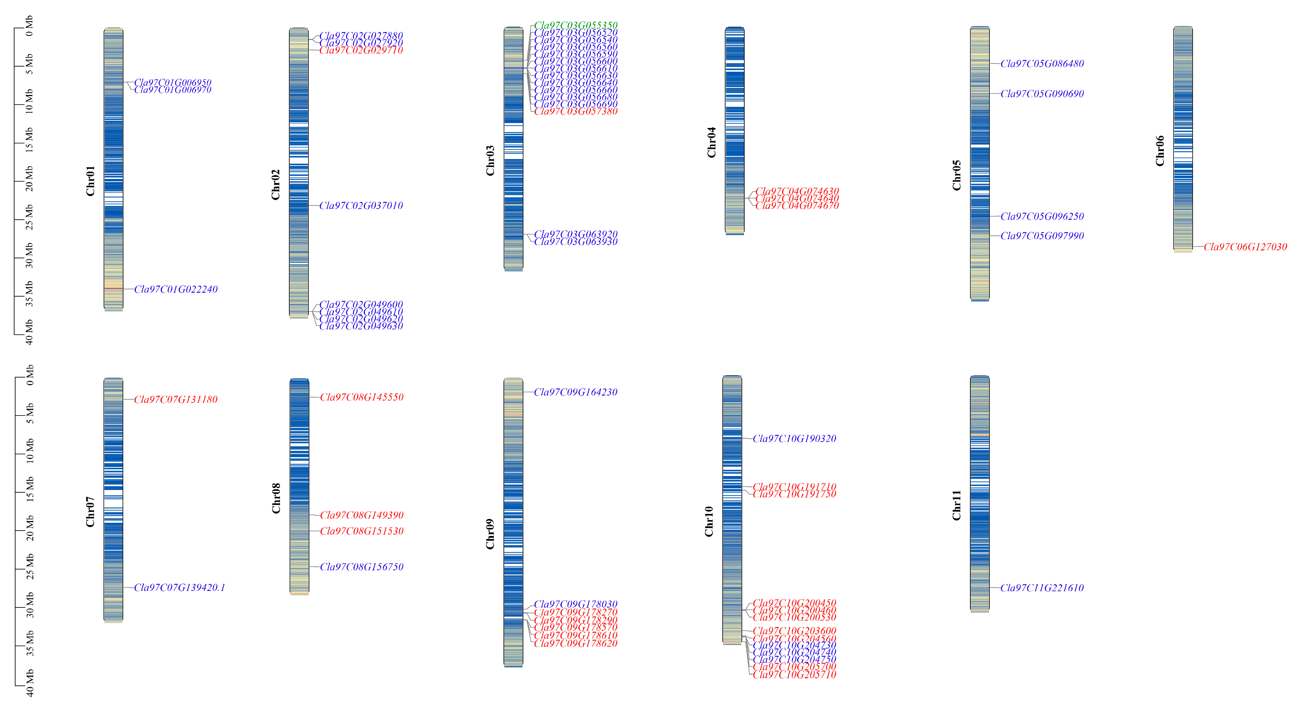
2.2. Chromosomal Location and Gene Duplication of ClaLecRLK Genes

The position of ClaLecRLK genes in the genome showed that the members of this family were distributed in clusters and were unevenly distributed on the watermelon’s chromosomes (Figure 1). Most of the ClaLecRLK genes were located on chromosome 3 with 15 ClaLecRLK genes and chromosome 10 with 13 ClaLecRLK genes. On the other hand, the G-type ClaLecRLK genes were mainly distributed on chromosome 2 with 7 genes and chromosome 3 with 13 genes. The L-type ClaLecRLK genes were mainly distributed on chromosome 9 with six genes and chromosome 10 with nine genes. The C-type ClaLecRLK genes were distributed on chromosome 3.
During the long process of evolution, gene duplication (tandem duplication and segmental duplication) is considered to be the main contributor to the generation of gene clusters and expansion of gene families [18,19]. Therefore, we investigated whether gene duplication occurred in the ClaLecRLK gene family. To our surprise, only one pair of duplicate genes were identified, Cla97C03G056520 and Cla97C03G056540, and their divergence occurred about 11.85 million years ago. The Ka/Ks value of Cla97C03G056520 and Cla97C03G056540 was 0.29, suggesting purifying selection acting on the ClaLecRLK gene family. This result indicated that the duplication event of the LecRLK gene family during the evolution of the watermelon genome was rare.

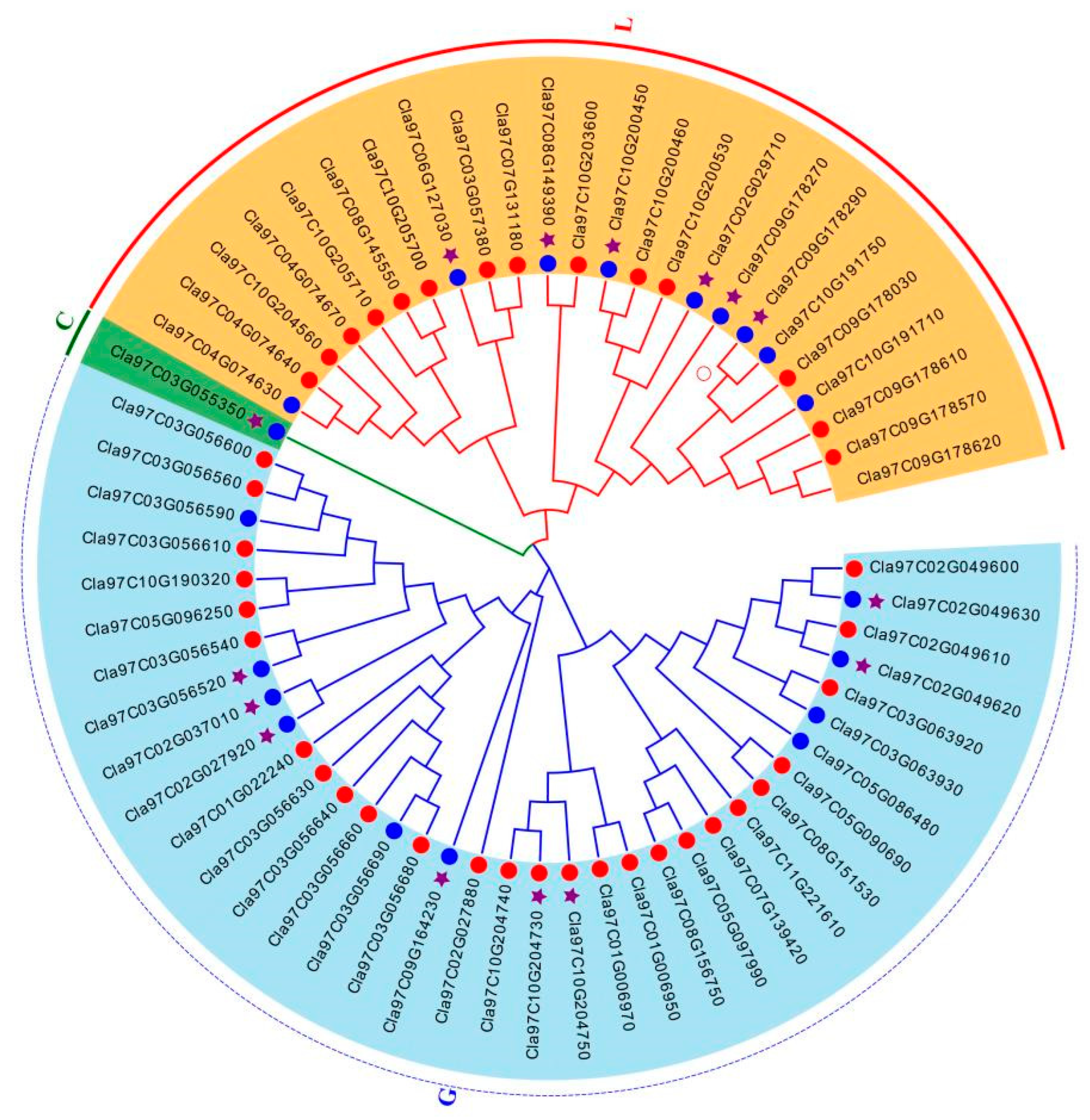
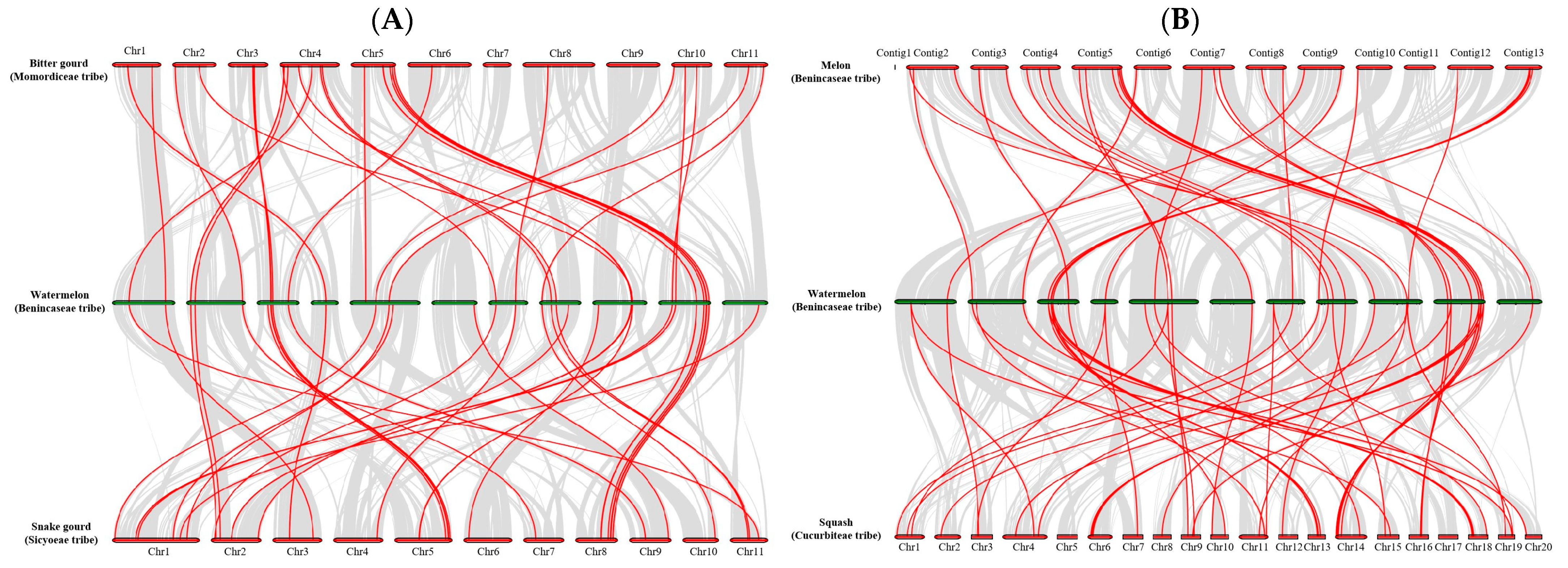
2.3. Analysis of Phylogenetic and Synteny in ClaLecRLK Genes

Based on the protein sequence of ClaLecRLK genes, a phylogenetic tree was constructed to analyze the evolutionary relationship of the ClaLecRLK family (Figure 2). As the same as the cucumber LecRLK gene family, although the ClaLecRLK gene family can be divided into multiple subgroups, three types were clearly distinguished on the phylogenetic tree; L-type ClaLecRLK and C-type ClaLecRLK groups had a closer evolutionary relationship. Combined with the analysis of protein structure, it was found that ClaLecRLK proteins that distributed in clusters on chromosomes were more likely to cluster into one subgroup than those that were structurally similar. The phylogenetic tree based on the kinase domain showed that three types of ClaLecRLK proteins were still distributed on three different branches (Supplementary Figure S1), but L-type ClaLecRLK and G-type ClaLecRLK were more closely related. This suggested that in addition to the N-terminal lectin domain, the C-terminal kinase domains from different subfamilies were still conserved, and different types of ClaLecRLK protein may have different interacting proteins in cells.
To further reveal the origin and evolution of LecRLK family members in Cucurbitaceae plants, the MCScanX method [20] was used to analyze the collinearity of LecRLK genes among the Siraitieae, Momordiceae, Sicyoeae, Benincaseae, and Cucurbiteae tribes (Figure 3 and Supplementary Table S2). We found that 40 LecRLK genes had a collinear relationship with the homologous genes of melon and watermelon. There was a collinearity between 42 LecRLK genes in watermelon and 53 LecRLK genes in squash. At the same time, we also found 43 LecRLK genes in watermelon and 44 LecRLK genes in snake gourd, having a collinear relationship. On the other hand, there was a collinear relationship between the 33 LecRLK gene pairs in watermelon and bitter gourd. Meanwhile, 24 LecRLK genes had a collinear relationship with the homologous genes of monk fruit and watermelon. Among them, 10 LecRLK genes had a collinear relationship in all six plants (named Cucurbitaceae LecRLK genes); these results indicated that these 10 LecRLK genes were relatively conserved in the evolution of Cucurbitaceae plants, and the homology of this gene family in Cucurbitaceae has no tribe preference. Finally, we found that only two Cucurbitaceae LecRLK genes had a collinear relationship with the homologous genes of rice (Table 2), which indicated that these two LecRLK genes were relatively conserved in the evolution of monocots and dicots.

2.4. Gene Structure and Protein Motif Analysis of ClaLecRLK Genes

The genomic sequence and corresponding cDNA sequence of the ClaLecRLK genes were submitted together to the TBtools-II software (v2.001) for analyzing their gene structure (Figure 4). The number of exons of ClaLecRLK genes varied from one to eight. Similar to the structure of the LecRLK family in other plant genomes, ClaLecRLK genes generally lack introns. In total, 85% of the studied ClaLecRLK genes had less than three exons. Except for Cla97C04G074630, which contained three exons, all L-type ClaLecRLK genes contained only one or two exons, and the C-type ClaLecRLK gene (Cla97C03G055350) contained five exons. The G-type ClaLecRLK genes contained one to eight exons. In this group, Cla97C02G027920 contains eight exons, which has the highest number of exons in all analyzed ClaLecRLK genes.
The ten most conserved motifs were identified in ClaLecRLK proteins using the MEME program (Figure 4). The results showed that motif 6, 7, and 10 were only present in G-type ClaLecRLKs, located on the B-Lectin, Pan, and EGF domains, respectively. The remaining seven motifs were located in the kinase domain, and although each motif appeared nearly 50 times, they had 19 different combination forms, of which were G-type ClaLecRLKs containing 16 motif combinations and L-type ClaLecRLKs containing 11 motif combinations. These different motif combinations can increase the diversity of the kinase domain. Motif 2–motif 1–motif 3 was the most conserved motif combination, which appeared in the kinase domain of 41 ClaLecRLKs. In the kinase domain, C-type ClaLecRLKs contained 4 motifs, G-type ClaLecRLKs contained 5.8 motifs, and L-type ClaLecRLKs contained 5.8 motifs, indicating that the conservativeness in the kinase domain of the three types of ClaLecRLKs was similar. By aligning the LecRLK protein sequences of cucumber, rice, and watermelon, it was found that most motifs have been mutated or lost during evolution, and only motif 1, motif 2, motif 3, and motif 5 were relatively intact in these plants, indicating that these four motifs were highly conserved during the evolution of monocots and dicots.
Although bioinformatic predictions suggested that almost all ClaLecRLK proteins were localized on the plasma membrane, the analysis of the protein structure of the ClaLecRLK protein showed that in addition to the three basic domains (N-terminal ligand recognition domain, C-terminal kinase domain, and transmembrane domain), there were various domain differences among LecRLKs, which may affect protein localization. For example, Cla97C09G178270 did not contain a signal peptide, but contained two transmembrane domains located in the N-terminal and middle regions of the protein. Cla97C10G204750 contained a signal peptide and two consecutive transmembrane domains located in the middle of the protein, whereas Cla97C01G022240 contained a signal peptide and transmembrane domain located in the middle of the protein. Subcellular localization experiments showed that these proteins were localized on the cell membrane; Cla97C05G086480, Cla97C01G022240, and Cla97C09G178270 were uniformly expressed on the membrane, whereas Cla97C10G204750 was expressed in a punctate manner (Figure 5).

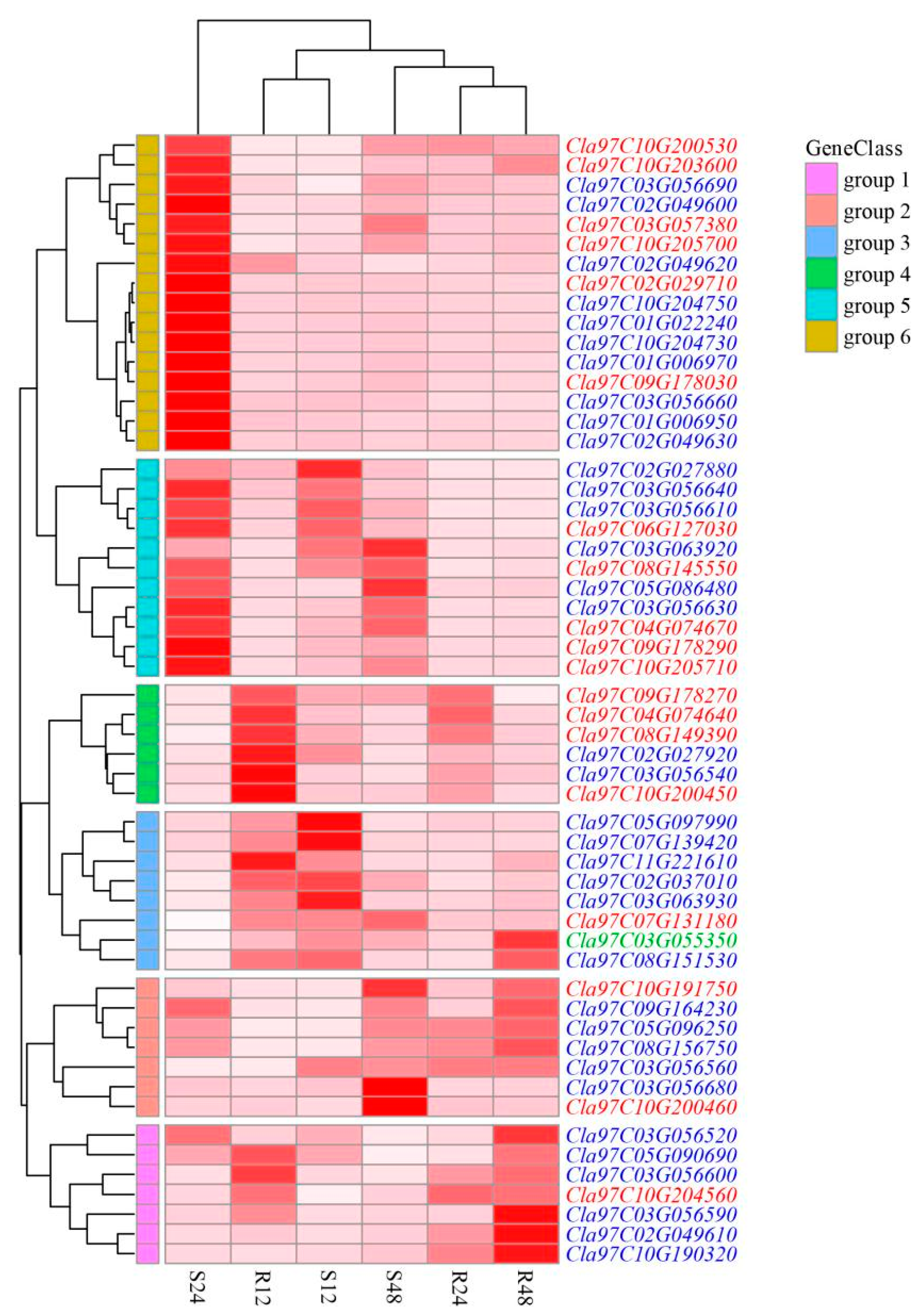
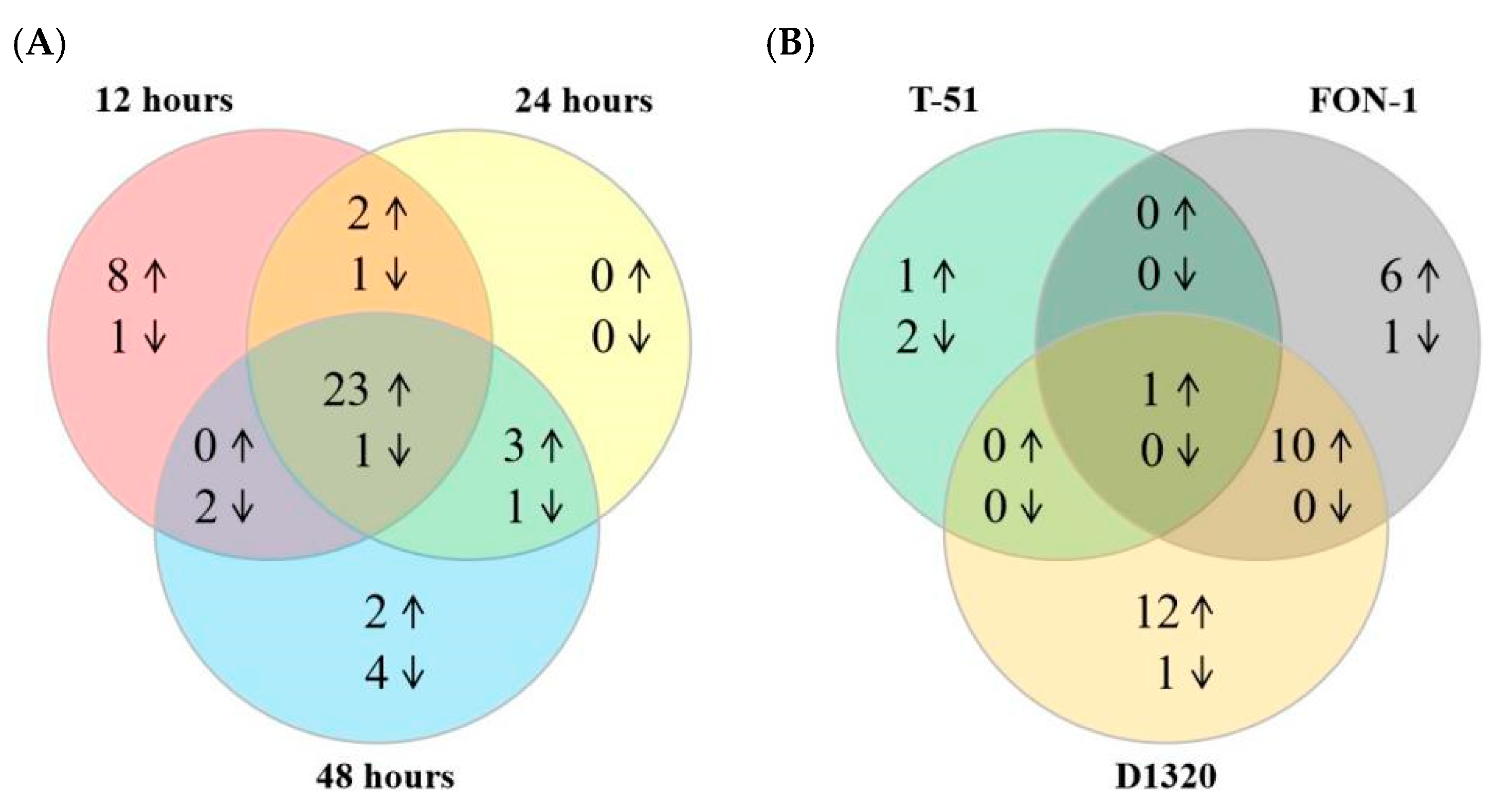
2.5. Response of LecRLK Genes to Fungal Infection in Watermelon

The LecRLK gene has been proven in several studies to be involved in responses to biotic stress. During the production process, cucurbitaceae crops are exposed to a variety of fungi, such as Fusarium oxysporum f. sp. niveum, which is an obligate parasite of watermelon [21]; Stagonosporopsis sp, which has a broad spectrum of infection to cucurbit crops [22]; and Trichoderma koningiopsis, which can promote plant growth [23]. In this study, transcriptome data were used to analyze the response characteristics of the watermelon ClaLecRLK gene family to different fungi.
Under T-51 infection stress, the expression levels of most ClaLecRLK genes did not differ significantly in watermelon (Table 3 and Supplementary Table S3). The expression levels of Cla97C10G203600 and Cla97C08G151530 were increased by 1.8- and 1.6-fold, respectively, whereas the expression levels of Cla97C03G057380 and Cla97C02G049610 were decreased by 3.6- and 18.4-fold, respectively, following infection.
The ClaLecRLK genes also responded to Fusarium wilt (FW) stress, caused by FON-1 (Supplementary Table S3). The expression levels of 18 ClaLecRLK genes were significantly different after inoculation with FON-1, among which only 1 gene was downregulated, whereas the other 17 genes were upregulated, with a change in the expression level ranging from 1.6- to 303.2-fold (Table 3 and Supplementary Table S3). Notably, the expression of Cla97C03G056680 was barely detected in the roots of the uninfected control group.
The transcriptional levels of ClaLecRLK genes were investigated at different time points after the infection of D1320. The expression levels of most ClaLecRLK genes changed significantly, while some genes showed the same expression trend at different time points (Figure 6 and Supplementary Table S3). After a 12 h inoculation with D1320, there were 33 ClaLecRLK genes with upregulated expression and 7 ClaLecRLK genes with downregulated expression in susceptible material (W4K-1 inbred lines), and 37 ClaLecRLK genes with upregulated expression and 7 ClaLecRLK genes with downregulated expression in resistant materials (au-s inbred lines). Interestingly, all upregulated ClaLecRLK genes in the susceptible materials were equally significantly upregulated in the resistant material. After a 24 h inoculation, 34 and 35 ClaLecRLK genes in resistant and susceptible materials were significantly upregulated, respectively; among them, 28 ClaLecRLK genes were upregulated simultaneously. In addition, seven and eight ClaLecRLK genes in resistant and susceptible materials were significantly downregulated, respectively. Meanwhile, after a 48 h inoculation, there were 34 ClaLecRLK genes that were upregulated in both susceptible and resistant materials, and among them 28 ClaLecRLK genes were upregulated in both materials. The Venn diagram shows that a total of 23 ClaLecRLK genes were upregulated at all three time points in both resistant and susceptible materials (Figure 7A), indicating that these genes were involved in watermelon response to D1320. Notably, although Cla97C10G204740 had a low background expression level, it was specifically upregulated in resistant materials at three time points, and Cla97C10G190320 was specifically upregulated in resistant materials at 24 h and 48 h, suggesting that these two genes might be involved in watermelon resistance to D1320.
The expression levels of 11 ClaLecRLK genes were increased after inoculation with FON-1 and D1320 (three time points in both materials) (Table 3 and Figure 7), among which Cla97C08G151530 expression was also upregulated after inoculation with T-51 (Table 3 and Figure 7B), suggesting that this gene might play an important role in watermelon’s immune response to fungi.

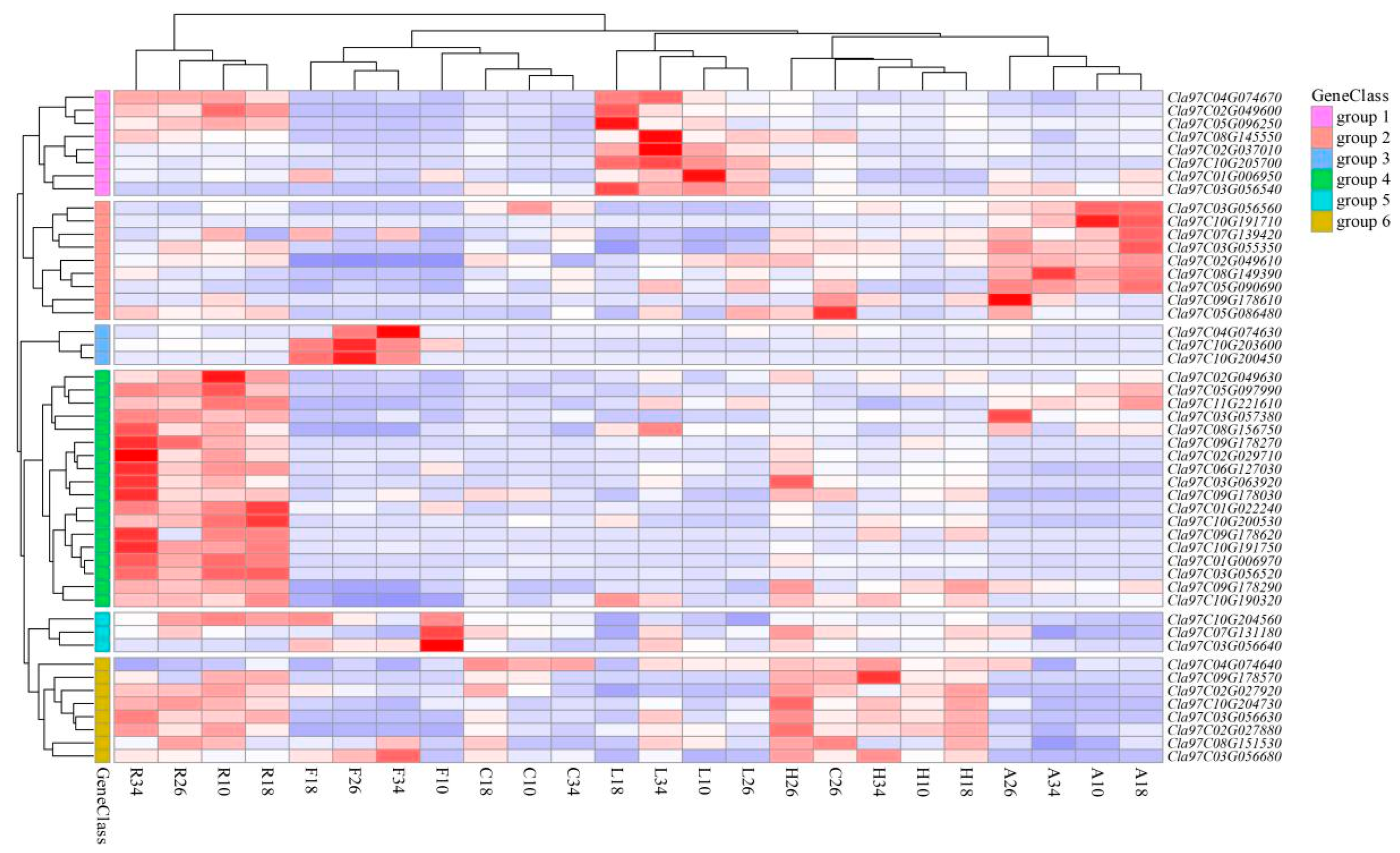
2.6. Tissue-Specific Expression of ClaLecRLK Genes

RNA-seq data (PRJNA532463) from the CuGenDB database were used to investigate the tissue-specific expression of the ClaLecRLK gene family. Each tissue contained at least 20 constitutively expressed genes (average FPKM ≥ 1) (Figure 8 and Supplementary Table S4) and 10 ClaLecRLK genes were expressed in all tissues at different time points, indicating that the ClaLecRLK gene family plays an important role in watermelon development. Some ClaLecRLK genes exhibited distinct tissue and temporal expression patterns. For example, Cla97C01G006970 was highly expressed in the roots (average FPKM ≈ 21.35) but was barely expressed in the fruits and hypocotyls. Cla97C03G056540 was hardly detected in the fruit, but maintained a relatively high expression level in the leaves (FPKM ≈ 17.46). Cla97C10G200450 was mainly expressed in the fruit and was significantly upregulated 18 days after pollination. The roots had the most widely expressed LecRLK genes, and the average FPKM values of these 38 genes were greater than 1. The fruits had the lowest expression levels of LecRLK genes with only 20 genes showing constitutive expression, indicating that the expression of all members of the gene family had a tissue bias.
Based on these results, we selected eight typical ClaLecRLK genes to verify their tissue-specific expression patterns using reverse transcription–quantitative polymerase chain reaction (Figure 9). The expression of most of these genes was consistent with the RNA-seq results, and the expression level of Cla97C01G022240 was high in the fruits and leaves but lower in other tissues. The expression level of Cla97C06G127030 was high in the roots but relatively low in other tissues. Cla97C08G151530, Cla97C03G055350, and Cla97C04G074640 were uniformly expressed in all tissues. The expression level of Cla97C10G203600 was the highest in the fruits but was lower in other tissues. The expression level of Cla97C10G205700 was the highest in the leaves but was lower in other tissues.

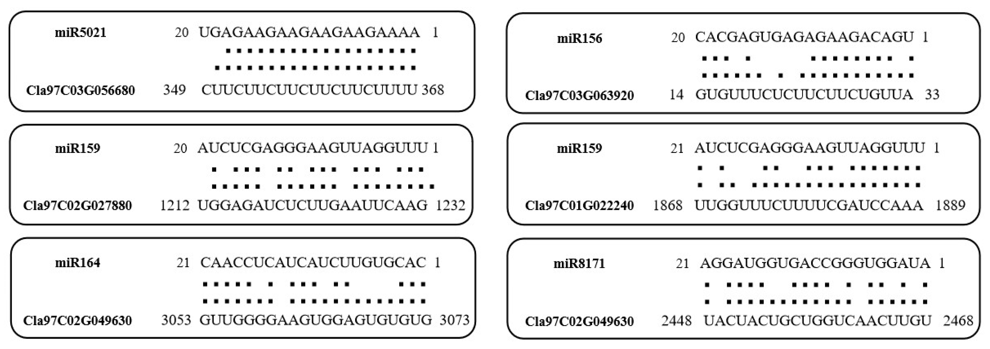
2.7. Prediction Analysis of ClaLecRLK Genes Targeted by miRNA

Through the psRNATarget website, we predict the binding sites of published miRNAs targeting the ClaLecRLK genes (Supplementary Table S5 and Figure 10). The results showed that a total of 171 miRNAs were involved in the regulation of the ClaLecRLK genes. Among 171 miRNAs, except a few miRNAs regulating only one ClaLecRLK gene, most of them could regulate multiple ClaLecRLK genes. For example, miR5021 had the most regulatory targets, and could regulate 17 ClaLecRLK genes; miR156, a famous miRNA in plants, could regulate 8 ClaLecRLK genes. In addition, 10 genes included were targeted by miR159. On the other hand, one ClaLecRLK gene can also be regulated by multiple miRNAs. For example, Cla97C02G049630 can be regulated by 25 miRNAs; Cla97C02G049610 had binding sites of 15 miRNAs.

3. Discussion

Generally, LecRLKs are localized on the cell membrane, where they can recognize extracellular signals through their extracellular ligand recognition domains. This helps plants respond accurately and quickly to external stimuli. Utilizing bioinformatics data and biological experiments can provide deeper insights into the signals recognized by LecRLKs and their associated biological pathways. On this basis, integrating new breeding technologies, particularly gene editing, can facilitate faster and more precise breeding for resistance to biotic and abiotic stresses.
At present, genome-wide analyses of the LecRLK gene family have been reported for some plants [17,24], but only a few such studies have been reported on Cucurbitaceae crops [4,9]. Therefore, a comprehensive understanding of the LecRLK genes of the Cucurbitaceae genome is still lacking. In this study, 61 LecRLK genes were identified in watermelon. As for subfamilies, there were 36 G-type ClaLecRLK genes, 24 L-type ClaLecRLK genes, and just 1 C-type ClaLecRLK gene. At the same time, we also counted the number of LecRLK genes of different Cucurbitaceae plants, such as melon, C. maxima, bitter gourd, and monk fruit; the results showed that they had significant differences in the number of LecRLK genes. This suggests that there might not be a direct relationship between LecRLK gene family expansion and genome size. According to a previous study [25], chromosome 2 and 3 of watermelon have a collinear relationship with chromosome 1, 2, 3, and 4 of cucumber, and chromosome 9 and 10 of watermelon have a collinear relationship with chromosome 3 and 7 of cucumber. In watermelon, the G-type ClaLecRLK genes were mainly distributed on chromosome 2 and chromosome 3, and the L-type ClaLecRLK genes were mainly distributed on chromosome 9 and chromosome 10. In cucumber, the G-type ClaLecRLK genes were mainly distributed on chromosome 1 and chromosome 4, and the L-type ClaLecRLK genes were mainly distributed on chromosome 3 and chromosome 7 [4]. These results suggest that although the LecRLK gene family originated from the same ancestors, they experienced relatively independent expansion in different Cucurbitaceae plants.
Most of the conserved motifs (seven motifs) were distributed in C-terminals (kinase domain) of ClaLecRLK proteins, and had 19 combinations in different ClaLecRLK proteins. Each subfamily contained approximately the same amount of motifs, which ensures that each subfamily does not have functional redundancy due to the unicity of the N-terminal. The phylogenetic tree showed that the full-length C-type ClaLecRLK protein was more closely related to L-type ClaLecRLK protein, while the phylogenetic tree based on the kinase domain showed that L-type ClaLecRLK proteins were more closely related to G-type ClaLecRLK proteins, which may be due to the N-terminal of G-type ClaLecRLK proteins often carrying multiple other domains (such as EGF, Pan, and/or S domains), and the kinase domains of C-type ClaLecRLK protein are generally considered to have different kinase activities compared with the other two types of ClaLecRLK proteins [2]. But L-type ClaLecRLK proteins and G-type ClaLecRLK proteins did not cross each other in the branches of the kinase domain phylogenetic tree, indicating that the kinase domain of each ClaLecRLK subfamily is also somewhat conservative.
After Cucurbitaceae crops experienced four additional genome duplication (WGD) events during their evolution, significant differentiation occurred among different genera [13]. This differentiation is also reflected in the collinearity analysis of the LecRLK gene family among Cucurbitaceae plants, which is positively correlated with their phylogenetic relationships. The number of LecRLK genes that had a collinear relationship between watermelon (Cucurbiteae tribe) and melon (Benincaseae tribe), squash (Cucurbiteae tribe), or snake gourd (Sicyoeae tribe) was significantly higher than the number of genes that had a collinear relationship between watermelon and bitter gourd (Momordiceae tribe) or monk fruit (Momordiceae tribe). On the other hand, the collinear relationship between watermelon and squash was not one to one, and there were only 10 collinearity LecRLK genes among different Cucurbitaceae tribes, again indicating that the evolution of this gene family in different Cucurbitaceae crops was independent. The LecRLK family plays important roles in stress response, microbial associations, and plant development. Relatively independent evolution represents a plant-specific adaptation for the perception and propagation of the extracellular signals [26]. Among 10 Cucurbitaceae collinearity LecRLK genes, only 2 genes had a collinear relationship with the homologous genes of rice. This result is consistent with previous reports that the LecRLK gene family was established before the monocot–dicot split [26]. After the monocot–dicot split, the LecRLK gene family experienced uncoordinated expansion in different crops.
As a kind of cell-surface receptor, LecRLK has been confirmed in previous reports to be associated with various biotic and abiotic stresses. Watermelon, a kind of land-cultivated crop, engages in symbiosis with multiple fungi during its growth. In this study, we selected three types of fungi as research targets to explore the role of LecRLK in watermelon resistance to biotic stress. FW, caused by Fusarium oxysporum f. sp. niveum, affects water transport efficiency by damaging the vascular system [27]. Gummy stem blight (GSB) is caused by Didymella bryonia, which can parasitize many kinds of Cucurbitaceae crops, including watermelon, and cause leaf and vine decay [28]. According to a previous study [29], Trichoderma koningiopsis isolate T-51 can improve the immunity of plant hosts to pathogens by activating the immune system. Our RNA-seq data further confirmed that the LecRLK family is related to fungal disease resistance. For example, the expression levels of 18 ClaLecRLK genes significantly changed after inoculation with FON-1, among which 17 ClaLecRLK genes were upregulated. Additionally, the expression levels of 24 genes were significantly upregulated after GSB (D1320) infection, of which 23 genes were upregulated. Combined with the above two sets of data, 11 genes showed upregulated expression after inoculation with the two fungi pathogens, indicating that they may play an important role in disease resistance. The inoculation of T-51, as a promising biocontrol agent, also upregulated the expression levels of two ClaLecRLK genes, among which Cla97C08G151530 was also upregulated under the induction of FW and GSB, indicating that this gene plays a role in the non-specific immune response system of watermelon. The expression levels of Cla97C03G056640, Cla97C03G056680, Cla97C09G178030, and Cla97C09G178290, coming from two gene clusters in chromosome 3 and chromosome 9, were all increased after inoculation with both FW and GSB pathogens, indicating that these gene clusters are functionally conserved. However, Cla97C03G056680 and Cla97C09G178030 were barely expressed in the non-inoculated roots and leaves, suggesting that some genes of clusters may not be expressed in certain tissues due to redundancy, but are significantly upregulated when performing specific biological functions. On the other hand, FW infects watermelon primarily through the roots, and GSB mainly affects the leaves and stems. Combined with transcriptome data in different tissues, it can be found that ClaLecRLK genes, which were highly expressed in roots, were also mostly responsive to FW inoculation, such as Cla97C09G178030, Cla97C09G178270, Cla97C09G178290, Cla97C10G190320, and Cla97C10G204730, having relatively higher baseline expression levels in watermelon roots compared to other tissues throughout the life cycle, and they were upregulated following FW inoculation. Similarly, some ClaLecRLK genes highly expressed in the leaves were also upregulated after GSB inoculation, such as Cla97C02G037010, Cla97C04G074670, Cla97C05G096250, and Cla97C08G145550. This suggests that there is an obvious linear relationship between the tissue specificity of ClaLecRLK genes and their resistance to biological stress. LecRLK genes are believed to participate in immunity, development, and reproductive processes via their diverse extracellular ligand domain in plants [30]. Although the sizes of the LecRLK gene family vary among plant species, G-type LecRLK is the biggest subfamily of the LecRLK gene in most plants; they are believed to expand over evolutionary time in response to pathogen pressure [31]. In this study, we found that G-type ClaLecRLK genes respond more strongly to FW and GSB inoculation compared to the other two ClaLecRLK gene subfamilies, indicating that G-type LecRLK genes may primarily be involved in sensing pathogen signals. L-type ClaLecRLK genes have significantly higher expression in fruits than G-type and C-type ClaLecRLK genes, suggesting that L-type LecRLK genes are mainly involved in the development of plant organs. C-type LecRLK genes have always been a mysterious subfamily because there are usually only one or two of them in most plants, and their mutants are rarely discovered. Recent reports indicate that C-type LecRLK genes are involved in the development of trichomes [32] and resistance to aphids [33] in cucumber. These results indicate that each subfamily has a distinct role in the plant’s life cycle.
miRNA (microRNA) is a kind of widely existing, non-coding small RNA with regulatory function in plants and animals. In plants, they are widely involved in regulating growth and development, signal transduction, and stress response [34]. At present, the research of some star miRNAs has been very in-depth, such as miR156, miR160, miR171, and so on. Despite the fact that most of LecRLK genes’ function has not yet been studied extensively, analyzing correspondences of miRNAs and genes will provide clues. In our study, most miRNA could regulate multiple ClaLecRLK genes. For example, miR156 can affect pollen development and adventitious root formation by promoting cell proliferation [35], and could regulate eight ClaLecRLK genes, suggesting that these ClaLecRLK genes may be involved in the regulation of plant development. The C-type LecRLK gene is the most mysterious subfamily, with only one member in most plants, and studies on its function are very rare. In our study, only one C-type LecRLK was identified in watermelon, and it was found that it may be the target of four miRNAs. Among these miRNAs, miR396 targets six Growth-Regulating Factor genes with roles in plant leaf growth in Arabidopsis [36], and miR773 is involved in response to fungal pathogens in Arabidopsis [37]. These results may provide a strong basis for us to further explore the function of the C-type LecRLK.

4. Materials and Methods

4.1. Plant Materials and Fungal Culture

The watermelon inbred line ‘W28-7’ was used to investigate the spatiotemporal expression and response of ClaLecRLK genes to Trichoderma koningiopsis isolate T-51 and Fusarium oxysporum f. sp. niveum race 1 (FON-1). The watermelon inbred lines ‘4k-1’ (susceptible material) and ‘au-s’ (resistant material) were used to investigate the response of ClaLecRLK genes to Stagonosporopsis sp. D1320. Seedlings of watermelon were grown in an incubator without any pathogen (16/8 h light–dark cycle, 25 °C, humidity was 70%) prior to treatment. When the first true leaf fully unfolded, the seedlings were placed in a glass greenhouse for further cultivation, and the controlled environment growth chamber was programed for 16 h light at 25 °C and 8 h dark at 20 °C. The strains of FW (FON-1) and Stagonosporopsis sp. D1320 in this study came from the experimental field of our laboratory (Shanghai, China), and have been separated and purified for many generations, which have good infectivity to the experimental materials, and ensure that there is no contamination of stray bacteria or fungi.
The Trichoderma koningiopsis isolate T-51 was cultured on solid potato dextrose agar (PDA) plates at 20 °C for 10 days under light. The conidia were washed from the culture using sterilized water and the conidial concentration was determined using a hemocytometer and adjusted to 1 × 10⁷ conidia/mL. Fusarium oxysporum f. sp. niveum race 1 (FON-1) was cultured on a solid PDA medium, inoculated into a 500 mL liquid PDA medium, and incubated with shaking at 25 °C for 10 days. The resulting mixture was filtered through a four-layer cheesecloth to remove mycelial fragments from the conidial suspensions. The conidial concentration was determined using a hemocytometer and adjusted to 1 × 10⁶ conidia/mL. Stagonosporopsis sp. D1320 was cultured on a PDA solid medium at 25 °C for 7 days in the dark. All strains used in this study were obtained from our laboratory’s preservation.

4.2. Identification of LecRLK Genes in Different Plants

The whole-genome and protein sequence data of different Cucurbitaceae plants were downloaded from a public Cucurbitaceae database (http://cucurbitgenomics.org/v2/ (accessed on 11 September 2023)), including watermelon (Citrullus lanatus, 97103 genome v2.5, accessed on 11 September 2023), bitter gourd (Momordica charantia, Dali-11 genome v1, accessed on 11 September 2023), squash (Cucurbita moschata, Rifu genome v1, accessed on 16 October 2017), melon (Cucumis melon, DHL92 genome v4, accessed on 11 September 2023), monk fruit (Siraitia grosvenorii, Qingpiguo genome v1, accessed on 28 December 2021), and snake gourd (Trichosanthes cucumerina, genome v1, accessed on 11 May 2021). The LecRLK family sequence data of rice and cucumber were obtained from the literature [2,4].
The hidden Markov model (HMM) was used to identify LecRLK candidates, and the HMM profiles of the LecRLKs were downloaded from the Pfam protein database (http://pfam.xfam.org/ (accessed on 11 May 2022)) [38]. The models used for these files were L-type (Lectin_legB PF00139), G-type (B_lectin PF01453), and C-type (Lectin_C PF00059). We used HMMER 3.0 [39] to search for the three types of LecRLK genes from different plant protein sequence data with an e-value cutoff of <0.001. We then submitted the whole-protein sequences of these genes to two bioinformatics websites, Pfam [40] (http://pfam.xfam.org/ (accessed on 11 May 2022)) and SMART [41] (http://smart.embl.de/ (accessed on 26 October 2020)), with an e-value cutoff of 1.0, while retaining genes that contained the lectin, transmembrane, and kinase domains.

4.3. Analysis of Phylogenetic and Gene Structure

The full-length and kinase domain sequences of ClaLecRLK proteins were aligned using the MUSCLE program (http://www.ebi.ac.uk/Tools/msa/muscle/ (accessed on 11 October 2023)) with the default parameters [42]. The Neighbor-Joining (NJ) method contained in MEGAX_software v.10.1.8 was used to construct phylogenetic trees with the following parameters: the Poisson model, pairwise deletion, and 1000 bootstrap replications. TBtools-II (v2.001) was used to show the exon–intron structures of ClaLecRLK genes.

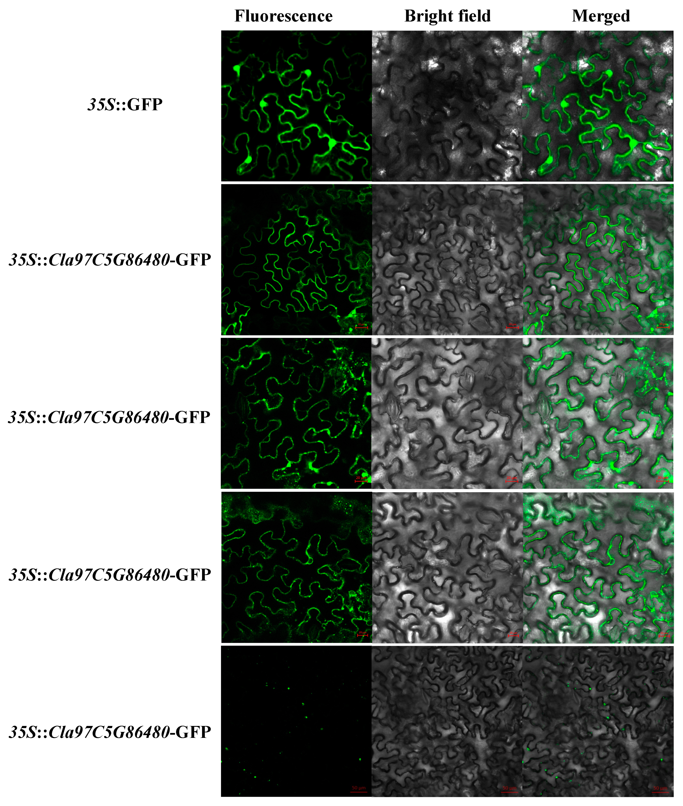
4.4. Tissue-Specific Expression Pattern Analysis and Subcellular Localization

Total RNA (contained clone of genes) was extracted using the RNeasy Plant Mini Kit (Cwbio, Beijing, China) from plants of watermelon. First-strand cDNA was prepared using a PrimeScript RT Reagent Kit with gDNA Eraser (Cwbio).
Public transcriptome data (PRJNA532463) [25], obtained from Cucurbit Expression Atlas Cucurbit Genomics Database (CuGenDB), were used to investigate expression patterns of the ClaLecRLK gene family. The spatiotemporal expression patterns of selected ClaLecRLK genes were confirmed by qRT-PCR in multiple tissues, including the roots (4-week-old seedlings), hypocotyls (4-week-old seedlings), apical point (10-week-old seedlings), carpopodium (10-week-old cucumber plants), fruits (unfertilized 10-week-old cucumber plants), and young leaves (10-week-old cucumber plants, from the top node). Three biological replicates were established in this experiment, and every tissue sample was collected from at least eight plants in each replicate.
The full-length cDNA sequences of different ClaLecRLK genes were cloned and fused to a pHB vector (CaMV35S–GFP). Complete vectors were injected into tobacco (Nicotiana tabacum) leaf epidermal cells using the Agrobacterium-mediated method [43]. Two days after injection, the green fluorescence signal was detected using an Olympus BX51 fluorescence microscope to acquire fluorescent images. Green fluorescent protein (GFP) imaging was performed at an excitation wavelength of 488 nm. At least three independent replicates were performed and the results of one representative experiment are shown. The primers used are listed in Supplementary Table S1.

4.5. Chromosome Localization and Duplication Analyses

A series of in-house Perl scripts were used to retrieve the location information for the ClaLecRLK gene and the length of each chromosome from the whole-genome sequence data of watermelon downloaded from the Cucurbitaceae public database (http://cucurbitgenomics.org/v2/ (accessed on 11 October 2023)). According to the gene position, the chromosome locations of the ClaLecRLK genes were mapped using TBtools-II (v2.001). Adobe Illustrator CS6 was used to enhance the images for a detailed analysis.
We used two methods to identify duplication events among the LecRLKs in watermelon. In the first method, gene duplication was confirmed according to the following three criteria using in-house Perl scripts: (a) the shorter aligned gene covers >70% of the length of the longer gene, (b) the similarity of aligned sequences is >70% [44], and (c) two genes located in the same chromosomal fragment of less than 100 kb represent tandem duplicated genes [18]. The second method was based on the Multiple Collinearity Scan toolkit (MCScanX) to analyze gene duplication events with default parameters [20].
The synonymous substitution (Ks) and nonsynonymous substitution (Ka) rates of tandemly duplicated genes were calculated using the method of Nei and Gojobori as implemented in the KaKs_calculator [45] based on the coding sequence alignments. The divergence time was calculated based on the formula T = Ks/2r, where Ks is the synonymous substitution rate per site and r is the rate of divergence of nuclear genes from plants; r was taken to be 1.5 × 10⁻⁸ synonymous substitutions per site per year for dicotyledonous plants [46].

4.6. Analysis of Conserved Motif and Synteny

The conserved motif prediction of all ClaLecRLK proteins was performed using the MEME program v.5.1.1 [47] (http://meme-suite.org/ (accessed on 11 October 2023)). The parameters were set as any number of repetitions and an optimum motif width of 10–210 residues. The maximum search time was set as 36,000 s, and the site distribution mode was ‘zoops’. The syntenic analyses were constructed by a series of the R program (v4.2.2) to exhibit the collinear relationship of the orthologous LecRLK genes among different plants.

4.7. RNA-Seq of Fungal Inoculation

For inoculation with T-51 and FON-1, the watermelon seedlings were root-dipped into the conidial suspension for 30 min and all seedlings were replanted in sterilized soil in a greenhouse. The roots were subjected to RNA-seq 2 and 10 days after T-51 and FON-1 inoculation, respectively. For inoculation with D1320, a mycelial plug (5 mm diameter) from the PDA plate was inoculated on the first true leaf of watermelon seedlings, followed by cultivation at 25 °C and relative humidity > 99%. The first true leaves were subjected to RNA-seq 0, 12, 24, and 48 h after D1320 inoculation. The high-quality RNA samples were sequenced by Biomarker Bioinformatics Technology Co., Ltd. (Beijing, China), and the cDNA library was subjected to high-throughput sequencing (RNA-seq) on the Illumina HiSeq™ 2500 platform. Raw data were deposited in the SRA of the NCBI website under accession numbers PRJNA995348 and PRJNA995420. The methods of the sequence assembly and differential expression gene analysis for RNA-seq used in this study have been described previously [32]. Tissue-specific expression and fungi inoculation analyses of watermelon LecRLK genes were performed and the results were visualized as a heatmap using the R program (4.2.2).

4.8. Prediction of Binding Sites of the ClaLecRLK Genes Targeted by miRNA

The miRNA sequences were downloaded from psRNATarget (https://www.zhaolab.org/psRNATarget/ (accessed on 16 September 2023)) [48]. The selected cDNA library is ‘Citrullus lanatus (watermelon), transcript, Cucurbit Genomics Database, version 1’, and other parameters are set to default values.

5. Conclusions

A total of 61 LecRLK genes of watermelon were identified in our study. Members of the ClaLecRLK gene family are distributed in clusters on chromosomes, and members of the same subfamily are mostly clustered together. The results of the phylogenetic tree and conserved motif analysis revealed that in addition to the N-terminal kinase domain, the C-terminal kinase domain was also conserved in different subfamilies. The synteny analysis showed that the ClaLecRLK gene family may have been established before the monocot–dicot split, and also has undergone relatively independent evolution in Cucurbitaceae plants. Transcriptome data demonstrated distinct expression patterns of ClaLecRLK genes in various tissues and response to multiple fungal pathogens. The prediction of mirNA-targeted binding sites revealed that each ClaLecRLK gene may be regulated by at least three miRNAs. In conclusion, the information obtained in our study could help to provide an in-depth understanding of basic features of the ClaLecRLK gene family; this knowledge could be applied to enhance breeding programs and improve management strategies for biotic and abiotic stress resistance in watermelon.

Supplementary Materials

The following supporting information can be downloaded at: https://www.mdpi.com/article/10.3390/ijms25158257/s1.

Author Contributions

D.L. and G.W. performed the experiments and completed the writing of the manuscript. J.Y. contributed to data analysis. L.Z. and H.Y. took care of the plants. W.G. offered good advice in the process of the experiments. B.C. took clear pictures and developed the manuscript. W.G. and C.L., the corresponding authors, supervised all the processes of the experiments and proposed the overarching ideas of the study. All authors have read and agreed to the published version of the manuscript.

Funding

This study was supported by the Shanghai Melon and Fruit Industry Technology System [Shanghai Agricultural Science (2024) No.1]. the Excellent Team of Shanghai Academy of Agricultural Sciences, Watermelon and Melon Innovation Team (2022),020. Postdoctoral Science Foundation of China (2023M732328).

Institutional Review Board Statement

Not applicable.

Informed Consent Statement

Not applicable.

Data Availability Statement

The original contributions presented in the study are publicly available. These data can be found at the National Center for Biotechnology Information (NCBI) BioProject database under accession number PRJNA995348 and PRJNA995420.

Conflicts of Interest

The authors declare that the research was conducted in the absence of any commercial or financial relationships that could be construed as a potential conflict of interest.

References

  1. Guo, S.; Zhao, S.; Sun, H.; Wang, X.; Wu, S.; Lin, T.; Ren, Y.; Gao, L.; Deng, Y.; Zhang, J.; et al. Resequencing of 414 cultivated and wild watermelon accessions identifies selection for fruit quality traits. Nat. Genet. 2019, 51, 1616–1623. [Google Scholar] [CrossRef] [PubMed]
  2. Vaid, N.; Macovei, A.; Tuteja, N. Knights in action: Lectin receptor-like kinases in plant development and stress responses. Mol. Plant 2013, 6, 1405–1418. [Google Scholar] [CrossRef]
  3. Shiu, S.H.; Bleecker, A.B. Receptor-like kinases from Arabidopsis form a monophyletic gene family related to animal receptor kinases. Proc. Natl. Acad. Sci. USA 2001, 98, 10763–10768. [Google Scholar] [CrossRef] [PubMed]
  4. Lv, D.; Wang, G.; Xiong, L.R.; Sun, J.X.; Chen, Y.; Guo, C.L.; Yu, Y.; He, H.L.; Cai, R.; Pan, J.S. Genome-Wide Identification and Characterization of Lectin Receptor-like Kinase Gene Family in Cucumber and Expression Profiling Analysis under Different Treatments. Genes 2020, 11, 1032. [Google Scholar] [CrossRef] [PubMed]
  5. Tanksley, S.D.; Loaiza-Figueroa, F. Gametophytic self-incompatibility is controlled by a single major locus on chromosome 1 in Lycopersicon peruvianum. Proc. Natl. Acad. Sci. USA 1985, 82, 5093–5096. [Google Scholar] [CrossRef]
  6. Cambi, A.; Koopman, M.; Figdor, C.G. How C-type lectins detect pathogens. Cell Microbiol. 2005, 7, 481–488. [Google Scholar] [CrossRef] [PubMed]
  7. Chen, X.; Shang, J.; Chen, D.; Lei, C.; Zou, Y.; Zhai, W.; Liu, G.; Xu, J.; Ling, Z.; Cao, G.; et al. A B-lectin receptor kinase gene conferring rice blast resistance. Plant J. 2006, 46, 794–804. [Google Scholar] [CrossRef] [PubMed]
  8. Hervé, C.; Serres, J.; Dabos, P.; Canut, H.; Barre, A.; Rougé, P.; Lescure, B. Characterization of the Arabidopsis lecRK-a genes: Members of a superfamily encoding putative receptors with an extracellular domain homologous to legume lectins. Plant Mol. Biol. 1999, 39, 671–682. [Google Scholar] [CrossRef] [PubMed]
  9. Guo, C.; Yang, X.; Wang, Y.; Nie, J.; Yang, Y.; Sun, J.; Du, H.; Zhu, W.; Pan, J.; Chen, Y.; et al. Identification and mapping of ts (tender spines), a gene involved in soft spine development in Cucumis sativus. Theor. Appl. Genet. 2018, 131, 1–12. [Google Scholar] [CrossRef]
  10. Li, C.H.; Wang, G.; Zhao, J.L.; Zhang, L.Q.; Ai, L.F.; Han, Y.F.; Sun, D.Y.; Zhang, S.W.; Sun, Y. The Receptor-like Kinase SIT1 Mediates Salt Sensitivity by Activating MAPK3/6 and Regulating Ethylene Homeostasis in Rice. Plant Cell 2014, 26, 2538–2553. [Google Scholar] [CrossRef]
  11. Kanzaki, H.; Saitoh, H.; Takahashi, Y.; Berberich, T.; Ito, A.; Kamoun, S.; Terauchi, R. NbLRK1, a lectin-like receptor kinase protein of Nicotiana benthamiana, interacts with Phytophthora infestans INF1 elicitin and mediates INF1-induced cell death. Planta 2008, 228, 977–987. [Google Scholar] [CrossRef] [PubMed]
  12. Bonaventure, G. The Nicotiana attenuata LECTIN RECEPTOR KINASE 1 is involved in the perception of insect feeding. Plant Signal Behav. 2011, 6, 2060–2063. [Google Scholar] [CrossRef] [PubMed]
  13. Ma, L.; Wang, Q.; Zheng, Y.; Guo, J.; Yuan, S.; Fu, A.; Bai, C.; Zhao, X.; Zheng, S.; Wen, C.; et al. Cucurbitaceae genome evolution, gene function and molecular breeding. Hortic. Res. 2022, 9, uhab057. [Google Scholar] [CrossRef] [PubMed]
  14. Schaefer, H.; Heibl, C.; Renner, S.S. Gourds afloat: A dated phylogeny reveals an Asian origin of the gourd family (Cucurbitaceae) and numerous oversea dispersal events. Proc. Biol. Sci. 2009, 276, 843–851. [Google Scholar] [CrossRef] [PubMed]
  15. Denna, D.W. Expression of determinate habit in cucumbers (Cucumis sativus L.). Amer Soc. Hort. Sci. J. 1971, 96, 277–279. [Google Scholar] [CrossRef]
  16. Nie, J.T.; Wang, Y.L.; He, H.L.; Guo, C.L.; Zhu, W.Y.; Pan, J.; Li, D.D.; Lian, H.L.; Pan, J.S.; Cai, R. Loss-of-Function Mutations in Confer Durable Powdery Mildew Resistance in Cucumber (L.). Front. Plant Sci. 2015, 6, 1155. [Google Scholar] [CrossRef] [PubMed]
  17. Yang, Y.; Labbé, J.; Muchero, W.; Yang, X.; Jawdy, S.S.; Kennedy, M.; Johnson, J.; Sreedasyam, A.; Schmutz, J.; Tuskan, G.A.; et al. Genome-wide analysis of lectin receptor-like kinases in Populus. BMC Genom. 2016, 17, 699. [Google Scholar] [CrossRef] [PubMed]
  18. Mehan, M.R.; Freimer, N.B.; Ophoff, R.A. A genome-wide survey of segmental duplications that mediate common human genetic variation of chromosomal architecture. Hum. Genom. 2004, 1, 335–344. [Google Scholar] [CrossRef]
  19. Kent, W.J.; Baertsch, R.; Hinrichs, A.; Miller, W.; Haussler, D. Evolution’s cauldron: Duplication, deletion, and rearrangement in the mouse and human genomes. Proc. Natl. Acad. Sci. USA 2003, 100, 11484–11489. [Google Scholar] [CrossRef]
  20. Wang, Y.; Tang, H.; Debarry, J.D.; Tan, X.; Li, J.; Wang, X.; Lee, T.H.; Jin, H.; Marler, B.; Guo, H.; et al. MCScanX: A toolkit for detection and evolutionary analysis of gene synteny and collinearity. Nucleic Acids Res. 2012, 40, e49. [Google Scholar] [CrossRef]
  21. Lambel, S.; Lanini, B.; Vivoda, E.; Fauve, J.; Patrick Wechter, W.; Harris-Shultz, K.R.; Massey, L.; Levi, A. A major QTL associated with Fusarium oxysporum race 1 resistance identified in genetic populations derived from closely related watermelon lines using selective genotyping and genotyping-by-sequencing for SNP discovery. Theor. Appl. Genet. 2014, 127, 2105–2115. [Google Scholar] [CrossRef] [PubMed]
  22. Jeong, Y.J.; Kwon, O.K.; Jeong, A.R.; Lee, H.; Moon, H.; Lee, O.N.; Hong, J.K.; Park, C.J. Population Structure of Stagonosporopsis Species Associated with Cucurbit Gummy Stem Blight in Korea. Plant Pathol. J. 2022, 38, 522–532. [Google Scholar] [CrossRef] [PubMed]
  23. You, J.; Li, G.; Li, C.; Zhu, L.; Yang, H.; Song, R.; Gu, W. Biological Control and Plant Growth Promotion by Volatile Organic Compounds of Trichoderma koningiopsis T-51. J. Fungi 2022, 8, 131. [Google Scholar] [CrossRef] [PubMed]
  24. Vaid, N.; Pandey, P.K.; Tuteja, N. Genome-wide analysis of lectin receptor-like kinase family from Arabidopsis and rice. Plant Mol. Biol. 2012, 80, 365–388. [Google Scholar] [CrossRef] [PubMed]
  25. Guo, S.; Zhang, J.; Sun, H.; Salse, J.; Lucas, W.J.; Zhang, H.; Zheng, Y.; Mao, L.; Ren, Y.; Wang, Z.; et al. The draft genome of watermelon (Citrullus lanatus) and resequencing of 20 diverse accessions. Nat. Genet. 2013, 45, 51–58. [Google Scholar] [CrossRef] [PubMed]
  26. Shiu, S.H.; Karlowski, W.M.; Pan, R.; Tzeng, Y.H.; Mayer, K.F.; Li, W.H. Comparative analysis of the receptor-like kinase family in Arabidopsis and rice. Plant Cell 2004, 16, 1220–1234. [Google Scholar] [CrossRef] [PubMed]
  27. Zhou, X.G.; Everts, K.L. Suppression of Fusarium Wilt of Watermelon by Soil Amendment with Hairy Vetch. Plant Dis. 2004, 88, 1357–1365. [Google Scholar] [CrossRef] [PubMed]
  28. Keinath, A.P. From Native Plants in Central Europe to Cultivated Crops Worldwide: The Emergence of Didymella bryoniae as a Cucurbit Pathogen. HortScience 2011, 46, 532–535. [Google Scholar] [CrossRef]
  29. You, J.; Zhang, J.; Wu, M.; Yang, L.; Li, G. Multiple Criteria-based Screening of Trichoderma isolates for Biological Control of Botrytis cinerea on Tomato. Biol. Control 2016, 101, 31–38. [Google Scholar] [CrossRef]
  30. Ngou, B.P.M.; Wyler, M.; Schmid, M.W.; Kadota, Y.; Shirasu, K. Evolutionary trajectory of pattern recognition receptors in plants. Nat. Commun. 2024, 15, 308. [Google Scholar] [CrossRef]
  31. Ngou, B.P.M.; Heal, R.; Wyler, M.; Schmid, M.W.; Jones, J.D.G. Concerted expansion and contraction of immune receptor gene repertoires in plant genomes. Nat. Plants 2022, 8, 1146–1152. [Google Scholar] [CrossRef] [PubMed]
  32. Lv, D.; Wang, G.; Zhang, Q.; Yu, Y.; Qin, P.C.; Pang, J.A.; Sun, J.X.; Zhang, K.Y.; He, H.L.; Cai, R. Comparative Transcriptome Analysis of Hard and Tender Fruit Spines of Cucumber to Identify Genes Involved in the Morphological Development of Fruit Spines. Front. Plant Sci. 2022, 13, 797433. [Google Scholar] [CrossRef] [PubMed]
  33. Yang, S.; Xue, S.; Shan, L.; Fan, S.; Sun, L.; Dong, Y.; Li, S.; Gao, Y.; Qi, Y.; Yang, L.; et al. The CsTM alters multicellular trichome morphology and enhances resistance against aphid by interacting with CsTIP1;1 in cucumber. J. Adv. Res. 2024. [Google Scholar] [CrossRef] [PubMed]
  34. Xie, M.; Zhang, S.; Yu, B. microRNA biogenesis, degradation and activity in plants. Cell Mol. Life Sci. 2015, 72, 87–99. [Google Scholar] [CrossRef] [PubMed]
  35. Zheng, C.; Ye, M.; Sang, M.; Wu, R. A Regulatory Network for miR156-SPL Module in Arabidopsis thaliana. Int. J. Mol. Sci. 2019, 20, 6166. [Google Scholar] [CrossRef] [PubMed]
  36. Liebsch, D.; Palatnik, J.F. MicroRNA miR396, GRF transcription factors and GIF co-regulators: A conserved plant growth regulatory module with potential for breeding and biotechnology. Curr. Opin. Plant Biol. 2020, 53, 31–42. [Google Scholar] [CrossRef] [PubMed]
  37. Li, Y.; Zhang, Q.; Zhang, J.; Wu, L.; Qi, Y.; Zhou, J.M. Identification of microRNAs involved in pathogen-associated molecular pattern-triggered plant innate immunity. Plant Physiol. 2010, 152, 2222–2231. [Google Scholar] [CrossRef] [PubMed]
  38. Mistry, J.; Chuguransky, S.; Williams, L.; Qureshi, M.; Salazar, G.A.; Sonnhammer, E.L.L.; Tosatto, S.C.E.; Paladin, L.; Raj, S.; Richardson, L.J.; et al. Pfam: The protein families database in 2021. Nucleic Acids Res. 2021, 49, D412–D419. [Google Scholar] [CrossRef] [PubMed]
  39. Johnson, L.S.; Eddy, S.R.; Portugaly, E. Hidden Markov model speed heuristic and iterative HMM search procedure. BMC Bioinform. 2010, 11, 431. [Google Scholar] [CrossRef]
  40. Finn, R.D.; Coggill, P.; Eberhardt, R.Y.; Eddy, S.R.; Mistry, J.; Mitchell, A.L.; Potter, S.C.; Punta, M.; Qureshi, M.; Sangrador-Vegas, A.; et al. The Pfam protein families database: Towards a more sustainable future. Nucleic Acids Res. 2016, 44, D279–D285. [Google Scholar] [CrossRef]
  41. Letunic, I.; Bork, P. 20 years of the SMART protein domain annotation resource. Nucleic Acids Res. 2018, 46, D493–d496. [Google Scholar] [CrossRef]
  42. Edgar, R.C. MUSCLE: Multiple sequence alignment with high accuracy and high throughput. Nucleic Acids Res. 2004, 32, 1792–1797. [Google Scholar] [CrossRef]
  43. Sparkes, I.A.; Runions, J.; Kearns, A.; Hawes, C. Rapid, transient expression of fluorescent fusion proteins in tobacco plants and generation of stably transformed plants. Nat. Protoc. 2006, 1, 2019–2025. [Google Scholar] [CrossRef]
  44. Gu, Z.; Cavalcanti, A.; Chen, F.C.; Bouman, P.; Li, W.H. Extent of gene duplication in the genomes of Drosophila, nematode, and yeast. Mol. Biol. Evol. 2002, 19, 256–262. [Google Scholar] [CrossRef]
  45. Zhang, Z.; Li, J.; Zhao, X.Q.; Wang, J.; Wong, G.K.; Yu, J. KaKs_Calculator: Calculating Ka and Ks through model selection and model averaging. Genom. Proteom. Bioinform. 2006, 4, 259–263. [Google Scholar] [CrossRef]
  46. Koch, M.A.; Haubold, B.; Mitchell-Olds, T. Comparative evolutionary analysis of chalcone synthase and alcohol dehydrogenase loci in Arabidopsis, Arabis, and related genera (Brassicaceae). Mol. Biol. Evol. 2000, 17, 1483–1498. [Google Scholar] [CrossRef]
  47. Bailey, T.L.; Williams, N.; Misleh, C.; Li, W.W. MEME: Discovering and analyzing DNA and protein sequence motifs. Nucleic Acids Res. 2006, 34, W369–W373. [Google Scholar] [CrossRef]
  48. Dai, X.; Zhuang, Z.; Zhao, P.X. psRNATarget: A plant small RNA target analysis server (2017 release). Nucleic Acids Res. 2018, 46, W49–W54. [Google Scholar] [CrossRef]
Figure 1. The chromosomal distribution of ClaLecRLK genes. The red ID represents L-type ClaLecRLK genes, the green ID represents C-type ClaLecRLK genes, and the blue ID represents G-type ClaLecRLK genes. The genetic distances of seven chromosomes are represented by the scale in megabases (Mb) on the left.
Ijms 25 08257 g001
Figure 2. The phylogenetic tree analysis of ClaLecRLK proteins. The L, G, and C, respectively, represent the L-type, G-type, and C-type LecRLK subfamilies. The red circles in front of ClaLecRLK proteins represent proteins with a signal peptide; the blue circles in front of ClaLecRLK proteins represent protein without a signal peptide. The purple star in front of ClaLecRLK proteins represents protein that has at least two transmembrane domains.
Ijms 25 08257 g002
Figure 3. The collinear relationship analysis of the LecRLK gene in Cucurbitaceae plants. The collinear gene pairs with LecRLK genes between different species are highlighted by the red lines. (A) The collinear gene pairs with LecRLK genes among bitter gourd, watermelon, and snake gourd. (B) The collinear gene pairs with LecRLK genes among melon, watermelon, and squash.
Ijms 25 08257 g003
Figure 4. The gene structure and conserved motif analysis of the ClaLecRLK family. (A) Gene structure: The lines represent the introns. The green square represents the exon. (B) Protein structure: Different colors represent different motifs. The red ID represents L-type ClaLecRLK genes, the green ID represents C-type ClaLecRLK genes, and the blue ID represents G-type ClaLecRLK genes.
Ijms 25 08257 g004
Figure 5. Subcellular localization of ClaLecRLK–GFP fusion proteins in tobacco leaves. Scale bars are 50 µm.
Ijms 25 08257 g005
Figure 6. The heatmap of the ClaLecRLK gene family to D1320. The expression of ClaLecRLK genes is shown on the heatmap using a log2foldchange value. The red ID represents L-type ClaLecRLK genes, and the blue ID represents G-type ClaLecRLK genes. Genes highly expressed in tissues are colored red, and genes not expressed in tissues are colored white. The letters at the bottom indicate R: resistant material and S: susceptible material. The numbers (12, 24, 48) indicate inoculation time.
Ijms 25 08257 g006
Figure 7. The Venn diagram of differentially expressed ClaLecRLK gene response to different fungi. (A) Differentially expressed ClaLecRLK gene response to D1320 in three time points. (B) Differentially expressed ClaLecRLK gene response to different fungi. The number in the circle represents the number of differentially expressed genes, the upward arrow indicates genes that are upregulated, and the downward arrow indicates genes that are downregulated.
Ijms 25 08257 g007
Figure 8. The heatmap of the ClaLecRLK gene family in different tissues. The red represents L-type ClaLecRLKs, the green represents C-type ClaLecRLKs, and the blue represents G-type ClaLecRLKs. Genes highly expressed in tissues are colored red, and genes not expressed in tissues are colored blue. The letters at the bottom indicate F: fruit; L: leaf; R: root; A: apical point; C: carpopodium; and H: hypocotyl. The numbers at the bottom indicate the day after pollination.
Ijms 25 08257 g008
Figure 9. The expression analysis of selected ClaLecRLK genes. The orange line represents the value of mean FPKM (fragments per kilobase per million) and the black column represents the expression levels analyzed by qRT-PCR. (A): Fruit, (B): Leaf, (C): Root, (D): Apical point, (E): Carpopodium, (F): Hypocotyl.
Ijms 25 08257 g009
Figure 10. The predicted miRNA information and their binding site on the ClaLecRLK genes. One dot indicates that there is a pairing between U and G in the secondary structure, two dots indicate pairing successfully between bases, and a blank space indicates that two bases failed to be paired.
Ijms 25 08257 g010
Table 1. The basic character of ClaLecRLK genes and their encoded proteins.
Gene IDGeneCDSProteinMWpILocalizationExonIntron
(bp)(bp)(aa)(KDa)
C-typeCla97C03G0553502533168956263.49.26PM54
L-typeCla97C02G0297102349207669177.76.25PM21
Cla97C03G0573802052205268375.76.13PM10
Cla97C04G07463070758819522.110.04PM32
Cla97C04G0746401983198366073.46.66PM10
Cla97C04G0746705405204668176.26.49PM21
Cla97C06G1270301971197165673.25.16PM10
Cla97C07G1311802055205568476.05.58PM10
Cla97C08G1455501992199266373.86.12PM10
Cla97C08G1493903450224774883.06.74PM21
Cla97C09G1780302031203167674.95.44PM10
Cla97C09G1782702121212170679.76.18PM10
Cla97C09G1782902130213070979.26.22PM10
Cla97C09G1785701971197165673.25.93PM10
Cla97C09G1786101944194464773.16.99PM10
Cla97C09G1786201974197465773.65.89PM10
Cla97C10G1917102602167755862.56.27PM21
Cla97C10G1917502162207969277.35.97PM21
Cla97C10G2004501989198966274.27.92PM10
Cla97C10G2004602001200166674.45.88PM10
Cla97C10G2005302028202867575.25.32PM10
Cla97C10G2036002178217872578.25.79PM10
Cla97C10G2045602148205268374.96.45PM21
Cla97C10G2057002004200466774.26.65PM10
Cla97C10G2057102046204668175.25.78PM10
Range707–5405588–2247195–74822.1–835.16–10.04 1–31–2
Average2221196865573.06.33 1.30.3
G-typeCla97C01G0069502466246682191.68.37Ec10
Cla97C01G0069702394239479788.98.72Ec10
Cla97C01G0222403318258085996.25.30PM76
Cla97C02G0278802493249383092.36.77PM10
Cla97C02G027920374030961031116.25.65PM87
Cla97C02G0370102772243381092.08.35PM54
Cla97C02G0496002424242480790.15.70PM10
Cla97C02G0496102409240980290.15.90PM10
Cla97C02G0496202481248182692.76.00PM10
Cla97C02G049630574945571518171.16.41Ec21
Cla97C03G0565204511255385096.37.37PM76
Cla97C03G0565404213250583494.06.87PM76
Cla97C03G0565603185242780890.25.78PM76
Cla97C03G0565903255242180691.57.49PM76
Cla97C03G0566003111241880590.75.95PM76
Cla97C03G0566103314244581492.16.17PM76
Cla97C03G0566303007246682192.86.19PM76
Cla97C03G0566403119253284395.77.99PM76
Cla97C03G05666010,301204968277.05.86PM65
Cla97C03G0566803185255385095.96.00PM76
Cla97C03G0566902598214571480.87.77PM43
Cla97C03G0639202448244881591.25.20PM10
Cla97C03G0639302361236178689.85.88PM10
Cla97C05G0864801979192664172.56.12PM21
Cla97C05G0906905800260486795.45.83PM54
Cla97C05G0962504180243381090.76.78PM76
Cla97C05G0979902466246682190.35.74PM10
Cla97C07G1394202869246081992.37.99PM21
Cla97C08G1515302556255685192.75.94PM10
Cla97C08G1567502466246682190.55.70PM10
Cla97C09G16423041172703900102.66.08PM76
Cla97C10G19032015,727245781892.68.21PM76
Cla97C10G2047302436243681191.15.74PM10
Cla97C10G2047402439243981291.35.14PM10
Cla97C10G2047502391239179689.56.19PM10
Cla97C11G2216104269260486795.66.08PM21
Range1979–15,7271926–4557641–151872.5–171.15.14–8.72 1–80–7
Average3682251783894.16.48 3.902.90
Range 707–15,727588–4557195–151863.4–171.15.14–10.04
Average 3088228776185.36.46
Table 2. Collinear relationship analysis of LecRLK gene.
SpeciesDicotMonocot
Bitter GourdSnake GourdMonk GourdMelonWatermelonSquashRice
TribeMomordiceaeSicyoeaeSiraitieaeBenincaseaeBenincaseaeCucurbiteae
Gene IDMC02G0084Tan0002591Sgr014680MELO3C014624Cla97C02G049630CmoCh20G000680
MC03Gnew0349Tan0014355Sgr029435MELO3C002524Cla97C03G056520CmoCh13G007160
MC03G0790Tan0016767Sgr019525MELO3C002606Cla97C03G057380CmoCh13G006610
CmoCh18G003480
MC01G0434Tan0008913Sgr026531MELO3C003146Cla97C04G074670CmoCh07G010510
MC11G0930Tan0015648Sgr028234MELO3C018168Cla97C05G096250CmoCh01G012240Os01G0784700
CmoCh09G009010Os04G0633200
MC08G0987Tan0015848Sgr015781MELO3C017881Cla97C07G139420CmoCh01G001500
MC04G1328Tan0003615Sgr014359MELO3C008394Cla97C08G149390CmoCh04G025490
MC10G0972Tan0002296Sgr020073MELO3C016574Cla97C10G190320CmoCh16G008700Os05G0501400
MC10G0515Tan0018666Sgr020674MELO3C014953Cla97C10G191750CmoCh04G009160
CmoCh16G007980
MC05Gnew0379Tan0005166Sgr022715MELO3C009237Cla97C10G204560CmoCh06G002850
CmoCh14G001210
Table 3. Response of differentially expressed ClaLecRLK genes to fungi.
SubfamilyGene IDFungiRegulated Direction
T51FON-1D1320UpDown
L-typeCla97C10G205710
Cla97C09G178290
Cla97C02G029710
Cla97C04G074670
Cla97C08G145550
Cla97C10G200450
Cla97C09G178030
Cla97C10G191750
Cla97C09G178270
Cla97C10G205700
Cla97C10G203600
Cla97C08G149390
Cla97C03G057380
G-typeCla97C05G096250
Cla97C01G022240
Cla97C02G037010
Cla97C02G027880
Cla97C03G056660
Cla97C03G056640
Cla97C08G151530
Cla97C10G204750
Cla97C01G006970
Cla97C10G204730
Cla97C03G063920
Cla97C09G164230
Cla97C02G049610
Cla97C03G063930
Cla97C03G056630
Cla97C05G086480
Cla97C03G056680
Cla97C03G056690
Cla97C10G190320
Cla97C08G156750
Cla97C02G049610
Disclaimer/Publisher’s Note: The statements, opinions and data contained in all publications are solely those of the individual author(s) and contributor(s) and not of MDPI and/or the editor(s). MDPI and/or the editor(s) disclaim responsibility for any injury to people or property resulting from any ideas, methods, instructions or products referred to in the content.

