Article

Native and Alien Antarctic Grasses as a Habitat for Fungi

1
Institute of Biochemistry and Biophysics, Polish Academy of Sciences, Pawińskiego 5A, 02-106 Warsaw, Poland
2
Botanical Garden—Center for Biological Diversity Conservation, Polish Academy of Sciences, Prawdziwka 2, 02-973 Warsaw, Poland
*
Authors to whom correspondence should be addressed.
These authors contributed equally to this work.
Int. J. Mol. Sci. 2024, 25(15), 8475; https://doi.org/10.3390/ijms25158475
Submission received: 12 July 2024 / Revised: 31 July 2024 / Accepted: 1 August 2024 / Published: 3 August 2024
(This article belongs to the Section Molecular Microbiology)

Abstract

:
Biological invasions are now seen as one of the main threats to the Antarctic ecosystem. An example of such an invasion is the recent colonization of the H. Arctowski Polish Antarctic Station area by the non-native grass Poa annua. This site was previously occupied only by native plants like the Antarctic hair grass Deschampsia antarctica. To adapt successfully to new conditions, plants interact with soil microorganisms, including fungi. The aim of this study was to determine how the newly introduced grass P. annua established an interaction with fungi compared to resident grass D. antarctica. We found that fungal diversity in D. antarctica roots was significantly higher compared with P. annua roots. D. antarctica managed a biodiverse microbiome because of its ability to recruit fungal biocontrol agents from the soil, thus maintaining a beneficial nature of the endophyte community. P. annua relied on a set of specific fungal taxa, which likely modulated its cold response, increasing its competitiveness in Antarctic conditions. Cultivated endophytic fungi displayed strong chitinolysis, pointing towards their role as phytopathogenic fungi, nematode, and insect antagonists. This is the first study to compare the root mycobiomes of both grass species by direct culture-independent techniques as well as culture-based methods.

1. Introduction

Fungi form widespread symbiotic associations with plants. Their colonization of plants is promoted by the limited availability of phosphorus and/or nitrogen, as fungi play a major role in plant nutrition, growth, water absorption, nutrient cycling, and protection against pathogens [1]. Thanks to mycorrhizal fungi, colonized plants gain access to a wide range of nutrients such as phosphorus, nitrogen, and microelements (S, K, Ca, Fe, Cu, Zn) from a larger source area via roots expanded by branched fungal hyphae [2,3]. Additionally, the extraradical mycelium absorbs mineral nutrients from the soil and transports them to the host plant. Thus, fungi mitigate the effects of abiotic stresses such as drought, nutrient imbalance, and unfavorable temperature on plant growth [4]. Plants actively recruit fungi from their environment using strigolactones released by roots to induce fungal spore germination and hyphal growth towards the host roots [5].
Endophytic fungi colonize plant tissues internally and have the potential to act as control agents, such as biological agents or elicitors in the process of induced resistance and in attenuating abiotic stresses [3,6]. Endophytic fungi inhabit a similar ecological niche to that occupied by phytopathogens, thus being able to protect their environment and control them through competition, production of antagonistic substances, direct parasitism, and even inducing resistance or tolerance in plants [6].
Fungi also benefit from symbiosis with plants by taking nutrients, carbohydrates, and lipids from their hosts [1,7,8,9]. Up to 20% of photoassimilates in host plants flow to mycorrhizal root systems to support the mutualistic interaction between hosts and fungi [1]. Evidence suggests that the fungus receives carbon from the plant in the form of sugars (hexoses); however, plants could transfer alternative carbon sources such as lipids [1,7,8,9]. In addition, Helber et al. [8] supported the hypothesis that the exchange of carbon for phosphate between plants and fungi is tightly linked.
Fungi are believed to be able to colonize a large number of host plant species, indicating a lack of host specificity [7]. Different combinations of host plants and fungi have different effects on various aspects of symbiosis, suggesting a high functional diversity of mycorrhizal fungi [10]. Different community compositions of fungi affect plants differently and play a major role in ecosystem biodiversity, sustainability, and productivity.
Traditionally, fungi are viewed as plain decomposers of complex organic compounds in the environment. However, mycorrhizal fungi should be redefined from decomposers to producers or assistant producers of plants, increasing the productivity and fitness of plants in ecosystems. The protective properties of mycorrhizal fungi are particularly important in the adaptation of plants to the extremely difficult environment of Antarctica [4]. The soils of the H. Arctowski Polish Antarctic Station are characterized by low nutrient availability, imposing an additional stress that plants must deal with in order to survive [11]. These extreme conditions also influence fungi. They can function in the coldest environment owing to their great efficiency of adaptation and combination of strategies such as the production of cold-active enzymes, unsaturated lipids, melanin, antifreeze proteins, and the cryoprotectants glycerol, trehalose, and sugar alcohols (mannitol, arabitol, erythritol) [12].
Only two species of grasses are currently found in Antarctica, i.e., Deschampsia antarctica Desv. (Antarctic hair grass) and Poa annua L. (annual bluegrass), both belonging to the family Poaceae.
The global occurrence of D. antarctica is very limited. It has been found in South America, Orkney, South Shetland, and along the western Antarctic Peninsula, while P. annua, an opportunistic species, has spread worldwide. The species was initially recorded in the vicinity of the Polish Antarctic Station in the austral summer of 1985/86 [13]. Amplified fragment length polymorphism (AFLP) analysis showed that the Antarctic population of P. annua was a subpopulation of the Polish one [14,15].
Both grasses have adapted to the extreme Antarctic conditions by symbiosis with bacteria and fungi [3,4,11]. Znój et al. [11] draw attention to Flavobacteriaceae suspected to be major bacterial contributors to the ecological success of P. annua, especially in harsh Antarctic conditions. Some authors point out that endophyte-infected grasses are more competitive than non-infected grasses and thrive better than non-infected grasses with limited resources [3,4].
In this study, we investigated root-associated fungi from D. antarctica and P. annua collected in the vicinity of the H. Arctowski Polish Antarctic Station, located at the western shore of Admiralty Bay, King George Island (South Shetlands Islands, maritime Antarctica). The aim of this study was to compare the communities of fungi inhabiting the roots of two different grasses and to learn how the fungal microbiome of a grass adapting to new environmental conditions is shaped. Our working hypothesis states that the composition of the fungal endorhizome communities of both investigated grass species differs considerably from those associated with D. antarctica roots, displaying a more uniform structure, while the structure of those associated with P. annua roots differs depending on the sampling site.

2. Results

2.1. Fungal Diversity in Antarctic Grasses and the Rhizosphere Soil

To characterize the fungal microbiome associated with roots of D. antarctica and P. annua, we analyzed the ITS1 (internal transcribed spacer) region of rDNA in total DNA obtained from samples of grass roots and surrounding soil collected in the Ecology Glacier foreland and at two locations in the Arctowski Station area.
Obtained ASV (Amplicon Sequence Variant) numbers varied between 31 and 346. In the soil samples, they equated to an av. of 74.3 ± 39.7, in D. antarctica roots, an av. of 314.6 ± 35.6, and in P. annua roots, an av. of 96.7 ± 94.7. Sites 2 and 3 displayed the highest and lowest fungal phylogenetic diversity, respectively (Figure 1A). ASV numbers in D. antarctica roots were significantly higher than those found in P. annua roots at p < 0.05 (Figure 1B). Ascomycota dominated all the samples in terms of sequence contribution (av. 75.4 ± 10.3%) with Basidiomycota as the second most numerous phylum (av. 14.2 ± 9.5%). Ascomycota’s relative abundance fluctuated considerably in the soil samples (av. 79.2 ± 13.7%) and P. annua roots (av. 74.7 ± 13.6%) when compared with D. antarctica roots (av. 72.4 ± 3.2%). Basidiomycota’s contribution displayed a similar pattern as follows: soil—av. 14.4 ± 11.3%, P. annua roots—av. 16.2 ± 14.7%, and D. antarctica—av. 11.9 ± 1.3%. Mortierellomycota maintained a stable persistence only in D. antarctica roots (av. 2.3 ± 0.9%) while displaying varying contributions to soil (av. 1.8 ± 3.1%) and P. annua root (av. 3.9 ± 3.8%) communities. The phylum Rozellomycota was present in roots in relatively small amounts (av. 0.8 ± 0.9%) while being almost absent in the soil samples (av. 0.003 ± 0.005%) (Figure 1C).
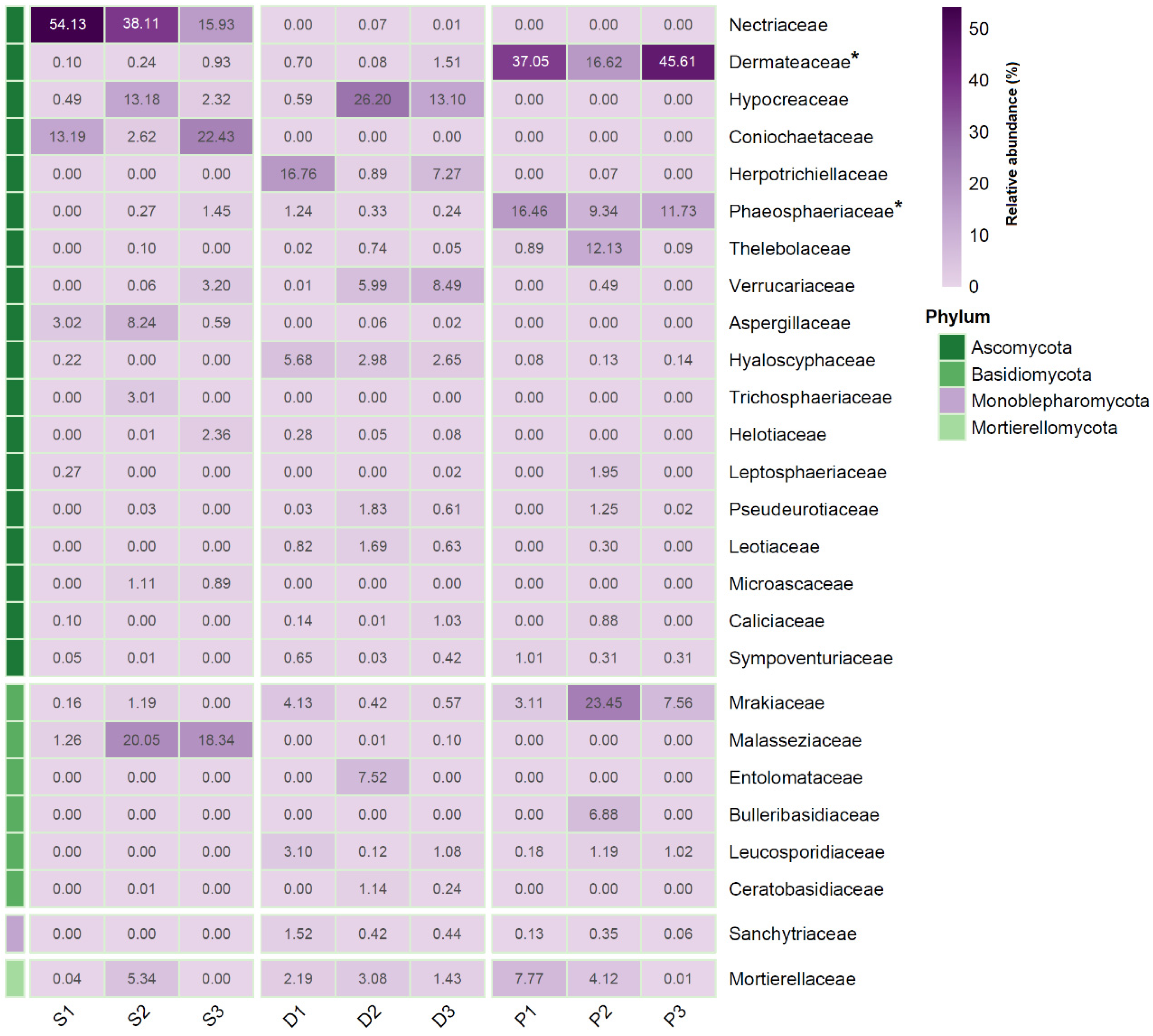
The soil samples were most abundant in the Ascomycota families of Nectriaceae (av. 36.1 ± 19.2%) and Coniochaetaceae (av. 12.7 ± 9.9%), with the Basidiomycota family of Malasseziaceae (av. 13.2 ± 10.4%) also displaying a considerable contribution. Several Ascomycota families displayed a high contribution on average to D. antarctica root fungal communities, albeit their abundance differed considerably among sampling sites as follows: Hypocreaceae—av. 13.3 ± 12.8%, Herpetrichiellaceae—av. 8.3 ± 7.9%, and Verrucariaceae—av. 4.8 ± 4.3%. However, the sequence contribution of the family Mortierellaceae displayed a somewhat even distribution in D. antarctica roots (av. 2.2 ± 0.8%). The most abundant fungal families within P. annua roots were Dermataceae (av. 33.1 ± 14.9%) and Phaeospaeriaceae (av. 12.5 ± 3.6%). The relative abundances of those two families were significantly higher (p < 0.05) in P. annua roots than in the roots of D. antarctica and in the examined soil samples. P. annua roots also harbored considerable amounts of the Basidiomycota family Mrakiaceae (av. 11.4 ± 10.7%) (Figure 2).
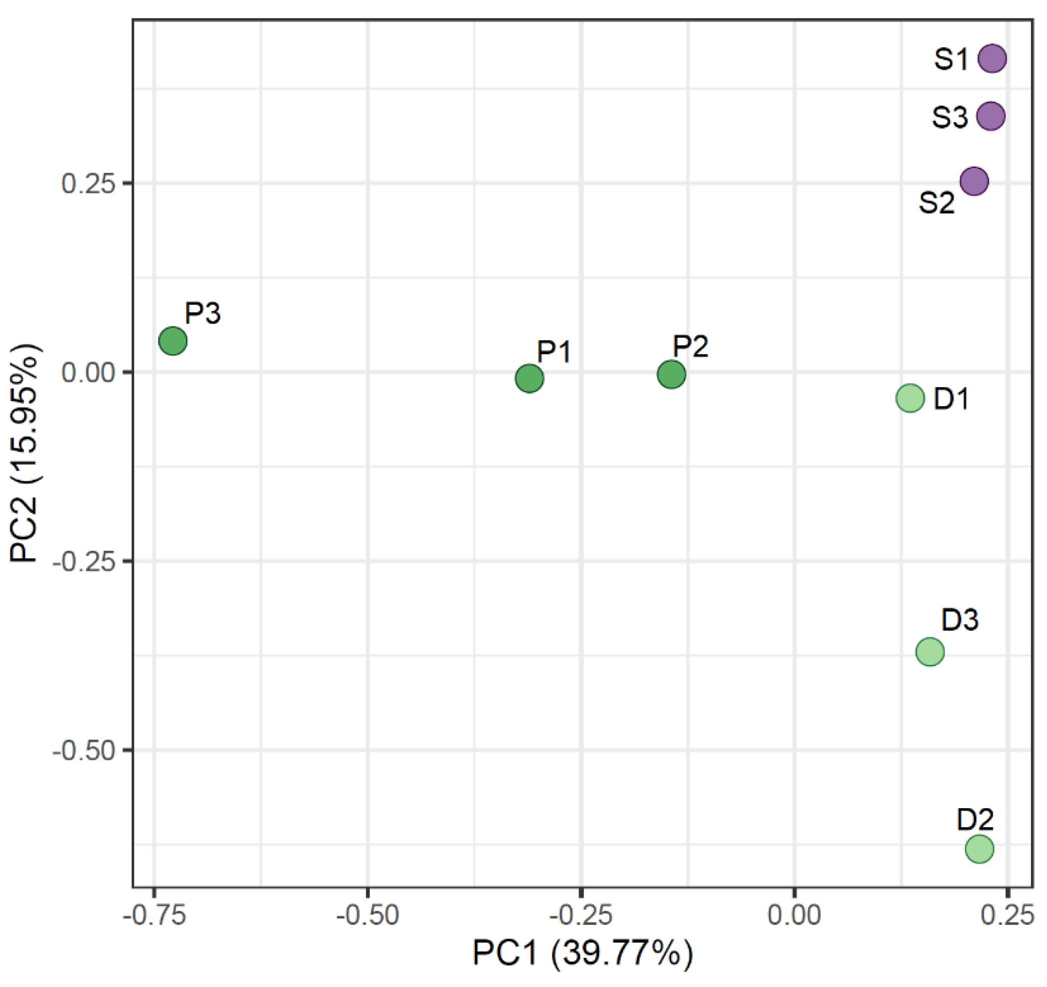
The Principal Component Analysis (PCA) clustered the samples based on ASV abundances (Figure 3). The soil samples are clustered tightly in the right upper corner of the graph. The root samples showed a less organized placement on the PCA, showcasing a high degree of variation not only among grass species but also among sampling sites.
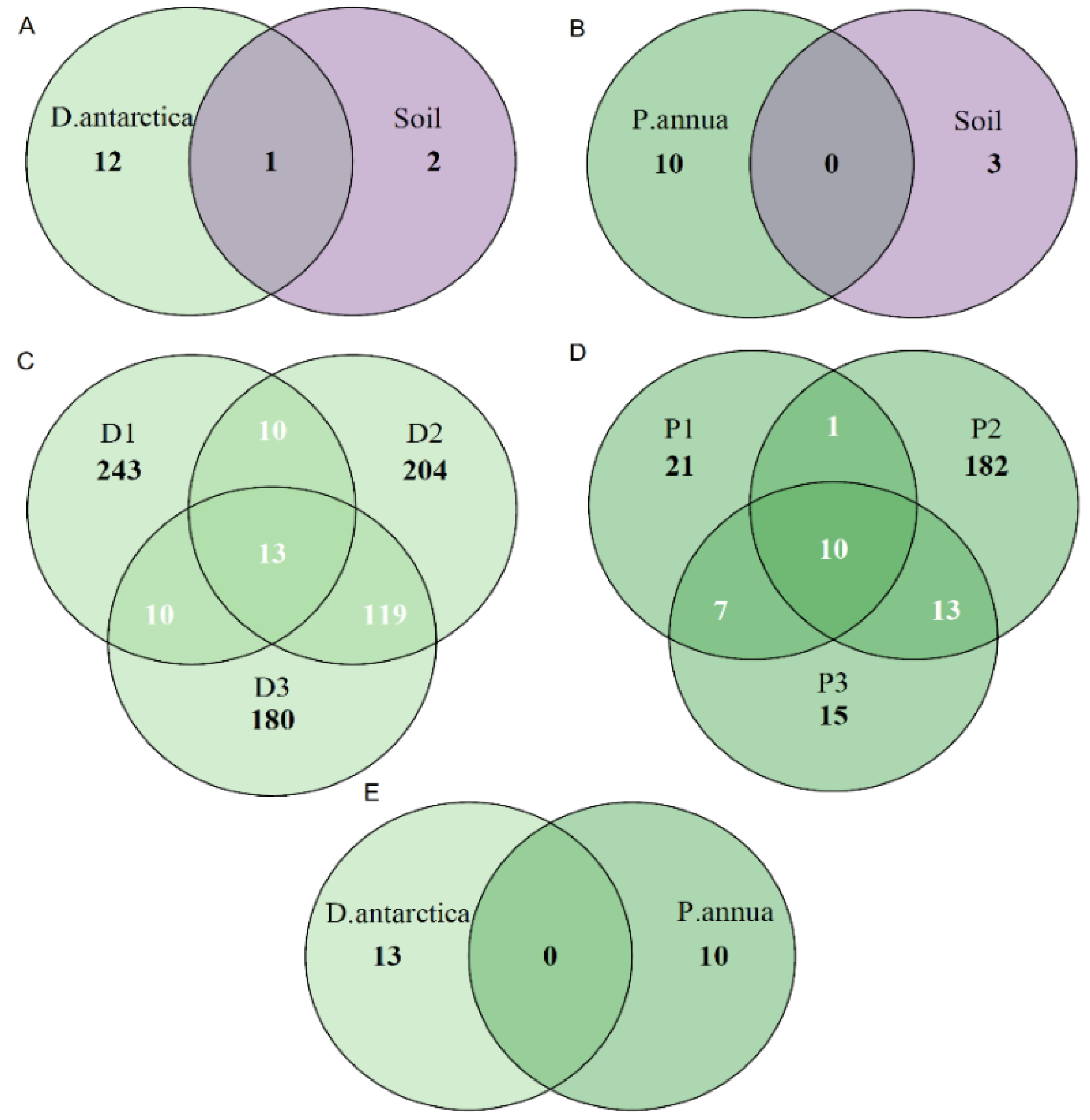
The Venn diagrams presented in Figure 4 show the amount of ASVs shared among the samples and sample sets. While D. antarctica shared 1 ASV between its root community and the examined Antarctic soil community (Figure 4A), P. annua shared none (Figure 4B). The fungal core microbiome of D. antarctica roots consisted of 13 ASVs (Figure 4C), while in the case of P. annua, the core microbiome comprised 10 ASVs (Figure 4D). No ASVs were shared between core microbiomes of the two grass species (Figure 4E).
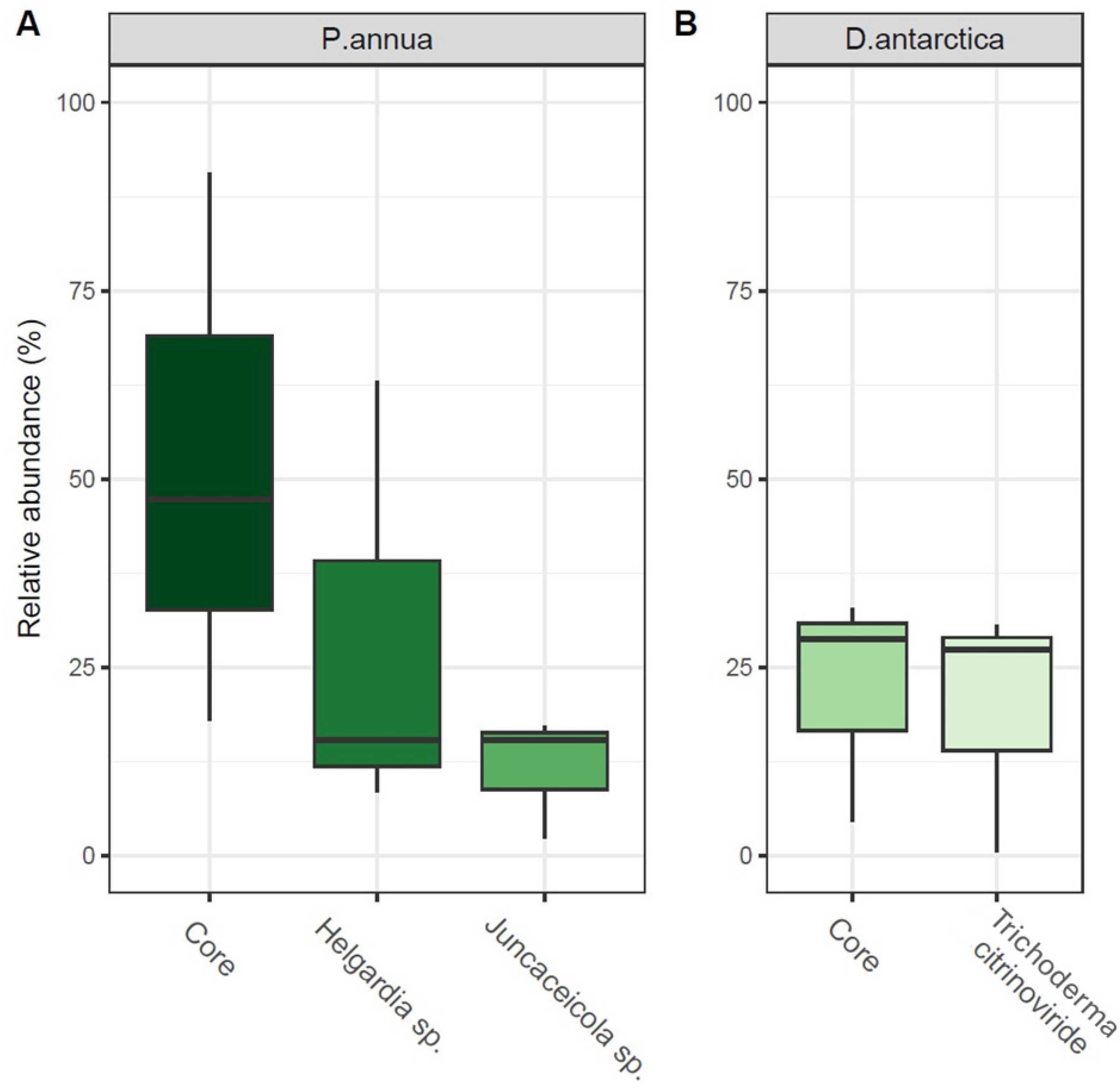
The fungal core microbiome of P. annua roots (10 ASVs) comprised between 18 and 90% of the community’s relative abundance. Its most numerous members were identified as Helgardia sp. (f. Dermataceae)—8.5 to 63% and Juncaceicola sp. (f. Phaeospaeriaceae)—2.3 to 15.3% (Figure 5A). The core microbiome of D. antarctica roots (13 ASVs) comprised between 4.5 and 32.9% of the total fungal community. The most abundant ASV (0.5–30.6% of total community) was identified as the species Trichoderma citrinoviride (f. Hypocreaceae), which represents the one ASV shared between D. antarctica roots and the Antarctic soil (Figure 5B).

2.2. Fungi Identified to the Species Level from Grass Roots and Their Characteristics

Forty-two and eighty-nine fungal sequences associated with P. annua and D. antarctica roots, respectively, were identified to the species level. Overall, 65.1% of the identified fungi belonged to the phylum Ascomycota, 26.4% to Basidiomycota, and 8.6% to Mortierellomycota.
Twenty-five species were recruited by both grasses. Among the most abundant genus Mortierella (Mortierellomycota), represented by nine species, M. amoeboidea, M. jenkinii, M. minutissima, and M. basiparvispora were identified in D. antarctica, and M. angusta, M. hyalina and M. gamsii were associated with P. annua roots. Both grasses recruited M. antarctica and M. elongatula. Mortierella was the only identified genus belonging to the Mortierellomycota phylum. Although many more species were identified in the roots of D. antarctica compared with P. annua, the relative abundance of phylum members in the roots of both grasses was almost the same (Figure 6, Table S1).

2.3. Cultivable Fungi Inhabiting Grasses in Antarctica and Their Hydrolytic Activity

To study the enzymatic activities of the Antarctic fungi experimentally, they were cultured directly from the grass and soil samples from all the locations. Few fungi from the roots of D. antarctica or P. annua from Arctowski Station 2 could be cultivated, while all of those from the soil came from Arctowski Station 2 and the Ecology Glacier foreland (Table 1 and Table S2).
Most of the cultivable fungi belonged to Ascomycota, and only four and three species to Basidiomycota and Mortierellomycota, respectively.
To survive in the Antarctic, soil fungi have to be able to utilize complex carbon sources, which they achieve by producing polymer-degrading enzymes. To analyze the hydrolytic potential of cultivable fungi, we determined their ability to grow on plant-derived polysaccharides such as cellulose, xylan, starch, and pectin, which can be found in the soil and on chitin potentially derived from the cell wall of fungi or arthropods exoskeleton [35,36,37].
The availability of nutrients in cold ecosystems depends strongly on the soil temperature and moisture and the activity of soil microbiota, which in turn control nitrogen fixation, mineralization, and organic matter decomposition [38,39,40,41,42]. Plant growth can be limited by nitrogen availability. The ability of fungi to secrete proteases and make the inaccessible organic nitrogen pool available to plants was analyzed. We determined the proteolytic potential of the cultivable grass root and soil fungi (Figure 7).
Phosphorus is often the limiting nutrient present in the soil in an insoluble form inaccessible to plants. Notably, some fungi secrete organic acids that mobilize the insoluble phosphates. We determined the ability of some of the grass-associated and soil fungi to convert insoluble phosphorite (Ca3(PO4)2) to an absorbable form (Figure 7).
Seventeen fungal strains isolated from the examined samples were identified phylogenetically and subjected to agar plate-based evaluation of seven metabolic features (Figure 7). Seven strains were isolated from grass roots, while ten were isolated from the Antarctic soil. All root-derived strains were capable of chitin digestion, with Microdochium phragmitis P4 being the most potent chitinolytic strain. Seven soil-derived strains also displayed this ability albeit to a lesser extent, with Mrakia robertii AI14 showing the greatest chitin dissolution activity within this group. Five root and seven soil strains produced cellulases, most notably, Herpotrichia juniperi D31 (roots) and Cladosporium herbarum AZI (soil). Pectin was digested by five root strains and eight soil strains with the highest scores noted for Helgardia sp. D1-1 and Botrytis cinerea AI3, respectively, for root and soil isolates. Starch was digested by six root strains and seven soil strains. Helgardia sp. D1-1 and Botrytis cinerea AI3 were the most potent amylolytic strains. Among the five root strains that degraded xylan, Herpotrichia juniperi D31 was the most active, while among the seven soil xylan-degrading strains, isolate AI10 (un. Leucosporidiales) was the most potent. Tricalcium phosphate was solubilized by only two root-derived isolates, including Helgardia sp. D1-1 and Sporobolomyces roseus D1A, while seven soil strains displayed this ability, with strain AI10 displaying the highest activity. Casein was enzymatically degraded by three root isolates and six soil isolates, where Ophiosphaerella sp. D21 and Sporobolomyces roseus D1A were the most active among root strains and Cladosporium herbarum AZI was the most active among soil strains. Two soil strains (Oidiodendron truncatum 21I and Pseudoeurotium sp. 25I) and one root strain (Helgardia sp. D1-1) displayed all seven of the examined traits.

3. Discussion

Deschampsia antarctica has been present in the Antarctic region since at least the late Pliocene (≈3.6 Mya) [43]. In this study, the reported high phylogenetic diversity in D. antarctica roots (based on ASV numbers) is likely explained by the long history of interaction between it and Antarctic soil microflora [44]. The relatively stable proportions of phylum-rank sequences in its roots also corroborate the notion of an established microbiome [45]. However, PCA clustering suggests a dynamic reorganization of the fungal endophyte community depending on the sampling site. Despite significant discrepancies in the phylogenetic structure, a core fungal microbiome was recognized, composed largely of one ASV, which was identified as Trichoderma citrinoviride. Interestingly, the relative abundance of the core microbiome taxa within D. antarctica roots was as low as 4.5%, suggesting that those endophytes are not an indispensable part of the community, i.e., not pivotal for the growth and development of the host plant. In this regard, Upson et al. [46] showed large variations in fungal endophyte colonization in D. antarctica roots procured from the South Shetland Islands. Furthermore, there was a continued presence of this T. citrinoviride ASV in the examined Antarctic soil samples, meaning that the fungus was not exclusively housed in hair grass roots, with soil being its primary and most accessible reservoir. The role of T. citrinoviride in the tissues of D. antarctica remains elusive, as this is the first record of its presence in this plant species and even in Antarctica. However, what is known about T. citrinoviride and, in general, about the Trichoderma genus is their ability to parasitize other fungi, most notably, severe plant pathogens, acting as a biocontrol agent and, consequently, promoting plant health [47]. The relative abundance of the T. citrinoviride ASV displayed a positive correlation with the fungal phylogenetic diversity (ASV numbers) in D. antarctica roots, implying that no antagonistic processes were engaged. However, a study from 2018 showed that pathogen-stressed tomato and cucumber plants promoted the growth of Trichoderma harzianum, while their root exudates induced positive chemotropic responses in germinating T. harzianum spores [48]. This could be a plausible scenario for D. antarctica roots, considering that the relative abundance of Hypocreaceae (Trichoderma parent taxon) displayed positive correlations with several other fungal families residing in D. antarctica roots, most notably, the family Pseudeurotiaceae, which largely consisted of sequences identified as Pseudogymnoascus destructans in this study. Although this species of fungus is the causative agent of the bat disease called white-nose syndrome, a study by Meteyer et al. [49] concluded that the ancestral state of the clade containing P. destructans was a plant-associated fungus with invasion strategies compatible with ancestral contributions from plant pathogens. Interestingly, endophyte-free specimens of D. antarctica displayed a higher Relative Competition Index when grown with P. annua than those infected with native fungal endophytes, albeit this phenomenon was only observed for the ones sampled from maritime Antarctica [50]. Presumably, D. antarctica, because of its evolutionary adaptations to Antarctic conditions, does not have to depend on any specific set of fungal endophytes to survive, yet it tolerates a large number of fungal species in its roots, promoting inter-species competition to maintain homeostasis. Similarly, Znój et al. [51] found a biodiverse bacterial community within the boundaries of the D. antarctica root system, with the root endosphere colonized by the known plant pathogen Clavibacter michiganensis and also by substantial amounts of bacterial Polyangiaceae family members, most of which are documented to exhibit a predatory lifestyle and secrete antimicrobial secondary metabolites [52].
P. annua is not native to Antarctica [53]. Its first sightings in the H. Arctowski station area were made during the austral summer of 1985–86 [54]. Soils imported for greenhouse cultivation were the likely source of its introduction. Since then, P. annua has established its presence in the general area, with new genetic material being incorporated into the population because of ongoing human activity [15]. Despite the systematic eradication of this species, it persists in the region [55]. In this study the fungal community in the roots of P. annua displayed site-specific variations both in phylogenetic diversity as well as structure, suggesting a relatively labile community, largely dependent on the present edaphic conditions. However, a core microbiome was distinguishable within the examined P. annua roots, albeit its relative abundance varied considerably (18–90%). Interestingly, the core taxa contribution negatively correlated with the general fungal diversity, hinting towards a possible antagonistic interaction. The two major contributors to the P. annua core community (two ASVs identified as Helgardia sp. and Juncaceicola sp.) were not present in the core communities of D. antarctica roots nor the Antarctic soil samples, suggesting specific affinity towards tissues of the annual bluegrass. A study by Molina-Montenegro et al. [50] on fungal involvement in the invasiveness of P. annua in Antarctica clearly showed a significant (p < 0.0001) positive influence of native endosymbionts on seed germination, survival, and flower production. As the potting soil in this experiment was sterile, the endosymbionts presumably aided the grass mainly in coping with abiotic environmental stressors by modulating plant gene expression and triggering the plant stress response. Consequently, beneficial plant–microbial symbioses should increase in prevalence in more stressful environments. Indeed, the contribution of Helgardia sp. and Juncaceicola sp. to the P. annua root microbiome was the highest in site 3, which was located at the forefield of Ecology Glacier, characterized by limited organic matter as well as frequent freeze–thaw and wet–dry cycles [56]. This is similar to the findings of Znój et al. [11], where the P. annua root endospheric bacteriome of Ecology Glacier forefield specimens was dominated by only three bacterial families, most notably, Flavobacteriaceae. Members of this family are known to produce ACC deaminase, which lowers the concentration of the plant stress hormone ethylene, a substance that inflicts a significant reduction in plant growth and development, especially in cold-stressed plants [57,58]. Representatives of the genera Helgardia and Juncaceicola were isolated from Antarctic materials, including soils and D. antarctica leaves and roots [59,60]; however, their role as endophytes has not been elucidated. Furthermore, it is not clear if these two core fungal ASVs in question are native to the Antarctic environment or if they were embedded in the original seeds that started the examined P. annua population. Further research is needed on this matter; however, it is apparent that P. annua has a different strategy for managing its fungal endophyte community than the Antarctic native D. antarctica.
Some fungi were identified to the genus/species level in the microbiome analysis, and some of them were also identified among cultivable fungi. The Mortierellaceae family was represented in the roots of both grasses, and most of the fungi from this family identified to the species level belong to the Mortierella genus (Supplementary Table S2). Mortierella spp. are widespread in Antarctica and have been found in lake waters, soil, glaciers, plants, algae, lichens, and mosses [20,21]. They are acid-tolerant producers of fatty acids and other organic acids needed to solubilize rock phosphate [22]. This ability allows Mortierella to grow in environments deficient in soluble inorganic phosphorus and makes it important in sediment decomposition and nutrient recycling [23]. Phosphate-solubilizing microorganisms convert insoluble phosphates to a soluble form available for absorption and use by other organisms, mainly plants. An additional ecological role has been proposed for Mortierella spp. because of the high activities of chitinases and serine proteases of the cold-tolerant Mortierella species, indicating an insecticidal potential [24]. Our results showed that M. antarctica and M. fimbricystis were efficiently growing on casein and insoluble phosphate-containing medium (Supplementary Figure S1).
Besides Mortierella spp., the fungal communities isolated from Antarctic lakes contained other endemic as well as cold-adapted cosmopolitan genera such as Mrakia and Penicillium [18,19]. In this study, some fungi species from these genera were found exclusively in D. antarctica roots (Penicillium melanoconidium) and some in both grasses (Mrakia frigida). Mrakia has been found in many cold environments throughout the world; notably, Mrakia is the dominant yeast genus in Antarctic soil [25]. The optimal growth temperature for Mrakia spp. is approximately 12–15 °C, and they produce cold-active enzymes responsible for the involvement in nutrient cycling in glacier habitats [25].
In P. annua roots, we found another Ascomycete yeast, Vishniacozyma victoriae, previously isolated from Antarctic lichens [26]. Lichens are mainly associated with rocky substrates that have to be solubilized to mobilize the biogenic elements. V. victoriae was able to release tartaric and acetic acids and, similarly to Mortierella spp., revealed high phosphate solubilization activity. This yeast genus was also found as endophytes of some fruits, conferring resistance to the pathogenic fungus Botrytis cinerea [27]. In this study, this pathogen was found in the soil of the Ecology Glacier foreland (Table 1).
Besides the Vishniacozyma and Mrakia species, several other yeast species were identified in the grasses studied here. It has been shown that Leucosporidium creatinivorum (roots of P. annua) and L. fragarium (roots of both grasses) are characterized by a high content of polyunsaturated fatty acids, which is a typical adaptation to a cold environment [32]. Recently, L. creatinivorum was analyzed with other Antarctic yeasts (including V. victoriae) for the production of antifreeze proteins, fatty acids, and ergosterol [32]. L. creatinovorum and V. victoriae contained high amounts of ergosterol compared with the other yeast species. Interestingly, no significant changes in total sterol content were observed when this yeast was grown at different temperatures [32].
Some of the grass-associated fungi identified in the presented study, including Volucrispora graminea (Ypsilina graminea), and Oculimacula yallundae (roots from both grasses), have been described as cereal pathogens [33,34] or, in the case of Fusicladium genus (roots from both grasses), as plant pathogens infecting fruit trees (pear, peach, almond, pecan, apple) [35]. Fusicladium sp. has not been reported from Antarctica to date.
Microdochium fragmitis, the airborne fungus found in P. annua roots, was recognized as a Fusarium-like fungus that can be a pathogen of grasses, including cereals [38].
Some highly specialized fungi were recruited only by D. antarctica, e.g., the black yeast Cladophialophora minutissima, one of the most tolerant multiextremophiles on the planet [39]. It is able to adapt to and reproduce in the harshest conditions such as extreme dryness, high UV and ionizing radiation, oil contamination, high and low temperatures, and oligotrophy. It was previously found in Antarctic soil [40]. It is perfectly adapted to life on and within rocks [41].
Another fungus found only in D. antarctica, Clitopilus sp., has been reported to establish an ectomycorrhizal association with plants in the presence of organic nitrogen, as was shown for C. hobsonii and its host Populus tomentosa [42].
In conclusion, root-associated mycobiomes differed between the Antarctic-native D. antarctica and the Antarctic-invasive P. annua in phylogenetic diversity, community structure, and responses to changing edaphic conditions. D. antarctica, while not having an indispensable, fixed set of beneficial fungal taxa, tolerated a biodiverse microbiome, likely containing species with beneficial and pathogenic traits alike. Because of its long history of interaction with Antarctic soil microbiota, it was able to recruit biocontrol agents to promote inter-species competition, thus shifting the structure of its resident community towards the dominance of growth-promoting or at least benign endophytes. On the other hand, in P. annua roots, a core mycobiome was present that always substantially contributed to the endophyte community. Although its role is not clear, based on available data, we can assume that it alleviates cold-induced stress by modulating innate host plant responses, thus increasing the competitiveness of P. annua in Antarctic settings. Both grass species housed a plethora of fungal strains with different plant-beneficial traits, most notably, those with strong chitinolytic abilities that can be crucial in controlling phytopathogenic fungi, nematodes, and insects. Cold-adapted enzymes secreted by those endophytes could be of value in biotechnology, especially in light of greenhouse gas emission reduction.

4. Material and Methods

4.1. Sites and Sampling

Samples were collected during the austral summer season of 2017–2018 from three sites on King George Island, South Shetland Islands, and maritime Antarctica (Table 2). Roots of D. antarctica and P. annua and their surrounding soil were collected from two locations within the Arctowski Station area and from the Ecology Glacier foreland. Two locations within Arctowski Station were chosen because they clearly differed in the presence of animals and, therefore, in the availability of nutrients for plants. Four to six specimens of the particular grass species were collected per site with the adjacent soil with the use of sterile tools, which were placed into sterile plastic containers and transported frozen (−20 °C) to the Institute of Biochemistry and Biophysics, Polish Academy of Sciences (Warsaw, Poland).

4.2. DNA Extraction, ITS1 Amplicon Library Preparation, and Sequencing

Three grass specimens of a particular species were analyzed per site. Three root samples per specimen were subjected to DNA extraction. For surface sterilization, the roots were immersed in 70% ethanol for 2 min followed by 5 min. in 5% sodium hypochlorite, and then rinsed three times (1 min. each) with sterile distilled water [42]. After drying on sterile filter paper, the roots were cut, and 100 mg samples were homogenized in liquid nitrogen and used for DNA isolation using Plant DNA Mini Kit (Syngen Biotech, Wrocław, Poland), following the manufacturer’s protocol. The quality of the DNA was checked in 1% TAE-agarose gel stained with ethidium bromide. The resulting root DNA sample solutions (54 in total) were stored at −20 °C until further processing. The roots were also collected for the cultivation of fungi for further analysis.
Six soil samples per site were processed (three samples from the vicinity of each grass species occurrence). Rhizosphere soil DNA was extracted using the PowerSoil® DNA isolation kit (QIAGEN GmbH, Hilden, Germany) according to the manufacturer’s protocol. Approx. 0.2 g of soil was used in triplicates per sample. DNA was kept in solution at −20 °C for further analysis. To guarantee the best DNA quality, different kits were used to isolate DNA from plants and soil, dedicated specifically to this type of sample.
Because of the low yield of the obtained DNA, the samples were pooled and concentrated, resulting in 18 root DNA samples (3 specimens × 2 species × 3 sites) and 6 soil DNA samples (2 species × 3 sites). Those samples were subjected to PCR amplification and high-throughput sequencing. Fungal taxa were identified based on the Internal Transcribed Spacer 1 sequence (ITS1) within the nuclear rDNA region [61,62]. The ITS1 region was amplified in total volume of 25 µL using 5 µL of 1 µM primers ITS1FI2 and 5.8S (Table 3) [63,64]. PCR reactions were performed using 2.5 µL (5 ng/µL) of DNA template, 12.5 µL of 2xKAPA HiFi Hot Start Ready Mix (Kapa Biosystems, Wilmington, MA, USA), and the following conditions: 95 °C for 3 min, followed by 25 cycles of 95 °C for 30 s, 55 °C for 30 s, 72 °C for 30 s, with a final extension step at 72 °C for 5 min. Sequencing of the PCR products was performed by Genomed (Warsaw, Poland) using an Illumina MiSeq Instrument (Analityk, Warsaw, Poland) and paired-end (2 × 250 bp) mode with the V2 Illumina kit (Analityk, Warsaw, Poland) [65]. A control reaction was performed without added DNA.

4.3. Cultivation of Fungi from Roots and Soil and DNA Extraction

To culture root-inhabiting fungi, root samples collected as described above were transferred to Petri dishes containing PDA medium (Potato Dextrose Agar) supplemented with chloramphenicol (0.01%), to inhibit bacterial growth, and Bengal Rose (0.005%), to prevent overgrowth by rapidly growing molds and to facilitate isolation of slow-growing fungi [67], followed by cultivation at 10 °C. Single fungal colonies were sub-cultured on separate Petri dishes containing PDA medium to obtain pure isolates for further studies. Pure culture isolates were deposited at the culture collection of the Laboratory of Fungal Biology IBB PAS, Warsaw, Poland.
To cultivate fungi from rhizosphere soil, a 2 g soil sample was taken to the 50 mL Falcon tube and 4 mL of sterile water was added. After vigorous vortexing, a portion of the liquid was transferred to a Petri dish containing PDA, chloramphenicol, and Bengal Rose and cultivated as above.
For DNA extraction, mycelia were cultivated in PDB medium (Potato Dextrose Broth) at 10 °C on a rotary shaker (250 rpm) in 250 mL shake flasks containing 100 mL of medium. DNA was isolated using the Wizard Genomic DNA Purification kit (Promega, Mannheim, Germany). Extracted DNA was used as the template for the amplification of the ITS region using universal primers ITS1 and ITS4 and a standard PCR protocol [64,66]. Amplified products were separated by electrophoresis on an agarose gel, isolated from the gel by Qiaex II Gel extraction kit (Qiagen), and sequenced. DNA sequences were analyzed using the NCBI database and the BLAST algorithm [68].
Identified species were classified according to the MycoBank fungal database (nomenclature and species bank) http://www.mycobank.org (accessed on 11 May 2024). Sequences were deposited in the GenBank database under the accession numbers PP761231-PP761248.

4.4. Screening for Phosphate Solubilization Ability

The ability of fungi to perform phosphate solubilization was assayed on a solid medium pH = 7 containing 1 L: 10 g glucose; 5 g MgCl2 × 6H2O; 0.25 g MgSO4 × 7H2O: 0.2 g KCl; 0.1 g (NH4)2SO4; and 5 g of Ca3(PO4)2 as the sole phosphate source. Only fungi capable of releasing soluble phosphate from insoluble tricalcium phosphate grew on such a medium. The formation of a halo indicative of the activity in question was evaluated after four weeks of incubation at 10 °C [26]. The screening was carried out in triplicate.

4.5. Evaluation of Proteolytic Activity of Fungi

Proteolytic activity was analyzed by growing fungi on a solid medium containing 1% skimmed milk in 50 mM Tris-HCl buffer and 1% agar. A 0.5 cm agar disc containing fungal culture was put in the center of the plate and incubated at 10 °C for 4 weeks. Proteolytic activity was indicated by the formation of a clear halo around the colony [69]. The diameter of the fungal colony and the colony plus halo was measured every week.

4.6. Screening for Chitinase, Xylanase, Amylase, Cellulase, and Pectinase Activity

Cultivable fungal strains were analyzed for their ability to produce specific extracellular hydrolases by cultivation on minimal medium containing 1 L: 1 g MgSO4 × 7H2O, 6 g (NH4)2SO4, 10 g KH2PO4, 3 g sodium citrate × 2H2O, and trace elements (25 mg FeSO4 × 7H2O, 2.7 mg MnCl2 × 4H2O, 6.2 mg ZnSO4 × 7H2O, 14 mg CaCl2 × 2H2O) per liter and 20 g agar and 1 g chitin or 5 g xylan or 10 g starch or 10 g carboxymetylcellulose or 5 g pectin as a carbon source [70,71,72,73]. A five-millimeter agar disc containing fungal culture was put in the center of the plate and incubated at 10 °C for 4 weeks. The activities were detected as a clear zone around the colony. The clear zones indicating cellulolytic, chitinolytic, or xylanolytic activity were visualized by developing the plate with 0.1% Congo Red. The clear zones indicating amylolytic or pectinolytic activity were visualized by developing the plates with Lugol’s iodine solution.

4.7. Data Curation and Analysis

Illumina sequence reads were initially quality-checked using the FastQC toolkit [74] and further processed using the Qiime2 pipeline [75]. In brief, paired reads were merged and denoised using DADA2, and Amplicon Sequence Variants (ASVs) were constructed. Sequences were then taxonomically classified using a Bayesian Naïve classifier pre-trained on the Unite v.9.0 database (https://unite.ut.ee/ (accessed on 1 July 2024)). Non-target (non-fungal) ASVs were filtered out. The core microbiome was defined as a set of specific fungal ASVs that occurred in every sample within a particular group (soil, D. antarctica roots, P. annua roots). Downstream analysis was performed in the R environment. Raw Illumina reads were deposited in the NCBI Sequence Read Archive (SRA) as BioProject PRJNA1122116. All results were compiled using Excel 2016 (MS Office) for Windows. The unpaired two-sample Wilcoxon test (also known as the Wilcoxon rank sum test or Mann–Whitney test) was used in all tests where significance (p < 0.05) was presented. This test is a non-parametric alternative to the unpaired two-sample t-test, which can be used to compare two independent groups of samples. It is used when data are not normally distributed. p values were adjusted using the Bonferroni correction method. Principal Component Analysis (PCA) of fungal communities based on ASV abundances was performed using the singular value decomposition method. Data visualization and statistical analysis were performed using R software (R v.4.0.2) and the following packages: ggplot2, pheatmap, ggvenn, fmsb, Hmisc, ggpubr, corrplot, and autoplot (R Core Team 2002) [76].

5. Conclusions

In this study, we compared the fungal communities inhabiting the roots of two Antarctic grasses, D. antarctica—a native to Antarctic—and the alien grass—P. annua. Root-associated mycobiomes differed between the Antarctic-native D. antarctica and the Antarctic-invasive P. annua in phylogenetic diversity, community structure, and responses to changing edaphic conditions. D. antarctica recruited more fungi compared with P. annua. ASV numbers in D. antarctica roots were significantly higher than those found in P. annua roots, and the fluctuations between samples were much lower for the samples from D. antarctica. Overall, D. antarctica, which has a long history in Antarctica, appears to have developed associations with a wide variety of fungi, and its root microbiome is more stable compared with P. annua. D. antarctica, while not having an indispensable, fixed set of beneficial fungal taxa, tolerated a biodiverse microbiome, likely containing species with beneficial and pathogenic traits alike. It was able to recruit biocontrol agents to promote inter-species competition, thus shifting the structure of its resident community towards the dominance of growth-promoting or at least benign endophytes. On the other hand, in P. annua roots, a core mycobiome was present that always substantially contributed to the endophyte community. Although its role is not clear, based on available data, we can assume that it alleviates cold-induced stress by modulating innate host plant responses, thus increasing the competitiveness of P. annua in Antarctic settings. Both grass species housed a plethora of fungal strains with different plant-beneficial traits, most notably, those with strong chitinolytic abilities, which can be crucial in controlling phytopathogenic fungi, nematodes, and insects. Cold-adapted enzymes secreted by those endophytes could be of value in biotechnology, especially in light of greenhouse gas emission reduction.

Supplementary Materials

The supporting information can be downloaded at: https://www.mdpi.com/article/10.3390/ijms25158475/s1.

Author Contributions

Conceptualization: J.S.K.; methodology: J.S.K., J.G. (Jan Gawor) and J.G. (Jakub Grzesiak); investigation: S.P., U.P.-L., A.J., P.S., A.Z., J.G. (Jan Gawor) and J.G. (Jakub Grzesiak); writing—original draft preparation: J.S.K. and J.G. (Jakub Grzesiak); writing—review and editing: J.S.K. and J.G. (Jakub Grzesiak); supervision: J.S.K.; project administration: J.S.K.; funding acquisition: A.Z. and J.G. (Jakub Grzesiak). All authors have read and agreed to the published version of the manuscript.

Funding

These studies were supported by The Institute of Biochemistry and Biophysics Polish Academy of Sciences and the National Science Center, Poland (Grant 2016/21/N/NZ9/01536), Warsaw, Poland.

Institutional Review Board Statement

Not applicable.

Informed Consent Statement

Not applicable.

Data Availability Statement

Data is contained within the article and supplementary material.

Acknowledgments

Samples and data were obtained by the scientific facility of H. Arctowski. Polish Antarctic Station.

Conflicts of Interest

The authors declare no conflicts of interest.

References

  1. Wang, W.; Shi, J.; Xie, Q.; Jiang, Y.; Yu, N.; Wang, E. Nutrient exchange and regulation in arbuscular mycorrhizal symbiosis. Mol. Plant 2017, 10, 2247–2258. [Google Scholar] [CrossRef] [PubMed]
  2. Chen, S.; Zhao, H.; Zou, C.; Li, Y.; Chen, Y.; Wang, Z.; Jiang, A.; Zhao, P.; Wang, M.; Ahammed, G.J. Combined inoculation with multiple arbuscular mycorrhizal fungi improves growth, nutrient uptake and photosynthesis in cucumber seedlings. Front. Microbiol. 2017, 8, 2516. [Google Scholar] [CrossRef] [PubMed]
  3. Malinowski, D.P.; Belesky, D.P. Adaptations of endophyte-infected cool-season grasses to environmental stresses: Mechanisms of drought and mineral stress tolerance. Crop Sci. 2000, 40, 923–940. [Google Scholar] [CrossRef]
  4. Thangavel, P.; Anjum, N.A.; Muthukumar, T.; Sridevi, G.; Vasudhaven, P.; Maruthupandian, A. Arbuscular mycorrhizae: Natural modulators of plant–nutrient relation and growth in stressful environments. Arch. Microbiol. 2022, 204, 264. [Google Scholar] [CrossRef] [PubMed]
  5. Waters, M.T.; Gutjahr, C.; Bennett, T.; Nelson, D.C. Strigolactone signaling and evolution. Annu. Rev. Plant Biol. 2017, 68, 291–322. [Google Scholar] [CrossRef] [PubMed]
  6. Fontana, D.C.; de Paula, S.; Torres, A.G.; de Souza, V.H.M.; Pascholati, S.F.; Schmidt, D.; Neto, D.D. Endophytic fungi: Biological control and induced resistance to phytopathogens and abiotic stresses. Pathogens 2021, 10, 570. [Google Scholar] [CrossRef]
  7. Avio, L.; Pellegrino, E.; Bonari, E.; Giovannetti, M. Functional diversity of arbuscular mycorrhizal fungal isolates in relation to extraradical mycelial networks. New Phytol. 2006, 172, 347–357. [Google Scholar] [CrossRef] [PubMed]
  8. Helber, N.; Wippel, K.; Sauer, N.; Schaarschmidt, S.; Hause, B.; Requena, N. A versatile monosaccharide transporter that operates in the arbuscular mycorrhizal fungus Glomus sp. is crucial for the symbiotic relationship with plants. Plant Cell 2011, 23, 3812–3823. [Google Scholar] [CrossRef]
  9. Jiang, Y.; Wang, W.; Xie, Q.; Liu, N.; Wang, D.; Zhang, X.; Yang, C.; Chen, X.; Tang, D.; Wang, E. Plants transfer lipids to sustain colonization by mutualistic mycorrhizal and parasitic fungi. Science 2017, 356, 1172–1175. [Google Scholar] [CrossRef]
  10. Lee, E.-H.; Eo, J.-K.; Ka, K.-H.; Eom, A.-H. Diversity of arbuscular mycorhizal fungi and their role in ecosystems. Mycobiology 2013, 41, 121–125. [Google Scholar] [CrossRef]
  11. Znój, A.; Grzesiak, J.; Gawor, J.; Gromadka, R.; Chwedorzewska, K.J. Bacterial Communities Associated with Poa annua Roots in Central European (Poland) and Antarctic Settings (King George Island). Microorganisms 2021, 9, 811. [Google Scholar] [CrossRef] [PubMed]
  12. Robinson, C.H. Cold adaptation in Arctic and Antarctic fungi. New Physiol. 2001, 151, 341–353. [Google Scholar] [CrossRef]
  13. Olech, M. Human impact on terrestrial ecosystems in west Antarctica. Proc. NIPR Symp. Polar Biol. 1996, 9, 299–306. [Google Scholar]
  14. Chwedorzewska, K. Poa annua L. in Antarctic: Searching for the source of introduction. Polar Biol. 2008, 33, 263–268. [Google Scholar] [CrossRef]
  15. Chwedorzewska, K.; Bednarek, P.T. Genetic and epigenetic variation in a cosmopolitan grass Poa annua from Antarctic and Polish populations. Polish Polar Res. 2012, 33, 63–80. [Google Scholar] [CrossRef]
  16. Crous, P.W.; Braun, U.; McDonald, B.A.; Lennox, C.L.; Edwards, J.; Mann, R.C.; Zaveri, A.; Linde, C.C.; Dyer, P.S.; Groenewald, J.Z. Redefining genera of cereal pathogens: Oculimacula, Rhynchosporium and Spermospora. Fungal Syst. Evol. 2020, 7, 67–98. [Google Scholar] [CrossRef] [PubMed]
  17. Palicova, J.; Matusinsky, P.; Dumalasova, V.; Hanzalova, A.; Svacinova, I.; Chrpova, J. The use of Real-Time PCR for the pathogen quantification in breeding winter wheat varieties resistant to eyespot. Plants 2022, 11, 1495. [Google Scholar] [CrossRef] [PubMed]
  18. Schneider, M.; Grung, C.R.; Holdenrieder, O.; Sieber, T.N. Cryptic speciation and community structure of Herpotrichia juniperi, the causal agent of brown felt blight of conifers. Mycol. Res. 2009, 113, 887–896. [Google Scholar] [CrossRef]
  19. Barahona, S.; Yuivar, Y.; Socias, G.; Alcaino, J.; Cifuentes, V.; Baeza, M. Identification and characterization of yeasts isolated from sedimentary rocks of Union Glacier at the Antarctica. Extremophiles 2016, 20, 479–491. [Google Scholar] [CrossRef]
  20. Zhang, F.; Huo, Y.; Cobb, A.B.; Luo, G.; Zhou, J.; Yang, G.; Wilson, G.W.T.; Zhang, Y. Trichoderma biofertilizer links to altered soil chemistry, altered microbial communities, and improved grassland biomass. Front. Microbiol. 2018, 9, 848. [Google Scholar] [CrossRef]
  21. Ozimek, E.; Jaroszuk-Ściseł, J.; Bohacz, J.; Korniłłowicz-Kowalska, T.; Tyśkiewicz, R.; Słomka, A.; Nowak, A.; Hanaka, A. Synthesis of indoleacetic acid, gibberellic acid and ACC-deaminase by Mortierella strains promote winter wheat seedlings growth under different conditions. Int. J. Mol. Sci. 2018, 19, 3218. [Google Scholar] [CrossRef] [PubMed]
  22. Ernst, M.; Neubert, K.; Mendgen, K.W.; Wirsel, S.G.R. Niche differentiation of two sympatric species of Microdochium colonizing the roots of common reed. BMC Microbiol. 2011, 11, 242. [Google Scholar] [CrossRef] [PubMed]
  23. Martinez, J.F.I.; Flores, F.J.; Koch, A.R.; Garzon, C.D.; Walker, N.R. Multiplex end-point PCR for the detection of three species of Ophiosphaerella causing spring dead spot of Bermudagrass. Plant Dis. 2019, 103, 2010–2014. [Google Scholar] [CrossRef] [PubMed]
  24. Tang, K.H.D.; Kristanti, R.A. Bioremediation of perfluorochemicals: Current state and the way forward. Bioprocess Biosyst. Eng. 2022, 45, 1093–1109. [Google Scholar] [CrossRef] [PubMed]
  25. Shi, T.; Li, X.-Q.; Wang, Z.-M.; Zheng, L.; Yu, Y.-Y.; Dai, J.-J.; Shi, D.-Y. Bioactivity-guided screening of antimicrobial secondary metabolites from Antarctic cultivable fungus Acrostalagmus luteoalbus CH-6 combined with molecular networking. Mar. Drugs 2022, 20, 334. [Google Scholar] [CrossRef] [PubMed]
  26. Badet, T.; Peyraud, R.; Raffaele, S. Common protein sequence signatures associate with Sclerotinia borealis lifestyle and secretion in fungal pathogens of the Sclerotiniaceae. Front. Plant Sci. 2015, 6, 776. [Google Scholar] [CrossRef] [PubMed]
  27. Dhume, G.M.; Maharana, A.K.; Tsuji, M.; Srivastave, A.K.; Singh, S.M. Cold-tolerant endoglucanase producing ability of Mrakia robertii A2-3 isolated from cryoconites, Hamtha glacier, Himalaya. J. Basic. Microbiol. 2019, 59, 667–679. [Google Scholar] [CrossRef] [PubMed]
  28. Laich, F.; Chavex, R.; Vaca, I. Leucosporidium escuderoi f.a., sp. nov., a basidiomycetous yeast associated with an Antarctic marine sponge. Antonie Van Leeuwenhoek 2014, 105, 593–601. [Google Scholar] [CrossRef] [PubMed]
  29. Vero, S.; Garmendia, G.; Gonzalez, M.B.; Bentancur, O.; Wisniewski, M. Evaluation of yeasts obtained from Antarctic soil samples as biocontrol agents for the management of postharvest diseases of apple (Malus × domestica). FEMS Yeast Res. 2013, 13, 189–199. [Google Scholar] [CrossRef]
  30. Perini, L.; Andrejasic, K.; Gostincar, G.; Gunde-Cimerman, N.; Zalar, P. Greenland and Svalbard glaciers host unknown basidiomycetes: The yeast Camptobasidium arcticum sp. nov. and the dimorphic Psychromyces glacialis gen. and sp. nov. Int. J. Syst. Evol. Microbiol. 2021, 71, 004655. [Google Scholar] [CrossRef]
  31. Porto, B.A.; da Silva, T.H.; Machado, M.R.; Soares de Oliveira, F.S.; Rosa, C.A.; Rosa, L.H. Diversity and distribution of cultivable fungi present in acid sulphate soils in chronosequence under para-periglacial conditions in King George Island, Antarctic. Extremophiles 2020, 24, 797–807. [Google Scholar] [CrossRef] [PubMed]
  32. Li, L.; Li, D.; Luan, Y.; Gu, Q.; Zhu, T. Cytotoxic metabolites from the Antarctic psychrophilic fungus Oidiodendron truncatum. J. Nat. Prod. 2012, 75, 920–927. [Google Scholar] [CrossRef] [PubMed]
  33. Rusman, Y.; Wilson, J.M.; Held, B.W.; Balnchette, R.A.; Anderson, B.N.; Lupfer, C.R.; Salomon, C.E. Antifungal norditerpene oidiolactones from the fungus Oidiodendron truncatum, a potential biocontrol agent for white-nose syndrome in bats. J. Nat. Prod. 2020, 83, 344–353. [Google Scholar] [CrossRef] [PubMed]
  34. Held, B.W.; Salomon, C.E.; Blanchette, R.A. Diverse subterranean fungi of an underground iron ore mine. PLoS ONE 2020, 15, e0234208. [Google Scholar] [CrossRef] [PubMed]
  35. Curry, T.M.; Pena, M.J.; Urbanowicz, B.R. An update on xylan structure, biosynthesis, and potential commercial applications. Cell Surf. 2023, 9, 100101. [Google Scholar] [CrossRef] [PubMed]
  36. Somerville, C. Cellulose synthesis in high plants. Annu. Rev. Cell Dev. Biol. 2006, 22, 53–78. [Google Scholar] [CrossRef] [PubMed]
  37. Mohnen, D. Pectin structure and biosynthesis. Curr. Opi. Plant Biol. 2008, 11, 266–277. [Google Scholar] [CrossRef] [PubMed]
  38. Gago, J.; Nadal, M.; Clemente-Moreno, M.J.; Figueroa, C.M.; Medeiros, D.B.; Cubo-Ribas, N.; Cavieres, L.A.; Gulias, J.; Fernie, A.R.; Flexas, J.; et al. Nutrient availability regulates Deschampsia antarctica photosynthetic and stress tolerance performance in Antarctica. J. Exp. Bot. 2023, 74, 2620–2637. [Google Scholar] [CrossRef] [PubMed]
  39. Caldwell, B.A.; Jumpponen, A.; Trappe, J.M. Utilization of major detrital substrates by dark-septate root endophytes. Mycologia 2000, 92, 230–232. [Google Scholar] [CrossRef]
  40. Roberts, P.; Newsham, K.K.; Bardgett, R.D.; Farrar, J.F.; Jones, D.L. Vegetation cover regulates the quantity, quality and temporal dynamics of dissolved organic carbon and nitrogen in Antarctic soils. Polar Biol. 2009, 32, 999–1008. [Google Scholar] [CrossRef]
  41. Wang, T.; Tian, Z.; Tunlid, A.; Persson, P. Nitrogen acquisition from mineral-associated proteins by an ectomycorrhizal fungus. New Phytol. 2020, 228, 697–711. [Google Scholar] [CrossRef] [PubMed]
  42. Santiago, I.F.; Rosa, C.A.; Rosa, L.H. Endophytic symbiont yeasts associated with the Antarctic angiosperms Deschampsia antarctica and Colobanthus quitensis. Polar Biol. 2017, 40, 177–183. [Google Scholar] [CrossRef]
  43. Mosyakin, S.L.; Bezusko, L.G.; Mosyakin, A.S. Origins of native vascular plants of Antarctica: Comments from a historical phytogeography viewpoint. Cytol. Genet. 2007, 41, 308–316. [Google Scholar] [CrossRef]
  44. Koyama, A.; Maherali, H.; Antunes, P.M. Plant geographic origin and phylogeny as potential drivers of community structure in root-inhabiting fungi. J. Ecol. 2019, 107, 1720–1736. [Google Scholar] [CrossRef]
  45. Chauhan, P.; Sharma, N.; Tapwal, A.; Kumar, A.; Verma, G.S.; Meena, M.; Swapnil, P. Soil microbiome: Diversity, benefits and interactions with plants. Sustainability 2023, 15, 14643. [Google Scholar] [CrossRef]
  46. Upson, R.; Newsham, K.K.; Read, D.J. Root-fungal associations of Colobanthus quitensis and Deschampsia antarctica in the maritime and subantarctic. Arctic Antarct. Alp. Res. 2008, 40, 592–599. [Google Scholar] [CrossRef]
  47. Guo, Q.; Shi, L.; Wang, X.; Li, D.; Yin, Z.; Zhang, J.; Ding, G.; Chen, L. Structures and biological activities of secondary metabolites from the Trichoderma genus (covering 2018–2022). J. Agric. Food Chem. 2023, 71, 13612–13632. [Google Scholar] [CrossRef]
  48. Lombardi, N.; Vitale, S.; Turrà, D.; Reverberi, M.; Fanelli, C.; Vinale, F.; Lorito, M. Root exudates of stressed plants stimulate and attract Trichoderma soil fungi. Mol. Plant-Microbe Int. 2018, 31, 982–994. [Google Scholar] [CrossRef]
  49. Meteyer, C.U.; Dutheil, J.Y.; Keel, M.K.; Boyles, J.G.; Stukenbrock, E.H. Plant pathogens provide clues to the potential origin of bat white-nose syndrome Pseudogymnoascus destructans. Virulence 2022, 13, 1020–1031. [Google Scholar] [CrossRef]
  50. Molina-Montenegro, M.A.; Ballesteros, G.I.; Acuña-Rodríguez, I.S.; Pertierra, L.R.; Greve, M.; Richardson, D.M.; Newsham, K.K. The “Trojan horse” strategy: Seed fungal endophyte symbiosis helps to explain the invasion success of the grass, Poa annua, in Maritime Antarctica. Divers. Distrib. 2023, 29, 1432–1444. [Google Scholar] [CrossRef]
  51. Znój, A.; Grzesiak, J.; Gawor, J.; Gromadka, R.; Chwedorzewska, K.J. Highly specialized bacterial communities within three distinct rhizocompartments of Antarctic hairgrass (Deschampsia antarctica Desv.). Polar Biol. 2022, 45, 833–844. [Google Scholar] [CrossRef]
  52. Garcia, R.; Müller, R. The family Polyangiaceae. In The Prokaryotes; Rosenberg, E., DeLong, E.F., Lory, S., Stackebrandt, E., Thompson, F., Eds.; Springer: Berlin, Germany, 2014. [Google Scholar]
  53. Chwedorzewska, K.J.; Giełwanowska, I.; Olech, M.; Molina-Montenegro, M.A.; Wódkiewicz, M.; Galera, H. Poa annua L. in the maritime Antarctic: An overview. Polar Rec. 2015, 51, 637–643. [Google Scholar] [CrossRef]
  54. Olech, M.; Chwedorzewska, K.J. Short note: The first appearance and establishment of an alien vascular plant in natural habitats on the forefield of a retreating glacier in Antarctica. Antarct. Sci. 2011, 23, 153–154. [Google Scholar] [CrossRef]
  55. Galera, H.; Wódkiewicz, M.; Czyż, E.; Łapiński, S.; Kowalska, M.E.; Pasik, M.; Chwedorzewska, K.J. First step to eradication of Poa annua L. from point Thomas oasis (King George Island, south Shetlands, Antarctica). Polar Biol. 2017, 40, 939–945. [Google Scholar] [CrossRef]
  56. Zdanowski, M.K.; Żmuda-Baranowska, M.J.; Borsuk, P.; Świątecki, A.; Górniak, D.; Wolicka, D.; Grzesiak, J. Culturable bacteria community development in postglacial soils of Ecology Glacier, King George Island, Antarctica. Polar Biol. 2013, 36, 511–527. [Google Scholar] [CrossRef]
  57. Araya, M.A.; Valenzuela, T.; Inostroza, N.G.; Maruyama, F.; Jorquera, M.A.; Acuña, J.J. Isolation and characterization of cold-tolerant hyper-ACC-degrading bacteria from the rhizosphere, endosphere, and phyllosphere of antarctic vascular plants. Microorganisms 2020, 8, 1788. [Google Scholar] [CrossRef]
  58. Tian, Y.; Gao, L. Bacterial diversity in the rhizosphere of cucumbers grown in soils covering a wide range of cucumber cropping histories and environmental conditions. Microb. Ecol. 2014, 68, 794–806. [Google Scholar] [CrossRef] [PubMed]
  59. De Andrade, G.A.K.; Cañón, E.R.P.; Alves, R.P.; Schmitz, D.; Schünemann, A.L.; De Albuquerque, M.P.; Putzke, J.; Pereira, A.B.; De Carvalho Victoria, F. First Record of Juncaceicola as Endophytic Fungi Associated with Deschampsia antarctica Desv. Diversity 2018, 10, 107. [Google Scholar] [CrossRef]
  60. Upson, R.; Newsham, K.K.; Bridge, P.D.; Pearce, D.A.; Read, D.J. Taxonomic affinities of dark septate root endophytes of Colobanthus quitensis and Deschampsia antarctica, the two native Antarctic vascular plant species. Fungal Ecol. 2009, 2, 184–196. [Google Scholar] [CrossRef]
  61. Koljalg, U.; Nilson, R.H.; Abarenkov, K.; Tedersoo, L.; Taylor, A.F.; Bahram, M.; Bates, S.T.; Bruns, T.D.; Bengtsson-Palme, J.; Callaghan, T.M.; et al. Towards a unified paradigm for sequence-based identification of fungi. Mol. Ecol. 2013, 22, 5271–5277. [Google Scholar] [CrossRef]
  62. Martin, K.J.; Rygiewicz, P.T. Fungal-specific PCR primers developed for analysis of the ITS region of environmental DNA extracts. BMC Microbiol. 2005, 5, 28. [Google Scholar] [CrossRef] [PubMed]
  63. Schmidt, P.A.; Balint, M.; Greshake, B.; Bandow, C.; Rombke, J.; Schmitt, I. Illumina metabarcoding of a soil fungal community. Soil. Biol. Biochem. 2013, 65, 128–132. [Google Scholar] [CrossRef]
  64. Vilgalys, R.; Hester, M. Rapid genetic identification and mapping of enzymatically amplified ribosomal DNA from several Cryptococcus species. J. Bacteriol. 1990, 17, 4238–4246. [Google Scholar] [CrossRef] [PubMed]
  65. Balint, M.; Schidt, P.A.; Sharma, R.; Thines, M.; Schmidt, I. An Illumina metabarcoding pipeline for fungi. Ecol. Evol. 2014, 4, 2642–2653. [Google Scholar] [CrossRef] [PubMed]
  66. White, T.J.; Bruns, T.; Lee, S.; Taylor, J.W. Amplification and direct sequencing of fungal ribosomal RNA genes for phylogenetics. In PCR Protocols: A Guide to Methods and Applications; Innis, M.A., Gelfand, D.H., Sninsky, J.J., White, T.J., Eds.; Academic Press Inc.: New York, NY, USA, 1990; pp. 315–322. [Google Scholar]
  67. King, A.D., Jr.; Hocking, A.D.; Pitt, J.I. Dichloran-rose Bengal medium for enumeration and isolation of molds from foods. Appl. Environm. Microbiol. 1979, 37, 959–964. [Google Scholar] [CrossRef] [PubMed]
  68. Altschul, S.F.; Madden, T.L.; Schaffer, A.A.; Zhang, J.; Zhang, Z.; Miller, W.; Lipman, D.J. Gapped BLAST and PSI-BLAST: A new generation of protein database search programs. Nucleic Acids Res. 1997, 25, 3389–3402. [Google Scholar] [CrossRef] [PubMed]
  69. Pineda-Castellanos, M.L.; Rodriquez-Segura, Z.; Villabobos, F.J.; Hernandez, L.; Lina, L.; Nunez-Valdez, M.E. Pathogenicity of isolates of Serratia marcescens towards larvae of the scarab Phyllophaga balnchardi (Coleoptera). Pathogens 2015, 4, 210–228. [Google Scholar] [CrossRef] [PubMed]
  70. Verma, K.; Garg, N. Detection of chitinase on chitin agar plates. Int. J. Sci. Res. 2019, 8, 1186–1189. [Google Scholar]
  71. Nair, M.G.; Sindhu, R.; Shashidhar, S. Fungal xylanase production under solid state and submerged fermentation conditions. Afr. J. Microbiol. Res. 2008, 2, 82–86. [Google Scholar]
  72. Aarti, C.; Khusro, A.; Agastian, P.; Darwich, N.M.; Al Farraj, D.A. Molecular diversity and hydrolytic enzymes production abilities of soil bacteria. Saudi J. Biol. Sci. 2020, 27, 3235–3248. [Google Scholar] [CrossRef]
  73. Sahay, S.; Hamid, B.; Singh, P.; Ranjan, K.; Chauhan, D.; Rana, R.S.; Chaurse, V.K. Evaluation of pectinolytic activities for oenological uses from psychrotrophic yeasts. Lett. Appl. Microbiol. 2013, 57, 115–121. [Google Scholar] [CrossRef] [PubMed]
  74. Andrews, S. FastQC: A Quality Control Tool for High Throughput Sequence Data. 2010. Available online: http://www.bioinformatics.babraham.ac.uk/projects/fastqc/ (accessed on 1 July 2024).
  75. Bolyen, E.; Rideout, J.R.; Dillon, M.R.; Bokulich, N.A.; Abnet, C.C.; Al-Ghalith, G.A.; Alexander, H.; Alm, E.J.; Arumugam, M.; Asnicar, F.; et al. Reproducible, interactive, scalable and extensible microbiome data science using QIIME 2. Nat. Biotechnol. 2019, 37, 852–857. [Google Scholar] [CrossRef] [PubMed]
  76. Wen, T.; Niu, G.; Chen, T.; Shen, Q.; Yuan, J.; Liu, Y.-X. The best practice for microbiome analysis using R. Protein Cell 2023, 14, 713–725. [Google Scholar] [CrossRef] [PubMed]
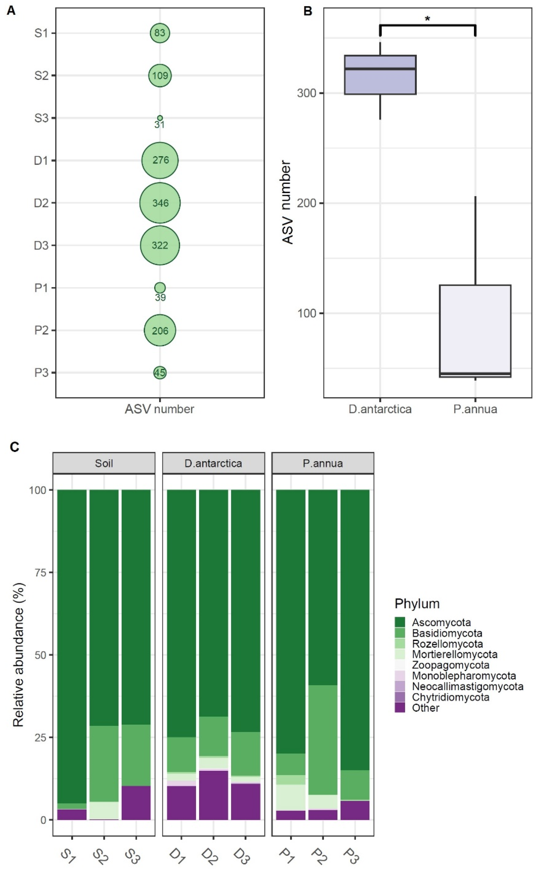
Figure 1. Fungal phylogenetic diversity in P. annua roots, D. antarctica roots, and the associated soil: (A) average ASV numbers per site; (B) differences in ASV numbers between P. annua and D. antarctica roots; and (C) relative abundance of fungal sequences identified to the phylum level. S1–S3—soil samples; D1–D3—D. antarctica root samples; P1–P3—P. annua root samples; *—significant at p < 0.05.
Ijms 25 08475 g001
Figure 2. Relative abundance of fungal sequences identified to the family level. *—families that significantly (p < 0.05) differed in abundance between P. annua and D. antarctica/soil samples; S1–S3—soil samples; D1–D3—D. antarctica root samples; P1–P3—P. annua root samples.
Ijms 25 08475 g002
Figure 3. Principal Component Analysis (PCA) based on relative ASV abundances of obtained fungal sequences. S1–S3—soil samples; D1–D3—D. antarctica root samples; P1–P3—P. annua root samples.
Ijms 25 08475 g003
Figure 4. Venn diagrams displaying shared ASVs. (A) ASVs shared between core fungal communities of D. antarctica and the associated soil communities; (B) ASVs shared between core fungal communities of P. annua and the associated soil communities; (C) ASVs shared within the fungal communities of D. antarctica from different sites; (D) ASVs shared within the fungal communities of P. annua from different sites; and (E) ASVs shared between the core communities of D. antarctica and P. annua. S1–S3—soil samples; D1–D3—D. antarctica root samples; P1–P3—P. annua root samples. The core microbiome was defined as a set of specific ASVs that occurred in every sample within a particular group.
Ijms 25 08475 g004
Figure 5. Size of fungal core communities and their major contributors in (A) P. annua roots and (B) D. antarctica roots. The size of the fungal core microbiome (core) is displayed as the relative sequence abundance (%) of 10 shared ASVs within P. annua roots and 13 shared ASVs within D. antarctica roots. Major contributors to the core microbiomes are displayed as relative sequence abundances of particular ASVs that made a substantial contribution to their respective core communities. Phylogenetic affiliations of those ASVs are displayed as identified by the Unite v.9.0 database.
Ijms 25 08475 g005
Figure 6. Fungal species identified in P. annua and D. antarctica roots: (A) phylum-rank affiliation of identified species (as % contribution within the group); (B) number of individual and shared identified species; and (C) presence (green circles)–absence (white circles) matrix of selected fungal species in P. annua (P1–P3) and D. antarctica (D1–D3) roots. The full list of identified strains is provided in Table S1.
Ijms 25 08475 g006
Figure 7. Taxonomic affiliation of fungal strains isolated from Antarctic soil and grass roots with annotated metabolic features. CaPO4—tricalcium phosphate; CMCellulose—carboxymethylcellulose; green—P. annua strains; yellow-green—D. antarctica strains; purple—soil strains (Ecology Glacier); violet—soil strains (Arctowski Station).
Ijms 25 08475 g007
Table 1. Cultivable fungi from Antarctic grasses and soil.
Fungal SpeciesPhylumCharacteristicsReference
D. antarctica roots
Helgardia anguioides-like
(Oculimacula)
Ascomycotacauses eyespot disease of wheat[16,17]
Herpotrichia juniperiAscomycotacausal agent of brown felt blight of conifers[18]
Sporobolomyces roseusBasidiomycotasynthesizes carotenoids and mycosporines to absorb UV radiation; secretes antimicrobial protein[19]
Ophiosphaerella sp.Ascomycotagrass pathogen [20]
P. annua roots
Mortierella antarctica-likeMortierellomycotapromotes winter wheat seedling growth[21]
Microdochium phragmitisAscomycotaendophyte colonizing roots of common reed[22]
Ophiosphaerella sp.Ascomycota causes spring dead spot of bermudagrass[23]
Soil—Ecology Glacier foreland
Cladosporium herbarumAscomycota source of antimicrobial compounds;
effective in bioremediation of perfluorochemicals
[24,25]
Botrytis cinereaAscomycotacosmopolitan broad host-range plant pathogen[26]
Mrakia robertiiBasidiomycotaproducer of cold-tolerant enzymes [27]
Leucosporidium sp.
Camptobasidium gelus
BasidiomycotaLeucosporidium sp.—yeast from Antarctic marine sponge, can biocontrol B. cinerea infection
Camptobasidium gelus-yeast found in Svalbard and Greenland glacial
[28,29,30]
Leucosporidiales sp.Basidiomycota
Soil—Arctowski Station 2
Mortierella fimbricystisMortierellomycota tolerates low pH, low temperature, high concentration of metals, and low organic matter[31]
Oidiodendron truncatumAscomycotaproducer of antifungal metabolites[32,33]
Pseudoeurotium sp.Ascomycotacandidate for bioremediation of polluted soil [24]
Pseudoeurotium sp.Ascomycotacandidate for bioremediation of polluted soil[24]
Linnemannia hialina
(Mortierella hyalina)
Mortierellomycotaidentified in iron ore mine as heavy metal-tolerating [34]
Cladosporium tenuissimumAscomycotacandidate for bioremediation of polluted soil[24]
Table 2. Main characteristics of the sampling sites. S1–S3—soil samples; D1–D3—D. antarctica root samples; P1–P3—P. annua root samples.
SiteGeographical CoordinatesDistance to the SeaSample IDLandform and Habitat
Arctowski Station 162°09′35″ S
58°28′26″ W
120 mS1, D1, P1Scientific station area; soil mechanically altered by human activities; Skeletic Eutric Fluvisol (Turbic).
The site is strongly influenced by marine aerosols; it is moist and located further from sources of nutrients, with large human influence.
Arctowski Station 262°09′33″ S
58°28′25″ W
100 mS2, D2, P2Scientific station area; soil mechanically altered by human activities; Skeletic Eutric Fluvisol (Turbic).
The site is strongly influenced by marine aerosols; it is moist and close to animal breeding colonies, with large human influence, especially related to using heavy vehicles.
Ecology Glacier foreland62°10′05″ S
58°27′46″ W
20 mS3, D3, P3Fluted moraine; Eutric Skeletic Protic Regosol (Turbic).
Dry site, sheltered from sources of nutrients, with little influence of marine aerosols.
Table 3. Primers used for the amplification of rDNA regions.
Primer Name5′-Sequence-3′PurposeReference
ITS1FI2GAACCWGCGGARGGATCAFungal-specific ITS1 amplification in the metagenomic study[63]
5.8SCGCTGCGTTCTTCATCG[64]
ITS1TCCGTAGGTGAACCTGCGGIdentification of cultured fungi[66]
ITS4TCCTCCGCTTATTGATATGC
Disclaimer/Publisher’s Note: The statements, opinions and data contained in all publications are solely those of the individual author(s) and contributor(s) and not of MDPI and/or the editor(s). MDPI and/or the editor(s) disclaim responsibility for any injury to people or property resulting from any ideas, methods, instructions or products referred to in the content.