Article

Biological Properties of Oleanolic Acid Derivatives Bearing Functionalized Side Chains at C-3

by
Gianfranco Fontana
1,2,
Natale Badalamenti
1,2,
Maurizio Bruno
1,2,3,
Filippo Maggi
4,*,
Federica Dell’Annunziata
5,6,
Nicoletta Capuano
5,
Mario Varcamonti
7 and
Anna Zanfardino
7
1
Department of Biological, Chemical and Pharmaceutical Sciences and Technologies (STEBICEF), University of Palermo, Viale delle Scienze, 90128 Palermo, Italy
2
NBFC, National Biodiversity Future Center, 90133 Palermo, Italy
3
Centro Interdipartimentale di Ricerca “Riutilizzo Bio-Based degli Scarti da Matrici Agroalimentari” (RIVIVE), University of Palermo, Viale delle Scienze, 90128 Palermo, Italy
4
Chemistry Interdisciplinary Project (ChIP) Research Center, University of Camerino, Via Madonna delle Carceri, 62032 Camerino, Italy
5
Department of Medicine, Surgery and Dentistry, “Scuola Medica Salernitana”, University of Salerno, 84081 Baronissi, Italy
6
Department of Experimental Medicine, University of Campania Luigi Vanvitelli, 80138 Naples, Italy
7
Department of Biology, University of Naples, Federico II, Via Cinthia, 80126 Naples, Italy
*
Author to whom correspondence should be addressed.
Int. J. Mol. Sci. 2024, 25(15), 8480; https://doi.org/10.3390/ijms25158480 (registering DOI)
Submission received: 3 July 2024 / Revised: 27 July 2024 / Accepted: 1 August 2024 / Published: 3 August 2024

Abstract

:
Triterpene acids are a class of pentacyclic natural carboxylic compounds endowed with a variety of biological activities including antitumor, antimicrobial, and hepatoprotective effects. In this work, several oleanolic acid derivatives were synthesized by structurally modifying them on the C-3 position. All synthesized derivatives were evaluated for possible antibacterial and antiviral activity, and among all the epimers, 6 and 7 demonstrated the best biological activities. Zone-of-inhibition analyses were conducted against two strains, E. coli as a Gram-negative and S. aureus as a Gram-positive model. Subsequently, experiments were performed using the microdilution method to determine the minimum inhibitory concentration (MIC). The results showed that only the derivative with reduced hydrogen bonding ability on ring A possesses remarkable activity toward E. coli. The conversion from acid to methyl ester implies a loss of activity, probably due to a reduced affinity with the bacterial membrane. Before the antiviral activity, the cytotoxicity of triterpenes was evaluated through a 3-(4,5-dimethylthiazol-2-yl)-2,5-diphenyltetrazolium bromide (MTT) assay. Samples 6 and 7 showed less than 50% cytotoxicity at 0.625 and 1 mg/mL, respectively. The antiviral activity against SARS-CoV-2 and PV-1 did not indicate that triterpene acids had any inhibitory capacity in the sub-toxic concentration range.

1. Introduction

Triterpene acids are a class of pentacyclic natural carboxylic acids endowed with a variety of biological activities including antitumor, antimicrobial, and hepatoprotective effects. They possess a basic set of three functional groups that are the carboxylic moiety at C-28, an alkene moiety at C-11,12, and a β-hydroxyl fragment at C-3. Several other functional groups, including hydroxyl and carboxyl, can decorate other positions of the pentacyclic carbon framework [1]. Even today, despite the important results obtained through drugs of synthetic origin, naturally sourced products are fundamental for the development and discovery of new pharmacological agents. In fact, approximately 80% of existing anticancer agents are inspired by or come from metabolism of plants, algae, or sponges [2]. Interest in triterpenoids has significantly increased in the last twenty years, precisely due to their chemical complexity, the presence of different functionalities, and their biological versatility [3]. Many triterpenes such as bryonolic acid, butyrospermol, lupeol, 1β-hydroxyaleuritolic acid 3-p-hydroxybenzoate, and isotirucallol have proven to be excellent antiproliferative [4,5,6], antineoplastic [7,8], and antiviral agents [9,10].
In this contest, we have been working for a couple of years on the antitumor activity of semisynthetic derivatives of oleanolic acid (1). It has been demonstrated that the introduction of a side chain at C-3 of the natural compound can deeply modify the biological efficacy of the molecule by a complex pattern of effects that include solubility, biological membrane permeability, and interaction with specific target proteins [1]. For example, an inverse relationship was observed between the hydrophilicity of a three-carbon side chain introduced in place of the H-3 of 1 and the cytotoxic effect of the molecule on HL60 cells, both resistant and sensitive [1]. The compound with a 2′,3′-dihydroxypropyl- moiety at C-3 was less active than the one bearing an allyl moiety. Furthermore, conjugation with appropriate peptides, and, in particular, the p10 peptide, has also been shown to improve the bioavailability, permeability, and pharmacological efficacy of oleanolic acid [11].
The antibacterial activity of triterpene acids has been widely investigated by several authors. This activity is exerted through a rather complex mechanism of action that includes influences on the gene expression and regulation of the peptide–glycans turnover, biofilm formation, and cell autolysis [12]. Other studies have shown that triterpene acids exert their antibacterial activity through the disruption of the bacterial cell membrane. Rebamang et al. highlighted how some terpenes isolated from the stem bark of Protorhus longifolia (Benrh.) Engl. affect the integrity of microbial cell membrane [13]. Additionally, some studies suggest that triterpene acids may have a synergistic effect when combined with traditional antibiotics or with each other, enhancing the overall efficacy of the treatment. For example, Wang et al. demonstrated that ursolic acid owns a synergistic effect with ampicillin and tetracycline against B. cereus and S. aureus bacterial strains [14].
It has been highlighted that the presence of hydroxyl functionalities at different positions of the A ring of triterpene oleane and ursolic acid derivatives can sensibly improve the antibacterial activity of these derivatives toward Gram-positive bacteria [15]. Other modifications at the C-3 of ring A were investigated to improve the antibacterial activity; these include the introduction of nitrogen-containing heterocyclic side chains [16] as well as nitrogen-containing side chains at C-28 [17,18]. Moreover, several studies have shown that some triterpenoids (oleanane, ursane, lupane, dammarane, lanostane, and cycloartane) recorded antiviral potential, with the high performance associated with tetracyclic and pentacyclic triterpenoids [19,20,21]. Sasaki et al. showed that glycyrrhizin acid, a pentacyclic triterpenoid isolated from licorice root, effectively inhibited HIV replication by increasing the number of OKT4 lymphocytes and reducing protein kinase C activity [22].
Considering these data, it was decided to study the antibacterial potential of a series of synthetic oleanolic acid derivatives, with modifications that focused on the C-3 position, against two strains of E. coli as a Gram-negative and S. aureus as a Gram-positive model by evaluating the minimum inhibitory concentration (MIC) for bacterial growth, and the possible antiviral activity against SARS-CoV2 and PV-1. Part of the molecules in the set were already available from previous investigations and some others were designed and synthesized to have a panel of compounds with a modified oleanane carbon structure by adding a three-carbon side chain at C-3 with different lipophilicity, hydrogen bonding capacity, and flexibility.

2. Results and Discussion

2.1. Preparation of Oleanolic Acid Derivatives

Methyl ester 2 was obtained by the treatment of compound 1 with an ethereal solution of CH2N2 at 0 °C. Acetate 3 was obtained from 1 with a standard acetylation procedure by treatment with Ac2O/Py, as reported previously [23]. The 3-oxoderivatives 4 and 5 were obtained by the oxidation of 1 [23] followed by CH2N2 methylation under the same conditions described above. The two epimeric 3-allyl derivatives 6 and 7 were prepared by Barbier–Grignard allylation with allyl magnesium bromide as previously reported [23]. Then, the new methyl esters 8 and 9 were obtained by the methylation of compounds 6 and 7, respectively. The epimer 7 was obtained as a largely minor product in the Grignard reaction. Alternatively, 7 can be obtained, as we demonstrated within this work for the first time, by the Barbier–Grignard allylation of compound 4 performed with allyl bromide and indium in THF. This procedure is compatible with the presence of the ester function at C-28, which, on the other side, is incompatible with the strong basicity of the organo-magnesium reagents. Compounds 10 and 11 were prepared by osmylation, as described previously [1], while new compounds 12 and 13 were obtained by the same procedure applied on the methyl ester 9 (Scheme 1).
Finally, the two new spyro tetrahydrofuryl derivatives 14 and 15 were obtained by a new procedure developed in this work that implies the tosylation of the less hindered primary hydroxyl group in 13 to convert it into a leaving group suitable for intramolecular substitution accompanied by the spyrotetrahydrofuran-forming cyclization process depicted in Scheme 2.
To the best of our knowledge, this kind of cyclization is reported for the first time. Other methods are available to obtain this spyro system starting from an alkene that involves quite different conditions, i.e., the use of electrophilic reagents such as hypervalent iodine [24], dibutyltin oxide on triols [25], cerium chloride [26], or perchloric acid [27] on epoxyalcohols.
All of the previously unknown compounds obtained in this work were fully characterized by complete spectroscopic analysis, particularly 1D and 2D NMR spectroscopy. The spectra of compounds 8, 9, 12, and 13 were similar to those of their unmethylated counterparts and further exhibit the NMR resonances of the carbomethoxy fragment at δC 51.7, 51.7, 51.7, and 52.0 ppm, respectively, and δH 3.61, 3.62, 3.60, and 3.62 ppm in the above cited order (Table 1). The confirmation of the spyro-tetrahydrofuryl structure for compounds 14 and 15 was less trivial and was supported by the long-range C-H hetero-correlation, in particular between the signals of C-3 (88.4 ppm) and H-3′ on the side chain (3.81 ppm) (Figure 1).

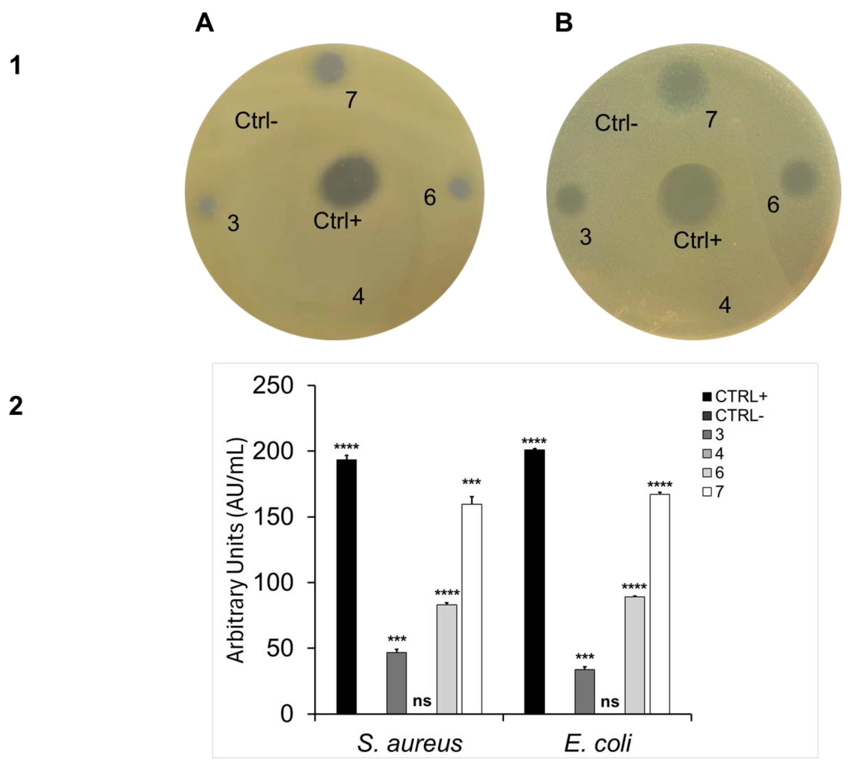
2.2. Antibacterial Activity

All triterpene compounds were tested for their antibacterial activity against two different bacteria, with E. coli DH5α as a Gram-negative and S. aureus ATCC6538P as a Gram-positive model.
The preliminary results, shown in Figure 2, highlight that compounds 6 and 7 were promising candidates for further investigation.
As can be observed in Figure 2 (panel 1A against S. aureus, panel 1B against E. coli), these two compounds appear to have the highest antimicrobial activity, as can be seen from both the largest diameter halos (panel 1) and the graph of arbitrary units per milliliter (panel 2). It is known that the larger the diameter of the zone, the greater the antimicrobial potential of the tested molecule (as shown in Figure 2) [28]. Among the two terpenoids, it appears that 7 was the most active against both S. aureus and E. coli. It shows potential antibacterial activity almost comparable to the positive control, ampicillin. All other compounds exhibited poor antibacterial activity since, after the development of the assay, no significant inhibition zones were observed. Specifically, 3 and 4 behaved similarly to the negative control, DMSO, indicating a lack of antibacterial activity and above all indicating how the small structural variations on C-3 did not improve their antibacterial activity. Figure 2 does not show the effects of compounds 2, 5, 8–15, as they did not result in inhibition. The literature confirms the biological importance of oleanolic acid (1). This triterpene is known for its antimicrobial, hepatoprotective, anti-inflammatory, antiallergic, antiviral, and cytotoxic activities. It is able to enhance the bioavailability of active ingredients of pharmaceutical formulations [29]. It has been found to be an active ingredient of Olea ferruginea Royle extract, to which antimicrobial activity is attributed. It has also been found to be inhibitory against the growth of intestinal bacteria such as Bacteroides fragilis, Clostridium clostridiiforme, C. perfringens, C. paraputrificum, Escherichia coli, Enterobacter cloacae, and Salmonella typhimurium. It shows antioxidant and pro-oxidant properties. It protects mammalian and bacterial cells from cytotoxicity induced by hydroperoxides [29].
To confirm the data obtained from the previous assay and to better investigate the intrinsic antibacterial activity of the various derivatives, the minimum inhibitory concentration (MIC) was estimated. The calculation of the MIC is essential for determining the efficacy of antibacterial agents against specific bacteria. In fact, it represents the lowest concentration of an antimicrobial, or in this case, a potential antimicrobial molecule, necessary to inhibit the visible growth of a specific bacterium. This parameter has various applications and significance in microbiology and medicine. For example, knowing the MIC can help personalize the dosage of antibiotics and/or antimicrobial molecules for patients, optimizing therapeutic efficacy and minimizing toxicity risks [30].
Compound 7 possessed a notable MIC value of 10 mg/mL. In contrast, samples 2, 3, 4, 6, 8, and 9 exhibited a MIC higher than 10 mg/mL, as shown in Table 2. These results confirmed that the most promising sample is indeed sample 7. This is worth noting as only the molecules bearing fewer polar and non-hydrogen-donating side chains are endowed of antibacterial activity. Further, the substitution of the hydrogen by the methyl group on the carboxy function (8 vs. 6 and 9 vs. 7) implies a complete loss of activity as shown by the complete lack of an inhibition zone for both 8 and 9.
The lack of antibacterial activity of the natural precursor 1 toward E. coli confirms the results of Kuete et al. [31], while this is in contrast with the data reported by other authors that obtained a MIC of 50 µg/mL [32]. Similar differences in the reported evidence regarding the antibacterial activity of 1 toward S. aureus ATCC 25923 vary from the very good MIC value of 8 µg/mL [33] to 64 µg/mL [34]. Compound 1 was instead rather efficient against a number of other clinically relevant strains such as M. tuberculosis and E. faecalis [10].
It is also worth noting that the stereochemistry at C-3 is quite relevant for the antibacterial activity as it emerged by comparing the activities of 6 and 7, the α-OH epimer being the most potent. For this reason, chemical modifications were devoted to obtaining further derivatives with 3-OH group with an α orientation. However, subsequent antibacterial tests at lower concentrations confirmed only compound 7 as the most promising lead compound, also confirming the presence of an apolar side chain in a β configuration as the best structural feature, together with the presence of the free carboxy functionality on C-28.

2.3. Cytotoxic Activity

The cytotoxic effects of 6 and 7 were performed on VERO-76 via the MTT assay. The cell monolayer was exposed to compounds ranging from 2 mg/mL to 0.0156 mg/mL for 24 h. DMSO (100%), used as a positive control (CTRL+), induced complete toxicity in the cell monolayer. On the other hand, cells treated with 2, 1, 0.5, 0.25, and 0.125% DMSO (corresponding to the solvent concentrations of DMSO in each treatment) showed cytotoxicity levels of about 28.3, 10.5, 8.1, 2.1, and 0.3%, respectively. Compound 6 recorded a cytotoxicity rate greater than 50% in the 2 to 0.25 mg/mL range. At 0.125 mg/mL the cytotoxicity was 48.9% and decreased in a dose-dependent manner at lower concentrations (Figure 3, histogram A). Compound 7 induced 57.8% cell death at 2 mg/mL, while at lower doses no relevant cytotoxic effect was verified (Figure 3, histogram B). Cytotoxicity data were normalized to the cell control rather than the solvent control, meaning that solvent cytotoxicity is included in the cytotoxicity data reported for each compound.

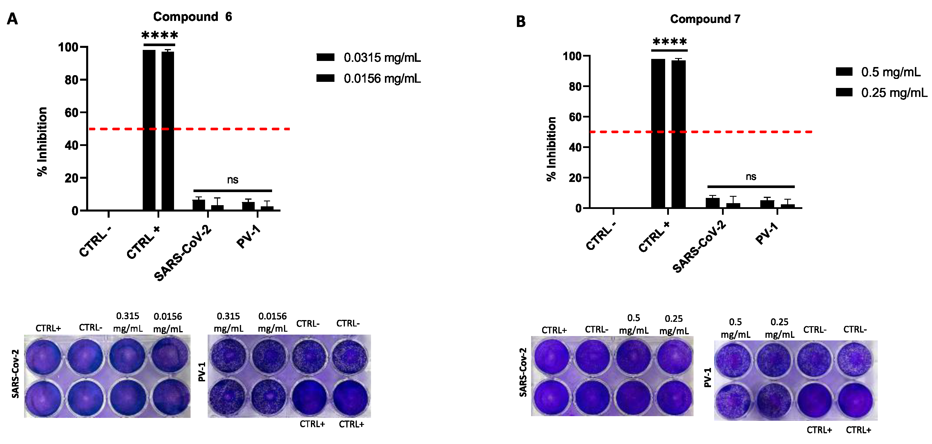
2.4. Antiviral Activity

The antiviral efficacy of compounds 6 and 7 was evaluated in the co-treatment phase, through the plaque reduction assay in infected cells, against SARS-CoV2 and PV-1. Neither compound showed significant antiviral activity. In detail, at the concentrations tested (0.5 and 0.250 mg/mL for 7 and 0.031 and 0.015 mg/mL for 6) the antiviral activity was less than 6% (Figure 4). Several studies have proven the antiviral activity of different terpenes against various viruses such as HIV, herpes simplex virus (HSV), hepatitis viruses, and influenza viruses. For example, betulinic acid, a well-studied triterpene, has shown a broad-spectrum antiviral activity, but its effectiveness can vary widely depending on modifications made to its structure and the specific viral strain [35]. For triterpenes that do not show antiviral activity, detailed studies and reviews often focus on their other pharmacological properties, such as cytotoxicity against tumor cells or anti-inflammatory effects [36,37]. These studies highlight that, although many triterpenes are potent antivirals, efficacy is not universal for all viruses or all triterpene derivatives.

3. Materials and Methods

3.1. General Procedures

Thin layer chromatography was performed on silica gel plates (Merck 60, F254, 0.2 mm) to monitor reaction progress. The NMR spectra were recorded on a Bruker Avance II 400 apparatus at 400.15 and 100.63 MHz for 1H and 13C, respectively, at the temperature of 300 K. The instrument was equipped with an inverse broadband probe (BBI). For the acquisition of 1H spectra, an 11.87 µs 90° pulse, a delay time of 5 s, and 16 scans were used. 13C spectra were acquired using a 90° pulse of 12.2 µs, a decoupling pulse of 80 µs, and a delay time of 3 s. The 2D NOESY spectra were acquired at the same temperature using a spin-lock pulse of 300 ms, eight scans, and 256 experiments. The 2D HSQC correlation spectra were acquired at the same temperature through a double inept transfer pulse sequence; the decoupling intervals were set as follows: a 90° pulse of 11.87 µs for 1H, with eight scans and 256 experiments. The stationary phase for flash chromatography (FC) was silica gel Merck (Kiesegel 60/230–400 mesh). Microanalysis data (C, H) were obtained by an Elemental Vario EI. III apparatus and are consistent with ± 0.4% of the theoretical value. Dry THF was obtained by suspension of THF onto 3 Å molecular sieves for 4 h. All chemical reagents were purchased from Merck s.r.l. (Sigma Aldrich, Milan, Italy). The solution of diazomethane in diethyl ether was obtained by Diazald® basic decomposition with KOH following the standard procedure [38,39].

3.2. Preparation of Oleanolic Derivatives

3.2.1. Synthesis of Compounds 2, 5, 8, and 9

To a 0.5 M solution of the reactant compound in dry THF, an ethereal solution of CH2N2 was added dropwise at 0 °C until the resulting solution assumed a persistent pale-yellow color. Then, the volatiles were removed by the rotary evaporator and the residual was purified by chromatographic column giving compounds 2, 5, 8, and 9 in quantitative yield (86.3, 94.7, 91.8, and 93.2%, respectively). The spectroscopic data of compounds 2 and 5 are in total agreement with the literature [40,41]. Methyl (3α)-allyl-(3β)-hydroxyolean-12-en-28-ate (8): white amorphous solid, anal. C 79.98%, H 10.70%; calcd. for C34H54O3; 1H NMR, and 13C NMR: see Table 1, Figures S1 and S2.

3.2.2. Synthesis of Compound 3

An amount of 300 mg of compound 1 was dissolved in 6 mL of mixture of pyridine/acetic anhydride 2:1 and left to react overnight at room temperature (rt). Then, the volatiles were removed by co-evaporation with toluene and the residual was dissolved in 3 mL of THF and treated with 2 mL of NaHCO3 for 1 h in order to hydrolyze the residual mixed anhydride. The mixture was partitioned between water and CHCl3; the organic phase was dried onto Na2SO4 and the solvent was removed by rotary evaporation. The residue was purified by chromatographic column (CHCl3/acetone, 0.3% in acetone) giving 282 mg (86%) of pure 3. The spectroscopic data of compound 3 is in total agreement with the literature [42].

3.2.3. Synthesis of Compound 4

To a 0.5 M solution of compound 1 in THF, an excess of Jones reagent was added dropwise at 0 °C until a light green persistent color developed. The insoluble residuals were removed by filtration and the solution was partitioned between water and CHCl3. The organic phase was neutralized with NaHCO3sat. and dried onto Na2SO4. The solvent was evaporated, and the residual was purified by chromatographic column (light petroleum/acetone 5% in acetone) giving pure 4 (80%). The spectroscopic data of compound 3 is in total agreement with the literature [43].

3.2.4. Synthesis of Compounds 6 and 7

(a) Allylmagnesium bromide procedure. To a 0.5 M solution of ketone 4 in dry THF, an excess of 2,5 eq. of a 2.0 M solution of allyl-MgCl in THF was added via syringe at 0 °C under an argon atmosphere. The resulting solution was stirred for 48 h and then the reaction was quenched by treatment with iced 1:1 HClaq: until the pH of the mixture was adjusted to 2. The mixture was extracted with CHCl3 and the resulting organic phase was neutralized with NaHCO3sat. and dried onto Na2SO4. The solvent was removed by the rotary evaporator and the residue was purified by chromatographic column (cyclohexane/acetone 9:1) giving pure 6 (45%) and 7 (10%).
(b) Allyl bromide and indium procedure. An amount of 1 mmol of compound 4 was solubilized in 10 mL of THF and 600 µL (7.0 mmol) of allyl-Br and 800 mg (7.0 mmol) of powdered In were added in one portion. The resulting heterogeneous mixture was stirred vigorously for 24 h at 30 °C. Then, the reaction was quenched by adding 10 mL of 1:10 HClaq. The mixture was extracted with CHCl3 and the organic phase was dried onto Na2SO4. The solvent was removed by evaporation and the residue was purified by chromatographic column (cyclohexane/acetone 9:1 to obtain pure 7, 65%; cyclohexane/EtOAc 14:1 to obtain pure 6). The spectroscopic data of compounds 6 and 7 are in total agreement with the literature [23,44].

3.2.5. Synthesis of Compounds 10–13

An amount of 0.5 mmol of the starting compounds 7 and 9 and 2.0 mmol (241.5 mg) of 97% N-methylmorpholine-N-oxide (NMO) were solubilized in 15 mL of acetone/THF/H2O 6:7:1 and treated with 1.5 mL (0.3 eq.) of a 2.5% w/w solution of OsO4 in in 2-methyl-2-propanol. The resulting solution was stirred for 48 h at rt and then quenched with 2 mL of Na2S3O3sat. The mixture was stirred for further 10 min and then extracted with CHCl3/EtOH 2:1. The organic phase was dried and evaporated giving a residue that was purified by chromatographic column (cyclohexane/EtOAc 1:1 to give pure 12, 40% yield and 13, 56% yield; cyclohexane/EtOAc 1:3 to give pure 10 and 11). The spectroscopic data of compounds 10 and 11 are in total agreement with the literature [1].
Methyl (3β)-2′S,3-dihydroxypropyl-(3α)-hydroxyolean-12-en-28-oate (12): amorphous white solid, anal. C 74.98%, H 10.37%; calcd. for C34H56O5, C 74.96%, H 10.36%. 1H and 13C-NMR: see Table 1; Methyl (3β)-2′R,3-dihydroxypropyl-(3α)-hydroxyolean-12-en-28-oate (13): amorphous white solid, anal. C 74.98%, H 10.36%; calcd. for C34H56O5, C 74.96%, H 10.36%. 1H and 13C NMR: see Table 1, Figures S3–S8.

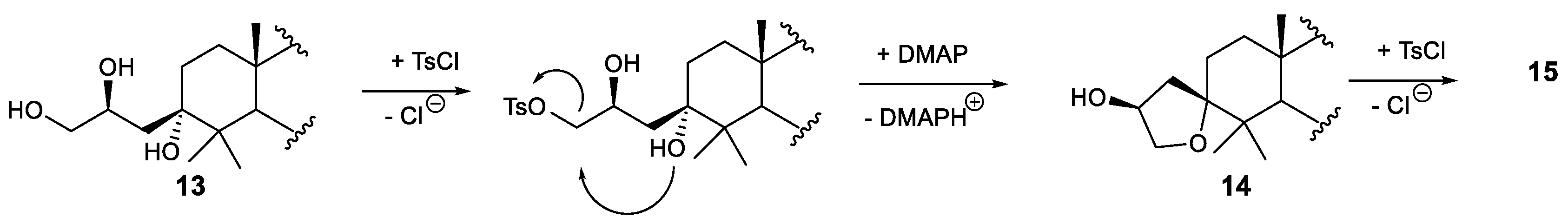
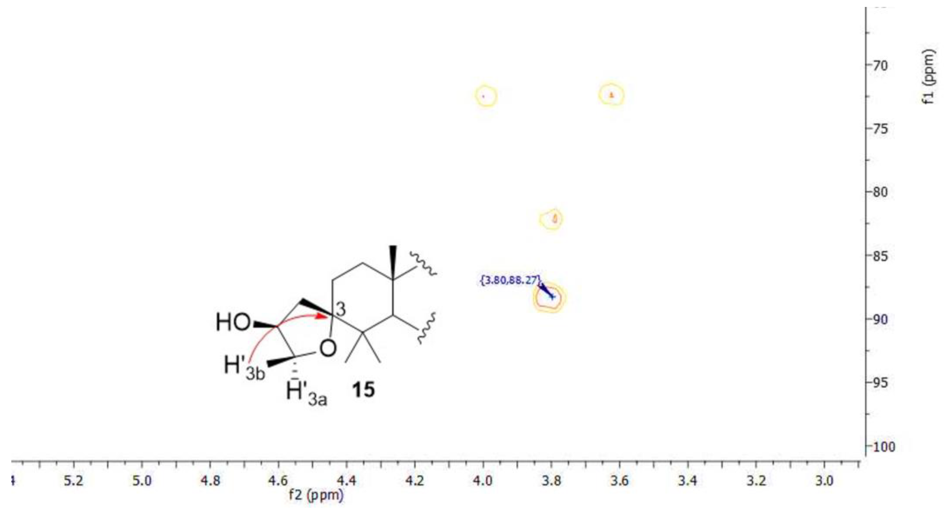
3.2.6. Synthesis of Compounds 14 and 15

The procedure to synthesized 14 as major compound: 300 mg (0.551 mmol) of compound 13 was dissolved in 30 mL of dry dichloromethane. Then, 1.05 gr (5.51 mmol, 10 eq.) of tosyl chloride, 766 µL (5.51 mmol, 10 eq.) of triethylamine, and 135 mg (1.1 mmol, 2 eq.) of 4-(N,N-dimethylamino)pyridine (DMAP) were added in one portion at rt. The mixture was stirred for 48 h, then a second portion of 10 eq. of DMAP was added and the mixture was stirred for further 6 h. Then, the reaction was quenched with 50 mL of H2O and extracted with CHCl3. The organic phase was dried, and the solvent was evaporated. The residue was purified by chromatographic column (cyclohexane/EtOAc 10:1) to give pure 14 (80%) and 15 (10%).
In the procedure to synthesized 15 as a major component: 100 mg (0.184 mmol) of compound 13 was dissolved in 10 mL of dry dichloromethane. Then, 53 mg (1.5 eq.) of tosyl chloride, 25.5 µL (1 eq.) of triethylamine were added in one portion at rt. Then, the mixture was stirred for 72 h and then further 8.5 eq. of tosyl chloride, 9 eq. of triethylamine, and 2 eq. of DMAP were added. The reaction mixture was stirred for further 6 h and then quenched with 10 mL of water. The organic phase was dried, and the solvent was evaporated. The residue was purified by chromatographic column (cyclohexane/EtOAc 10:1) to give pure 14 (23%) and pure 15 (75%).
Compound 14: amorphous white solid, anal. C 72.34%, H 8.90%; calcd. for C41H60SO6, C 72.31%, H 8.88% S 4.71%. 1H and 13C NMR: see Table 1; compound 15: amorphous white solid, anal. C 77.55%, H 10.35%; calcd. for C34H54O4, C 77.52%, H 10.33%. 1H and 13C-NMR: see Table 1, Figures S9–S12.

3.3. Bacterial Strains and Antimicrobial Activity

To evaluate the antimicrobial activity on all compounds 1–15, the Gram-negative Escherichia coli DH5α strain and the Gram-positive Staphylococcus aureus ATCC6538P strain were used. The antimicrobial properties of compounds 3, 4, 6, and 7 were detected using the agar diffusion test following the Kirby–Bauer method [45] with some modifications. An amount of 5 μL of the various samples was placed on Luria–Bertani agar plates and then covered with ~10 mL of soft agar (0.7%) premixed with 10 μL of the E. coli and S. aureus strains. The negative control was dimethyl sulfoxide (DMSO 80%) used to resuspend the samples; the positive control was the antibiotic ampicillin (10 µL) at a concentration of 0.1 mg/mL.
The plates were incubated overnight at 37 °C and the antimicrobial activity was evaluated by calculating arbitrary units based on the width of the halo formed, according to equation [46,47]:
A U / m L = D i a m e t e r   o f   t h e   z o n e   o f   c l e a r a n c e m m × 1000 V o l u m e   t a k e n   i n   t h e   w e l l   ( μ L )

3.4. Determination of Minimal Inhibitory Concentration

The microdilution method established by the Clinical and Laboratory Standards Institute (CLSI) was used to determine the minimal inhibitory concentration (MIC) of the samples against the chosen strains. Briefly, 5 × 10⁵ CFU/mL of each bacterial strain were added to 95 µL of Mueller–Hinton broth (CAM-HB; Fisher Scientific, Segrate, Italy), supplemented or not with the various compounds (3, 4, 6, 7, 8, and 9) used at various concentrations (0.5, 1, 2, 4, 8, or 10 mg/mL). DMSO was used as a negative control and the antibiotic ampicillin as a positive control [47]. After overnight incubation at 37 °C, MIC100 values were determined as the lowest concentration associated with no visible bacterial growth at 600 nm.

3.5. Cell Culture Condition

The cell line used for cytotoxicity testing originated from the renal epithelium of the African green monkey (Cercopithecus aethiops) (VERO-76 CRL-1587, Manassas, VA, USA). The cells were grown in Dulbecco’s modified Eagle medium (DMEM) containing 4.5 g/L glucose (Gibco Life Technologies, Paisley, UK), supplemented with 2 mM L-glutamine (Gibco Life Technologies, Paisley, UK), 100 IU/mL penicillin–streptomycin solution (Gibco Life Technologies, Paisley, United Kingdom), and 10% fetal bovine serum (FBS) (Gibco Life Technologies, Paisley, United Kingdom). For the assays, the cells were seeded into 96-well plates, with final volumes of 0.1 μL, respectively. Incubation occurred at 37 °C with 5% CO2 in a humidified environment.

3.6. Viruses

The viruses used in this study were poliovirus type 1 (PV-1 strain CHAT) and SARS-CoV-2, strain VR PV10734, kindly donated by the Lazzaro Spallanzani Hospital in Rome, Italy. Both viral strains were propagated on a VERO-76 cell line. The original titers (PFU/mL) were 108 and 109 plaque-forming units (PFU)/mL, for PV-1 and SARS-CoV-2, respectively.

3.7. Cell Cytotoxicity Assay

Cytotoxicity assessment was performed on all compounds (1–15) using the 3-[4,5-dimethylthiazol-2-yl]-2,5-diphenyl tetrazolium bromide (MTT) method. VERO-76 cells were meticulously seeded in 96-well plates at a density of 2 × 10⁴ cells/well in a final volume of 0.1 mL. Compounds 6 and 7 were subjected to serial dilutions, ensuring cell exposure to a concentration range of 2 to 0.156 mg/mL for 24 h. Cells cultured with DMSO and solvent represented positive (CTRL+) and negative (CTRL−) controls, respectively. After exposure, cell viability was assessed by adding 100 μL of MTT solution (0.03 mg/mL) to cell monolayers for 3 h at 37 °C. Then, the medium was removed, and the formazan salts were solubilized using 100 μL of DMSO. The cytotoxicity rate was quantified by measuring absorbance at 570 nm with a microplate reader (Tecan life science, Männedorf, SW).

3.8. Antiviral Activity Assays

The antiviral efficacy of triterpenes was assessed by co-treatment assays using plaque reduction assays in infected cells, as previously described [48,49]. VERO-76 cells were seeded at a density of 2 × 10⁵ cells/well in a 12-well plate and incubated overnight at 37 °C in a humidified environment with 5% CO2. The compounds were serially diluted in a concentration range of 500–250 mg/mL for 7 and 0.625–0.312 mg/mL for 6. The cells were co-treated with viruses at a multiplicity of infection (MOI) of 0.01 and terpenes at selected concentrations for 1 h at 37 °C. Then, the virus–compound mixture was removed, and a complete medium (10% FBS) supplemented with 5% carboxymethylcellulose (Sigma, C5678, C5013) was added. The cells were incubated for 24 h at 37 °C and then fixed with 4% formaldehyde. Thus, the cell monolayer was stained with 0.5% crystal violet and the number of plaques was normalized to untreated infected cells. Pleconaril (2 µg/mL) and Ivermectin (10 μM) were used as positive controls (CTRL+) for PV-1 and SARS-CoV-2, respectively.

3.9. Statistical Analysis

Antimicrobial assays were performed in biological triplicates and data were expressed as mean ± error standard (SE). The significance of the difference between treated samples and CTRL− for each assay was evaluated by two-tailed paired t-test. Cytotoxicity and antiviral assays were performed in biological and technical duplicates and data were expressed as mean ± standard deviation (SD). The significance of the difference between treated samples and CTRL− for each assay was evaluated by Dunnett’s test. For cytotoxicity and antiviral assays were used Graph Pad Prism 9.0 software (San Diego, CA, USA). A p-value < 0. 05 was considered significant.

4. Conclusions

In this study, we focused on the antibacterial and antiviral effects of different C-3 modified oleanolic acid derivatives. In particular, methylation, acetylation, oxidation, allylation, and osmylation reactions were performed; two new spyro tetrahydrofuryl derivatives were also prepared to evaluate the impact of these structural modifications on the biological activities investigated. Compound 7 is the most active antimicrobial, with a MIC value of 10 mg/mL. The stereochemistry of C-3 is quite relevant for the antibacterial activity, with the α-OH epimer 7 being the most potent compound. Also, the presence of a non-polar side chain in the β configuration, together with the presence of the free carboxyl functionality on C-28, is a fundamental structural feature necessary to preserve the antibacterial activity.

Supplementary Materials

The following supporting information can be downloaded at: https://www.mdpi.com/article/10.3390/ijms25158480/s1.

Author Contributions

Conceptualization, G.F. and N.C.; methodology, G.F.; software, G.F. and N.B.; validation, G.F., A.Z. and F.D.; formal analysis, G.F.; investigation, A.Z., G.F. and N.B.; resources, G.F., N.B. and M.B.; data curation, G.F.; writing—original draft preparation, G.F., A.Z. and M.V.; writing—review and editing, G.F., N.B. and A.Z.; visualization, N.B.; supervision, M.B., F.M. and A.Z. All authors have read and agreed to the published version of the manuscript.

Funding

This research received external funding from the National Biodiversity Future Center S.c.a.r.l., Piazza Marina 61 (c/o Palazzo Steri) Palermo, Italy, C.I. CN00000033\u2014CUP UNIPA B73C22000790001. This work was supported by a grant from the European Union\u2014Next Generation EU (PRIN-PNRR); Project Code P2022CKMPW_002\u2014CUP B53D23025620001.

Institutional Review Board Statement

Not applicable.

Informed Consent Statement

Not applicable.

Data Availability Statement

Data are contained within the article and Supplementary Materials.

Acknowledgments

We thank the National Biodiversity Future Center, S.c.a.r.l., Piazza Marina 61 (c/o Palazzo Steri), Palermo, Italy, for their support. Thanks also to Professor Dario Antonini for his support with revisions of the paper.

Conflicts of Interest

The authors declare no conflicts of interest.

References

  1. Fontana, G.; Badalamenti, N.; Bruno, M.; Castiglione, D.; Notarbartolo, M.; Poma, P.; Spinella, A.; Tutone, M.; Labbozzetta, M. Synthesis, In Vitro and In Silico Analysis of New Oleanolic Acid and Lupeol Derivatives against Leukemia Cell Lines: Involvement of the NF-κB Pathway. Int. J. Mol. Sci. 2022, 23, 6594. [Google Scholar] [CrossRef] [PubMed]
  2. Cragg, G.M.; Grothaus, P.G.; Newman, D.J. Impact of natural products on developing new anti-cancer agents. Chem. Rev. 2009, 109, 3012–3043. [Google Scholar] [CrossRef] [PubMed]
  3. Ovesná, Z.; Vachálková, A.; Horváthová, K.; Tóthová, D. Pentacyclic triterpenoic acids: New chemoprotective compounds. Minirev. Neoplasma 2004, 51, 327–333. [Google Scholar]
  4. Gill, B.S.; Kumar, S. Triterpenes in cancer: Significance and their influence. Mol. Biol. Rep. 2016, 43, 881–896. [Google Scholar] [CrossRef] [PubMed]
  5. Liby, K.T.; Yore, M.M.; Sporn, M.B. Triterpenoids and rexinoids as multifunctional agents for the prevention and treatment of cancer. Nat. Rev. Cancer 2007, 7, 357–369. [Google Scholar] [CrossRef] [PubMed]
  6. Setzer, W.N.; Setzer, M.C. Plant-derived triterpenoids as potential antineoplastic agents. Mini Rev. Med. Chem. 2003, 3, 540–556. [Google Scholar] [CrossRef] [PubMed]
  7. Bradford, P.G.; Awad, A.B. Phytosterols as anticancer compounds. Mol. Nutr. Food Res. 2007, 51, 161–170. [Google Scholar] [CrossRef]
  8. Hisham Shady, N.; Youssif, K.A.; Sayed, A.M.; Belbahri, L.; Oszako, T.; Hassan, H.M.; Abdelmohsen, U.R. Sterols and Triterpenes: Antiviral Potential Supported by In-Silico Analysis. Plants 2021, 10, 41. [Google Scholar] [CrossRef]
  9. Liu, Y.; Yang, L.; Wang, H.; Xiong, Y. Recent Advances in Antiviral Activities of Triterpenoids. Pharmaceuticals 2022, 15, 1169. [Google Scholar] [CrossRef]
  10. Wolska, K.; Grudniak, A.; Fiecek, B.; Kraczkiewicz-Dowjat, A.; Kurek, A. Antibacterial Activity of Oleanolic and Ursolic Acids and Their Derivatives. Open Life Sci. 2010, 5, 543–553. [Google Scholar] [CrossRef]
  11. Wang, L.; Geng, J.; Wang, H. Delivery of Oleanolic Acid with Improved Antifibrosis Efficacy by a Cell Penetrating Peptide P10. ACS Pharmacol. Transl. Sci. 2023, 6, 1006–1014. [Google Scholar] [CrossRef] [PubMed]
  12. Rebamang, A.M.; Mandlakayise, L.N.; Thandeka, V.D.; Andy, R.O. Antibacterial Activity of Two Triterpenes from Stem Bark of Protorhus longifolia. J. Med. Plants Res. 2014, 8, 686–702. [Google Scholar] [CrossRef]
  13. Wang, C.-M.; Chen, H.-T.; Wu, Z.-Y.; Jhan, Y.-L.; Shyu, C.-L.; Chou, C.-H. Antibacterial and Synergistic Activity of Pentacyclic Triterpenoids Isolated from Alstonia scholaris. Molecules 2016, 21, 139. [Google Scholar] [CrossRef] [PubMed]
  14. Fontana, A.; Albarella, L.; Scognamiglio, G.; Uriz, M.; Cimino, G. Structural and Stereochemical Studies of C-21 Terpenoids from Mediterranean Spongiidae Sponges. J. Nat. Prod. 1996, 59, 869–872. [Google Scholar] [CrossRef]
  15. Medina-O’Donnell, M.; Rivas, F.; Reyes-Zurita, F.J.; Cano-Muñoz, M.; Martinez, A.; Lupiañez, J.A.; Parra, A. Oleanolic Acid Derivatives as Potential Inhibitors of HIV-1 Protease. J. Nat. Prod. 2019, 82, 2886–2896. [Google Scholar] [CrossRef] [PubMed]
  16. Blanco-Cabra, N.; Vega-Granados, K.; Moya-Andérico, L.; Vukomanović, M.; Parra, A.; Álvarez de Cienfuegos, L.; Torrents, E. Novel Oleanolic and Maslinic Acids derivatives as a promising treatment against bacterial biofilm in nosocomial infections: An in Vitro and in Vivo study. ACS Infect. Dis. 2019, 5, 1581–1589. [Google Scholar] [CrossRef] [PubMed]
  17. Kazakova, O.; Rubanik, L.; Smirnova, I.; Poleschuk, N.; Petrova, A.; Kapustsina, Y.; Baikova, I.; Tret’yakova, E.; Khusnutdinova, E. Synthesis and in vitro activity of oleanolic acid derivatives against Chlamydia trachomatis and Staphylococcus aureus. Med. Chem. Res. 2021, 30, 1408–1418. [Google Scholar] [CrossRef]
  18. Fontana, G.; Bruno, M.; Notarbartolo, M.; Labbozzetta, M.; Poma, P.; Spinella, A.; Rosselli, S. Cytotoxicity of oleanolic and ursolic acid derivatives toward hepatocellular carcinoma and evaluation of NF-κB involvement. Bioorg. Chem. 2019, 90, 103054. [Google Scholar] [CrossRef]
  19. Talybov, G.M.; Mamedbeyli, E.G.; Yusubov, F.V. Synthesis of Propyne(Ene)Oxy-Substituted Spirotetrahydrofurans. Russ. J. Gen. Chem. 2018, 88, 2684–2688. [Google Scholar] [CrossRef]
  20. Baltina, L.A.; Tasi, Y.T.; Huang, S.H.; Lai, H.C.; Baltina, L.A.; Petrova, S.F.; Yunusov, M.S.; Lin, C.W. Glycyrrhizic acid derivatives as Dengue virus inhibitors. Bioorg. Med. Chem. Lett. 2019, 29, 126645. [Google Scholar] [CrossRef]
  21. Hattori, T.; Ikematsu, S.; Koito, A.; Matsushita, S.; Maeda, Y.; Hada, M.; Fujimaki, M.; Takatsuki, K. Preliminary evidence for inhibitory effect of glycyrrhizin on HIV replication in patients with AIDS. Antivir. Res. 1989, 11, 255–261. [Google Scholar] [CrossRef] [PubMed]
  22. Sasaki, H.; Takei, M.; Kobayashi, M.; Pollard, R.B.; Suzuki, F. Effect of glycyrrhizin, an active component of licorice roots, on HIV replication in cultures of peripheral blood mononuclear cells from HIV-seropositive patients. Pathobiology 2003, 70, 229–236. [Google Scholar] [CrossRef] [PubMed]
  23. Lahmadi, G.; Horchani, M.; Dbeibia, A.; Mahdhi, A.; Romdhane, A.; Lawson, A.M.; Daïch, A.; Harrath, A.H.; Ben Jannet, H.; Othman, M. Novel Oleanolic Acid-Phtalimidines Tethered 1,2,3 Triazole Hybrids as Promising Antibacterial Agents: Design, Synthesis, In Vitro Experiments and In Silico Docking Studies. Molecules 2023, 28, 4655. [Google Scholar] [CrossRef] [PubMed]
  24. Gamedze, M.P.; Nkambule, C.M. Dibutyltin Oxide Mediated Diastereoselective Cyclodehydration/Sulfonylation of 1,2,4-Triols. Tetrahedron Lett. 2015, 56, 1825–1829. [Google Scholar] [CrossRef]
  25. Gowravaram Sabitha, V.; Rama Subba Rao, K.; Sudhakar, M.; Raj Kumar, E.; Venkata Reddy, J.S. Study of conventional versus microwave-assisted reactions of 3,4-epoxyalcohols by CeCl3·7H2O: Synthesis of tetrahydrofurans and 1-chloro-3-substituted-2-propanols. J. Mol. Catal. A Chem. 2008, 280, 16–19. [Google Scholar] [CrossRef]
  26. Chirskaya, M.V.; Vasil’ev, A.A.; Shorshnev, S.V.; Sviridov, S.I. Transformation of homoallylic alcohol oxides into 3-hydroxytetrahydrofurans in aqueous HClO4. Russ. Chem. Bull. 2006, 55, 1300–1303. [Google Scholar] [CrossRef]
  27. Bubonja-Šonje, M.; Knežević, S.; Abram, M. Challenges to Antimicrobial Susceptibility Testing of Plant-Derived Polyphenolic Compounds. Arh. Hig. Rada. Toksikol. 2020, 71, 300–311. [Google Scholar] [CrossRef] [PubMed]
  28. Magréault, S.; Jauréguy, F.; Carbonnelle, E.; Zahar, J.-R. When and How to Use MIC in Clinical Practice? Antibiotics 2022, 11, 1748. [Google Scholar] [CrossRef] [PubMed]
  29. Sultana, N.; Ata, A. Oleanolic acid and related derivatives as medicinally important compounds. J. Enzyme Inhib. Med. Chem. 2008, 23, 739–756. [Google Scholar] [CrossRef]
  30. Zhang, Y.-Z.; Zhang, W.; Hong, D.; Shi, L.; Shen, Q.; Li, J.-Y.; Li, J.; Hu, L.-H. Oleanolic acid and its derivatives: New inhibitor of protein tyrosine phosphatase 1B with cellular activities. Bioorg. Med. Chem. 2008, 16, 8697–8705. [Google Scholar] [CrossRef]
  31. Kuete, V.; Wabo, G.F.; Ngameni, B.; Mbaveng, A.T.; Metuno, R.; Etoa, F.X. Antimicrobial activity ofthe methanolic extract, fractions and compounds from the stem bark of Irvingia gabonensis (Ixonanthaceae). J. Ethnopharmacol. 2007, 114, 54–60. [Google Scholar] [CrossRef] [PubMed]
  32. Woldemichael, G.M.; Franzblau, S.G.; Zhang, F.; Wang, Y.; Timmerman, B.N. Inhibitory effect of sterols from Ruprechtia triflora and diterpenes from Calceolaria pinnifolia on the growth of Mycobacterium tuberculosis. Planta Med. 2003, 69, 628–631. [Google Scholar]
  33. Farina, C.; Pinza, M.; Pifferi, G. Synthesis and antiulcer activity of new derivatives of glycyrrhetic, oleanolic and ursolic acids. Pharmacology 1998, 53, 22–32. [Google Scholar]
  34. Fontanay, S.; Grare, M.; Mayer, J.; Finance, C.; Duval, R.M. Ursolic, oleanolic and betulic acids: Antibacterial spectra and selectivity indexes. J. Ethnopharmacol. 2008, 120, 272–276. [Google Scholar] [CrossRef] [PubMed]
  35. Gnoatto, S.C.B.; Vechia, L.D.; Lencina, C.L.; Dassonville-Klimpt, A.; Da Nascimento, S.; Mossalayi, D.; Guillon, J.; Gosmann, G.; Sonnet, P. Synthesis and preliminary evaluation of new ursolic and oleanolic acids derivatives as antileishmanial agents. J. Enzyme Inhib. Med. Chem. 2008, 23, 604–610. [Google Scholar] [CrossRef]
  36. Lou, H.; Li, H.; Zhang, S.; Lu, H.; Chen, Q. A Review on Preparation of Betulinic Acid and Its Biological Activities. Molecules 2021, 26, 5583. [Google Scholar] [CrossRef] [PubMed]
  37. Ogbole, O.; Adeniji, J.; Ajaiyeoba, E.; Kamdem, R.; Choudhary, M. Anthraquinones and triterpenoids from Senna siamea (Fabaceae) Lam inhibit poliovirus activity. Afr. J. Microbiol. Res. 2014, 8, 2955–2963. [Google Scholar]
  38. Badalamenti, N.; Rosselli, S.; Zito, P.; Bruno, M. Phytochemical profile and insecticidal activity of Drimia pancration (Asparagaceae) against adults of Stegobium paniceum (Anobiidae). Nat. Prod. Res. 2021, 35, 4468–4478. [Google Scholar] [CrossRef]
  39. Candela, R.G.; Lazzara, G.; Piacente, S.; Bruno, M.; Cavallaro, G.; Badalamenti, N. Conversion of Organic Dyes into Pigments: Extraction of Flavonoids from Blackberries (Rubus ulmifolius) and Stabilization. Molecules 2021, 26, 6278. [Google Scholar] [CrossRef]
  40. Chatow, L.; Nudel, A.; Eyal, N.; Lupo, T.; Ramirez, S.; Zelinger, E.; Nesher, I.; Boxer, R. Terpenes and cannabidiol against human corona and influenza viruses-anti-inflammatory and antiviral in vitro evaluation. Biotechnol. Rep. Amst. 2024, 41, e00829. [Google Scholar] [CrossRef]
  41. Bauer, A.W.; Kirby, W.M.; Sherris, J.C.; Turck, M. Antibiotic susceptibility testing by a standardized single-disk method. J. Clin. Pathol. 1966, 45, 493–496. [Google Scholar] [CrossRef]
  42. Wen, X.; Sun, H.; Liu, J.; Cheng, K.; Zhang, P.; Zhang, L.; Hao, J.; Zhang, L.; Ni, P.; Zographos, S.E.; et al. Naturally Occurring Pentacyclic Triterpenes as Inhibitors of Glycogen Phosphorylase: Synthesis, Structure−Activity Relationships, and X-ray Crystallographic Studies. J. Med. Chem. 2008, 51, 3540–3554. [Google Scholar] [CrossRef] [PubMed]
  43. Seo, S.; Tomita, Y.; Tori, K. Carbon-13 NMR spectra of urs-12-enes and application to structural assignments of components of Isodon japonicus hara tissue cultures. Tetrahedron Lett. 1975, 16, 7–10. [Google Scholar] [CrossRef]
  44. Castagliuolo, G.; Di Napoli, M.; Vaglica, A.; Badalamenti, N.; Antonini, D.; Varcamonti, M.; Bruno, M.; Zanfardino, A.; Bazan, G. Thymus richardii Subsp. nitidus (Guss.) Jalas Essential Oil: An Ally against Oral Pathogens and Mouth Health. Molecules 2023, 28, 4803. [Google Scholar] [CrossRef] [PubMed]
  45. Iyapparaj, P.; Maruthiah, T.; Ramasubburayan, R.; Prakash, S.; Kumar, C.; Immanuel, G.; Palavesam, A. Optimization of Bacteriocin Production by Lactobacillus Sp. MSU3IR against Shrimp Bacterial Pathogens. Aquat. Biosyst. 2013, 9, 12. [Google Scholar] [CrossRef]
  46. Di Napoli, M.; Castagliuolo, G.; Badalamenti, N.; Vaglica, A.; Ilardi, V.; Varcamonti, M.; Bruno, M.; Zanfardino, A. Chemical Composition, Antimicrobial and Antioxidant Activities of the Essential Oil of Italian Prangos trifida (Mill.) Herrnst. & Heyn. Nat. Prod. Res. 2023, 37, 3772–3786. [Google Scholar] [CrossRef] [PubMed]
  47. Di Napoli, M.; Castagliuolo, G.; Pio, S.; Di Nardo, I.; Russo, T.; Antonini, D.; Notomista, E.; Varcamonti, M.; Zanfardino, A. Study of the Antimicrobial Activity of the Human Peptide SQQ30 against Pathogenic Bacteria. Antibiotics 2024, 13, 145. [Google Scholar] [CrossRef] [PubMed]
  48. Dell’Annunziata, F.; Sellitto, C.; Franci, G.; Marcotullio, M.C.; Piovan, A.; Della Marca, R.; Folliero, V.; Galdiero, M.; Filippelli, A.; Conti, V.; et al. Antiviral Activity of Ficus rubiginosa Leaf Extracts against HSV-1, HCoV-229E and PV-1. Viruses 2022, 14, 2257. [Google Scholar] [CrossRef]
  49. Dell’Annunziata, F.; Morone, M.V.; Gioia, M.; Cione, F.; Galdiero, M.; Rosa, N.; Franci, G.; De Bernardo, M.; Folliero, V. Broad-Spectrum Antimicrobial Activity of Oftasecur and Visuprime Ophthalmic Solutions. Microorganisms 2023, 11, 503. [Google Scholar] [CrossRef]
Scheme 1. Reagents and conditions: i. CH2N2, diethyl ether, 0 °C, 30′; ii. Ac2O/Py 1:2 rt, overnight; iii. Jones reagent, acetone, 0 °C, 3 h; iv. allyl-MgBr, dry THF, 0 °C, 48 h; v. allyl-Br, In, THF, rt, 24 h; vi. OsO4 cat., NMO, acetone/H2O 6:1, rt, 48 h; vii. TsCl, TEA, DMAP, dry DCM, 48 h.
Ijms 25 08480 sch001
Scheme 2. Formation of compounds 14 and 15 from 13.
Ijms 25 08480 sch002
Figure 1. Long-range C-H hetero-correlation (HMBC) between the signals of C-3 (88.4 ppm) and H-3′b on the side chain (3.81 ppm) of compound 15.
Ijms 25 08480 g001
Figure 2. Bacterial growth inhibition: the inhibition halo of samples 3, 4, 6, and 7 (panel 1) against (A) S. aureus and (B) E. coli. The positive control is ampicillin; the negative control is dimethyl sulfoxide. Panel 2 reports arbitrary units in milliliters for both bacterial strains. The assays were performed in three independent experiments (n = 3). Statistical analysis was performed using a two-tailed paired t-test (ns is not significant, *** p ≤ 0.005, **** p ≤ 0.001) versus the negative control.
Ijms 25 08480 g002
Figure 3. Cytotoxicity of 6 (A) and 7 (B) on the VERO-76 cell line after 24 h. The data represent the mean ± SD. Dunnett’s multiple comparisons test: **** p < 0.0001; *** p < 0.001; ** p < 0.01; ns p > 0.05.
Ijms 25 08480 g003
Figure 4. Antiviral activity of 6 (A) and 7 (B) against SARS-CoV-2 and PV-1. The data represent the mean ± SD. Dunnett’s multiple comparisons test: **** p < 0.0001; ns p > 0.05.
Ijms 25 08480 g004
Table 1. Spectroscopic data of compounds 8, 9, 12, 13, 14, and 15 expressed in ppm (coupling constant J expressed in Hz).
Comp.8912131415
C/HCHCHCHCHCHCH
136.9 36.7 36.91.25 m
1.27 m
34.4 34.81.29 m
1.31 m
34.7
232.6 28.4 32.41.49 m
1.65 m
33.9 31.31.45 m
1.66 m
32.4
375.0 76.2 77.1 75.9 85.1 88.2
441.2 40.6 41.7 41.7 42.0 41.7
550.91.21 m51.11.19 m51.51.17 m51.01.18 m51.61.24 m51.51.22 m
618.8 18.9 18.81.37 m
1.43 m
20.8 18.71.40 m
1.42 m
18.8
733.8 32.9 32.71.20 m
1.29 m
30.7 32.51.20 m
1.22 m
32.5
839.2 39.3 39.2 37.0 37.1 36.8
947.5 47.6 47.71.62 m47.6 46.51.62 m47.4
1030.7 37.1 36.7 36.8 31.2 30.9
1123.5 23.4 23.01.88 m
1.90 m
23.2 23.31.83 m
1.84 m
23.2
12122.45.29 t
(3.4)
122.35.29 t
(3.4)
122.35.29 t br
(3.5)
122.25.28 t
(3.5)
122.45.28 m122.45.28 m
13143.8 143.8 143.8 143.7 143.4 143.7
1441.7 41.6 40.8 39.2 40.7 40.4
1527.6 27.7 27.91.04 m
1.60 m
27.6 27.91.00 m
1.58 m
27.6
1623.2 23.1 23.51.61 m
1.96 m
23.0 23.21.60 m
1.87 m
23.0
1746.7 46.0 45.8 45.8 41.5 45.8
1840.92.86 dd
(14.4, 4.4)
41.32.87 dd (14.0, 4.6)41.22.85 dd
(13.4, 4.3)
41.32.85 dd
(13.9, 4.1)
41.32.85 m
41.22.84 dd (14.1, 4.3)
1945.8 46.8 46.71.13 m
1.15 m
46.7 46.51.10 m
1.12 m
46.7
2030.7 30.7 30.7 30.2 30.8 30.7
2134.3 33.9 33.81.17 m
1.32 m
32.4 34.01.15 m
1.17 m
33.8
2232.4 32.4 34.41.20 m
1.45 m
32.7 29.21.65 m
1.73 m
29.1
2320.70.91 s19.50.87 s16.80.73 s16.80.71 s17.20.71 s16.80.70 s
2423.00.82 s24.10.95 s23.60.93 s23.80.92 s23.70.91 s23.40.92 s
2514.90.91 s15.80.91 s14.90.90 s14.90.90 s15.10.93 s15.00.90 s
2616.80.73 s16.90.73 s20.70.78 s18.80.79 s20.40.76 s20.50.75 s
2726.11.15 s26.01.16 s26.01.14 s26.11.14 s26.21.12 s26.11.14 s
28178.3 178.3 178.3 178.4 178.4 178.3
2933.10.93 s33.10.91 s33.10.90 s33.10.90 s33.20.91 s33.10.89 s
3023.60.94 s23.60.93 s23.70.94 s23.60.92 s23.80.76 s23.60.92 s
1′40.52.11 dd
(13.8, 6.8)
2.41 dd
(13.8, 8.2)
41.12.23 dd
(13.5, 7.2)
2.48 dd
(14.0, 7.4)
36.81.25 m
1.87 m
40.91.85 m
1.90 m
43.82.00 m
2.24 dd m
(14.1, 6.6)
39.21.67 m
1.70 m
2′134.75.93 ddt
(17.1, 10.0, 7.3)
135.05.91 ddt. (17.0, 10.2, 7.5)69.04.11 m69.64.04 m67.34.44 m82.15.02 m
3′118.95.12 m
5.16 m
118.05.10 dd (17.0, 2.3)
5.16 dd (10.2, 2.3)
67.43.44 dd
(11.2, 6.7)
3.59 m
67.63.48 dd
(10.9, 6.5)
3.58 m
78.23.81 m72.33.81 m
3.90 m
1″ 133.9
2″ = 6″ 129.97.34 d (8.5)
3″ = 5″ 127.77.79 m
4″ 144.8
Tosyl-CH3 21.62.45 s
-OMe51.53.63 s51.53.62 s50.73.63 s51.63.62 s51.53.62 s51.53.62 s
Table 2. The minimal inhibitory concentration (MIC) of samples 3, 4, 6, 7, 8 and 9 against E. coli. The values were obtained from minimum of three independent experiments.
StrainCompoundsMIC100 [mg/mL]
E. coli DH5α1>10
3>10
4>10
6>10
710
8>10
9>10
All the other compounds were substantially inactive toward the bacterial strains tested.
Disclaimer/Publisher’s Note: The statements, opinions and data contained in all publications are solely those of the individual author(s) and contributor(s) and not of MDPI and/or the editor(s). MDPI and/or the editor(s) disclaim responsibility for any injury to people or property resulting from any ideas, methods, instructions or products referred to in the content.

