Article

Reconstruction and Analysis of a Genome-Scale Metabolic Model of Acinetobacter lwoffii

by
Nan Xu
1,
Jiaojiao Zuo
1,
Chenghao Li
1,
Cong Gao
2,* and
Minliang Guo
1,*
1
College of Bioscience and Biotechnology, Yangzhou University, Yangzhou 225009, China
2
School of Biotechnology, Jiangnan University, Wuxi 214122, China
*
Authors to whom correspondence should be addressed.
Int. J. Mol. Sci. 2024, 25(17), 9321; https://doi.org/10.3390/ijms25179321
Submission received: 30 June 2024 / Revised: 31 July 2024 / Accepted: 22 August 2024 / Published: 28 August 2024
(This article belongs to the Section Molecular Microbiology)

Abstract

:
Acinetobacter lwoffii is widely considered to be a harmful bacterium that is resistant to medicines and disinfectants. A. lwoffii NL1 degrades phenols efficiently and shows promise as an aromatic compound degrader in antibiotic-contaminated environments. To gain a comprehensive understanding of A. lwoffii, the first genome-scale metabolic model of A. lwoffii was constructed using semi-automated and manual methods. The iNX811 model, which includes 811 genes, 1071 metabolites, and 1155 reactions, was validated using 39 unique carbon and nitrogen sources. Genes and metabolites critical for cell growth were analyzed, and 12 essential metabolites (mainly in the biosynthesis and metabolism of glycan, lysine, and cofactors) were identified as antibacterial drug targets. Moreover, to explore the metabolic response to phenols, metabolic flux was simulated by integrating transcriptomics, and the significantly changed metabolism mainly included central carbon metabolism, along with some transport reactions. In addition, the addition of substances that effectively improved phenol degradation was predicted and validated using the model. Overall, the reconstruction and analysis of model iNX811 helped to study the antimicrobial systems and biodegradation behavior of A. lwoffii.

1. Introduction

Acinetobacter lwoffii is a Gram-negative bacterium of the genus Acinetobacter, class Gammaproteobacteria. While Acinetobacter baumannii is the most common cause of infections among 38 species of this Acinetobacter genus [1], there have been increasing reports that A. lwoffii has been identified as a serious human pathogen. A. lwoffii causes nosocomial infections, such as bacteremia, pneumonia, and meningitis [2]. A. lwoffii possesses resistance to multiple drugs and disinfectants, which allows it to survive in clinical and hospital environments. Whole-genome sequencing on clinical isolates of A. lwoffii has been used to annotate antibiotic resistance genes [3]. Comparative genomics studies of the phylogeny of A. lwoffii and other Acinetobacter strains [4,5,6] have detected more resistance genes on plasmids in A. lwoffii strains compared to A. baumannii strains [7]. The challenge of eliminating bacterial cells in the environment is exacerbated by multidrug resistance, which renders infections more harmful. Although it has not yet been fully utilized, metabolism is a promising source of possible medication adjuvants for harmful bacteria [8]. A systems-level framework would help to explore the relationship between microbial metabolism and antimicrobials, and to identify drug targets for treating the pathogens [9].
In addition to pathogenicity and drug resistance, the biodegradation potential of A. lwoffii is currently recognized. Like other Acinetobacter bacteria, A. lwoffii is ubiquitous in soil and water environments, as well as in animal and human clinical samples. For example, A. lwoffii DNS32 can degrade atrazine (100 mg/L) [10], and its degradation capacity has been optimized in a microbial consortium [11]. Adaptive laboratory evolution produced A. lwoffii NL115, capable of degrading 1500 mg/L phenol [12]. A. lwoffii MG04 can degrade pyrethroids [13]. Environmental strains of A. lwoffii contain heavy metal resistance genes, allowing the organism to survive in high concentrations of heavy metals [14]. A system-level platform would help to uncover the metabolic mechanisms underlying biodegradation, and to design optimal conditions that would enhance biodegradation processes.
Genome-scale metabolic models (GEMs) are effective systemic platforms for elucidating the associations among metabolites, genes, and reactions in living organisms [15]. For the genus Acinetobacter, five models described for A. baumannii and one for A. baylyi have been used to explore microbial growth phenotypes, essential metabolism, antimicrobial development, metabolic alterations under certain conditions, and other metabolic characteristics [16,17,18,19,20,21]. Despite the large number of genomes and draft sequences of A. lwoffii, a systematic and comprehensive metabolic model is still lacking. In this study, we describe the reconstruction of the first genome-scale metabolic model, designated iNX811. A. lwoffii NL1 is resistant to multiple compounds and effectively degrades phenol [22]. The application of the model iNX811 was in the two relevant metabolic and physiological features. The essential metabolism of A. lwoffii was analyzed to identify in silico drug targets. The metabolic response under phenol stress was evaluated by the integration of transcriptomic data into the model iNX811. The addition of TCA cycle intermediates and alanine to improve phenol degradation was predicted and validated using constraint-based analyses and biochemical studies.

2. Results and Discussion

2.1. Characteristics of the Genome-Scale Metabolic Model iNX811

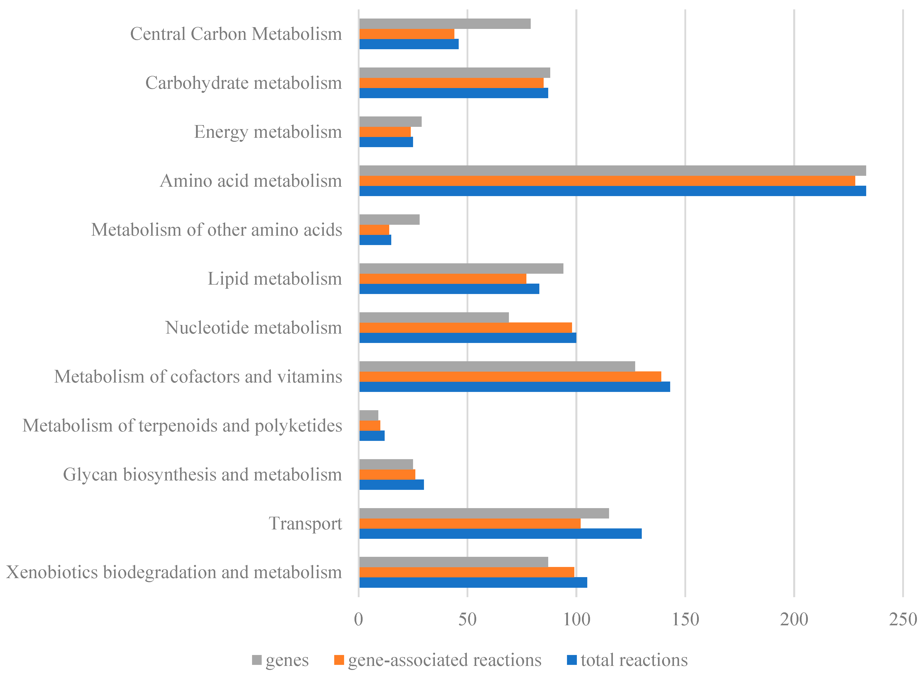
As shown in Figure 1, reconstruction of the genome-scale metabolic model for A. lwoffii mainly included three steps: (1) The ModelSEED model for A. lwoffii NL1 was built using annotation findings from the RAST server [23], and it included 1292 reactions, 1353 metabolites, and 631 genes. Moreover, 679 and 599 genes of A. lwoffii NL1 were matched to the protein sequences in the models iATCC19606 and iAbaylyiV4, respectively, and utilized to extract model contents from the two models based on gene–protein–reaction relationships. (2) After combining the SEED model and homologous models, metabolites and gene names were unified into their own formats, and duplicate reactions were removed. The draft model was modified trivially and manually, and the improved draft model had 696 genes, 1022 metabolites, and 1084 reactions. (3) Metabolic gaps in the biosynthesis pathways for biomass components were filled using the literature, biochemical databases, and gene reannotation. After passing biomassPrecursorCheck in the COBRA toolbox, 102 transporters from A. lwoffii NL1 were introduced to the model, with nutrient uptake rates serving as constraints. Then, growth phenotypes were modeled and compared to the experimental data. Iterative manual curation of the disagreements was required to fill in metabolic gaps, change the direction of reactions, and correct incorrectly anticipated reactions. Finally, there were 811 genes, 1071 metabolites, and 1155 reactions in the model iNX811 (Tables S1 and S2 and Supplementary File S2), which had a 52% total MEMOTE score [24] (Supplementary File S3). This model accounts for 76 metabolic pathways in 16 metabolic subsystems, including the biosynthesis and biodegradation of primary and secondary metabolites. Except for exchange, tRNA biosynthesis, general reactions, and biomass formation, the other 12 metabolic subsystems are generally shown in Figure 2. The average ratio of gene–protein–reaction (GPR) association reactions per subsystem was 92.8%, reflecting adequate genome annotation using the model iNX811. Amino acid metabolism comprised the highest number of reactions and genes, consistent with the capability of A. lwoffii to synthesize 20 amino acids. Xenobiotic biodegradation and metabolism had 103 reactions, with 94.3% GPR associations. They included 23 chlorocyclohexane and chlorobenzene degradation reactions, 17 xylene degradation reactions, 17 benzoate degradation reactions, 13 fluorobenzoate degradation reactions, 13 toluene degradation reactions, 5 reactions each for aminobenzoate, polycyclic aromatic hydrocarbon, and atrazine degradation, and 5 scatter reactions. The findings revealed genes with potential value in degrading a wide range of xenobiotics.
To identify the characteristics of the model iNX811, genome-scale metabolic models of A. baumannii iATCC1960 [15] and A. baylyi ADP1 [20] were compared. The coverage of the annotated open reading frames of the three models was approximately 23.2% (iNX811), 23.4% (iATCC1960), and 23.1% (ADP1). A total of 468 metabolites shared by the three models comprised 518 metabolic reactions, excluding exchange reactions. These represent the core metabolism of Acinetobacter, including common primary metabolism, ten reactions in the metabolism of terpenoids and polyketides, and ten xenobiotic metabolic reactions (Table S3). In the model iNX811, 800 and 495 metabolites coexisted in the iATCC19606 and A. baylyi ADP1 models (Table S3), respectively, which had similar trends to the genome homology of A. lwoffii with A. baumannii (74.13%) and A. baylyi (68.3%). Compared to the other two models, 64 unique metabolites in the model iNX811 participated in 65 unique reactions (Table S4), excluding exchange reactions. They were mainly distributed in xenobiotic biodegradation and metabolism (44 reactions), as well as amino acid and alternative carbon metabolism.

2.2. Model Validation by Cell Growth Phenotypes and Genotypes

The utilization of 39 carbon and nitrogen sources was simulated using the model iNX811 (Table 1). The in silico growth coincided well with the experimental results, except for when glycerol was employed as the carbon source. A. lwoffii NL1 did not utilize glycerol as the sole carbon source, which may have been related to over-reduction. Adaptive laboratory evolution could overcome this barrier and enable in silico growth on glycerol MM medium [25]. This model could reflect the metabolic pathways of various carbon and nitrogen sources, including common sugars, amino acids, alcohols, carboxylic acids, and uncommon aromatic xenobiotics. The carbon source spectrum of A. lwoffii is relatively broad, and the metabolites did not support cell growth, mainly because of the loss of relevant transporters or metabolic enzymes. For example, A. lwoffii NL1 can grow on fructose, but not on glucose, because the hexokinase in A. lwoffii NL1 cannot convert glucose to glucose-6-phosphate. These intermediates in glycolysis and the TCA cycle, such as acetate, ethanol, citrate, and succinate, can support cell growth as sole carbon sources. In addition, the observation that A. lwoffii NL1 could use simple phenols and phenolic acids as sole carbon sources suggests that A. lwoffii has a good biodegradability capacity [26,27,28].
In silico single-gene deletions were induced using the model iNX811. Compared with the essential genes identified by transposon insertion mutants, the prediction accuracy of essential genes was 79.6% on LB medium and 75.9% on succinate as the sole carbon source. The high accuracy of essential gene prediction validated that the model iNX811 can reflect the metabolic characteristics of A. lwoffii NL1. Precisely 100 genes were predicted to be essential for cell growth in the LB medium (Table S4). Of these genes, 95 were validated as essential in the DEG database and participated in 175 biochemical reactions involving 34 metabolic pathways. A total of 164 genes were predicted to be essential for cell growth with succinate as the sole carbon source (Table S4). Among these, 157 genes (95.7%) were validated in the DEG database and participated in 270 biochemical reactions involving 41 metabolic pathways.

2.3. Essential Metabolite Analysis for In Silico Drug Targets

A. lwoffii is an opportunistic pathogen that causes nosocomial infections in humans [3]. A recent study found that the NL1 strain has numerous antibiotic resistances [21], making it difficult to kill the bacterial cells. As a result, identifying feasible therapeutics is an urgent and substantial task [29]. One approach to address this issue has been to use systems-level computational models, specifically genome-scale metabolic models [30]. Pathogenic bacteria’s GEMs have recently been reconstructed, revealing genes and pathways that are essential to the survival and spread of zoonotic and human infections [31,32,33]. GEMs, as powerful computational tools, can predict cellular essential genes, reactions, and metabolites. Essential metabolites are required for cellular growth, and their absence leads to cell death. Therapeutic targets can be created based on the structural analogues of essential metabolites of pathogens, hence eliminating the need for labor-intensive random chemical library screening. The essential metabolite filtering method (EMFilter) was used for discovering effective drug targets by removing currency metabolites, metabolites consumed by only one outgoing reaction, metabolites present in human GEMs, and the essential metabolites consumed by homologous enzymes with human genomes. The EMFilter procedure was employed in developing therapeutic strategies against various pathogens, such as A. baumannii [18], Vibrio vulnificus [34], and Cryptococcus neoformans [35]. Using the validated model iNX811, eighty-five genes were predicted to be essential for cell growth on arbitrary complex media (see Table S7) and were related to 148 reactions and 216 metabolites. According to the EMFilter procedure [18], 34 currency metabolites, 85 metabolites involved in only one consumption reaction, and 138 metabolites in the human metabolic model Recon3D were eliminated. Twelve of the essential metabolites identified were predicted to be potential drug targets (Table 2). These were mainly distributed in glycan biosynthesis, lysine biosynthesis, and the metabolism of cofactors and vitamins.
Specifically, four essential metabolites (D-glucosamine 1-phosphate, D-arabinose 5-phosphate, dTDP-glucose, and alpha-trehalose 6-phosphate) were involved in glycan biosynthesis, which affects the cell wall and membrane structures of A. lwoffii. D-glucosamine 1-phosphate 1,6-phosphomutase (GlmM) catalyzes the reversible isomerization of D-glucosamine 1-phosphate and D-glucosamine 6-phosphate. The phosphoglucosamine mutase (GlmM) in Legionella pneumophila was discovered as a candidate therapeutic target enzyme after docking the substrate for the most favorable binding of S-mercaptocystein [36]. Glucosamine-1-phosphate-acetyltransferase (GlmU) is involved in bacterial cell wall biosynthesis, making it an appealing target for creating antibacterial drugs for Aspergillus terreus [37] and Haemophilus influenzae [38]. D-arabinose-5-phosphate can be reversibly converted to d-ribulose 5-phosphate by D-arabinose-5-phosphate isomerase, which has been utilized as an antibacterial target in Bacteroides fragilis [39], Pseudomonas aeruginosa [40], and Burkholderia pseudomallei [41]. D-arabinose-5-phosphate and phosphoenolpyruvate form 3-deoxy-d-manno-octulosonate 8-phosphate, which is a precursor of lipopolysaccharides that are necessary for the growth and virulence of Acinetobacter [42]. dTDP-Glucose synthase (glucose-1-phosphate thymidylyltransferase) was predicted to be a drug target for A. lwoffii, having previously been identified as a possible target for antibacterial medicines in M. tuberculosis [43] and P. aeruginosa [44]. It has been found that, in pathogenic fungi like Candida albicans, dTDP-glucose 4,6-hydrolyase preserves cell wall integrity and virulence [45]. dTDP-Glucose 4,6-dehydratase from Streptococcus mutants (serotype c) was found in the DrugBank database (www.drugbank.com). Trehalose 6-phosphate has been discovered as an essential metabolite of A. lwoffii. Some potential drug targets were found in trehalose metabolism [46]. Trehalose 6-phosphate synthase, for example, has been investigated as an antifungal target in C. albicans [47] and Cryptococcus neoformans [48].
Four essential metabolites (dehydrodipicolinate, N-succinyl-LL-2,6-diaminoheptanedioate, meso-2,6-diaminoheptanedioate, and L-Aspartate 4-semialdehyde) are involved in the lysine biosynthesis of A. lwoffii. The lysine biosynthesis pathway is of special interest in pharmacology because the absence of DAP in mammalian cells allows for using lysine biosynthesis genes as bacteria-specific drug targets [49]. Dehydrodipicolinate synthase (DHDPS), dihydrodipicolinate reductase (DapB), diaminopimelate epimerase (DapF), succinyldiaminopimelate transaminase, succinyl-diaminopimelate desuccinylase (DapE), UDP-N-acetylmuramoyl-L-alanyl-D-glutamate-2,6-diaminopimelate ligase (MurE), aspartate-semialdehyde dehydrogenase (ASADH), and 4-hydroxy-tetrahydrodipicolinate synthase (DapA) are relevant enzymes that catalyze reactions containing these essential metabolites. Potential antibacterials, inhibitors of DHDPS, were created for Bacillus anthracis [50] and Campylobacter jejuni [51]. Studies on DapB inhibition were conducted on M. tuberculosis [52] and Staphylococcus aureus [53]. Research has been carried out on the importance of DapF as a therapeutic target in Bordetella pertussis [54] and Enterococcus faecalis [55]. Succinyldiaminopimelate transaminase in M. tuberculosis was studied to develop novel antibacterial medications [56]. DapE has emerged as a promising bacterial enzyme target, and its related inhibitors have been identified in H. influenzae [57], A. baumannii [58], and a few pathogens [59]. A. baumannii employed MurE, which was found in the DrugBank database (www.drugbank.com), as a therapeutic target [60]. The inhibition of ASADHs has been discussed as a possible therapeutic target for producing novel antibacterial drugs [61]. The structures of DapA from M. tuberculosis [62], P. aeruginosa [63], and S. aureus [64] were examined to develop drugs that inhibit the DapA family.
In folate biosynthesis, the essential metabolites 4-aminobenzoate and 2-amino-4-hydroxy-6-hydroxymethyl-7,8-dihydropteridine were also found in the opportunistic pathogen V. vulnificus, and structural analog 24837 of 4-aminobenzoate has antibacterial effects [34]. Chorismate, as a popular pharmacological target, has been effectively employed against pathogenic bacteria [65,66]. 1-Deoxy-D-xylulose 5-phosphate, predicted as the essential metabolite, was involved in the metabolism of thiamine and vitamin B6. Related enzymes, 1-deoxy-D-xylulose-5-phosphate synthase and pyridoxine 5′-phosphate synthase, were predicted to be drug targets in pathogens [67,68,69]. Therefore, essential metabolite analysis can aid in predicting possible pharmacological targets for antimicrobial treatments. Some metabolites and enzymes have been shown to be efficient targets in pathogenic bacteria or fungi. The inhibition of pathogens illustrates the GEM’s utility in discovering therapeutic targets, making it exceedingly viable to produce antimicrobials against A. lwoffii in future studies.

2.4. Integration of Transcriptomic Data in the Genome-Scale Metabolic Model

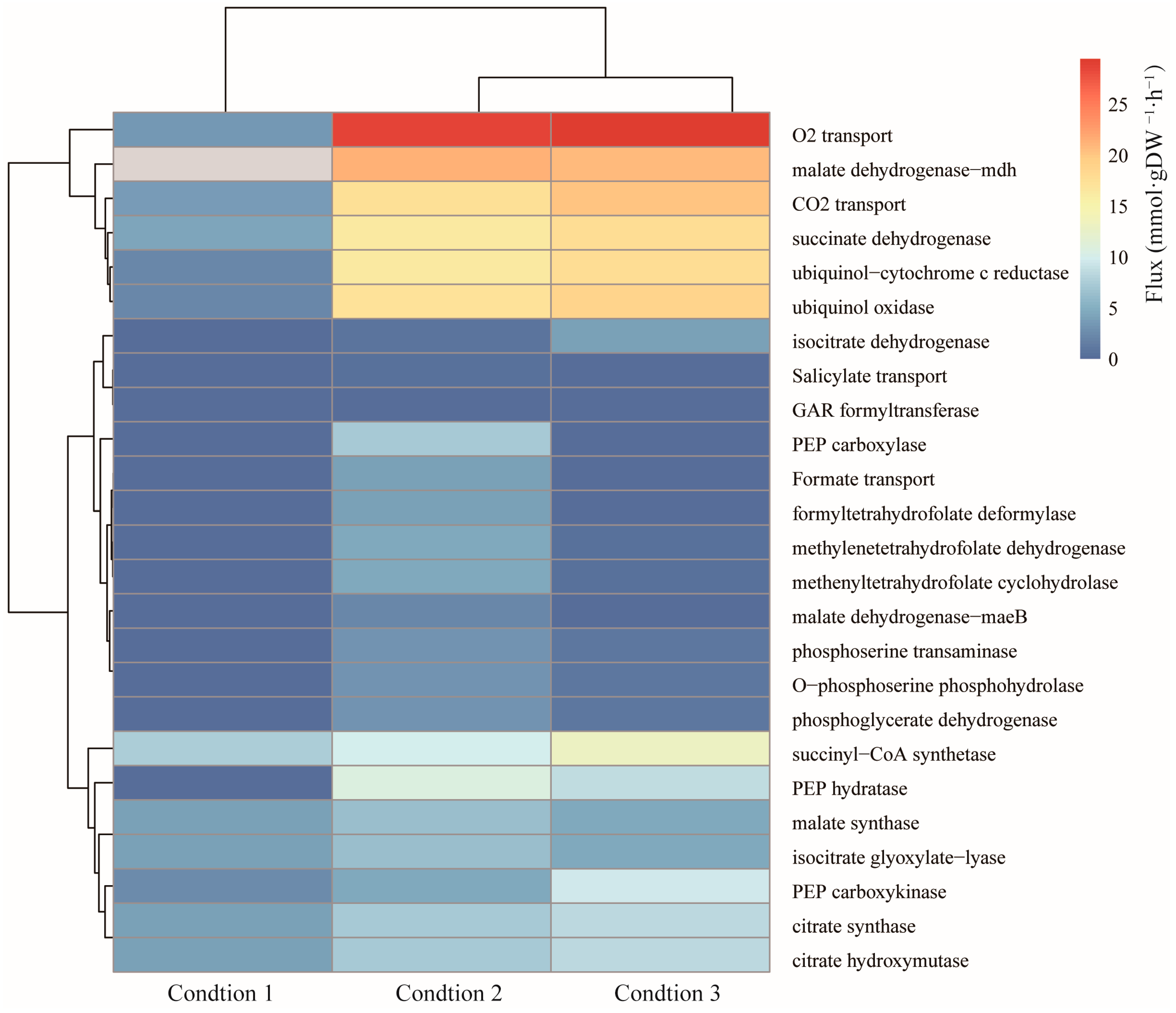
A. lwoffii NL1 is one of the most efficient degraders of phenols. GEMs for microbial biodegradation were reconstructed to analyze the degradation pathways of non-conventional substrates [70,71], to improve biodegradation by modeling-based identification of media supplements [72,73], to predict degradation behavior by imposing more constraints [74], and to simulate interactions between degraders and non-degraders in consortium systems [75]. Modern omics data integrated into GEMs contribute to the identification of novel metabolic insights into this microbe and are increasingly being utilized to promote a better understanding of complex biological systems [76,77]. In this study, the Gene Inactivity Moderated by Metabolism and Expression (GIMME) algorithm [78] was used to construct context-specific GEMs of A. lwoffii. Gene expression data of A. lwoffii on sodium acetate (NaAc) as the sole carbon source and NaAc with 0.5 or 1.5 g/L phenol as a carbon source were integrated into the model iNX811, and the three relevant context-specific GEMs included 1050 reactions and 1016 metabolites, 1045 reactions and 1017 metabolites, and 1077 reactions and 1016 metabolites, respectively (see Supplementary File S4). After introducing transcriptional constraints, the reaction flux of the phenol ortho-cleavage pathway was narrowed under the three environmental conditions (Figure 3) using random sampling. Integrating transcriptomic data into the model iNX811 reduces the number of metabolic states [79]. The metabolic flux of the three context-specific GEMs was compared to investigate the metabolic differences under phenol stress. Reactions exhibiting a significant flux change (added phenol/without phenol) were assumed to change under phenol stress (Figure 4). Specifically, the in silico metabolic flux for complexes II and IV of oxidative phosphorylation and D-phosphoglycerate 2,3-phosphomutase in glycolysis and the conversion of acetyl-CoA to malate (citrate synthetase, citrate hydroxymutase, isocitrate dehydrogenase, succinyl-CoA synthetase, succinate dehydrogenase, and fumarase) in the TCA cycle were enhanced, suggesting accelerated energy conversion in response to phenol stress. The conversion between 3-phosphoglycerate and phosphoenolpyruvate, the glyoxylate cycle, and the outflow from malate to pyruvate were downregulated under greater phenol stress. The oxaloacetate anaplerotic reaction became active under high phenol stress with increased phosphoenolpyruvate (PEP) carboxykinase and decreased PEP carboxylase. Although no salicylate was present in the medium, the expression of the salicylate exporter was activated under phenol stress, which might weaken the competition for catechol metabolism in the beta-ketoadipate branch. Under phenol stress, there was an increase in CO2 permeability and oxygen absorption level. It was found that Pseudomonas CF600 in continuous culture responded significantly to oxygen in terms of phenol consumption [80]. The flux from 3-phosphoglycerate to serine decreased. Folate downregulates one carbon pool as the phenol concentration increases [81]. The transcriptome-constrained GEMs may be used to investigate reactions with large flux changes at different phenol levels, allowing us to gain a better understanding of the metabolic response to phenol stress in A. lwoffii and identify potential metabolic modules to enhance biodegradation.

2.5. Prediction of Targets for Increased Phenol Degradation

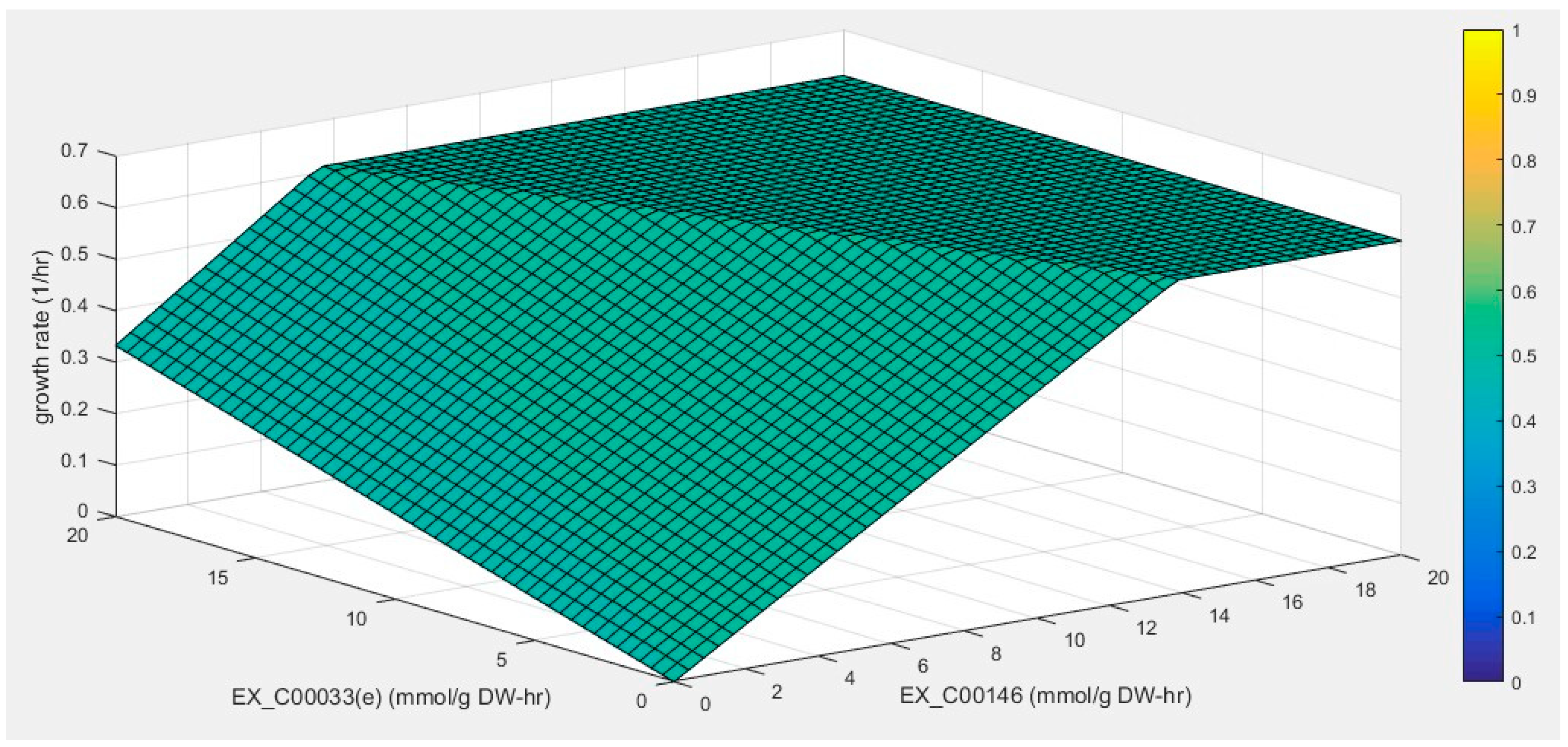
Furthermore, the model iNX811 was used to find some exogenous substances capable of improving phenol breakdown by simulating the interaction between phenol and supplement consumption and cell growth. Exogenous substances that benefit phenol degradation were selected from the test carbon sources of A. lwoffii. As previously reported, adding sodium acetate can shorten the time taken to reach maximum biomass compared to its growth on phenol as the sole carbon source (Figure S1). The phenotype phase-plane analysis was used to investigate the interaction of the two substrate utilizations and their effects on cell growth. Two distinct surfaces (I and II) are shown in Figure 5. In plane I, cell growth was affected by the uptake of both substrates. Cell growth increased linearly with the increase in sodium acetate or phenol when the uptake rate of the other substrate was zero. When one parameter was fixed, cell growth increased with the uptake rate of other substrates. Cell growth reached its maximum value on the two-phase boundary line. In plane II, cell growth remained unchanged with the uptake of the two substrates. The metabolic traits of available carboxylic acids, saccharides, and amino acids were investigated using the model iNX811. The PHPP results for certain carbon sources exhibited the same mode as that of sodium acetate (Figure S2A–O), which theoretically benefits cell growth in the presence of phenol. Fructose, pyruvate, malate, and succinate were chosen as candidates based on the PHPP results.
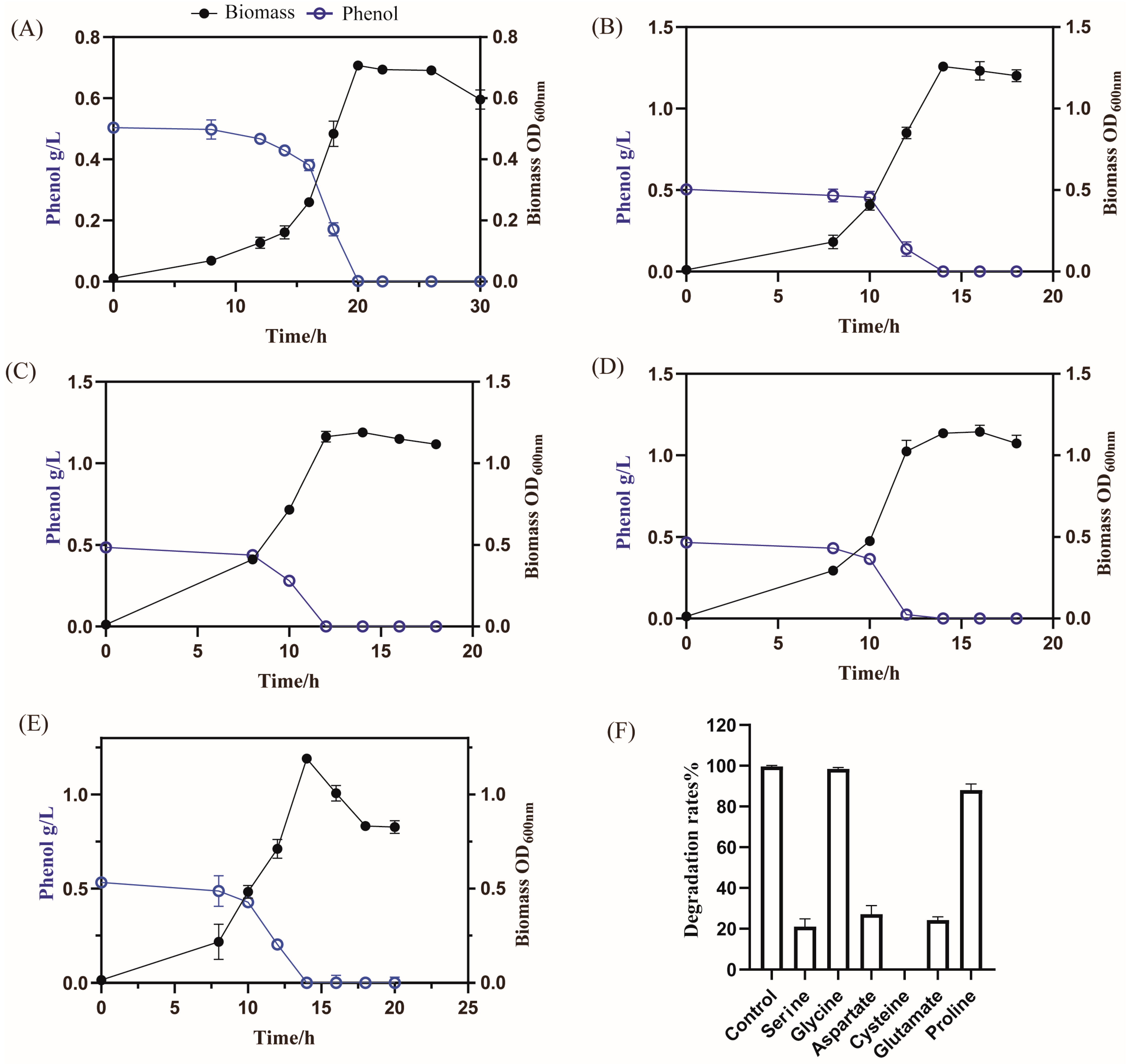
Thus, the effects of the four carbon sources on phenol degradation and cell growth were evaluated. When cultivated in these organic acids, the capacity for phenol degradation was significantly improved, consistent with the enhancement of oxidative phosphorylation and the TCA cycle response to phenol stress using GIMME analysis of the model iNX811. However, the addition of fructose did not promote phenol degradation. As shown in Figure 6, after adding sodium pyruvate, A. lwoffii NL1 reached its highest biomass at 14 h, and phenol was completely degraded after 15 h. After adding malate, the growth period of A. lwoffii NL1 (12 h) was shortened, and the highest biomass increased by 0.3 at OD600. After adding succinate, A. lwoffii NL1 reached its highest biomass at 16 h, and phenol was completely degraded. Considering that alanine can be converted into pyruvate by oxidation, the effect of alanine on phenol degradation was investigated. After adding alanine, A. lwoffii NL1 quickly entered the logarithmic growth phase at 5 h and reached its highest biomass at 14 h. Simultaneously, phenol was completely degraded after 14 h. Alanine promoted phenol degradation; however, its PHPP results were different from those of sodium acetate. In silico cell growth generally increased with the uptake rates of alanine and phenol but did not increase at uptake rates of alanine <4 mM and phenol >14 mM. The limitation of cell growth seemed to be eliminated by increasing the alanine supply. These substances (serine, glycine, aspartate, cysteine, proline, and glutamate) were investigated and showed a similar PHPP mode to alanine (Figure S2I–E). After adding these substances, the phenol degradation rate did not reach 100% at 20 h. Four exogenous substances supporting phenol degradation were identified using the model iNX811. Malate addition resulted in the most significant improvements in phenol degradation and biomass formation.

3. Materials and Methods

3.1. Reconstruction of the Genome-Scale Metabolic Model

3.1.1. Construction of the Draft Model

A draft model of A. lwoffii NL1 was constructed using automated construction using ModelSEED [82] and manual reconstruction based on homologous comparison. The reference models were from A. baumannii [15] and A. baylyi [20] according to their close phylogenetic relationships. Gene–protein–reaction associations of the reference organisms were acquired by the local sequence similarity search (BLASTp). The criteria for the BLASTp were identity > 40% and match length > 70% [83]. The model contents from the two methods were manually curated by deleting duplicate annotations and unifying the reaction formula. Cytoplasmic and extracellular compartments were considered in the draft model of A. lwoffii NL1. The transport proteins of A. lwoffii NL1 were reannotated using transporters in the Transporter Classification Database (TCDB) [84]. The refined model was transformed into a mathematical model on the MATLAB 2019a platform using the xls2model.m program in the COBRA toolbox [85].

3.1.2. Biomass Formation

The macromolecular composition of A. lwoffii NL1 was referenced to that of A. baumannii AYE [18]. Details of the biomass composition are provided in Table S6. DNA, mRNA, and amino acid compositions were calculated from the genome and protein sequences of A. lwoffii NL1. The contents of free fatty acids [25] and phospholipids [26] were extracted from the literature.
The biomassPrecursorCheck program verified the formation of biomacromolecules in the draft model. Metabolic gaps in the draft model led to an inability to synthesize a few biomacromolecules. These gaps were identified using the gapAnalysis.m program and filled by adding missing biochemical reactions to the draft model. The reaction pool for gap filling was from the KEGG pathway. The genes related to these reactions were annotated by comparing the proteomic data of A. lwoffii NL1 with the protein sequences in UniProtKB/Swiss-Prot [86].

3.2. Model Simulation and Analysis

3.2.1. Cell Growth Simulation

Biomass formation was set as an objective function to simulate cell growth. Flux balance analysis (FBA) [87] was used to optimize the biomass equation using the GUROBI 8.0.0 (Gurobi Optimization Inc., Houston, TX, USA) mathematical optimization solver [88] on the MATLAB interface. Genes required for biomass biosynthesis were defined as essential genes and were analyzed on Luria–Bertani and minimal medium using succinate as the sole carbon source (see Table S7). The in silico essential genes (see Table S5) were evaluated by comparison with previously reported transposon insertion mutants of Acinetobacter [89,90] and the Database of Essential Genes (DEG) [91].

3.2.2. Prediction of Drug Targets

Metabolites related to essential genes extracted by gene–protein–reaction associations in the model iNX811 were considered to be essential metabolites. The EMFilter framework reduces the anticipated important metabolites to a manageable quantity for in silico drug targets [18]. Common metabolites, such as cofactors and intermediates in the tricarboxylic acid (TCA) cycle, were excluded from the essential metabolites. Metabolites that coexist in humans were excluded to avoid drug side effects [92]. In addition, the drug targets should be endogenous metabolites of A. lwoffii that contain at least two consumption reactions.

3.2.3. Context-Specific Metabolic Models

Transcriptomic datasets (SRR25444162, SRR25444164, and SRR25444166) under three conditions were integrated into the model iNX811 using the Gene Inactivity Moderated by Metabolism and Expression (GIMME) algorithm [78]. The three cultivation conditions were NaAc as the sole carbon source and NaAc with 0.5 or 1.5 g/L phenol as an additional carbon source (see Table S7). The first quartile of expression values of the metabolic genes was 13.04, 9.89, and 3.35 under the three conditions, respectively. The expression values of each gene above and below the expression threshold represented its presence (1) or absence (0) in the model iNX811 [93]. Three context-specific metabolic models were generated using CreateTissueSpecificModel.m (see Supplementary File S4). Feasible metabolic states of the models were explored by random sampling, and then the feasible flux distribution for each reaction with/without transcriptional constraints was plotted using plotSampleHist.m. Reaction fluxes (x values) of gimmeSolution were calculated using solveGimme.m. Reactions with a flux change (added phenol/without phenol) greater than twofold or less than one-half were thought to differ considerably with phenol concentration, and the reaction flux was visualized using the pheatmap package (https://cran.r-project.org/web/packages/pheatmap/index.html (accessed on 20 August 2023)) on the R platform (4.0.5).

3.2.4. Phenotype Phase-Plane (PHPP) Analysis

PHPP analysis was used to calculate all possible variations in the two constraining variables to investigate the effects of substrate variables on cell growth [94]. The uptake rates of phenol (x-axis) and other substrates (y-axis) varied from 0 to 20 mmol/g dry cell weight/h. The in silico cell growth was plotted along the z-axis. All points on the x-y plane represent the optimal cell growth allowable for each pair of substrate uptake rates.

3.3. Model Validation

3.3.1. Microbial Cultivation

The A. lwoffii NL1 used in this study was preserved in the China Center for Type Culture Collection (CCTCC NO: M2014329). A. lwoffii NL1 from a Luria broth (LB) agar slant was inoculated in 5 mL of liquid LB medium. Overnight cultures of A. lwoffii NL1 were collected and washed with sterile phosphate-buffered saline to achieve an optical density at 600 nm (OD600) of 0.5. Culture in liquid minimal mineral (MM) medium supplemented with specific carbon sources was performed at 28 °C and 200 rpm.

3.3.2. Cell Growth and Phenol Utilization

Aliquots of bacterial suspension were inoculated (2% v/v) in test tubes containing MM supplemented with an individual carbon and nitrogen source. Cell growth was observed at 36 h to determine the utilization of each carbon and nitrogen source. The molar masses of the individual carbon and nitrogen sources were equal to those of phenol (0.5 g of phenol and 1.0 g of NH4Cl, respectively). The carbon sources included glucose, fructose, xylose, arabinose, mannitol, glycerol, ethanol, acetate, citrate, succinate, malate, phenol, 4-hydroxybenzoic acid, salicylate, benzoic acid, toluene, mandelate, benzene, phthalate, alanine, glycine, proline, serine, arginine, glutamate, glutamine, aspartate, threonine, valine, cysteine, tryptophan, methionine, leucine, phenylalanine, histidine, and lysine. The nitrogen sources included nitrate, NH4Cl, and urea.
In another series of experiments, aliquots of bacterial suspension were inoculated (2% v/v) in 20 mL of MM medium containing 0.5 g/L phenol and another substrate (0.02 moles of carbon) in 100 mL flasks. The effects of fructose, pyruvate, succinate, malate, and alanine on cell growth and phenol degradation were investigated by determining the OD600 of the biomass and the OD510 of the supernatants every few hours via a modified 4-aminoantipyrine colorimetric method [95]. Phenol concentration was also measured 20 h after adding serine, glycine, aspartate, cysteine, proline, or glutamate to 20 mL of MM containing 0.5 g/L phenol.

4. Conclusions

In this study, we constructed the first genome-scale metabolic model of A. lwoffii NL1 by combining manual and semi-automatic construction. The model iNX811 accurately predicted growth characteristics in different cultures. A. lwoffii NL1 showed multiple drug resistances and could effectively degrade phenol. Therefore, we used the model for antimicrobial systems and phenol biodegradation in A. lwoffii. Essential metabolite analysis predicted 12 potential target compounds for cell wall, lysine, vitamin, and fatty acid biosynthesis. Integrating transcriptomic data with model iNX811 provides a fundamental understanding of metabolic changes in response to phenol stress. These changes focused on central carbon metabolism, including enhancing oxidative phosphorylation, the TCA cycle, the oxaloacetate anaplerotic reaction, and the decreased glyoxylate cycle outflow from malate to pyruvate. A few transporters, such as salicylate exporters and oxygen uptake, were strengthened. In addition, the relationship between exogenous substances, phenol uptake, and cell growth was evaluated using PHPP analysis. Succinate, malate, alanine, and pyruvate enhanced phenol degradation in flask experiments.

Supplementary Materials

The supporting information can be downloaded at https://www.mdpi.com/article/10.3390/ijms25179321/s1.

Author Contributions

N.X.: formal analysis, conceptualization, writing—original draft. J.Z.: methodology, data curation. C.L.: methodology. C.G.: writing—original draft, review and editing. M.G.: supervision, writing—review and editing. All authors have read and agreed to the published version of the manuscript.

Funding

This work was supported by the National Key R&D Program of China (2020YFA0908300), the National Natural Science Foundation of China (22278350), the High-end Talent Support Program of Yangzhou University, and Yangzhou University’s ‘Qinglan Project’.

Institutional Review Board Statement

Not applicable.

Informed Consent Statement

Not applicable.

Data Availability Statement

Data and materials are available from the corresponding author.

Conflicts of Interest

The authors declare that they have no competing interests.

References

  1. Visca, P.; Seifert, H.; Towner, K.J. Acinetobacter infection—An emerging threat to human health. IUBMB Life 2011, 63, 1048–1054. [Google Scholar] [CrossRef] [PubMed]
  2. Cao, S.; Geng, Y.; Yu, Z.; Deng, L.; Gan, W.; Wang, K.; Ou, Y.; Chen, D.; Huang, X.; Zuo, Z.; et al. Acinetobacter lwoffii, an emerging pathogen for fish in Schizothorax genus in China. Transbound. Emerg. Dis. 2018, 65, 1816–1822. [Google Scholar] [CrossRef] [PubMed]
  3. Hu, Y.; Zhang, W.; Liang, H.; Liu, L.; Peng, G.; Pan, Y.; Yang, X.; Zheng, B.; Gao, G.F.; Zhu, B.; et al. Whole-genome sequence of a multidrug-resistant clinical isolate of Acinetobacter lwoffii. J. Bacteriol. 2011, 193, 5549–5550. [Google Scholar] [CrossRef]
  4. Touchon, M.; Cury, J.; Yoon, E.-J.; Krizova, L.; Cerqueira, G.C.; Murphy, C.; Feldgarden, M.; Wortman, J.; Clermont, D.; Lambert, T.; et al. The Genomic Diversification of the Whole Acinetobacter Genus: Origins, Mechanisms, and Consequences. Genome Biol. Evol. 2014, 6, 2866–2882. [Google Scholar] [CrossRef] [PubMed]
  5. Zhao, Y.; Wei, H.M.; Yuan, J.L.; Xu, L.; Sun, J.Q. A comprehensive genomic analysis provides insights on the high environmental adaptability of Acinetobacter strains. Front. Microbiol. 2023, 14, 1177951. [Google Scholar] [CrossRef] [PubMed]
  6. Almeida, O.G.G.; Furlan, J.P.R.; Stehling, E.G.; De Martinis, E.C.P. Comparative phylo-pangenomics reveals generalist lifestyles in representative Acinetobacter species and proposes candidate gene markers for species identification. Gene 2021, 791, 145707. [Google Scholar] [CrossRef]
  7. Mindlin, S.; Petrenko, A.; Kurakov, A.; Beletsky, A.; Mardanov, A.; Petrova, M. Resistance of Permafrost and Modern Acinetobacter lwoffii Strains to Heavy Metals and Arsenic Revealed by Genome Analysis. BioMed Res. Int. 2016, 2016, 3970831. [Google Scholar] [CrossRef]
  8. Alonso-Vásquez, T.; Fondi, M.; Perrin, E. Understanding antimicrobial resistance using genome-scale metabolic modeling. Antibiotics 2023, 12, 896. [Google Scholar] [CrossRef]
  9. Nazarshodeh, E.; Marashi, S.A.; Gharaghani, S. Structural systems pharmacology: A framework for integrating metabolic network and structure-based virtual screening for drug discovery against bacteria. PLoS ONE 2021, 16, e0261267. [Google Scholar] [CrossRef]
  10. Han, S.; Tao, Y.; Cui, Y.; Xu, J.; Ju, H.; Fan, L.; Zhang, L.; Zhang, Y. Lanthanum-modified polydopamine loaded Acinetobacter lwoffii DNS32 for phosphate and atrazine removal: Insights into co-adsorption and biodegradation mechanisms. Bioresour. Technol. 2023, 368, 128266. [Google Scholar] [CrossRef]
  11. Han, S.; Tao, Y.; Zhao, L.; Cui, Y.; Zhang, Y. Metabolic insights into how multifunctional microbial consortium enhances atrazine removal and phosphorus uptake at low temperature. J. Hazard. Mater. 2024, 461, 132539. [Google Scholar] [CrossRef]
  12. Xu, N.; Yang, X.; Yang, Q.; Guo, M. Comparative Genomic and Transcriptomic Analysis of Phenol Degradation and Tolerance in Acinetobacter lwoffii through Adaptive Evolution. Int. J. Mol. Sci. 2023, 24, 16529. [Google Scholar] [CrossRef]
  13. Uzma, B.; Ali, F.; Qureshi, N.A.; Shakeela, Q.; Asima, B.; Ahmed, S.; Hayat, A.; Rehman, M.U. Isolation and characterization of synthetic pyrethroids-degrading bacterial strains from agricultural soil. Braz. J. Biol. 2023, 83, e271790. [Google Scholar] [CrossRef] [PubMed]
  14. Walter, T.; Klim, J.; Jurkowski, M.; Gawor, J.; Köhling, I.; Słodownik, M.; Zielenkiewicz, U. Plasmidome of an environmental Acinetobacter lwoffii strain originating from a former gold and arsenic mine. Plasmid 2020, 110, 102505. [Google Scholar] [CrossRef] [PubMed]
  15. Xu, N.; Ye, C.; Liu, L.M. Genome-scale biological models for industrial microbial systems. Appl. Microbiol. Biotechnol. 2018, 102, 3439–3451. [Google Scholar] [CrossRef] [PubMed]
  16. Zhu, Y.; Zhao, J.; Maifiah, M.H.M.; Velkov, T.; Schreiber, F.; Li, J. Metabolic Responses to Polymyxin Treatment in Acinetobacter baumannii ATCC 19606: Integrating Transcriptomics and Metabolomics with Genome-Scale Metabolic Modeling. Msystems 2019, 4, e00157-18. [Google Scholar] [CrossRef]
  17. Zhao, J.X.; Zhu, Y.; Han, J.R.; Lin, Y.W.; Aichem, M.; Wang, J.P.; Chen, K.; Velkov, T.; Schreiber, F.; Li, J. Genome-Scale Metabolic Modeling Reveals Metabolic Alterations of Multidrug-Resistant Acinetobacter baumannii in a Murine Bloodstream Infection Model. Microorganisms 2020, 8, 18. [Google Scholar] [CrossRef]
  18. Norsigian, C.J.; Kavvas, E.; Seif, Y.; Palsson, B.O.; Monk, J.M. iCN718, an Updated and Improved Genome-Scale Metabolic Network Reconstruction of Acinetobacter baumannii AYE. Front. Genet. 2018, 9, 121. [Google Scholar] [CrossRef]
  19. Kim, H.U.; Kim, T.Y.; Lee, S.Y. Genome-scale metabolic network analysis and drug targeting of multi-drug resistant pathogen Acinetobacter baumannii AYE. Mol. Biosyst. 2010, 6, 339–348. [Google Scholar] [CrossRef]
  20. Zhu, Y.; Lu, J.; Zhao, J.; Zhang, X.; Yu, H.H.; Velkov, T.; Li, J. Complete genome sequence and genome-scale metabolic modelling of Acinetobacter baumannii type strain ATCC 19606. Int. J. Med. Microbiol. 2020, 310, 151412. [Google Scholar] [CrossRef]
  21. Durot, M.; Le Fèvre, F.; de Berardinis, V.; Kreimeyer, A.; Vallenet, D.; Combe, C.; Smidtas, S.; Salanoubat, M.; Weissenbach, J.; Schachter, V. Iterative reconstruction of a global metabolic model of Acinetobacter baylyi ADP1 using high-throughput growth phenotype and gene essentiality data. BMC Syst. Biol. 2008, 2, 85. [Google Scholar] [CrossRef] [PubMed]
  22. Xu, N.; Qiu, C.; Yang, Q.; Zhang, Y.; Wang, M.; Ye, C.; Guo, M. Analysis of phenol biodegradation in antibiotic and heavy metal resistant Acinetobacter lwoffii NL1. Front. Microbiol. 2021, 12, 725755. [Google Scholar] [CrossRef]
  23. Aziz, R.K.; Bartels, D.; Best, A.A.; DeJongh, M.; Disz, T.; Edwards, R.A.; Formsma, K.; Gerdes, S.; Glass, E.M.; Kubal, M.; et al. The RAST server: Rapid annotations using subsystems technology. BMC Genom. 2008, 9, 75. [Google Scholar] [CrossRef]
  24. Lieven, C.; Beber, M.E.; Olivier, B.G.; Bergmann, F.T.; Ataman, M.; Babaei, P.; Bartell, J.A.; Blank, L.M.; Chauhan, S.; Correia, K.; et al. MEMOTE for standardized genome-scale metabolic model testing. Nat. Biotechnol. 2020, 38, 504. [Google Scholar] [CrossRef] [PubMed]
  25. Ibarra, R.U.; Edwards, J.S.; Palsson, B.O. Escherichia coli K-12 undergoes adaptive evolution to achieve predicted optimal growth. Nature 2002, 420, 186–189. [Google Scholar] [CrossRef]
  26. Imron, M.F.; Titah, H.S. Biodegradation of Diesel by Acinetobacter lwoffii and Vibrio alginolyticus Isolated from Ship Dismantling Facility in Tanjungjati Coast, Madura, Indonesia. J. Appl. Biol. Sci. 2019, 12, 1–8. [Google Scholar]
  27. Yang, F.; Jiang, Q.; Zhu, M.; Zhao, L.; Zhang, Y. Effects of biochars and MWNTs on biodegradation behavior of atrazine by Acinetobacter lwoffii DNS32. Sci. Total Environ. 2017, 577, 54–60. [Google Scholar] [CrossRef] [PubMed]
  28. Liu, Y.; Wan, Y.Y.; Wang, C.; Ma, Z.; Liu, X.; Li, S. Biodegradation of n-alkanes in crude oil by three identified bacterial strains. Fuel 2020, 275, 117897. [Google Scholar] [CrossRef]
  29. Guilhelmelli, F.; Vilela, N.; Albuquerque, P.; Derengowski Lda, S.; Silva-Pereira, I.; Kyaw, C.M. Antibiotic development challenges: The various mechanisms of action of antimicrobial peptides and of bacterial resistance. Front. Microbiol. 2013, 4, 353. [Google Scholar] [CrossRef]
  30. Dunphy, L.J.; Papin, J.A. Biomedical applications of genome-scale metabolic network reconstructions of human pathogens. Curr. Opin. Biotechnol. 2018, 51, 70–79. [Google Scholar] [CrossRef]
  31. Kim, S.K.; Lee, M.; Lee, Y.Q.; Lee, H.J.; Rho, M.; Kim, Y.; Seo, J.Y.; Youn, S.H.; Hwang, S.J.; Kang, N.G.; et al. Genome-scale metabolic modeling and in silico analysis of opportunistic skin pathogen Cutibacterium acnes. Front. Cell. Infect. Microbiol. 2023, 13, 1099314. [Google Scholar] [CrossRef] [PubMed]
  32. Díaz Calvo, T.; Tejera, N.; McNamara, I.; Langridge, G.C.; Wain, J.; Poolman, M.; Singh, D. Genome-scale metabolic modelling approach to understand the metabolism of the opportunistic human pathogen Staphylococcus epidermidis RP62A. Metabolites 2022, 12, 136. [Google Scholar] [CrossRef]
  33. Veith, N.; Solheim, M.; van Grinsven, K.W.; Olivier, B.G.; Levering, J.; Grosseholz, R.; Hugenholtz, J.; Holo, H.; Nes, I.; Teusink, B.; et al. Using a genome-scale metabolic model of Enterococcus faecalis V583 to assess amino acid uptake and its impact on central metabolism. Appl. Environ. Microbiol. 2015, 81, 1622–1633. [Google Scholar] [CrossRef]
  34. Kim, H.U.; Kim, S.Y.; Jeong, H.; Kim, T.Y.; Kim, J.J.; Choy, H.E.; Yi, K.Y.; Rhee, J.H.; Lee, S.Y. Integrative genome-scale metabolic analysis of Vibrio vulnificus for drug targeting and discovery. Mol. Syst. Biol. 2011, 7, 460. [Google Scholar] [CrossRef] [PubMed]
  35. Tezcan, E.F.; Demirtas, Y.; Cakar, Z.P.; Ulgen, K.O. Comprehensive genome-scale metabolic model of the human pathogen Cryptococcus neoformans: A platform for understanding pathogen metabolism and identifying new drug targets. Front. Bioinform. 2023, 3, 1121409. [Google Scholar] [CrossRef]
  36. Hasan, A.; Mazumder, H.H.; Khan, A.; Hossain, M.U.; Chowdhury, H.K. Molecular Characterization of Legionellosis Drug Target Candidate Enzyme Phosphoglucosamine Mutase from Legionella pneumophila (strain Paris): An In Silico Approach. Genom. Inf. 2014, 12, 268–275. [Google Scholar] [CrossRef]
  37. Sharma, R.; Lambu, M.R.; Jamwal, U.; Rani, C.; Chib, R.; Wazir, P.; Mukherjee, D.; Chaubey, A.; Khan, I.A. Escherichia coli N-Acetylglucosamine-1-Phosphate-Uridyltransferase/Glucosamine-1-Phosphate-Acetyltransferase (GlmU) Inhibitory Activity of Terreic Acid Isolated from Aspergillus terreus. J. Biomol. Screen. 2016, 21, 342–353. [Google Scholar] [CrossRef]
  38. Stokes, S.S.; Albert, R.; Buurman, E.T.; Andrews, B.; Shapiro, A.B.; Green, O.M.; McKenzie, A.R.; Otterbein, L.R. Inhibitors of the acetyltransferase domain of N-acetylglucosamine-1-phosphate-uridylyltransferase/glucosamine-1-phosphate-acetyltransferase (GlmU). Part 2: Optimization of physical properties leading to antibacterial aryl sulfonamides. Bioorg. Med. Chem. Lett. 2012, 22, 7019–7023. [Google Scholar] [CrossRef] [PubMed]
  39. Chiu, H.J.; Grant, J.C.; Farr, C.L.; Jaroszewski, L.; Knuth, M.W.; Miller, M.D.; Elsliger, M.A.; Deacon, A.M.; Godzik, A.; Lesley, S.A.; et al. Structural analysis of arabinose-5-phosphate isomerase from Bacteroides fragilis and functional implications. Acta Crystallogr. D Biol. Crystallogr. 2014, 70 Pt 10, 2640–2651. [Google Scholar] [CrossRef]
  40. Airoldi, C.; Sommaruga, S.; Merlo, S.; Sperandeo, P.; Cipolla, L.; Polissi, A.; Nicotra, F. Targeting bacterial membranes: Identification of Pseudomonas aeruginosa D-arabinose-5P isomerase and NMR characterisation of its substrate recognition and binding properties. Chembiochem 2011, 12, 719–727. [Google Scholar] [CrossRef]
  41. Jenkins, C.H.; Scott, A.E.; O’Neill, P.A.; Norville, I.H.; Prior, J.L.; Ireland, P.M. The Arabinose 5-Phosphate Isomerase KdsD Is Required for Virulence in Burkholderia pseudomallei. J. Bacteriol. 2023, 205, e0003423. [Google Scholar] [CrossRef]
  42. Tillery, L.M.; Barrett, K.F.; Dranow, D.M.; Craig, J.; Shek, R.; Chun, I.; Barrett, L.K.; Phan, I.Q.; Subramanian, S.; Abendroth, J.; et al. Toward a structome of Acinetobacter baumannii drug targets. Protein Sci. 2020, 29, 789–802. [Google Scholar] [CrossRef] [PubMed]
  43. Sha, S.; Zhou, Y.; Xin, Y.; Ma, Y. Development of a colorimetric assay and kinetic analysis for Mycobacterium tuberculosis D-glucose-1-phosphate thymidylyltransferase. J. Biomol. Screen. 2012, 17, 252–257. [Google Scholar] [CrossRef] [PubMed]
  44. Alphey, M.S.; Pirrie, L.; Torrie, L.S.; Boulkeroua, W.A.; Gardiner, M.; Sarkar, A.; Maringer, M.; Oehlmann, W.; Brenk, R.; Scherman, M.S.; et al. Allosteric competitive inhibitors of the glucose-1-phosphate thymidylyltransferase (RmlA) from Pseudomonas aeruginosa. ACS Chem. Biol. 2013, 8, 387–396. [Google Scholar] [CrossRef] [PubMed]
  45. Sen, M.; Shah, B.; Rakshit, S.; Singh, V.; Padmanabhan, B.; Ponnusamy, M.; Pari, K.; Vishwakarma, R.; Nandi, D.; Sadhale, P.P. UDP-glucose 4, 6-dehydratase activity plays an important role in maintaining cell wall integrity and virulence of Candida albicans. PLoS Pathog. 2011, 7, e1002384. [Google Scholar] [CrossRef] [PubMed]
  46. Vanaporn, M.; Titball, R.W. Trehalose and bacterial virulence. Virulence 2020, 11, 1192–1202. [Google Scholar] [CrossRef]
  47. Miao, Y.; Tenor, J.L.; Toffaletti, D.L.; Maskarinec, S.A.; Liu, J.; Lee, R.E.; Perfect, J.R.; Brennan, R.G. Structural and In Vivo Studies on Trehalose-6-Phosphate Synthase from Pathogenic Fungi Provide Insights into Its Catalytic Mechanism, Biological Necessity, and Potential for Novel Antifungal Drug Design. mBio 2017, 8, e00643-17. [Google Scholar] [CrossRef]
  48. Washington, E.J.; Zhou, Y.; Hsu, A.L.; Petrovich, M.; Tenor, J.L.; Toffaletti, D.L.; Guan, Z.; Perfect, J.R.; Borgnia, M.J.; Bartesaghi, A.; et al. Structures of trehalose-6-phosphate synthase, Tps1, from the fungal pathogen Cryptococcus neoformans: A target for antifungals. Proc. Natl. Acad. Sci. USA 2024, 121, e2314087121. [Google Scholar] [CrossRef]
  49. Kale, M.; Mohd Sayeed, S. Drug discovery of newer analogs of anti-microbials through enzyme-inhibition: A review. Int. J. Pharm. Pharm. Sci. 2014, 6, 27–35. [Google Scholar]
  50. Mitsakos, V.; Dobson, R.C.; Pearce, F.G.; Devenish, S.R.; Evans, G.L.; Burgess, B.R.; Perugini, M.A.; Gerrard, J.A.; Hutton, C.A. Inhibiting dihydrodipicolinate synthase across species: Towards specificity for pathogens? Bioorg. Med. Chem. Lett. 2008, 18, 842–844. [Google Scholar] [CrossRef]
  51. Skovpen, Y.V.; Conly, C.J.; Sanders, D.A.; Palmer, D.R. Biomimetic Design Results in a Potent Allosteric Inhibitor of Dihydrodipicolinate Synthase from Campylobacter jejuni. J. Am. Chem. Soc. 2016, 138, 2014–2020. [Google Scholar] [CrossRef] [PubMed]
  52. Angrish, N.; Lalwani, N.; Khare, G. In silico virtual screening for the identification of novel inhibitors against dihydrodipicolinate reductase (DapB) of Mycobacterium tuberculosis, a key enzyme of diaminopimelate pathway. Microbiol. Spectr. 2023, 11, e01359-23. [Google Scholar] [CrossRef] [PubMed]
  53. Dommaraju, S.R.; Dogovski, C.; Czabotar, P.E.; Hor, L.; Smith, B.J.; Perugini, M.A. Catalytic mechanism and cofactor preference of dihydrodipicolinate reductase from methicillin-resistant Staphylococcus aureus. Arch. Biochem. Biophys. 2011, 512, 167–174. [Google Scholar] [CrossRef] [PubMed]
  54. Singh, S.; Praveen, A.; Khanna, S.M. Computational modelling, functional characterization and molecular docking to lead compounds of Bordetella Pertussis diaminopimelate epimerase. Appl. Biochem. Biotech. 2023, 195, 6675–6693. [Google Scholar] [CrossRef]
  55. Chaudhary, J.; Singh, N.; Srivastava, V.K.; Jyoti, A.; Kaushik, S. Exploring the significance of diaminopimelate epimerase as a drug target in multidrug resistant Enterococcus faecalis. Vegetos 2023, 36, 1–9. [Google Scholar] [CrossRef]
  56. Weyand, S.; Kefala, G.; Weiss, M.S. The three-dimensional structure of N-succinyldiaminopimelate aminotransferase from Mycobacterium tuberculosis. J. Mol. Biol. 2007, 367, 825–838. [Google Scholar] [CrossRef]
  57. Nocek, B.P.; Gillner, D.M.; Fan, Y.; Holz, R.C.; Joachimiak, A. Structural basis for catalysis by the mono- and dimetalated forms of the dapE-encoded N-succinyl-L,L-diaminopimelic acid desuccinylase. J. Mol. Biol. 2010, 397, 617–626. [Google Scholar] [CrossRef]
  58. Kelley, E.H.; Minasov, G.; Konczak, K.; Shuvalova, L.; Brunzelle, J.S.; Shukla, S.; Beulke, M.; Thabthimthong, T.; Olsen, K.W.; Inniss, N.L.; et al. Biochemical and Structural Analysis of the Bacterial Enzyme Succinyl-Diaminopimelate Desuccinylase (DapE) from Acinetobacter baumannii. ACS Omega 2024, 9, 3905–3915. [Google Scholar] [CrossRef]
  59. Terrazas-López, M.; Lobo-Galo, N.; Aguirre-Reyes, L.G.; Cuen-Andrade, J.L.; de la Rosa, L.A.; Alvarez-Parrilla, E.; Martínez-Martínez, A.; Díaz-Sánchez, Á.G. Interaction of N-succinyl-diaminopimelate desuccinylase with flavonoids. Biochimie 2020, 177, 198–212. [Google Scholar] [CrossRef]
  60. Amera, G.M.; Khan, R.J.; Pathak, A.; Jha, R.K.; Muthukumaran, J.; Singh, A.K. Computer aided ligand based screening for identification of promising molecules against enzymes involved in peptidoglycan biosynthetic pathway from Acinetobacter baumannii. Microb. Pathog. 2020, 147, 104205. [Google Scholar] [CrossRef]
  61. Kumar, R.; Rajkumar, R.; Diwakar, V.; Khan, N.; Kumar Meghwanshi, G.; Garg, P. Structural-functional analysis of drug target aspartate semialdehyde dehydrogenase. Drug Discov. Today 2024, 29, 103908. [Google Scholar] [CrossRef] [PubMed]
  62. Rehman, A.; Akhtar, S.; Siddiqui, M.H.; Sayeed, U.; Ahmad, S.S.; Arif, J.M.; Khan, M.K. Identification of potential leads against 4-hydroxytetrahydrodipicolinate synthase from Mycobacterium tuberculosis. Bioinformation 2016, 12, 400–407. [Google Scholar] [CrossRef] [PubMed]
  63. Impey, R.E.; Panjikar, S.; Hall, C.J.; Bock, L.J.; Sutton, J.M.; Perugini, M.A.; Soares da Costa, T.P. Identification of two dihydrodipicolinate synthase isoforms from Pseudomonas aeruginosa that differ in allosteric regulation. FEBS J. 2020, 287, 386–400. [Google Scholar] [CrossRef]
  64. Girish, T.S.; Sharma, E.; Gopal, B. Structural and functional characterization of Staphylococcus aureus dihydrodipicolinate synthase. FEBS Lett. 2008, 582, 2923–2930. [Google Scholar] [CrossRef] [PubMed]
  65. Hossain, M.M.; Roy, P.K.; Mosnaz, A.T.; Shakil, S.K.; Hasan, M.M.; Prodhan, S.H. Structural analysis and molecular docking of potential ligands with chorismate synthase of Listeria monocytogenes: A novel antibacterial drug target. Indian J. Biochem. Biophys. 2015, 52, 45–59. [Google Scholar] [PubMed]
  66. Dias, M.V.; Ely, F.; Palma, M.S.; de Azevedo, W.F., Jr.; Basso, L.A.; Santos, D.S. Chorismate synthase: An attractive target for drug development against orphan diseases. Curr. Drug Targets 2007, 8, 437–444. [Google Scholar] [CrossRef]
  67. Ball, H.S.; Girma, M.B.; Zainab, M.; Soojhawon, I.; Couch, R.D.; Noble, S.M. Characterization and Inhibition of 1-Deoxy-d-Xylulose 5-Phosphate Reductoisomerase: A Promising Drug Target in Acinetobacter baumannii and Klebsiella pneumoniae. ACS Infect. Dis. 2021, 7, 2987–2998. [Google Scholar] [CrossRef]
  68. Parveez Zia, M.; Singh, E.; Jain, M.; Muthukumaran, J.; Singh, A.K. Structural and functional characterization of 1-deoxy-D-xylulose-5-phosphate synthase (DXS) from Acinetobacter baumannii: Identification of promising lead molecules from virtual screening, molecular docking and molecular dynamics simulations. J. Biomol. Struct. Dyn. 2023, 41, 11598–11611. [Google Scholar] [CrossRef]
  69. Ahmad, S.; Raza, S.; Qurat-ul-Ain; Uddin, R.; Rungrotmongkol, T.; Azam, S.S. From phylogeny to protein dynamics: A computational hierarchical quest for potent drug identification against an emerging enteropathogen “Yersinia enterocolitica”. J. Mol. Liq. 2018, 265, 372–389. [Google Scholar] [CrossRef]
  70. Bordel, S.; Martín-González, D.; Börner, T.; Muñoz, R.; Santos-Beneit, F. Genome-scale metabolic model of the versatile bacterium Paracoccus denitrificans Pd1222. mSystems 2024, 9, e01077-23. [Google Scholar] [CrossRef]
  71. Sohn, S.B.; Kim, T.Y.; Park, J.M.; Lee, S.Y. In silico genome-scale metabolic analysis of Pseudomonas putida KT2440 for polyhydroxyalkanoate synthesis, degradation of aromatics and anaerobic survival. Biotechnol. J. 2010, 5, 739–750. [Google Scholar] [CrossRef] [PubMed]
  72. Dhakar, K.; Zarecki, R.; van Bommel, D.; Knossow, N.; Medina, S.; Öztürk, B.; Aly, R.; Eizenberg, H.; Ronen, Z.; Freilich, S. Strategies for Enhancing in vitro Degradation of Linuron by Variovorax sp. Strain SRS 16 Under the Guidance of Metabolic Modeling. Front. Bioeng. Biotechnol. 2021, 9, 602464. [Google Scholar] [CrossRef] [PubMed]
  73. Ofaim, S.; Zarecki, R.; Porob, S.; Gat, D.; Lahav, T.; Kashi, Y.; Aly, R.; Eizenberg, H.; Ronen, Z.; Freilich, S. Genome-scale reconstruction of Paenarthrobacter aurescens TC1 metabolic model towards the study of atrazine bioremediation. Sci. Rep. 2020, 10, 13019. [Google Scholar] [CrossRef] [PubMed]
  74. Scheibe, T.D.; Mahadevan, R.; Fang, Y.; Garg, S.; Long, P.E.; Lovley, D.R. Coupling a genome-scale metabolic model with a reactive transport model to describe in situ uranium bioremediation. Microb. Biotechnol. 2009, 2, 274–286. [Google Scholar] [CrossRef]
  75. Zhuang, K.; Izallalen, M.; Mouser, P.; Richter, H.; Risso, C.; Mahadevan, R.; Lovley, D.R. Genome-scale dynamic modeling of the competition between Rhodoferax and Geobacter in anoxic subsurface environments. ISME J. 2011, 5, 305–316. [Google Scholar] [CrossRef] [PubMed]
  76. Rai, A.; Saito, K. Omics data input for metabolic modeling. Curr. Opin. Biotechnol. 2016, 37, 127–134. [Google Scholar] [CrossRef]
  77. Rau, M.H.; Gaspar, P.; Jensen, M.L.; Geppel, A.; Neves, A.R.; Zeidan, A.A. Genome-Scale Metabolic Modeling Combined with Transcriptome Profiling Provides Mechanistic Understanding of Streptococcus thermophilus CH8 Metabolism. Appl. Environ. Microb. 2022, 88, e00780-22. [Google Scholar] [CrossRef]
  78. Becker, S.A.; Palsson, B.O. Context-specific metabolic networks are consistent with experiments. PLoS Comput. Biol. 2008, 4, e1000082. [Google Scholar] [CrossRef]
  79. Jenior, M.L.; Moutinho, T.J., Jr.; Dougherty, B.V.; Papin, J.A. Transcriptome-guided parsimonious flux analysis improves predictions with metabolic networks in complex environments. PLoS Comput. Biol. 2020, 16, e1007099. [Google Scholar] [CrossRef]
  80. Kapley, A.; Tolmare, A.; Purohit, H.J. Role of oxygen in the utilization of phenol by CF600 in continuous culture. World J. Microbiol. Biotechnol. 2001, 17, 801–804. [Google Scholar] [CrossRef]
  81. Sánchez-Del-Campo, L.; Sáez-Ayala, M.; Chazarra, S.; Cabezas-Herrera, J.; Rodríguez-López, J.N. Binding of natural and synthetic polyphenols to human dihydrofolate reductase. Int. J. Mol. Sci. 2009, 10, 5398–5410. [Google Scholar] [CrossRef] [PubMed]
  82. Seaver, S.M.D.; Liu, F.; Zhang, Q.; Jeffryes, J.; Faria, J.P.; Edirisinghe, J.N.; Mundy, M.; Chia, N.; Noor, E.; Beber, M.E.; et al. The ModelSEED Biochemistry Database for the integration of metabolic annotations and the reconstruction, comparison and analysis of metabolic models for plants, fungi and microbes. Nucleic Acids Res. 2021, 49, D575–D588. [Google Scholar] [CrossRef] [PubMed]
  83. Altschul, S.F.; Gish, W.; Miller, W.; Myers, E.W.; Lipman, D.J. Basic local alignment search tool. J. Mol. Biol. 1990, 215, 403–410. [Google Scholar] [CrossRef] [PubMed]
  84. Saier, M.H., Jr.; Tran, C.V.; Barabote, R.D. TCDB: The Transporter Classification Database for membrane transport protein analyses and information. Nucleic Acids Res. 2006, 34, D181–D186. [Google Scholar] [CrossRef] [PubMed]
  85. Heirendt, L.; Arreckx, S.; Pfau, T.; Mendoza, S.N.; Richelle, A.; Heinken, A.; Haraldsdóttir, H.S.; Wachowiak, J.; Keating, S.M.; Vlasov, V.; et al. Creation and analysis of biochemical constraint-based models using the COBRA Toolbox v.3.0. Nat. Protoc. 2019, 14, 639–702. [Google Scholar] [CrossRef]
  86. Boutet, E.; Lieberherr, D.; Tognolli, M.; Schneider, M.; Bairoch, A. UniProtKB/Swiss-Prot. Methods Mol. Biol. 2007, 406, 89–112. [Google Scholar]
  87. Orth, J.D.; Thiele, I.; Palsson, B.Ø. What is flux balance analysis? Nat. Biotechnol. 2010, 28, 245–248. [Google Scholar] [CrossRef]
  88. Gurobi Optimization. Gurobi Optimizer Reference Manual; Gurobi Optimization, LLC: Beaverton, OR, USA, 2014. [Google Scholar]
  89. Gallagher, L.A.; Ramage, E.; Weiss, E.J.; Radey, M.; Hayden, H.S.; Held, K.G.; Huse, H.K.; Zurawski, D.V.; Brittnacher, M.J.; Manoil, C.; et al. Resources for Genetic and Genomic Analysis of Emerging Pathogen Acinetobacter baumannii. J. Bacteriol. 2015, 197, 2027–2035. [Google Scholar] [CrossRef]
  90. de Berardinis, V.; Vallenet, D.; Castelli, V.; Besnard, M.; Pinet, A.; Cruaud, C.; Samair, S.; Lechaplais, C.; Gyapay, G.; Richez, C.; et al. A complete collection of single-gene deletion mutants of Acinetobacter baylyi ADP1. Mol. Syst. Biol. 2008, 4, 174. [Google Scholar] [CrossRef]
  91. Luo, H.; Lin, Y.; Liu, T.; Lai, F.L.; Zhang, C.T.; Gao, F.; Zhang, R. DEG 15, an update of the Database of Essential Genes that includes built-in analysis tools. Nucleic Acids Res. 2021, 49, D677–D686. [Google Scholar] [CrossRef]
  92. Brunk, E.; Sahoo, S.; Zielinski, D.C.; Altunkaya, A.; Dräger, A.; Mih, N.; Gatto, F.; Nilsson, A.; Preciat Gonzalez, G.A.; Aurich, M.K.; et al. Recon3D enables a three-dimensional view of gene variation in human metabolism. Nat. Biotechnol. 2018, 36, 272–281. [Google Scholar] [CrossRef] [PubMed]
  93. Nogales, J.; Agudo, L. A Practical Protocol for Integration of Transcriptomics Data into Genome-Scale Metabolic Reconstructions. In Hydrocarbon and Lipid Microbiology Protocols; Springer: Berlin/Heidelberg, Germany, 2015; pp. 135–152. [Google Scholar]
  94. Duarte, N.C.; Palsson, B.O.; Fu, P. Integrated analysis of metabolic phenotypes in Saccharomyces cerevisiae. BMC Genom. 2004, 5, 63. [Google Scholar] [CrossRef] [PubMed]
  95. Lacoste, R.J.; Venable, S.H.; Stone, J.C. Modified 4-Aminoantipyrine Colorimetric Method for Phenols. Application to Acrylic Monomer. Anal. Chem. 1959, 31, 1246–1249. [Google Scholar] [CrossRef]
Figure 1. The genome-scale model reconstruction process for A. lwoffii.
Ijms 25 09321 g001
Figure 2. Genes, reactions, and gene-associated reactions for each metabolic subsystem of the model iNX811. Genes indicate how many genes there are in each metabolic subsystem, total reactions show how many reactions there are in each metabolic subsystem, and gene-associated reactions show which metabolic reactions have their encoding genes annotated.
Ijms 25 09321 g002
Figure 3. The metabolic states feasible through the phenol ortho-cleavage pathway of A. lwoffii under different cultivation conditions: (AC) Different cultivation conditions, i.e., NaAc as the sole carbon source, NaAc with 0.5 g/L phenol as carbon sources, and NaAc with 1.5 g/L phenol as carbon sources. The range of flux distributions with and without the regulatory constraints is shown in blue and in orange, respectively. (D) The phenol ortho-cleavage pathway in A. lwoffii. All of the reactions (R numbers) were from the KEGG database.
Ijms 25 09321 g003
Figure 4. Flux comparison of three context-specific genome-scale models constrained by differential transcriptomic data. Conditions 1–3 involved using NaAc as the only carbon source, adding 0.5 g/L of phenol as a carbon source, and adding 1.5 g/L of phenol as a carbon source, respectively. GAR, phosphoribosylglycinamide; PEP, phosphoenolpyruvate.
Ijms 25 09321 g004
Figure 5. Effects of phenol and sodium acetate on cell growth using phenotype phase-plane analysis. The Ex_C00146 and Ex_C00033(e) axes represent the phenol uptake rate and sodium acetate uptake rate, respectively. The colored legend signifies cell growth rates.
Ijms 25 09321 g005
Figure 6. Effects of the addition of various substances on phenol degradation: (AE) the dynamic curves of cell growth and phenol contents after adding pyruvate (B), malate (C), succinate (D), and alanine (E). The chemicals contained the same number of carbon atoms (0.02 moles). (A) Control. (F) Their breakdown rates at 20 h after introducing six amino acids.
Ijms 25 09321 g006
Table 1. Growth phenotypes of A. lwoffii NL1 on sole carbon and nitrogen sources.
Substrates In SilicoIn Vivo
SaccharidesGlucose--
Fructose++
Xylose--
Arabinose--
AlcoholMannitol--
Glycerol+-
Ethanol++
Carboxylic acidsAcetate++
Citrate++
Succinate++
Malate++
Aromatic xenobioticsPhenol++
4-Hydroxybenzoic acid++
Salicylate++
Benzoic acid++
Toluene++
Mandelate++
Benzene++
Phthalate++
Amino acidsL-Alanine++
Glycine++
Proline++
Serine++
Arginine++
Glutamate++
Glutamine++
Aspartate++
Threonine++
Valine++
Cysteine++
Tryptophan--
Methionine--
Leucine++
Phenylalanine++
Histidine--
Lysine--
Nitrogen sourcesNaNO3--
NH4Cl++
Urea++
The symbol + represents cells that can thrive only on this kind of carbon source or nitrogen source. The symbol - refers to cells that cannot grow only on this kind of carbon source or nitrogen source.
Table 2. Drug target prediction according to essential metabolite analysis.
Pathway Essential MetabolitesEnzymes Genes
Amino sugar and nucleotide sugar metabolismalpha-D-Glucosamine 1-phosphateD-Glucosamine 1,6-phosphomutase,
glucosamine-1-phosphate-acetyltransferase
LNSL_0126 LNSL_2808
Lipopolysaccharide biosynthesisD-arabinose-5-phosphateD-Arabinose-5-phosphate isomerase,
3-deoxy-8-phosphooctulonate synthase
LNSL_ 1272
LNSL_ 1680
Polysaccharide biosynthesisdTDP-glucosedTDP-Glucose 4,6-hydro-lyase,
dTDP-glucose 4-epimerase,
dTDP-glucose synthase
LNSL_2851
LNSL_2820
LNSL_2850
Starch and sucrose metabolismTrehalose 6-phosphateTrehalose 6-phosphate synthaseLNSL_0763
Lysine biosynthesisDehydrodipicolinateDehydrodipicolinate synthase,
dihydrodipicolinate reductase
LNSL_ 0056
LNSL_ 2865
N-Succinyl-LL-2,6-diaminoheptanedioateSuccinyldiaminopimelate transaminase,
succinyl-diaminopimelate desuccinylase
LNSL_ 1115
LNSL_ 2239
meso-2,6-DiaminoheptanedioateDiaminopimelate epimerase,
UDP-N-acetylmuramoylalanyl-D-glutamate-2,6-diaminopimelate ligase
LNSL_ 2102
LNSL_ 2621
L-Aspartate 4-semialdehydeAspartate-semialdehyde dehydrogenase,
4-hydroxy-tetrahydrodipicolinate synthase
LNSL_0359
LNSL_ 0056
Folate biosynthesis4-Aminobenzoate4-Amino-4-deoxychorismate pyruvate-lyase,
dihydropteroate synthase
LNSL_2017
LNSL_2140
2-Amino-4-hydroxy-6-hydroxymethyl-7,8-dihydropteridine7,8-Dihydroneopterin aldolase,
dihydropteroate synthase
LNSL_1717
LNSL_2140
ChorismateAminodeoxychorismate synthaseLNSL_0563
Thiamine metabolism1-Deoxy-D-xylulose 5-phosphate1-Deoxy-D-xylulose-5-phosphate synthaseLNSL_2536
Vitamin B6 metabolism1-Deoxy-D-xylulose 5-phosphatePyridoxine 5′-phosphate synthaseLNSL_2003
Disclaimer/Publisher’s Note: The statements, opinions and data contained in all publications are solely those of the individual author(s) and contributor(s) and not of MDPI and/or the editor(s). MDPI and/or the editor(s) disclaim responsibility for any injury to people or property resulting from any ideas, methods, instructions or products referred to in the content.