Article

Large Extracellular Vesicles Derived from Natural Killer Cells Affect the Functions of Monocytes

1
Federal State Budgetary Scientific Institution “The Research Institute of Obstetrics, Gynecology and Reproductology Named after D.O. Ott”, 199034 St. Petersburg, Russia
2
Saint-Petersburg Pasteur Institute, 197101 St. Petersburg, Russia
*
Author to whom correspondence should be addressed.
Int. J. Mol. Sci. 2024, 25(17), 9478; https://doi.org/10.3390/ijms25179478 (registering DOI)
Submission received: 3 June 2024 / Revised: 13 August 2024 / Accepted: 28 August 2024 / Published: 31 August 2024

Abstract

:
Communication between natural killer cells (NK cells) and monocytes/macrophages may play an important role in immunomodulation and regulation of inflammatory processes. The aim of this research was to investigate the impact of NK cell-derived large extracellular vesicles on monocyte function because this field is understudied. We studied how NK-cell derived large extracellular vesicles impact on THP-1 cells characteristics after coculturing: phenotype, functions were observed with flow cytometry. In this study, we demonstrated the ability of large extracellular vesicles produced by NK cells to integrate into the membranes of THP-1 cells and influence the viability, phenotype, and functional characteristics of the cells. The results obtained demonstrate the ability of large extracellular vesicles to act as an additional component in the immunomodulatory activity of NK cells in relation to monocytes.

1. Introduction

Monocytes, macrophages, and NK cells are essential components of the innate immune system. NK cells are CD3-negative large granular lymphocytes of the innate immune system that make up 10–15% of all circulating lymphocytes in human blood [1,2]. Monocytes are myeloid cells that circulate in blood for up to three days before migrating to tissues, where they differentiate into macrophages in response to specific environmental factors [3].
During the immune response, cells interact with each other in a specific manner, creating a unique microenvironment that either activates or inhibits the activity of other cells [4,5]. This interaction is primarily achieved through direct intercellular contact or by the activation of various biologically active molecules, primarily cytokines [4,6].
NK cells are able to influence the differentiation of monocytes in various ways. For example, in rheumatoid arthritis, CD56bright CD16dim/− NK cells have been shown to contribute to the differentiation of monocytes into osteoclasts, which leads to increased bone destruction in this condition [7]. In addition, these same cells in lymph nodes and inflamed tissues have been found to direct the differentiation of monocytes into dendritic cells by secreting GM-CSF, a cytokine that promotes this process. The secretion of GM-CSF is enhanced via the activation of NK cells through the action of IL-15. IL-15 is produced in various cell types, including monocytes [8]. This mutual activation can also occur through the interaction between the NKp80 receptor on NK cells and the activation-induced C-type lectin CLEC2B on monocytes. This interaction leads to an increased production of TNFα in both cell types, as well as increased expression of IFNγ via NK cells [9].
NK cells are able to destroy hyperactivated macrophages [10]. In response to the excessive activation of macrophages, the expression of ligands for NKG2D receptors on NK cells, MICA and MICB, increases on the surface of NK cells. This leads to ligand-receptor interaction between NK cells and macrophages, resulting in the lysis of the macrophages. This mechanism is likely aimed at eliminating overstimulated macrophages to prevent endotoxic shock [11]. The lack of NK cell regulation of macrophages can lead to inflammation and various pathological conditions [10].
It has been shown that IFNγ, produced via NK cells, regulates the polarization of macrophages, directing them towards the pro-inflammatory (M1) phenotype [12,13]. However, other NK cell-derived factors (GM-CSF, TNFα, and MIP1αß) can also activate macrophages [5,14]. It was also found that the number of macrophages in an inflammatory focus is dependent on the number of NK cells present, with a decrease in NK cell numbers leading to a reduction in macrophage and neutrophil infiltration, as well as a shift in macrophage polarization towards the M2 phenotype [15].
Cells release membrane vesicles of various types into the extracellular space with a membrane that both has an endosomal origin (exosomes) and forms from the plasma outer membrane of the cell (ectosomes) or apoptotic bodies [16,17,18]. The diameter of the exosomes is up to 100 nm, the diameter of the ectosomes ranges from 100 to 1000 nm [19], and the diameter of apoptotic bodies is above 1000 nm. The methods of the formation, composition, and functions of all of these vesicles are different. There are no recognized markers specific for ectosomes that are not present on exosomes. For example, CD63, and CD9 were considered to confirm that the isolated extracellular vesicles were exosomes [20]. However, Kowal J. et al., 2016 [21] showed that CD63 and CD9, previously considered to be specific exosome markers, were present on extracellular vesicles obtained after centrifugation at 2000× g (apoptotic bodies), 10,000× g (apoptotic bodies and ectosomes), and 20,000× g (ectosomes). Therefore, at present, one of the most adequate ways to obtain a pure ectosome fraction is the method of differential centrifugation [22,23]. In accordance with the Minimal Information for Studies of Extracellular Vesicles (MISEV2023) guidelines [18], “ectosomes” can only be used as proof for subcellular origin extracellular vesicles (EVs). In the absence of a clear-cut body of evidence, at present, the MISEV2023 guidelines delineate the categorization of such entities based on their compositional makeup, dimensions, and other attributes. According to the MISEV2023 guidelines, the dimensions of the resulting particulates may fluctuate during the course of differential centrifugation procedures. Based on the diameter of the separated particles, Small EVs (SEVs) are often described as being <200 nm in diameter and large EVs (LEVs) are often described as being >200 nm in diameter.
Currently, it is hypothesized that extracellular vesicles, including ectosomes, may function as an additional method of communication between cells [24]. Ectosomes are small, membranous particles that are released via different types of cells, including NK cells. It has been demonstrated that ectosomes contain various intracellular molecules from the source cell, as well as receptors, adhesion molecules, and other proteins [25,26]. The presence of these diverse biologically active substances in ectosomes, along with the wide range of receptors on their surface suggests that these particles may play a role in various physiological and pathological processes within the body, such as inflammation and the immune response [27,28,29].
Due to the fact that NK cells are a small population of leukocytes, there is a lack of data on the characteristics and functions of their ectosomes\LEVs. However, it has been established that NK cells can secrete extracellular vesicles, including ectosomes. It has been demonstrated that molecules specific to the source cells are expressed both within and on the surface of NK cell-derived ectosomes [30]. This suggests that the effects of NK cell-derived ectosomes and the NK cells themselves on target cells may be similar [31,32]. In particular, the ability of NK cells to produce ectosomes in order to enhance the expression of costimulatory molecules such as CD80 and CD86 on the surface of peripheral blood monocytes has been shown [33]. To date, there is a lack of data on the effect of EVs produced via NK cells, and especially ectosomes, on macrophages. However, it is assumed that this mechanism of communication between NK cells and monocytes/macrophages may play an important role in immunomodulation and the regulation of inflammatory processes.
Until now, the overall understanding of the interactions between NK cells and monocytes/macrophages has failed to consider the potential for these cells to communicate with each other through ectosomes. Consequently, the aim of this research was to investigate the impact of NK cell-derived ectosomes\LEVs on monocyte function.

2. Results

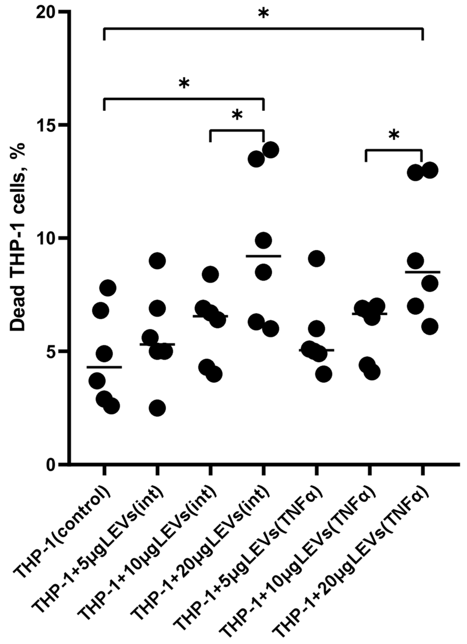
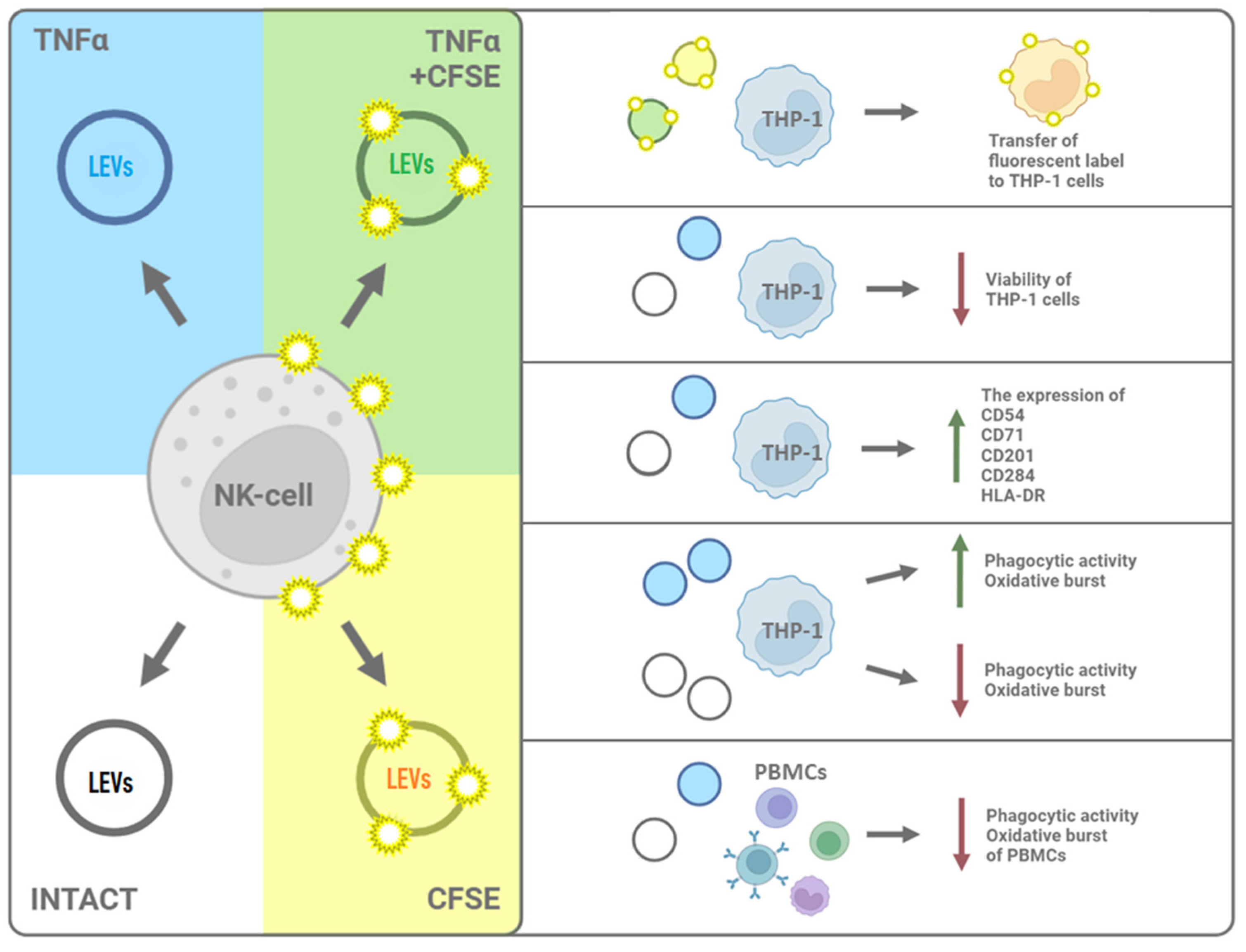
2.1. A High Concentration of LEVs Derived from NK Cells Causes a Decrease in the Viability of THP-1 Cells

In this study, we used TNFα as an inductor since previously the analysis of the cytotoxic activity of NK-92 cells in an in vitro NK-cell assay using K562 target cells had demonstrated that LEVs derived from TNFα-stimulated NK-92 cells increased the cytotoxicity of those cells. This coincides with the previous finding of an increased content of granzyme B in LEVs derived from TNFα-stimulated cells of the NK-92 line. This is described in more detail in our paper [28]. First, we tested different concentrations of LEVs (5, 10, or 20 μg of protein per 100 μL) and found that only the highest concentration of LEVs (20 μg/100 μL) derived from NK-92 cells was a decrease in the viability of THP-1 cells caused (Figure 1, Supplementary Data, Table S2). In order to avoid a cytotoxic effect on THP-1 cells, all further experiments were carried out using a LEVs concentration of 10 µg.

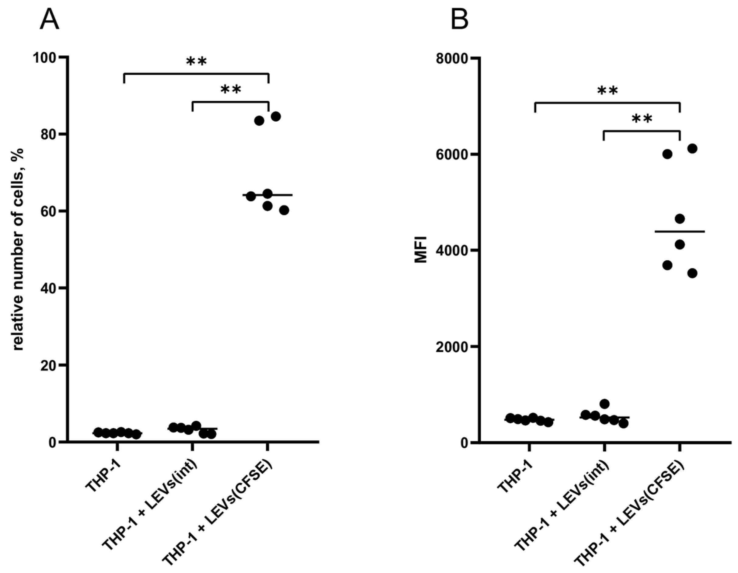
2.2. LEVs Derived from CFSE-Stained NK Cells Transfer a Fluorescent Label to THP-1 Cells

THP-1 cells were cultured in the presence of LEVs derived from CFSE-treated NK-92 cells (LEVs (CFSE)). As a result, there was an increase in the level of fluorescence of THP-1 cells compared to the autofluorescence level of intact THP-1 cells (negative control) and the fluorescence level of THP-1 cells cultured with LEVs derived from intact NK-92 cells (LEVs(int)) (Figure 2, Supplementary Data, Table S1).

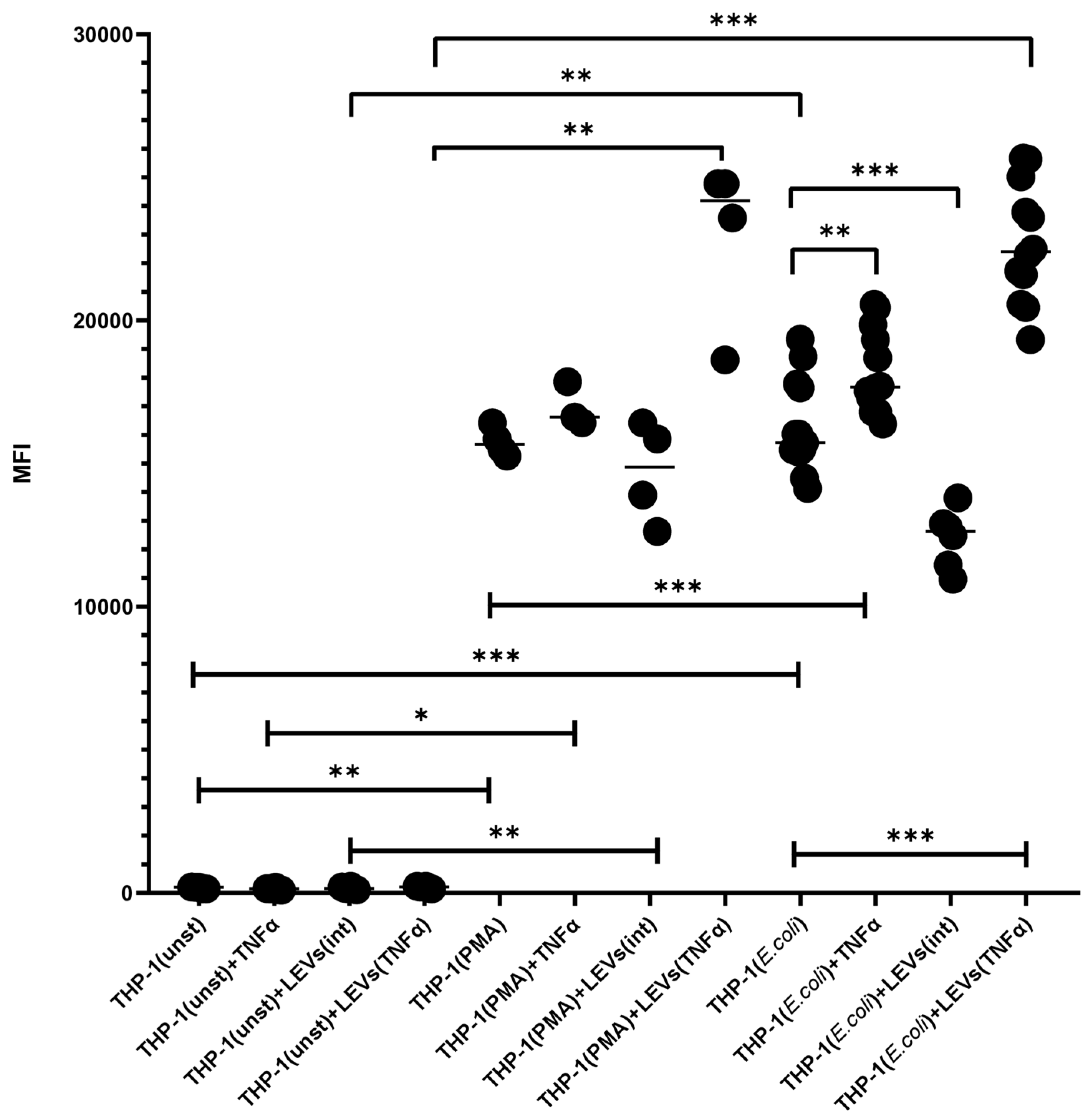
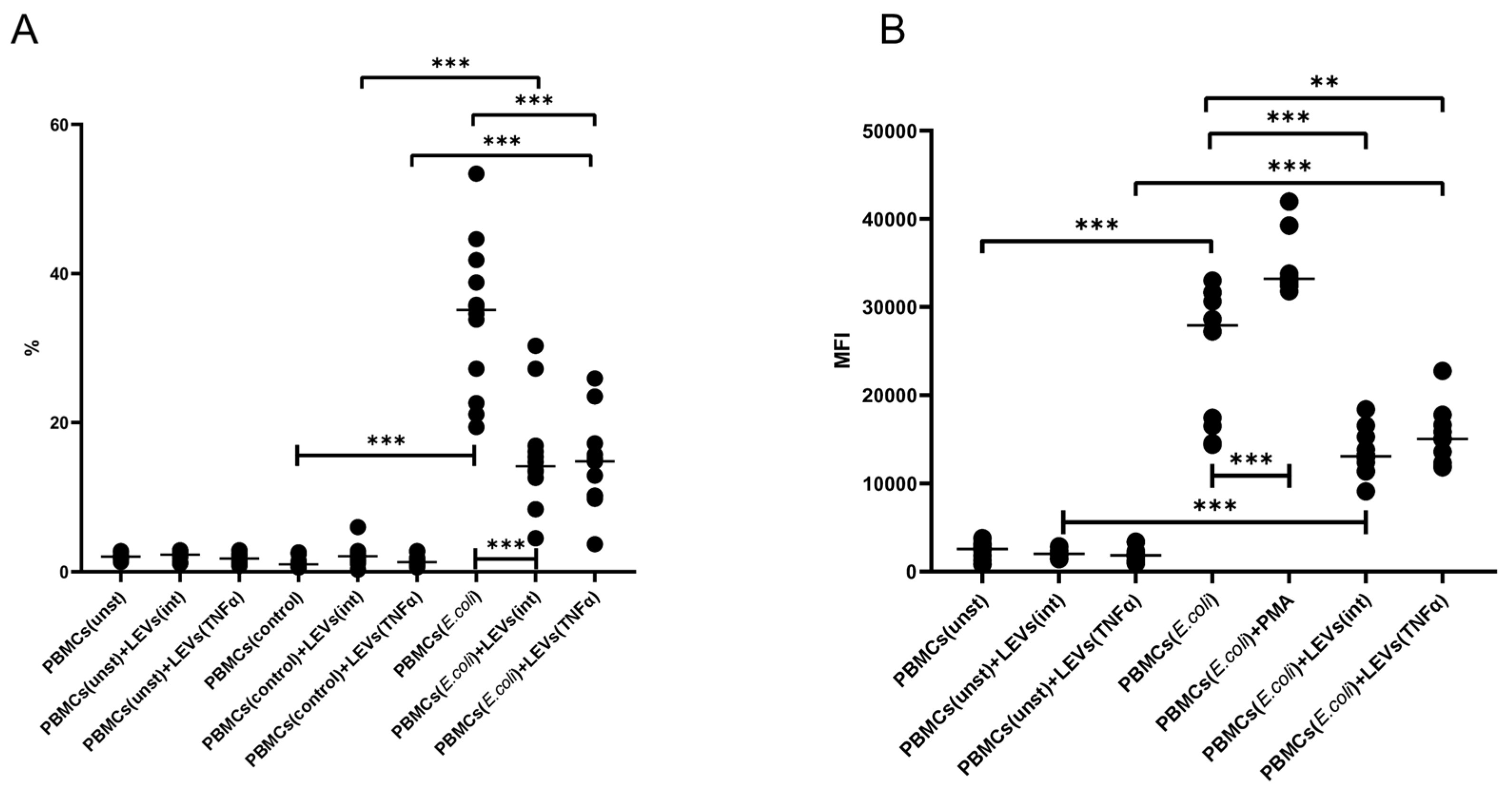
2.3. TNFα and LEVs Derived from TNFα-Activated NK Cells Increase the Phagocytic Activity and the Oxidative Burst of THP-1 Cells

We demonstrated that the activation of THP-1 cells with TNFα leads to an increase in their phagocytic capacity, as compared to the level of phagocytosis of intact THP-1 cells (Figure 3). Prior incubation of THP-1 cells with LEVs(int) caused a decrease in phagocytic activity compared to the spontaneous level (Figure 3). However, the incubation of THP-1 cells with LEVs produced via TNFα-activated NK-92 cells (LEVs(TNFα)) resulted in an increased phagocytic activity compared to both the control group and the level shown for cells incubated with LEVs(int) (Figure 3, Supplementary Data, Table S3).
The activation of THP-1 cells with TNFα also resulted in an increased oxidative burst compared to the level seen in intact THP-1 cells (Figure 4, Supplementary Data, Table S4). The pretreatment of THP-1 cells with LEVs(int) decreased the level of the oxidative burst in THP-1 cells compared to the level seen in intact THP-1 cells. At the same time, the treatment of THP-1 cells with LEVs(TNFα) enhanced the intensity of the oxidative burst in THP-1 cells compared to the level seen in intact THP-1 cells and also compared to the level in THP-1 cells pre-treated with LEVs(int) (Figure 4).

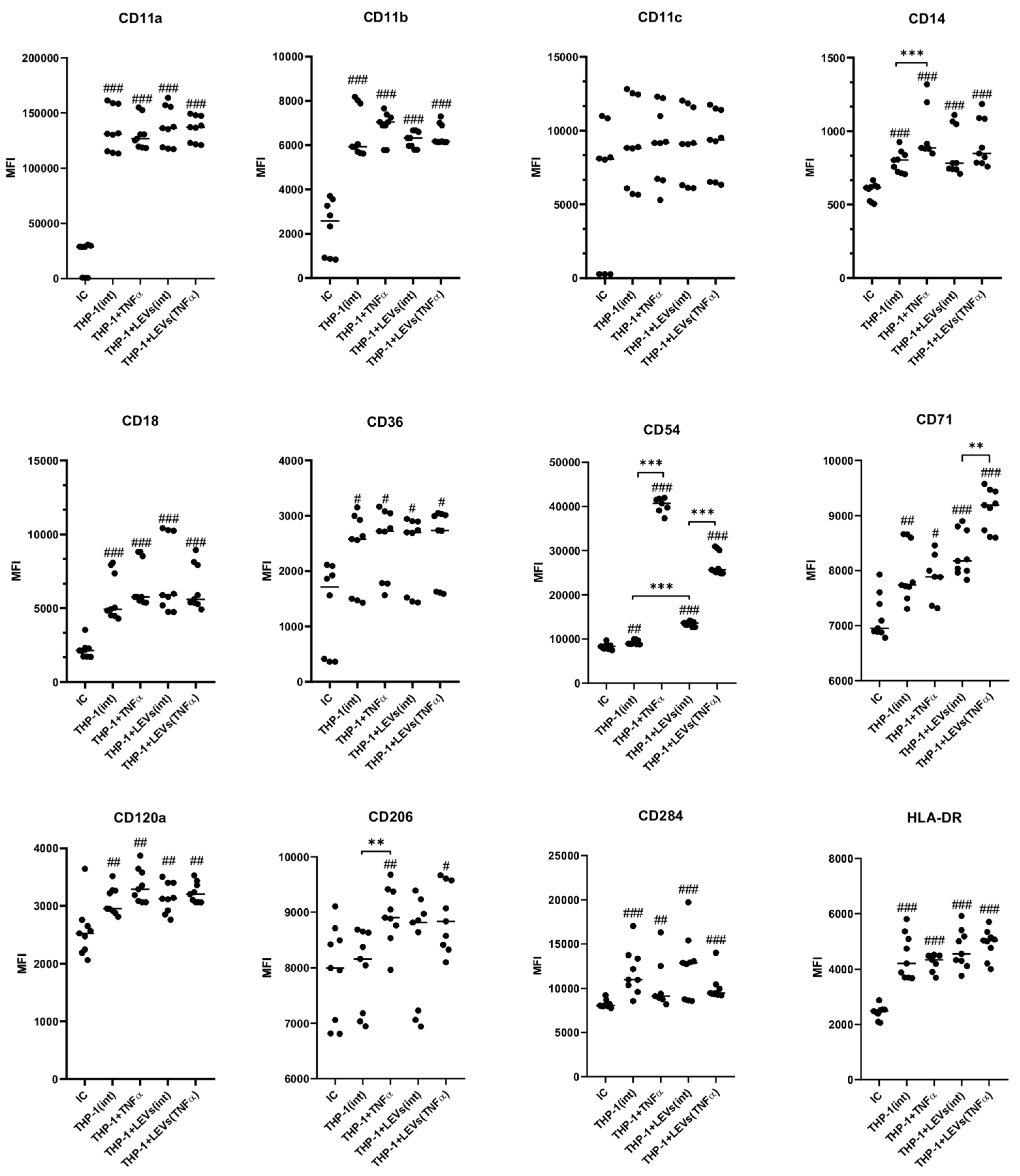
2.4. LEVs Derived from NK-92 Cells Alter the Phenotype of THP-1 Cells

It has been determined that intact THP-1 cells express the following surface molecules: CD54, CD71, CD206, CD120a, CD14, CD36, CD11a, -b and -c, and CD18. The expression of HLA-DR and CD284 in unstimulated cells was not detected (Figure 5 and Figure 6). The stimulation of THP-1 cells with TNFα led to an increased expression of CD284 as well as a higher number of cells expressing CD54, CD71, and CD206 compared to unstimulated cells (Figure 5). Additionally, the stimulation also enhanced the expression intensity of CD54 and CD206 receptors (Figure 6). A co-culture of THP-1 cells with LEVs(int) resulted in an upregulation of HLA-DR expression (Figure 5 and Figure 6) and an increase in the number of CD54+ and CD71+ cells (Figure 5). Additionally, there was an increase in the intensity of CD54 expression (Figure 6) when compared to the control. A co-culture of THP-1 cells with LEVs(TNFα) led to an increase in the number of cells expressing HLA-DR, CD284, CD206, CD54, and CD71, compared to the number observed co-cultured with LEVs(int). In addition, a higher expression intensity of CD54 and CD71 was seen when THP-1 cells were co-cultured with LEVs(TNFα), compared to the level shown when co-cultured with LEVs(int) (Figure 6). Different cultivation conditions did not affect the number of THP-1 cells expressing CD14, CD36, CD18, CD11a, CD11b, and CD11c (Figure 5, Supplementary Data, Table S5), or the intensity of expression of CD120a, CD284, CD36, CD18, CD11a, CD11b, and CD11c (Figure 6, Supplementary Data, Table S6).

2.5. LEVs Derived from NK-92 Cells Decrease the Phagocytic Activity and the Oxidative Burst of PBMCs

It was found that the cultivation of PBMCs with LEVs(int) or LEVs(TNFα) resulted in a decreased phagocytic capacity of PBMCs and reduced intensity of oxidative burst, compared to baseline levels (Figure 7, and Supplementary Data, Tables S7 and S8).

3. Discussion

Currently, the role of different types of extracellular vesicles in facilitating intercellular communication is a topic of active research [24]. It has been shown that almost all cells of the body, including NK cells [34], are capable of producing EVs [35]. In this study we investigated the impact of NK cells on the functional status and phenotypic characteristics of monocyte-like THP-1 cells. NK-92 cells were chosen as a source of EVs, as they exhibit the phenotypic and functional features of NK cells [36].
Due to their ability to transport biologically active molecules, ectosomes\LEVs are considered to be subcellular messengers that transmit signals to surrounding cells [28,37]. The contact of ectosomes\LEVs with the recipient cells leads to signal transmission and occurs through both ligand-receptor interactions and the release of ectosomes\LEVs content into the extracellular space [38]. In addition, ectosomes\LEVs are able to enter the cells via endocytosis, and fuse with the plasma membrane of recipient cells and transfer their contents directly into the cytosol [38]. These mechanisms allow ectosomes\LEVs to trigger various intracellular signaling pathways, altering the functional and phenotypic characteristics of recipient cells [39]. We found that the incubation of THP-1 cells with a medium containing LEVs derived from fluorescently labeled NK-92 cells leads to the transfer of an intracellular protein labeled with a fluorescent dye to monocyte-like cells. These findings are consistent with the results of numerous researchers who have demonstrated the ability of EVs, including ectosomes\LEVs, from various cell types, to internalize within recipient cells [40,41,42].
To assess the functional capabilities of LEVs derived from NK cells in relation to monocyte-like cells, we investigated the effect of NK-92-derived LEVs on the viability of THP-1 cells. We found that the highest tested concentration of LEVs (20 μg/100 μL) was able to reduce the viability of approximately 15% of THP-1 cells. This result could be attributed to the presence of perforin and granzyme B in ectosomes\LEVs [28,43]. These proteins, which are also part of the lytic granules of NK cells, together with granulysin, FasL, and TRAIL, are released upon contact with a target cell causing its apoptosis [44]. It is possible that in our study high concentrations of LEVs could have caused a decrease in cell viability due to the apoptosis of THP-1 cells. The results we obtained regarding the reduction in the viability of monocyte-like cells and the cytotoxic activity of LEVs produced via NK cells, as described previously [28], which may support the non-classical theory of cell lysis [28]. At present, it is not entirely clear how granzyme B is transported to target cells. In addition to the well-established theory of cellular cytolysis, which states that perforin forms pores in the cell membrane, allowing granzyme B to enter the cytoplasm [44], there is another theory that suggests that LEVs act as carriers for perforin and granzyme B within target cells [31]. Perforin then forms pores in the endosomal membrane, and granzyme B enters the cytoplasm, initiating apoptosis [43]. Therefore, the presence of lytic proteins in NK cells [28] suggests that they may constitute a novel mechanism through which these cells are able to exert their cytotoxic function, including in relation to monocytes. In the absence of inflammation and stimulation via factors such as LPS, TNFα, and IL-1β, monocytes perceive apoptotic signals from surrounding cells, including NK cells, and are eliminated from tissues [45,46]. In this context, the observed reduction in the viability of THP-1 cells caused by LEVs derived from NK cells can be considered an additional mechanism for immunoregulating the monocyte function by NK cells, as well as for maintaining the number of tissue-resident monocytes available for immune responses.
The primary function of monocytes is to provide innate immunity, which protects against pathogens and maintains tissue homeostasis [47]. The regulation of monocyte activity is provided via various cells of the innate and adaptive immune systems [48,49]. Specific interactions between monocytes and these cells occur through ligand-receptor communication or through the secretion of biologically active substances. These interactions result in the establishment of a unique microenvironment that defines the outcome of inflammation [5]. Currently, the role of various types of EVs in the process of communication during inflammation is being actively investigated. However, there is a lack of data on the effect of LEVs derived from NK cells on monocytes. Therefore, we set out to analyze the potential of NK cells to modulate the phenotypic and functional characteristics of monocytes through LEVs.
The ability of LEVs produced via NK cells to alter the phenotypic features of monocytes was assessed through the analysis of the expression of various functional receptors (see the table in Section 4). THP-1 cells, which reflect the characteristics of promonocytic cells, are able to differentiate into various populations of monocytes and macrophages when subjected to different inducers [50,51]. The results obtained from this study regarding the spontaneous expression of molecules via intact THP-1 cells align with previously published data on their phenotypic characteristics. In particular, more than half of the cells expressed CD14, which is a classic marker for monocytes [50,52], as well as TNF receptor (CD120a) [53] and integrins CD11a, CD11b, and CD18 [54]. At the same time, these cells exhibited a weak expression of CD71, CD206, and CD54 molecules [54,55,56], and they also did not express HLA-DR [57,58] and CD284 molecules [59]. This indicates their undifferentiated state and is consistent with the literature. The expression of many surface markers on THP-1 cells can be triggered via treatment with PMA, LPS, or cytokines [50,51]. To simulate the conditions of monocyte activation during the immune response, THP-1 cells were activated with the pro-inflammatory cytokine TNFα. As expected, we observed an increased level of the expression of the CD54, CD71, and CD206 molecules, which are characteristic of activated monocytes. Additionally, the number of THP-1 cells expressing CD120a and CD284 increased, consistent with previous reports on the inducibility of these surface markers in THP-1 cells [53,54,55,56,59]. The activation of THP-1 cells by TNFα apparently induced the differentiation of these cells into mature monocytes, as evidenced by the results of the functional tests.
After that, we assessed the ability of LEVs produced via NK cells to alter the phenotypic features of THP-1 cells. To accomplish this, we cultured THP-1 cells with LEVs derived from intact or TNFα-activated NK-92. The activation with TNFα was used in order to simulate the inflammatory activation of NK cells [1]. It was found that the co-culture with LEVs derived from intact NK cells resulted in the emergence of THP-1 cells expressing HLA-DR, as well as an increase in the number of cells expressing CD54, CD71, and CD206. The co-culture with LEVs derived from the TNFα-stimulated NK cell led to the emergence of CD284+ cells and an even higher increase in the expression of HLA-DR, CD284, CD206, CD54, and CD71 relative to the levels observed in the presence of LEVs(int). These results are likely connected to the varied internal and surface molecules of LEVs, which can be transmitted to target cells through fusion or endocytosis [27,33,38]. Although the exact mechanisms of the interaction between monocyte cells and LEVs have not yet been determined, we found that LEVs derived from NK cells are able to alter the phenotypic characteristics of THP-1 cells. Furthermore, the observed effect is similar to that seen when THP-1 themselves are activated via TNFα. As previously stated, CD54, CD71, CD206, and CD120a are inducible markers whose expression increases on monocytes upon activation and polarization via various cytokines and activating agents [53,54,55,56,59]. In addition to cytotoxic proteins, numerous proinflammatory and immunomodulatory molecules have been found in LEVs derived from NK cells: interferon-gamma (IFN-γ), interleukin-1 beta (IL-1β), and tumor necrosis factor alpha (TNFα) [60], as well as various microRNAs [61]. The evidence for the presence of these cytokines in LEVs is supported by the appearance of human leukocyte antigen HLA-DR expression on the surface of THP-1 cells after cultivation with LEVs, but not with TNFα. According to the literature, HLA-DR is not expressed through unstimulated THP-1 cells, however, its expression is observed when the cells are stimulated via cytokines [57,58]. Furthermore, HLA-DR expression can only be induced in the presence of both TNFα and IFNγ [58]. Therefore, the induction of HLA-DR on monocyte-like cells appears to be associated with the presence of IFNγ and TNFα in the LEVs derived from NK cells. It has been suggested that the ability of NK cells to generate vesicles with different internal compositions, depending on their activation status, ref. [33] may explain the variation in the level of expression of other markers observed on THP-1 cells. It should also be noted that LEVs and receptors on their surfaces can be incorporated into the membranes of target cells [42]. Consequently, the increased expression of the CD284 receptor on THP-1 cells after their incubation with LEVs may be linked to the integration of the membranes of LEVs derived from NK-92 cells, which are known to express CD284 (see table in Section 4.8).
The ability of NK cells to influence the phagocytic activity and the oxidative burst of monocytes has been evaluated through the analysis of the interaction between THP-1 cells and FITC-labeled E. coli after the pre-incubation of cells with LEVs derived from intact or TNFα-activated NK cells. The results obtained for the baseline levels of phagocytosis and oxidative burst in intact THP-1 cells are consistent with the literature’s data [50,51]. The increased phagocytic activity and oxidative response induced via TNFα-activated THP-1 cells also corresponds to the classical theory of pro-inflammatory monocyte activation [5,14,62]. The increased functional activity of TNFα-activated THP-1 cells is likely related to an increase in the number of cells expressing the mannose receptor, CD206, on their surface. This protein plays an important role in facilitating the initial stages of phagocytosis by facilitating the uptake of bacterial cell wall components [56,63].
Pre-incubation of THP-1 cells with LEVs derived from intact NK-92 cells led to a decrease in the intensity of phagocytosis by THP-1 cells and a reduction in the production of reactive oxygen species (ROS) compared to the spontaneous levels. This effect was also observed in PBMCs from healthy donors. These findings suggest that LEVs produced via NK-92 cells can transmit inhibitory signals to phagocytes, potentially leading to anti-inflammatory responses. These effects can be mediated through various substances transported in vesicles, specifically anti-inflammatory cytokines (IL-10 and TGFβ) or microRNAs, that affect the expression of genes in target cells [61]. According to the literature data, EVs derived from NK cells can influence various signaling pathways in target cells, although the mechanisms underlying this interaction are not fully understood. Specifically, NK cell-derived exosomes have been shown to influence the activation of phosphatidylinositol-3 kinase (PI3K) [60], which plays a crucial role in activation of actin cytoskeletal rearrangements and the formation of phagocytic cup upon contact with pathogens [64]. A reduction in the intensity of oxidative burst is a direct result of a reduction in phagocytic activity, as the assembly of NADPH oxidase complexes, which produce ROS, occurs on membranes formed via phagolysosomes [64,65].
The pre-treatment of THP-1 cells with LEVs produced through TNFα-activated NK-92 cells resulted in increased phagocytic activity and oxidative burst, compared to both the spontaneous levels and the levels shown in TNFα-activated THP-1 cells. These findings are consistent with the observed changes in the phenotypical properties of the cells. Therefore, the activation of THP-1 cells via the components or surface molecules of LEVs affects the phenotypic features of the cells and directly influences their functional abilities. It is noteworthy that, despite the fact that the functional outcomes observed during the incubation of THP-1 cells with TNFα or LEVs produced via TNFα-activated NK cells are comparable, in the latter case, there was a greater level of phagocytic activity and ROS production. This result can also be explained by the diversity of components in LEVs, including pro-inflammatory cytokines (IFNy, IL-1β, and TNFα), which can stimulate monocyte-like cells to phagocytose [33]. Additionally, the increased activity of THP-1 cells may be linked to an increase in the expression level of TLR4 (CD284) after co-culturing with LEVs derived from TNFα-activated NK cells [45,64].
It should be noted that, in contrast to THP-1 cells, the pre-incubation of PBMCs with LEVs produced via TNFα-activated NK-92 cells led to a decrease in the intensity of phagocytosis and ROS production. This result can be explained by the effect of LEVs on other cell populations of PBMCs, which could alter the effect of LEVs on monocytes. Further study is required to clarify the mechanisms through which cells of the microenvironment modulate the effects of LEVs on target cells.

4. Materials and Methods

4.1. Cell Lines

NK-92 cells (ATCC, Manassas, VA, USA), which are a model for activated NK cells [36,66,67], and THP-1 cells, representing monocytes, [51] were used. The cells were cultured in accordance with ATCC guidelines. All experiments were conducted in a humidified atmosphere at 37 °C and 5% CO2. Cell viability was assessed using trypan blue and propidium iodide solutions [68], and it was at least 95%. To work with EVs, all solutions, media, and fetal bovine serum (FBS) (F2442, Sigma-Aldrich, Saint Louis, MO, USA) were filtered through 0.2-micron pore filters (Sigma-Aldrich Chemical Co., Saint Louis, MO, USA) to ensure sterility [69]. The manufacturer guarantees the triple filtration of the serum through 0.1-micron membrane under aseptic conditions. Due to protocols for the isolation of EVs from biological fluids published by Gelderman, M.P., and Simak, J. [70], Ectosomes\LEVs are unstable extracellular structures; freezing and defrosting the samples lead to the destruction of ectosomes\LEVs. Therefore, in this work, we used sequentially frozen and then thawed FCS, which was further inactivated according to the standard protocol. These procedures additionally guaranteed the absence of contaminant ectosomes\LEVs from the serum. We did not perform fetal bovine serum (FBS) ultracentrifugation, as according to some researchers [71,72,73]. FBS depleted via ultracentrifugation may affect and support cell growth in a different way in comparison to regular FBS.

4.2. LEVs Isolation

LEVs produced via NK-92 were isolated using the method of differential centrifugation of the culture medium [23,74]. NK-92 cells were incubated at a concentration of 4 × 10⁵ cells per mL for 24 h in a volume of 40 mL of fresh medium. Some of these cells were incubated in the presence of 100 U/mL of TNFα (R&D, Santa Clara, CA, USA). Following this, LEVs were isolated from the remaining supernatant via successive centrifugations at 500× g (4 °C, 10 min), 9900× g (4 °C, 10 min), and 19,800× g (4 °C, 20 min).The method used allows for the isolation of LEVs with sufficient purity and a minimal loss of biological material [21,75]. Protein content in the LEVs samples was measured using the Bradford assay [76] with a NanoDrop One spectrophotometer, from Thermo Scientific (Waltham, MA, USA). The protein content in LEVs isolated from the medium of control NK-92 cells and activated with TNFα NK-92 cells was 3.3 ± 0.3 μg per 106 cells and 2.8 ± 0.3 μg per 106 cells, respectively. Previously, we described the phenotype ([28,34]), content ([43,77]), and effects of LEVs produced via NK-92 on target cells ([42,78]).

4.3. Laser Correlation Analysis

Granulometric analysis was conducted to monitor the size of isolated LEVs using a laser correlation spectrometer, Zetasizer NanoZS (Malvern Instruments, Malvern, UK). The size of LEVs isolated from the culture medium of NK-92 cells ranged from 232 to 556 nanometers, with a peak distribution at 339 nanometers, which is consistent with previously reported data [43,69,74,79]. Previously, using the methods of Laser Correlation Analysis, atomic force microscopy, electron microscopy, and flow cytofluorometry, we showed that the LEVs of NK-92 cells, which we obtain using differential centrifugation (Section 4.2), have similar results when describing the sizes [80]. Using electron microscopy, we showed the spherical symmetry of the LEVs produced by NK-92 cell line and the presence of a membrane made of a lipid bilayer [80].

4.4. Evaluation of the Transfer of a Fluorescent Label from LEVs Derived from NK-92 Cells to THP-1 Cells

The method described previously was utilized [42]. Prior to the experiment, the THP-1 culture medium was refreshed. THP-1 cells were seeded into the wells of a 24-well plate for suspension cultures at a concentration of 5 × 10⁵ cells per 500 μL of a complete medium for THP-1 cells supplemented with 10% FBS and incubated for 24 h. To label intracellular proteins, NK-92 cells were incubated with a solution of 5(6)-carboxy-fluorescein diacetate succinimidyl ester (CFSE) (Sigma-Aldrich Chemical Co., USA) at a concentration of 50 μM in accordance with the manufacturer’s guidelines. Intact and stained NK-92 cells were then cultured for 24 h in 75 cm2 flasks in 40 mL of complete culture medium at a concentration of 4 × 10⁵ cells per mL. LEVs were isolated as previously described. The LEVs were then seeded into the wells containing THP-1 cells at a concentration of 10 μg of protein per 100 μL, and co-cultured for 24 h. The cells were then washed three times with versene solution, resuspended in a Hank’s solution without Ca2+ and Mg2+, and centrifuged at 200× g for 10 min to remove the supernatant. The incorporation of the fluorescent form of CFSE into the membrane of THP-1 cells was assessed using a FACSCanto II flow cytometer (BD, Franklin Lakes, NJ, USA), as previously described (Supplementary Data, Figure S1) [42]. The experiments were conducted in triplicate.

4.5. Evaluation of the Effect of LEVs of NK-92 Cells on the THP-1 Cells Viability

THP-1 cells were prepared as we mentioned in Section 4.4. Intact and activated via 400 U/mL of TNFα NK-92 cells were cultured for 24 h in 75 cm2 flasks in 40 mL of complete culture medium at a concentration of 4 × 10⁵ per mL. LEVs were then isolated as described above. Following isolation, LEVs from the NK-92 cells were added to the wells containing THP-1 cells at a concentration of 5, 10, or 20 μg of protein per 100 μL. The cells were then cultured for 24 h. Then, the THP-1 cells were washed twice with Hank’s solution via centrifugation at 200× g for 10 min. After that, the cells were treated with a 7-amino-actinomycin D solution (7-AAD), in accordance with the manufacturer’s instructions (BD, USA). The fluorescence intensity was measured using a FACS Canto II flow cytometer (BD, USA). All experiments were conducted in triplicate, with three replicates for each concentration of LEVs.

4.6. Evaluation of the Effect of LEVs Derived from NK-92 Cells on Phagocytic Activity and Oxidative Burst in THP-1 Cells

THP-1 cells were prepared as we mentioned in Section 4.4 and Section 4.5. LEVs were isolated as described above and added to wells containing intact THP-1 cells at a concentration of 10 μg of protein per 100 μL. TNFα was also added to a separate group of THP-1 cells as a control for phagocytosis activation and oxidative burst. Intact cells were used as a control. After 24 h of incubation, the plate with THP-1 cells was centrifuged at 200× g for 10 min. Then 150 μL of the supernatant from each well was collected. The cells were then resuspended in 50 μL of medium and transferred to Eppendorf tubes. The phagocytic activity of the cells was evaluated using commercial kits (IngoFlowEx, Exbio, Vestec-Jesenice u Prahy, Czech Republic) with a BD FACSCanto II flow cytometer (BD, USA). The IngoFlowEx Kit is designed for the quantification of phagocytic activity by measuring the ingestion of fluorescently labeled (FITC) E. coli bacteria. The oxidative activity of the cells was evaluated using commercial kits (FagoFlowEx, Exbio, Czech Republic)with a BD FACSCanto II flow cytometer (BD, USA). The FagoFlowEx Kit is intended for the examination of phagocytic activity by measuring the respiratory (oxidative) burst after stimulation with E. coli bacteria in human heparinized whole blood using flow cytometry. Reactive oxygen species (ROS) production after E. coli incorporation via phagocytes was measured using dihydrorhodamine 123 which was then oxidized to rodamine 123 and then became fluorescent (Supplementary Data, Figures S2 and S4) Thus, the MFI of phagocytic cells is proportional to intracellular ROS activity (Figure 4). All experiments were conducted in triplicate, with three replicates for each cultivation condition.

4.7. Evaluation of the Effect of LEVs Derived from NK-92 Cells on Phagocytic Activity and the Realization of Oxidative Explosion via the PBMCs of Healthy Donors

Peripheral blood mononuclear cells (PBMCs) were isolated from the peripheral blood of 12 healthy donors through Ficoll density gradient centrifugation (ρ = 1.077 g/mL, Biolot, St. Petersburg, Russia), following standard procedures. The isolated PBMCs were seeded into 96-well plates at a concentration of 2 × 10⁵ cells/200 μL of complete cell culture medium. After 24 h LEVs produced by either intact or TNFα-activated NK-92 cells were added to wells at a concentration of 10 μg of protein per 100 μL. The phagocytic and oxidative activities of the PBMCs were then evaluated using commercial kits (IngoFlowEx and FagoFlowEx, Exbio, Czech Republic) and a BD FACS Canto II flow cytometer. The study was reviewed and approved by the local ethics committee of the Federal State Budgetary Scientific Institution “The Research Institute of Obstetrics, Gynecology and Reproductology named after D.O. Ott” (Protocol No. 107, dated 15 March 2021). Donors whose data were used in the work provided informed consent to their participation in the study. Subsequently, the data were de-identified and the results were analyzed.

4.8. Evaluation of the Effect of LEVs Derived from NK-92 Cells on the Phenotype of THP-1 Cells

THP-1 cells were prepared as we mentioned in Section 4.4 and Section 4.5. LEVs were isolated as described above and added to wells containing intact THP-1 cells at a concentration of 10 μg of protein per 100 μL and co-cultured for 24 h. THP-1 cells that were intact and activated via 400 U/mL of TNFα were used as controls. The cells were then washed three times with a versene solution, resuspended in Hank’s solution without Ca2+ and Mg2+, and centrifuged at 200× g for 10 min to remove the supernatant. A Fc-blocking reagent was used to block Fc-receptors according to the manufacturer’s instructions (Miltenyi Biotec, Gaithersburg, MD, USA). After incubation with LEVs, the THP-1 cells were stained with monoclonal antibodies for CD11a, CD11b, CD11c, CD18, CD54, HLA-DR, CD120a, CD14, CD36, CD284, and CD71 in accordance with the manufacturer’s instructions (BD Biosciences, San Jose, CA, USA). Isotypic antibodies were used as control for non-specific binding (BD, USA). The choice of antibodies was made based on our data, as well as the literature’s data on alterations to the phenotype of THP-1 cells after exposure to cytokines [53,81,82,83]. Additionally, we took into account the phenotype of NK-92 cells and their LEVs [28,42,74]; the data are summarized in Table 1.
Fluorescence was measured using a BD FACSCanto II flow cytometer (BD, USA) (Supplementary Data, Figure S3). The experiments were conducted in triplicate.

4.9. Statistical Analysis

Statistical analysis was conducted using the GraphPad Prism 8 software. The nonparametric Mann-Whitney U test was used to compare the obtained data. The results were reported as “median (lower quartile, upper quartile)”.

5. Conclusions

In this study, we demonstrated the ability of LEVs produced via NK cells to be integrated into the membranes of THP-1 cells and influence the viability, phenotype, and functional characteristics of the cells. We supposed that the increased expression of CD54, CD71, and CD206 via THP-1 cells, as well as the appearance of CD284 and increased phagocytic and oxidative activity after a co-culture with LEVs produced through TNFα-activated NK cells, suggests that THP-1 cells acquire the characteristics of classically activated M1 macrophages [102,103]. On the contrary, in the presence of LEVs produced via intact NK cells THP-1 cells acquired the characteristics of alternatively activated macrophages (M2) [104]. The stated hypothesis requires further experimental confirmation. These cells demonstrated reduced phagocytic activity and oxidative burst intensity, as well as low expression of the M1-specific markers CD206 and CD284 [103]. However, a high number of CD54+ and CD71+ cells were observed, which is not fully consistent with the hypothesis. Therefore, further investigation is needed to determine the molecular mechanisms underlying the interaction between LEVs derived from NK cells and monocytes. The results obtained demonstrate the ability of LEVs to act as an additional component in the immunomodulatory activity of NK cells in relation to monocytes. Figure 8 represents the main results of the research.

Supplementary Materials

The supporting information can be downloaded at: https://www.mdpi.com/article/10.3390/ijms25179478/s1.

Author Contributions

Conceptualization, D.S.; methodology, A.G., E.T., P.G. and M.Z.; validation, D.S.; formal analysis, E.T., D.S.; investigation, A.G., E.T., P.G. and M.Z.; resources, D.S.; data curation, D.S.; writing—original draft preparation, D.S.; writing—review and editing, D.S. and P.G.; visualization, A.G. and D.S.; supervision, D.S.; project administration, D.S.; funding acquisition, D.S., I.K. and A.T. All authors have read and agreed to the published version of the manuscript.

Funding

This research was funded by the Russian Science Foundation, grant number № 24-15-00002.

Institutional Review Board Statement

The study was conducted in accordance with the Declaration of Helsinki, and approved by the Institutional Ethics Committee of the Federal State Budgetary Scientific Institution “The Research Institute of Obstetrics, Gynecology and Reproductology named after D.O. Ott” (Protocol No. 107, dated 15 March 2021).

Informed Consent Statement

Informed consent was obtained from all subjects involved in the study.

Data Availability Statement

The data presented in this study are available upon request from the corresponding author.

Acknowledgments

The team of authors expresses gratitude to laboratory assistant Semenov V.A. for methodological assistance.

Conflicts of Interest

The authors declare no conflict of interest.

References

  1. Del Zotto, G.; Marcenaro, E.; Vacca, P.; Sivori, S.; Pende, D.; Della Chiesa, M.; Moretta, F.; Ingegnere, T.; Mingari, M.C.; Moretta, A.; et al. Markers and function of human NK cells in normal and pathological conditions. Cytom. Part B Clin. Cytom. 2017, 92, 100–114. [Google Scholar] [CrossRef]
  2. Mandal, A.; Viswanathan, C. Natural killer cells: In health and disease. Hematol./Oncol. Stem Cell Ther. 2015, 8, 47–55. [Google Scholar] [CrossRef]
  3. Auffray, C.; Sieweke, M.H.; Geissmann, F. Blood monocytes: Development, heterogeneity, and relationship with dendritic cells. Annu. Rev. Immunol. 2009, 27, 669–692. [Google Scholar] [CrossRef]
  4. Walajtys-Rode, E.; Dzik, J.M. Monocyte/Macrophage: NK Cell Cooperation-Old Tools for New Functions. Results Probl. Cell Differ. 2017, 62, 73–145. [Google Scholar] [CrossRef]
  5. Lapaque, N.; Walzer, T.; Meresse, S.; Vivier, E.; Trowsdale, J. Interactions between human NK cells and macrophages in response to Salmonella infection. J. Immunol. 2009, 182, 4339–4348. [Google Scholar] [CrossRef]
  6. Empson, V.G.; McQueen, F.M.; Dalbeth, N. The natural killer cell: A further innate mediator of gouty inflammation? Immunol. Cell Biol. 2010, 88, 24–31. [Google Scholar] [CrossRef]
  7. Söderström, K.; Stein, E.; Colmenero, P.; Purath, U.; Müller-Ladner, U.; de Matos, C.T.; Tarner, I.H.; Robinson, W.H.; Engleman, E.G. Natural killer cells trigger osteoclastogenesis and bone destruction in arthritis. Proc. Natl. Acad. Sci. USA 2010, 107, 13028–13033. [Google Scholar] [CrossRef]
  8. Zhang, A.L.; Colmenero, P.; Purath, U.; Teixeira de Matos, C.; Hueber, W.; Klareskog, L.; Tarner, I.H.; Engleman, E.G.; Söderström, K. Natural killer cells trigger differentiation of monocytes into dendritic cells. Blood 2007, 110, 2484–2493. [Google Scholar] [CrossRef]
  9. Welte, S.; Kuttruff, S.; Waldhauer, I.; Steinle, A. Mutual activation of natural killer cells and monocytes mediated by NKp80-AICL interaction. Nat. Immunol. 2006, 7, 1334–1342. [Google Scholar] [CrossRef]
  10. Nedvetzki, S.; Sowinski, S.; Eagle, R.A.; Harris, J.; Vely, F.; Pende, D.; Trowsdale, J.; Vivier, E.; Gordon, S.; Davis, D.M. Reciprocal regulation of human natural killer cells and macrophages associated with distinct immune synapses. Blood 2007, 109, 3776–3785. [Google Scholar] [CrossRef]
  11. Schulz, U.; Kreutz, M.; Multhoff, G.; Stoelcker, B.; Köhler, M.; Andreesen, R.; Holler, E. Interleukin-10 promotes NK cell killing of autologous macrophages by stimulating expression of NKG2D ligands. Scand. J. Immunol. 2010, 72, 319–331. [Google Scholar] [CrossRef]
  12. Cichocki, F.; Sitnicka, E.; Bryceson, Y.T. NK cell development and function--plasticity and redundancy unleashed. Semin. Immunol. 2014, 26, 114–126. [Google Scholar] [CrossRef]
  13. Cooper, M.A.; Fehniger, T.A.; Caligiuri, M.A. The biology of human natural killer-cell subsets. Trends Immunol. 2001, 22, 633–640. [Google Scholar] [CrossRef]
  14. Michel, T.; Hentges, F.; Zimmer, J. Consequences of the crosstalk between monocytes/macrophages and natural killer cells. Front. Immunol. 2013, 3, 403. [Google Scholar] [CrossRef]
  15. Wu, M.; Zhou, C.; Li, M.; Yu, H.; Zhao, D.; Xue, W.; Qin, L.; Peng, A. Depletion of NK cells attenuates paraquat-induced acute lung injury by manipulating macrophage polarization. Int. Immunopharmacol. 2020, 86, 106698. [Google Scholar] [CrossRef]
  16. Raposo, G.; Stoorvogel, W. Extracellular vesicles: Exosomes, microvesicles, and friends. J. Cell Biol. 2013, 200, 373–383. [Google Scholar] [CrossRef]
  17. Borges, F.T.; Reis, L.A.; Schor, N. Extracellular vesicles: Structure, function, and potential clinical uses in renal diseases. Braz. J. Med. Biol. Res. 2013, 46, 824–830. [Google Scholar] [CrossRef]
  18. Welsh, J.A.; Goberdhan, D.C.I.; O’Driscoll, L.; Buzas, E.I.; Blenkiron, C.; Bussolati, B.; Cai, H.; Di Vizio, D.; Driedonks, T.A.p.; Erdbrügger, U.; et al. Minimal information for studies of extracellular vesicles (MISEV2023): From basic to advanced approaches. J. Extracell. Vesicles 2024, 13, e12404. [Google Scholar] [CrossRef]
  19. Muralidharan-Chari, V.; Clancy, J.; Plou, C.; Romao, M.; Chavrier, P.; Raposo, G.; D’Souza-Schorey, C. ARF6-regulated shedding of tumor cell-derived plasma membrane microvesicles. Curr. Biol. CB 2009, 19, 1875–1885. [Google Scholar] [CrossRef]
  20. Li, P.; Kaslan, M.; Lee, S.H.; Yao, J.; Gao, Z. Progress in Exosome Isolation Techniques. Theranostics 2017, 7, 789–804. [Google Scholar] [CrossRef]
  21. Kowal, J.; Arras, G.; Colombo, M.; Jouve, M.; Morath, J.P.; Primdal-Bengtson, B.; Dingli, F.; Loew, D.; Tkach, M.; Théry, C. Proteomic comparison defines novel markers to characterize heterogeneous populations of extracellular vesicle subtypes. Proc. Natl. Acad. Sci. USA 2016, 113, E968–E977. [Google Scholar] [CrossRef]
  22. Simak, J.; Gelderman, M.P. Cell Membrane Microparticles in Blood and Blood Products: Potentially Pathogenic Agents and Diagnostic Markers. Transfus. Med. Rev. 2006, 20, 1–26. [Google Scholar] [CrossRef]
  23. Simak, J.; Gelderman, M.P.; Yu, H.; Wright, V.; Baird, A.E. Circulating endothelial microparticles in acute ischemic stroke: A link to severity, lesion volume and outcome. J. Thromb. Haemost. 2006, 4, 1296–1302. [Google Scholar] [CrossRef]
  24. Stahl, P.D.; Raposo, G. Extracellular Vesicles: Exosomes and Microvesicles, Integrators of Homeostasis. Physiology 2019, 34, 169–177. [Google Scholar] [CrossRef]
  25. Camussi, G.; Deregibus, M.C.; Bruno, S.; Grange, C.; Fonsato, V.; Tetta, C. Exosome/microvesicle-mediated epigenetic reprogramming of cells. Am. J. Cancer Res. 2011, 1, 98–110. [Google Scholar]
  26. György, B.; Szabó, T.G.; Pásztói, M.; Pál, Z.; Misják, P.; Aradi, B.; László, V.; Pállinger, É.; Pap, E.; Kittel, Á.; et al. Membrane vesicles, current state-of-the-art: Emerging role of extracellular vesicles. Cell. Mol. Life Sci. CMLS 2011, 68, 2667–2688. [Google Scholar] [CrossRef]
  27. Burbano, C.; Rojas, M.; Vasquez, G.; Castano, D. Microparticles That Form Immune Complexes as Modulatory Structures in Autoimmune Responses. Mediat. Inflamm. 2015, 2015, 267590. [Google Scholar] [CrossRef]
  28. Sokolov, D.I.; Markova, K.L.; Mikhailova, V.A.; Vyazmina, L.P.; Milyutina, Y.P.; Kozyreva, A.R.; Zhdanova, A.A.; Malygina, D.A.; Onokhin, K.V.; Ivanova, A.N.; et al. Phenotypic and Functional Characteristics of Microvesicles Produced by Natural Killer Cells. Med. Immunol. 2019, 21, 669–688. [Google Scholar] [CrossRef]
  29. Vajen, T.; Mause, S.F.; Koenen, R.R. Microvesicles from platelets: Novel drivers of vascular inflammation. Thromb. Haemost. 2015, 114, 228–236. [Google Scholar] [CrossRef]
  30. Lugini, L.; Cecchetti, S.; Huber, V.; Luciani, F.; Macchia, G.; Spadaro, F.; Paris, L.; Abalsamo, L.; Colone, M.; Molinari, A.; et al. Immune surveillance properties of human NK cell-derived exosomes. J. Immunol. 2012, 189, 2833–2842. [Google Scholar] [CrossRef]
  31. Burnett, L.A.; Nowak, R.A. Exosomes mediate embryo and maternal interactions at implantation and during pregnancy. Front. Biosci. (Sch. Ed) 2016, 8, 79–96. [Google Scholar] [CrossRef]
  32. Tong, M.; Chamley, L.W. Placental extracellular vesicles and feto-maternal communication. Cold Spring Harb. Perspect. Med. 2015, 5, a023028. [Google Scholar] [CrossRef]
  33. Federici, C.; Shahaj, E.; Cecchetti, S.; Camerini, S.; Casella, M.; Iessi, E.; Camisaschi, C.; Paolino, G.; Calvieri, S.; Ferro, S.; et al. Natural-Killer-Derived Extracellular Vesicles: Immune Sensors and Interactors. Front. Immunol. 2020, 11, 262. [Google Scholar] [CrossRef]
  34. Mikhailova, V.A.; Belyakova, K.L.; Vyazmina, L.P.; Sheveleva, A.R.; Selkov, S.A.; Sokolov, D.I. Evaluation of Microvesicles Formed by Natural Killer (Nk) Cells Using Flow Cytometry. Med. Immunol. 2018, 20, 251–254. [Google Scholar] [CrossRef]
  35. Sedgwick, A.E.; D’Souza-Schorey, C. The biology of extracellular microvesicles. Traffic 2018, 19, 319–327. [Google Scholar] [CrossRef]
  36. Gong, J.H.; Maki, G.; Klingemann, H.G. Characterization of a human cell line (NK-92) with phenotypical and functional characteristics of activated natural killer cells. Leuk. Off. J. Leuk. Soc. Am. Leuk. Res. Fund UK 1994, 8, 652–658. [Google Scholar]
  37. Tannetta, D.; Dragovic, R.; Alyahyaei, Z.; Southcombe, J. Extracellular vesicles and reproduction-promotion of successful pregnancy. Cell Mol. Immunol. 2014, 11, 548–563. [Google Scholar] [CrossRef]
  38. Burger, D.; Schock, S.; Thompson, C.S.; Montezano, A.C.; Hakim, A.M.; Touyz, R.M. Microparticles: Biomarkers and beyond. Clin. Sci. 2013, 124, 423–441. [Google Scholar] [CrossRef]
  39. Tricarico, C.; Clancy, J.; D’Souza-Schorey, C. Biology and biogenesis of shed microvesicles. Small GTPases 2017, 8, 220–232. [Google Scholar] [CrossRef]
  40. Fuhrmann, G.; Serio, A.; Mazo, M.; Nair, R.; Stevens, M.M. Active loading into extracellular vesicles significantly improves the cellular uptake and photodynamic effect of porphyrins. J. Control Release 2015, 205, 35–44. [Google Scholar] [CrossRef]
  41. Deng, S.Z.; Lai, M.F.; Li, Y.P.; Xu, C.H.; Zhang, H.R.; Kuang, J.G. Human marrow stromal cells secrete microRNA-375-containing exosomes to regulate glioma progression. Cancer Gene Ther. 2020, 27, 203–215. [Google Scholar] [CrossRef]
  42. Markova, K.L.; Mikhailova, V.A.; Korenevsky, A.V.; Milyutina, Y.P.; Rodygina, V.V.; Aleksandrova, E.P.; Markov, A.S.; Balabas, O.A.; Selkov, S.A.; Sokolov, D.I. Microvesicles produced by natural killer cells of the NK-92 cell line affect the phenotype and functions of endothelial cells of the EA.Hy926 cell line. Med. Immunol. 2020, 22, 249–268. [Google Scholar] [CrossRef]
  43. Korenevskii, A.V.; Milyutina, Y.P.; Zhdanova, A.A.; Pyatygina, K.M.; Sokolov, D.I.; Sel’kov, S.A. Mass-Spectrometric Analysis of Proteome of Microvesicles Produced by NK-92 Natural Killer Cells. Bull. Exp. Biol. Med. 2018, 165, 564–571. [Google Scholar] [CrossRef]
  44. Krzewski, K.; Coligan, J.E. Human NK cell lytic granules and regulation of their exocytosis. Front. Immunol. 2012, 3, 335. [Google Scholar] [CrossRef]
  45. Daigneault, M.; Preston, J.A.; Marriott, H.M.; Whyte, M.K.; Dockrell, D.H. The identification of markers of macrophage differentiation in PMA-stimulated THP-1 cells and monocyte-derived macrophages. PLoS ONE 2010, 5, e8668. [Google Scholar] [CrossRef]
  46. Mangan, D.F.; Welch, G.R.; Wahl, S.M. Lipopolysaccharide, tumor necrosis factor-alpha, and IL-1 beta prevent programmed cell death (apoptosis) in human peripheral blood monocytes. J. Immunol. 1991, 146, 1541–1546. [Google Scholar] [CrossRef]
  47. Titov, L.P. Monocytes, Macrophages, Dendritic and Myeloid Suppressor Cells: Genesis, Classification, Immunobiological Properties. Proc. Natl. Acad. Sci. Belarus Med. Ser. 2018, 15, 363–382. [Google Scholar] [CrossRef]
  48. Navegantes, K.C.; de Souza Gomes, R.; Pereira, P.A.T.; Czaikoski, P.G.; Azevedo, C.H.M.; Monteiro, M.C. Immune modulation of some autoimmune diseases: The critical role of macrophages and neutrophils in the innate and adaptive immunity. J. Transl. Med. 2017, 15, 36. [Google Scholar] [CrossRef]
  49. Goldszmid, R.S.; Caspar, P.; Rivollier, A.; White, S.; Dzutsev, A.; Hieny, S.; Kelsall, B.; Trinchieri, G.; Sher, A. NK cell-derived interferon-gamma orchestrates cellular dynamics and the differentiation of monocytes into dendritic cells at the site of infection. Immunity 2012, 36, 1047–1059. [Google Scholar] [CrossRef]
  50. Chanput, W.; Mes, J.J.; Wichers, H.J. THP-1 cell line: An in vitro cell model for immune modulation approach. Int. Immunopharmacol. 2014, 23, 37–45. [Google Scholar] [CrossRef]
  51. Tsuchiya, S.; Yamabe, M.; Yamaguchi, Y.; Kobayashi, Y.; Konno, T.; Tada, K. Establishment and characterization of a human acute monocytic leukemia cell line (THP-1). Int. J. Cancer 1980, 26, 171–176. [Google Scholar] [CrossRef] [PubMed]
  52. Gordon, S.; Taylor, P.R. Monocyte and macrophage heterogeneity. Nat. Rev. Immunol. 2005, 5, 953–964. [Google Scholar] [CrossRef]
  53. Moh, M.C.; Lorenzini, P.A.; Gullo, C.; Schwarz, H. Tumor necrosis factor receptor 1 associates with CD137 ligand and mediates its reverse signaling. FASEB J. 2013, 27, 2957–2966. [Google Scholar] [CrossRef] [PubMed]
  54. Baqui, A.A.; Meiller, T.F.; Kelley, J.I.; Turng, B.F.; Falkler, W.A. Antigen activation of THP-1 human monocytic cells after stimulation with lipopolysaccharide from oral microorganisms and granulocyte-macrophage colony-stimulating factor. J. Periodontal Res. 1999, 34, 203–213. [Google Scholar] [CrossRef] [PubMed]
  55. Mitachi, T.; Kouzui, M.; Maruyama, R.; Yamashita, K.; Ogata, S.; Kojima, H.; Itagaki, H. Some non-sensitizers upregulate CD54 expression by activation of the NLRP3 inflammasome in THP-1 cells. J. Toxicol. Sci. 2019, 44, 213–224. [Google Scholar] [CrossRef]
  56. Gonzalez, N.; Quintana, J.A.; García-Silva, S.; Mazariegos, M.; de la Aleja, A.G.; Nicolás-Ávila, J.A.; Walter, W.; Adrover, J.M.; Crainiciuc, G.; Kuchroo, V.K.; et al. Phagocytosis imprints heterogeneity in tissue-resident macrophages. J. Exp. Med. 2017, 214, 1281–1296. [Google Scholar] [CrossRef]
  57. Popēna, I.; Ābols, A.; Saulīte, L.; Pleiko, K.; Zandberga, E.; Jēkabsons, K.; Endzeliņš, E.; Llorente, A.; Linē, A.; Riekstiņa, U. Effect of colorectal cancer-derived extracellular vesicles on the immunophenotype and cytokine secretion profile of monocytes and macrophages. Cell Commun. Signal 2018, 16, 17. [Google Scholar] [CrossRef]
  58. Portillo, G.; Turner, M.; Chantry, D.; Feldmann, M. Effect of cytokines on HLA-DR and IL-1 production by a monocytic tumour, THP-1. Immunology 1989, 66, 170–175. [Google Scholar]
  59. Ubanako, P.; Xelwa, N.; Ntwasa, M. LPS induces inflammatory chemokines via TLR-4 signalling and enhances the Warburg Effect in THP-1 cells. PLoS ONE 2019, 14, e0222614. [Google Scholar] [CrossRef]
  60. Wu, F.; Xie, M.; Hun, M.; She, Z.; Li, C.; Luo, S.; Chen, X.; Wan, W.; Wen, C.; Tian, J. Natural Killer Cell-Derived Extracellular Vesicles: Novel Players in Cancer Immunotherapy. Front. Immunol. 2021, 12, 658698. [Google Scholar] [CrossRef]
  61. Wang, L.; Wang, Y.; Quan, J. Exosomal miR-223 derived from natural killer cells inhibits hepatic stellate cell activation by suppressing autophagy. Mol. Med. 2020, 26, 81. [Google Scholar] [CrossRef]
  62. Ma, W.T.; Gao, F.; Gu, K.; Chen, D.K. The Role of Monocytes and Macrophages in Autoimmune Diseases: A Comprehensive Review. Front. Immunol. 2019, 10, 1140. [Google Scholar] [CrossRef]
  63. Kerrigan, A.M.; Brown, G.D. C-type lectins and phagocytosis. Immunobiology 2009, 214, 562–575. [Google Scholar] [CrossRef]
  64. Uribe-Querol, E.; Rosales, C. Phagocytosis: Our Current Understanding of a Universal Biological Process. Front. Immunol. 2020, 11, 1066. [Google Scholar] [CrossRef]
  65. Babior, B.M. NADPH oxidase. Curr. Opin. Immunol. 2004, 16, 42–47. [Google Scholar] [CrossRef]
  66. Hannan, N.J.; Paiva, P.; Dimitriadis, E.; Salamonsen, L.A. Models for study of human embryo implantation: Choice of cell lines? Biol. Reprod. 2010, 82, 235–245. [Google Scholar] [CrossRef] [PubMed]
  67. Komatsu, F.; Kajiwara, M. Relation of natural killer cell line NK-92-mediated cytolysis (NK-92-lysis) with the surface markers of major histocompatibility complex class I antigens, adhesion molecules, and Fas of target cells. Oncol. Res. 1998, 10, 483–489. [Google Scholar]
  68. Crowley, L.C.; Scott, A.P.; Marfell, B.J.; Boughaba, J.A.; Chojnowski, G.; Waterhouse, N.J. Measuring Cell Death by Propidium Iodide Uptake and Flow Cytometry. Cold Spring Harb. Protoc. 2016, 2016, pdb-prot087163. [Google Scholar] [CrossRef] [PubMed]
  69. Van Der Pol, E.; Coumans, F.A.; Grootemaat, A.E.; Gardiner, C.; Sargent, I.L.; Harrison, P.; Sturk, A.; van Leeuwen, T.G.; Nieuwland, R. Particle size distribution of exosomes and microvesicles determined by transmission electron microscopy, flow cytometry, nanoparticle tracking analysis, and resistive pulse sensing. J. Thromb. Haemost. 2014, 12, 1182–1192. [Google Scholar] [CrossRef] [PubMed]
  70. Gelderman, M.P.; Simak, J. Flow Cytometric Analysis of Cell Membrane Microparticles. In Functional Proteomics; Methods in Molecular Biology; Humana Press: New York, NY, USA, 2008; pp. 79–93. [Google Scholar]
  71. Kornilov, R.; Puhka, M.; Mannerström, B.; Hiidenmaa, H.; Peltoniemi, H.; Siljander, P.; Seppänen-Kaijansinkko, R.; Kaur, S. Efficient ultrafiltration-based protocol to deplete extracellular vesicles from fetal bovine serum. J. Extracell. Vesicles 2018, 7, 1422674. [Google Scholar] [CrossRef]
  72. Eitan, E.; Zhang, S.; Witwer, K.W.; Mattson, M.P. Extracellular vesicle-depleted fetal bovine and human sera have reduced capacity to support cell growth. J. Extracell. Vesicles 2015, 4, 26373. [Google Scholar] [CrossRef] [PubMed]
  73. Liao, Z.; Muth, D.C.; Eitan, E.; Travers, M.; Learman, L.N.; Lehrmann, E.; Witwer, K.W. Serum extracellular vesicle depletion processes affect release and infectivity of HIV-1 in culture. Sci. Rep. 2017, 7, 2558. [Google Scholar] [CrossRef] [PubMed]
  74. Sokolov, D.I.; Ovchinnikova, O.M.; Korenkov, D.A.; Viknyanschuk, A.N.; Benken, K.A.; Onokhin, K.V.; Selkov, S.A. Influence of peripheral blood microparticles of pregnant women with preeclampsia on the phenotype of monocytes. Transl. Res. J. Lab. Clin. Med. 2016, 170, 112–123. [Google Scholar] [CrossRef] [PubMed]
  75. Dragovic, R.A.; Collett, G.P.; Hole, P.; Ferguson, D.J.; Redman, C.W.; Sargent, I.L.; Tannetta, D.S. Isolation of syncytiotrophoblast microvesicles and exosomes and their characterisation by multicolour flow cytometry and fluorescence Nanoparticle Tracking Analysis. Methods 2015, 87, 64–74. [Google Scholar] [CrossRef] [PubMed]
  76. Bradford, M.M. A rapid and sensitive method for the quantitation of microgram quantities of protein utilizing the principle of protein-dye binding. Anal. Biochem. 1976, 72, 248–254. [Google Scholar] [CrossRef] [PubMed]
  77. Korenevsky, A.V.; Shcherbitskaia, A.D.; Berezkina, M.E.; Markova, K.L.; Alexandrova, E.P.; Balabas, O.A.; Selkov, S.A.; Sokolov, D.I. MALDI-TOF mass spectrometric protein profiling of microvesicles produced by the NK-92 natural killer cell line. Med. Immunol. 2020, 22, 633–646. [Google Scholar] [CrossRef]
  78. Sokolov, D.; Gorshkova, A.; Markova, K.; Milyutina, Y.; Pyatygina, K.; Zementova, M.; Korenevsky, A.; Mikhailova, V.; Selkov, S. Natural Killer Cell Derived Microvesicles Affect the Function of Trophoblast Cells. Membranes 2023, 13, 213. [Google Scholar] [CrossRef]
  79. Evans-Osses, I.; Reichembach, L.H.; Ramirez, M.I. Exosomes or microvesicles? Two kinds of extracellular vesicles with different routes to modify protozoan-host cell interaction. Parasitol. Res. 2015, 114, 3567–3575. [Google Scholar] [CrossRef]
  80. Markova, K.L.; Kozyreva, A.R.; Gorshkova, A.A.; Aleksandrova, E.p.; Berezkina, M.E.; Mikhailova, V.A.; Ivanova, A.N.; Kaputkina, S.Y.; Onokhin, K.V.; Benken, K.A.; et al. Methodological Approaches to Assessing the Size and Morphology of Microvesicles of Cell Lines. Bull. Exp. Biol. Med. 2020, 169, 586–595. [Google Scholar] [CrossRef] [PubMed]
  81. Al-Numani, D.; Segura, M.; Doré, M.; Gottschalk, M. Up-regulation of ICAM-1, CD11a/CD18 and CD11c/CD18 on human THP-1 monocytes stimulated by Streptococcus suis serotype 2. Clin. Exp. Immunol. 2003, 133, 67–77. [Google Scholar] [CrossRef]
  82. Tobias, P.S.; Soldau, K.; Kline, L.; Lee, J.D.; Kato, K.; Martin, T.P.; Ulevitch, R.J. Cross-linking of lipopolysaccharide (LPS) to CD14 on THP-1 cells mediated by LPS-binding protein. J. Immunol. 1993, 150, 3011–3021. [Google Scholar] [CrossRef] [PubMed]
  83. Zingg, J.M.; Azzi, A.; Meydani, M. α-Tocopheryl Phosphate Induces VEGF Expression via CD36/PI3Kgamma in THP-1 Monocytes. J. Cell Biochem. 2017, 118, 1855–1867. [Google Scholar] [CrossRef] [PubMed]
  84. Schierloh, P.; Alemán, M.; Yokobori, N.; Alves, L.; Roldan, N.; Abbate, E.; María del, C.S.; de la Barrera, S. NK cell activity in tuberculosis is associated with impaired CD11a and ICAM-1 expression: A regulatory role of monocytes in NK activation. Immunology 2005, 116, 541–552. [Google Scholar] [CrossRef] [PubMed]
  85. Lima, M.; Almeidab, J.; Teixeira, M.d.A.; Queirós, M.L.; Justiçaa, B.; Orfãob, A. The “ex vivo” patterns of CD2/CD7, CD57/CD11c, CD38/CD11b, CD45RA/CD45RO, and CD11a/HLA-DR expression identify acute/early and chronic/late NK-cell activation states. Blood Cells Mol. Dis. 2002, 28, 181–190. [Google Scholar] [CrossRef] [PubMed]
  86. Cheng, M.; Ma, J.; Chen, Y.; Zhang, J.; Zhao, W.; Zhang, J.; Wei, H.; Ling, B.; Sun, R.; Tian, Z. Establishment, characterization, and successful adaptive therapy against human tumors of NKG cell, a new human NK cell line. Cell Transplant. 2011, 20, 1731–1746. [Google Scholar] [CrossRef]
  87. Wu, Z.; Zhang, Z.; Lei, Z.; Lei, P. CD14: Biology and role in the pathogenesis of disease. Cytokine Growth Factor Rev. 2019, 48, 24–31. [Google Scholar] [CrossRef]
  88. Swaim, C.D.; Scott, A.F.; Canadeo, L.A.; Huibregtse, J.M. Extracellular ISG15 Signals Cytokine Secretion through the LFA-1 Integrin Receptor. Mol. Cell 2017, 68, 581–590.e5. [Google Scholar] [CrossRef]
  89. Bosch, N.C.; Voll, R.E.; Voskens, C.J.; Gross, S.; Seliger, B.; Schuler, G.; Schaft, N.; Dorrie, J. NF-kappaB activation triggers NK-cell stimulation by monocyte-derived dendritic cells. Ther. Adv. Med Oncol. 2019, 11, 1758835919891622. [Google Scholar] [CrossRef]
  90. Komatsu, F.; Yoshida, S. Characteristics of human T-lymphotropic virus type-1 (HTLV-1)-infected cell line MT-2, which is not killed by a natural killer cell line NK-92 but is killed by lymphokine-activated killer cells. Oncol. Res. 1999, 11, 213–218. [Google Scholar]
  91. Sakaguchi, H.; Ashikaga, T.; Miyazawa, M.; Kosaka, N.; Ito, Y.; Yoneyama, K.; Sono, S.; Itagaki, H.; Toyoda, H.; Suzuki, H. The relationship between CD86/CD54 expression and THP-1 cell viability in an in vitro skin sensitization test--human cell line activation test (h-CLAT). Cell Biol. Toxicol. 2009, 25, 109–126. [Google Scholar] [CrossRef]
  92. Salzberger, W.; Martrus, G.; Bachmann, K.; Goebels, H.; Hess, L.; Koch, M.; Langeneckert, A.; Lunemann, S.; Oldhafer, K.J.; Pfeifer, C.; et al. Tissue-resident NK cells differ in their expression profile of the nutrient transporters Glut1, CD98 and CD71. PLoS ONE 2018, 13, e0201170. [Google Scholar] [CrossRef]
  93. Ma, H.; Shu, Q.; Li, D.; Wang, T.; Li, L.; Song, X.; Lou, K.; Xu, H. Accumulation of Intracellular Ferrous Iron in Inflammatory-Activated Macrophages. Biol. Trace Elem Res. 2023, 201, 2303–2310. [Google Scholar] [CrossRef] [PubMed]
  94. Mason, A.T.; McVicar, D.W.; Smith, C.A.; Young, H.A.; Ware, C.F.; Ortaldo, J.R. Regulation of NK cells through the 80-kDa TNFR (CD120b). J. Leukoc. Biol. 1995, 58, 249–255. [Google Scholar] [CrossRef]
  95. Azad, A.K.; Rajaram, M.V.; Schlesinger, L.S. Exploitation of the Macrophage Mannose Receptor (CD206) in Infectious Disease Diagnostics and Therapeutics. J. Cytol. Mol. Biol. 2014, 1, 1000003. [Google Scholar] [CrossRef]
  96. Bahrami, M.; Haji Molla Hoseini, M.; Rezaei, M.; Ziai, S.A. Umbelliprenin Increases the M1/M2 Ratio of Macrophage Polarization and Improves the M1 Macrophage Activity in THP-1 Cells Cocultured with AGS Cells. Evid. Based Complement. Altern. Med. 2021, 2021, 9927747. [Google Scholar] [CrossRef] [PubMed]
  97. Duriez, M.; Quillay, H.; Madec, Y.; El Costa, H.; Cannou, C.; Marlin, R.; de Truchis, C.; Rahmati, M.; Barre-Sinoussi, F.; Nugeyre, M.T.; et al. Human decidual macrophages and NK cells differentially express Toll-like receptors and display distinct cytokine profiles upon TLR stimulation. Front. Microbiol. 2014, 5, 316. [Google Scholar] [CrossRef] [PubMed]
  98. Souza-Fonseca-Guimaraes, F.; Parlato, M.; Philippart, F.; Misset, B.; Cavaillon, J.-M.; Adib-Conquy, M.; the Captain study group. Toll-like receptors expression and interferon-gamma production by NK cells in human sepsis. Crit. Care 2012, 16, R206. [Google Scholar] [CrossRef] [PubMed]
  99. Xie, X.; Ma, L.; Zhou, Y.; Shen, W.; Xu, D.; Dou, J.; Shen, B.; Zhou, C. Polysaccharide enhanced NK cell cytotoxicity against pancreatic cancer via TLR4/MAPKs/NF-κB pathway in vitro/vivo. Carbohydr. Polym. 2019, 225, 115223. [Google Scholar] [CrossRef]
  100. Kim, Y.K.; Hwang, J.H.; Lee, H.T. Differential susceptibility to lipopolysaccharide affects the activation of toll-like-receptor 4 signaling in THP-1 cells and PMA-differentiated THP-1 cells. Innate Immun. 2022, 28, 122–129. [Google Scholar] [CrossRef]
  101. Lertjuthaporn, S.; Somkird, J.; Lekmanee, K.; Atipimonpat, A.; Sukapirom, K.; Sawasdipokin, H.; Tiewcharoen, S.; Pattanapanyasat, K.; Khowawisetsut, L. Extracellular Vesicles from Naegleria fowleri Induce IL-8 Response in THP-1 Macrophage. Pathogens 2022, 11, 632. [Google Scholar] [CrossRef]
  102. Gordon, S.; Martinez-Pomares, L. Physiological roles of macrophages. Pflügers Arch. Eur. J. Physiol. 2017, 469, 365–374. [Google Scholar] [CrossRef] [PubMed]
  103. Allden, S.J.; Ogger, P.P.; Ghai, P.; McErlean, P.; Hewitt, R.; Toshner, R.; Walker, S.A.; Saunders, P.; Kingston, S.; Molyneaux, P.L.; et al. The Transferrin Receptor CD71 Delineates Functionally Distinct Airway Macrophage Subsets during Idiopathic Pulmonary Fibrosis. Am. J. Respir. Crit. Care Med. 2019, 200, 209–219. [Google Scholar] [CrossRef] [PubMed]
  104. Italiani, P.; Boraschi, D. From Monocytes to M1/M2 Macrophages: Phenotypical vs. Functional Differentiation. Front. Immunol. 2014, 5, 514. [Google Scholar] [CrossRef] [PubMed]
Figure 1. The percentage of dead THP-1 cells after their co-culturing with LEVs derived from NK-92 cells. THP-1(control)–THP-1 cells stained with 7-AAD; 5 μg/10 μg/20 μg LEVs (int)–THP-1 cells stained with 7-AAD after a co-culture with LEVs(int) at three different concentrations (5, 10, or 20 μg of protein per 100 μL); and 5 μg/10 μg/20 μg LEVs(TNFα)—THP-1 cells stained with 7-AAD after a co-culture with LEVs(TNFα) at three different concentrations (5, 10, or 20 μg of protein per 100 μL). Significant differences: *—p < 0.05.
Ijms 25 09478 g001
Figure 2. The percentage (A) and mean fluorescence intensity (MFI) (B) of THP-1 cells, incubated with LEVs derived from NK-92 cells. THP-1 intact cells; THP-1 + LEVs(int)—THP-1 cells cultivated in the presence of LEVs(int); and THP-1 + LEVs(CFSE)—THP-1 cells cultivated in the presence of LEVs(CFSE). Significant differences: **—p < 0.01.
Ijms 25 09478 g002
Figure 3. The percentage of FITC-positive THP-1 cells after the phagocytosis of FITC-labeled E. coli. THP-1(unst)—intact THP-1 cells; THP-1(control)—THP-1 cells stained with tripan blue (TB) and propidium iodide (PI); THP-1(E. coli)—THP-1 cells stained with TB and PI after incubation with FITC-labeled E. coli; +TNFα—THP-1 cells pre-incubated with TNFα; +LEVs(int)—THP-1 cells pre-incubated with LEVs(int); and +LEVs(TNFα)—THP-1 cells pre-incubated with LEVs(TNFα). Significant differences: *—p < 0.05; **—p < 0.01; and ***—p < 0.001.
Ijms 25 09478 g003
Figure 4. The mean fluorescent intensity (MFI) of THP-1 cells after the phagocytosis of unstained E. coli in the presence of dihydrorhodamine 123. THP-1(unst)—intact THP-1 cells; THP-1(PMA)—THP-1 cells treated with phorbol-12-myristate-13-acetate (PMA); THP(E. coli)—THP-1 cells that have undergone oxidative explosion after activation with FITC-labeled E. coli; +TNFα—THP-1 cells pre-incubated with TNFα; +LEVs(int)—THP-1 cells pre-incubated with LEVs(int); and +LEVs(TNFα)—THP-1 cells pre-incubated with LEVs(TNFα). Significant differences: *—p < 0.05; **—p < 0.01; and ***—p < 0.001.
Ijms 25 09478 g004
Figure 5. The percentage of THP-1 cells expressing receptors in the presence of LEVs(int) or LEVs(TNFα). IC—isotypic control; THP(int)—intact THP-1 cells; THP + TNFα—THP-1 cells pre-incubated with TNFα; THP + LEVs(int)—THP-1 cells pre-incubated with LEVs(int); and THP + LEVs(TNFα)—THP-1 cells pre-incubated with LEVs(TNFα). Significant differences: *—p < 0.05; **—p < 0.01; ***—p < 0.001; #–the difference with isotypic control; #—p < 0.05; ##—p < 0.01; and ###—p < 0.001.
Ijms 25 09478 g005
Figure 6. The mean fluorescence intensity (MFI) (B) of THP-1 cells expressing receptors in the presence of LEVs(int) or LEVs(TNFα). IC—isotypic control; THP(int)—intact THP-1 cells; THP + TNFα—THP-1 cells pre-incubated with TNFα; THP + LEVs(int)—THP-1 cells pre-incubated with LEVs(int); and THP + LEVs(TNFα)—THP-1 cells pre-incubated with LEVs(TNFα). Significant differences: **—p < 0.01; ***—p < 0.001; #–the difference with isotypic control; #—p < 0.05; ##—p < 0.01; and ###—p < 0.001.
Ijms 25 09478 g006
Figure 7. The percentage of FITC-positive PBMCs after the phagocytosis of FITC-labeled E.coli (A) and the mean fluorescent intensity (MFI) of PBMCs after the phagocytosis of unstained E. coli in the presence of dihydrorhodamine 123 (B) in the presence of LEVs(int) or LEVs(TNFα). PBMC(unst)–oxidative burst of intact PBMCs; PBMC(E. coli)—baseline level of oxidative burst of PBMCs stimulated by FITC-labeled E. coli; +PMA—oxidative burst in the presence of PMA (phorbol-12-myristate-13-acetate); and +LEVs—PBMCs treated with LEVs(int); +LEVs(TNFα)—PBMCs treated with LEVs(TNFα). Significant differences: **—p < 0.01; ***—p < 0.001.
Ijms 25 09478 g007
Figure 8. Summarizing the key findings of the study. The left side shows groups of LEVs used in the study; the right side—shows their effects on THP-1/PBMC.
Ijms 25 09478 g008
Table 1. The expression of membrane receptors in THP-1 cells, NK cells, NK-92 cells and their LEVs.
ReceptorSurface ExpressionReceptor’s Function
NK CellsNK-92 CellsLEVsTHP-1
CD11aYes [84]Yes [28,36]Yes [28]Yes [81]Integrin
CD11bYes [85]Yes [28,86]Yes [28]No [81]Integrin
CD11cYes [85]Yes [28,86]Yes [28]Yes [81]Integrin
CD14No [87]No [28,36]No dataYes [82]LPS receptor, coreceptor for TLR4
CD18Yes [85]Yes [28,88]Yes [28]Yes [81]Integrin
CD36No dataNo dataNo dataYes [83]Scavenger receptor
CD54 (ICAM-1)Yes [89]Yes [28,90]Yes [28]Yes [91]Adhesion molecule, cell activation marker
CD71Yes [92]No dataNo dataYes [93]Transferrin receptor
CD120a (TNFR1)Yes [94]No dataNo dataYes [53]TNFα receptor
CD206No [95]No dataNo dataYes [96]Mannose receptor
CD284 (TLR4)Yes [97,98]Yes [99]No dataYes, after activation [100]TLR4
HLA-DRYes [85], after activationNo [36]No dataYes, after activation [101]MHC class II cell surface receptor
Disclaimer/Publisher’s Note: The statements, opinions and data contained in all publications are solely those of the individual author(s) and contributor(s) and not of MDPI and/or the editor(s). MDPI and/or the editor(s) disclaim responsibility for any injury to people or property resulting from any ideas, methods, instructions or products referred to in the content.

