
Peripheral Inflammation Featuring Eosinophilia or Neutrophilia Is Associated with the Survival and Infiltration of Eosinophils within the Tumor among Various Histological Subgroups of Patients with NSCLC
 , , , , and
Abstract
:1. Introduction
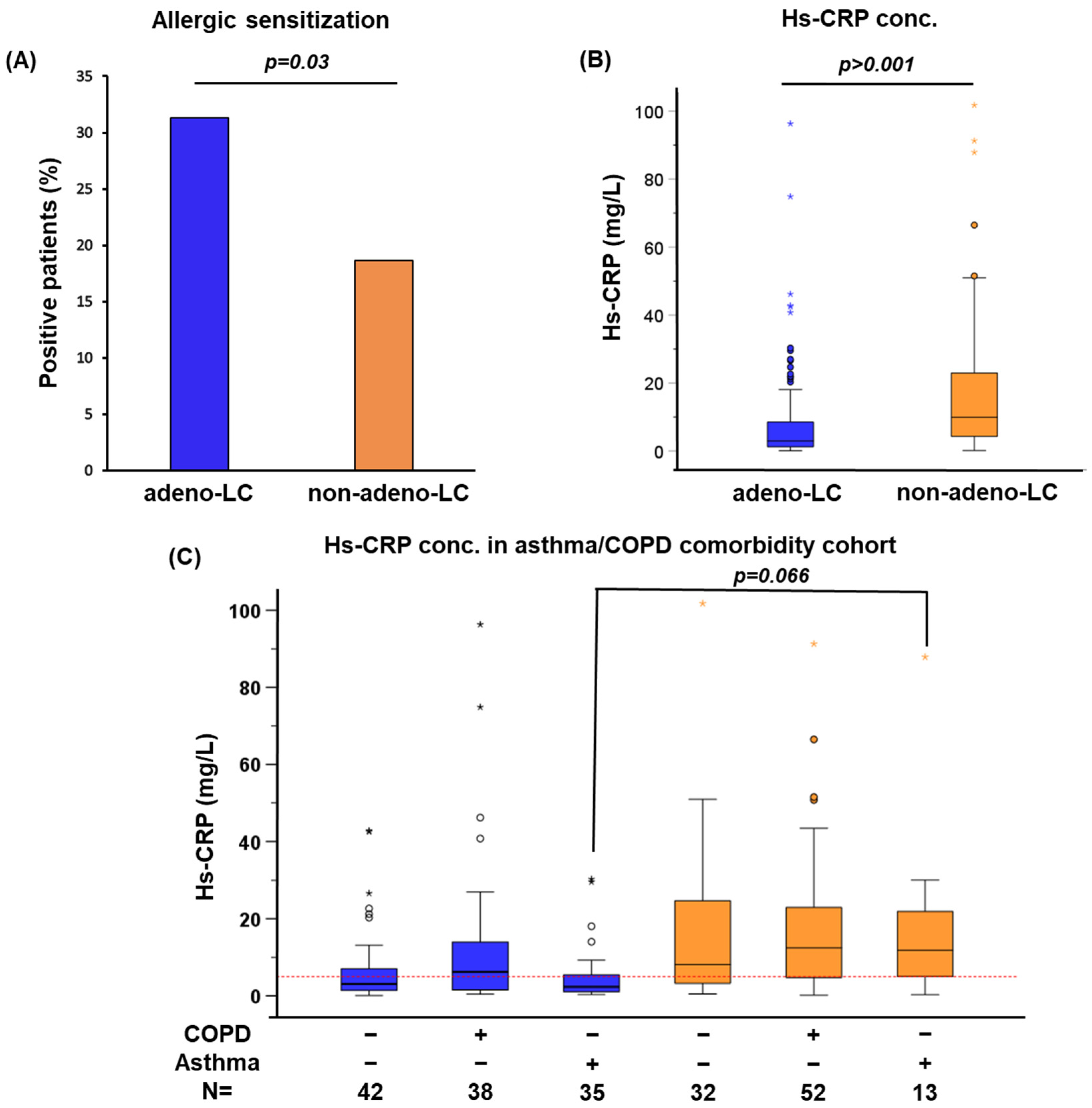
2. Results
3. Discussion
4. Methods
4.1. Study Design and Study Population
- Lung cancer cohort: Retrospective, including 3,143 patients with NSCLC diagnosed between 2010 and 2020. Of these, 2,113 had adeno-LC and 1030 had non-adeno-LC, which was primarily small squamous cell carcinoma histology. Inclusion criteria: (1) histological classification (adeno-LC and non-adeno-LC) according to the WHO Classification of Lung Tumors [53]; and (2) available data on pre-therapeutic differential white blood cell count. This cohort was used to study the association of blood eosinophilia and neutrophilia and survival of patients diagnosed with NSCLC (adeno-LC or non-adeno-LC). The demographic data are detailed in Table 1.
- Asthma/COPD comorbidity sub-cohort: A subgroup of 212 NSCLC patients was selected from the Lung Biobank Heidelberg cancer cohort, of which 115 had adeno-LC and 97 had non-adeno-LC. Inclusion criteria: (1) asthma (n = 48) or COPD (n = 90) comorbidities; and (2) available pre-therapeutic serum samples. A control group (n = 74) comprised patients with NSCLC without a diagnosis of asthma or COPD. The three groups (no asthma or COPD; COPD; asthma) were matched for gender, age, histology, stage, and date of diagnosis; the demographic data are detailed in Table 4. This sub-cohort was used to study sensitization and subclinical inflammation in adeno-LC and non-adeno-LC, by measuring the sIgE and hs-CRP.
- Tissue eosinophilia sub-cohort: Selected to assess the infiltration of immune cells (eosinophils, mast cells, and T cells) into the peritumoral and intratumoral regions. Inclusion criteria: (1) available formalin-fixed paraffin-embedded tissue sample from resected patients; and (2) pre-surgical white blood cell count. Six groups were analyzed (n ≥ 9 patients each) based on histology (adeno-LC vs. non-adeno-LC), blood eosinophil count (split at 500 cells/µL), neutrophil count (split at 7700 cells/µL), and blood CRP level (split at 5 mg/l). Details on the groups’ distribution are provided in Table 5.
4.2. Blood Measurements from the Asthma/COPD Comorbidity Cohort
4.3. Immunohistochemistry and Giemsa Staining from the Tissue Eosinophilia Sub-Cohort
4.4. Statistics
5. Conclusions
Supplementary Materials
Author Contributions
Funding
Institutional Review Board Statement
Informed Consent Statement
Data Availability Statement
Conflicts of Interest
Abbreviations
| NSCLC | non-small cell lung cancer |
| TH2 | T helper type 2 |
| hs-CRP | high-sensitivity C-reactive protein |
| adeno-LC | adenocarcinoma lung cancer |
| non-adeno-LC | non- adenocarcinoma lung cancer |
| ZfKD | German Centre for Cancer Registry Data |
| COPD | chronic obstructive pulmonary disease |
| sIgE | allergen-specific immunoglobulin E |
| HIS | Hospital Information System |
| HPFs | high-power fields |
| BMI | body mass index |
| HR | Hazard Ratio |
| TME | tumor microenvironment |
| PDAC | pancreatic ductal adenocarcinoma |
References
- Wong, M.C.S.; Lao, X.Q.; Ho, K.H.; Goggins, W.B.; Tse, S.L.A. Incidence and mortality of lung cancer: Global trends and association with socioeconomic status. Sci. Rep. 2017, 71, 14300. [Google Scholar] [CrossRef]
- Krebs-Lung Cancer. Available online: https://www.krebsdaten.de/Krebs/EN/Content/Cancer_sites/Lung_cancer/lung_cancer_node.html (accessed on 31 July 2023).
- Duma, N.; Santana-Davila, R.; Molina, J.R. Non–Small Cell Lung Cancer: Epidemiology, Screening, Diagnosis, and Treatment. Mayo Clin. Proc. 2019, 94, 1623–1640. [Google Scholar] [CrossRef] [PubMed]
- Travis, W.D.; Brambilla, E.; Burke, A.P.; Marx, A.; Nicholson, A.G. Introduction to the 2015 World Health Organization Classification of Tumors of the Lung, Pleura, Thymus, and Heart. J. Thorac. Oncol. 2015, 10, 1240–1242. [Google Scholar] [CrossRef]
- Mirhadi, S.; Tam, S.; Li, Q.; Moghal, N.; Pham, N.-A.; Tong, J.; Golbourn, B.J.; Krieger, J.R.; Taylor, P.; Li, M.; et al. Integrative analysis of non-small cell lung cancer patient-derived xenografts identifies distinct proteotypes associated with patient outcomes. Nat. Commun. 2022, 13, 1811. [Google Scholar] [CrossRef] [PubMed]
- Tammemagi, C.M.; Neslund-Dudas, C.; Simoff, M.; Kvale, P. Impact of comorbidity on lung cancer survival. Int. J. Cancer 2003, 103, 792–802. [Google Scholar] [CrossRef]
- Islam, K.M.M.; Jiang, X.; Anggondowati, T.; Lin, G.; Ganti, A.K. Comorbidity and survival in lung cancer patients. Cancer Epidemiol. Biomarkers Prev. 2015, 24, 1079–1085. [Google Scholar] [CrossRef]
- Bossert, J.; Ludwig, M.; Wronski, P.; Koetsenruijter, J.; Krug, K.; Villalobos, M.; Jacob, J.; Walker, J.; Thomas, M.; Wensing, M. Lung cancer patients’ comorbidities and attendance of German ambulatory physicians in a 5-year cross-sectional study. npj Prim. Care Respir. Med. 2021, 31, 2. [Google Scholar] [CrossRef] [PubMed]
- Sigel, K.; Wisnivesky, J.P. Comorbidity profiles of patients with lung cancer: A new approach to risk stratification? Ann. Am. Thorac. Soc. 2017, 14, 1512–1513. Available online: www.atsjournals.org (accessed on 30 July 2023). [CrossRef] [PubMed]
- Jensen-Jarolim, E.; Bax, H.J.; Bianchini, R.; Capron, M.; Corrigan, C.; Castells, M.; Dombrowicz, D.; Daniels-Wells, T.R.; Fazekas, J.; Fiebiger, E.; et al. AllergoOncology—The impact of allergy in oncology: EAACI position paper. Allergy 2017, 72, 866–887. [Google Scholar] [CrossRef]
- Hoste, E.; Cipolat, S.; Watt, F.M. Understanding allergy and cancer risk: What are the barriers? Nat. Rev. Cancer 2015, 15, 131–132. Available online: https://www.nature.com/articles/nrc3909 (accessed on 30 July 2023). [CrossRef]
- Jensen-Jarolim, E.; Pawelec, G. The nascent field of AllergoOncology. Cancer Immunol. Immunother. 2012, 61, 1355–1357. [Google Scholar] [CrossRef]
- Jensen-Jarolim, E.; Bax, H.J.; Bianchini, R.; Crescioli, S.; Daniels-Wells, T.R.; Dombrowicz, D.; Fiebiger, E.; Gould, H.J.; Irshad, S.; Janda, J.; et al. AllergoOncology: Opposite outcomes of immune tolerance in allergy and cancer. Allergy 2018, 73, 328–340. [Google Scholar] [CrossRef]
- Helby, J.; Bojesen, S.E.; Nielsen, S.F.; Nordestgaard, B.G. IgE and risk of cancer in 37747 individuals from the general population. Ann. Oncol. 2015, 26, 1784–1790. [Google Scholar] [CrossRef] [PubMed]
- Allin, K.H.; Bojesen, S.E.; Nordestgaard, B.G. Baseline C-reactive protein is associated with incident cancer and survival in patients with cancer. J. Clin. Oncol. 2009, 27, 2217–2224. [Google Scholar] [CrossRef] [PubMed]
- Shiels, M.S.; Pfeiffer, R.M.; Hildesheim, A.; Engels, E.A.; Kemp, T.J.; Park, J.H.; Katki, H.A.; Koshiol, J.; Shelton, G.; Caporaso, N.E.; et al. Circulating Inflammation Markers and Prospective Risk for Lung Cancer. JNCI J. Natl. Cancer Inst. 2013, 105, 1871–1880. [Google Scholar] [CrossRef]
- McDonald, L.; Carroll, R.; Harish, A.; Tanna, N.; Mehmud, F.; Alikhan, R.; Ramagopalan, S.V. Suspected cancer symptoms and blood test results in primary care before a diagnosis of lung cancer: A case-control study. Futur. Oncol. 2019, 15, 3755–3762. [Google Scholar] [CrossRef] [PubMed]
- Oppenheimer, J.; Hoyte, F.C.L.; Phipatanakul, W.; Silver, J.; Howarth, P.; Lugogo, N.L. Allergic and eosinophilic asthma in the era of biomarkers and biologics: Similarities, differences and misconceptions. Ann. Allergy Asthma Immunol. 2022, 129, 169–180. [Google Scholar] [CrossRef] [PubMed]
- Takemura, M.; Matsumoto, H.; Niimi, A.; Ueda, T.; Matsuoka, H.; Yamaguchi, M.; Jinnai, M.; Muro, S.; Hirai, T.; Ito, Y.; et al. High sensitivity C-reactive protein in asthma. Eur. Respir. J. 2006, 27, 908–912. [Google Scholar] [CrossRef]
- Yen, M.L.; Yang, C.Y.; Yen, B.L.; Ho, Y.L.; Cheng, W.C.; Bai, C.H. Increased high sensitivity C-reactive protein and neutrophil count are related to increased standard cardiovascular risk factors in healthy Chinese men. Int. J. Cardiol. 2006, 110, 191–198. [Google Scholar] [CrossRef]
- Leduc, C.; Antoni, D.; Charloux, A.; Falcoz, P.E.; Quoix, E. Comorbidities in the management of patients with lung cancer. Eur. Respir. J. 2017, 49, 1601721. [Google Scholar] [CrossRef]
- Acevedo, N.; Escamilla-Gil, J.M.; Espinoza, H.; Regino, R.; Ramírez, J.; Florez de Arco, L.; Dennis, R.; Torres-Duque, C.A.; Caraballo, L. Chronic Obstructive Pulmonary Disease Patients Have Increased Levels of Plasma Inflammatory Mediators Reported Upregulated in Severe COVID-19. Front. Immunol. 2021, 12, 678661. [Google Scholar] [CrossRef] [PubMed]
- Wouters, E.F.M.; Reynaert, N.L.; Dentener, M.A.; Vernooy, J.H.J. Systemic and local inflammation in asthma and chronic obstructive pulmonary disease is there a connection? Proc. Am. Thorac. Soc. 2009, 6, 638–647. [Google Scholar] [CrossRef] [PubMed]
- Gigon, L.; Fettrelet, T.; Yousefi, S.; Simon, D.; Simon, H.U. Eosinophils from A to Z. Allergy 2023, 78, 1810–1846. [Google Scholar] [CrossRef]
- Akdis, C.A.; Arkwright, P.D.; Brüggen, M.C.; Busse, W.; Gadina, M.; Guttman-Yassky, E.; Kabashima, K.; Mitamura, Y.; Vian, L.; Wu, J.; et al. Type 2 immunity in the skin and lungs. Allergy 2020, 75, 1582–1605. [Google Scholar] [CrossRef]
- Rittmeyer, D.; Lorentz, A. Relationship between Allergy and Cancer: An Overview. Int. Arch. Allergy Immunol. 2012, 159, 216–225. [Google Scholar] [CrossRef] [PubMed]
- Kantor, E.D.; Hsu, M.; Du, M.; Signorello, L.B. Allergies and asthma in relation to cancer risk. Cancer Epidemiol. Biomarkers Prev. 2019, 28, 1395. [Google Scholar] [CrossRef]
- Ji, J.; Shu, X.; Li, X.; Sundquist, K.; Sundquist, J.; Hemminki, K. Cancer risk in hospitalised asthma patients. Br. J. Cancer 2009, 100, 829–833. [Google Scholar] [CrossRef]
- Vandentorren, S.; Baldi, I.; Annesi Maesano, I.; Charpin, D.; Neukirch, F.; Filleul, L.; Cantagrel, A.; Tessier, J.F. Long-term mortality among adults with or without asthma in the PAARC study. Eur. Respir. J. 2003, 21, 462–467. [Google Scholar] [CrossRef]
- Karagiannis, S.N.; Bracher, M.G.; Beavil, R.L.; Beavil, A.J.; Hunt, J.; McCloskey, N.; Thompson, R.G.; East, N.; Burke, F.; Sutton, B.J.; et al. Role of IgE receptors in IgE antibody-dependent cytotoxicity and phagocytosis of ovarian tumor cells by human monocytic cells. Cancer Immunol. Immunother. 2008, 57, 247–263. [Google Scholar] [CrossRef]
- Zheng, J.M.; Lou, C.X.; Huang, Y.L.; Song, W.T.; Luo, Y.C.; Mo, G.Y.; Tan, L.Y.; Chen, S.W.; Li, B.J. Associations between immune cell phenotypes and lung cancer subtypes: Insights from mendelian randomization analysis. BMC Pulm. Med. 2024, 24, 242. [Google Scholar] [CrossRef] [PubMed] [PubMed Central]
- Josephs, D.H.; Spicer, J.F.; Corrigan, C.J.; Gould, H.J.; Karagiannis, S.N. Epidemiological associations of allergy, IgE and cancer. Clin. Exp. Allergy 2013, 43, 1110–1123. [Google Scholar] [CrossRef] [PubMed]
- Kasayama, S.; Tanemura, M.; Koga, M.; Fujita, K.; Yamamoto, H.; Miyatake, A. Asthma is an independent risk for elevation of plasma C-reactive protein levels. Clin. Chim. Acta. 2009, 399, 79–82. [Google Scholar] [CrossRef]
- Kilic, H.; Karalezli, A.; Hasanoglu, H.C.; Erel, O.; Ates, C. The relationship between hs-CRP and asthma control test in asthmatic patients. Allergol. Immunopathol. 2012, 40, 362–367. [Google Scholar] [CrossRef]
- Heikkilä, K.; Harris, R.; Lowe, G.; Rumley, A.; Yarnell, J.; Gallacher, J.; Ben-Shlomo, Y.; Ebrahim, S.; Lawlor, D.A. Associations of circulating C-reactive protein and interleukin-6 with cancer risk: Findings from two prospective cohorts and a meta-analysis. Cancer Causes Control 2009, 20, 15–26. [Google Scholar] [CrossRef] [PubMed]
- Ko, Y.J.; Kwon, Y.M.; Kim, K.H.; Choi, H.C.; Chun, S.H.; Yoon, H.J.; Goh, E.; Cho, B.; Park, M. High-Sensitivity C-Reactive Protein Levels and Cancer Mortality. Cancer Epidemiol. Biomarkers Prev. 2012, 21, 2076–2086. [Google Scholar] [CrossRef] [PubMed]
- McMillan, D.C.; Elahi, M.M.; Sattar, N.; Angerson, W.J.; Johnstone, J.; McArdle, C.S. Measurement of the Systemic Inflammatory Response Predicts Cancer-Specific and Non-Cancer Survival in Patients with Cancer. Nutr. Cancer 2011, 41, 64–69. [Google Scholar] [CrossRef]
- Muller, D.C.; Larose, T.L.; Hodge, A.; Guida, F.; Langhammer, A.; Grankvist, K.; Meyer, K.; Cai, Q.; Arslan, A.A.; Zeleniuch-Jacquotte, A.; et al. Circulating high sensitivity C reactive protein concentrations and risk of lung cancer: Nested case-control study within Lung Cancer Cohort Consortium. BMJ 2019, 364, 23. [Google Scholar] [CrossRef]
- Simon, S.C.S.; Utikal, J.; Umansky, V. Opposing roles of eosinophils in cancer. Cancer Immunol. Immunother. 2018, 68, 823–833. [Google Scholar] [CrossRef]
- Wong, D.T.W.; Bowen, S.M.; Elovic, A.; Gallagher, G.T.; Weller, P.F. Eosinophil ablation and tumor development. Oral. Oncol. 1999, 35, 496–501. [Google Scholar] [CrossRef]
- Xie, F.; Liu, L.B.; Shang, W.Q.; Chang, K.K.; Meng, Y.H.; Mei, J.; Yu, J.J.; Li, D.J.; Li, M.Q. The infiltration and functional regulation of eosinophils induced by TSLP promote the proliferation of cervical cancer cell. Cancer Lett. 2015, 364, 106–117. [Google Scholar] [CrossRef]
- von Wasielewski, R.; Seth, S.; Franklin, J.; Fischer, R.; Hübner, K.; Hansmann, M.L.; Diehl, V.; Georgii, A. Tissue eosinophilia correlates strongly with poor prognosis in nodular sclerosing Hodgkin’s disease, allowing for known prognostic factors. Blood 2000, 95, 1207–1213. [Google Scholar] [CrossRef]
- Li, F.; Du, X.; Lan, F.; Li, N.; Zhang, C.; Zhu, C.; Wang, X.; He, Y.; Shao, Z.; Chen, H.; et al. Eosinophilic inflammation promotes CCL6-dependent metastatic tumor growth. Sci. Adv. 2021, 7, 5943–5969. [Google Scholar] [CrossRef] [PubMed]
- Hanahan, D.; Weinberg, R.A. Hallmarks of cancer: The next generation. Cell 2011, 144, 646–674. [Google Scholar] [CrossRef]
- Shaul, M.E.; Fridlender, Z.G. Tumour-associated neutrophils in patients with cancer. Nat. Rev. Clin. Oncol. 2019, 16, 601–620. [Google Scholar] [CrossRef] [PubMed]
- Hedrick, C.C.; Malanchi, I. Neutrophils in cancer: Heterogeneous and multifaceted. Nat. Rev. Immunol. 2021, 22, 173–187. [Google Scholar] [CrossRef]
- Grisaru-Tal, S.; Itan, M.; Klion, A.D.; Munitz, A. A new dawn for eosinophils in the tumour microenvironment. Nat. Rev. Cancer 2020, 20, 594–607. [Google Scholar] [CrossRef]
- Schernberg, A.; Mezquita, L.; Boros, A.; Botticella, A.; Caramella, C.; Besse, B.; Escande, A.; Planchard, D.; Le Péchoux, C.; Deutsch, E. Neutrophilia as prognostic biomarker in locally advanced stage III lung cancer. PLoS ONE 2018, 13, e0204490. [Google Scholar] [CrossRef] [PubMed]
- Rapoport, B.L.; Theron, A.J.; Vorobiof, D.A.; Langenhoven, L.; Hall, J.M.; Van Eeden, R.I.; Smit, T.; Chan, S.W.; Botha, M.C.; Raats, J.I.; et al. Prognostic significance of the neutrophil/lymphocyte ratio in patients undergoing treatment with nivolumab for recurrent non-small-cell lung cancer. Lung Cancer Manag. 2020, 9, 37–1758. [Google Scholar] [CrossRef] [PubMed]
- Bremnes, R.M.; Al-Shibli, K.; Donnem, T.; Sirera, R.; Al-Saad, S.; Andersen, S.; Stenvold, H.; Camps, C.; Busund, L.T. The Role of Tumor-Infiltrating Immune Cells and Chronic Inflammation at the Tumor Site on Cancer Development, Progression, and Prognosis: Emphasis on Non-small Cell Lung Cancer. J. Thorac. Oncol. 2011, 6, 824–833. [Google Scholar] [CrossRef]
- Smolkova, B.; Cierna, Z.; Kalavska, K.; Miklikova, S.; Plava, J.; Minarik, G.; Sedlackova, T.; Cholujova, D.; Gronesova, P.; Cihova, M.; et al. Increased Stromal Infiltrating Lymphocytes Are Associated with the Risk of Disease Progression in Mesenchymal Circulating Tumor Cell-Positive Primary Breast Cancer Patients. Int. J. Mol. Sci. 2020, 21, 9460. [Google Scholar] [CrossRef] [PubMed] [PubMed Central]
- Pyo, J.S.; Son, B.K.; Lee, H.Y.; Oh, I.H.; Chung, K.H. Prognostic Implications of Intratumoral and Peritumoral Infiltrating Lymphocytes in Pancreatic Ductal Adenocarcinoma. Curr. Oncol. 2021, 28, 4367–4376. [Google Scholar] [CrossRef] [PubMed] [PubMed Central]
- Nicholson, A.G.; Tsao, M.S.; Beasley, M.B.; Borczuk, A.C.; Brambilla, E.; Cooper, W.A.; Dacic, S.; Jain, D.; Kerr, K.M.; Lantuejoul, S.; et al. The 2021 WHO Classification of Lung Tumors: Impact of Advances Since 2015. J. Thorac. Oncol. 2022, 17, 362–387. [Google Scholar] [CrossRef] [PubMed]
- Skevaki, C.; Tafo, P.; Eiringhaus, K.; Timmesfeld, N.; Weckmann, M.; Happle, C.; Nelson, P.P.; Maison, N.; Schaub, B.; Ricklefs, I.; et al. Allergen extract- and component-based diagnostics in children of the ALLIANCE asthma cohort. Clin. Exp. Allergy 2021, 51, 1331–1345. [Google Scholar] [CrossRef]
- Stark, M.; Nicolai, M.; Tatura, M.; Keber, C.U.; Kaufmann, A.; Chung, H.R.; Slater, E.P.; Heeschen, C.; Lawlor, R.T.; Scarpa, A.; et al. Dissecting the role of toll-like receptor 7 in pancreatic cancer. Cancer Med. 2023, 12, 8542–8556. [Google Scholar] [CrossRef] [PubMed]
- R: A Language and Environment for Statistical Computing|BibSonomy. Available online: https://www.bibsonomy.org/bibtex/7469ffee3b07f9167cf47e7555041ee7 (accessed on 31 July 2023).
- Gerds, T.A.; Kattan, M.W. Medical Risk Prediction Models: With Ties to Machine Learning, 1st ed.; CRC: New York, NY, USA, 2021; Available online: https://www.taylorfrancis.com/books/mono/10.1201/9781138384484/medical-risk-prediction-models-thomas-gerds-michael-kattan (accessed on 31 July 2023).


| Lung Cancer Cohort | Total | Non-Adeno-LC N (%) | Adeno-LC n (%) | p-Value | |
|---|---|---|---|---|---|
| Total | 3143 (100%) | 1030 (100%) | 2113 (100%) | N/A | |
| Sex | m | 1910 (60.8%) | 759 (73.7%) | 1151 (54.5%) | p < 0.001 * |
| f | 1233 (39.2%) | 271 (26.3%) | 962 (45.5%) | ||
| Age | Median (min, max) | N/A | 66 (40, 91) | 65 (22, 90) | p = 0.001 ** |
| BMI | Median | 26 (100%) | 25.4 | p = 0.004 ** | |
| (min, max) | (13.8, 54.1) | (14.7, 57.8) | |||
| Best Stage (if available p-Stage, otherwise c-Stage) | I | 522 (16.6%) | 159 (15.4%) | 363 (17.2%) | p < 0.001 * |
| II | 341 (10.8%) | 174 (16.9%) | 167 (7.9%) | ||
| III | 885 (28.2%) | 435 (42.2%) | 450 (21.3%) | ||
| IV | 1395 (44.4%) | 262 (25.4%) | 1133 (53.6%) | ||
| Smoking status | No | 336 (10.7%) | 40 (3.9%) | 296 (14%) | p < 0.001 * |
| Yes | 1105 (35.2%) | 390 (37.9%) | 715 (33.8%) | ||
| Ex/former | 1534 (48.8%) | 561 (54.5%) | 973 (46%) | ||
| N/A | 168 (5.3%) | 39 (3.8%) | 129 (6.1%) | ||
| Eosinophilia/Neutrophilia | Neutrophilia (Neutrophils > 7700/µL and eosinophils ≤ 500/µL) | 770 (24.5%) | 289 (28.1%) | 481 (22.8%) | p = 0.002 * |
| Eosinophilia (Eosinophils > 500/µL and Neutrophils ≤ 7700/µL) | 108 (3.4%) | 41 (3.9%) | 67 (3.2%) | ||
| Eosinophilia and Neutrophilia (Eosinophils > 500/µL and Neutrophils > 7700/µL) | 75 (2.4%) | 17 (1.7%) | 58 (2.7%) | ||
| No Eosinophilia or Neutrophilia (Eosinophils ≤ 500/µL and Neutrophils ≤ 7700/µL) | 2190 (69.7%) | 683 (66.3%) | 1507 (71.3%) | ||
| Adeno-LC | Non-Adeno-LC | ||||||
|---|---|---|---|---|---|---|---|
| Hazard Ratio | 95%CI | p Multivariate | Hazard Ratio | 95%CI | p Multivariate | ||
| Age | 1.01 | 1.01–1.02 | 1.43 × 10⁻⁵ | 1.03 | 1.02–1.04 | 1.29 × 10⁻⁸ | |
| Sex | 0.74 | 0.66–0.83 | 4.23 × 10⁻⁷ | 0.94 | 0.78–1.12 | 0.50 | |
| Stage | I | 1 | 1 | ||||
| II | 1.27 | 0.86–1.89 | 0.24 | 1.02 | 0.71–1.46 | 0.92 | |
| III | 3.48 | 2.64–4.58 | 7.68 × 10⁻¹⁹ | 2.69 | 2.01–3.58 | 1.90 × 10⁻¹¹ | |
| IV | 7.70 | 5.95–9.95 | 1.07 × 10−54 | 6.27 | 4.64–8.47 | 5.92 × 10⁻³³ | |
| BMI | 1.00 | 0.99–1.01 | 0.87 | 0.98 | 0.96–0.99 | 0.0099 | |
| Smoking status | N0 | 1 | 1 | ||||
| Ex/former | 1.35 | 1.16–1.58 | 0.0001 | 0.87 | 0.66–1.14 | 0.31 | |
| Yes | 1.50 | 1.28–1.77 | 1.05 × 10⁻⁶ | 1.03 | 0.78–1.36 | 0.86 | |
| EOS/Neutros | No Eosinophilia or Neutrophilia | 1 | 1 | ||||
| Neutrophilia | 1.39 | 1.22–1.57 | 5.04 × 10⁻⁷ | 1.55 | 1.30–1.84 | 6.64 × 10⁻⁷ | |
| Eosinophilia | 1.35 | 1.01–1.81 | 0.044 | 0.95 | 0.64–1.41 | 0.80 | |
| Eosinophilia and Neutrophilia | 2.03 | 1.51–2.72 | 2.45 × 10⁻⁶ | 1.34 | 0.71–2.52 | 0.37 | |
| All NSCLC | ||||
|---|---|---|---|---|
| n | MS (mos) | 5-yr-S (%) | p (Multivariate) | |
| Normal Eosinophils and Neutrophils | 2190 | 28.0 | 29.0 | |
| Neutrophilia | 770 | 13.4 | 19.0 | 3.0506 × 10⁻²³ |
| Eosinophilia | 108 | 16.8 | 16.5 | 0.003 |
| Eosinophilia and Neutrophilia | 75 | 8.2 | n.def. | 1.4714 × 10⁻⁹ |
| Adeno-LC | ||||
| n | MS (mos) | 5-yr-S (%) | p (Multivariate) | |
| Normal Eosinophils and Neutrophils | 1507 | 29.5 | 29.2 | |
| Neutrophilia | 481 | 13.4 | 19.5 | 5.04 × 10⁻⁷ |
| Eosinophilia | 67 | 16.1 | n.def. | 0.044 |
| Eosinophilia and Neutrophilia | 58 | 5.7 | n.def. | 2.45 × 10⁻⁶ |
| Non-Adeno-LC | ||||
| n | MS (mos) | 5-yr-S (%) | p (Multivariate) | |
| Normal Eosinophils and Neutrophils | 683 | 25.5 | 28.6 | |
| Neutrophilia | 289 | 13.5 | 17.8 | 6.64 × 10⁻⁷ |
| Eosinophilia | 41 | 18.0 | 24.7 | 0.80 |
| Eosinophilia and Neutrophilia | 17 | 14.6 | n.def. | 0.37 |
| Total | NSCLC No Asthma/No COPD N (%) | NSCLC COPD N (%) | NSCLC Asthma N (%) | ||
|---|---|---|---|---|---|
| Patients number | 212 (100%) | 74 (34.9%) | 90 (42.5%) | 48 (22.6%) | |
| Histology (% from Histology) | adeno-LC | 115 (54.2%) | 42 (56.8%) | 38 (42.2%) | 35 (72.9%) |
| non-adeno-LC | 97 (45.8%) | 32 (43.2%) | 52 (57.8%) | 13 (27.1%) | |
| Sex | m/f | 101/111 | 36/38 | 47/43 | 18/30 |
| Age | Median (min-max) | 63 (39–87) | 64 (44–87) | 63 (47–80) | 62.5 (39–84) |
| BMI | Median | 25.8 | 25.8 | 25.6 | 26.2 |
| Stage (% from comorbidity group) | 1/2 | 128 (60.4%) | 34 (45.9%) | 64 (71.1%) | 30 (62.5%) |
| 3/4 | 84 (39.6%) | 40 (54.1%) | 26 (28.9%) | 18 (37.5%) | |
| Smoking in patient history | yes (ever) | 183 | 61 | 87 | 35 |
| no | 21 | 11 | 1 | 9 | |
| n/a | 8 | 2 | 2 | 4 | |
| Blood | ||||
|---|---|---|---|---|
| NSCLC | N | Eosinophils (cells/µL) | Neutrophils (cells/µL) | CRP (mg/L) |
| adeno-LC | 10 | >500 | ≤7700 | ≤5 |
| non-adeno-LC | 10 | >500 | ≤7700 | ≤5 |
| adeno-LC | 9 | ≤500 | >7700 | >5 |
| non-adeno-LC | 10 | ≤500 | >7700 | >5 |
| Control adeno-LC | 10 | ≤500 | ≤7700 | ≤5 |
| Control non-adeno-LC | 10 | ≤500 | ≤7700 | ≤5 |
Disclaimer/Publisher’s Note: The statements, opinions and data contained in all publications are solely those of the individual author(s) and contributor(s) and not of MDPI and/or the editor(s). MDPI and/or the editor(s) disclaim responsibility for any injury to people or property resulting from any ideas, methods, instructions or products referred to in the content. |
© 2024 by the authors. Licensee MDPI, Basel, Switzerland. This article is an open access article distributed under the terms and conditions of the Creative Commons Attribution (CC BY) license (https://creativecommons.org/licenses/by/4.0/).
Share and Cite
Alashkar Alhamwe, B.; Yuskaeva, K.; Wulf, F.; Trinkmann, F.; Kriegsmann, M.; Thomas, M.; Keber, C.U.; Strandmann, E.P.v.; Herth, F.J.; Kolahian, S.; et al. Peripheral Inflammation Featuring Eosinophilia or Neutrophilia Is Associated with the Survival and Infiltration of Eosinophils within the Tumor among Various Histological Subgroups of Patients with NSCLC. Int. J. Mol. Sci. 2024, 25, 9552. https://doi.org/10.3390/ijms25179552
Alashkar Alhamwe B, Yuskaeva K, Wulf F, Trinkmann F, Kriegsmann M, Thomas M, Keber CU, Strandmann EPv, Herth FJ, Kolahian S, et al. Peripheral Inflammation Featuring Eosinophilia or Neutrophilia Is Associated with the Survival and Infiltration of Eosinophils within the Tumor among Various Histological Subgroups of Patients with NSCLC. International Journal of Molecular Sciences. 2024; 25(17):9552. https://doi.org/10.3390/ijms25179552
Chicago/Turabian StyleAlashkar Alhamwe, Bilal, Kadriya Yuskaeva, Friederike Wulf, Frederik Trinkmann, Mark Kriegsmann, Michael Thomas, Corinna Ulrike Keber, Elke Pogge von Strandmann, Felix J. Herth, Saeed Kolahian, and et al. 2024. "Peripheral Inflammation Featuring Eosinophilia or Neutrophilia Is Associated with the Survival and Infiltration of Eosinophils within the Tumor among Various Histological Subgroups of Patients with NSCLC" International Journal of Molecular Sciences 25, no. 17: 9552. https://doi.org/10.3390/ijms25179552