Communication

A Computational Approach in the Systematic Search of the Interaction Partners of Alternatively Spliced TREM2 Isoforms †

by
Junyi Liang
1,
Aditya Menon
2,
Taylor Tomco
1,
Nisha Bhattarai
3,
Iris Nira Smith
1,
Maria Khrestian
1,
Shane V. Formica
1,
Charis Eng
1,4,5,
Matthias Buck
3,* and
Lynn M. Bekris
1,4,5,*
1
Genomic Medicine Institute, Cleveland Clinic, Cleveland, OH 44195, USA
2
Department of Biomedical Engineering, Case Western Reserve University, Cleveland, OH 44106, USA
3
Department of Physiology and Biophysics, Case Western Reserve University, Cleveland, OH 44106, USA
4
Cleveland Clinic Lerner College of Medicine, Cleveland, OH 44195, USA
5
Department of Neurosciences, Lerner Research Institute, Cleveland Clinic, Cleveland, OH 44195, USA
*
Authors to whom correspondence should be addressed.
Dr. Charis Eng passed away during this paper’s review and the other authors also publish this paper in celebration of her career and contribution to research.
Int. J. Mol. Sci. 2024, 25(17), 9667; https://doi.org/10.3390/ijms25179667
Submission received: 5 August 2024 / Revised: 30 August 2024 / Accepted: 2 September 2024 / Published: 6 September 2024
(This article belongs to the Special Issue Molecular Informatics and Genomics of Alzheimer’s Disease)

Abstract

:
Alzheimer’s disease is the most common form of dementia, characterized by the pathological accumulation of amyloid-beta (Aβ) plaques and tau neurofibrillary tangles. Triggering receptor expressed on myeloid cells 2 (TREM2) is increasingly recognized as playing a central role in Aβ clearance and microglia activation in AD. The TREM2 gene transcriptional product is alternatively spliced to produce three different protein isoforms. The canonical TREM2 isoform binds to DAP12 to activate downstream pathways. However, little is known about the function or interaction partners of the alternative TREM2 isoforms. The present study utilized a computational approach in a systematic search for new interaction partners of the TREM2 isoforms by integrating several state-of-the-art structural bioinformatics tools from initial large-scale screening to one-on-one corroborative modeling and eventual all-atom visualization. CD9, a cell surface glycoprotein involved in cell–cell adhesion and migration, was identified as a new interaction partner for two TREM2 isoforms, and CALM, a calcium-binding protein involved in calcium signaling, was identified as an interaction partner for a third TREM2 isoform, highlighting the potential role of cell adhesion and calcium regulation in AD.

1. Introduction

Alzheimer’s disease (AD) is a progressive brain disorder that affects a person’s memory, thinking, and behavior [1,2]. It typically has a significant impact on the individual patient and their ability to function independently as well as on their family and caregivers and also poses a significant economic burden on society due to the high cost of care [3,4].
Triggering receptor expressed on myeloid cells 2 (TREM2) is a protein that is primarily expressed in myeloid cell types in the brain and has emerged as a significant player in AD and other neurodegenerative diseases [5,6,7] as well as a promising therapeutic target [8,9]. TREM2 RNA undergoes alternative splicing to generate three TREM2 isoforms in addition to the canonical TREM2, specifically producing TREM2△exon2, TREM2△exon4, and TREM2△exon5 [7,10]. Canonical TREM2 is membrane-bound, owing to exon 4, which is home to its transmembrane domain (Figure 1A). TREM2 has a cytoplasmic tail [11,12] that interacts with intracellular signaling molecules and, in turn, transduces signals into the cell [13,14]. TREM2△exon2 is believed to be a membrane protein not unlike canonical TREM2, but it differs from the canonical protein because it lacks the ligand-binding domain encoded by exon 2, while all other exons are intact (Figure 1B) [15,16]. TREM2△exon4 skips exon 4, causing a frameshift and an alternation to exon5 (Figure 1C) [17]. TREM2△exon5 has an untranslated exon 5 region and harbors a changed sequence in exon 4 (Figure 1D) [7,18,19]. TREM2△exon4 and TREM2△exon5 are likely soluble due to the absence of a functional transmembrane domain and are reportedly exported to the extracellular environment (Figure 1C,D) [7].
Canonical TREM2 engages in protein–protein interactions that play an integral role in cell signaling and the function of microglia, the immune cells of the central nervous system, as well as monocytes, dendritic cells, and macrophages in the peripheral system [19]. Canonical TREM2 may interact with lipids and lipoproteins [20], including high-density lipoprotein and low-density lipoprotein [21], as well as apolipoproteins in the extracellular environment, where apolipoprotein (ApoE) plays a critical role in lipid handling and clearance of cellular debris [11,22,23,24]. The direct interaction of the canonical TREM2 ligand-binding region and Aβ42 has also been described [25]. The interaction between canonical TREM2 and DAP12 is known to initiate important immune responses that are crucial for phagocytosis that, in turn, plays a critical role in the clearance of Aβ [26,27,28]. The development of small molecules that stimulate the interaction between canonical TREM2 and DAP12 and downstream signaling is being pursued as a new therapeutic opportunity [29].
In addition, soluble TREM2 generated by alternative splicing is detectable in various biological samples, including plasma and cerebrospinal fluid [30,31,32,33]. Studies have demonstrated its presence in both Alzheimer’s patients and non-patients. In Alzheimer’s patients, both TREM2△exon4 and TREM2△exon5 are less abundant than the canonical TREM2 in the brain [17,34]. In addition to these presumed soluble TREM2 protein isoforms produced by alternative splicing, another soluble TREM2 protein is produced by α-secretase cleavage of the canonical isoform [34,35]. Remarkably, soluble TREM2 levels exhibit distinct patterns in the different stages of AD progression. Cerebrospinal fluid-soluble TREM2 is notably elevated in the early stages of AD, but its levels tend to decrease as AD progresses [30,35,36,37], and is presumed to hold important regulatory functions in the extracellular environment through its interaction with biomolecules in the cerebrospinal fluid [38]. For example, soluble TREM2 is proposed to be neurologically beneficial in the setting of AD due to its ability to bind Aβ and lessen Aβ oligomerization-dependent neuronal damage [39,40]. TREM2 is also reported to be cleavable by γ-secretase, which processes amyloid-beta precursor protein (APP), the precursor to Aβ [41]. However, little is known about alternatively spliced TREM2 isoforms’ function or interaction with other biomolecules.
We hypothesized that, in addition to canonical TREM2, alternatively spliced TREM2 isoforms have interaction partners other than previously reported DAP12, Aβ42, or ApoE. This study aimed to implement a systematic computational search for new interaction partners of these TREM2 isoforms by employing several state-of-the-art biocomputational tools in a series of three sequential steps, (1) initial large-scale screening, (2) one-on-one corroborative modeling, and (3) fine structural visualization, as shown in the workflow diagram of Figure 2.
Specifically, this computational approach was designed in order to, firstly, perform large-scale screening upfront in the Homo sapiens proteome to find prospective interaction partners for TREM2 variants utilizing ProteinPrompt (https://proteinformatics.uni-leipzig.de/protein_prompt/, accessed on 2 September 2024), an emerging software particularly adapted at performing protein–protein interaction (PPI) screening at scale [42]. Then, secondly, one-on-one modeling was performed on the top five ProteinPrompt predictions for each TREM2 variant as a validation of the authenticity of the newly identified interaction. For this step we used Pipe for the Extraction of Predicted Protein–protein Interactions, otherwise known as PEPPI (https://zhanggroup.org/PEPPI/, accessed on 2 September 2024), a pipeline designed for predicting PPI [43]. Lastly, AlphaFold2 Multimer is an AI engine pre-trained to build dimeric complexes [41,44,45] and was used to visualize the heterodimer, including the dimer interface, formed by alternatively spliced TREM2 isoforms and their newfound interaction partners. In the end, two new interaction partners for certain alternatively spliced TREM2 isoforms were discovered that highlight the potential role of cell adhesion and calcium regulation in AD.

2. Results and Discussion

2.1. ProteinPrompt Predictions from the Human Proteome

Utilizing the ProteinPrompt random forest (RF) machine learning algorithm, the five top predictions (candidate interaction partners) were selected from the proteome of Homo sapiens as the most likely interaction partners for canonical TREM2 and alternatively spliced TREM2 isoforms. (1) DAP12, GBRL2, KP412, 4EBP1, and PRAF1 were the top five predictions for canonical TREM2; (2) CCL2, RL40, CALM, KP412, and CD9 were the top five predictions for TREM2△exon2; (3) KP412, CD9, DYL1, PRAF1, and 4EBP1 were the top five predictions for TREM2△exon4; and (4) CALM, KP412, 4EBP1, PRAF1, and DYL1 were TREM2△exon5’s top five predictions, as listed in Figure 3A. There is some overlap worthy of mentioning: for example, CD9 is one of ProteinPrompt’s top five predictions for both TREM2△exon2 and TREM2△exon4, and CALM is among ProteinPrompt’s top five predictions for both TREM2△exon2 and TREM2△exon5 (Figure 3B). Here, the identified top five predictions for each alternatively spliced TREM2 isoform were the starting point for the next step of one-on-one modeling. Note that not all the known interaction partners of the TREM2 variants are in the top five results of ProteinPrompt (e.g., Aβ is not). This might be caused by the built-in filters, which were meant to reduce false positives, inadvertently diminishing the ranking of these true interactions.

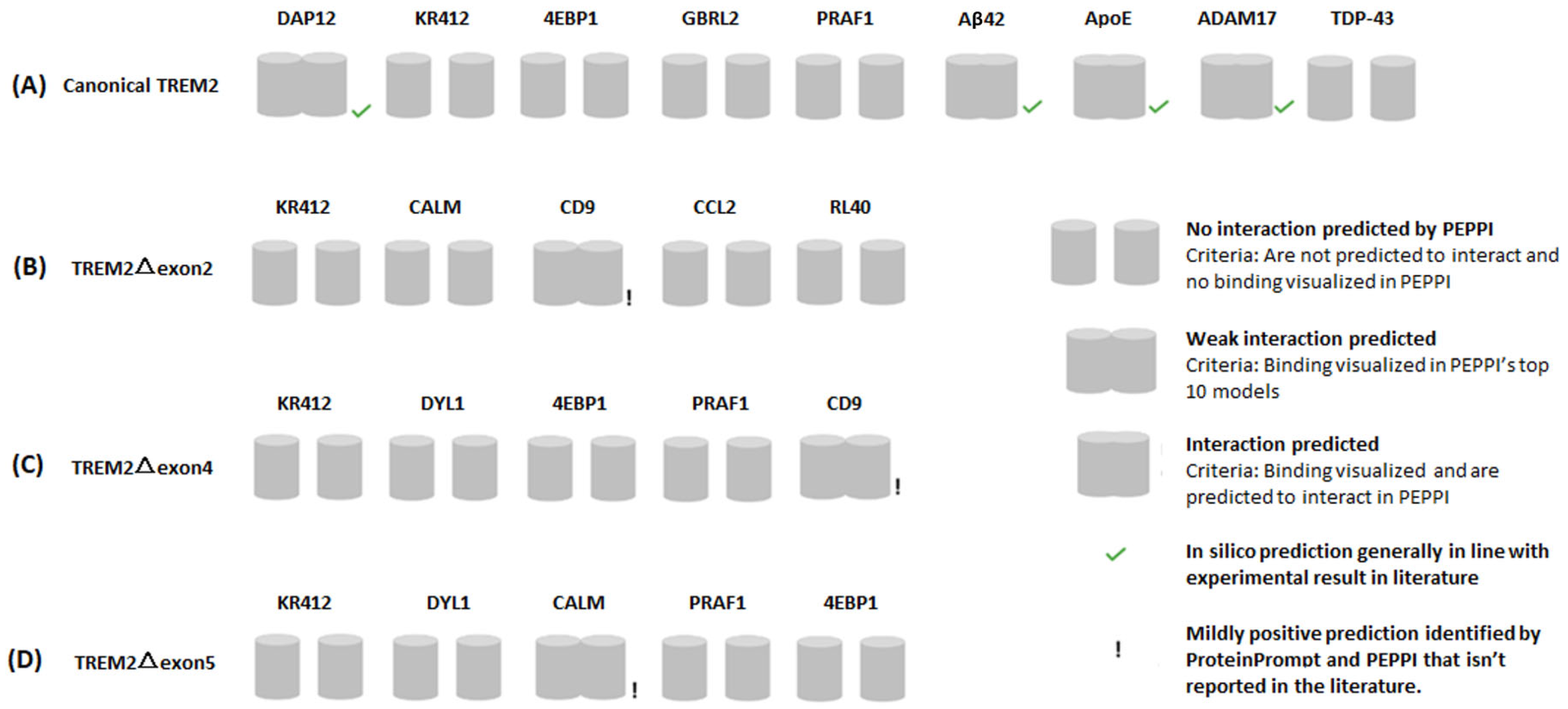
2.2. PEPPI Validation of Top Five Protein Prompt Predictions

The PEPPI analyses reproduced some known interactions of canonical TREM2, namely, its interaction with Aβ42 and ApoE. DAP12, one of ProteinPrompt’s top five likely interaction partners for canonical TREM2, is known to interact with canonical TREM2. Indeed, DAP12 and canonical TREM2 binding was visualized by PEPPI (Figure 4A).
PEPPI verified the validity of CD9 (cluster of differentiation 9; Uniprot ID: P21926) as an interaction partner, as binding was visible in the modeling of both TREM2△exon2:CD9 and TREM2△exon4:CD9 (Figure 4B,C).
A look into the biology of CD9 is intriguing because it is involved in cell adhesion and membrane organization. It is expressed in microglia, in platelets and in immune cells such as T cells [46,47]. The implication for AD is that TREM2-expressing microglia are involved in Aβ clearance through phagocytosis. CD9, through its involvement in membrane organization and cell adhesion, could potentially influence the interaction between microglia and Aβ plaques via its interaction with TREM2. CD9 has also been implicated in cell migration [48,49]. Moreover, TREM2 has been proposed to mediate microglial migration and interactions with other cell types [50]. Taken together, this suggests that an interaction between CD9 and TREM2 may influence microglial migration and positioning and their interactions with other cells in the brain. It is also worth mentioning that the interaction of CD9 and canonical TREM2, as evidenced by their colocalization, has already been observed using immunofluorescence staining in lipid-associated macrophages from mice on a high-fat diet [51] (CD9 is ranked as sixth among the interaction partners for canonical TREM2 in ProteinPrompt).
CALM (calmodulin 2; Uniprot ID: P62158) binding with TREM2△exon5 was replicated by PEPPI (Figure 4D). Interestingly, in AD, calcium dysregulation is observed, leading to altered calcium homeostasis in neurons and glial cells [52]. It is plausible that an interaction with TREM2 could potentially modulate CALM’s function in microglia. Also, both CALM and TREM2 have been implicated in synaptic function and plasticity. CALM interacts with proteins involved in neurotransmitter release and synaptic plasticity [53,54]. Disruptions in synaptic function and plasticity are early pathological features of AD [55,56,57]. TREM2 has been associated with synaptic pruning and modulating synaptic connectivity [58,59,60]. Furthermore, it has been suggested that TREM2 contains a CALM-binding motif [61]. Taken together, this suggests that an interaction between CALM and TREM2 may influence synaptic function in the brain.

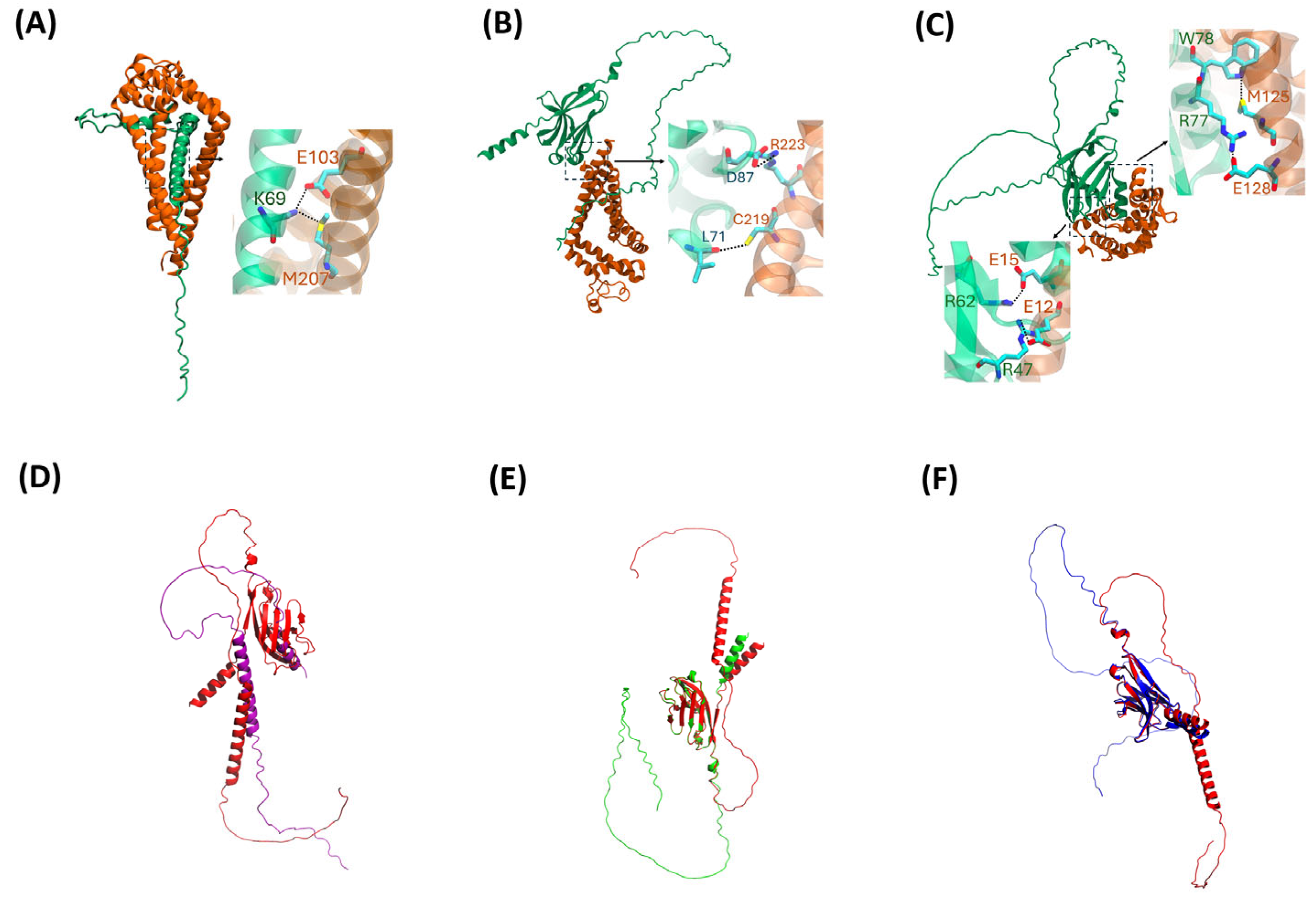
2.3. AlphaFold2 Visualization

ProteinPrompt identified the likely interaction partners, and PEPPI validated some of these putative interaction partners. However, neither method offers any molecular details regarding the manner of the interaction. AlphaFold2 Multimer predicted structures for the three computationally mined novel interactions identified by ProteinPrompt and validated by PEPPI (Figure 5). The heterodimeric complexes formed by TREM2△exon2 and CD9 (Figure 5A), TREM2△exon4 and CD9 (Figure 5B), and TREM2△exon5 and CALM (Figure 5C) indicate distinct structural interactions with the alternatively spliced TREM2 isoforms. Notably, each TREM2 isoform was predicted to interact differently with its newfound interaction partner.
In AlphaFold2 Multimer’s top model, CD9 binds to the stalk and TM regions of TREM2△exon2, the latter of which is encoded by exon 4. Therefore, in AlphaFold2 Multimer’s TREM2Δexon2:CD9 model, the TM region is conducive to its interaction with CD9. In AlphaFold2 Multimer’s heterodimeric complex TREM2△exon4:CD9, CD9 binds to TREM2△exon4 in its immunoglobulin domain (Ig domain), a domain known to mediate PPI important for immune functions [22,25]. TREM2’s Ig domain is encoded by exon 2 and also binds with well-known ligands such as Aβ42 and ApoE. It is plausible that TREM2△exon4’s binding with CD9 and its binding to known ligands may be mutually exclusive due to the overlap of the binding regions.
It should be pointed out that CD9 binds to TREM2△exon2’s stalk and TM region but not TREM2△exon4’s stalk region in the AlphaFold2 complexes, despite both isoforms sharing exon 3. This could be related to the major difference in solubility between TREM2△exon2 and TREM2△exon4, as the former is membrane-bound and the latter is soluble. By contrast, CALM also binds to TREM2△exon5’s immunoglobulin domain, which may compete with the binding of natural ligands. The superposition between predicted canonical TREM2 and its alternatively spliced isoforms structures (Figure 5D–F) also shows a correlated and yet distinct conformational posture, underscoring the structural differences leading to CD9 and CALM to bind to specific regions on each isoform in the AlphaFold2 models.
The TREM2 isoforms:CD9 complexes, as shown in Figure 5A,B, could potentially impact processes such as immune surveillance in AD. An interaction between CALM and TREM2 isoform and the resulting complex (Figure 5C) may influence synaptic processes, potentially impacting synaptic dysfunction through calcium signaling in AD. The specific interacting residues in each of the AlphaFold2 Multimer’s three complexes are listed in Table 1. However, it should be noted that AlphaFold2 results come with the caveat that the pLDDTs of the interfaces of the three predicted complexes are all less than 70%, indicating that AlphaFold2 is only mildly confident about the accuracy of the interfaces modeled. Nevertheless, AlphaFold2 predictions for relatively small interfaces, such as those between transmembrane helices that form (hetero)dimers in type-I receptors, were at 40–60% and were found to be remarkably accurate in one recent study [62].

3. Materials and Methods

3.1. Protein Prompt

ProteinPrompt is a web server that utilizes machine learning algorithms to calculate protein–protein interactions [42]. The platform offers three distinct computational methods—a random forest (RF), a graph neural network (GNN), and a consensus method (CM)—for the determination of top predictions. This software was used to scan Homo sapiens proteome libaries in order to identify prospective binders for canonical TREM2 (ENST00000373113; Uniprot ID: Q9NZC2), where all three methods were used one-by-one for TREM2△exon2, TREM2△exon4 (ENST00000338469), and TREM2△exon5 (ENST00000373122). The graph neural network and consensus methods returned an overabundance of likely binders, owing to score saturation. In preparation for the next step, candidate binders for each TREM2 family member were obtained utilizing random forest top 5 predictions. This online platform can be accessed here: https://proteinformatics.uni-leipzig.de/protein_prompt/, accessed on 2 September 2024.

3.2. Pipe for the Extraction of Predicted Protein–Protein Interactions (PEPPI)

PEPPI is a software program operated by Michigan University for predicting the likelihood that a given pair of polypeptide chains interact directly. This tool integrates structural similarity, sequence similarity, functional association data, and machine learning-based classification [43]. The potential interactions between canonical TREM2 and its top 5 candidate binders obtained in ProteinPrompt RF were simulated by PEPPI, one pair at a time. The same calculations were performed with TREM2△exon2, TREM2△exon4, and TREM2△exon5. PEPPI can be accessed online via this link: https://zhanggroup.org/PEPPI/, accessed on 2 September 2024. The inputs for PEPPI are the primary structures of the two polypeptide chains; in our work, the inputs are the sequence of one of the TREM2 isoforms and the sequence of one of its top 5 candidate binders as predicted by ProteinPrompt. The coordinate files for the 10 models deemed by PEPPI to be most probable were the output.

3.3. AlphaFold2 Multimer

AlphaFold2 Multimer v3 is an extension of AlphaFold2, a computational structural biology program developed by Google’s artificial intelligence arm Deepmind and a newer iteration of AlphaFold [41]. AlphaFold2 Multimer was used to model the conformation of protein complexes. UCSF chimera, a molecular graphics system, was used as the environment to access ColabFold, a faster version of AlphaFold2 for complex building [63,64]. The input to AlphaFold2 Multimer is the primary structure of the two polypeptide chains. Thus, in our work, the inputs were the sequences of TREM2△exon2 and CD9, the sequences of TREM2△exon4 and CD9, and the sequences of TREM2△exon5 and CALM. The most likely complex model’s coordinate file for each pair is the output.

4. Conclusions

In summary, we carried out a proteome-wide search of alternatively spliced TREM2 isoform interaction partners in silico. The investigation involved both large-scale screening of the proteomes of Homo sapiens as well as conformational one-on-one modeling by state-of-the-art in silico tools. CD9, a cell surface glycoprotein belonging to the tetraspanin family, was identified as a novel interaction partner for TREM2△exon2 and TREM2△exon4. CD9 is known as a key player in cell adhesion and is found on the surface of various cell types that are an integral part of the immune system, such as white blood cells and platelets. The newly discovered interaction between CD9 and TREM2 isoforms raises the prospect that TREM2 isoforms could be playing a role in cell–cell interaction within the same cell type or between different types of cells relevant to brain immunomodulation and, by extension, to AD.
CALM, a calcium-binding protein, is a novel interaction partner for TREM2△exon5 identified in this work. Calcium is a key element involved in maintaining neuronal health and the interaction between CALM and TREM2 isoform suggests that TREM2 may also influence calcium dynamics.
All in all, the protein interaction partners determined in this computational study for alternatively spliced TREM2 isoforms suggest possible new functions of TREM2 isoforms with respect to Alzheimer’s disease, including the role of TREM2 isoform in cell adhesion and calcium signaling. Remarkably, this in silico approach is an experiment in accelerating the study of the protein-protein interactions of a key protein of urgent biological interest, compared with typically slower biochemical approaches. It will be important to conduct a comprehensive biophysical characterization of the interaction between TREM2 isoforms and their newfound binders by biocompatible imaging approaches, such as fluorescent protein technology [62,65,66,67,68,69], to understand the manner these predicted binders interact with TREM2 isoforms biologically.

Author Contributions

J.L. and L.M.B. designed the study. Led by J.L., the team of three (J.L., A.M. and T.T.) performed the modeling and analyzed the models. N.B. and M.B. performed additional modeling at the High Performance Computing Cluster at Case Western Reserve University. I.N.S. and C.E. looked at certain models. J.L. prepared the first draft and helped finalize the manuscript. M.K. and S.V.F. refined the reference. M.B. and L.M.B. rewrote the manuscript into its publishable version. M.B. and L.M.B. also supervised this study. A.M., T.T. and N.B. contributed equally to this work. All authors have read and agreed to the published version of the manuscript.

Funding

This work was mainly supported by the Aging Mind Foundation (L.M.B.). M.B. and group are supported by grant R01EY029169, R21AG084065, and N.B. has a slot on NCI T32 (T32CA094186). I.N.S. is funded, in part, by the Ambrose Monell Cancer Genomic Medicine Fellowship and the NIH National Institute of General Medical Sciences (NIGMS) Maximizing Opportunities for Scientific and Academic Independent Careers (MOSAIC) K99/R00 grant—1K99GM143552. C.E. was funded in part by the Ambrose Monell Foundation PTEN-Switch Grant and a grant allocation of computing time from the Ohio Supercomputing Center (PCCF0020) [both to C.E.]. C.E. was an American Cancer Society Clinical Research Professor and the Sondra J. and Stephen R. Hardis Endowed Chair of Cancer Genomic Medicine at the Cleveland Clinic.

Institutional Review Board Statement

Not applicable.

Informed Consent Statement

Not applicable.

Data Availability Statement

The data presented in this study are available upon reasonable request to the corresponding authors.

Conflicts of Interest

The authors declare no conflicts of interest.

References

  1. Long, J.M.; Holtzman, D.M. Alzheimer Disease: An Update on Pathobiology and Treatment Strategies. Cell 2019, 179, 312–339. [Google Scholar] [CrossRef] [PubMed]
  2. Knopman, D.S.; Amieva, H.; Petersen, R.C.; Chételat, G.; Holtzman, D.M.; Hyman, B.T.; Nixon, R.A.; Jones, D.T. Alzheimer disease. Nat. Rev. Dis. Primer 2021, 7, 33. [Google Scholar] [CrossRef] [PubMed]
  3. Li, X.; Feng, X.; Sun, X.; Hou, N.; Han, F.; Liu, Y. Global, regional, and national burden of Alzheimer’s disease and other dementias, 1990–2019. Front. Aging Neurosci. 2022, 14, 937486. [Google Scholar] [CrossRef] [PubMed]
  4. Nandi, A.; Counts, N.; Chen, S.; Seligman, B.; Tortorice, D.; Vigo, D.; Bloom, D.E. Global and regional projections of the economic burden of Alzheimer’s disease and related dementias from 2019 to 2050: A value of statistical life approach. eClinicalMedicine 2022, 51, 101580. [Google Scholar] [CrossRef]
  5. Jay, T.R.; von Saucken, V.E.; Landreth, G.E. TREM2 in Neurodegenerative Diseases. Mol. Neurodegener. 2017, 12, 56. [Google Scholar] [CrossRef]
  6. Gratuze, M.; Leyns, C.E.G.; Holtzman, D.M. New insights into the role of TREM2 in Alzheimer’s disease. Mol. Neurodegener. 2018, 13, 66. [Google Scholar] [CrossRef]
  7. Filipello, F.; Goldsbury, C.; You, S.F.; Locca, A.; Karch, C.M.; Piccio, L. Soluble TREM2: Innocent bystander or active player in neurological diseases? Neurobiol. Dis. 2022, 165, 105630. [Google Scholar] [CrossRef]
  8. Deczkowska, A.; Weiner, A.; Amit, I. The Physiology, Pathology, and Potential Therapeutic Applications of the TREM2 Signaling Pathway. Cell 2020, 181, 1207–1217. [Google Scholar] [CrossRef]
  9. George, J. TREM2 as an evolving therapeutic target in Alzheimer’s disease. Neural Regen. Res. 2023, 18, 2680–2681. [Google Scholar] [CrossRef]
  10. Moutinho, M.; Coronel, I.; Tsai, A.P.; Di Prisco, G.V.; Pennington, T.; Atwood, B.K.; Puntambekar, S.S.; Smith, D.C.; Martinez, P.; Han, S.; et al. TREM2 splice isoforms generate soluble TREM2 species that disrupt long-term potentiation. Genome Med. 2023, 15, 11. [Google Scholar] [CrossRef]
  11. Bailey, C.C.; DeVaux, L.B.; Farzan, M. The Triggering Receptor Expressed on Myeloid Cells 2 Binds Apolipoprotein E. J. Biol. Chem. 2015, 290, 26033–26042. [Google Scholar] [CrossRef] [PubMed]
  12. Zhu, Z.; Zhang, X.; Dong, W.; Wang, X.; He, S.; Zhang, H.; Wang, X.; Wei, R.; Chen, Y.; Liu, X.; et al. TREM2 suppresses the proinflammatory response to facilitate PRRSV infection via PI3K/NF-κB signaling. PLoS Pathog. 2020, 16, e1008543. [Google Scholar] [CrossRef]
  13. Lessard, C.B.; Malnik, S.L.; Zhou, Y.; Ladd, T.B.; Cruz, P.E.; Ran, Y.; Mahan, T.E.; Chakrabaty, P.; Holtzman, D.M.; Ulrich, J.D.; et al. High-affinity interactions and signal transduction between Aβ oligomers and TREM 2. EMBO Mol. Med. 2018, 10, e9027. [Google Scholar] [CrossRef]
  14. Yao, H.; Coppola, K.; Schweig, J.E.; Crawford, F.; Mullan, M.; Paris, D. Distinct Signaling Pathways Regulate TREM2 Phagocytic and NFκB Antagonistic Activities. Front. Cell. Neurosci. 2019, 13, 457. [Google Scholar] [CrossRef] [PubMed]
  15. Kiianitsa, K.; Kurtz, I.; Beeman, N.; Matsushita, M.; Chien, W.-M.; Raskind, W.H.; Korvatska, O. Novel TREM2 splicing isoform that lacks the V-set immunoglobulin domain is abundant in the human brain. J. Leukoc. Biol. 2021, 110, 829–837. [Google Scholar] [CrossRef]
  16. Shaw, B.C.; Snider, H.C.; Turner, A.K.; Zajac, D.J.; Simpson, J.F.; Estus, S. An Alternatively Spliced TREM2 Isoform Lacking the Ligand Binding Domain is Expressed in Human Brain. J. Alzheimers Dis. 2022, 87, 1647–1657. [Google Scholar] [CrossRef] [PubMed]
  17. Del-Aguila, J.L.; Benitez, B.A.; Li, Z.; Dube, U.; Mihindukulasuriya, K.A.; Budde, J.P.; Farias, F.H.G.; Fernández, M.V.; Ibanez, L.; Jiang, S.; et al. TREM2 brain transcript-specific studies in AD and TREM2 mutation carriers. Mol. Neurodegener. 2019, 14, 18. [Google Scholar] [CrossRef]
  18. Olufunmilayo, E.O.; Holsinger, R.M.D. Variant TREM2 Signaling in Alzheimer’s Disease. J. Mol. Biol. 2022, 434, 167470. [Google Scholar] [CrossRef]
  19. Colonna, M. The biology of TREM receptors. Nat. Rev. Immunol. 2023, 23, 580–594. [Google Scholar] [CrossRef]
  20. Ulland, T.K.; Colonna, M. TREM2—A key player in microglial biology and Alzheimer disease. Nat. Rev. Neurol. 2018, 14, 667–675. [Google Scholar] [CrossRef]
  21. Wang, Y.; Cella, M.; Mallinson, K.; Ulrich, J.D.; Young, K.L.; Robinette, M.L.; Gilfillan, S.; Krishnan, G.M.; Sudhakar, S.; Zinselmeyer, B.H.; et al. TREM2 Lipid Sensing Sustains the Microglial Response in an Alzheimer’s Disease Model. Cell 2015, 160, 1061–1071. [Google Scholar] [CrossRef]
  22. Song, W.; Hooli, B.; Mullin, K.; Jin, S.C.; Cella, M.; Ulland, T.K.; Wang, Y.; Tanzi, R.E.; Colonna, M. Alzheimer’s disease-associated TREM2 variants exhibit either decreased or increased ligand-dependent activation. Alzheimers Dement. 2017, 13, 381–387. [Google Scholar] [CrossRef] [PubMed]
  23. Atagi, Y.; Liu, C.-C.; Painter, M.M.; Chen, X.-F.; Verbeeck, C.; Zheng, H.; Li, X.; Rademakers, R.; Kang, S.S.; Xu, H.; et al. Apolipoprotein E Is a Ligand for Triggering Receptor Expressed on Myeloid Cells 2 (TREM2). J. Biol. Chem. 2015, 290, 26043–26050. [Google Scholar] [CrossRef] [PubMed]
  24. Mai, Z.; Wei, W.; Yu, H.; Chen, Y.; Wang, Y.; Ding, Y. Molecular recognition of the interaction between ApoE and the TREM2 protein. Transl. Neurosci. 2022, 13, 93–103. [Google Scholar] [CrossRef] [PubMed]
  25. Yeh, F.L.; Wang, Y.; Tom, I.; Gonzalez, L.C.; Sheng, M. TREM2 Binds to Apolipoproteins, Including APOE and CLU/APOJ, and Thereby Facilitates Uptake of Amyloid-Beta by Microglia. Neuron 2016, 91, 328–340. [Google Scholar] [CrossRef]
  26. Zhao, Y.; Wu, X.; Li, X.; Jiang, L.-L.; Gui, X.; Liu, Y.; Sun, Y.; Zhu, B.; Piña-Crespo, J.C.; Zhang, M.; et al. TREM2 Is a Receptor for β-Amyloid that Mediates Microglial Function. Neuron 2018, 97, 1023–1031.e7. [Google Scholar] [CrossRef]
  27. Konishi, H.; Kiyama, H. Microglial TREM2/DAP12 Signaling: A Double-Edged Sword in Neural Diseases. Front. Cell. Neurosci. 2018, 12, 206. [Google Scholar] [CrossRef]
  28. Cui, X.; Qiao, J.; Liu, S.; Wu, M.; Gu, W. Mechanism of TREM2/DAP12 complex affecting β-amyloid plaque deposition in Alzheimer’s disease modeled mice through mediating inflammatory response. Brain Res. Bull. 2021, 166, 21–28. [Google Scholar] [CrossRef]
  29. Haure-Mirande, J.-V.; Audrain, M.; Ehrlich, M.E.; Gandy, S. Microglial TYROBP/DAP12 in Alzheimer’s disease: Transduction of physiological and pathological signals across TREM2. Mol. Neurodegener. 2022, 17, 55. [Google Scholar] [CrossRef]
  30. Gallo, C.; Manzo, E.; Barra, G.; Fioretto, L.; Ziaco, M.; Nuzzo, G.; d’Ippolito, G.; Ferrera, F.; Contini, P.; Castiglia, D.; et al. Sulfavant A as the first synthetic TREM2 ligand discloses a homeostatic response of dendritic cells after receptor engagement. Cell. Mol. Life Sci. 2022, 79, 369. [Google Scholar] [CrossRef]
  31. Bekris, L.M.; Khrestian, M.; Dyne, E.; Shao, Y.; Pillai, J.A.; Rao, S.M.; Bemiller, S.M.; Lamb, B.; Fernandez, H.H.; Leverenz, J.B. Soluble TREM2 and biomarkers of central and peripheral inflammation in neurodegenerative disease. J. Neuroimmunol. 2018, 319, 19–27. [Google Scholar] [CrossRef] [PubMed]
  32. Heslegrave, A.; Heywood, W.; Paterson, R.; Magdalinou, N.; Svensson, J.; Johansson, P.; Öhrfelt, A.; Blennow, K.; Hardy, J.; Schott, J.; et al. Increased cerebrospinal fluid soluble TREM2 concentration in Alzheimer’s disease. Mol. Neurodegener. 2016, 11, 3. [Google Scholar] [CrossRef]
  33. Piccio, L.; Buonsanti, C.; Cella, M.; Tassi, I.; Schmidt, R.E.; Fenoglio, C.; Rinker, J.; Naismith, R.T.; Panina-Bordignon, P.; Passini, N.; et al. Identification of soluble TREM-2 in the cerebrospinal fluid and its association with multiple sclerosis and CNS inflammation. Brain 2008, 131, 3081–3091. [Google Scholar] [CrossRef] [PubMed]
  34. Schlepckow, K.; Kleinberger, G.; Fukumori, A.; Feederle, R.; Lichtenthaler, S.F.; Steiner, H.; Haass, C. An Alzheimer-associated TREM2 variant occurs at the ADAM cleavage site and affects shedding and phagocytic function. EMBO Mol. Med. 2017, 9, 1356–1365. [Google Scholar] [CrossRef]
  35. Feuerbach, D.; Schindler, P.; Barske, C.; Joller, S.; Beng-Louka, E.; Worringer, K.A.; Kommineni, S.; Kaykas, A.; Ho, D.J.; Ye, C.; et al. ADAM17 is the main sheddase for the generation of human triggering receptor expressed in myeloid cells (hTREM2) ectodomain and cleaves TREM2 after Histidine 157. Neurosci. Lett. 2017, 660, 109–114. [Google Scholar] [CrossRef] [PubMed]
  36. Diaz-Lucena, D.; Kruse, N.; Thüne, K.; Schmitz, M.; Villar-Piqué, A.; Da Cunha, J.E.G.; Hermann, P.; López-Pérez, Ó.; Andrés-Benito, P.; Ladogana, A.; et al. TREM2 expression in the brain and biological fluids in prion diseases. Acta Neuropathol. 2021, 141, 841–859. [Google Scholar] [CrossRef] [PubMed]
  37. Bonomi, C.G.; Assogna, M.; Di Donna, M.G.; Bernocchi, F.; De Lucia, V.; Nuccetelli, M.; Fiorelli, D.; Loizzo, S.; Mercuri, N.B.; Koch, G.; et al. Cerebrospinal Fluid sTREM-2, GFAP, and β-S100 in Symptomatic Sporadic Alzheimer’s Disease: Microglial, Astrocytic, and APOE Contributions Along the Alzheimer’s Disease Continuum. J. Alzheimers Dis. JAD 2023, 92, 1385–1397. [Google Scholar] [CrossRef]
  38. Hok-A-Hin, Y.S.; Del Campo, M.; Boiten, W.A.; Stoops, E.; Vanhooren, M.; Lemstra, A.W.; Van Der Flier, W.M.; Teunissen, C.E. Neuroinflammatory CSF biomarkers MIF, sTREM1, and sTREM2 show dynamic expression profiles in Alzheimer’s disease. J. Neuroinflammation 2023, 20, 107. [Google Scholar] [CrossRef]
  39. Liu, D.; Cao, B.; Zhao, Y.; Huang, H.; McIntyre, R.S.; Rosenblat, J.D.; Zhou, H. Soluble TREM2 changes during the clinical course of Alzheimer’s disease: A meta-analysis. Neurosci. Lett. 2018, 686, 10–16. [Google Scholar] [CrossRef]
  40. Morenas-Rodríguez, E.; Li, Y.; Nuscher, B.; Franzmeier, N.; Xiong, C.; Suárez-Calvet, M.; Fagan, A.M.; Schultz, S.; Gordon, B.A.; Benzinger, T.L.S.; et al. Soluble TREM2 in CSF and its association with other biomarkers and cognition in autosomal-dominant Alzheimer’s disease: A longitudinal observational study. Lancet Neurol. 2022, 21, 329–341. [Google Scholar] [CrossRef]
  41. Hur, J.-Y. γ-Secretase in Alzheimer’s disease. Exp. Mol. Med. 2022, 54, 433–446. [Google Scholar] [CrossRef] [PubMed]
  42. Zhong, L.; Xu, Y.; Zhuo, R.; Wang, T.; Wang, K.; Huang, R.; Wang, D.; Gao, Y.; Zhu, Y.; Sheng, X.; et al. Soluble TREM2 ameliorates pathological phenotypes by modulating microglial functions in an Alzheimer’s disease model. Nat. Commun. 2019, 10, 1365. [Google Scholar] [CrossRef]
  43. Brown, G.C.; St George-Hyslop, P. Does Soluble TREM2 Protect Against Alzheimer’s Disease? Front. Aging Neurosci. 2022, 13, 834697. [Google Scholar] [CrossRef] [PubMed]
  44. Canzler, S.; Fischer, M.; Ulbricht, D.; Ristic, N.; Hildebrand, P.W.; Staritzbichler, R. ProteinPrompt: A webserver for predicting protein–protein interactions. Bioinform. Adv. 2022, 2, vbac059. [Google Scholar] [CrossRef]
  45. Zhu, W.; Shenoy, A.; Kundrotas, P.; Elofsson, A. Evaluation of AlphaFold-Multimer prediction on multi-chain protein complexes. Bioinformatics 2023, 39, btad424. [Google Scholar] [CrossRef]
  46. Pettersen, E.F.; Goddard, T.D.; Huang, C.C.; Meng, E.C.; Couch, G.S.; Croll, T.I.; Morris, J.H.; Ferrin, T.E. UCSF ChimeraX: Structure visualization for researchers, educators, and developers. Protein Sci. 2021, 30, 70–82. [Google Scholar] [CrossRef]
  47. Mirdita, M.; Schütze, K.; Moriwaki, Y.; Heo, L.; Ovchinnikov, S.; Steinegger, M. ColabFold: Making protein folding accessible to all. Nat. Methods 2022, 19, 679–682. [Google Scholar] [CrossRef]
  48. Kawakami, Y.; Kawakami, K.; Steelant, W.F.A.; Ono, M.; Baek, R.C.; Handa, K.; Withers, D.A.; Hakomori, S. Tetraspanin CD9 Is a “Proteolipid”, and Its Interaction with α3 Integrin in Microdomain Is Promoted by GM3 Ganglioside, Leading to Inhibition of Laminin-5-dependent Cell Motility. J. Biol. Chem. 2002, 277, 34349–34358. [Google Scholar] [CrossRef] [PubMed]
  49. Reyes, R.; Cardeñes, B.; Machado-Pineda, Y.; Cabañas, C. Tetraspanin CD9: A Key Regulator of Cell Adhesion in the Immune System. Front. Immunol. 2018, 9, 863. [Google Scholar] [CrossRef]
  50. Powner, D.; Kopp, P.M.; Monkley, S.J.; Critchley, D.R.; Berditchevski, F. Tetraspanin CD9 in cell migration. Biochem. Soc. Trans. 2011, 39, 563–567. [Google Scholar] [CrossRef]
  51. Suwatthanarak, T.; Tanaka, M.; Miyamoto, Y.; Miyado, K.; Okochi, M. Inhibition of cancer-cell migration by tetraspanin CD9-binding peptide. Chem. Commun. 2021, 57, 4906–4909. [Google Scholar] [CrossRef] [PubMed]
  52. Mazaheri, F.; Snaidero, N.; Kleinberger, G.; Madore, C.; Daria, A.; Werner, G.; Krasemann, S.; Capell, A.; Trümbach, D.; Wurst, W.; et al. TREM 2 deficiency impairs chemotaxis and microglial responses to neuronal injury. EMBO Rep. 2017, 18, 1186–1198. [Google Scholar] [CrossRef] [PubMed]
  53. Jaitin, D.A.; Adlung, L.; Thaiss, C.A.; Weiner, A.; Li, B.; Descamps, H.; Lundgren, P.; Bleriot, C.; Liu, Z.; Deczkowska, A.; et al. Lipid-Associated Macrophages Control Metabolic Homeostasis in a Trem2-Dependent Manner. Cell 2019, 178, 686–698.e14. [Google Scholar] [CrossRef]
  54. Ge, M.; Zhang, J.; Chen, S.; Huang, Y.; Chen, W.; He, L.; Zhang, Y. Role of Calcium Homeostasis in Alzheimer’s Disease. Neuropsychiatr. Dis. Treat. 2022, 18, 487–498. [Google Scholar] [CrossRef]
  55. DeLorenzo, R.J. Calmodulin in neurotransmitter release and synaptic function. Fed. Proc. 1982, 41, 2265–2272. [Google Scholar] [CrossRef]
  56. O’Day, D.H. Calmodulin Binding Proteins and Alzheimer’s Disease: Biomarkers, Regulatory Enzymes and Receptors That Are Regulated by Calmodulin. Int. J. Mol. Sci. 2020, 21, 7344. [Google Scholar] [CrossRef]
  57. Sheng, M.; Sabatini, B.L.; Sudhof, T.C. Synapses and Alzheimer’s Disease. Cold Spring Harb. Perspect. Biol. 2012, 4, a005777. [Google Scholar] [CrossRef]
  58. Marsh, J.; Alifragis, P. Synaptic dysfunction in Alzheimer’s disease: The effects of amyloid beta on synaptic vesicle dynamics as a novel target for therapeutic intervention. Neural Regen. Res. 2018, 13, 616. [Google Scholar]
  59. Meftah, S.; Gan, J. Alzheimer’s disease as a synaptopathy: Evidence for dysfunction of synapses during disease progression. Front. Synaptic Neurosci. 2023, 15, 1129036. [Google Scholar] [CrossRef] [PubMed]
  60. Qin, Q.; Wang, M.; Yin, Y.; Tang, Y. The Specific Mechanism of TREM2 Regulation of Synaptic Clearance in Alzheimer’s Disease. Front. Immunol. 2022, 13, 845897. [Google Scholar] [CrossRef]
  61. O’Day, D.H.; Huber, R.J. Calmodulin binding proteins and neuroinflammation in multiple neurodegenerative diseases. BMC Neurosci. 2022, 23, 10. [Google Scholar] [CrossRef] [PubMed]
  62. Sahoo, A.R.; Souza, P.C.T.; Meng, Z.; Buck, M. Transmembrane dimers of type 1 receptors sample alternate configurations: MD simulations using coarse grain Martini 3 versus AlphaFold2 Multimer. Structure 2023, 31, 735–745.e2. [Google Scholar] [CrossRef] [PubMed]
  63. Bell, E.W.; Schwartz, J.H.; Freddolino, P.L.; Zhang, Y. PEPPI: Whole-proteome Protein-protein Interaction Prediction through Structure and Sequence Similarity, Functional Association, and Machine Learning. J. Mol. Biol. 2022, 434, 167530. [Google Scholar] [CrossRef] [PubMed]
  64. Evans, R.; O’Neill, M.; Pritzel, A.; Antropova, N.; Senior, A.; Green, T.; Žídek, A.; Bates, R.; Blackwell, S.; Yim, J.; et al. Protein complex prediction with AlphaFold-Multimer. bioRxiv 2021. [Google Scholar]
  65. Liang, J.; Qin, M.; Xu, R.; Gao, X.; Shen, Y.; Xu, Q.; Cao, Y.; Wang, W. A genetically encoded copper(i) sensor based on engineered structural distortion of EGFP. Chem. Commun. 2012, 48, 3890. [Google Scholar] [CrossRef]
  66. Liang, J.; Yang, Y.; Yin, P.; Ding, Y.; Shen, Y.; Qin, M.; Wang, J.; Xu, Q.; Cao, Y.; Wang, W. A Yellow Fluorescent Protein with Reduced Chloride Sensitivity Engineered by Loop-Insertion. ChemBioChem 2013, 14, 1423–1426. [Google Scholar] [CrossRef]
  67. Liang, J.; Guo, L.; Ding, Y.; Xia, L.; Shen, Y.; Qin, M.; Xu, Q.; Cao, Y.; Wang, W. Genetically encoded red fluorescent copper(I) sensors for cellular copper(I) imaging. Biochem. Biophys. Res. Commun. 2014, 443, 894–898. [Google Scholar] [CrossRef]
  68. Liang, J.; Seghiri, M.; Singh, P.K.; Seo, H.G.; Lee, J.Y.; Jo, Y.; Song, Y.B.; Park, C.; Zalicki, P.; Jeong, J.-Y.; et al. The β2-adrenergic receptor associates with CXCR4 multimers in human cancer cells. Proc. Natl. Acad. Sci. USA 2024, 121, e2304897121. [Google Scholar] [CrossRef]
  69. Liang, J.; Smith, A.W. The Oligomeric State of Vasorin in the Plasma Membrane Measured Non-Invasively by Quantitative Fluorescence Fluctuation Spectroscopy. Int. J. Mol. Sci. 2024, 25, 4115. [Google Scholar] [CrossRef]
Figure 1. Schematic introduction to the TREM2 isoforms: canonical TREM2 and TREM2 isoforms produced by alternative splicing. (A) The primary structure of canonical TREM2, including its composition with respect to the exons and corresponding localization in the cellular setting, and the same depiction for (B) TREM2△exon2. (C) TREM2△exon4 and (D) TREM2△exon5. Red stands for signaling peptide, yellow stands for IgV-set domain, green stands for stalk region, blue stands for transmembrane domain/TM domain, purple stands for cytosolic tail, grey stands for an exon5 altered from the original exon 5, and navy blue stands for an exon 4 altered from the original exon 4.
Ijms 25 09667 g001
Figure 2. General workflow diagram. In step 1, proteome-wide screening by ProteinPrompt was used to identify the prospective interaction partners for TREM2 isoforms. In step 2, independent modeling by PEPPI was used to validate step 1’s predictions. In step 3, the interaction partners identified in step 1 and validated in step 2 were built into a heterodimeric complex with the corresponding TREM isoform by AlphaFold2 Multimer.
Ijms 25 09667 g002
Figure 3. ProteinPrompt’s prediction on the prospective interaction partners of canonical TREM2 and alternatively spliced TREM2 isoforms. (A) ProteinPrompt’s top 5 predictions for each of the TREM2 are shown and ranked according to the score in ProteinPrompt, which in turn was calculated by a majority vote in ProteinPrompt’s random forest method. Note that a couple of canonical TREM2’s known interaction partners, such as TDP-43 and ADAM17, were not among the top 5 predictions in ProteinPrompt. (B) ProteinPrompt’s prediction for each TREM2 variant’s 5 most likely interaction partners in the proteome of Homo sapiens was obtained with the random forest method. The notable overlaps are the following: KP412 is predicted by ProteinPrompt to interact with all four TREM2 variants, while CALM interacts with TREM2△exon2 and TREM2△exon5; CD9 interacts with TREM2△exon2 and TREM2△exon4; PRAF1 and 4EBP1 interacts with TREM2△exon2, TREM2△exon4, and with TREM2△exon5, (canonical TREM2: red; TREM2△exon2: purple; TREM2△exon4: green; TREM2△exon5: blue).
Ijms 25 09667 g003
Figure 4. PEPPI modeling results for (A) canonical TREM2, (B) TREM2△exon2, (C) TREM2△exon3, (D) TREM2△exon4. A cross-check on ProteinPrompt’s top 5 predictions. PEPPI succeeded in reproducing some known interactions of canonical TREM2, namely, its interaction with Aβ42 and ApoE. Canonical TREM2’s interaction with ADAM17 as well as DAP12 was reproduced by PEPPI. We note, however, that PEPPI did not reproduce the interaction between canonical TREM2 and TDP-43. CD9, one of ProteinPrompt’s top predictions for TREM2△exon2, was shown to bind with this isoform by PEPPI. CD9 was predicted to bind TREM2△exon4, and the interaction with CALM, another top prediction for TREM2△exon5, was also shown by PEPPI. However, similar to the canonical TREM2, PEPPI provided no confirmation of ProteinPrompt’s other top predictions for the splice variants.
Ijms 25 09667 g004
Figure 5. AlphaFold2 Multimer visualization of the dimeric complexes formed by TREM2 alternatively spliced isoforms and the newfound interaction partners with part of the interacting interface zoomed in: (A) TREM2△exon2 (dark green) and CD9 (orange), (B) TREM2△exon4 (dark green) and CD9 (orange), (C) TREM2△exon5 (dark green) and CALM (orange)—and structural superposition between canonical TREM2 (red) and the isoforms—(D) between canonical TREM2 and TREM2△exon2, (E) between canonical TREM2 and TREM2△exon4, (F) between canonical TREM2 and TREM2△exon5.
Ijms 25 09667 g005
Table 1. The interacting interfaces in AlphaFold2 Multimer’s three heterodimeric complexes.
Interacting Residues
TREM2△exon2:CD9TREM△exon4:CD9TREM2△exon5:CALM
S57:T58L71:C209; L71:M215R47:E12
K69:M207D87:R223R62:E15; R62:A11
K69:E103L89:I216H67:E15
I8:R36L75:R222R77:E128
D14:K42F74:C219R98:E121
I68:A24; I68:V28 H114:E8
F17:A72; L62:I102 W78:K116; W78:M125; W78:L117
L59:A106 W70:F93; F74:M110
M77:L69; L75:L113
Disclaimer/Publisher’s Note: The statements, opinions and data contained in all publications are solely those of the individual author(s) and contributor(s) and not of MDPI and/or the editor(s). MDPI and/or the editor(s) disclaim responsibility for any injury to people or property resulting from any ideas, methods, instructions or products referred to in the content.

