Review

Utilizing Molecular Dynamics Simulations, Machine Learning, Cryo-EM, and NMR Spectroscopy to Predict and Validate Protein Dynamics

by
Ahrum Son
1,
Woojin Kim
2,
Jongham Park
2,
Wonseok Lee
2,
Yerim Lee
2,
Seongyun Choi
3 and
Hyunsoo Kim
2,3,4,5,*
1
Department of Molecular Medicine, Scripps Research, San Diego, CA 92037, USA
2
Department of Bio-AI Convergence, Chungnam National University, 99 Daehak-ro, Yuseong-gu, Daejeon 34134, Republic of Korea
3
Department of Convergent Bioscience and Informatics, Chungnam National University, 99 Daehak-ro, Yuseong-gu, Daejeon 34134, Republic of Korea
4
Protein AI Design Institute, Chungnam National University, 99 Daehak-ro, Yuseong-gu, Daejeon 34134, Republic of Korea
5
SCICS (Sciences for Panomics), 99 Daehak-ro, Yuseong-gu, Daejeon 34134, Republic of Korea
*
Author to whom correspondence should be addressed.
Int. J. Mol. Sci. 2024, 25(17), 9725; https://doi.org/10.3390/ijms25179725
Submission received: 1 August 2024 / Revised: 6 September 2024 / Accepted: 7 September 2024 / Published: 8 September 2024
(This article belongs to the Special Issue Advanced Research on Protein Structure and Protein Dynamics)

Abstract

:
Protein dynamics play a crucial role in biological function, encompassing motions ranging from atomic vibrations to large-scale conformational changes. Recent advancements in experimental techniques, computational methods, and artificial intelligence have revolutionized our understanding of protein dynamics. Nuclear magnetic resonance spectroscopy provides atomic-resolution insights, while molecular dynamics simulations offer detailed trajectories of protein motions. Computational methods applied to X-ray crystallography and cryo-electron microscopy (cryo-EM) have enabled the exploration of protein dynamics, capturing conformational ensembles that were previously unattainable. The integration of machine learning, exemplified by AlphaFold2, has accelerated structure prediction and dynamics analysis. These approaches have revealed the importance of protein dynamics in allosteric regulation, enzyme catalysis, and intrinsically disordered proteins. The shift toward ensemble representations of protein structures and the application of single-molecule techniques have further enhanced our ability to capture the dynamic nature of proteins. Understanding protein dynamics is essential for elucidating biological mechanisms, designing drugs, and developing novel biocatalysts, marking a significant paradigm shift in structural biology and drug discovery.

1. Introduction

Proteins are the fundamental workhorses of cellular processes, playing critical roles in signal transduction, cell division, metabolism, and countless other biological functions [1]. While early structural biology studies portrayed proteins as static entities, it has become increasingly clear that proteins are dynamic molecules undergoing complex motions across multiple timescales [2]. These motions range from femtosecond vibrations of individual atoms to large-scale domain movements occurring over microseconds to milliseconds [3]. Understanding protein dynamics is crucial for elucidating how proteins carry out their diverse functions and how those motions relate to disease states and drug interactions [4].
The field of protein dynamics has seen remarkable progress in recent years, driven by advances in biophysical techniques, computational methods, and the integration of artificial intelligence (AI) approaches [5]. Experimental methods such as nuclear magnetic resonance (NMR) spectroscopy, X-ray crystallography, and cryo-electron microscopy (cryo-EM) have provided unprecedented insights into protein structure and dynamics at atomic resolution [6]. Notably, cryo-EM has emerged as a powerful technique to probe larger-scale dynamics of protein complexes and assemblies, allowing researchers to capture different conformational states and understand how structural rearrangements occur on a broader scale [7,8]. Recent advancements in time-resolved cryo-EM have enabled the capture of transient intermediate states, revealing dynamic processes at near-atomic resolution [9,10]. Similarly, time-resolved X-ray crystallography has been developed to study rapid conformational changes in proteins, offering insights into the atomic details of functional motions occurring on timescales from femtoseconds to milliseconds [11,12,13]. These techniques are revolutionizing our understanding of protein dynamics by allowing the visualization of fleeting states that are crucial for biological function. NMR relaxation experiments have enabled the characterization of protein motions across a wide range of timescales, from picoseconds to seconds [14]. Meanwhile, fluorescence-based techniques, including single-molecule fluorescence resonance energy transfer (smFRET) and high-speed atomic force microscopy (HS-AFM), allow researchers to observe protein motions in real time, revealing rare conformations and heterogeneous behaviors that may be masked in ensemble measurements [15,16,17].
Complementing these experimental approaches, molecular dynamics (MD) simulations have emerged as a powerful tool for studying protein dynamics in atomistic detail [18]. MD simulations can provide a continuous trajectory of protein motions, allowing researchers to observe conformational changes and transient states that may be difficult to capture experimentally [19]. Recent advances in computing power and specialized hardware have enabled simulations to reach biologically relevant timescales of microseconds to milliseconds [18]. The integration of machine learning and AI techniques with traditional biophysical methods has opened up new avenues for studying protein dynamics [6]. Deep-learning models, such as AlphaFold2, have revolutionized protein structure prediction and are now being applied to predict protein dynamics and conformational ensembles [20]. These experimental and computational advances have significantly enhanced our understanding of the biological implications of protein dynamics. One key area is allosteric regulation, where binding at one site affects protein function at a distant site, often involving subtle conformational changes and dynamic processes [21,22].
Advanced NMR techniques and MD simulations have revealed how allosteric signals propagate through protein structures, providing insights into the design of allosteric drugs and the evolution of protein function [23]. Protein dynamics also play a crucial role in enzyme catalysis [24]. While the static lock-and-key model of enzyme–substrate interactions has long been abandoned, recent studies have shown that enzyme dynamics can contribute to catalysis by promoting the formation of reactive conformations, facilitating the sampling of transition states, and modulating the free energy landscape of the reaction [25,26,27]. Understanding these dynamic contributions is essential for rational enzyme design and the development of novel biocatalysts [28].
Furthermore, the study of intrinsically disordered proteins (IDPs) and intrinsically disordered regions (IDRs) has further highlighted the importance of protein dynamics [29]. These proteins and regions lack a stable three-dimensional structure under physiological conditions but play critical roles in cellular signaling and regulation [30]. Advanced NMR techniques, single-molecule fluorescence, and computational methods have revealed how IDPs/IDRs exploit their conformational flexibility to mediate diverse interactions and functions [31]. In the field of structural biology, the recognition of protein dynamics has led to a shift from static structural models to ensemble representations [32]. Ensemble refinement in X-ray crystallography and integrative structural biology approaches are techniques that utilize data from various experimental sources to generate more precise and dynamic models of protein structure and function. These methods aim to better align with experimental data by capturing the different conformations of proteins [33,34,35,36].
Finally, integrating protein dynamics into drug discovery and design has led to new strategies for developing more effective and selective therapeutics [37]. Computational approaches that account for protein flexibility, such as ensemble docking and molecular dynamics-based virtual screening, have improved the ability to identify novel drug candidates and predict their binding modes [38]. Additionally, targeting specific dynamic states or allosteric sites of proteins has emerged as a promising approach for modulating protein function and developing drugs for previously “undruggable” targets [39].

2. Experimental Techniques for Studying Protein Dynamics

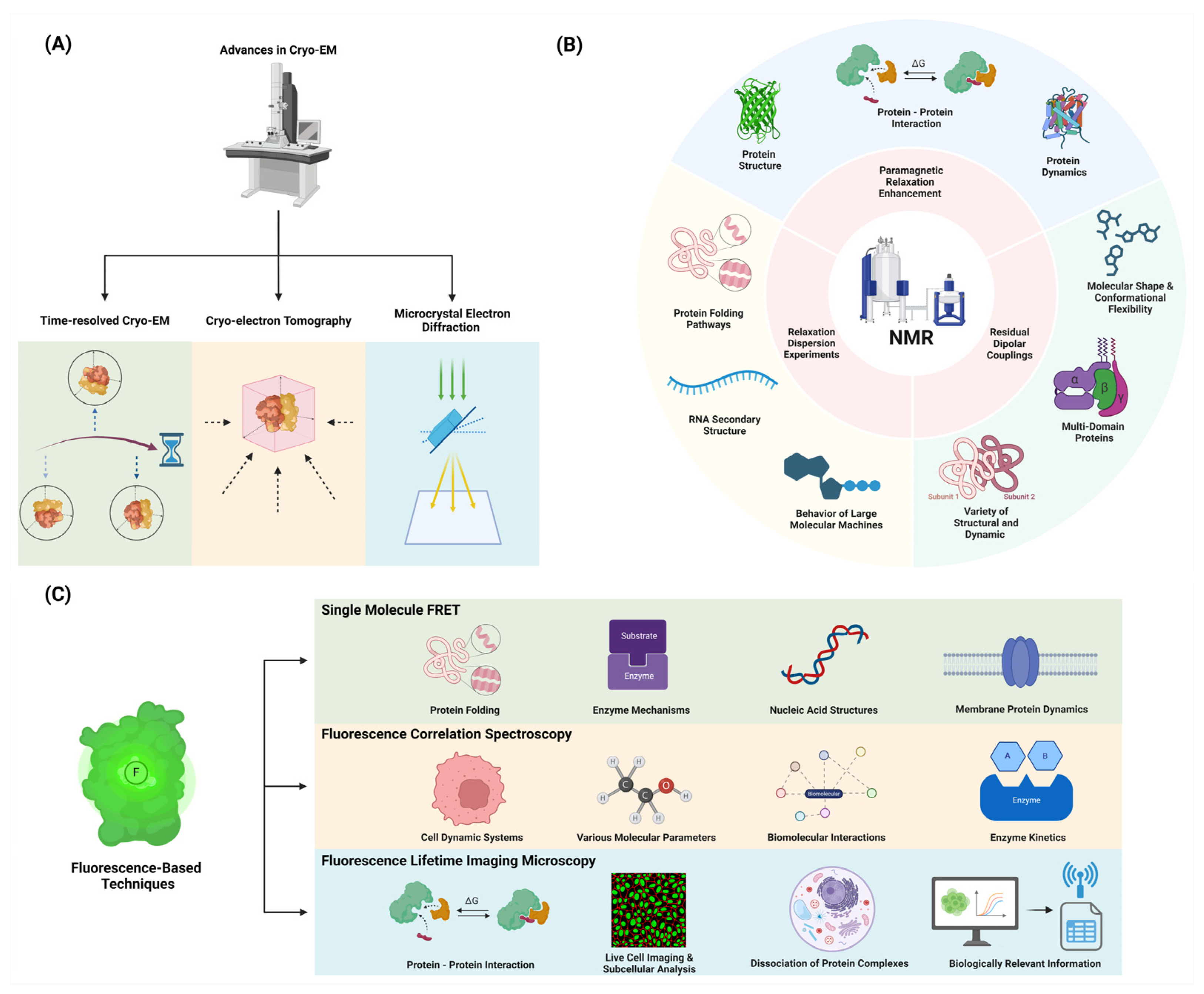
2.1. Innovations in Cryo-EM and X-ray Crystallography for Dynamics Studies

Cryogenic electron microscopy (cryo-EM) has revolutionized structural biology by enabling the visualization of proteins in near-native states without the need for crystallization. Recent developments in cryo-EM have provided unprecedented insights into protein dynamics, particularly for large, flexible complexes (Figure 1).

2.1.1. Time-Resolved Cryo-EM: Capturing Protein Motions at Different Time Points

Time-resolved cryogenic electron microscopy (cryo-EM) is an emerging technique in structural biology that allows researchers to capture structural states that are too transient for standard methods [9]. This technique has revolutionized the field by enabling the visualization of proteins in near-native states without the need for crystallization [40]. Recent developments in cryo-EM have provided unprecedented insights into protein dynamics, particularly for large, flexible complexes [41]. By freezing samples at different time points, time-resolved cryo-EM can trap non-equilibrium states and determine conformations present after defined periods, typically in the millisecond time frame [42]. This approach has been instrumental in elucidating the mechanics of molecular machines such as ribosomes and polymerases, which undergo complex, multistep processes during their functional cycles [41]. Methods such as microsecond time-resolved cryo-EM have enabled observations of protein dynamics, revealing detailed pictures of conformational changes that occur on short timescales [40]. These advancements highlight the potential of time-resolved cryo-EM to fundamentally advance our understanding of protein function and dynamics [9,42].

2.1.2. Cryo-Electron Tomography: Visualizing Proteins in Their Cellular Context

Cryo-electron tomography (Cryo-ET) is a cutting-edge technique that enables the visualization of proteins within their native cellular environments, offering unprecedented insights into their structural organization and interactions within the complex milieu of living cells [43]. This method combines the principles of electron tomography with cryogenic preservation, allowing researchers to capture three-dimensional images of biological samples without the need for crystallization or chemical fixation, thus preserving their native states [44]. The ability to visualize intact cells and tissues while maintaining the spatial relationships between proteins and other cellular components represents a significant advancement over traditional imaging methods [45]. Recent technological advancements in Cryo-ET have significantly improved the resolution and data acquisition speeds, facilitating the observation of dynamic processes at molecular scales [46]. Furthermore, tools such as subtomogram averaging have enhanced the high-resolution analysis of large molecular complexes, providing crucial information about protein localization and conformational states within their cellular contexts [47]. As research progresses, Cryo-ET holds immense potential for advancing our understanding of biological systems by enabling the direct observation of proteins functioning in situ [48].

2.1.3. Time-Resolved X-ray Crystallography: Studying Room Temperature and Computational Modeling

Recent advancements in time-resolved X-ray crystallography have significantly enhanced our understanding of protein dynamics, particularly through studies conducted at room temperature and the development of new computational approaches for modeling conformational ensembles. Time-resolved X-ray diffraction methods, such as those leveraging X-ray free-electron lasers (XFELs), allow researchers to capture transient intermediate states, offering insights into the dynamic processes that occur on timescales from femtoseconds to milliseconds [49,50]. These techniques have been instrumental in visualizing rapid conformational changes in proteins, which are crucial for understanding their functional mechanisms [12]. Room-temperature crystallography has emerged as a key approach, providing more physiologically relevant data by avoiding the artifacts introduced by cryogenic cooling [51,52]. This method enhances the detection of low-occupancy conformational states and allosteric sites that are often masked in cryogenic conditions [52]. Alongside these experimental advancements, computational methods such as automated multiconformer model building have been developed to better interpret the ensemble data obtained from these studies [36]. Tools like qFit v3.0 (https://github.com/ExcitedStates/qfit-3.0) facilitate the modeling of protein conformational heterogeneity, improving the accuracy of structural models and providing deeper insights into the relationship between protein dynamics and function [36]. Collectively, these advancements are revolutionizing structural biology by enabling a more comprehensive understanding of the dynamic nature of proteins and their roles in biological processes.

2.1.4. Microcrystal Electron Diffraction (MicroED): Studying Small Protein Dynamics

Microcrystal electron diffraction (MicroED) has emerged as a powerful technique for studying protein dynamics, offering unique advantages in structural biology. This method allows for the determination of high-resolution protein structures from micro- or nanosized crystals, which are significantly smaller than those required for traditional X-ray crystallography [10,53]. MicroED is particularly valuable for proteins that are difficult to crystallize or form only small crystals, such as membrane proteins, which are crucial for drug development [54]. Recent advancements have enabled the capture of conformational dynamics in protein microcrystals on femto- to microsecond timescales, providing insights into transient states that are essential for understanding protein function [55]. The technique’s ability to deliver atomic-resolution structures from minute crystals facilitates the study of challenging targets, including ligand-bound protein complexes, which are pivotal in drug discovery [56]. Moreover, MicroED’s integration with computational modeling allows for the accurate interpretation of conformational ensembles, enhancing our understanding of protein dynamics and their implications in biological processes [10]. As the field continues to evolve, MicroED is poised to complement existing structural biology methods, offering new opportunities for the rapid and detailed analysis of macromolecular structures [10,53].

2.2. Nuclear Magnetic Resonance (NMR) Spectroscopy

NMR spectroscopy remains a powerful tool for studying protein dynamics, offering atomic-level resolution and the ability to probe motions across a wide range of timescales. Recent advances include the following.

2.2.1. Relaxation Dispersion Experiments: Detecting and Characterizing Excited States

Relaxation dispersion NMR spectroscopy is a powerful technique used to detect and characterize low-populated, transient, excited states of biomolecules by quantifying the broadening of NMR resonance lines due to chemical exchange between ground and excited states [57,58]. This method relies on the exchange between highly populated, NMR-visible ground states and sparsely populated, NMR-invisible excited states, transferring information about magnetic resonance properties such as relaxation parameters, chemical shifts, and residual dipolar couplings from the invisible state to the observable species [58]. The technique provides detailed kinetic and thermodynamic data, enabling the study of the structural and dynamic properties of these excited states on the millisecond timescale [59]. Various experiments, including Carr–Purcell–Meiboom–Gill (CPMG) and rotating frame relaxation dispersion (R1ρ) methods, are employed to probe these exchange processes, often requiring isotopic labeling of the macromolecules under study [59,60]. Applications of relaxation dispersion NMR have revealed critical insights into protein-folding pathways, RNA secondary structure dynamics, and the behavior of large molecular machines, offering a high-resolution view of intermediate states that are otherwise challenging to study [61,62]. This approach is complementary to other biophysical techniques and can be performed in the absence of denaturants, making it a versatile tool for studying biomolecular dynamics in native-like conditions [63].

2.2.2. Paramagnetic Relaxation Enhancement (PRE): Probing Long-Range Interactions

Paramagnetic relaxation enhancement (PRE) is a powerful technique in nuclear magnetic resonance (NMR) spectroscopy that utilizes the magnetic dipolar interactions between unpaired electrons in a paramagnetic center and nearby nuclei to increase nuclear relaxation rates [64,65]. This effect is measurable at long distances, making it valuable for probing transient, lowly populated states and long-range interactions in macromolecules. PRE can be applied using intrinsic paramagnetic centers in metalloproteins or by introducing paramagnetic labels through chemical modification [65]. The technique is particularly useful for studying protein dynamics, structures, and interactions, as it can provide information on sparsely populated states and conformational changes that are difficult to detect using other methods [66,67]. PRE measurements typically involve comparing nuclear relaxation rates between paramagnetic and diamagnetic samples, with transverse (Γ2) PRE rates generally providing the most reliable and accurate data [65]. Recent advancements have expanded the application of PRE to include solvent accessibility studies, nanostructure determination in materials, and enhanced temporal resolution in NMR experiments [68,69]. The versatility of PRE has made it an invaluable tool in structural biology, materials science, and other fields where understanding molecular interactions and dynamics is crucial [70].

2.2.3. Residual Dipolar Couplings (RDCs): Characterizing Domain Orientations and Flexibility

Residual dipolar couplings (RDCs) are a valuable tool in nuclear magnetic resonance (NMR) spectroscopy for characterizing the relative orientations and flexibility of molecular domains. RDCs arise when molecules in solution exhibit partial alignment, causing an incomplete averaging of spatially anisotropic dipolar couplings, which provides orientation-dependent restraints that are crucial for structural determination [71]. This partial alignment can be achieved using alignment media such as liquid crystalline phases or stretched polymer gels, which create an anisotropic environment necessary for RDC measurements [72]. RDCs are particularly useful for studying multi-domain proteins, as they provide information on the relative orientation of domains and their dynamic behavior [73]. By measuring the dipolar couplings between NMR-active nuclei, RDCs deliver insights into the global molecular shape and conformational flexibility, which are essential for understanding the functional motions and interactions of biomolecules. The technique has been successfully applied to a variety of structural and dynamic studies [74], including the analysis of protein–substrate interactions and the determination of quaternary structures of oligomers in equilibrium with monomers [75]. Recent advancements in RDC measurement and analysis have further expanded its applications, making it an indispensable tool in structural biology and related fields [74].

2.3. Fluorescence-Based Techniques

Fluorescence-based methods offer high sensitivity and the ability to study proteins in solution or in living cells.

2.3.1. Single-Molecule FRET: Probing Conformational Changes in Individual Molecules

Single-molecule Förster resonance energy transfer (smFRET) is a powerful technique for studying the conformational dynamics and interactions of individual biomolecules with high spatial and temporal resolution [15,76,77]. This method relies on measuring the energy transfer efficiency between donor and acceptor fluorophores attached to specific sites on a molecule of interest [76,78]. smFRET can reveal heterogeneous populations, transient intermediates, and dynamic fluctuations that are often masked in ensemble measurements [77,79]. Recent advances have improved the precision and accuracy of smFRET measurements, with studies reporting distance uncertainties of ±2–5 Å [15,80]. The technique has been successfully applied to investigate protein folding, enzyme mechanisms, nucleic acid structures, and membrane protein dynamics [76,77,78]. Developments in multicolor FRET schemes allow the probing of more complex biomolecular systems [77]. Additionally, progress in data analysis methods, including hidden Markov modeling and photon-by-photon approaches, enables the extraction of kinetic information on microsecond to millisecond timescales [79]. While challenges remain in achieving higher temporal resolution and applying smFRET in cellular environments, ongoing innovations continue to expand its capabilities for elucidating biomolecular structure and function at the single-molecule level [15,80].

2.3.2. Fluorescence Correlation Spectroscopy (FCS): Analyzing Diffusion and Binding Kinetics

Fluorescence correlation spectroscopy (FCS) is a powerful technique for quantifying molecular dynamics and has been widely applied in diverse fields such as biomedicine, biophysics, and chemistry [81]. By analyzing the time correlation of fluorescence fluctuations induced by molecules diffusing through a focused light, FCS can quantitatively evaluate the concentration, diffusion coefficient, and interaction of molecules both in vitro and in vivo [81,82]. The technique measures the spatial and temporal correlation of individual molecules, providing a bridge between classical ensemble and contemporary single-molecule measurements [82]. Typically implemented using a fluorescence microscope, FCS samples femtoliter volumes, making it especially useful for characterizing small dynamic systems such as biological cells. FCS can investigate various molecular parameters, including diffusion coefficients, chemical rate constants, molecular concentrations, and fluorescence brightness. The method’s sensitivity allows for the analysis of extremely low-concentration biomolecules, with applications ranging from studying diffusion and chemical dynamics to monitoring biomolecular interactions and enzyme kinetics. Recent advancements in FCS include dual-color cross-correlation, multi-focus FCS, and scanning FCS, which enhance its capability to probe complex biological environments and interactions [81,83]. Despite its requirement for high signal-to-noise ratios and long time traces, FCS remains a versatile tool, with ongoing developments aimed at improving its temporal resolution and reducing phototoxic effects on living samples [84].

2.3.3. Fluorescence Lifetime Imaging Microscopy (FLIM): Mapping Protein Interactions in Cells

Fluorescence lifetime imaging microscopy (FLIM) is a powerful technique that measures the time-resolved fluorescence decay of fluorophores to generate contrast in microscopy images, providing information beyond traditional intensity-based imaging [85]. FLIM exploits the characteristic excited-state lifetime of fluorophores, which is sensitive to the local molecular environment, enabling the technique to probe various cellular parameters, such as pH, viscosity, and protein interactions. The method typically employs time-correlated single-photon counting (TCSPC) to measure fluorescence lifetimes with picosecond resolution, allowing for precise quantification of molecular dynamics. FLIM is particularly valuable for studying protein–protein interactions through Förster resonance energy transfer (FRET), where the fluorescence lifetime of a donor fluorophore decreases in the presence of a nearby acceptor, indicating molecular proximity within 1–10 nm [86]. Recent advances in FLIM instrumentation, including the development of fast FLIM techniques and improved spatial resolution, have expanded its capabilities for live-cell imaging and subcellular analysis. Computational developments in FLIM data analysis, such as phasor approaches and machine-learning algorithms, have enhanced the extraction of biologically relevant information from complex lifetime datasets [87]. FLIM has found widespread applications in cell biology, including the study of protein conformational changes, ligand–receptor interactions, and the spatiotemporal dynamics of signaling complexes in living cells [86]. The technique’s ability to discriminate between free and bound states of fluorescently labeled proteins makes it particularly suited to mapping the formation and dissociation of protein complexes in various cellular compartments. As FLIM continues to evolve, its integration with other advanced microscopy techniques and the development of novel fluorescent probes promise to further expand its utility in unraveling the intricate molecular interactions that underlie cellular function [87].

3. Computational Approaches to Protein Dynamics

3.1. Molecular Dynamics Simulations

Molecular dynamics (MD) simulations have become an indispensable tool for studying protein dynamics at atomic resolution (Figure 2).

3.1.1. Fundamentals of Molecular Dynamics (MD) Simulations: Force Fields and Integration Algorithms

Molecular dynamics (MD) simulations rely heavily on force fields to model the interactions within and between molecules, providing a computational framework for studying molecular systems. A force field is a set of equations and parameters that describe the potential energy of a system, typically decomposed into bonded interactions (including bond stretching, angle bending, and dihedral torsions) and non-bonded interactions (such as van der Waals and electrostatic forces) [88,89]. Bonded interactions are often represented by harmonic potentials, while non-bonded interactions use Lennard–Jones and Coulombic potentials [88,89]. The MD algorithm integrates Newton’s equations of motion to simulate the trajectories of atoms over time, with common integration methods including the Verlet and leapfrog algorithms [36,90]. The Verlet algorithm is favored for its simplicity and time-reversibility, although it does not explicitly calculate velocities, which can be derived separately. The leapfrog algorithm, an alternative, calculates velocities at half-time steps, improving the accuracy in velocity calculations. Choosing an appropriate time step is crucial to balance accuracy and computational efficiency; larger time steps increase the sampling efficiency but can introduce errors if too large [89,91]. Typically, time steps are chosen to be small enough to accurately capture the fastest motions in the system, such as bond vibrations, while being large enough to efficiently explore the phase space. These principles and methods form the backbone of MD simulations, enabling detailed studies of molecular dynamics across various fields [92,93].

3.1.2. Long-Timescale Simulations: Accessing Biologically Relevant Timescales (Ms–S)

Long-timescale molecular dynamics simulations have emerged as a powerful tool for studying biological processes occurring on millisecond to second timescales, overcoming traditional limitations of atomistic simulations [94]. Advances in hardware, including specialized supercomputers and graphics processing units (GPUs), have enabled microsecond to millisecond simulations of protein folding, conformational changes, and ligand binding [94,95]. Enhanced sampling techniques like accelerated molecular dynamics, replica exchange, and Markov state models allow more efficient exploration of conformational space [94,96]. Coarse-graining approaches sacrifice atomic detail but permit much longer effective timescales [97]. The development of polarizable force fields and quantum mechanics/molecular mechanics (QM/MM) methods has improved the accuracy of modeling electronic effects [95,97]. Machine-learning potentials trained on quantum mechanical data show promise for combining accuracy and speed [98]. Distributed computing projects like Folding@home have aggregated enormous computational resources for long timescales. Despite these advances, challenges remain in force field accuracy, sampling rare events, and bridging timescales from femtoseconds to seconds [94,96]. Careful validation against experimental data and assessment of convergence are critical for reliable results [99]. As the field progresses, long-timescale simulations are providing unprecedented insight into the dynamics and mechanisms of biomolecular systems [94,95,98].

3.1.3. Enhanced Sampling Techniques: Exploring Rare Events and Conformational Transitions

Enhanced sampling techniques have significantly advanced molecular dynamics (MD) simulations by enabling the exploration of rare events and conformational transitions, which are crucial for understanding complex biological systems. Metadynamics is a key method that modifies the potential energy landscape by adding a history-dependent bias potential, effectively “filling” free energy wells to facilitate transitions over energy barriers. This approach allows for the detailed exploration of the free energy landscape, as demonstrated in studies of protein folding and ligand binding, where metadynamics has provided insights into the underlying mechanisms of these processes [100,101]. Replica-exchange molecular dynamics (REMD) enhances sampling by simulating multiple replicas of a system at different temperatures, allowing for temperature swaps that help the system overcome energy barriers. This method has been successfully applied to study peptide aggregation and protein folding, revealing detailed conformational landscapes and transition mechanisms [102,103]. Adaptive biasing force (ABF) methods dynamically flatten the free-energy landscape, enabling efficient mapping of complex free-energy surfaces without prior knowledge of the potential energy surface. This approach has been applied to calculate free-energy differences in prototypical molecular systems, providing reliable estimates of free-energy landscapes [104,105]. Recent advancements also include integrating machine-learning techniques, such as neural networks, to identify collective variables and approximate free-energy surfaces. These methods have improved the efficiency and accuracy of enhanced sampling, as seen in the generation of dynamic protein conformational ensembles, which has enhanced our understanding of protein dynamics and function [106,107]. Collectively, these enhanced sampling techniques expand the capabilities of MD simulations, allowing for the study of processes that occur over extended timescales and providing detailed insights into the dynamic behavior of biomolecules [108].

3.1.4. Coarse-Grained Models: Simulating Large Systems and Complex Assemblies

Coarse-grained (CG) models have become indispensable for simulating large systems and complex assemblies by reducing the number of degrees of freedom, thereby enabling the exploration of larger spatial and temporal scales than all-atom simulations [109,110]. These models achieve computational efficiency by averaging out fast local motions and focusing on slow collective dynamics, which is particularly useful for studying biological processes such as protein folding, membrane dynamics, and large biomolecular complexes [111]. The development of CG models involves defining pseudoatoms that represent groups of atoms and parameterizing the interactions between them, often guided by all-atom simulations or experimental data [112]. Recent advancements in machine learning have further enhanced CG models by improving the mapping from all-atom to CG representations and enhancing the accuracy of CG force fields, although there is still a long way to go toward achieving fully accurate coarse-gained simulations [109,113]. Techniques such as variational auto-encoders and neural network potentials have been employed to create more accurate and generalizable CG models, capable of capturing essential thermodynamic and kinetic properties of the system. These models have been successfully applied to simulate processes that occur on timescales ranging from microseconds to milliseconds, providing insights into the dynamics of large protein complexes, lipid membranes, and other biomolecular assemblies [110]. Despite their advantages, CG models must be carefully validated against experimental data and all-atom simulations to ensure their reliability and accuracy [111,114]. As the field progresses, the integration of CG models with multiscale simulation approaches promises to bridge the gap between molecular and macroscopic descriptions of biological systems, enhancing our understanding of complex biological phenomena [114,115].

3.2. Machine Learning and AI in Protein Dynamics

The integration of machine learning and AI approaches has opened new avenues for understanding protein dynamics.

3.2.1. Deep Learning for Feature Extraction: Identifying Relevant Collective Variables

The integration of machine learning (ML) and AI approaches has opened new avenues for understanding protein dynamics, particularly through deep learning for feature extraction and identifying relevant collective variables (CVs). Deep-learning techniques, such as convolutional neural networks (CNNs) and recurrent neural networks (RNNs), have proven effective in extracting meaningful features from large-scale protein data, enabling the prediction of protein structures and dynamics with improved accuracy [116]. These methods leverage vast protein databases to identify patterns and correlations that are not apparent through traditional approaches, thus enhancing our ability to model protein behavior [117]. One notable application is the use of variational auto-encoders and neural network potentials to discover CVs that capture the essential dynamics of protein systems, facilitating the exploration of conformational changes and rare events in molecular simulations [107]. For instance, machine-learning models have been trained on molecular dynamics (MD) simulation data to generate physically realistic conformational ensembles, significantly reducing the computational costs and improving the efficiency of sampling techniques [118,119]. This approach has been particularly beneficial for studying intrinsically disordered proteins (IDPs), which exhibit high conformational variability and are challenging to model using conventional methods. By integrating machine learning with MD simulations, researchers can now achieve a more comprehensive understanding of protein-folding mechanisms, energy landscapes, and the thermodynamics and kinetics of protein interactions [117]. As these techniques continue to evolve, they promise to further enhance our ability to predict and manipulate protein functions, with broad implications for drug discovery and protein engineering [116,118].

3.2.2. Generative Models: Predicting Protein Conformations and Dynamics

The integration of generative models in understanding protein dynamics has significantly advanced the prediction of protein conformations and dynamics [120]. Generative models, such as variational auto-encoders and generative adversarial networks, have been employed to learn low-dimensional representations of protein conformational spaces, which can then be used to generate novel protein structures and dynamic ensembles [107]. These models are trained on extensive datasets from molecular dynamics (MD) simulations, allowing them to capture the complex energy landscapes and conformational transitions of proteins [120,121]. By learning the underlying probability distributions of protein conformations, generative models can efficiently sample new conformations, thereby reducing the computational cost associated with traditional MD simulations [107]. This approach has been particularly effective in studying intrinsically disordered proteins (IDPs), which exhibit a high degree of conformational variability and are challenging to model using conventional methods [121]. The application of generative models has also facilitated the exploration of rare events and large-scale conformational changes, providing deeper insights into the thermodynamics and kinetics of protein folding and function [120]. As these techniques continue to evolve, they promise to enhance our ability to predict and manipulate protein dynamics, with broad implications for drug discovery and protein engineering [107,122].

3.2.3. Reinforcement Learning: Optimizing Sampling Strategies in MD Simulations

Reinforcement learning (RL) has emerged as a powerful approach for optimizing sampling strategies in molecular dynamics (MD) simulations, enabling more efficient exploration of protein conformational landscapes and rare events [123,124]. RL-based methods, such as REAP (reinforcement learning-based adaptive sampling) and AdaptiveBandit, address the exploration–exploitation dilemma by balancing the sampling of known metastable states with the discovery of new, potentially important regions of the conformational space. These algorithms typically define reward functions that encourage exploration of undersampled areas while also prioritizing regions of interest, such as those with low free energy or specific structural features. For example, FAST (fluctuation amplification of specific traits) uses a multi-armed bandit-inspired approach to initialize simulations that optimize a given property while maintaining exploration of poorly sampled states [108,125]. Similarly, AdaptiveBandit employs the UCB1 algorithm to find the minimum free energy configurations by balancing expected rewards with uncertainty in sampling. Tree search molecular dynamics (TS-MD) applies the upper confidence bounds for trees (UCT) algorithm to efficiently sample transition pathways between initial and target protein configurations [123]. These RL-based methods have demonstrated success in enhancing the sampling of complex systems, such as protein folding and conformational changes, often achieving better coverage of the conformational space compared to traditional MD approaches. Moreover, the integration of RL with other machine-learning techniques, such as deep learning for feature extraction and dimensionality reduction, has further improved the efficiency and accuracy of MD simulations [126]. As the field progresses, RL-optimized sampling strategies promise to play an increasingly important role in unraveling the intricacies of protein dynamics and facilitating drug discovery efforts [123,126].

4. Applications and Insights from Protein Dynamics Studies

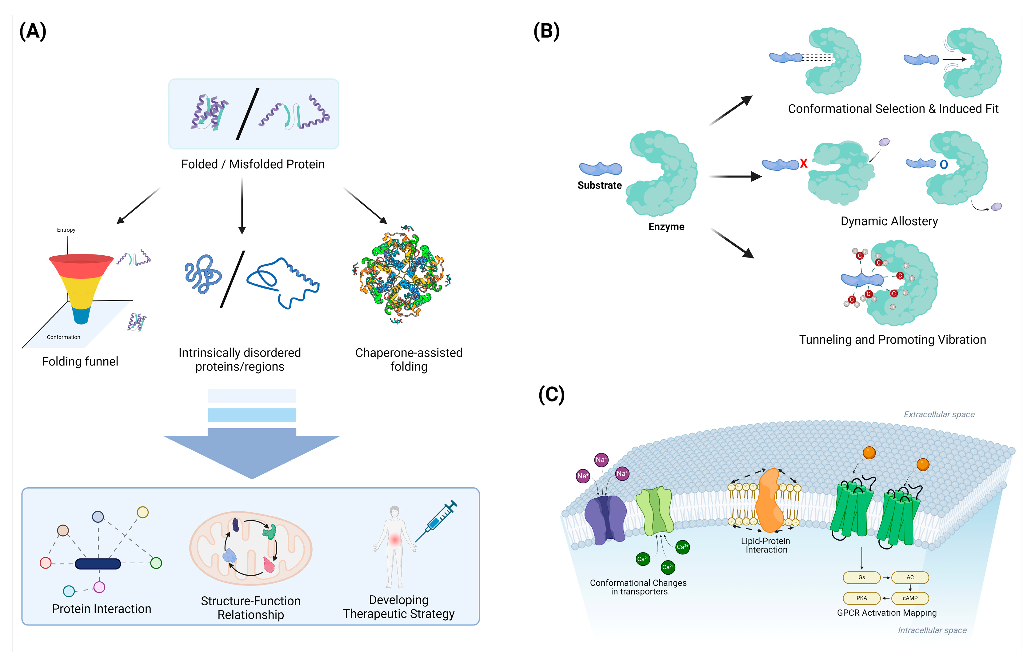
4.1. Protein Folding and Misfolding

Combining experimental and computational approaches has led to significant progress in understanding protein-folding mechanisms and the factors contributing to misfolding and aggregation (Figure 3).

4.1.1. Folding Funnels and Energy Landscapes: Characterizing the Thermodynamics and Kinetics of Folding

Folding funnels and energy landscapes provide a powerful framework for understanding the thermodynamics and kinetics of protein folding [127,128,129]. The funnel-shaped energy landscape concept, introduced in the 1990s, revolutionized our understanding of how proteins navigate the vast conformational space to reach their native states [127]. This model depicts folding as a process of descending a rugged energy landscape, where the vertical axis represents free energy and the horizontal axes represent conformational degrees of freedom [128,130]. The overall funnel shape arises from the principle of minimal frustration, which posits that evolution has selected sequences that minimize conflicting interactions, thereby creating a bias toward the native state [129,131]. Quantitatively, the folding funnel can be characterized by parameters such as the energy gap between the native state and the average of misfolded states, the ruggedness of the landscape, and the configurational entropy [128,132]. These parameters influence the folding rate and mechanism, with smoother funnels generally leading to faster folding [130,133]. The presence of local minima in the energy landscape can give rise to folding intermediates and multiple folding pathways, explaining the complexity observed in folding kinetics [132]. Advanced experimental techniques, such as single-molecule spectroscopy and hydrogen exchange mass spectrometry, have provided empirical support for the funnel model by mapping energy landscapes and detecting transient intermediates [131,133]. Computational methods, including molecular dynamics simulations and statistical mechanical models, have further refined our understanding of folding funnels and their relationship to sequence–structure correlations [129,132]. The folding funnel concept has also been extended to explain protein–protein interactions, allostery, and the evolution of new protein functions, demonstrating its broad applicability in structural biology [127,131].

4.1.2. Intrinsically Disordered Proteins: Recognizing the Functional Importance of Structural Flexibility

Intrinsically disordered proteins (IDPs) and intrinsically disordered regions (IDRs) have emerged as crucial components of cellular signaling and regulation, challenging the traditional structure–function paradigm of proteins [29,134]. These proteins lack a stable three-dimensional structure under physiological conditions, instead existing as dynamic ensembles of interconverting conformations [135,136]. The structural flexibility of IDPs/IDRs enables them to interact with multiple binding partners, facilitating their roles as hubs in protein interaction networks and allowing for fine-tuned regulation through post-translational modifications [29]. This conformational plasticity is encoded in their amino acid sequences, which are typically characterized by a low content of bulky hydrophobic residues and a high proportion of polar and charged amino acids [125,136]. IDPs/IDRs are particularly enriched in eukaryotic proteomes and are involved in various cellular processes, including transcriptional control, cell signaling, and the formation of membrane-less organelles through phase separation [29,134,137]. The functional significance of protein disorder extends to enzymes, where IDRs can play important roles in allosteric regulation and catalysis [135,138]. Recent advances in experimental techniques, such as NMR spectroscopy and single-molecule FRET, combined with computational methods, have greatly enhanced our understanding of the structural ensembles and functional mechanisms of IDPs [136,139]. The recognition of the widespread occurrence and functional importance of intrinsic disorder has led to the establishment of the “disorder-function paradigm”, complementing the classical structure–function relationship in protein science [134,140].

4.1.3. Chaperone-Assisted Folding: Elucidating the Role of Cellular Machinery in Protein Folding

Chaperone-assisted protein folding is a critical cellular process that ensures the proper folding and functionality of proteins, preventing misfolding and aggregation, which can lead to cellular dysfunction [141,142]. Molecular chaperones, such as those from the Hsp70 and chaperonin families, play a pivotal role in this process by stabilizing unfolded or partially folded proteins and facilitating their correct folding through ATP-dependent cycles of binding and release [141,143]. These chaperones recognize exposed hydrophobic regions on nascent or stress-denatured proteins, preventing inappropriate interactions that could lead to aggregation [142,144]. For instance, Hsp70 chaperones not only prevent aggregation but also actively remodel misfolded proteins, promoting their refolding into native conformations [143,145]. Additionally, chaperonins like GroEL/GroES provide a protected environment for protein folding, encapsulating substrate proteins and allowing them to fold without interference from the crowded cellular milieu [146,147]. Recent studies using advanced techniques such as single-molecule fluorescence and cryo-electron microscopy have provided detailed insights into the dynamic interactions between chaperones and their substrate proteins, revealing how these molecular machines guide proteins through their folding landscapes [144,148]. The chaperone system is also integral to the cellular stress response, with heat shock proteins being upregulated to manage increased loads of misfolded proteins under stress conditions [142,149]. Understanding the mechanisms of chaperone-assisted folding not only elucidates fundamental aspects of cellular proteostasis but also has significant implications for developing therapeutic strategies against diseases caused by protein misfolding and aggregation [141,147,149].

4.2. Enzyme Catalysis and Allostery

Studies of protein dynamics have revealed the critical role of motions in enzyme function and regulation.

4.2.1. Conformational Selection vs. Induced Fit: Understanding Substrate Binding Mechanisms

Conformational selection and induced fit are two fundamental mechanisms that explain how proteins interact with ligands, each offering distinct insights into the dynamics of molecular recognition [150,151]. In conformational selection, proteins exist in a pre-existing ensemble of conformations, and the ligand selectively binds to and stabilizes a subset of these conformations, as supported by techniques like NMR and single-molecule FRET [150,152]. Conversely, induced fit posits that the initial protein–ligand interaction triggers a conformational change in the protein to optimize the binding interface, a concept first proposed by Koshland to explain enzymatic specificity [151,153]. These mechanisms are not mutually exclusive and can coexist within the same system, with their relative contributions often dependent on factors such as the ligand concentration and specific protein characteristics [150,154]. Advanced computational methods, including molecular dynamics simulations and enhanced sampling techniques, have been instrumental in elucidating the energy landscapes that govern these binding processes [153,154]. Understanding these mechanisms is crucial for drug design and protein engineering, as it provides insights into how proteins achieve specificity and efficiency in their interactions [150,155]. For instance, in enzyme design, considering both conformational selection and induced fit can lead to more effective catalysts by optimizing both the pre-existing conformational ensemble and the induced changes upon substrate binding [153,154]. In the context of drug discovery, this understanding can guide the development of more potent and selective inhibitors by targeting specific conformational states or by designing ligands that can induce favorable conformational changes [151,154]. Recent studies have also highlighted the role of these mechanisms in allosteric regulation and the function of intrinsically disordered proteins, further expanding their relevance in biological processes [150,154].

4.2.2. Dynamic Allostery: Recognizing the Importance of Entropy in Allosteric Regulation

Dynamic allostery refers to the phenomenon whereby the binding of a ligand leads to conformational changes in a protein that can influence the dynamics of other distant sites within the protein or within associated proteins [156,157]. This process highlights the significance of entropic changes during allosteric regulation, emphasizing that allosteric effects are not merely a consequence of structural changes but also involve shifts in the entropy of the protein states [158]. Entropy plays a crucial role in allosteric regulation as it governs the stability of various protein conformations; thus, changes in entropy upon ligand binding can enhance or inhibit the functional responses of the protein [157]. Additionally, advances in experimental techniques, such as nuclear magnetic resonance (NMR) spectroscopy and molecular dynamics simulations, have demonstrated how conformational flexibility and entropic fluctuations are essential for understanding allosteric mechanisms [158]. This recognition of the importance of entropy in allostery has implications for drug design, providing insights into how small molecules can induce conformational changes that modulate protein activity [159].

4.2.3. Tunneling and Promoting Vibrations: Exploring Quantum Effects in Enzyme Catalysis

Quantum mechanical effects, such as tunneling and promoting vibrations, play a crucial role in enzyme catalysis by influencing the reaction rates and mechanisms involved. Hydrogen tunneling, a process where hydrogen atoms pass through energy barriers rather than over them, has been shown to be significant in enzymatic C–H bond cleavage, linking protein dynamics to catalysis and highlighting the importance of the donor–acceptor distance and active-site electrostatics [160]. Promoting vibrations, which are specific localized motions within the enzyme, enhance catalytic efficiency by coupling directly to the reaction coordinate and optimizing the compression along the donor–acceptor distance, thus facilitating hydrogen tunneling [161,162]. These vibrations are believed to be a universal feature of enzymes, contributing to their pre-organized active sites and enabling efficient biochemical transformations. Recent theoretical and experimental studies have demonstrated that these quantum effects are not only significant at cryogenic temperatures but also under physiological conditions, challenging classical transition state theory and emphasizing the dynamic nature of enzyme catalysis [163]. Computational models, particularly quantum mechanical/molecular mechanical (QM/MM) simulations, have provided valuable insights into these mechanisms, underscoring the interplay between electronic structure and protein dynamics in enzyme function [164].

4.3. Membrane Protein Dynamics

Advances in studying membrane protein dynamics have provided insights into transport mechanisms and signal transduction.

4.3.1. Lipid–Protein Interactions: Characterizing the Influence of the Membrane Environment

Lipid–protein interactions are fundamental in regulating the structure and function of membrane proteins, influencing processes such as protein conformational changes and membrane domain formation [165,166]. These interactions can be specific, involving direct binding of lipids to proteins, or nonspecific, affecting the general properties of the membrane, such as the thickness and lateral pressure [167,168]. Experimental techniques, including cryo-electron microscopy and mass spectrometry, have advanced our understanding of these interactions by providing high-resolution structures and identifying lipid compositions in complex membranes [165,166]. Molecular dynamics (MD) simulations have complemented these experimental approaches, offering detailed insights into the dynamics and energetics of lipid–protein interactions at atomic resolution [168,169]. For instance, MD simulations have revealed how the lipid composition and membrane properties modulate the conformational equilibrium of ion channels, such as potassium channels, by altering the lateral pressure and membrane thickness [169]. Additionally, specific lipid-binding events have been shown to modulate protein–protein interactions and influence the functional states of membrane proteins, further underscoring the complexity and specificity of lipid–protein interactions [165,167]. Overall, the integration of experimental and computational methods continues to enhance our understanding of the intricate roles that lipid–protein interactions play in cellular membranes [166,168].

4.3.2. Conformational Changes in Transporters: Elucidating Alternating Access Mechanisms

Conformational changes in transporters are essential for elucidating the alternating access mechanisms that facilitate substrate translocation across cell membranes. The alternating access model posits that transporters cycle between outward-facing (OF) and inward-facing (IF) conformations, allowing substrates to bind on one side of the membrane and be released on the other [170,171]. This mechanism has been extensively studied in various transporters, including the Na+/Ca2+ exchanger NCX_Mj, which demonstrates significant structural asymmetry between its topological repeats, with transmembrane helices TM1–TM6 undergoing substantial displacement during the transition [170]. Crystallographic studies of the sodium-hydantoin transporter Mhp1 have captured the transporter in multiple states, providing a detailed mechanism for how it switches from the OF to the IF state through an occluded intermediate [171]. Molecular dynamics (MD) simulations have further elucidated these transitions, revealing that substrate binding induces conformational changes that facilitate the alternating access mechanism, as observed in SWEET family transporters. Recent advancements in computational approaches, such as AlphaFold2, have enabled the prediction of multiple conformational states of transporters, highlighting the dynamic nature of these proteins and their ability to adopt diverse conformations [172]. The integration of experimental and computational methods continues to enhance our understanding of the alternating access mechanism, underscoring the importance of conformational plasticity in transporter function [173].

4.3.3. GPCR Activation: Mapping the Energy Landscape of Receptor Activation

The activation of G protein-coupled receptors (GPCRs) involves complex conformational changes that can be conceptualized as transitions across an energy landscape [174,175]. This landscape is characterized by multiple energy wells representing distinct conformational states, including inactive, intermediate, and active configurations. Agonist binding modifies the shape of this energy landscape, lowering energy barriers and stabilizing active conformations [174]. The activation process typically involves outward movement of transmembrane helix 6 (TM6), a hallmark of GPCR activation that creates space for G-protein coupling [176]. Studies have revealed a common activation pathway comprising 34 residue pairs and 35 residues that links the ligand-binding pocket to the G-protein coupling region, unifying previously identified key motifs such as CWxP, DRY, and NPxxY. This pathway allows for the decoupling of ligand binding site and G-protein binding region evolution, potentially facilitating the diversification of GPCRs. Advanced techniques like X-ray crystallography, cryo-electron microscopy, and molecular dynamics simulations have been instrumental in mapping these energy landscapes and identifying key conformational intermediates [177,178]. The energy landscape framework provides insights into mechanisms of constitutive activity, inverse agonism, and allosteric modulation, offering a powerful tool for understanding GPCR function and drug design [174,179]. Recent studies have also explored the vibrational energy landscapes of GPCRs, providing graphical representations of residue-level energy distributions in active and inactive states [180].

5. Integrating Complementary Techniques for Comprehensive Understanding of Protein Dynamics

Understanding protein dynamics requires a combination of experimental and computational methods, each providing unique insights that, when integrated, offer a more comprehensive view. Experimental techniques such as cryo-electron microscopy (cryo-EM), nuclear magnetic resonance (NMR) spectroscopy, and fluorescence-based methods are powerful for capturing structural and dynamic details at varying resolutions and timescales. Computational approaches, including molecular dynamics (MD) simulations and machine-learning algorithms, offer detailed atomic-level insights and predictive power, complementing experimental observations [181,182].

5.1. Cryo-EM and MD Simulations: Synergy in Structural and Dynamic Studies

Cryo-EM has transformed structural biology by enabling the visualization of macromolecular complexes in near-native states without the need for crystallization. Recent advancements, such as time-resolved cryo-EM, allow for capturing snapshots of transient states, providing a static but high-resolution view of dynamic processes. However, these snapshots alone cannot capture the continuous motion of proteins. MD simulations fill this gap by modeling the movements of atoms over time, providing a dynamic picture that complements the static images from cryo-EM. By using cryo-EM-derived structures as starting points for MD simulations, researchers can validate and refine computational models, ensuring that the simulated dynamics correspond to biologically relevant conformations observed experimentally. For example, the conformational changes of ribosomes during translation have been extensively studied using a combination of cryo-EM and MD, revealing the sequence of motions at a level of detail that neither method alone could achieve [181,182].

5.2. NMR Spectroscopy and Fluorescence Techniques: Complementary Insights into Protein Dynamics

NMR spectroscopy offers atomic-level resolution for studying protein dynamics, and it is particularly suited to observing conformational changes and interactions over a range of timescales, from picoseconds to seconds. Techniques such as relaxation dispersion and paramagnetic relaxation enhancement (PRE) can detect low-populated, transient states that are often invisible to other structural methods. Meanwhile, fluorescence techniques like single-molecule Förster resonance energy transfer (smFRET) provide real-time data on protein conformational changes and interactions in live cells or under physiologically relevant conditions. When used together, NMR provides detailed structural information about specific states, while smFRET can validate these states and monitor transitions between them in a more dynamic context. For instance, studying the folding pathways of intrinsically disordered proteins (IDPs) benefits greatly from this combination, with NMR identifying the ensemble of conformations and smFRET elucidating the folding kinetics and pathways in real time [183,184].

5.3. Computational and Experimental Integration: A Holistic Approach to Protein Dynamics

Molecular dynamics (MD) simulations are invaluable for understanding the movements and flexibility of proteins at atomic resolution, particularly over timescales that are challenging for experimental techniques to probe directly. Enhanced sampling techniques in MD simulations, such as metadynamics and replica exchange, allow researchers to explore rare events and conformational transitions that are critical for understanding biological functions. These simulations can predict how proteins will respond to different conditions or mutations, which can then be experimentally validated. Additionally, machine-learning models have emerged as powerful tools to predict protein conformations and identify key dynamic regions, which can guide the design of experiments using cryo-EM or NMR to test these predictions. This iterative cycle of prediction, experimentation, and validation accelerates the discovery process and deepens our understanding of protein dynamics.

5.4. Applications in Drug Discovery and Protein Engineering

The integration of experimental and computational approaches has substantial implications for drug discovery and protein engineering. For example, in the context of drug discovery, understanding the dynamics of G protein-coupled receptors (GPCRs) requires mapping the conformational landscape accessible to these proteins. Combining cryo-EM to capture different receptor states with MD simulations to model the transitions between these states provides a comprehensive view of how potential drugs could stabilize certain conformations. This integrated approach allows for the identification of novel allosteric sites and the design of more effective therapeutics that can modulate protein activity in a controlled manner. Similarly, in protein engineering, combining NMR data on protein flexibility and MD simulations helps design proteins with desired dynamic properties, such as increased stability or enhanced binding affinity.

6. Future Directions and Challenges

As the field of protein dynamics continues to evolve, several key areas for future research and development have emerged (Figure 4).

6.1. Limitations of Experimental Techniques for Studying Protein Dynamics

Experimental techniques such as cryo-electron microscopy (cryo-EM), nuclear magnetic resonance (NMR) spectroscopy, and fluorescence-based methods have significantly advanced our understanding of protein dynamics, each with distinct strengths and limitations. Cryo-EM has become a cornerstone in structural biology for studying proteins in near-native states, particularly large, flexible complexes, but it faces challenges in resolving smaller proteins and capturing transient states at a high resolution. Despite improvements like time-resolved cryo-EM, capturing dynamics at millisecond timescales remains difficult due to sample heterogeneity and radiation damage during data acquisition [7,185,186]. Cryo-electron tomography (Cryo-ET) extends the capabilities of cryo-EM by visualizing proteins within their cellular context, providing insights into their organization and interactions in situ, but suffers from lower resolution and longer data acquisition times compared to single-particle cryo-EM [186]. Time-resolved X-ray crystallography and microcrystal electron diffraction (MicroED) offer high-resolution structural data and can capture transient states but require crystallization, which may introduce artifacts and fail to represent the full range of physiological conditions [12,187]. NMR spectroscopy, particularly through techniques like relaxation dispersion experiments, paramagnetic relaxation enhancement (PRE), and residual dipolar couplings (RDCs), excels in providing atomic-level insights into protein dynamics across a range of timescales but often requires isotopic labeling and faces sensitivity and resolution challenges, especially for large proteins and complexes [184,188]. Fluorescence-based techniques such as single-molecule Förster resonance energy transfer (smFRET), fluorescence correlation spectroscopy (FCS), and fluorescence lifetime imaging microscopy (FLIM) offer real-time insights into protein dynamics in solution or living cells but are limited by photobleaching, fluorophore stability, and the complexity of data interpretation [81,189,190]. Thus, while these experimental techniques have revolutionized our understanding of protein dynamics, each is constrained by specific limitations that affect their applicability to different types of proteins and dynamic processes.

6.2. Limitations of Molecular Dynamics Simulations

Molecular dynamics (MD) simulations have become indispensable for studying protein dynamics at atomic resolution, but they are also subject to several critical limitations. One major challenge is the accuracy of force fields, which are the mathematical models used to approximate the forces between atoms; inaccuracies in force field parameters can lead to significant errors in simulated protein conformations and dynamics [91,191]. Moreover, the sampling efficiency is a persistent issue in MD simulations, especially for capturing rare events and transitions between different conformational states, which often requires enhanced sampling techniques like metadynamics or replica exchange molecular dynamics (REMD) to overcome high energy barriers [95,191]. The computational cost of MD simulations also remains a significant hurdle, particularly for long-timescale simulations that aim to capture biologically relevant processes occurring on millisecond to second timescales. Although advances in hardware, such as the use of GPUs and specialized supercomputers, have enabled longer simulations, these still require significant resources and often provide limited sampling due to the immense configurational space of proteins [95,192]. Additionally, coarse-grained models, which simplify the representation of molecular systems to extend simulations to longer timescales, often sacrifice atomic detail, leading to potential inaccuracies in describing specific interactions and dynamics critical for understanding protein function [95,193]. Therefore, while MD simulations are powerful tools for exploring protein dynamics, they require careful consideration of their inherent limitations in force field accuracy, sampling efficiency, and computational feasibility.

6.3. Integration of Multi-Scale Approaches: Combining Atomistic Simulations with Coarse-Grained Models and Experimental Data to Bridge Timescales and Length Scales

Future directions in computational biology point toward the integration of multi-scale approaches that combine atomistic simulations with coarse-grained models and experimental data to bridge timescales and length scales [194]. Atomistic molecular dynamics simulations provide detailed insights into biomolecular structure and dynamics at the nanoscale but are limited in the timescales they can access [111]. Coarse-grained models allow for simulations of larger systems over longer timescales by reducing the number of degrees of freedom but sacrifice atomic-level detail [195]. Integrating these approaches with experimental data can leverage the strengths of each method while overcoming individual limitations [196]. Machine-learning techniques are emerging as powerful tools to develop accurate coarse-grained models from atomistic simulation data and to bridge between scales [197]. Multiscale modeling frameworks that dynamically couple different levels of theory show promise for capturing processes that span multiple spatiotemporal scales [115]. The key challenges include developing robust methods to systematically connect different scales, quantifying uncertainties across scales, and efficiently sampling rare events [197]. Overcoming these hurdles will enable more comprehensive and predictive models of complex biological systems across scales.

6.4. In-Cell Dynamics: Developing Methods to Study Protein Motions in Their Native Cellular Environment

Developing methods to study protein motions in their native cellular environment remains a major challenge and frontier in the field of protein dynamics [198]. While significant progress has been made in studying protein dynamics in vitro, the complex and crowded cellular milieu can significantly alter protein behavior, necessitating in-cell approaches [199]. Recent advances in in-cell NMR spectroscopy have enabled the study of the protein structure and dynamics directly within living cells, providing insights into how the cellular environment impacts protein function. Fluorescence-based techniques like single-molecule FRET and tracking of transfected biomolecules are also emerging as powerful tools to probe protein conformational dynamics and interactions in cellular contexts with high spatiotemporal resolution [198]. Cryo-electron tomography combined with computational approaches like tomoDRGN shows promise for capturing protein structural heterogeneity and dynamics in their native cellular context [200]. However, significant challenges remain, including improving the sensitivity and resolution of in-cell measurements, developing methods to study low-abundance proteins, and integrating data from multiple techniques to build comprehensive models of protein behavior in cells. Overcoming these hurdles will require continued innovation in labeling strategies, instrumentation, and computational analysis methods [177]. As the field progresses, in-cell dynamic studies have the potential to reveal new principles of protein function and regulation within the complex cellular environment.

6.5. AI-Driven Discovery: Leveraging Machine Learning to Predict Functional Motions and Design Proteins with Specific Dynamic Properties

AI-driven discovery is poised to revolutionize our understanding of protein dynamics and enable the design of proteins with specific dynamic properties. Machine-learning approaches have demonstrated remarkable success in predicting protein structures and are now being extended to capture the dynamic nature of proteins [106]. By integrating sequence, structure, and dynamics information, these methods can provide insights into conformational ensembles and functional motions that are crucial for protein function [119]. Deep-learning models, such as those based on geometric deep learning, show promise in predicting protein dynamics from static structures, potentially accelerating the exploration of conformational landscapes [119]. Furthermore, generative models are emerging as powerful tools for designing novel proteins with tailored dynamic properties, opening up new possibilities for protein engineering [201]. The integration of molecular dynamics simulations with machine-learning frameworks allows for more accurate predictions of protein flexibility and allosteric communication pathways. Recent advances in physics-informed neural networks offer the potential to incorporate physical constraints and improve the interpretability of AI-driven predictions in protein science [106]. As these methods continue to evolve, they are expected to play an increasingly important role in understanding protein function at a molecular level and designing proteins with specific dynamic properties for biotechnological and therapeutic applications.

6.6. Dynamics in Complex Assemblies: Extending Our Understanding to Large Macromolecular Complexes and Cellular Machines

Future directions in the study of dynamics in complex assemblies focus on extending our understanding to large macromolecular complexes and cellular machines. The dynamic nature of these assemblies significantly impacts their molecular and cellular functions, necessitating advanced methods to study their conformational changes, interactions, and spatial-temporal dynamics within the cell [202]. Recent progress in coarse-grained (CG) modeling has facilitated the study of large biological systems by bridging the gap between microscopic and macroscopic details, allowing for the simulation of complex macromolecular interactions over extended timescales [203]. Techniques such as cryo-electron microscopy (cryo-EM) have revolutionized structural biology by providing high-resolution models of large complexes, enabling the visualization of their dynamic processes during assembly, remodeling, and disassembly [202]. Single-molecule biophysical approaches have also advanced, offering unique insights into the stochastic behavior of individual molecules within these assemblies, thus revealing the non-linear and highly dynamic nature of cellular machinery [204]. However, challenges remain in improving the sensitivity and resolution of these methods to capture the full spectrum of dynamic behaviors in complex environments. Integrating experimental data with computational models, such as all-atom molecular dynamics (MD) simulations and CG models, is crucial for developing comprehensive and predictive models of macromolecular dynamics [203]. As these methods continue to evolve, they promise to enhance our understanding of the intricate dynamics of large macromolecular complexes and their roles in cellular processes [202,203,204].

6.7. Linking Dynamics to Function: Developing Quantitative Frameworks to Relate Protein Motions to Biological Function and Disease States

Future directions in linking protein dynamics to function involve developing quantitative frameworks to relate protein motions to biological function and disease states. Proteins are dynamic entities, and their functions are governed by their dynamic personalities, which include fluctuations ranging from atomic vibrations to large domain movements [1]. Understanding these dynamics is crucial, as disease-causing mutations can perturb protein structural networks, leading to altered functions [205]. Integrating experimental data with computational models, such as molecular dynamics (MD) simulations, can reveal how these mutations affect protein stability and function [206]. Techniques like cryo-electron microscopy and NMR spectroscopy have advanced our ability to capture dynamic processes in proteins, providing insights into conformational changes and interactions [1]. Network-based models have also emerged as powerful tools to study the complex spatiotemporal relationships between protein structures and their functions, particularly in the context of disease mutations [207]. These models can help identify allosteric pathways and residues involved in biological activities, linking structural changes to functional outcomes. As we continue to develop and refine these quantitative frameworks, we will enhance our ability to predict how protein dynamics contribute to function and disease, ultimately aiding in the design of targeted therapeutics [208].

7. Conclusions

The study of protein dynamics has advanced significantly in recent years, driven by technological innovations in relation to both experimental and computational approaches. As our understanding of protein motions continues to grow, we gain deeper insights into the fundamental mechanisms of life at the molecular level. The integration of diverse techniques, from cryo-EM to AI-powered simulations, promises to further accelerate progress in this field, with far-reaching implications for drug discovery, protein engineering, and our understanding of cellular processes.

Author Contributions

Conceptualization, investigation, writing, and original draft preparation, A.S. and H.K. Visualization and proofreading, W.K., J.P., W.L., Y.L. and S.C. Supervision, project administration, funding acquisition, review and editing, H.K. All authors have read and agreed to the published version of the manuscript.

Funding

This work was supported by the BK21 FOUR Program by Chungnam National University Research Grant, 2024. This work was partly supported by an Institute of Information and Communications Technology Planning and Evaluation (IITP) grant funded by the Korean government (MSIT) (No. RS-2022-00155857, Artificial Intelligence Convergence Innovation Human Resources Development (Chungnam National University)). This work was supported by the National Research Foundation of Korea (NRF) grant funded by the Korena government (MSIT) (RS-2023-00209456). This work was supported by the Korea Basic Science Institute (National research Facilities and Equipment Center) grant funded by the Korean government (MSIT) (RS-2024-00402298).

Acknowledgments

This manuscript was proofread and edited with the assistance of ChatGPT-4o, a language model by OpenAI. The figures were created using BioRender (BioRender.com).

Conflicts of Interest

The authors declare no conflicts of interest.

References

  1. Nam, K.; Wolf-Watz, M. Protein dynamics: The future is bright and complicated! Struct. Dyn. 2023, 10, 014301. [Google Scholar] [CrossRef]
  2. Jayaraman, V.; Toledo-Patino, S.; Noda-Garcia, L.; Laurino, P. Mechanisms of protein evolution. Protein Sci. 2022, 31, e4362. [Google Scholar] [CrossRef]
  3. Henzler-Wildman, K.A.; Lei, M.; Thai, V.; Kerns, S.J.; Karplus, M.; Kern, D. A hierarchy of timescales in protein dynamics is linked to enzyme catalysis. Nature 2007, 450, 913–916. [Google Scholar] [CrossRef]
  4. Klyshko, E.; Kim, J.S.; McGough, L.; Valeeva, V.; Lee, E.; Ranganathan, R.; Rauscher, S. Functional protein dynamics in a crystal. Nat. Commun. 2024, 15, 3244. [Google Scholar] [CrossRef]
  5. Roca-Martinez, J.; Lazar, T.; Gavalda-Garcia, J.; Bickel, D.; Pancsa, R.; Dixit, B.; Tzavella, K.; Ramasamy, P.; Sanchez-Fornaris, M.; Grau, I.; et al. Challenges in describing the conformation and dynamics of proteins with ambiguous behavior. Front. Mol. Biosci. 2022, 9, 959956. [Google Scholar] [CrossRef]
  6. Ghosh, D.; Biswas, A.; Radhakrishna, M. Advanced computational approaches to understand protein aggregation. Biophys. Rev. 2024, 5, 021302. [Google Scholar] [CrossRef]
  7. Chua, E.Y.D.; Mendez, J.H.; Rapp, M.; Ilca, S.L.; Tan, Y.Z.; Maruthi, K.; Kuang, H.; Zimanyi, C.M.; Cheng, A.; Eng, E.T.; et al. Better, Faster, Cheaper: Recent Advances in Cryo-Electron Microscopy. Annu. Rev. Biochem. 2022, 91, 1–32. [Google Scholar] [CrossRef]
  8. Krieger, J.M.; Sorzano, C.O.S.; Carazo, J.M.; Bahar, I. Protein dynamics developments for the large scale and cryoEM: Case study of ProDy 2.0. Acta Crystallogr. D Struct. Biol. 2022, 78, 399–409. [Google Scholar] [CrossRef]
  9. Maeots, M.E.; Enchev, R.I. Structural dynamics: Review of time-resolved cryo-EM. Acta Crystallogr. D Struct. Biol. 2022, 78, 927–935. [Google Scholar] [CrossRef]
  10. Bongiovanni, G.; Harder, O.F.; Voss, J.M.; Drabbels, M.; Lorenz, U.J. Near-atomic resolution reconstructions from in situ revitrified cryo samples. Acta Crystallogr. D Struct. Biol. 2023, 79, 473–478. [Google Scholar] [CrossRef]
  11. Srajer, V.; Royer, W.E., Jr. Time-resolved x-ray crystallography of heme proteins. Methods Enzym. 2008, 437, 379–395. [Google Scholar] [CrossRef]
  12. Hekstra, D.R. Emerging Time-Resolved X-Ray Diffraction Approaches for Protein Dynamics. Annu. Rev. Biophys. 2023, 52, 255–274. [Google Scholar] [CrossRef]
  13. Wolff, A.M.; Nango, E.; Young, I.D.; Brewster, A.S.; Kubo, M.; Nomura, T.; Sugahara, M.; Owada, S.; Barad, B.A.; Ito, K.; et al. Mapping protein dynamics at high spatial resolution with temperature-jump X-ray crystallography. Nat. Chem. 2023, 15, 1549–1558. [Google Scholar] [CrossRef]
  14. Palmer, A.G., 3rd. NMR characterization of the dynamics of biomacromolecules. Chem. Rev. 2004, 104, 3623–3640. [Google Scholar] [CrossRef]
  15. Hellenkamp, B.; Schmid, S.; Doroshenko, O.; Opanasyuk, O.; Kuhnemuth, R.; Rezaei Adariani, S.; Ambrose, B.; Aznauryan, M.; Barth, A.; Birkedal, V.; et al. Precision and accuracy of single-molecule FRET measurements-a multi-laboratory benchmark study. Nat. Methods 2018, 15, 669–676. [Google Scholar] [CrossRef]
  16. Ando, T. High-speed atomic force microscopy and its future prospects. Biophys. Rev. 2018, 10, 285–292. [Google Scholar] [CrossRef]
  17. Lerner, E.; Cordes, T.; Ingargiola, A.; Alhadid, Y.; Chung, S.; Michalet, X.; Weiss, S. Toward dynamic structural biology: Two decades of single-molecule Forster resonance energy transfer. Science 2018, 359, eaan1133. [Google Scholar] [CrossRef]
  18. Dror, R.O.; Dirks, R.M.; Grossman, J.P.; Xu, H.; Shaw, D.E. Biomolecular simulation: A computational microscope for molecular biology. Annu. Rev. Biophys. 2012, 41, 429–452. [Google Scholar] [CrossRef]
  19. Barredo, P.A.; Balanay, M.P. Recent Advances in Molecular Dynamics Simulations of Tau Fibrils and Oligomers. Membranes 2023, 13, 277. [Google Scholar] [CrossRef]
  20. Jumper, J.; Evans, R.; Pritzel, A.; Green, T.; Figurnov, M.; Ronneberger, O.; Tunyasuvunakool, K.; Bates, R.; Zidek, A.; Potapenko, A.; et al. Highly accurate protein structure prediction with AlphaFold. Nature 2021, 596, 583–589. [Google Scholar] [CrossRef]
  21. Grutsch, S.; Bruschweiler, S.; Tollinger, M. NMR Methods to Study Dynamic Allostery. PLoS Comput. Biol. 2016, 12, e1004620. [Google Scholar] [CrossRef]
  22. Tzeng, S.R.; Kalodimos, C.G. Protein dynamics and allostery: An NMR view. Curr. Opin. Struct. Biol. 2011, 21, 62–67. [Google Scholar] [CrossRef] [PubMed]
  23. Guo, J.; Zhou, H.X. Protein Allostery and Conformational Dynamics. Chem. Rev. 2016, 116, 6503–6515. [Google Scholar] [CrossRef]
  24. Schwartz, S.D. Protein Dynamics and Enzymatic Catalysis. J. Phys. Chem. B 2023, 127, 2649–2660. [Google Scholar] [CrossRef]
  25. Kohen, A. Role of dynamics in enzyme catalysis: Substantial versus semantic controversies. Acc. Chem. Res. 2015, 48, 466–473. [Google Scholar] [CrossRef]
  26. McGeagh, J.D.; Ranaghan, K.E.; Mulholland, A.J. Protein dynamics and enzyme catalysis: Insights from simulations. Biochim. Biophys. Acta 2011, 1814, 1077–1092. [Google Scholar] [CrossRef]
  27. Otten, R.; Liu, L.; Kenner, L.R.; Clarkson, M.W.; Mavor, D.; Tawfik, D.S.; Kern, D.; Fraser, J.S. Rescue of conformational dynamics in enzyme catalysis by directed evolution. Nat. Commun. 2018, 9, 1314. [Google Scholar] [CrossRef]
  28. Warshel, A.; Bora, R.P. Perspective: Defining and quantifying the role of dynamics in enzyme catalysis. J. Chem. Phys. 2016, 144, 180901. [Google Scholar] [CrossRef]
  29. Wright, P.E.; Dyson, H.J. Intrinsically disordered proteins in cellular signalling and regulation. Nat. Rev. Mol. Cell Biol. 2015, 16, 18–29. [Google Scholar] [CrossRef]
  30. Babu, M.M. The contribution of intrinsically disordered regions to protein function, cellular complexity, and human disease. Biochem. Soc. Trans. 2016, 44, 1185–1200. [Google Scholar] [CrossRef]
  31. Naudi-Fabra, S.; Blackledge, M.; Milles, S. Synergies of Single Molecule Fluorescence and NMR for the Study of Intrinsically Disordered Proteins. Biomolecules 2021, 12, 27. [Google Scholar] [CrossRef]
  32. van den Bedem, H.; Fraser, J.S. Integrative, dynamic structural biology at atomic resolution--it’s about time. Nat. Methods 2015, 12, 307–318. [Google Scholar] [CrossRef] [PubMed]
  33. Burnley, B.T.; Afonine, P.V.; Adams, P.D.; Gros, P. Modelling dynamics in protein crystal structures by ensemble refinement. Elife 2012, 1, e00311. [Google Scholar] [CrossRef] [PubMed]
  34. Rout, M.P.; Sali, A. Principles for Integrative Structural Biology Studies. Cell 2019, 177, 1384–1403. [Google Scholar] [CrossRef]
  35. Boehr, D.D.; Nussinov, R.; Wright, P.E. The role of dynamic conformational ensembles in biomolecular recognition. Nat. Chem. Biol. 2009, 5, 789–796. [Google Scholar] [CrossRef] [PubMed]
  36. Wankowicz, S.A.; Ravikumar, A.; Sharma, S.; Riley, B.; Raju, A.; Hogan, D.W.; Flowers, J.; van den Bedem, H.; Keedy, D.A.; Fraser, J.S. Automated multiconformer model building for X-ray crystallography and cryo-EM. Elife 2024, 12, RP90606. [Google Scholar] [CrossRef]
  37. Verkhivker, G.M.; Agajanian, S.; Hu, G.; Tao, P. Allosteric Regulation at the Crossroads of New Technologies: Multiscale Modeling, Networks, and Machine Learning. Front. Mol. Biosci. 2020, 7, 136. [Google Scholar] [CrossRef]
  38. Govindaraj, R.G.; Thangapandian, S.; Schauperl, M.; Denny, R.A.; Diller, D.J. Recent applications of computational methods to allosteric drug discovery. Front. Mol. Biosci. 2022, 9, 1070328. [Google Scholar] [CrossRef]
  39. Hu, G.; Doruker, P.; Li, H.; Demet Akten, E. Editorial: Understanding Protein Dynamics, Binding and Allostery for Drug Design. Front. Mol. Biosci. 2021, 8, 681364. [Google Scholar] [CrossRef]
  40. Harder, O.F.; Barrass, S.V.; Drabbels, M.; Lorenz, U.J. Fast viral dynamics revealed by microsecond time-resolved cryo-EM. Nat. Commun. 2023, 14, 5649. [Google Scholar] [CrossRef]
  41. Amann, S.J.; Keihsler, D.; Bodrug, T.; Brown, N.G.; Haselbach, D. Frozen in time: Analyzing molecular dynamics with time-resolved cryo-EM. Structure 2023, 31, 4–19. [Google Scholar] [CrossRef] [PubMed]
  42. Klebl, D.P.; Aspinall, L.; Muench, S.P. Time resolved applications for Cryo-EM; approaches, challenges and future directions. Curr. Opin. Struct. Biol. 2023, 83, 102696. [Google Scholar] [CrossRef] [PubMed]
  43. Baumeister, W. Cryo-electron tomography: A long journey to the inner space of cells. Cell 2022, 185, 2649–2652. [Google Scholar] [CrossRef] [PubMed]
  44. Lucic, V.; Rigort, A.; Baumeister, W. Cryo-electron tomography: The challenge of doing structural biology in situ. J. Cell Biol. 2013, 202, 407–419. [Google Scholar] [CrossRef]
  45. Golding, C.G.; Lamboo, L.L.; Beniac, D.R.; Booth, T.F. The scanning electron microscope in microbiology and diagnosis of infectious disease. Sci. Rep. 2016, 6, 26516. [Google Scholar] [CrossRef]
  46. Bauerlein, F.J.B.; Baumeister, W. Towards Visual Proteomics at High Resolution. J. Mol. Biol. 2021, 433, 167187. [Google Scholar] [CrossRef]
  47. Guaita, M.; Watters, S.C.; Loerch, S. Recent advances and current trends in cryo-electron microscopy. Curr. Opin. Struct. Biol. 2022, 77, 102484. [Google Scholar] [CrossRef]
  48. Turk, M.; Baumeister, W. The promise and the challenges of cryo-electron tomography. FEBS Lett. 2020, 594, 3243–3261. [Google Scholar] [CrossRef] [PubMed]
  49. Lee, Y.; Oang, K.Y.; Kim, D.; Ihee, H. A comparative review of time-resolved x-ray and electron scattering to probe structural dynamics. Struct. Dyn. 2024, 11, 031301. [Google Scholar] [CrossRef]
  50. Schmidt, M. Practical considerations for the analysis of time-resolved x-ray data. Struct. Dyn. 2023, 10, 044303. [Google Scholar] [CrossRef]
  51. Thorne, R.E. Determining biomolecular structures near room temperature using X-ray crystallography: Concepts, methods and future optimization. Acta Crystallogr. D Struct. Biol. 2023, 79, 78–94. [Google Scholar] [CrossRef] [PubMed]
  52. Hough, M.A.; Prischi, F.; Worrall, J.A.R. Perspective: Structure determination of protein-ligand complexes at room temperature using X-ray diffraction approaches. Front. Mol. Biosci. 2023, 10, 1113762. [Google Scholar] [CrossRef]
  53. Shi, D.; Nannenga, B.L.; Iadanza, M.G.; Gonen, T. Three-dimensional electron crystallography of protein microcrystals. Elife 2013, 2, e01345. [Google Scholar] [CrossRef]
  54. Gallenito, M.J.; Gonen, T. Studying membrane proteins with MicroED. Biochem. Soc. Trans. 2022, 50, 231–239. [Google Scholar] [CrossRef]
  55. Du, D.X.; Simjanoska, M.; Fitzpatrick, A.W.P. Four-dimensional microED of conformational dynamics in protein microcrystals on the femto-to-microsecond timescales. J. Struct. Biol. 2023, 215, 107941. [Google Scholar] [CrossRef] [PubMed]
  56. Danelius, E.; Patel, K.; Gonzalez, B.; Gonen, T. MicroED in drug discovery. Curr. Opin. Struct. Biol. 2023, 79, 102549. [Google Scholar] [CrossRef]
  57. Walinda, E.; Morimoto, D.; Sugase, K. Overview of Relaxation Dispersion NMR Spectroscopy to Study Protein Dynamics and Protein-Ligand Interactions. Curr. Protoc. Protein Sci. 2018, 92, e57. [Google Scholar] [CrossRef] [PubMed]
  58. Clore, G.M. NMR spectroscopy, excited states and relevance to problems in cell biology—transient pre-nucleation tetramerization of huntingtin and insights into Huntington’s disease. J. Cell Sci. 2022, 135, jcs258695. [Google Scholar] [CrossRef] [PubMed]
  59. Neudecker, P.; Lundstrom, P.; Kay, L.E. Relaxation dispersion NMR spectroscopy as a tool for detailed studies of protein folding. Biophys. J. 2009, 96, 2045–2054. [Google Scholar] [CrossRef]
  60. Dreydoppel, M.; Lichtenecker, R.J.; Akke, M.; Weininger, U. (1)H R(1rho) relaxation dispersion experiments in aromatic side chains. J. Biomol. NMR 2021, 75, 383–392. [Google Scholar] [CrossRef]
  61. Overbeck, J.H.; Kremer, W.; Sprangers, R. A suite of (19)F based relaxation dispersion experiments to assess biomolecular motions. J. Biomol. NMR 2020, 74, 753–766. [Google Scholar] [CrossRef]
  62. Xue, Y.; Kellogg, D.; Kimsey, I.J.; Sathyamoorthy, B.; Stein, Z.W.; McBrairty, M.; Al-Hashimi, H.M. Characterizing RNA Excited States Using NMR Relaxation Dispersion. Methods Enzym. 2015, 558, 39–73. [Google Scholar] [CrossRef]
  63. Vallurupalli, P.; Hansen, D.F.; Kay, L.E. Structures of invisible, excited protein states by relaxation dispersion NMR spectroscopy. Proc. Natl. Acad. Sci. USA 2008, 105, 11766–11771. [Google Scholar] [CrossRef] [PubMed]
  64. Clore, G.M.; Iwahara, J. Theory, practice, and applications of paramagnetic relaxation enhancement for the characterization of transient low-population states of biological macromolecules and their complexes. Chem. Rev. 2009, 109, 4108–4139. [Google Scholar] [CrossRef] [PubMed]
  65. Clore, G.M. Practical Aspects of Paramagnetic Relaxation Enhancement in Biological Macromolecules. Methods Enzym. 2015, 564, 485–497. [Google Scholar] [CrossRef]
  66. Kocman, V.; Di Mauro, G.M.; Veglia, G.; Ramamoorthy, A. Use of paramagnetic systems to speed-up NMR data acquisition and for structural and dynamic studies. Solid. State Nucl. Magn. Reson. 2019, 102, 36–46. [Google Scholar] [CrossRef]
  67. Lenard, A.J.; Mulder, F.A.A.; Madl, T. Solvent paramagnetic relaxation enhancement as a versatile method for studying structure and dynamics of biomolecular systems. Prog. Nucl. Magn. Reson. Spectrosc. 2022, 132–133, 113–139. [Google Scholar] [CrossRef]
  68. Schlagnitweit, J.; Tang, M.; Baias, M.; Richardson, S.; Schantz, S.; Emsley, L. Nanostructure of Materials Determined by Relayed Paramagnetic Relaxation Enhancement. J. Am. Chem. Soc. 2015, 137, 12482–12485. [Google Scholar] [CrossRef] [PubMed]
  69. Bara-Estaun, A.; Harder, M.C.; Lyall, C.L.; Lowe, J.P.; Suturina, E.; Hintermair, U. Paramagnetic Relaxation Agents for Enhancing Temporal Resolution and Sensitivity in Multinuclear FlowNMR Spectroscopy. Chemistry 2023, 29, e202300215. [Google Scholar] [CrossRef]
  70. Swartjes, A.; White, P.B.; Bruekers, J.P.J.; Elemans, J.; Nolte, R.J.M. Paramagnetic relaxation enhancement NMR as a tool to probe guest binding and exchange in metallohosts. Nat. Commun. 2022, 13, 1846. [Google Scholar] [CrossRef]
  71. Chen, K.; Tjandra, N. The use of residual dipolar coupling in studying proteins by NMR. Top. Curr. Chem. 2012, 326, 47–67. [Google Scholar] [CrossRef] [PubMed]
  72. Born, A.; Henen, M.A.; Nichols, P.J.; Vogeli, B. On the use of residual dipolar couplings in multi-state structure calculation of two-domain proteins. Magn. Reson. Lett. 2022, 2, 61–68. [Google Scholar] [CrossRef] [PubMed]
  73. Lemak, A.; Wu, B.; Yee, A.; Houliston, S.; Lee, H.W.; Gutmanas, A.; Fang, X.; Garcia, M.; Semesi, A.; Wang, Y.X.; et al. Structural characterization of a flexible two-domain protein in solution using small angle X-ray scattering and NMR data. Structure 2014, 22, 1862–1874. [Google Scholar] [CrossRef] [PubMed]
  74. Shi, L.; Traaseth, N.J.; Verardi, R.; Gustavsson, M.; Gao, J.; Veglia, G. Paramagnetic-based NMR restraints lift residual dipolar coupling degeneracy in multidomain detergent-solubilized membrane proteins. J. Am. Chem. Soc. 2011, 133, 2232–2241. [Google Scholar] [CrossRef]
  75. Poveda, A.; Fittolani, G.; Seeberger, P.H.; Delbianco, M.; Jimenez-Barbero, J. The Flexibility of Oligosaccharides Unveiled Through Residual Dipolar Coupling Analysis. Front. Mol. Biosci. 2021, 8, 784318. [Google Scholar] [CrossRef]
  76. Sasmal, D.K.; Pulido, L.E.; Kasal, S.; Huang, J. Single-molecule fluorescence resonance energy transfer in molecular biology. Nanoscale 2016, 8, 19928–19944. [Google Scholar] [CrossRef] [PubMed]
  77. Roy, R.; Hohng, S.; Ha, T. A practical guide to single-molecule FRET. Nat. Methods 2008, 5, 507–516. [Google Scholar] [CrossRef]
  78. Mazal, H.; Haran, G. Single-molecule FRET methods to study the dynamics of proteins at work. Curr. Opin. Biomed. Eng. 2019, 12, 8–17. [Google Scholar] [CrossRef]
  79. Meszaros, J.; Geggier, P.; Manning, J.J.; Asher, W.B.; Javitch, J.A. Methods for automating the analysis of live-cell single-molecule FRET data. Front. Cell Dev. Biol. 2023, 11, 1184077. [Google Scholar] [CrossRef]
  80. Agam, G.; Gebhardt, C.; Popara, M.; Machtel, R.; Folz, J.; Ambrose, B.; Chamachi, N.; Chung, S.Y.; Craggs, T.D.; de Boer, M.; et al. Reliability and accuracy of single-molecule FRET studies for characterization of structural dynamics and distances in proteins. Nat. Methods 2023, 20, 523–535. [Google Scholar] [CrossRef]
  81. Yu, L.; Lei, Y.; Ma, Y.; Liu, M.; Zheng, J.; Dan, D.; Gao, P. A Comprehensive Review of Fluorescence Correlation Spectroscopy. Front. Phys. 2021, 9, 644450. [Google Scholar] [CrossRef]
  82. Elson, E.L. Fluorescence correlation spectroscopy: Past, present, future. Biophys. J. 2011, 101, 2855–2870. [Google Scholar] [CrossRef]
  83. Wang, Z.; Zhang, H.; Jian, L.; Ding, B.; Huang, K.; Zhang, W.; Xiao, Q.; Huang, S. Principles of fluorescence correlation spectroscopy applied to studies of biomolecular liquid-liquid phase separation. Biophys. Rep. 2022, 8, 100–118. [Google Scholar] [CrossRef] [PubMed]
  84. Jazani, S.; Sgouralis, I.; Shafraz, O.M.; Levitus, M.; Sivasankar, S.; Presse, S. An alternative framework for fluorescence correlation spectroscopy. Nat. Commun. 2019, 10, 3662. [Google Scholar] [CrossRef]
  85. Datta, R.; Heaster, T.M.; Sharick, J.T.; Gillette, A.A.; Skala, M.C. Fluorescence lifetime imaging microscopy: Fundamentals and advances in instrumentation, analysis, and applications. J. Biomed. Opt. 2020, 25, 1–43. [Google Scholar] [CrossRef]
  86. Kaufmann, T.; Herbert, S.; Hackl, B.; Besold, J.M.; Schramek, C.; Gotzmann, J.; Elsayad, K.; Slade, D. Direct measurement of protein-protein interactions by FLIM-FRET at UV laser-induced DNA damage sites in living cells. Nucleic Acids Res. 2020, 48, e122. [Google Scholar] [CrossRef]
  87. Datta, R.; Gillette, A.; Stefely, M.; Skala, M.C. Recent innovations in fluorescence lifetime imaging microscopy for biology and medicine. J. Biomed. Opt. 2021, 26, 070603. [Google Scholar] [CrossRef]
  88. Edeling, W.; Vassaux, M.; Yang, Y.; Wan, S.; Guillas, S.; Coveney, P.V. Global ranking of the sensitivity of interaction potential contributions within classical molecular dynamics force fields. npj Comput. Mater. 2024, 10, 87. [Google Scholar] [CrossRef]
  89. González, M.A. Force fields and molecular dynamics simulations. École Thématique De. La. Société Française De. La. Neutron. 2011, 12, 169–200. [Google Scholar] [CrossRef]
  90. de Oliveira, C.A.; Hamelberg, D.; McCammon, J.A. Coupling Accelerated Molecular Dynamics Methods with Thermodynamic Integration Simulations. J. Chem. Theory Comput. 2008, 4, 1516–1525. [Google Scholar] [CrossRef]
  91. Hollingsworth, S.A.; Dror, R.O. Molecular Dynamics Simulation for All. Neuron 2018, 99, 1129–1143. [Google Scholar] [CrossRef] [PubMed]
  92. Chmiela, S.; Sauceda, H.E.; Muller, K.R.; Tkatchenko, A. Towards exact molecular dynamics simulations with machine-learned force fields. Nat. Commun. 2018, 9, 3887. [Google Scholar] [CrossRef] [PubMed]
  93. Vanommeslaeghe, K.; Guvench, O.; MacKerell, A.D., Jr. Molecular mechanics. Curr. Pharm. Des. 2014, 20, 3281–3292. [Google Scholar] [CrossRef] [PubMed]
  94. Durrant, J.D.; McCammon, J.A. Molecular dynamics simulations and drug discovery. BMC Biol. 2011, 9, 71. [Google Scholar] [CrossRef]
  95. Lazim, R.; Suh, D.; Choi, S. Advances in Molecular Dynamics Simulations and Enhanced Sampling Methods for the Study of Protein Systems. Int. J. Mol. Sci. 2020, 21, 6339. [Google Scholar] [CrossRef]
  96. Hospital, A.; Goni, J.R.; Orozco, M.; Gelpi, J.L. Molecular dynamics simulations: Advances and applications. Adv. Appl. Bioinform. Chem. 2015, 8, 37–47. [Google Scholar] [CrossRef]
  97. Mouvet, F.; Villard, J.; Bolnykh, V.; Rothlisberger, U. Recent Advances in First-Principles Based Molecular Dynamics. Acc. Chem. Res. 2022, 55, 221–230. [Google Scholar] [CrossRef]
  98. Bhati, A.P.; Hoti, A.; Potterton, A.; Bieniek, M.K.; Coveney, P.V. Long Time Scale Ensemble Methods in Molecular Dynamics: Ligand-Protein Interactions and Allostery in SARS-CoV-2 Targets. J. Chem. Theory Comput. 2023, 19, 3359–3378. [Google Scholar] [CrossRef] [PubMed]
  99. Henkelman, G.; Jónsson, H.; Lelièvre, T.; Mousseau, N.; Voter, A.F. Long-Timescale Simulations: Challenges, Pitfalls, Best Practices, for Development and Applications. In Handbook of Materials Modeling; Springer: Berlin/Heidelberg, Germany, 2018; pp. 825–834. [Google Scholar]
  100. Clayton, J.; Baweja, L.; Wereszczynski, J. Peptide Dynamics and Metadynamics: Leveraging Enhanced Sampling Molecular Dynamics to Robustly Model Long-Timescale Transitions. Methods Mol. Biol. 2022, 2405, 151–167. [Google Scholar] [CrossRef]
  101. Ray, D.; Parrinello, M. Kinetics from Metadynamics: Principles, Applications, and Outlook. J. Chem. Theory Comput. 2023, 19, 5649–5670. [Google Scholar] [CrossRef]
  102. Bernardi, R.C.; Melo, M.C.R.; Schulten, K. Enhanced sampling techniques in molecular dynamics simulations of biological systems. Biochim. Biophys. Acta 2015, 1850, 872–877. [Google Scholar] [CrossRef] [PubMed]
  103. Qi, R.; Wei, G.; Ma, B.; Nussinov, R. Replica Exchange Molecular Dynamics: A Practical Application Protocol with Solutions to Common Problems and a Peptide Aggregation and Self-Assembly Example. Methods Mol. Biol. 2018, 1777, 101–119. [Google Scholar] [CrossRef]
  104. Comer, J.; Gumbart, J.C.; Henin, J.; Lelievre, T.; Pohorille, A.; Chipot, C. The adaptive biasing force method: Everything you always wanted to know but were afraid to ask. J. Phys. Chem. B 2015, 119, 1129–1151. [Google Scholar] [CrossRef] [PubMed]
  105. Lesage, A.; Lelievre, T.; Stoltz, G.; Henin, J. Smoothed Biasing Forces Yield Unbiased Free Energies with the Extended-System Adaptive Biasing Force Method. J. Phys. Chem. B 2017, 121, 3676–3685. [Google Scholar] [CrossRef] [PubMed]
  106. Zheng, L.E.; Barethiya, S.; Nordquist, E.; Chen, J. Machine Learning Generation of Dynamic Protein Conformational Ensembles. Molecules 2023, 28, 4047. [Google Scholar] [CrossRef]
  107. Janson, G.; Valdes-Garcia, G.; Heo, L.; Feig, M. Direct generation of protein conformational ensembles via machine learning. Nat. Commun. 2023, 14, 774. [Google Scholar] [CrossRef]
  108. Mehdi, S.; Smith, Z.; Herron, L.; Zou, Z.; Tiwary, P. Enhanced Sampling with Machine Learning. Annu. Rev. Phys. Chem. 2024, 75, 347–370. [Google Scholar] [CrossRef]
  109. Majewski, M.; Perez, A.; Tholke, P.; Doerr, S.; Charron, N.E.; Giorgino, T.; Husic, B.E.; Clementi, C.; Noe, F.; De Fabritiis, G. Machine learning coarse-grained potentials of protein thermodynamics. Nat. Commun. 2023, 14, 5739. [Google Scholar] [CrossRef]
  110. Wang, W.; Gómez-Bombarelli, R. Coarse-graining auto-encoders for molecular dynamics. npj Comput. Mater. 2019, 5, 125. [Google Scholar] [CrossRef]
  111. Singh, N.; Li, W. Recent Advances in Coarse-Grained Models for Biomolecules and Their Applications. Int. J. Mol. Sci. 2019, 20, 3774. [Google Scholar] [CrossRef]
  112. Kmiecik, S.; Gront, D.; Kolinski, M.; Wieteska, L.; Dawid, A.E.; Kolinski, A. Coarse-Grained Protein Models and Their Applications. Chem. Rev. 2016, 116, 7898–7936. [Google Scholar] [CrossRef] [PubMed]
  113. Noid, W.G. Perspective: Coarse-grained models for biomolecular systems. J. Chem. Phys. 2013, 139, 090901. [Google Scholar] [CrossRef] [PubMed]
  114. Liwo, A.; Czaplewski, C.; Sieradzan, A.K.; Lipska, A.G.; Samsonov, S.A.; Murarka, R.K. Theory and Practice of Coarse-Grained Molecular Dynamics of Biologically Important Systems. Biomolecules 2021, 11, 1347. [Google Scholar] [CrossRef] [PubMed]
  115. Saunders, M.G.; Voth, G.A. Coarse-graining methods for computational biology. Annu. Rev. Biophys. 2013, 42, 73–93. [Google Scholar] [CrossRef]
  116. Lee, M. Recent Advances in Deep Learning for Protein-Protein Interaction Analysis: A Comprehensive Review. Molecules 2023, 28, 5169. [Google Scholar] [CrossRef]
  117. Bhakat, S. Collective variable discovery in the age of machine learning: Reality, hype and everything in between. RSC Adv. 2022, 12, 25010–25024. [Google Scholar] [CrossRef]
  118. Saharkhiz, S.; Mostafavi, M.; Birashk, A.; Karimian, S.; Khalilollah, S.; Jaferian, S.; Yazdani, Y.; Alipourfard, I.; Huh, Y.S.; Farani, M.R.; et al. The State-of-the-Art Overview to Application of Deep Learning in Accurate Protein Design and Structure Prediction. Top Curr. Chem. 2024, 382, 23. [Google Scholar] [CrossRef] [PubMed]
  119. Elia Venanzi, N.A.; Basciu, A.; Vargiu, A.V.; Kiparissides, A.; Dalby, P.A.; Dikicioglu, D. Machine Learning Integrating Protein Structure, Sequence, and Dynamics to Predict the Enzyme Activity of Bovine Enterokinase Variants. J. Chem. Inf. Model. 2024, 64, 2681–2694. [Google Scholar] [CrossRef]
  120. Langmead, C.J. Generative models of conformational dynamics. Adv. Exp. Med. Biol. 2014, 805, 87–105. [Google Scholar] [CrossRef]
  121. Zhu, J.; Li, Z.; Tong, H.; Lu, Z.; Zhang, N.; Wei, T.; Chen, H.F. Phanto-IDP: Compact model for precise intrinsically disordered protein backbone generation and enhanced sampling. Brief. Bioinform. 2023, 25, bbad429. [Google Scholar] [CrossRef]
  122. Lin, Z.; Akin, H.; Rao, R.; Hie, B.; Zhu, Z.; Lu, W.; Smetanin, N.; Verkuil, R.; Kabeli, O.; Shmueli, Y.; et al. Evolutionary-scale prediction of atomic-level protein structure with a language model. Science 2023, 379, 1123–1130. [Google Scholar] [CrossRef]
  123. Shin, K.; Tran, D.P.; Takemura, K.; Kitao, A.; Terayama, K.; Tsuda, K. Enhancing Biomolecular Sampling with Reinforcement Learning: A Tree Search Molecular Dynamics Simulation Method. ACS Omega 2019, 4, 13853–13862. [Google Scholar] [CrossRef] [PubMed]
  124. Noe, F.; De Fabritiis, G.; Clementi, C. Machine learning for protein folding and dynamics. Curr. Opin. Struct. Biol. 2020, 60, 77–84. [Google Scholar] [CrossRef] [PubMed]
  125. Zhang, L.; Wang, H.; E, W. Reinforced dynamics for enhanced sampling in large atomic and molecular systems. J. Chem. Phys. 2018, 148, 124113. [Google Scholar] [CrossRef] [PubMed]
  126. Chen, W.; Sidky, H.; Ferguson, A.L. Nonlinear discovery of slow molecular modes using state-free reversible VAMPnets. J. Chem. Phys. 2019, 150, 214114. [Google Scholar] [CrossRef] [PubMed]
  127. Eaton, W.A. Modern Kinetics and Mechanism of Protein Folding: A Retrospective. J. Phys. Chem. B 2021, 125, 3452–3467. [Google Scholar] [CrossRef]
  128. Chong, S.H.; Ham, S. Folding Free Energy Landscape of Ordered and Intrinsically Disordered Proteins. Sci. Rep. 2019, 9, 14927. [Google Scholar] [CrossRef]
  129. Wolynes, P.G. Evolution, energy landscapes and the paradoxes of protein folding. Biochimie 2015, 119, 218–230. [Google Scholar] [CrossRef]
  130. Nymeyer, H.; Garcia, A.E.; Onuchic, J.N. Folding funnels and frustration in off-lattice minimalist protein landscapes. Proc. Natl. Acad. Sci. USA 1998, 95, 5921–5928. [Google Scholar] [CrossRef]
  131. Ma, B.; Kumar, S.; Tsai, C.J.; Nussinov, R. Folding funnels and binding mechanisms. Protein Eng. 1999, 12, 713–720. [Google Scholar] [CrossRef]
  132. Tsai, C.J.; Ma, B.; Nussinov, R. Folding and binding cascades: Shifts in energy landscapes. Proc. Natl. Acad. Sci. USA 1999, 96, 9970–9972. [Google Scholar] [CrossRef]
  133. Bryngelson, J.D.; Onuchic, J.N.; Socci, N.D.; Wolynes, P.G. Funnels, pathways, and the energy landscape of protein folding: A synthesis. Proteins 1995, 21, 167–195. [Google Scholar] [CrossRef] [PubMed]
  134. Trivedi, R.; Nagarajaram, H.A. Intrinsically Disordered Proteins: An Overview. Int. J. Mol. Sci. 2022, 23, 14050. [Google Scholar] [CrossRef]
  135. DeForte, S.; Uversky, V.N. Not an exception to the rule: The functional significance of intrinsically disordered protein regions in enzymes. Mol. Biosyst. 2017, 13, 463–469. [Google Scholar] [CrossRef] [PubMed]
  136. Holehouse, A.S.; Kragelund, B.B. The molecular basis for cellular function of intrinsically disordered protein regions. Nat. Rev. Mol. Cell Biol. 2024, 25, 187–211. [Google Scholar] [CrossRef] [PubMed]
  137. Shin, Y.; Brangwynne, C.P. Liquid phase condensation in cell physiology and disease. Science 2017, 357, eaaf4382. [Google Scholar] [CrossRef] [PubMed]
  138. Pancsa, R.; Tompa, P. Structural disorder in eukaryotes. PLoS ONE 2012, 7, e34687. [Google Scholar] [CrossRef]
  139. Schuler, B.; Soranno, A.; Hofmann, H.; Nettels, D. Single-Molecule FRET Spectroscopy and the Polymer Physics of Unfolded and Intrinsically Disordered Proteins. Annu. Rev. Biophys. 2016, 45, 207–231. [Google Scholar] [CrossRef]
  140. Uversky, V.N. Intrinsically Disordered Proteins and Their “Mysterious” (Meta)Physics. Front. Phys. 2019, 7, 10. [Google Scholar] [CrossRef]
  141. Hartl, F.U.; Hayer-Hartl, M. Converging concepts of protein folding in vitro and in vivo. Nat. Struct. Mol. Biol. 2009, 16, 574–581. [Google Scholar] [CrossRef]
  142. Liberek, K.; Lewandowska, A.; Zietkiewicz, S. Chaperones in control of protein disaggregation. EMBO J. 2008, 27, 328–335. [Google Scholar] [CrossRef] [PubMed]
  143. Marzano, N.R.; Paudel, B.P.; van Oijen, A.M.; Ecroyd, H. Real-time single-molecule observation of chaperone-assisted protein folding. Sci. Adv. 2022, 8, eadd0922. [Google Scholar] [CrossRef]
  144. Horowitz, S.; Salmon, L.; Koldewey, P.; Ahlstrom, L.S.; Martin, R.; Quan, S.; Afonine, P.V.; van den Bedem, H.; Wang, L.; Xu, Q.; et al. Visualizing chaperone-assisted protein folding. Nat. Struct. Mol. Biol. 2016, 23, 691–697. [Google Scholar] [CrossRef] [PubMed]
  145. Shamsi, Z.; Cheng, K.J.; Shukla, D. Reinforcement Learning Based Adaptive Sampling: REAPing Rewards by Exploring Protein Conformational Landscapes. J. Phys. Chem. B 2018, 122, 8386–8395. [Google Scholar] [CrossRef] [PubMed]
  146. Balchin, D.; Hayer-Hartl, M.; Hartl, F.U. In vivo aspects of protein folding and quality control. Science 2016, 353, aac4354. [Google Scholar] [CrossRef]
  147. Bukau, B.; Weissman, J.; Horwich, A. Molecular chaperones and protein quality control. Cell 2006, 125, 443–451. [Google Scholar] [CrossRef]
  148. Tsutsui, Y.; Wintrode, P.L. Hydrogen/deuterium exchange-mass spectrometry: A powerful tool for probing protein structure, dynamics and interactions. Curr. Med. Chem. 2007, 14, 2344–2358. [Google Scholar] [CrossRef] [PubMed]
  149. Hartl, F.U. Molecular chaperones in cellular protein folding. Nature 1996, 381, 571–579. [Google Scholar] [CrossRef]
  150. Nussinov, R.; Ma, B.; Tsai, C.J. Multiple conformational selection and induced fit events take place in allosteric propagation. Biophys. Chem. 2014, 186, 22–30. [Google Scholar] [CrossRef]
  151. Paul, F.; Weikl, T.R. How to Distinguish Conformational Selection and Induced Fit Based on Chemical Relaxation Rates. PLoS Comput. Biol. 2016, 12, e1005067. [Google Scholar] [CrossRef]
  152. Morando, M.A.; Saladino, G.; D’Amelio, N.; Pucheta-Martinez, E.; Lovera, S.; Lelli, M.; Lopez-Mendez, B.; Marenchino, M.; Campos-Olivas, R.; Gervasio, F.L. Conformational Selection and Induced Fit Mechanisms in the Binding of an Anticancer Drug to the c-Src Kinase. Sci. Rep. 2016, 6, 24439. [Google Scholar] [CrossRef] [PubMed]
  153. Nam, K.; Shao, Y.; Major, D.T.; Wolf-Watz, M. Perspectives on Computational Enzyme Modeling: From Mechanisms to Design and Drug Development. ACS Omega 2024, 9, 7393–7412. [Google Scholar] [CrossRef]
  154. Wlodarski, T.; Zagrovic, B. Conformational selection and induced fit mechanism underlie specificity in noncovalent interactions with ubiquitin. Proc. Natl. Acad. Sci. USA 2009, 106, 19346–19351. [Google Scholar] [CrossRef] [PubMed]
  155. Henzler-Wildman, K.; Kern, D. Dynamic personalities of proteins. Nature 2007, 450, 964–972. [Google Scholar] [CrossRef] [PubMed]
  156. Motlagh, H.N.; Wrabl, J.O.; Li, J.; Hilser, V.J. The ensemble nature of allostery. Nature 2014, 508, 331–339. [Google Scholar] [CrossRef] [PubMed]
  157. Nussinov, R.; Tsai, C.J. Allostery in disease and in drug discovery. Cell 2013, 153, 293–305. [Google Scholar] [CrossRef]
  158. Kern, D.; Zuiderweg, E.R. The role of dynamics in allosteric regulation. Curr. Opin. Struct. Biol. 2003, 13, 748–757. [Google Scholar] [CrossRef]
  159. Tsai, C.J.; Nussinov, R. A unified view of “how allostery works”. PLoS Comput. Biol. 2014, 10, e1003394. [Google Scholar] [CrossRef]
  160. Klinman, J.P.; Kohen, A. Hydrogen tunneling links protein dynamics to enzyme catalysis. Annu. Rev. Biochem. 2013, 82, 471–496. [Google Scholar] [CrossRef]
  161. Schramm, V.L.; Schwartz, S.D. Promoting Vibrations and the Function of Enzymes. Emerging Theoretical and Experimental Convergence. Biochemistry 2018, 57, 3299–3308. [Google Scholar] [CrossRef]
  162. Chalopin, Y.; Piazza, F.; Mayboroda, S.; Weisbuch, C.; Filoche, M. Universality of fold-encoded localized vibrations in enzymes. Sci. Rep. 2019, 9, 12835. [Google Scholar] [CrossRef]
  163. Sutcliffe, M.J.; Scrutton, N.S. Enzymology takes a quantum leap forward. Philos. Trans. A Math. Phys. Eng. Sci. 2000, 358, 367–386. [Google Scholar] [CrossRef]
  164. Yang, Z.; Mehmood, R.; Wang, M.; Qi, H.W.; Steeves, A.H.; Kulik, H.J. Revealing quantum mechanical effects in enzyme catalysis with large-scale electronic structure simulation. React. Chem. Eng. 2019, 4, 298–315. [Google Scholar] [CrossRef] [PubMed]
  165. Corradi, V.; Sejdiu, B.I.; Mesa-Galloso, H.; Abdizadeh, H.; Noskov, S.Y.; Marrink, S.J.; Tieleman, D.P. Emerging Diversity in Lipid-Protein Interactions. Chem. Rev. 2019, 119, 5775–5848. [Google Scholar] [CrossRef] [PubMed]
  166. Tieleman, D.P.; Sejdiu, B.I.; Cino, E.A.; Smith, P.; Barreto-Ojeda, E.; Khan, H.M.; Corradi, V. Insights into lipid-protein interactions from computer simulations. Biophys. Rev. 2021, 13, 1019–1027. [Google Scholar] [CrossRef] [PubMed]
  167. Jiang, Y.; Thienpont, B.; Sapuru, V.; Hite, R.K.; Dittman, J.S.; Sturgis, J.N.; Scheuring, S. Membrane-mediated protein interactions drive membrane protein organization. Nat. Commun. 2022, 13, 7373. [Google Scholar] [CrossRef] [PubMed]
  168. Muller, M.P.; Jiang, T.; Sun, C.; Lihan, M.; Pant, S.; Mahinthichaichan, P.; Trifan, A.; Tajkhorshid, E. Characterization of Lipid-Protein Interactions and Lipid-Mediated Modulation of Membrane Protein Function through Molecular Simulation. Chem. Rev. 2019, 119, 6086–6161. [Google Scholar] [CrossRef]
  169. Gu, R.X.; de Groot, B.L. Lipid-protein interactions modulate the conformational equilibrium of a potassium channel. Nat. Commun. 2020, 11, 2162. [Google Scholar] [CrossRef]
  170. Marinelli, F.; Faraldo-Gomez, J.D. Conformational free-energy landscapes of a Na(+)/Ca(2+) exchanger explain its alternating-access mechanism and functional specificity. Proc. Natl. Acad. Sci. USA 2024, 121, e2318009121. [Google Scholar] [CrossRef]
  171. Weyand, S.; Shimamura, T.; Beckstein, O.; Sansom, M.S.; Iwata, S.; Henderson, P.J.; Cameron, A.D. The alternating access mechanism of transport as observed in the sodium-hydantoin transporter Mhp1. J. Synchrotron Radiat. 2011, 18, 20–23. [Google Scholar] [CrossRef]
  172. Del Alamo, D.; Sala, D.; McHaourab, H.S.; Meiler, J. Sampling alternative conformational states of transporters and receptors with AlphaFold2. Elife 2022, 11, e75751. [Google Scholar] [CrossRef] [PubMed]
  173. Badiee, S.A.; Isu, U.H.; Khodadadi, E.; Moradi, M. The Alternating Access Mechanism in Mammalian Multidrug Resistance Transporters and Their Bacterial Homologs. Membranes 2023, 13, 568. [Google Scholar] [CrossRef]
  174. Deupi, X.; Kobilka, B.K. Energy landscapes as a tool to integrate GPCR structure, dynamics, and function. Physiology 2010, 25, 293–303. [Google Scholar] [CrossRef]
  175. Lu, S.; He, X.; Yang, Z.; Chai, Z.; Zhou, S.; Wang, J.; Rehman, A.U.; Ni, D.; Pu, J.; Sun, J.; et al. Activation pathway of a G protein-coupled receptor uncovers conformational intermediates as targets for allosteric drug design. Nat. Commun. 2021, 12, 4721. [Google Scholar] [CrossRef] [PubMed]
  176. Zhou, Q.; Yang, D.; Wu, M.; Guo, Y.; Guo, W.; Zhong, L.; Cai, X.; Dai, A.; Jang, W.; Shakhnovich, E.I.; et al. Common activation mechanism of class A GPCRs. Elife 2019, 8, e50279. [Google Scholar] [CrossRef] [PubMed]
  177. Alenghat, F.J.; Golan, D.E. Membrane protein dynamics and functional implications in mammalian cells. Curr. Top. Membr. 2013, 72, 89–120. [Google Scholar] [CrossRef]
  178. Hauser, A.S.; Kooistra, A.J.; Munk, C.; Heydenreich, F.M.; Veprintsev, D.B.; Bouvier, M.; Babu, M.M.; Gloriam, D.E. GPCR activation mechanisms across classes and macro/microscales. Nat. Struct. Mol. Biol. 2021, 28, 879–888. [Google Scholar] [CrossRef]
  179. Fleetwood, O.; Matricon, P.; Carlsson, J.; Delemotte, L. Energy Landscapes Reveal Agonist Control of G Protein-Coupled Receptor Activation via Microswitches. Biochemistry 2020, 59, 880–891. [Google Scholar] [CrossRef]
  180. Poudel, H.; Wales, D.J.; Leitner, D.M. Vibrational Energy Landscapes and Energy Flow in GPCRs. J. Phys. Chem. B 2024, 128, 7568–7576. [Google Scholar] [CrossRef]
  181. Bock, L.V.; Igaev, M.; Grubmuller, H. Single-particle Cryo-EM and molecular dynamics simulations: A perfect match. Curr. Opin. Struct. Biol. 2024, 86, 102825. [Google Scholar] [CrossRef]
  182. Vant, J.W.; Sarkar, D.; Nguyen, J.; Baker, A.T.; Vermaas, J.V.; Singharoy, A. Exploring cryo-electron microscopy with molecular dynamics. Biochem. Soc. Trans. 2022, 50, 569–581. [Google Scholar] [CrossRef] [PubMed]
  183. Mockel, C.; Kubiak, J.; Schillinger, O.; Kuhnemuth, R.; Della Corte, D.; Schroder, G.F.; Willbold, D.; Strodel, B.; Seidel, C.A.M.; Neudecker, P. Integrated NMR, Fluorescence, and Molecular Dynamics Benchmark Study of Protein Mechanics and Hydrodynamics. J. Phys. Chem. B 2019, 123, 1453–1480. [Google Scholar] [CrossRef] [PubMed]
  184. Kovermann, M.; Rogne, P.; Wolf-Watz, M. Protein dynamics and function from solution state NMR spectroscopy. Q. Rev. Biophys. 2016, 49, e6. [Google Scholar] [CrossRef] [PubMed]
  185. Chari, A.; Stark, H. Prospects and Limitations of High-Resolution Single-Particle Cryo-Electron Microscopy. Annu Rev Biophys 2023, 52, 391–411. [Google Scholar] [CrossRef]
  186. Hylton, R.K.; Swulius, M.T. Challenges and triumphs in cryo-electron tomography. iScience 2021, 24, 102959. [Google Scholar] [CrossRef]
  187. Wilson, M.A. Mapping Enzyme Landscapes by Time-Resolved Crystallography with Synchrotron and X-ray Free Electron Laser Light. Annu. Rev. Biophys. 2022, 51, 79–98. [Google Scholar] [CrossRef]
  188. Toke, O.; Batta, G. Dynamic Structures of Bioactive Proteins as Determined by Nuclear Magnetic Resonance. Int. J. Mol. Sci. 2023, 25, 295. [Google Scholar] [CrossRef]
  189. Dos Santos Rodrigues, F.H.; Delgado, G.G.; Santana da Costa, T.; Tasic, L. Applications of fluorescence spectroscopy in protein conformational changes and intermolecular contacts. BBA Adv. 2023, 3, 100091. [Google Scholar] [CrossRef]
  190. Raghuraman, H.; Chatterjee, S.; Das, A. Site-Directed Fluorescence Approaches for Dynamic Structural Biology of Membrane Peptides and Proteins. Front. Mol. Biosci. 2019, 6, 96. [Google Scholar] [CrossRef]
  191. Ormeno, F.; General, I.J. Convergence and equilibrium in molecular dynamics simulations. Commun. Chem. 2024, 7, 26. [Google Scholar] [CrossRef]
  192. Racz, A.; Mihalovits, L.M.; Bajusz, D.; Heberger, K.; Miranda-Quintana, R.A. Molecular Dynamics Simulations and Diversity Selection by Extended Continuous Similarity Indices. J. Chem. Inf. Model. 2022, 62, 3415–3425. [Google Scholar] [CrossRef] [PubMed]
  193. Janakaloti Narayanareddy, B.R.; Allipeta, N.R.; Allard, J.; Gross, S.P. A new method to experimentally quantify dynamics of initial protein-protein interactions. Commun. Biol. 2024, 7, 311. [Google Scholar] [CrossRef]
  194. May, A.; Pool, R.; van Dijk, E.; Bijlard, J.; Abeln, S.; Heringa, J.; Feenstra, K.A. Coarse-grained versus atomistic simulations: Realistic interaction free energies for real proteins. Bioinformatics 2014, 30, 326–334. [Google Scholar] [CrossRef] [PubMed]
  195. van der Kamp, M.W.; Shaw, K.E.; Woods, C.J.; Mulholland, A.J. Biomolecular simulation and modelling: Status, progress and prospects. J. R. Soc. Interface 2008, 5 (Suppl. 3), S173–S190. [Google Scholar] [CrossRef]
  196. Lapatas, V.; Stefanidakis, M.; Jimenez, R.C.; Via, A.; Schneider, M.V. Data integration in biological research: An overview. J. Biol. Res. 2015, 22, 9. [Google Scholar] [CrossRef] [PubMed]
  197. Walpole, J.; Papin, J.A.; Peirce, S.M. Multiscale computational models of complex biological systems. Annu. Rev. Biomed. Eng. 2013, 15, 137–154. [Google Scholar] [CrossRef]
  198. Lippincott-Schwartz, J.; Snapp, E.; Kenworthy, A. Studying protein dynamics in living cells. Nat. Rev. Mol. Cell Biol. 2001, 2, 444–456. [Google Scholar] [CrossRef]
  199. Zalejski, J.; Sun, J.; Sharma, A. Unravelling the Mystery inside Cells by Using Single-Molecule Fluorescence Imaging. J. Imaging 2023, 9, 192. [Google Scholar] [CrossRef]
  200. Feng, R.; Gruebele, M.; Davis, C.M. Quantifying protein dynamics and stability in a living organism. Nat. Commun. 2019, 10, 1179. [Google Scholar] [CrossRef]
  201. Audagnotto, M.; Czechtizky, W.; De Maria, L.; Kack, H.; Papoian, G.; Tornberg, L.; Tyrchan, C.; Ulander, J. Machine learning/molecular dynamic protein structure prediction approach to investigate the protein conformational ensemble. Sci. Rep. 2022, 12, 10018. [Google Scholar] [CrossRef]
  202. Ficner, R. Highlight: Integrative structural biology of dynamic macromolecular assemblies. Biol. Chem. 2023, 404, 739. [Google Scholar] [CrossRef] [PubMed]
  203. Pak, A.J.; Voth, G.A. Advances in coarse-grained modeling of macromolecular complexes. Curr. Opin. Struct. Biol. 2018, 52, 119–126. [Google Scholar] [CrossRef] [PubMed]
  204. Monachino, E.; Spenkelink, L.M.; van Oijen, A.M. Watching cellular machinery in action, one molecule at a time. J. Cell Biol. 2017, 216, 41–51. [Google Scholar] [CrossRef] [PubMed]
  205. Prabantu, V.M.; Naveenkumar, N.; Srinivasan, N. Influence of Disease-Causing Mutations on Protein Structural Networks. Front. Mol. Biosci. 2020, 7, 620554. [Google Scholar] [CrossRef]
  206. Yang, L.Q.; Sang, P.; Tao, Y.; Fu, Y.X.; Zhang, K.Q.; Xie, Y.H.; Liu, S.Q. Protein dynamics and motions in relation to their functions: Several case studies and the underlying mechanisms. J. Biomol. Struct. Dyn. 2014, 32, 372–393. [Google Scholar] [CrossRef]
  207. Pacini, L.; Dorantes-Gilardi, R.; Vuillon, L.; Lesieur, C. Mapping Function from Dynamics: Future Challenges for Network-Based Models of Protein Structures. Front. Mol. Biosci. 2021, 8, 744646. [Google Scholar] [CrossRef]
  208. Ruiz, C.; Zitnik, M.; Leskovec, J. Identification of disease treatment mechanisms through the multiscale interactome. Nat. Commun. 2021, 12, 1796. [Google Scholar] [CrossRef]
Figure 1. Advanced experimental techniques and applications for studying protein dynamics. (A) Schematic diagram of advanced cryo-electron microscopy (cryo-EM) techniques, including time-resolved cryo-EM, cryo-electron tomography, and micro-crystal electron diffraction (MicroED). (B) Application fields of protein dynamics research using nuclear magnetic resonance (NMR) spectroscopy techniques, including relaxation dispersion experiments, paramagnetic relaxation enhancement (PRE), and residual dipolar couplings (RDCs). (C) Application fields of protein dynamics research using fluorescence-based techniques, including single-molecule FRET, fluorescence correlation spectroscopy (FCS), and fluorescence lifetime imaging microscopy (FLIM).
Ijms 25 09725 g001
Figure 2. Advances and applications of molecular dynamics simulations in structural proteomics. Proteins are dynamic molecules that exhibit motions and functions over multiple time scales. The development of time-scaling techniques is crucial for understanding various protein dynamics. Recently, the integration of rapidly advancing computing resources and artificial intelligence models with advanced quantum mechanics (QM)/molecular mechanics (MM) methods has led to significant advancements in molecular dynamics simulations technology. These advancements overcome existing limitations in studying biological processes occurring on time scales from milliseconds to seconds and can be applied to protein engineering and drug discovery, including the exploration of large-scale protein structural changes.
Ijms 25 09725 g002
Figure 3. Applications of protein dynamics studies through advanced experimental and computational approaches. (A) Schematic diagram of the application of folding protein dynamics through experimental and computational approaches. (B) Schematic diagram of the application of enzyme–substrate complexes using protein dynamics approaches. (C) Application of protein dynamics approaches to membrane proteins.
Ijms 25 09725 g003
Figure 4. Future directions and challenges in protein dynamics studies through experimental and computational approaches. (A) Integrating atomic simulations, coarse-grained models, and experimental data to connect temporal and spatial scales. (B) Developing methods to study protein motions within their native cellular environments using advanced imaging and spectroscopic techniques. (C) Utilizing machine learning and deep learning to predict functional protein dynamics and design custom proteins. (D) Extending dynamics studies to large macromolecular complexes and cellular machinery. (E) Employing advanced technologies to link protein motions to biological functions and design targeted therapeutics.
Ijms 25 09725 g004
Disclaimer/Publisher’s Note: The statements, opinions and data contained in all publications are solely those of the individual author(s) and contributor(s) and not of MDPI and/or the editor(s). MDPI and/or the editor(s) disclaim responsibility for any injury to people or property resulting from any ideas, methods, instructions or products referred to in the content.

