Article

NR5A1/SF-1 Collaborates with Inhibin α and the Androgen Receptor

by
Rawda Naamneh Elzenaty
1,2,3,
Chrysanthi Kouri
1,2,3,
Idoia Martinez de Lapiscina
1,2,4,5,6,7,
Kay-Sara Sauter
1,2,
Francisca Moreno
8,
Núria Camats-Tarruella
9 and
Christa E. Flück
1,2,*
1
Pediatric Endocrinology, Diabetology and Metabolism, Department of Pediatrics, Inselspital, Bern University Hospital, University of Bern, 3010 Bern, Switzerland
2
Department of BioMedical Research, University of Bern, 3008 Bern, Switzerland
3
Graduate School for Cellular and Biomedical Sciences, University of Bern, 3012 Bern, Switzerland
4
Biobizkaia Health Research Institute, Cruces University Hospital, University of the Basque, 48903 Barakaldo, Spain
5
CIBER de Diabetes y Enfermedades Metabólicas Asociadas (CIBERDEM), Instituto de Salud Carlos III, 28029 Madrid, Spain
6
CIBER de Enfermedades Raras (CIBERER), Instituto de Salud Carlos III, 28029 Madrid, Spain
7
Endo-ERN, 1081 HV Amsterdam, The Netherlands
8
Department of Pediatrics, Hospital Infantil La Fe, 46026 Valencia, Spain
9
Growth and Development Research Group, Vall d’Hebron Research Institute, 08035 Barcelona, Spain
*
Author to whom correspondence should be addressed.
Int. J. Mol. Sci. 2024, 25(18), 10109; https://doi.org/10.3390/ijms251810109
Submission received: 9 August 2024 / Revised: 13 September 2024 / Accepted: 18 September 2024 / Published: 20 September 2024
(This article belongs to the Special Issue Molecular Insights in Steroid Biosynthesis and Metabolism)

Abstract

:
Steroidogenic factor 1 (SF-1) is a nuclear receptor that regulates steroidogenesis and reproductive development. NR5A1/SF-1 variants are associated with a broad spectrum of phenotypes across individuals with disorders of sex development (DSDs). Oligogenic inheritance has been suggested as an explanation. SF-1 interacts with numerous partners. Here, we investigated a constellation of gene variants identified in a 46,XY severely undervirilized individual carrying an ACMG-categorized ‘pathogenic’ NR5A1/SF-1 variant in comparison to the healthy carrier father. Candidate genes were revealed by whole exome sequencing, and pathogenicity was predicted by different in silico tools. We found variants in NR1H2 and INHA associated with steroidogenesis, sex development, and reproduction. The identified variants were tested in cell models. Novel SF-1 and NR1H2 binding sites in the AR and INHA gene promoters were found. Transactivation studies showed that wild-type NR5A1/SF-1 regulates INHA and AR gene expression, while the NR5A1/SF-1 variant had decreased transcriptional activity. NR1H2 was found to regulate AR gene transcription; however, the NR1H2 variant showed normal activity. This study expands the NR5A1/SF-1 network of interacting partners, while not solving the exact interplay of different variants that might be involved in revealing the observed DSD phenotype. It also illustrates that understanding complex genetics in DSDs is challenging.

1. Introduction

Steroidogenic factor 1 (NR5A1/SF-1) is a nuclear receptor and a master regulator of steroidogenesis and reproductive development. NR5A1/SF-1 controls several steps of gonadal and adrenal development [1,2]. Therefore, the disruption of NR5A1/SF-1 may lead to abnormalities in steroidogenic and reproductive tissues. Nr5a1/Sf-1 knockout mice have a sex reversal phenotype and adrenocortical insufficiency, while heterozygous Nr5a1/Sf-1 mice exhibit hypoplasia of the adrenal glands and testes [3,4]. Human genetic variants in NR5A1/SF-1 may lead to disorders/differences of sex development (DSDs) associated with a wide range of phenotypes, and very few individuals with NR5A1/SF-1 variants show an adrenal phenotype. Mice models do not recapitulate the broad phenotype seen in humans [5,6,7]. NR5A1/SF-1 variants are mostly found in a heterozygous state and are scattered throughout the whole gene without any obvious hotspots [5,6,7]. To assess the pathogenicity of the identified NR5A1/SF-1 variants, numerous in vitro studies showed mixed results concerning confirmation of the disease-causing mechanism as required by the current guidelines of the American College of Medical Genetics and Genomics (ACMG) [6,8].
NR5A1/SF-1 has a wide network of interactions, including many transcription factors, co-modulators, posttranslational modulators, and signaling molecules [1]. Therefore, it was suggested that the broad phenotypes among patients with DSD may be explained by oligogenic inheritance, where multiple genetic variants together with NR5A1/SF-1 might contribute to a specific DSD phenotype of an individual [5,6,9,10,11,12,13,14]. Oligogenic causation has been reported for other endocrine disorders, for instance congenital hypogonadotropic hypogonadism or congenital hypothyroidism [15,16,17,18]. In both, a synergistic or collaborative role of different gene variants was assumed probable [15,16,17,18]. Similarly, NR5A1/SF-1 variants, in combination with other variants in DSD-related genes, were identified in several individuals using next-generation sequencing (NGS) methods [6,9,10,12,13,14,19,20,21,22]. However, a mechanistic confirmation of oligogenicity in DSDs related to NR5A1/SF-1 is missing.
Bioinformatic tools for testing combinatory variants are beneficial for identifying potential oligogenicity but are scarce [10,23,24,25]. Moreover, the contribution of the predicted variants needs to be confirmed experimentally by in vitro or ex vivo studies [26]. The activity of NR5A1/SF-1 as a transcription factor has classically been analyzed in cell models by testing its transactivation activity on the promoter constructs of targeted genes and by nuclear translocation studies [6]. These studies have enhanced our understanding of the effect of NR5A1/SF-1 on specific target genes. Therefore, in this study, we investigated the possible mechanism of interaction of genetic variants found in a 46,XY individual with a severe DSD phenotype carrying an NR5A1/SF-1 mutation in comparison with his healthy carrier father using bioinformatic and in vitro, cell-based methods. We performed whole exome sequencing (WES) and a comprehensive data analysis guided by the patient’s phenotype to identify candidate variants in additional genes, which were then investigated by transactivation studies in different cell models to elucidate their interaction with NR5A1/SF-1 and beyond.

2. Results

2.1. Phenotypic Characterization

The patient manifested at birth with a 46,XY DSD consisting of micropenis, scrotal hypospadias, bilateral cryptorchidism, and the absence of Müllerian ducts (previously reported in [9,27]). The patient had hypospadias repair at the age of 3.8 and 4.5 years and a right and left orchidopexy at the age of 2.5 and 5.7 years, respectively. An adrenocorticotropic hormone (ACTH) stimulation test was performed at the age of 3 years and revealed a normal cortisol response. At 11 years of age, ultrasound showed testes in the scrotum (volume of 1 cm3 and 0.8 cm3). The patient had spontaneous puberty at the age of 11.8 years with normal testosterone (T) and luteinizing hormone (LH) levels, but elevated follicle-stimulating hormone (FSH) levels (38.2 mIU/mL) for the Tanner stage. Normal ACTH and cortisol levels were confirmed. A testicular biopsy was taken at the age of 12.4 years, revealing seminiferous tubules devoid of germinal cells. An anthropometric evaluation at the age of 14.8 years indicated a weight of 57.9 kg (−0.28 SDS), height of 167.2 cm (−0.25 SDS), and BMI of 20.7 kg/m2 (−0.18 SDS). Growth velocity was 9.4 cm/year (3.45 SD). The patient had a testicular volume of 6 mL/8 mL, with Tanner stage 3 for pubic hair and genital status; breast development was B1. A further biochemical evaluation was performed at the age of 15 years, presented in Table 1. A family history revealed healthy parents and was unremarkable for DSDs.

2.2. Genotypic Characterization

The index patient and the father carry a heterozygous c.58G>C; p.(Val20Leu) variant in the NR5A1 gene [27]. The mother’s DNA was not available for genetic investigations. The c.58G>C; p.(Val20Leu) variant was previously classified as “pathogenic” according to the ACMG criteria and most of the in silico tools (Table 2). Because of the discrepancy in phenotype between father and son, WES was performed on both. A variant analysis was conducted using a tailored algorithm to search for the oligogenic etiology of DSDs linked to NR5A1/SF-1 [9]. A single heterozygous variant, c.675T>G; p.(Ser225Arg), in the INHA gene was found in the patient but not in the healthy father; this variant was classified as a variant of uncertain significance (VUS) [9]. Recently, the aforementioned algorithm was updated [11], and the WES data of the index patient were reanalyzed. This reanalysis revealed four additional heterozygous variants in the patient in different genes, NR1H2, TCF7L2, NIBAN1, and SCUBE2 (Table 2). The INHA variant was re-classified as benign (B) according to the ACMG criteria [28] and in silico tools (Table 2). Three of the newly identified candidate variants were classified as VUS, while in silico tools showed variable predictions. The variant in the TCF7L2 gene was classified as likely benign (LB) according to the ACMG and in silico tools (Table 2). Additionally, in ORVAL, the variants in the TCF7L2 c.1535C>G; p.(Pro512Arg), NIBAN1 c.929G>A; p.(Arg310His), and SCUBE2 c.692C>T; p.(Thr231Ile) genes were predicted to form a pathogenic oligogenic combination with the NR5A1 gene (Table 2).
To investigate the possible contribution of the newly identified variants to the DSD phenotype of the patient, we searched the literature for reported interactions between the different genes and NR5A1/SF-1 (Table 3). In addition, we searched for the phenotype associated with these variants in human and mice models (Table 3). Apart from the NR5A1/SF-1 gene, which is associated with a wide phenotypic spectrum of DSDs [5,6], we found that only two genes (NR1H2 and INHA) were involved in steroidogenesis, sex development, and/or reproduction. Therefore, the identified variants in the three other genes were excluded from further studies due to their different biological roles (see Table 3 for more details).

2.3. Characterization of the Identified Variants in NR5A1/SF-1, NR1H2, and INHA

We conducted a conservation comparison for each of the three variants NR5A1/SF-1 c.58G>C; p.(Val20Leu), c.515_516insCAA; p.(Arg171_Lys172insAsn) NR1H2/LXRβ, and c.675T>G; p.(Ser225Arg) INHA against their wild-type (WT) protein (Figure S1). Comparison for variant and WT SF-1 similarity across species revealed that the variants and the surrounding regions are highly conserved (Figure S1). Similar results were found for inhibin α. The insertion of the asparagine amino acid in the LXRβ variant protein (encoded by the NR1H2 gene) may affect two conserved amino acids across different species.

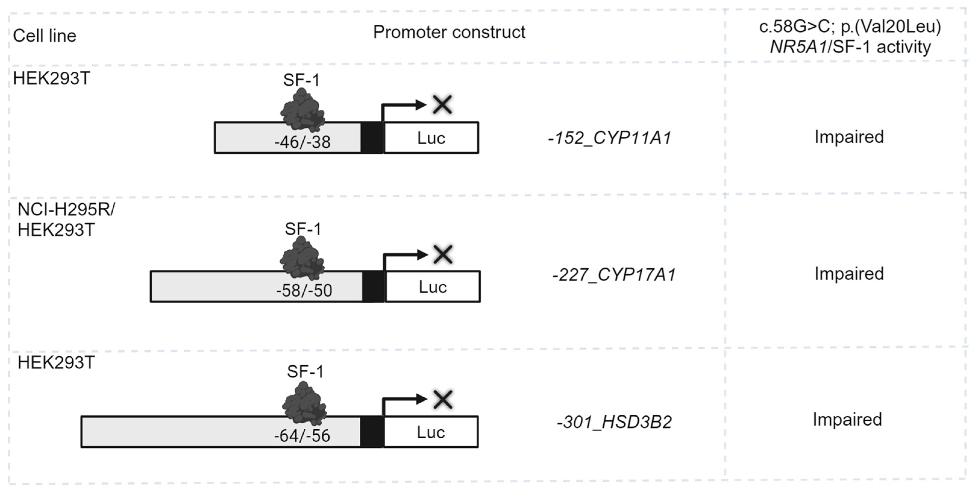
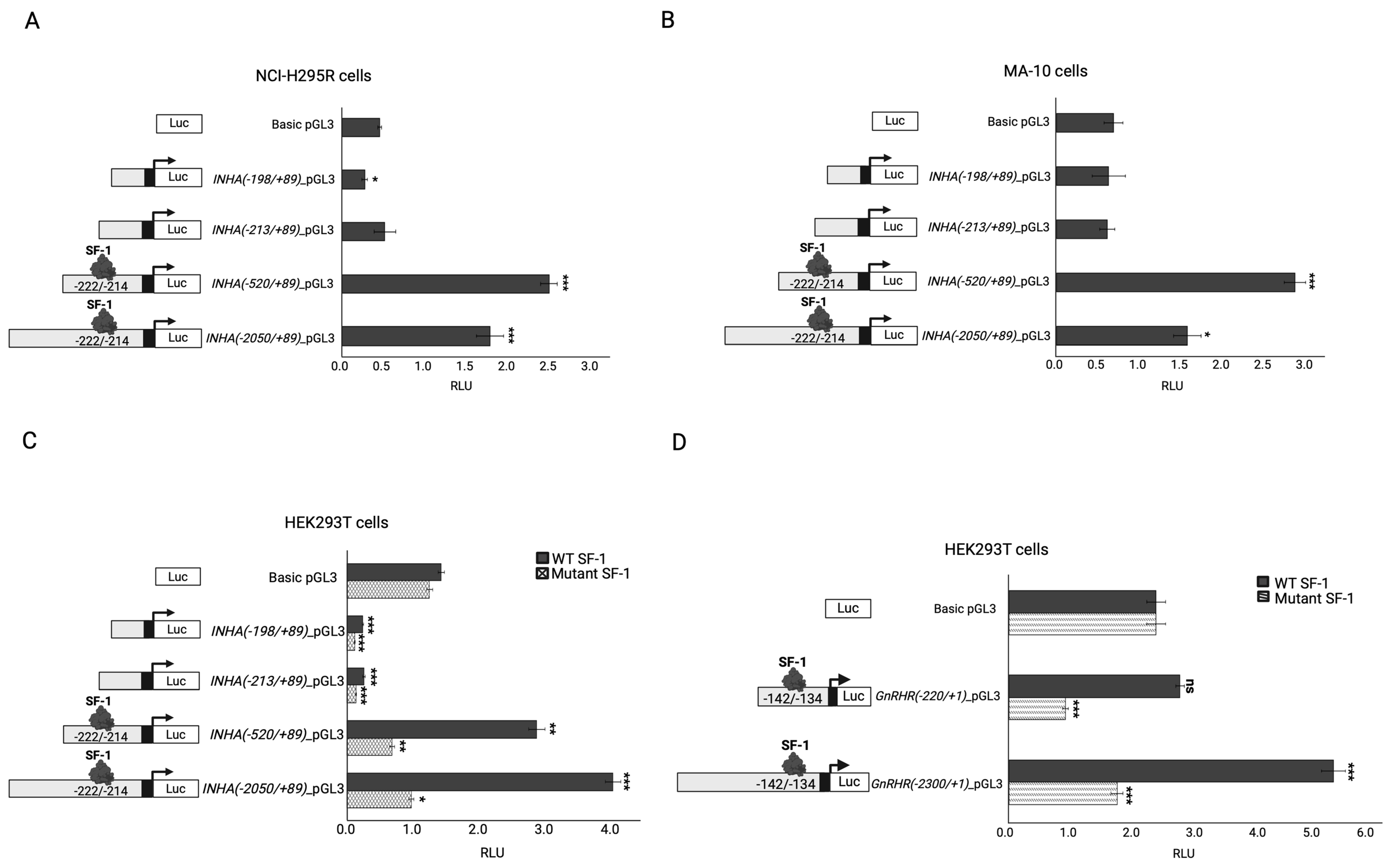
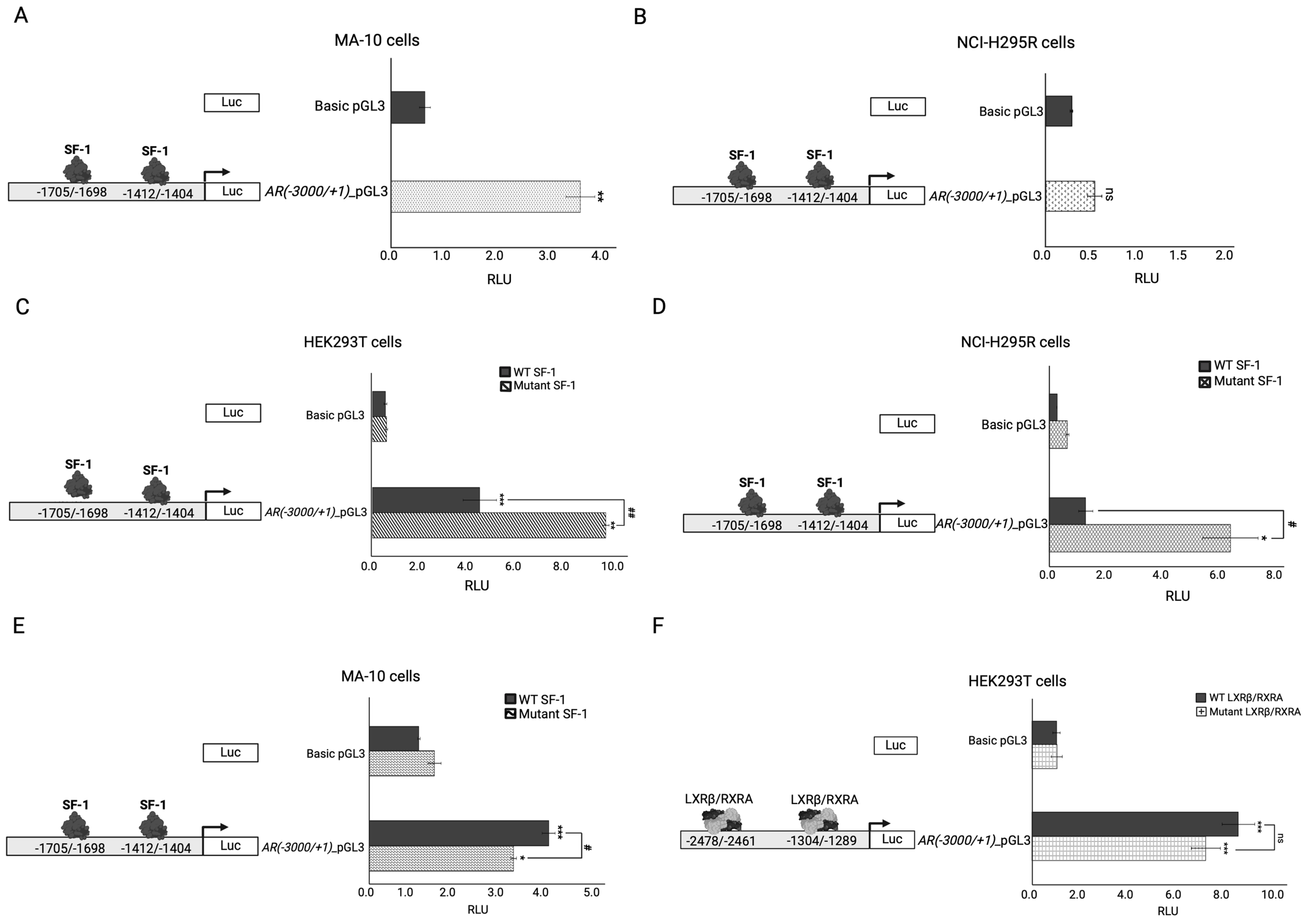
2.4. In Vitro Functional Testing of Selected Variants

The pathogenicity of the c.58G>C; p.(Val20Leu) NR5A1/SF-1 variant was previously assessed by activation testing on three different promoter constructs of three steroid enzymes (e.g., −152_CYP11A1, −227_CYP17A1 and −301_HSD3B2) in HEK293T and NCI-H295R cells, revealing impaired transcriptional activation on all three gene promoters (Figure 1; data reproduced from [27]). In pursuit of explaining the DSD phenotype of the patient in comparison with the healthy carrier father for the NR5A1/SF-1 variant, two additional variants in the INHA and NR1H2 genes were functionally studied in vitro for their possible disease-causing effect.
The transcriptional regulation of INHA by NR5A1/SF-1 was tested by transfecting four INHA promoter constructs in steroidogenic adrenal NCI-H295R cells and Leydig MA-10 cells, which both express endogenous NR5A1/SF-1. Only the two longer constructs, −520INHA and −2050INHA, containing a consensus NR5A1/SF-1 binding site (5′-TCATGGCCA-3′ at −222/−214) were activated by SF-1, while the two constructs lacking the NR5A1/SF-1 and/or cAMP-responsive element (5′-TGCGTCA-3′ at −205/−199) were not (Figure 2A,B). In order to confirm that this activation was specifically achieved by NR5A1/SF-1, the constructs were co-transfected with WT or variant c.58G>C; p.(Val20Leu) NR5A1/SF-1 in non-steroidogenic HEK293T cells that do not express NR5A1/SF-1. Similar results were found; only the constructs −520INHA and −2050INHA were activated by the WT NR5A1/SF-1 (Figure 2C). However, variant c.58G>C; p.(Val20Leu) NR5A1/SF-1 showed an impaired activation (Figure 2C) of INHA promoters. Overall, these results indicate that SF-1 is a transcriptional regulator of INHA expression.
As the role of INHA in sex development appears to be through the regulation of the hypothalamic–pituitary–gonadal (HPG) axis [37], we investigated the combined impact of NR5A1/SF-1, inhibin α, and activin A on GnRHR gene expression. WT SF-1 was found to activate the −2300GnRHR promoter construct harboring an NR5A1/SF-1 binding site at −142/−134, while mutant SF-1 showed a significantly lower activation (Figure 2D). By contrast, the addition of activin A, or the overexpression of WT INHA, in the absence or presence of NR5A1/SF-1, had no additional impact on −2300GnRHR promoter activation. To assess the impact of inhibin α on NR5A1 expression, we overexpressed INHA in adrenal NCI-H295R cells, which express endogenous NR5A1. However, neither WT nor mutant inhibin α had an effect on NR5A1 expression levels.
To test the impact of the identified variants in the NR1H2 and NR5A1/SF-1 genes, the androgen receptor (AR) was chosen as a target. The AR was reported to regulate NR1H2/LXβ expression [56] and to interact with NR5A1/SF-1 as part of the transcriptional machinery modulating the expression of specific genes (e.g., LHB) [57]. However, its regulation by these nuclear factors has not been reported so far. Therefore, we first tested whether the AR promoter is regulated by endogenous NR5A1/SF-1 (Figure 3A,B). The -3000AR promoter–reporter construct was significantly activated in steroidogenic MA-10 Leydig cells (Figure 3A); however, no activation was detected in the adrenal NCI-H295R cells (Figure 3B). The possible NR5A1/SF-1 binding sites in the AR promoter were searched manually, and we found 5′-TGACCTCT-3′ at −1705/−1698 and 5′-TGGCCTCC-3′ at −1412/−1404. Interestingly, the −3000AR construct was found to be differentially regulated by NR5A1/SF-1 overexpression in three different cell lines (Figure 3C–F). WT NR5A1/SF-1 significantly activated the −3000AR construct in HEK293T and MA-10 cells, but not in NCI-H295R cells (Figure 3C–E). Surprisingly, the mutant c.58G>C; p.(Val20Leu) NR5A1/SF-1 activated the AR construct in HEK293T and NCI-H295R cells (Figure 3C,D) more than in MA-10 Leydig cells (Figure 3E). Lastly, the AR was tested for its transcriptional regulation by LXRβ/RXRA in HEK293T cells. Both the WT and mutant c.515_516insCAA, p.(Arg171_Lys172insAsn) LXRβ/RXRA hetero-tetramers were able to significantly activate the −3000AR construct, but no significant difference was found for the variant (Figure 3F).

3. Discussion

NR5A1/SF-1 variants are reported in 46,XY and 46,XX individuals presenting with a variable severity of DSDs ranging from healthy to opposite sex phenotypes. So far, a genotype–phenotype correlation has not been found [5,6]. Oligogenic inheritance could be a possible explanation for this broad phenotype, where multiple gene variants may contribute to a unique DSD phenotype for each individual [5,6,9,10,12,27,58]. SF-1 has been reported to have many interacting partners. In several DSD individuals carrying NR5A1/SF-1 variants, additional variants in DSD-related genes have been described (Table S1). In this study, we investigated the possible genetic interplay of several variants of a 46,XY DSD individual. By conducting a WES analysis on both the healthy carrier father and the index patient, we identified five additional gene variants in the patient only. Four of these variants had not been previously reported. According to the literature, only the INHA and NR1H2 genes are involved in steroidogenesis, sex development, and/or reproduction, while the other genes are either involved in diabetes or cancer (Table 3).
NR5A1/SF-1 is a regulatory hub for numerous interacting partners [1]. Conducting functional assays, we were able to show that both INHA and NR1H2 are part of the NR5A1/SF-1 interaction network. A finding that was not previously reported.
The INHA gene encodes the α subunit needed for the assembly of the dimeric glycoproteins termed A and B inhibins that suppress FSH secretion from the pituitary and play an important role in modulating activin levels [37]. In addition, inhibins play a role in Sertoli and Leydig cell function, spermatogenesis, and sperm count [59]. The rat inhibin α subunit can be detected at a very early stage of testicular development following the formation of the testicular cord, and it is thought to play an important autocrine/paracrine role [60]. In mice, disruption of the Inha gene leads to the development of gonadal sex cord–stromal tumor and infertility [61]. In contrast, the human inhibin α subunit has been detected in the fetal testis only by 16 weeks of gestation following gonadal differentiation, specifically in interstitial and Sertoli cells; it contributes to normal testicular development [60]. Biallelic INHA variants were found to be associated with 46,XY DSD in humans [38,40]. A homozygous 2 bp deletion c.208_209delAG, p.(R70Gfs*3) in the INHA gene was found in two brothers with hypospadias, hypergonadotropic hypogonadism, gynecomastia, and azoospermia [38]. Still, the specific functional role of INHA in male sex development and reproduction is largely unknown.
In this study, we identified a regulatory NR5A1/SF-1 binding site in the human INHA gene promoter and show that INHA is transcriptionally regulated by NR5A1/SF-1. Relating this to the investigated DSD patient, his NR5A1/SF-1 variant showed reduced activity on the INHA promoter.
Investigating the specific contribution of the c.675T>G, p.(Ser225Arg) INHA variant to the patient’s DSD phenotype was more challenging. The c.675T>G, p.(Ser225Arg) INHA variant affects a highly conserved amino acid (Figure S1) located in the αN pro-domain in the inhibin α precursor protein, which is further processed to obtain its mature and active form [62]. Very little information is available regarding the function of this region and its underlying regulatory mechanisms. However, it is predicted to contribute to the proper folding, processing, and export of inhibins (predominantly inhibin B) from Sertoli cells in the testis to the serum [62,63,64]. It has been previously reported that Nr5a1/Sf-1 can stimulate Gnrhr expression in mice and humans [65,66]. Additionally, activin A was shown to enhance Gnrhr expression in mice; however, its role in regulating human GnRHR is unknown [67]. Therefore, we explored the potential collaborative activation of the human GnRHR gene by activin A and NR5A1/SF-1 and their inhibition by inhibin α. Unfortunately, we did not observe any additional increase in GnRHR promoter activity by activin A. Similarly, upon the addition of the WT inhibin α, GnRHR promoter activity was not affected in the presence or absence of activin A. Therefore, the mechanistic proof of the contribution of the c.675T>G, p.(Ser225Arg) INHA variant to the phenotype found in our patient remains elusive.
Another variant identified in the patient was in the NR1H2 gene, which encodes the liver X receptor β (LXRβ), an important modulator of lipid and cholesterol homeostasis [68]. It forms an obligate heterodimer with the retinoid X receptor (RXR) to govern gene transcription by binding to specific LXR-responsive elements [68]. The Nr1h2 gene was found to be strongly expressed at 16.5 days postcoitum (dpc) in the mouse embryonic testis, specifically in Sertoli cells, where its expression persists into adulthood [69]. lxrβ−/− knockout mice present with excessive cholesterol accumulation in Sertoli cells and dysregulated spermatogenesis, while lxrαβ−/− mice present with a severe infertility phenotype [70]. Similarly, lower expression levels of NR1H2 were detected in the testis of infertile men with azoospermia [35,36]. However, the specific function of NR1H2 in the human developing testis has not been elucidated.
Due to the fact that both NR1H2 and NR5A1/SF-1 play important roles in androgen homeostasis and male fertility [1,33,35,36,70,71,72], we tested their transcriptional activity on the AR promoter. Functional studies showed that NR5A1/SF-1 is a cell-specific transcriptional regulator of the AR in Leydig MA-10 cells but not in adrenal NCI-H295R cells. Overexpression of WT NR5A1/SF-1 enhanced the AR reporter activity.
Going back to our patient, we found that the mutant NR5A1/SF-1 was not able to activate the AR to the same degree as WT in MA-10 Leydig cells. By contrast, transactivation studies of the NR5A1/SF-1 variant in adrenal NCI-H295R and non-steroidogenic HEK293T cells revealed contradictory results, suggesting that the specific background of the Leydig cell is necessary for showing that specific interplay.
AR activity is regulated by complex mechanisms [73]. It is influenced by various transcription factors and coregulators involved in multiple cellular pathways [73,74,75]. The most recent study showed that AR activity is modulated by the transcription factor disheveled-associated activator of morphogenesis 2 (DAAM2), a cytoskeletal regulator of formin and actin. In vitro studies of genital skin-derived fibroblasts (GSFs) from patients with androgen insensitivity syndrome (AIS) type II and DAAM2 variants showed reduced dihydrotestosterone (DHT)-induced AR activity compared to WT GSFs [74]. Moreover, the AR is epigenetically regulated; alterations in methylated CpG regions within the proximal AR promoter were found to inhibit AR transcription in GSFs from several patients with AIS type II [75]. In our study, we show that the LXRβ/RXRA heterodimer is a transcriptional activator of the AR, strengthening NR1H2/LXRβ association with male fertility in line with previous reports [33,35,36,70]. However, the c.515_516insCAA; p.(Arg171_Lys172insAsn) NR1H2/LXRβ VUS had a similar transcriptional activity on the AR reporter as WT; thus, its contribution to the DSD phenotype is in doubt.
The proper reporting of oligogenic variant combinations requires thorough genetic testing and functional evidence of the pathogenicity of the causal variants [8,26]. Advancements in NGS methods (WES, whole genome sequencing) have significantly enhanced the yield from efforts to identify the possible genetic causes of DSDs [25], and this is especially true for gene variants that occur in combination with NR5A1/SF-1 variants. In fact, more than 70 different gene variants have been reported in association with NR5A1/SF-1 variants in individuals with DSD (Table S1) [6,9,11,12,13,14,58,76]. We performed a WES analysis on individuals with DSD and NR5A1/SF-1 variants as part of the SF1next study [5] and found several additional novel gene variants (unpublished data), suggesting digenic or oligogenic causation for the disease. To confirm an oligogenic disease mechanism can be difficult, as often, appropriate experimental models are missing to account for multiple genetic hits and/or the smaller effect size assumed for individual variants occurring in combination. In our 46,XY DSD index patient, five gene variants were identified, of which only the NR5A1/SF-1 variant was also found in the healthy father. While the variants in TCF7L2, NIBAN1, and SCUBE2 were deemed irrelevant for the observed phenotype, the NR1H2 and INHA genes were found to be interacting partners of NR5A1/SF-1. However, our efforts to show the disease-causing effects of the identified variants in the NR1H2 and INHA genes of our index case through functional studies were not successful.
Future studies using patient-derived biomaterials may help in assessing oligogenic mechanisms. The cellular reprogramming of induced pluripotent stem cells (iPSCs) carrying the specific individual’s genetic background may inform us about the variants’ effects on steroidogenesis and sex development. Recently, in vitro systems for the differentiation of iPSCs towards gonadal progenitors and Sertoli-like cells have been established [77,78]. Rescue experiments in iPSCs originating from a 46,XY DSD patient with an NR5A1/SF-1 variation showed the disease mechanism on sex determination [77]. Unfortunately, even these promising models have limitations, including the availability of patients’ biological materials and the variability and difficulty in obtaining the robust maturation of fully functional iPSC-derived somatic cells (e.g., Sertoli- and Leydig-like cells). Therefore, even these experiments may not fully recapitulate the phenotype when used for disease modeling. Moreover, the challenge to rescue multiple combined variants and assess their effect on the overall phenotype remains.
In conclusion, we provide new functional evidence that NR5A1/SF-1 regulates the transcription of the AR and INHA genes. In addition, we show that NR1H2/LXRβ, a modulator of lipid and cholesterol homeostasis in the testis, regulates the AR. Variants in NR5A1/SF-1, INHA, and AR have been previously reported to cause monogenic DSD. Our study was not able to provide functional proof of the disease-causing effects of specific variants in NR5A1/SF-1, INHA, and NR1H2/LXRβ identified in combination in a 46,XY DSD individual. Addressing the possible oligogenic mode of these mechanisms remains a challenge.

4. Materials and Methods

4.1. Participants

The patient and his father included in this work were part of two previous genetic studies [9,27] and the SF1next study [5].

4.2. In Silico Analyses and Variant Classification

The DNA of the index patient and the father were sequenced by WES (Novogene, Cambridge, UK) and analyzed with an in-house specific data-filtering algorithm for gene variants related to DSD and/or NR5A1/SF-1 [9,11]. We predicted the possible effect of the identified genetic variants on the structure and function of the protein using Polyphen-2, (Polymorphism Phenotyping v2, http://genetics.bwh.harvard.edu/pph2/) (accessed on 7 February 2023), Panther (Protein ANalysis THrough Evolutionary Relationships, http://www.pantherdb.org/tools/csnpScore.do) (accessed on 7 February 2023), SNPs and Go (https://snps-and-go.biocomp.unibo.it/snps-and-go/) (accessed on 7 February 2023), CADD (Combined Annotation Dependent Depletion, https://cadd.gs.washington.edu/) (accessed on 7 February 2023), and the calibrated scores given by VarSome [28] for Revel (Rare Exome Variant Ensemble Learner), SIFT (scale-invariant feature transform), Provean (Protein Variation Effect Analyzer), Mutation Taster, and M-CAP (Mendelian Clinically Applicable Pathogenicity). Variants were classified for pathogenicity according to the standards and guidelines of the ACMG using VarSome version v.11.9.0 [28]. We explored the possible pathogenicity of multiple variants’ combinatory effects using ORVAL version 2.2.0 (Oligogenic Resource for Variant AnaLysis) [79].

4.3. Plasmids

The human HA-tagged WT and the variant c.58G>C cDNA of NR5A1/SF-1 (NM_004959.5) in pcDNA3, empty control vector pcDNA3, and Renilla-TK (pRL-TK) were all available from previous work [27]. The human NR1H2 cDNA (NM_007121.5) in pCMV3-C-HA and RXRA cDNA (NM_002957.5) in the pCMV3 vector were purchased (Sino Biologicals Inc, Eschborn, Germany). The human NR1H2 cDNA was used as a template to generate the NR1H2 variant expression vector by PCR-based site-directed mutagenesis using the primers, forward (5′-CGGAAGAAGAAGATTCGGAACAAACAGCAGCAGGAG-3′), reverse (5′-CTCCTGCTGCTGTTTGTTCCGAATCTTCTTCTTCCG-3′), and the QuickChange protocol by Stratagene (Agilent Technologies Inc., Santa Clara, CA, USA).

4.4. Cloning

The 5′-untranslated region constructs of the different genes were produced by PCR using control human DNA extracted from blood leukocytes using the DNA isolation kit of Qiagen (QIAGEN, Aarhus, Denmark). The different forward primers used for PCR were as follows: −2056_INHA (5′-AGAGAGGGTACCTTGAGCACGAAGCCGCC-3′), −520_INHA (5′-AGA GAGGGTACCCTGAGGGGTGATGCACTTTGTC-3′), −213_INHA (5′-GAGGGTACCCA GACATCTGCGTCAGAGATAGGAG-3′), −198_INHA (5′-AGAGGGTACCGAGATAGGA GGTCTCAATGCCACG-3′). All had the KpnI restriction site included, while the following reverse primer, including the XhoI restriction site, was used in the four PCR reactions, (5′-GAGAGACTCGAGAGAACAAGTTCCCGGGCCAG-3′). For the generation of the −220_GnRHR construct, the forward primer including the KpnI restriction site (5′-AGAGGTACCGGCCTGCTCTGTTTTAGCACT-3′) and the reverse primer including the XhoI restriction site (5′-GAGCTCGAGATTTTCCCAGGACAGAGCTTCAAG-3′) were used. For the generation of the −3000_AR construct, the forward primer including the HindIII restriction site (5′-AGAGAAGCTTTAAACTTTGGAGTCTTTCAGACCCAG-3′), and the reverse primer including the XhoI restriction site (5′-GAGACTCGAGCCTTGAG CTTGGCTGAATCTTCC-3′) were used in the PCR reaction. All PCR products were digested with the indicated restriction enzymes and subcloned into the corresponding site in the pGL3 basic vector (Promega, Madison, WI, USA). The constructs were confirmed by direct sequencing. The −2300_GnRHR promoter construct in pGL3 was custom-made by Genscript (Rijswijk, the Netherlands).

4.5. In Vitro Testing of Transactivation Activity by Dual Luciferase Assay

Non-steroidogenic, human embryonic kidney HEK293T cells (ATCC CRL-1573), steroidogenic NCI-H295R adrenal cells (ATCC CRL-2128), and mouse Leydig MA-10 cells (ATCC CRL-3050) were cultured as previously described [27,80]. For all promoter activity experiments, cells were cultured on 12-well plates. For the INHA promoter activity experiments, NCI-H295R and MA-10 steroidogenic cells were transiently transfected with 950 ng from the different promoter luciferase reporter constructs, −2050INHA_pGL3, −520INHA_pGL3, −213INHA_pGL3, or −198INHA_pGL3; whereas HEK293T cells were transiently transfected with 200 ng WT or mutant NR5A1/SF-1 expression vectors, 800 ng of the different promoter luciferase reporter construct −2050INHA_pGL3, −520INHA_pGL3, −213INHA_pGL3, or −198INHA_pGL3 separately. For the GnRHR promoter activity experiment, HEK293T cells were transiently co-transfected with 200 ng WT or mutant NR5A1/SF-1 expression vectors and 800 ng of the promoter luciferase reporter constructs −2300GnRHR_pGL3 or −220GnRHR_pGL3. For the AR promoter activity experiments, MA-10 or NCI-H295R cells were transiently transfected with 950 ng of the −3000AR_pGL3 promoter luciferase reporter construct, while for the AR promoter experiments with overexpressed NR5A1/SF-1 or NR1H2/LXRβ, the cell lines were transiently transfected with 600 ng of the −3000AR_pGL3 promoter and 200 ng WT or mutant of NR5A1/SF-1 expression vector (in the three cell lines), or with NR1H2/LXRβ and RXRA expression vectors. Lastly, 50 ng of the pRL-TK vector was used as an internal control in all transfection experiments. All transfections were carried out with Lipofectamine 2000TM (Invitrogen, Glasgow, UK) in Opti-MEM (1X)-reduced serum medium (Gibco, Thermo Fisher Scientific, Waltham, MA, USA). Forty-eight hours after transfection, the cells were washed with PBS, lysed, and assayed for luciferase activity with a Dual-Luciferase assay using a microplate Luminometer reader (Fluoroskan Ascent® FL and Fluoroskan Ascent®, Thermo Fisher, Waltham, MA, USA). Specific Firefly luciferase readings were standardized against Renilla luciferase control readings. Experiments were repeated three to five times in duplicates, and the data were summarized, giving the mean ± standard error of the mean (SEM). Statistical significance was examined by Student’s t-test (GraphPad Prism, GraphPad Software version 9.4.1.681, Boston, MA, USA). Significance was assumed with a p-value of less than 0.05.

Supplementary Materials

The following supporting information can be downloaded at: https://www.mdpi.com/article/10.3390/ijms251810109/s1. References [81,82,83,84,85,86,87,88,89,90,91,92,93,94] are cited in the Supplementary Materials.

Author Contributions

Conceptualization, C.E.F.; methodology, R.N.E.; software, R.N.E. and C.K.; validation, R.N.E. and C.E.F.; formal analysis, R.N.E.; investigation, R.N.E.; resources, C.E.F.; data curation, R.N.E. and C.E.F.; writing—original draft preparation, R.N.E. and C.E.F.; writing—review and editing, R.N.E., C.K., I.M.d.L., K.-S.S., F.M., N.C.-T. and C.E.F.; visualization, R.N.E. and C.E.F.; supervision, C.E.F.; project administration, C.E.F.; funding acquisition, C.E.F. All authors have read and agreed to the published version of the manuscript.

Funding

This study was supported by a project grant from the Swiss National Science Foundation (320030-197725). I.M.d.L. is supported by a Postdoctoral Fellowship Grant from the Education Department of the Basque Government (Spain) (POS_2020_1_0034).

Institutional Review Board Statement

This study was conducted in accordance with the Declaration of Helsinki and approved by the Institutional Review Board of the Hospital Infantil La Fe, Valencia, Spain, and the Ethics Committee of Canton Bern, Switzerland (BASEC 2016-01210, approved on 15 July 2016).

Informed Consent Statement

Informed consent was obtained from all subjects involved in the study.

Data Availability Statement

Data were collected in a project-specific REDCap database governed by the Clinical Trials Unit (CTU) at the University of Bern, Switzerland. The genetic data are also stored on the servers of the University of Bern. These data can also be accessed upon reasonable request according to ethical considerations and informed consent.

Acknowledgments

We thank the patient and the family for providing their medical data and trust to this research study.

Conflicts of Interest

The authors declare no conflicts of interest.

References

  1. Schimmer, B.P.; White, P.C. Minireview: Steroidogenic factor 1: Its roles in differentiation, development, and disease. Mol. Endocrinol. 2010, 24, 1322–1337. [Google Scholar] [PubMed]
  2. Wong, M.; Ikeda, Y.; Luo, X.; Caron, K.M.; Weber, T.J.; Swain, A.; Schimmer, B.P.; Parker, K.L. Steroidogenic factor 1 plays multiple roles in endocrine development and function. Recent. Prog. Horm. Res. 1997, 52, 167–182, discussion 182–184. [Google Scholar]
  3. Luo, X.; Ikeda, Y.; Parker, K.L. A cell-specific nuclear receptor is essential for adrenal and gonadal development and sexual differentiation. Cell 1994, 77, 481–490. [Google Scholar]
  4. Bland, M.L.; Jamieson, C.A.; Akana, S.F.; Bornstein, S.R.; Eisenhofer, G.; Dallman, M.F.; Ingraham, H.A. Haploinsufficiency of steroidogenic factor-1 in mice disrupts adrenal development leading to an impaired stress response. Proc. Natl. Acad. Sci. USA 2000, 97, 14488–14493. [Google Scholar] [PubMed]
  5. Kouri, C.; Sommer, G.; Martinez de Lapiscina, I.; Elzenaty, R.N.; Tack, L.J.W.; Cools, M.; Ahmed, S.F.; Fluck, C.E.; SF1next Study Group. Clinical and genetic characteristics of a large international cohort of individuals with rare NR5A1/SF-1 variants of sex development. EBioMedicine 2024, 99, 104941. [Google Scholar]
  6. Naamneh Elzenaty, R.; Martinez de Lapiscina, I.; Kouri, C.; Sauter, K.S.; Sommer, G.; Castano, L.; Fluck, C.E.; SF1next Study Group. Characterization of 35 novel NR5A1/SF-1 variants identified in individuals with atypical sexual development: The SF1next study. J. Clin. Endocrinol. Metab. 2024. [Google Scholar] [CrossRef]
  7. Fabbri-Scallet, H.; de Sousa, L.M.; Maciel-Guerra, A.T.; Guerra-Junior, G.; de Mello, M.P. Mutation update for the NR5A1 gene involved in DSD and infertility. Hum. Mutat. 2020, 41, 58–68. [Google Scholar]
  8. Richards, S.; Aziz, N.; Bale, S.; Bick, D.; Das, S.; Gastier-Foster, J.; Grody, W.W.; Hegde, M.; Lyon, E.; Spector, E.; et al. Standards and guidelines for the interpretation of sequence variants: A joint consensus recommendation of the American College of Medical Genetics and Genomics and the Association for Molecular Pathology. Genet. Med. 2015, 17, 405–424. [Google Scholar]
  9. Camats, N.; Fernandez-Cancio, M.; Audi, L.; Schaller, A.; Fluck, C.E. Broad phenotypes in heterozygous NR5A1 46,XY patients with a disorder of sex development: An oligogenic origin? Eur. J. Hum. Genet. 2018, 26, 1329–1338. [Google Scholar]
  10. Camats, N.; Fluck, C.E.; Audi, L. Oligogenic Origin of Differences of Sex Development in Humans. Int. J. Mol. Sci. 2020, 21, 1809. [Google Scholar] [CrossRef]
  11. Martinez de Lapiscina, I.; Kouri, C.; Aurrekoetxea, J.; Sanchez, M.; Naamneh Elzenaty, R.; Sauter, K.S.; Camats, N.; Grau, G.; Rica, I.; Rodriguez, A.; et al. Genetic reanalysis of patients with a difference of sex development carrying the NR5A1/SF-1 variant p.Gly146Ala has discovered other likely disease-causing variations. PLoS ONE 2023, 18, e0287515. [Google Scholar]
  12. Martinez de LaPiscina, I.; Mahmoud, R.A.; Sauter, K.S.; Esteva, I.; Alonso, M.; Costa, I.; Rial-Rodriguez, J.M.; Rodriguez-Estevez, A.; Vela, A.; Castano, L.; et al. Variants of STAR, AMH and ZFPM2/FOG2 May Contribute towards the Broad Phenotype Observed in 46,XY DSD Patients with Heterozygous Variants of NR5A1. Int. J. Mol. Sci. 2020, 21, 8554. [Google Scholar] [CrossRef]
  13. Zidoune, H.; Ladjouze, A.; Chellat-Rezgoune, D.; Boukri, A.; Dib, S.A.; Nouri, N.; Tebibel, M.; Sifi, K.; Abadi, N.; Satta, D.; et al. Novel Genomic Variants, Atypical Phenotypes and Evidence of a Digenic/Oligogenic Contribution to Disorders/Differences of Sex Development in a Large North African Cohort. Front. Genet. 2022, 13, 900574. [Google Scholar]
  14. Wang, H.; Zhang, L.; Wang, N.; Zhu, H.; Han, B.; Sun, F.; Yao, H.; Zhang, Q.; Zhu, W.; Cheng, T.; et al. Next-generation sequencing reveals genetic landscape in 46, XY disorders of sexual development patients with variable phenotypes. Hum. Genet. 2018, 137, 265–277. [Google Scholar]
  15. Gach, A.; Pinkier, I.; Wysocka, U.; Salacinska, K.; Salachna, D.; Szarras-Czapnik, M.; Pietrzyk, A.; Sakowicz, A.; Nykel, A.; Rutkowska, L.; et al. New findings in oligogenic inheritance of congenital hypogonadotropic hypogonadism. Arch. Med. Sci. 2022, 18, 353–364. [Google Scholar]
  16. de Filippis, T.; Gelmini, G.; Paraboschi, E.; Vigone, M.C.; Di Frenna, M.; Marelli, F.; Bonomi, M.; Cassio, A.; Larizza, D.; Moro, M.; et al. A frequent oligogenic involvement in congenital hypothyroidism. Hum. Mol. Genet. 2017, 26, 2507–2514. [Google Scholar] [PubMed]
  17. Oliver-Petit, I.; Edouard, T.; Jacques, V.; Bournez, M.; Cartault, A.; Grunenwald, S.; Savagner, F. Next-Generation Sequencing Analysis Reveals Frequent Familial Origin and Oligogenism in Congenital Hypothyroidism With Dyshormonogenesis. Front. Endocrinol. 2021, 12, 657913. [Google Scholar]
  18. Sykiotis, G.P.; Plummer, L.; Hughes, V.A.; Au, M.; Durrani, S.; Nayak-Young, S.; Dwyer, A.A.; Quinton, R.; Hall, J.E.; Gusella, J.F.; et al. Oligogenic basis of isolated gonadotropin-releasing hormone deficiency. Proc. Natl. Acad. Sci. USA 2010, 107, 15140–15144. [Google Scholar]
  19. Hughes, L.A.; McKay-Bounford, K.; Webb, E.A.; Dasani, P.; Clokie, S.; Chandran, H.; McCarthy, L.; Mohamed, Z.; Kirk, J.M.W.; Krone, N.P.; et al. Next generation sequencing (NGS) to improve the diagnosis and management of patients with disorders of sex development (DSD). Endocr. Connect. 2019, 8, 100–110. [Google Scholar]
  20. Werner, R.; Monig, I.; Lunstedt, R.; Wunsch, L.; Thorns, C.; Reiz, B.; Krause, A.; Schwab, K.O.; Binder, G.; Holterhus, P.M.; et al. New NR5A1 mutations and phenotypic variations of gonadal dysgenesis. PLoS ONE 2017, 12, e0176720. [Google Scholar]
  21. Mazen, I.; Abdel-Hamid, M.; Mekkawy, M.; Bignon-Topalovic, J.; Boudjenah, R.; El Gammal, M.; Essawi, M.; Bashamboo, A.; McElreavey, K. Identification of NR5A1 Mutations and Possible Digenic Inheritance in 46,XY Gonadal Dysgenesis. Sex. Dev. 2016, 10, 147–151. [Google Scholar] [PubMed]
  22. de Oliveira, F.R.; Mazzola, T.N.; de Mello, M.P.; Francese-Santos, A.P.; Lemos-Marini, S.H.V.; Maciel-Guerra, A.T.; Hiort, O.; Werner, R.; Guerra-Junior, G.; Fabbri-Scallet, H. DHX37 and NR5A1 Variants Identified in Patients with 46,XY Partial Gonadal Dysgenesis. Life 2023, 13, 1093. [Google Scholar] [CrossRef] [PubMed]
  23. Audi, L.; Ahmed, S.F.; Krone, N.; Cools, M.; McElreavey, K.; Holterhus, P.M.; Greenfield, A.; Bashamboo, A.; Hiort, O.; Wudy, S.A.; et al. Genetics in Endocrinology: Approaches to molecular genetic diagnosis in the management of differences/disorders of sex development (DSD): Position paper of EU COST Action BM 1303 ‘DSDnet’. Eur. J. Endocrinol. 2018, 179, R197–R206. [Google Scholar] [PubMed]
  24. Alhomaidah, D.; McGowan, R.; Ahmed, S.F. The current state of diagnostic genetics for conditions affecting sex development. Clin. Genet. 2017, 91, 157–162. [Google Scholar]
  25. Delot, E.C.; Vilain, E. Towards improved genetic diagnosis of human differences of sex development. Nat. Rev. Genet. 2021, 22, 588–602. [Google Scholar]
  26. Papadimitriou, S.; Gravel, B.; Nachtegael, C.; De Baere, E.; Loeys, B.; Vikkula, M.; Smits, G.; Lenaerts, T. Toward reporting standards for the pathogenicity of variant combinations involved in multilocus/oligogenic diseases. HGG Adv. 2023, 4, 100165. [Google Scholar]
  27. Camats, N.; Pandey, A.V.; Fernandez-Cancio, M.; Andaluz, P.; Janner, M.; Toran, N.; Moreno, F.; Bereket, A.; Akcay, T.; Garcia-Garcia, E.; et al. Ten novel mutations in the NR5A1 gene cause disordered sex development in 46,XY and ovarian insufficiency in 46,XX individuals. J. Clin. Endocrinol. Metab. 2012, 97, E1294–E1306. [Google Scholar]
  28. Kopanos, C.; Tsiolkas, V.; Kouris, A.; Chapple, C.E.; Albarca Aguilera, M.; Meyer, R.; Massouras, A. VarSome: The human genomic variant search engine. Bioinformatics 2019, 35, 1978–1980. [Google Scholar] [PubMed]
  29. Lin, L.; Achermann, J.C. Steroidogenic factor-1 (SF-1, Ad4BP, NR5A1) and disorders of testis development. Sex. Dev. 2008, 2, 200–209. [Google Scholar]
  30. Bashamboo, A.; McElreavey, K. Human sex-determination and disorders of sex-development (DSD). Semin. Cell Dev. Biol. 2015, 45, 77–83. [Google Scholar]
  31. Budefeld, T.; Tobet, S.A.; Majdic, G. Altered position of cell bodies and fibers in the ventromedial region in SF-1 knockout mice. Exp. Neurol. 2011, 232, 176–184. [Google Scholar] [CrossRef] [PubMed]
  32. Zhao, C.; Dahlman-Wright, K. Liver X receptor in cholesterol metabolism. J. Endocrinol. 2010, 204, 233–240. [Google Scholar] [CrossRef] [PubMed]
  33. Robertson, K.M.; Schuster, G.U.; Steffensen, K.R.; Hovatta, O.; Meaney, S.; Hultenby, K.; Johansson, L.C.; Svechnikov, K.; Soder, O.; Gustafsson, J.A. The liver X receptor-beta is essential for maintaining cholesterol homeostasis in the testis. Endocrinology 2005, 146, 2519–2530. [Google Scholar] [CrossRef]
  34. Nilsson, M.; Stulnig, T.M.; Lin, C.Y.; Yeo, A.L.; Nowotny, P.; Liu, E.T.; Steffensen, K.R. Liver X receptors regulate adrenal steroidogenesis and hypothalamic-pituitary-adrenal feedback. Mol. Endocrinol. 2007, 21, 126–137. [Google Scholar] [CrossRef] [PubMed]
  35. Rondanino, C.; Ouchchane, L.; Chauffour, C.; Marceau, G.; Dechelotte, P.; Sion, B.; Pons-Rejraji, H.; Janny, L.; Volle, D.H.; Lobaccaro, J.M.; et al. Levels of liver X receptors in testicular biopsies of patients with azoospermia. Fertil. Steril. 2014, 102, 361–371. [Google Scholar] [CrossRef]
  36. Jarvis, S.; Williamson, C.; Bevan, C.L. Liver X Receptors and Male (In)fertility. Int. J. Mol. Sci. 2019, 20, 5379. [Google Scholar] [CrossRef]
  37. Barakat, B.; Itman, C.; Mendis, S.H.; Loveland, K.L. Activins and inhibins in mammalian testis development: New models, new insights. Mol. Cell Endocrinol. 2012, 359, 66–77. [Google Scholar] [CrossRef] [PubMed]
  38. Arslan Ates, E.; Eltan, M.; Sahin, B.; Gurpinar Tosun, B.; Seven Menevse, T.; Geckinli, B.B.; Greenfield, A.; Turan, S.; Bereket, A.; Guran, T. Homozygosity for a novel INHA mutation in two male siblings with hypospadias, primary hypogonadism, and high-normal testicular volume. Eur. J. Endocrinol. 2022, 186, K25–K31. [Google Scholar] [PubMed]
  39. Sundblad, V.; Chiauzzi, V.A.; Andreone, L.; Campo, S.; Charreau, E.H.; Dain, L. Controversial role of inhibin alpha-subunit gene in the aetiology of premature ovarian failure. Hum. Reprod. 2006, 21, 1154–1160. [Google Scholar] [CrossRef]
  40. Huhtaniemi, I. The first report on homozygous INHA inactivation in humans. Eur. J. Endocrinol. 2022, 187, C1–C2. [Google Scholar] [CrossRef]
  41. Matzuk, M.M.; Finegold, M.J.; Mather, J.P.; Krummen, L.; Lu, H.; Bradley, A. Development of cancer cachexia-like syndrome and adrenal tumors in inhibin-deficient mice. Proc. Natl. Acad. Sci. USA 1994, 91, 8817–8821. [Google Scholar] [CrossRef] [PubMed]
  42. Ito, M.; Park, Y.; Weck, J.; Mayo, K.E.; Jameson, J.L. Synergistic activation of the inhibin alpha-promoter by steroidogenic factor-1 and cyclic adenosine 3′,5′-monophosphate. Mol. Endocrinol. 2000, 14, 66–81. [Google Scholar] [PubMed]
  43. Korinek, V.; Barker, N.; Moerer, P.; van Donselaar, E.; Huls, G.; Peters, P.J.; Clevers, H. Depletion of epithelial stem-cell compartments in the small intestine of mice lacking Tcf-4. Nat. Genet. 1998, 19, 379–383. [Google Scholar] [CrossRef] [PubMed]
  44. Nateri, A.S.; Spencer-Dene, B.; Behrens, A. Interaction of phosphorylated c-Jun with TCF4 regulates intestinal cancer development. Nature 2005, 437, 281–285. [Google Scholar] [CrossRef] [PubMed]
  45. Lili, L.N.; Farkas, A.E.; Gerner-Smidt, C.; Overgaard, C.E.; Moreno, C.S.; Parkos, C.A.; Capaldo, C.T.; Nusrat, A. Claudin-based barrier differentiation in the colonic epithelial crypt niche involves Hopx/Klf4 and Tcf7l2/Hnf4-alpha cascades. Tissue Barriers 2016, 4, e1214038. [Google Scholar] [CrossRef]
  46. Angus-Hill, M.L.; Elbert, K.M.; Hidalgo, J.; Capecchi, M.R. T-cell factor 4 functions as a tumor suppressor whose disruption modulates colon cell proliferation and tumorigenesis. Proc. Natl. Acad. Sci. USA 2011, 108, 4914–4919. [Google Scholar] [CrossRef]
  47. Boj, S.F.; van Es, J.H.; Huch, M.; Li, V.S.; Jose, A.; Hatzis, P.; Mokry, M.; Haegebarth, A.; van den Born, M.; Chambon, P.; et al. Diabetes risk gene and Wnt effector Tcf7l2/TCF4 controls hepatic response to perinatal and adult metabolic demand. Cell 2012, 151, 1595–1607. [Google Scholar] [CrossRef]
  48. Helgason, A.; Palsson, S.; Thorleifsson, G.; Grant, S.F.; Emilsson, V.; Gunnarsdottir, S.; Adeyemo, A.; Chen, Y.; Chen, G.; Reynisdottir, I.; et al. Refining the impact of TCF7L2 gene variants on type 2 diabetes and adaptive evolution. Nat. Genet. 2007, 39, 218–225. [Google Scholar] [CrossRef]
  49. Freathy, R.M.; Weedon, M.N.; Bennett, A.; Hypponen, E.; Relton, C.L.; Knight, B.; Shields, B.; Parnell, K.S.; Groves, C.J.; Ring, S.M.; et al. Type 2 diabetes TCF7L2 risk genotypes alter birth weight: A study of 24,053 individuals. Am. J. Hum. Genet. 2007, 80, 1150–1161. [Google Scholar] [CrossRef]
  50. Gummow, B.M.; Winnay, J.N.; Hammer, G.D. Convergence of Wnt signaling and steroidogenic factor-1 (SF-1) on transcription of the rat inhibin alpha gene. J. Biol. Chem. 2003, 278, 26572–26579. [Google Scholar] [CrossRef]
  51. Diana, P.; Carvalheira, G.M.G. NIBAN1, Exploring its Roles in Cell Survival Under Stress Context. Front. Cell Dev. Biol. 2022, 10, 867003. [Google Scholar] [CrossRef] [PubMed]
  52. Sun, G.D.; Kobayashi, T.; Abe, M.; Tada, N.; Adachi, H.; Shiota, A.; Totsuka, Y.; Hino, O. The endoplasmic reticulum stress-inducible protein Niban regulates eIF2alpha and S6K1/4E-BP1 phosphorylation. Biochem. Biophys. Res. Commun. 2007, 360, 181–187. [Google Scholar] [CrossRef] [PubMed]
  53. Esmaeili, R.; Mohammadi, S.; Jafarbeik-Iravani, N.; Yadegari, F.; Olfatbakhsh, A.; Mazaheri, M.; Kaviani, A.; Rezaee, M.; Majidzadeh, A.K. Expression of SCUBE2 and BCL2 Predicts Favorable Response in ERalpha Positive Breast Cancer. Arch. Iran. Med. 2021, 24, 209–217. [Google Scholar] [CrossRef]
  54. Lin, Y.C.; Chen, C.C.; Cheng, C.J.; Yang, R.B. Domain and functional analysis of a novel breast tumor suppressor protein, SCUBE2. J. Biol. Chem. 2011, 286, 27039–27047. [Google Scholar] [CrossRef]
  55. Lin, Y.C.; Roffler, S.R.; Yan, Y.T.; Yang, R.B. Disruption of Scube2 Impairs Endochondral Bone Formation. J. Bone Miner. Res. 2015, 30, 1255–1267. [Google Scholar] [CrossRef]
  56. Krycer, J.R.; Brown, A.J. Cross-talk between the androgen receptor and the liver X receptor: Implications for cholesterol homeostasis. J. Biol. Chem. 2011, 286, 20637–20647. [Google Scholar] [CrossRef]
  57. Jorgensen, J.S.; Nilson, J.H. AR suppresses transcription of the LHbeta subunit by interacting with steroidogenic factor-1. Mol. Endocrinol. 2001, 15, 1505–1516. [Google Scholar] [PubMed]
  58. Mazen, I.; Mekkawy, M.; Kamel, A.; Essawi, M.; Hassan, H.; Abdel-Hamid, M.; Amr, K.; Soliman, H.; El-Ruby, M.; Torky, A.; et al. Advances in genomic diagnosis of a large cohort of Egyptian patients with disorders of sex development. Am. J. Med. Genet. A 2021, 185, 1666–1677. [Google Scholar] [CrossRef] [PubMed]
  59. O’Connor, A.E.; De Kretser, D.M. Inhibins in normal male physiology. Semin. Reprod. Med. 2004, 22, 177–185. [Google Scholar] [CrossRef]
  60. Majdic, G.; McNeilly, A.S.; Sharpe, R.M.; Evans, L.R.; Groome, N.P.; Saunders, P.T. Testicular expression of inhibin and activin subunits and follistatin in the rat and human fetus and neonate and during postnatal development in the rat. Endocrinology 1997, 138, 2136–2147. [Google Scholar] [CrossRef]
  61. Matzuk, M.M.; Finegold, M.J.; Su, J.G.; Hsueh, A.J.; Bradley, A. Alpha-inhibin is a tumour-suppressor gene with gonadal specificity in mice. Nature 1992, 360, 313–319. [Google Scholar] [CrossRef] [PubMed]
  62. Walton, K.L.; Makanji, Y.; Robertson, D.M.; Harrison, C.A. The synthesis and secretion of inhibins. Vitam. Horm. 2011, 85, 149–184. [Google Scholar] [PubMed]
  63. Walton, K.L.; Kelly, E.K.; Johnson, K.E.; Robertson, D.M.; Stanton, P.G.; Harrison, C.A. A Novel, More Efficient Approach to Generate Bioactive Inhibins. Endocrinology 2016, 157, 2799–2809. [Google Scholar] [CrossRef]
  64. Anawalt, B.D.; Bebb, R.A.; Matsumoto, A.M.; Groome, N.P.; Illingworth, P.J.; McNeilly, A.S.; Bremner, W.J. Serum inhibin B levels reflect Sertoli cell function in normal men and men with testicular dysfunction. J. Clin. Endocrinol. Metab. 1996, 81, 3341–3345. [Google Scholar] [PubMed]
  65. Duval, D.L.; Nelson, S.E.; Clay, C.M. A binding site for steroidogenic factor-1 is part of a complex enhancer that mediates expression of the murine gonadotropin-releasing hormone receptor gene. Biol. Reprod. 1997, 56, 160–168. [Google Scholar] [CrossRef] [PubMed]
  66. Ngan, E.S.; Cheng, P.K.; Leung, P.C.; Chow, B.K. Steroidogenic factor-1 interacts with a gonadotrope-specific element within the first exon of the human gonadotropin-releasing hormone receptor gene to mediate gonadotrope-specific expression. Endocrinology 1999, 140, 2452–2462. [Google Scholar] [CrossRef]
  67. Fernandez-Vazquez, G.; Kaiser, U.B.; Albarracin, C.T.; Chin, W.W. Transcriptional activation of the gonadotropin-releasing hormone receptor gene by activin A. Mol. Endocrinol. 1996, 10, 356–366. [Google Scholar]
  68. Bilotta, M.T.; Petillo, S.; Santoni, A.; Cippitelli, M. Liver X Receptors: Regulators of Cholesterol Metabolism, Inflammation, Autoimmunity, and Cancer. Front. Immunol. 2020, 11, 584303. [Google Scholar] [CrossRef]
  69. Annicotte, J.S.; Schoonjans, K.; Auwerx, J. Expression of the liver X receptor alpha and beta in embryonic and adult mice. Anat. Rec. A Discov. Mol. Cell Evol. Biol. 2004, 277, 312–316. [Google Scholar] [CrossRef]
  70. Volle, D.H.; Mouzat, K.; Duggavathi, R.; Siddeek, B.; Dechelotte, P.; Sion, B.; Veyssiere, G.; Benahmed, M.; Lobaccaro, J.M. Multiple roles of the nuclear receptors for oxysterols liver X receptor to maintain male fertility. Mol. Endocrinol. 2007, 21, 1014–1027. [Google Scholar] [CrossRef]
  71. Lee, J.H.; Gong, H.; Khadem, S.; Lu, Y.; Gao, X.; Li, S.; Zhang, J.; Xie, W. Androgen deprivation by activating the liver X receptor. Endocrinology 2008, 149, 3778–3788. [Google Scholar] [PubMed]
  72. Naamneh Elzenaty, R.; du Toit, T.; Fluck, C.E. Basics of androgen synthesis and action. Best. Pract. Res. Clin. Endocrinol. Metab. 2022, 36, 101665. [Google Scholar] [CrossRef] [PubMed]
  73. Hornig, N.C.; Holterhus, P.M. Molecular basis of androgen insensitivity syndromes. Mol. Cell Endocrinol. 2021, 523, 111146. [Google Scholar] [CrossRef] [PubMed]
  74. Knerr, J.; Werner, R.; Schwan, C.; Wang, H.; Gebhardt, P.; Grotsch, H.; Caliebe, A.; Spielmann, M.; Holterhus, P.M.; Grosse, R.; et al. Formin-mediated nuclear actin at androgen receptors promotes transcription. Nature 2023, 617, 616–622. [Google Scholar] [CrossRef] [PubMed]
  75. Hornig, N.C.; Rodens, P.; Dorr, H.; Hubner, N.C.; Kulle, A.E.; Schweikert, H.U.; Welzel, M.; Bens, S.; Hiort, O.; Werner, R.; et al. Epigenetic Repression of Androgen Receptor Transcription in Mutation-Negative Androgen Insensitivity Syndrome (AIS Type II). J. Clin. Endocrinol. Metab. 2018, 103, 4617–4627. [Google Scholar] [CrossRef]
  76. Fabbri-Scallet, H.; Werner, R.; Guaragna, M.S.; de Andrade, J.G.R.; Maciel-Guerra, A.T.; Hornig, N.C.; Hiort, O.; Guerra-Junior, G.; de Mello, M.P. Can Non-Coding NR5A1 Gene Variants Explain Phenotypes of Disorders of Sex Development? Sex. Dev. 2022, 16, 252–260. [Google Scholar] [CrossRef]
  77. Gonen, N.; Eozenou, C.; Mitter, R.; Elzaiat, M.; Stevant, I.; Aviram, R.; Bernardo, A.S.; Chervova, A.; Wankanit, S.; Frachon, E.; et al. In vitro cellular reprogramming to model gonad development and its disorders. Sci. Adv. 2023, 9, eabn9793. [Google Scholar] [CrossRef]
  78. Rodriguez Gutierrez, D.; Eid, W.; Biason-Lauber, A. A Human Gonadal Cell Model From Induced Pluripotent Stem Cells. Front. Genet. 2018, 9, 498. [Google Scholar] [CrossRef]
  79. Renaux, A.; Papadimitriou, S.; Versbraegen, N.; Nachtegael, C.; Boutry, S.; Nowe, A.; Smits, G.; Lenaerts, T. ORVAL: A novel platform for the prediction and exploration of disease-causing oligogenic variant combinations. Nucleic Acids Res. 2019, 47, W93–W98. [Google Scholar] [CrossRef]
  80. Udhane, S.S.; Pandey, A.V.; Hofer, G.; Mullis, P.E.; Fluck, C.E. Retinoic acid receptor beta and angiopoietin-like protein 1 are involved in the regulation of human androgen biosynthesis. Sci. Rep. 2015, 5, 10132. [Google Scholar] [CrossRef]
  81. Eggers, S.; Sadedin, S.; van den Bergen, J.A.; Robevska, G.; Ohnesorg, T.; Hewitt, J.; Lambeth, L.; Bouty, A.; Knarston, I.M.; Tan, T.Y.; et al. Disorders of sex development: Insights from targeted gene sequencing of a large international patient cohort. Genome Biol. 2016, 17, 243. [Google Scholar] [CrossRef] [PubMed]
  82. Allali, S.; Muller, J.-B.; Brauner, R.; Lourenço, D.; Boudjenah, R.; Karageorgou, V.; Trivin, C.; Lottmann, H.; Lortat-Jacob, S.; Nihoul-Fékété, C.; et al. Mutation Analysis of NR5A1 Encoding Steroidogenic Factor 1 in 77 Patients with 46, XY Disorders of Sex Development (DSD) Including Hypospadias. PLoS ONE 2011, 6, e24117. [Google Scholar] [CrossRef] [PubMed]
  83. Robevska, G.; van den Bergen, J.A.; Ohnesorg, T.; Eggers, S.; Hanna, C.; Hersmus, R.; Thompson, E.M.; Baxendale, A.; Verge, C.F.; Lafferty, A.R.; et al. Functional characterization of novel NR5A1 variants reveals multiple complex roles in disorders of sex development. Hum. Mutat. 2018, 39, 124–139. [Google Scholar] [CrossRef]
  84. Köhler, B.; Lin, L.; Ferraz-de-Souza, B.; Wieacker, P.; Heidemann, P.; Schröder, V.; Biebermann, H.; Schnabel, D.; Grüters, A.; Achermann, J.C. Five novel mutations in steroidogenic factor 1 (SF1, NR5A1) in 46,XY patients with severe underandrogenization but without adrenal insufficiency. Hum. Mutat. 2008, 29, 59–64. [Google Scholar] [CrossRef] [PubMed]
  85. Eggers, S.; Smith, K.R.; Bahlo, M.; Looijenga, L.H.J.; Drop, S.L.S.; Juniarto, Z.A.; Harley, V.R.; Koopman, P.; Faradz, S.M.H.; Sinclair, A.H. Whole exome sequencing combined with linkage analysis identifies a novel 3 bp deletion in NR5A1. Eur. J. Hum. Genet. 2015, 23, 486–493. [Google Scholar] [CrossRef]
  86. Bashamboo, A.; Brauner, R.; Bignon-Topalovic, J.; Lortat-Jacob, S.; Karageorgou, V.; Lourenco, D.; Guffanti, A.; McElreavey, K. Mutations in the FOG2/ZFPM2 gene are associated with anomalies of human testis determination. Hum. Mol. Genet. 2014, 23, 3657–3665. [Google Scholar] [CrossRef]
  87. Sreenivasan, R.; Bell, K.; van den Bergen, J.; Robevska, G.; Belluoccio, D.; Dahiya, R.; Leong, G.M.; Dulon, J.; Touraine, P.; Tucker, E.J.; et al. Whole exome sequencing reveals copy number variants in individuals with disorders of sex development. Mol. Cell Endocrinol. 2022, 546, 111570. [Google Scholar] [CrossRef]
  88. Cheng, Y.; Chen, J.; Zhou, X.; Yang, J.; Ji, Y.; Xu, C. Characteristics and possible mechanisms of 46, XY differences in sex development caused by novel compound variants in NR5A1 and MAP3K1. Orphanet. J. Rare Dis. 2021, 16, 268. [Google Scholar] [CrossRef]
  89. Laan, M.; Kasak, L.; Timinskas, K.; Grigorova, M.; Venclovas, C.; Renaux, A.; Lenaerts, T.; Punab, M. NR5A1 c.991-1G > C splice-site variant causes familial 46,XY partial gonadal dysgenesis with incomplete penetrance. Clin. Endocrinol. (Oxf.) 2021, 94, 656–666. [Google Scholar] [CrossRef]
  90. Cannarella, R.; Condorelli, R.A.; Paolacci, S.; Barbagallo, F.; Guerri, G.; Bertelli, M.; La Vignera, S.; Calogero, A.E. Next-generation sequencing: Toward an increase in the diagnostic yield in patients with apparently idiopathic spermatogenic failure. Asian J. Androl. 2021, 23, 24–29. [Google Scholar] [CrossRef]
  91. Giannakopoulos, A.; Sertedaki, A.; Chrysis, D. A human paradigm of LHX4 and NR5A1 developmental gene interaction in the pituitary gland and ovary? Eur. J. Hum. Genet. 2022, 30, 1191–1194. [Google Scholar] [CrossRef] [PubMed]
  92. Gomes, N.L.; Batista, R.L.; Nishi, M.Y.; Lerario, A.M.; Silva, T.E.; de Moraes Narcizo, A.; Benedetti, A.F.F.; de Assis Funari, M.F.; Faria Junior, J.A.; Moraes, D.R.; et al. Contribution of Clinical and Genetic Approaches for Diagnosing 209 Index Cases With 46,XY Differences of Sex Development. J. Clin. Endocrinol. Metab. 2022, 107, e1797–e1806. [Google Scholar] [CrossRef] [PubMed]
  93. Oral, E.; Toksoy, G.; Sofiyeva, N.; Celik, H.G.; Karaman, B.; Basaran, S.; Azami, A.; Uyguner, Z.O. Clinical and Genetic Investigation of Premature Ovarian Insufficiency Cases from Turkey. J. Gynecol. Obstet. Hum. Reprod. 2019, 48, 817–823. [Google Scholar] [CrossRef]
  94. Wang, N.; Zhu, W.; Han, B.; Wang, H.; Zhu, H.; Chen, Y.; Chen, Y.; Liu, J.; Liu, Y.; Zhao, S.; et al. Inherited Missense Mutation Occurring in Arginine76 of the SRY Gene Does Not Account for Familial 46, XY Sex Reversal. J. Clin. Endocrinol. Metab. 2020, 105, 1355–1365. [Google Scholar] [CrossRef] [PubMed]
Figure 1. Previously reported transcriptional activity of the c.58G>C; p.(Val20Leu) NR5A1/SF-1 variant tested on three different steroidogenic promoter constructs in HEK293T and NCI-H295R cell lines, figure drawn based on data of [27].
Ijms 25 10109 g001
Figure 2. NR5A1/SF-1 regulates the expression of genes crucial for the function of steroidogenic tissues and the hypothalamic–pituitary–gonadal axis. (A,B) Endogenous NR5A1/SF-1 transcriptional activity on different INHA promoter–reporter constructs in steroidogenic cell lines: (A) adrenal NCI-H295R cells and (B) mouse Leydig MA-10 cells. Cells were transiently transfected only with the −198_INHA, −213_INHA, −520_INHA, −2050_INHA promoter luciferase reporter constructs. (C) The ability of the WT or mutant c.58G>C; p.(Val20Leu) NR5A1/SF-1 to activate four different promoter–reporter constructs of the INHA gene was tested in the non-steroidogenic HEK293T cell line. The cells were transiently co-transfected with WT or mutant c.58G>C; p.(Val20Leu) NR5A1/SF-1 and −198_INHA, −213_INHA, −520_INHA, −2050_INHA promoter luciferase reporter constructs. (D) The ability of the WT or mutant c.58G>C; p.(Val20Leu) NR5A1/SF-1 to activate the two different promoter–reporter constructs of the GnRHR gene was tested in HEK293T cells. Cells were transiently co-transfected with WT or mutant c.58G>C; p.(Val20Leu) NR5A1/SF-1 and −220_GnRHR, −2300_GnRHR promoter luciferase reporter constructs. In all experiments, the luciferase activity was measured with the Dual-Luciferase assay system (Promega). Results are shown as the mean ± standard error of the mean (SEM) of three to five independent experiments, all performed in duplicate. ns, not significant; *, p < 0.05; **, p < 0.01; ***, p < 0.001. RLU, relative light units.
Ijms 25 10109 g002
Figure 3. The transcriptional regulation of the AR in different cell lines. (A,B) The AR promoter construct’s transcriptional regulation was investigated in the steroidogenic cell line MA-10 (A,B) NCI-H295R. Cells were transiently transfected only with the −3000_AR promoter luciferase reporter construct. (C–E) The ability of the WT or mutant c.58G>C; p.(Val20Leu) NR5A1/SF-1 to activate the AR promoter reporter construct was tested in (C) HEK293T, (D) NCI-H295R, and (E) MA-10 cells. The cells were transiently co-transfected with WT or mutant c.58G>C; p.(Val20Leu) NR5A1/SF-1 and −3000_AR promoter luciferase reporter construct. (F) The ability of the WT or mutant c.515_516insCAA; p.(Arg171_Lys172insAsn) NR1H2/LXRβ and WT RXRA hetero-tetramer to activate the AR promoter–reporter constructs was tested in HEK293T cells. Cells were transiently co-transfected with WT or mutant c.515_516insCAA; p.(Arg171_Lys172insAsn) NR1H2/LXRβ, WT RXRA, and the −3000_AR promoter luciferase reporter construct. In all experiments, the luciferase activity was measured with the Dual-Luciferase assay system (Promega). Results are shown as the mean ± standard error of the mean (SEM) of three to five independent experiments, all performed in duplicate. RLU, relative light units. Significance of the experimental group vs. the control group: *, p < 0.05; **, p < 0.01; ***, p < 0.001. Significance between the experimental groups: #, p < 0.05; ##, p < 0.01.
Ijms 25 10109 g003
Table 1. Biochemical characterization of the index patient at 15 years of age.
Hormones/MarkersBiochemical ValueRangeUnits
Sex hormones
FSH85.10.95–11.95mU/mL
LH20.30.57–12.07mU/mL
Prolactin29.13.46–19.4 ng/mL
Testosterone4.151–12ng/mL
AMH5.1827–1141 pM
Adrenal function
ACTH53.79.0–40.0 pg/mL
Cortisol179 30–210ng/mL
DHEA-S2243 166–2427 ng/mL
ACTH, adrenocorticotropic hormone; AMH, anti-Müllerian hormone; DHEA-S, dehydroepiandrosterone sulfate; FSH, follicle-stimulating hormone; LH, luteinizing hormone. Values in bold are outside the range.
Table 2. Genetic characterization of the different gene variants identified in a complex DSD case.
Gene NameGene TranscriptVariantChromosome PositionType/
Consequence
ACMG Classification (Criteria)SIFTPolyphenMutation TasterPantherSNPs and GoM-CAPMutation AssessorREVELProveanCADD
Score
ORVAL—VarCoPP Score
NR5A1ENST00000373588.9c.58G>C; p.(Val20Leu)9:124503338SNV/missensePUncBUncPrdamDisPUncPUnc23.3ND
NR1H2ENST00000253727.10c.515_516insCAA; p.(Arg171_Lys172insAsn)19:50378563Ins/In-frame insertionVUSNDNDNDNDNDNDNDNDNDNDND
INHAENST00000243786.3c.675T>G; p.(Ser225Arg)2:219575100SNV/missenseBBBB NDDisUncUncB B15.40.5825
TCF7L2ENST00000355995.9c.1535C>G; p.(Pro512Arg)10:113165647SNV/missenseLBBBUncPrbenNeuUncBBB23.50.9825
NIBAN1ENST00000367511.4c.929G>A; p.(Arg310His)1:184823223SNV/missenseVUSUncPrdamUncPrdamNeuBUncBUnc25.20.8500
SCUBE2ENST00000649792.2c.692C>T; p.(Thr231Ile)11:9066765SNV/missenseVUSUncPrdamUncPrdamNeuBUncUncP24.60.8450
ACMG, American College of Medical Genetics and Genomics; B, benign; Dis, disease-causing; DSD, disorder of sex development; Ins, insertion; LB, likely benign; ND, not defined; Neu, neutral; P, pathogenic; Prben, probably benign; Prdam, probably damaging; SNV, single-nucleotide variant; Unc, uncertain; VUS, variant of uncertain significance.
Table 3. Relevant information on selected candidate genes from literature.
Gene/Protein Biological FunctionPhenotype Associated with This Gene in HumansThe Phenotype Associated with This Gene in Mice ModelsIn Vitro Studies (NR5A1-Related)A Possible Contribution of This Gene to the DSD Phenotype of the Patient?
NR5A1/SF-1
  • Necessary in the formation of the bipotential gonad; plays an important role in the expression of male-specific genes and participates in the ovarian development [29].
  • Main regulator of enzymes involved in adrenal and gonadal steroidogenesis [29].
  • Plays physiological roles in the central nervous system [29].
NR5A1 homozygous and heterozygous variants are associated with disorders of sex development including adrenal insufficiency and 46,XY gonadal dysgenesis, ambiguous genitalia, hypospadias, micropenis, spermatogenic failure with normal genitalia, and primary ovarian insufficiency [29,30].
  • XY mice lacking Nr5a1 have gonadal dysgenesis, adrenal insufficiency, and underdevelopment of the spleen [3].
  • Nr5a1−/− mice do not express luteinizing hormone or follicle-stimulating hormone and have a disorganized ventromedial nucleus of the hypothalamus [31].
The majority of heterozygous NR5A1/SF-1 variants located in the DNA-binding domain present with impaired functional activity on different human steroidogenic enzyme promoters, while variants located elsewhere in the SF-1 protein present with variable activity. Mostly, no genotype-phenotype correlation was found [6].Yes
NR1H2/LXRβPlays an important role as a modulator of lipid homeostasis and inflammation throughout the human body [32].Diseases associated with NR1H2 include type 2 diabetes and male infertility (azoospermia) [32,33,34,35,36].
  • Nr1h2−/− mice are glucose-intolerant due to impaired insulin secretion, with a lost ability to regulate cholesterol, lipids, and carbohydrates, and with a defective immune function [36].
  • Nr1h2−/− mice have an excessive cholesterol accumulation in Sertoli cells from 2.5 months and dysregulated spermatogenesis at the age of 10 months [33].
LXRβ is involved in the basal expression levels of CYP11A1, StAR, and NR5A1 in NCI-H295R adrenal cells [34].Yes
INHA/Inhibin αAntagonizes activin signaling in the reproductive hypothalamic-pituitary gonadal axis [37,38].Homozygous INHA variants are associated with decreased prenatal and postnatal testosterone production and infertility in males, and primary ovarian failure in women [38,39,40].INHA knockout mice develop mixed or incompletely differentiated gonadal stromal tumors and die from cachexia syndrome [38,41].Rat inhibin alpha gene expression is regulated by the synergistic activity of Nr5a1 and cAMP [42].Yes
TCF7L2/TCF-4
  • Plays a role in intestinal cancer through the WNT signaling pathway [43,44].
  • Involved in the development of the small intestinal and colonic epithelium tissue homeostasis in the adult intestine [45,46].
TCF7L2 variants are associated with an increased risk of type 2 diabetes [47,48,49].Tcf7l2 knockout causes neonatal death in mice [43]. Conditional inactivation of Tcf7l2 in the adult intestinal epithelium in mice causes impaired cell proliferation in the small intestines and colon [46].Tcf-4 is involved in rat inhibin alpha gene expression: Tcf-4 disrupts β-catenin’s ability to synergize with Sf-1 on the inhibin alpha promoter in a dose-dependent manner [50].Unlikely
NIBAN1/FAM129APlays an important role in apoptosis, preventing cell death and tumor progression under stress conditions [51,52].NIBAN1 expression has been described in several tumor subtypes, including microcarcinomas, papillary and follicular carcinoma, and prostate cancer, as well as in Hashimoto’s Thyroiditis [51].Niban1−/− mice are viable and show no obvious phenotype or any phenotypic abnormalities [52].Not foundUnlikely
SCUBE2/SCUB2Plays an important role as a tumor suppressor in different types of cancer [53,54].SCUBE2 expression is reduced in endometrial, breast, and colorectal cancers [53].Scube2(−/−) mice have a defective endochondral bone formation and impaired Indian hedgehog-dependent chondrocyte-mediated chondrocyte differentiation and proliferation [55].Not foundUnlikely
Disclaimer/Publisher’s Note: The statements, opinions and data contained in all publications are solely those of the individual author(s) and contributor(s) and not of MDPI and/or the editor(s). MDPI and/or the editor(s) disclaim responsibility for any injury to people or property resulting from any ideas, methods, instructions or products referred to in the content.

