Article

Antibody Responses in SARS-CoV-2-Exposed and/or Vaccinated Individuals Target Conserved Epitopes from Multiple CoV-2 Antigens

1
Department of Surgery, Faculty of Medicine and Dentistry, College of Health Sciences, University of Alberta, Edmonton, AB T6G 2R3, Canada
2
Department of Medicine, Faculty of Medicine and Dentistry, College of Health Sciences, University of Alberta, Edmonton, AB T6G 2R3, Canada
*
Author to whom correspondence should be addressed.
Int. J. Mol. Sci. 2024, 25(18), 9814; https://doi.org/10.3390/ijms25189814
Submission received: 28 August 2024 / Revised: 6 September 2024 / Accepted: 9 September 2024 / Published: 11 September 2024

Abstract

:
There is a need to investigate novel strategies in order to create an effective, broadly protective vaccine for current and future severe acute respiratory syndrome coronavirus 2 (SARS-CoV-2) outbreaks. The currently available vaccines demonstrate compromised efficacy against emerging SARS-CoV-2 variants of concern (VOCs), short-lived immunity, and susceptibility to immune imprinting due to frequent boosting practices. In this study, we examined the specificity of cross-reactive IgG antibody responses in mRNA-vaccinated, AstraZeneca-vaccinated, and unvaccinated donors to identify potentially conserved, cross-reactive epitopes to target in order to create a broadly protective SARS-CoV-2 vaccine. Our study provides evidence for cross-reactive IgG antibodies specific to eight different spike (S) variants. Furthermore, the specificities of these cross-variant IgG antibody titers were associated to some extent with spike S1- and S2-subunit-derived epitopes P1 and P2, respectively. In addition, nucleocapsid (N)- and membrane (M)-specific IgG antibody titers correlated with N- and M-derived epitopes conserved across beta-CoVs, P3–7. This study reveals conserved epitopes of viral antigens, targeted by natural and/or vaccine-induced human immunity, for future designs of next-generation COVID-19 vaccines.

1. Introduction

The global outbreak of the SARS-CoV-2 has infected >775 million people and caused >7 million deaths worldwide [1]. As of June 2024, a total of 5.47 billion vaccine doses of first-generation COVID-19 vaccines (Pfizer/BioNTech-BNT162b2, Moderna-mRNA-1273, AstraZeneca-ChAdOx1nCov-19, Johnson & Johnson-Ad26.COV2-S) and the newly approved bivalent boosters have been administered [1]. Thus far, vaccination has been the most effective strategy against SARS-CoV-2, as these vaccine efforts have mitigated the risk of severe disease outcomes, hospitalizations, and mortalities [2]. However, vaccine-induced immunity appears to be short-lived and wanes over time [3]. Furthermore, repeated booster shots with the latest spike variants are susceptible to back-boosting immunity as well as antigenic sin, which involves imprinting antibody responses against the original Wuhan-Hu-1 strain and limiting the generation of de novo antibody responses against new SARS-CoV-2 variants [4,5]. Taking account of these limitations, next-generation vaccines should target conserved and broadly protective epitopes while avoiding the pooling of S-proteins from different SARS-CoV-2 VOCs to establish cross-reactivity [6].
Exploring heterologous immunity could provide valuable insights for addressing the limitations of current vaccines. Heterologous immunity refers to the immune responses against one pathogen providing cross-protection against a different pathogen. This phenomenon partly relies on the cross-reactivity of adaptive immune responses against conserved epitopes shared between different pathogens [7]. While heterologous immunity has been well documented in murine models, its application to SARS-CoV-2, in identifying functionally conserved, cross-reactive epitopes, is poorly understood. The humoral arm of the immune system, which involves the production of neutralizing antibodies, has been associated with protection from SARS-CoV-2 infections [8]. Moreover, N- and M-specific antibody responses have shown protective effects against disease outcomes and have been found to reduce viral shedding [9,10]. Therefore, investigating cross-reactive antibodies capable of recognizing conserved epitopes against novel SARS-CoV-2 VOCs in individuals previously infected with SARS-CoV-2 and/or vaccinated with first-generation COVID-19 vaccines can facilitate the development of an effective vaccine that limits back-boosting, immune imprinting, and the antigenic sin phenomena, while promoting the generation of de novo antibody responses. Considering the high sequence homology of SARS-CoV-2 with other human CoVs, and the presence of highly conserved regions within SARS-CoV-2 structural proteins (S, N, and M), our study aims to support the development of vaccines that offer broader and longer-lasting protection. All in all, by understanding cross-reactive antibodies generated by natural infection and/or vaccination, we can better design vaccines to combat current and future SARS-CoV-2 variants and other emerging CoVs.
Our study investigates the specificity of cross-reactive IgG antibodies in individuals who have been infected and/or vaccinated with the first-generation COVID-19 vaccines. We hypothesize that understanding the specificity of cross-reactive antibody responses against conserved epitopes within the SARS-CoV-2 S (P1/P2), N (P3/P4/P5), and M (P6/P7) proteins can provide a basis for designing a pan-coronavirus vaccine candidate (Table 1). P1–7 epitopes were selected based on major histocompatibility complex (MHC) class I and II binding predictions, CD4+/CD8+ T cell and B cell epitope predictions, and conservativity scoring among CoVs (Table 1) [10,11,12,13,14,15,16,17,18,19,20,21]. Furthermore, we determined the IgG response against seven lipopeptide constructs (LP1–7), incorporating P1–7 epitopes conjugated with a palmitoyl moiety, to explore the potential immunogenicity of lipopeptide-based subunit vaccines with respect to human humoral immunity [22]. Ultimately, this work lays the foundation for the creation of a vaccine capable of providing long-term, cross-reactive immunity against a broad spectrum of CoVs, addressing the challenges posed by the continual evolution of SARS-CoV-2 and related viruses.

2. Results

2.1. Plasma Donor Demographics

In this study, plasma samples from a total of 41 individuals were used to assess IgG antibody titers from mRNA-vaccinated (n = 15), AstraZeneca-vaccinated (n = 11), and unvaccinated (n = 15) donors. Samples were collected between July 2020 and Jan 2022 (Table 2). Among the mRNA-vaccinated donors, there were 11 females (73.3%) and 4 males (26.7%), with a median age of 25 years. Moreover, the mRNA-vaccinated cohort had no donors reported with COVID-19 infections. The AstraZeneca-vaccinated donors had an approximately equal proportion of six females (54.5%) and five males (45.5%), with a median age of 58 years and six donors with reported COVID-19 infections. The unvaccinated donors had a median age of 42 years, and this sample was composed of 6 females (40.0%) and 9 males (60.0%), with 10 donors reported to be infected with COVID-19.

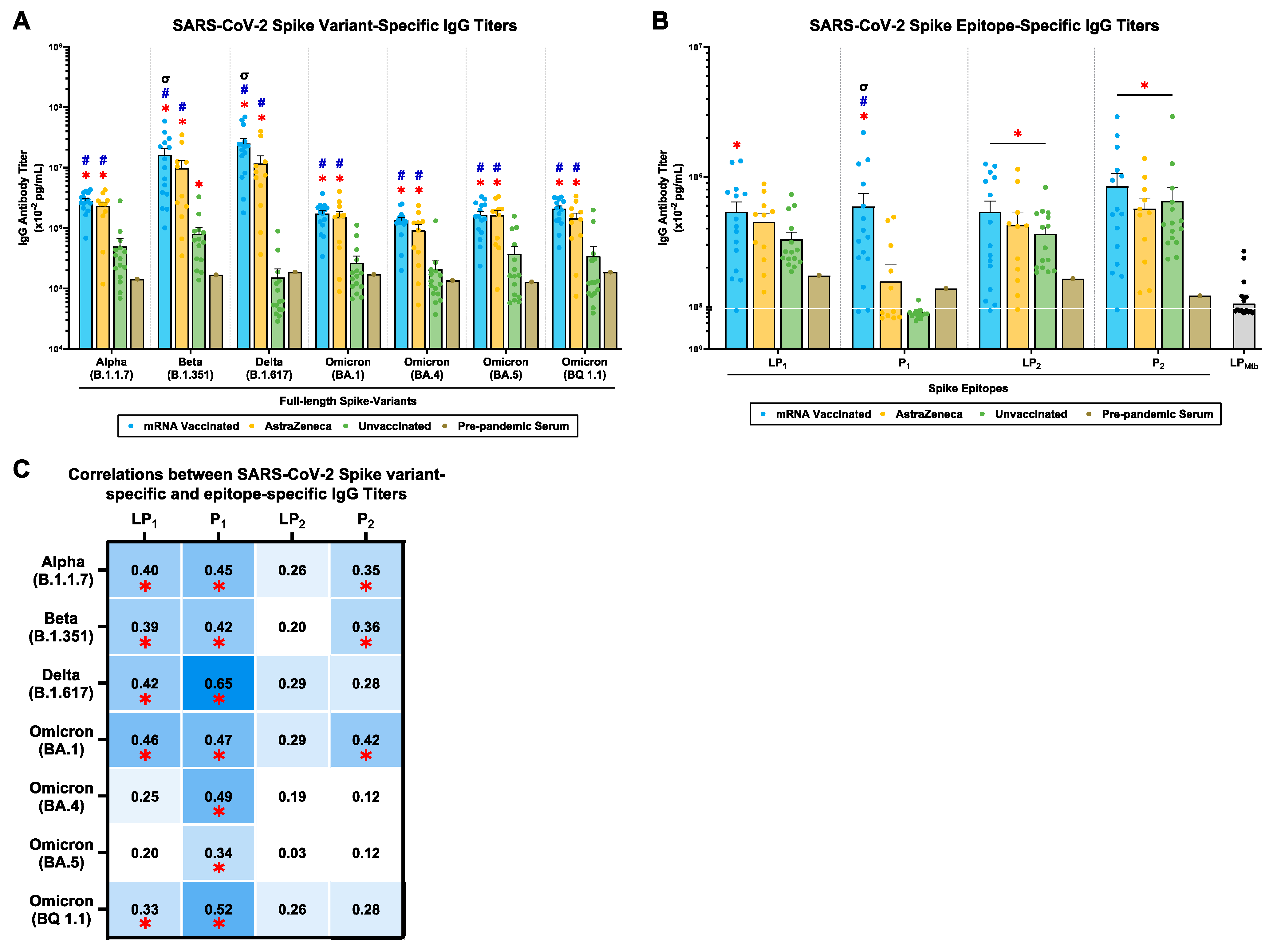
2.2. mRNA and AstraZeneca-Vaccinated Donors Generated Cross-Reactive IgG Antibody Titers against Multiple SARS-CoV-2 Spike Variants and P1/P2 Spike Epitopes

Plasma samples from mRNA-vaccinated, AstraZeneca-vaccinated, and unvaccinated donors were evaluated for IgG antibodies against the S-protein of the SARS-CoV-2 Alpha (B.1.1.7), Beta (B.1.351), Delta (B.1.617), and Omicron (BA.1, BA.4, BA.5, BQ1.1) variants (Figure 1). mRNA and AstraZeneca-vaccinated donors generated significantly higher cross-reactive IgG antibody titers across all SARS-CoV-2 variants compared to the unvaccinated donors and pre-pandemic serum (Figure 1A). Moreover, the mRNA-vaccinated group showed significantly higher IgG antibody titers against the Beta (B.1.351) and Delta (B.1.617) variants compared to the AstraZeneca-vaccinated group (Figure 1A). Notably, in the unvaccinated group, there were detectable levels of IgG antibody titers across all SARS-CoV-2 variants, with IgG antibody titers against the Beta (B.1.351) variants showing significance compared to pre-pandemic serum (Figure 1A). Also, there was a decline in IgG antibody titers against the latest Omicron (BA.1, BA.4, BA.5, BQ1.1) variants compared to the earlier Alpha (B.1.1.7), Beta (B.1.351), and Delta (B.1.617) variants. Overall, mRNA and AstraZeneca-vaccinated donors generated a cross-reactive IgG antibody response against the SARS-CoV-2 Alpha (B.1.1.7), Beta (B.1.351), Delta (B.1.617), and Omicron (BA.1, BA.4, BA.5, BQ1.1) variants.
Next, plasma samples were assayed for IgG antibody titers against spike lipopeptides (LP1 and LP2), and their peptide counterparts (P1 and P2). LP1-specific IgG antibody titers were highest in the mRNA-vaccinated group, followed by the AstraZeneca-vaccinated and unvaccinated groups (Figure 1B). Similarly, P1-specific IgG antibody titers were significantly higher in the mRNA-vaccinated group compared to the AstraZeneca, unvaccinated, and pre-pandemic serum (Figure 1B). Furthermore, LP2/P2-specific IgG antibody titers were significantly higher in the mRNA, AstraZeneca, and unvaccinated groups, compared to pre-pandemic serum (Figure 1B). As a control, all donor plasma samples were tested against a Mycobacterium tuberculosis (LPMtb)-derived lipopeptide to differentiate the peptide-specific binding of IgG antibody titers, which were tested against LP1 and LP2, and not towards the lipid tail of the lipopeptides. LPMtb-specific IgG antibody titers showed titers even lower than those in the pre-pandemic serum (Figure 1B). Altogether, plasma IgG antibodies were detected against all S-derived lipopeptides (LP1 and LP2) and peptides (P1 and P2), with mRNA-vaccinated groups showing the highest titer, followed by AstraZeneca-vaccinated and unvaccinated donors.
Moreover, correlation studies showed that LP1-specific IgG titers significantly correlated with IgG titers against the S-protein of the SARS-CoV-2 Alpha (B.1.1.7), Beta (B.1.351), Delta (B.1.617), and Omicron (BA.1, BQ1.1) variants (Figure 1C). P1-specific IgG titers significantly correlated with all tested SARS-CoV-2 variants (Figure 1C). However, LP2-specific IgG titers did not correlate significantly with any of the tested SARS-CoV-2 variants (Figure 1C). Finally, P2-specific IgG titers showed moderate correlations with SARS-CoV-2 Alpha (B.1.1.7), Beta (B.1.351), and Omicron (BA.1) variants (Figure 1C).
In conclusion, mRNA-vaccinated, AstraZeneca-vaccinated, and unvaccinated donors induced cross-reactive IgG antibody titers against multiple SARS-CoV-2 spike variants, which correlated with P1 and P2 epitope specificity (Figure 1).

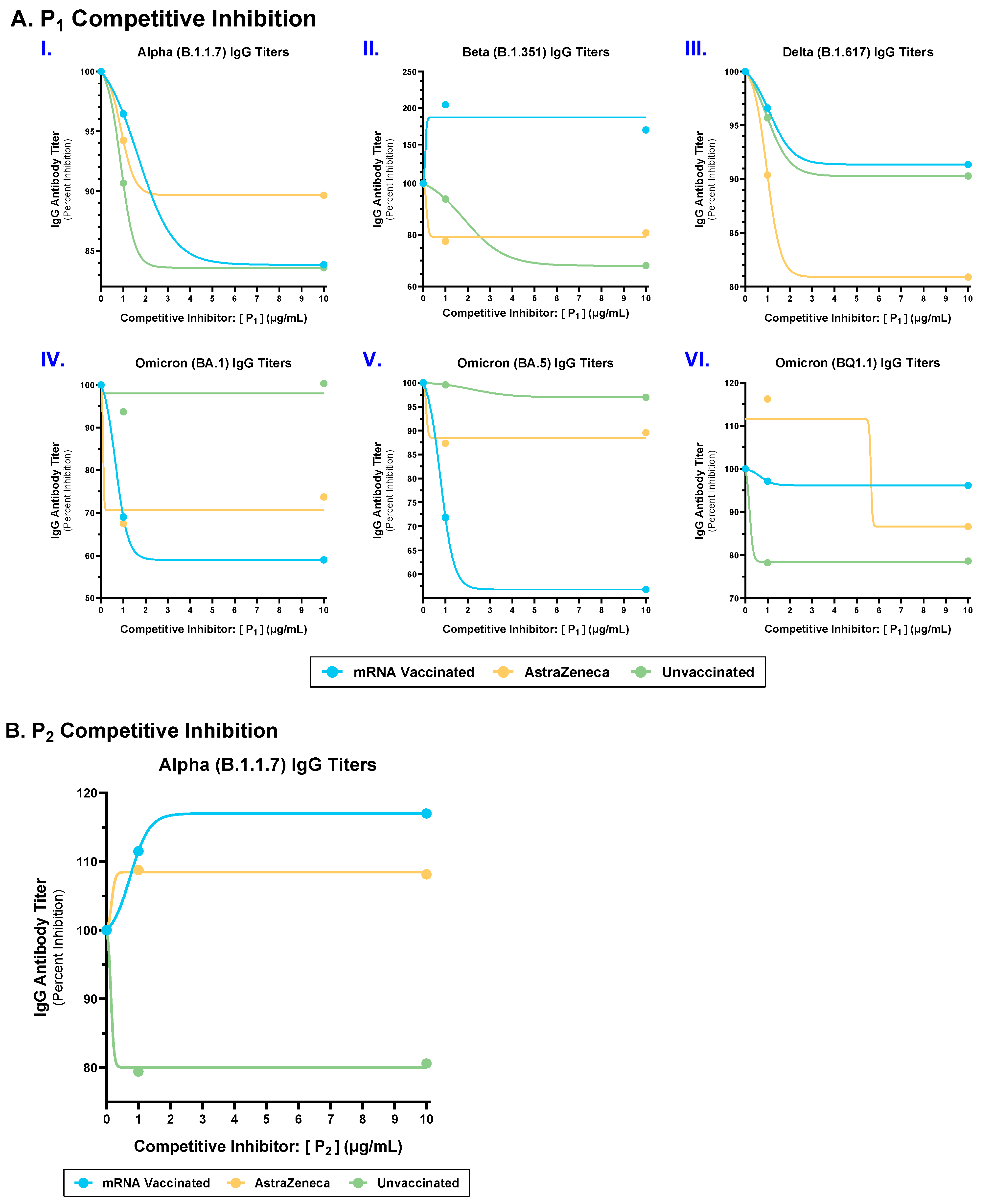
2.3. Competitive Inhibition with P1 and P2 Epitopes Reduced Cross-Reactive IgG Antibody Binding to Multiple SARS-CoV-2 Spike Variants in mRNA, AstraZeneca, and Unvaccinated Donors

To determine the specificity of cross-reactive SARS-CoV-2 spike variant-specific IgG antibody titers, we carried out competitive inhibition experiments using increasing concentrations of P1 and P2 epitopes and measured IgG binding to the SARS-CoV-2 Alpha (B.1.1.7), Beta (B.1.351), Delta (B.1.617), and Omicron (BA.1, BA.5, BQ1.1) variants (Figure 2). P1 epitope has acquired mutations in variants; however, it is structurally conserved among SARS-CoV-2 VOCs and is predicted to be a discontinuous B cell epitope, whereas P2 is sequentially and structurally conserved among CoVs [13,16,17,20,21]. In the mRNA-vaccinated group, competitive inhibition with P1 epitope partially reduced IgG binding to the SARS-CoV-2 Alpha (B.1.1.7), Delta (B.1.617), Omicron (BA.1), and Omicron (BA.5) variants (Figure 2(AI,III–V)). In contrast, competitive interactions with P1 epitope enhanced IgG binding to the SARS-CoV-2 Beta (B.1.351) variant in the mRNA-vaccinated group (Figure 2(AII)), whereas no reduction in IgG binding was observed against the SARS-CoV-2 Omicron (BQ1.1) variants (Figure 2(AVI)). Next, AstraZeneca-vaccinated donors showed a partial reduction in IgG binding against all tested SARS-CoV-2 variants (Figure 2(AI–VI)). Furthermore, the unvaccinated groups showed a reduction in IgG binding against the SARS-CoV-2 Alpha (B.1.1.7), Beta (B.1.351), Delta (B.1.617), and Omicron (BQ1.1) variants (Figure 2(AI–III,VI)). However, there was no reduction in IgG binding against the SARS-CoV-2 Omicron (BA.1), and Omicron (BA.5) variants (Figure 2(AIV,V)).
Considering P2 is a sequentially conserved epitope among SARS-CoV-2/CoV, we competitively inhibited plasma samples with the P2 epitope only against the SARS-CoV-2 Alpha (B.1.1.7) variant [23]. The unvaccinated group showed a 20% reduction in IgG binding, whereas the mRNA and AstraZeneca-vaccinated groups showed an increase in IgG binding against the SARS-CoV-2 Alpha (B.1.1.7) variant by 17% and 4%, respectively (Figure 2B).
In conclusion, these results suggest that in the human IgG repertoire against spike antigens, at least a fraction of antibodies are targeted against P1- and P2-conserved epitopes.

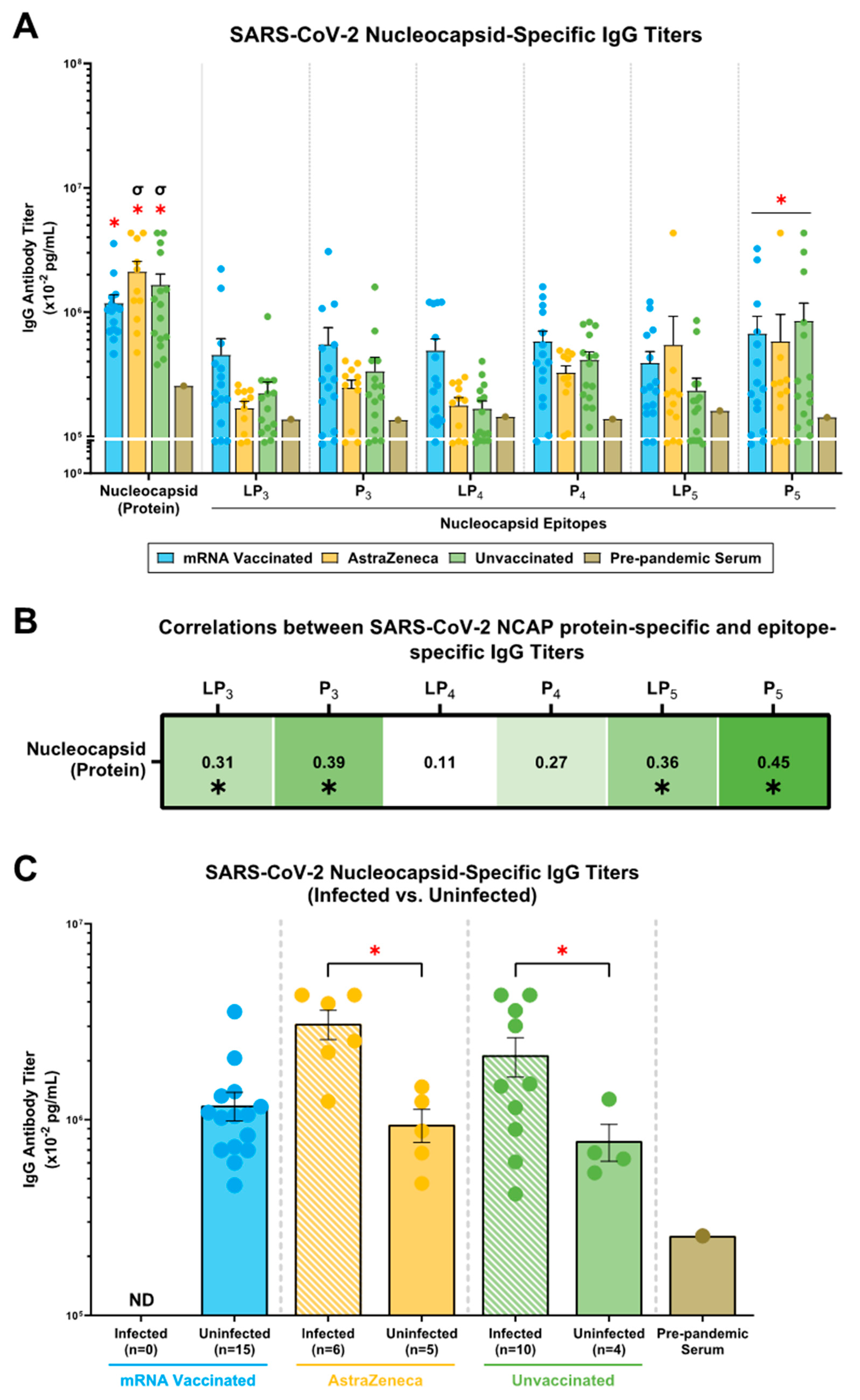
2.4. mRNA, AstraZeneca, and Unvaccinated Donors Showed IgG Antibody Titers against the SARS-CoV-2 N (Protein), N-Derived Lipopeptides (LP3, LP4, LP5) and Peptides (P3, P4, P5)

N (protein)-specific IgG antibody titers were significantly higher in the mRNA, AstraZeneca, and unvaccinated donors compared to the pre-pandemic serum (Figure 3A). Moreover, the AstraZeneca and unvaccinated donors showed higher N-specific IgG antibody titers compared to the mRNA-vaccinated group (Figure 3A). Next, regarding IgG antibody titers against N-derived lipopeptides and peptides, the mRNA-vaccinated group showed the highest titers against LP3, P3, LP4, and P4, followed by the unvaccinated and AstraZeneca group (Figure 3A). LP5-specific IgG antibody titers were higher in the mRNA and AstraZeneca-vaccinated groups, followed by the unvaccinated donors, which showed similar titers to pre-pandemic serum (Figure 3A). For all donors, the P5-specific IgG antibody titers were significantly higher compared to those in pre-pandemic serum; however, similarities were observed among the vaccinated and unvaccinated groups (Figure 3A). Lastly, correlation studies with N (protein)-specific IgG antibody titers showed significant moderate correlations with LP3-, P3-, LP5-, and P5-specific IgG antibody titers (Figure 3B).
Considering the mRNA and AstraZeneca vaccines are spike-based formulations, we hypothesized that N-specific IgG responses were generated through exposure to SARS-CoV-2 during the COVID-19 pandemic. We stratified donors by known infection with COVID-19 and assessed the prevalence of N (protein)-specific IgG antibody titers. For the AstraZeneca and unvaccinated groups, N (protein)-specific IgG antibody titers were significantly higher in donors with known COVID-19 infections compared to donors who did not report an infection (Figure 3C). The mRNA-vaccinated donors did not report any infection, and yet showed a similar N (protein)-specific IgG antibody titer compared to the uninfected AstraZeneca-vaccinated and unvaccinated donors (Figure 3C). Consistent with other studies, these results suggest that N-specific IgG responses in the donor plasma can be attributed to possible prior infection and/or exposure to COVID-19 [24,25,26].

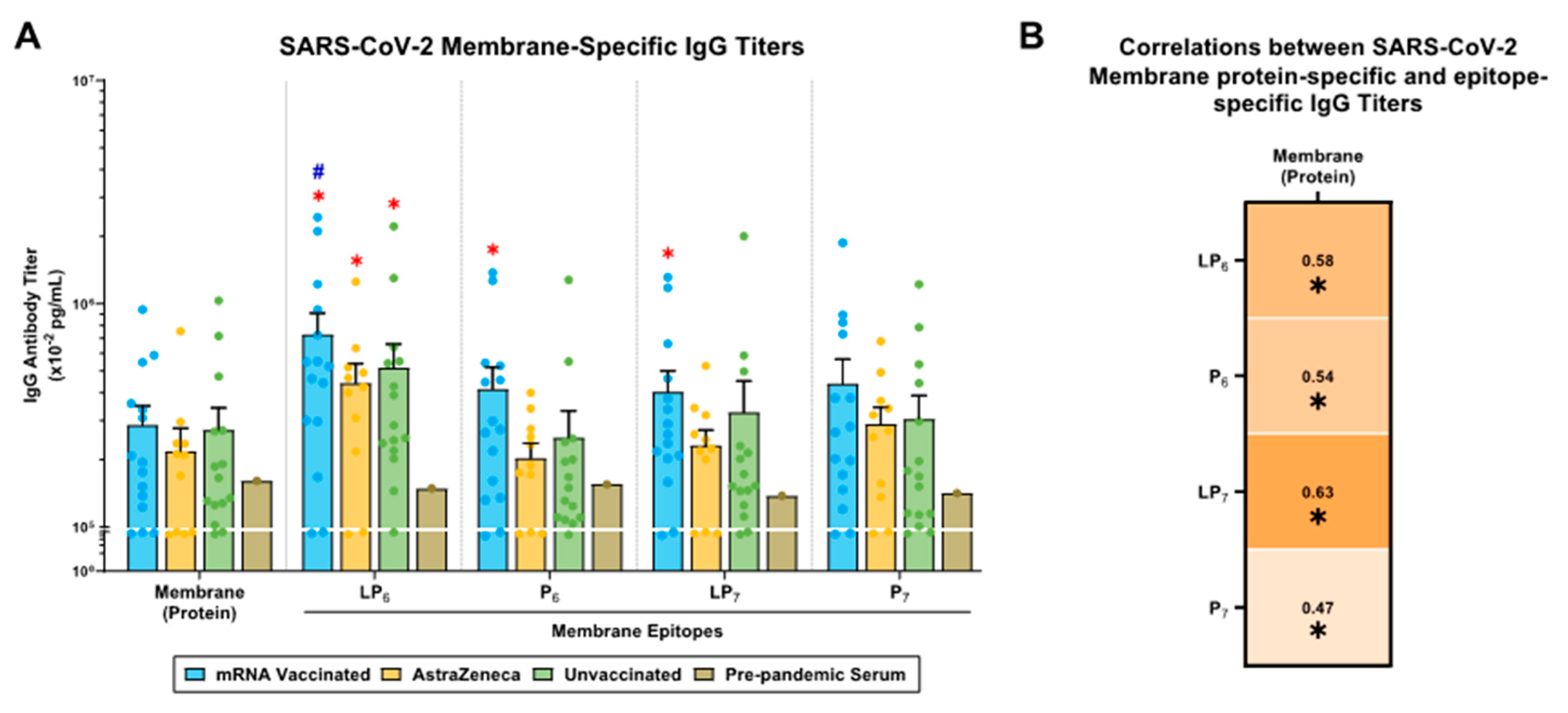
2.5. SARS-CoV-2 M-Specific IgG Antibody Responses in mRNA-Vaccinated, AstraZeneca-Vaccinated, and Unvaccinated Donors

In the mRNA-vaccinated, AstraZeneca-vaccinated, and unvaccinated groups, M (protein)-specific IgG titers were higher compared to those in pre-pandemic serum (Figure 4A). All M-derived lipopeptides (LP6,7)- and peptides (P6,7)-specific IgG responses showed similar titers, with the mRNA-vaccinated donors showing the highest IgG titers, followed by unvaccinated and AstraZeneca-vaccinated donors (Figure 4A). Notably, only the mRNA-vaccinated donors showed significant IgG titers against LP6, P6, and LP7, compared to pre-pandemic serum (Figure 4A). Moreover, the IgG titers in AstraZeneca and unvaccinated donors were significantly higher against LP6 compared to those in pre-pandemic serum (Figure 4A). Next, there were strong positive correlations between M (protein)-specific IgG antibody titers with LP6-, P6-, LP7-, and P7-specific IgG titers (Figure 4B).

3. Discussion

To develop a broadly protective vaccine that targets multiple SARS-CoV-2 variants, it is important to understand the antigenic homology between SARS-CoV-2, its variants, and other heterologous CoVs. Considering the SARS-CoV-2 S protein, several studies have reported sequence homology between SARS-CoV-2 VOCs (96.23–97.8%), SARS-CoVs (75–80%), MERS-CoVs (>70%), and common cold CoVs (14–30%) [27,28,29]. This sequence identity has contributed to the cross-reactive antibody titers observed in convalescent COVID-19 sera against several SARS-CoV-2 variants and other heterologous CoVs [30,31]. Here, we demonstrate that human plasma samples from mRNA-vaccinated, AstraZeneca-vaccinated, and unvaccinated donors generated cross-reactive IgG antibody titers against the SARS-CoV-2 Alpha (B.1.1.7), Beta (B.1.351), Delta (B.1.617), and Omicron (BA.1, BA.4, BA.5, BQ1.1) variants (Figure 1A). Interestingly, these plasma samples were collected before the emergence of the Omicron lineage (B.1.1.529); therefore, all donors are considered naïve against the tested Omicron variants (BA.1, BA.4, BA.5, and BQ1.1). This indicates that plasma IgG titers against the SARS-CoV-2 Omicron variants (BA.1, BA.4, BA.5, BQ1.1) were originally generated against the Wuhan-Hu-1 strain by the mRNA or AstraZeneca vaccines and/or by natural infection with the Alpha (B.1.1.7), Beta (B.1.351), and/or Delta (B.1.617) variants. This provides evidence of heterologous immunity induced in humans, supporting the idea that immune responses specifically targeted towards one pathogen can mount a response against a novel, never-seen-before pathogen [2]. However, it is important to note that the quantity and/or effectiveness of these cross-reactive antibody responses may not provide sufficient protection against newer SARS-CoV-2 variants, suggesting the need for a next-generation COVID-19 vaccine that enhances protection against multiple variants and prevents SARS-CoV-2 infections.
Conceptually, vaccine-induced and/or naturally acquired heterologous immunity relies on targeting shared epitopes between pathogens [7]. In mRNA-vaccinated, AstraZeneca-vaccinated, and unvaccinated donors, the specificity of the cross-reactive IgG titers significantly correlated with spike-derived epitopes, P1 and P2, which are structurally and/or sequentially conserved among SARS-CoV-2 VOCs (Figure 1B,C) [23]. Additionally, competitive inhibition of mRNA-vaccinated, AstraZeneca-vaccinated, and unvaccinated plasma samples, with the P1 epitope, led to a partial reduction in IgG binding to most of the examined SARS-CoV-2 variants (Figure 2A). Moreover, competitive inhibition with the P2 epitope led to a reduction in IgG binding to SARS-CoV-2 spike protein in the unvaccinated group; however, in the mRNA- and AstraZeneca-vaccinated donors, there was an increase in IgG binding (Figure 2B). The addition of P1 and P2 epitopes served the purpose of competitively inhibiting antibodies to spike proteins in donor plasma samples. However, in the mRNA- and AstraZeneca-vaccinated donors, the addition of P2 (epitope) enhanced binding to the spike protein (Figure 2B). Herein, P2 may have acted as an agonist for pre-existing IgG antibodies to enhance their binding ability against the SARS-CoV-2 spike antigen. Mouse studies have shown that antibodies induced upon immunization with P1 or P2 have neutralizing functionality [22]. Therefore, these results provide support for the notion that targeting sequentially conserved regions of the SARS-CoV-2 S-protein induces heterologous (cross-reactive) immunity that recognizes multiple SARS-CoV-2 variants. Applications of heterologous immunity can facilitate the design of next-generation vaccines for SARS-CoV-2, by continual screening for conserved, functional epitopes, such as P1 and P2, to achieve protection against a broad spectrum of CoVs. Our findings highlight the feasibility of designing a pan-coronavirus vaccine through identifying cross-reactive epitopes among multiple SARS-CoV-2 VOCs and other CoVs to maximize immune coverage against CoVs.
Current practices of updating the highly variable SARS-CoV-2 spike antigens to account for prevailing variants with repeated boosters is not a long-term solution. Furthermore, regularly updating COVID-19 vaccines may result in immune imprinting and antigenic sin, leading to compromised vaccine efficacy [32,33]. Pre-existing antibodies specific to the Wuhan-Hu-1 strain have been shown to be imprinted and increased with each homologous boost with the BNT162b2 mRNA vaccine [34,35]. Also, imprinting occurs with heterologous boosting by bivalent vaccines that incorporate S-proteins of novel SARS-CoV-2 VOCs [34,35]. Vaccine-induced imprinting results in back-boosting of original humoral immunity while limiting the generation of de novo antibodies against novel immunogens and conserved epitopes. Moreover, administering full-length S-proteins may focus immune responses towards immunodominant, hypervariable regions that promote imprinting, whereas next-generation vaccines need to direct responses toward conserved, cryptic domains, thereby establishing cross-reactivity, while limiting antigenic sin [36].
The majority of the current vaccine efforts against SARS-CoV-2 are limited by targeting the S-protein; however, it is important to consider N and M proteins as potential targets that will allow us to establish cross-reactive responses. Our results demonstrate that mRNA-vaccinated, AstraZeneca-vaccinated, and unvaccinated donors generate N- and M-specific IgG antibody titers. However, within the vaccinated groups, those with known COVID-19 infections showed significantly higher N-specific IgG titer (Figure 3 and Figure 4). Considering that the mRNA and AstraZeneca vaccines are spike-based formulations, the presence of N- and M-specific IgG antibody responses in uninfected donors can be explained by unreported/asymptomatic SARS-CoV-2 infections and/or cross-reactive IgG antibodies generated as a result of prior exposure to human CoVs. Many studies have demonstrated cross-reactive N-/M-specific humoral and cellular responses among common cold CoVs, MERS-CoV, and SARS-CoV that have contributed to sustainable, decade-long immunity in humans [12,15,37,38,39,40]. Previous studies [10,11,12,13,14,15,16,17,18,19,20,21] identified cross-reactive epitopes from the N- (P3/P4/P5) and M- (P6/P7) proteins that share sequence homology between beta-CoVs, and donor plasma were assayed for IgG against these epitopes. In this study, the N- and M-specific IgG titers showed specificity towards conserved N-derived epitopes (P3 and P5) and M-derived epitopes (P6 and P7), respectively (Figure 3 and Figure 4). Furthermore, N-specific antibody titers have correlated with improved clinical outcomes and protection against disease severity. Moreover, these antibodies neutralize viral shedding of the N-protein to restore chemokine function during SARS-CoV-2 infections [9,10,14]. The detection of M-specific antibody titers suggests that there is corresponding cellular immunity targeting the SARS-CoV-2 M antigen [41]. Moreover, antibodies can mediate effector functions, including phagocytosis, opsonization, complement activation, and antibody-dependent cellular cytotoxicity. Therefore, in the quest to find a pan-coronavirus vaccine candidate, targeting non-spike epitopes can facilitate broader coverage of CoVs, expand immune functionality, and provide a multidimensional response against multiple SARS-CoV-2 proteins.
In conclusion, this study has defined immune cross-reactivity and identified seven epitopes (P1–7) from the SARS-CoV-2 S-, N-, and M-proteins that can be further investigated for a multi-valent, pan-coronavirus vaccine candidate. Identification of peptide-epitope specificity of cross-reactive/heterologous humoral responses can serve as a significant milestone in the development of a pan-coronavirus vaccine.

4. Materials and Methods

4.1. Ethical Approval

The study was approved by the University of Alberta Human Research Ethics Board (HREB; ref. no. Pro0085358).

4.2. Human Plasma Donors

Patient samples were acquired from the Canadian BioSample Repository (CBSR), and the pre-pandemic (pooled) serum was collected prior to the COVID-19 pandemic. The serum was commercially purchased (Sigma-Aldrich; St. Louis, MO, USA).

4.3. SARS-CoV-2 Proteins, Lipopeptides, and Peptides

Synthetic lipopeptides (LP1–7), peptides (P1–7), and SARS-CoV-2 spike, nucleocapsid, and membrane proteins were purchased from Genscript Inc. (Piscataway, NJ, USA), with >96% purity (Table 1) [22]. Spike proteins of the Alpha (B.1.1.7), Beta (B.1.351), Delta (B.1.617), and Omicron (BA.1, BA.4, BA.5, and BQ1.1) variants were obtained. All lipopeptides, peptides, and SARS-CoV-2 proteins were stored at −20 °C and diluted in PBS before use.

4.4. Detection of Human IgG Antibodies and P1/P2 Competitive Antibody Assays

NUNC MaxiSorp 96-well flat-bottom plates (Thermo Scientific Nunc™; Waltham, MA, USA) were coated with synthetic lipopeptides (LP1–7), peptides (P1–7), SARS-CoV-2 spike proteins of the Alpha (B.1.1.7), Beta (B.1.351), Delta (B.1.617), and Omicron (BA.1, BA.4, BA.5, and BQ1.1) variants, nucleocapsid, and membrane proteins, in individual plates, at 1 µg/mL in PBS, and incubated overnight at 4 °C. Next, wells were blocked with PBS (1% BSA) for 1h, followed by the addition of plasma samples (1:100; triplicates), which were incubated for 2 h at room temperature (RT). For P1/P2 competitive ELISAs, diluted plasma samples (1:100) were pre-incubated with either P1 or P2 peptides (10, 1, 0 µg/mL), at 37 °C for 1 h. Next, the pre-incubated mixtures were transferred to coated plates and incubated for 2 h at RT. Following the incubation, a detection antibody, mouse anti-human IgG Fc-Alkaline Phosphatase (AP; Southern Biotech., Birmingham, AL, USA), was added for 1 h at RT. Subsequently, p-Nitrophenyl Phosphate (PNPP; Southern Biotech., Birmingham, AL, USA) was added, and color development was measured using a DTX 880 Plate Reader (Beckman Coulter, Brea, CA, USA) at 405 nm after 1 h. Using an IgG standard curve, optical density (O.D.) readings were interpolated into [IgG] (pg/mL). Bars are expressed as the mean [Ig] ± SEM of triplicate wells.

4.5. Statistical Analyses

Data analysis, statistical analysis, and figures were generated using GraphPad Prism Software 10.2.3 (San Diego, CA, USA). p ≤ 0.05 was used to determine significance.

Author Contributions

Conceptualization, B.A.; methodology, D.Y., R.S.P. and A.L.; data analysis, D.Y., R.S.P., A.L. and B.A.; writing—original draft preparation, D.Y., R.S.P. and A.L.; writing—review and editing, D.Y., R.S.P., A.L. and B.A.; supervision, B.A.; project administration, B.A.; funding acquisition, B.A.; human sample collection and BioBank, Q.G., C.S., A.H. and B.R.; D.Y. and R.S.P. are equal contributors. All authors have read and agreed to the published version of the manuscript.

Funding

This work was funded by project grants (PJT165854 and PS173314) from the Canadian Institutes of Health Research (CIHR) to B.A.

Institutional Review Board Statement

The study was approved by the University of Alberta Human Research Ethics Board (HREB; ref. no. Pro0085358).

Informed Consent Statement

Informed consent was obtained from all subjects involved in the study.

Data Availability Statement

The data supporting the findings in this article will be made available by the authors, without undue reservation, upon reasonable request.

Conflicts of Interest

The authors have no conflicts of interest to declare. The funders had no role in the design of the study; in the collection, analyses, or interpretation of data; in the writing of the manuscript; or in the decision to publish the results.

References

  1. World Health Organization COVID-19 Cases WHO COVID-19 Dashboard. Available online: https://data.who.int/dashboards/covid19/cases?n=o (accessed on 23 June 2024).
  2. Patel, R.S.; Agrawal, B. Heterologous Immunity Induced by 1st Generation COVID-19 Vaccines and Its Role in Developing a Pan-Coronavirus Vaccine. Front. Immunol. 2022, 13, 952229. [Google Scholar] [CrossRef] [PubMed]
  3. Echaide, M.; Chocarro de Erauso, L.; Bocanegra, A.; Blanco, E.; Kochan, G.; Escors, D. MRNA Vaccines against SARS-CoV-2: Advantages and Caveats. Int. J. Mol. Sci. 2023, 24, 5944. [Google Scholar] [CrossRef] [PubMed]
  4. Röltgen, K.; Nielsen, S.C.A.; Silva, O.; Younes, S.F.; Zaslavsky, M.; Costales, C.; Yang, F.; Wirz, O.F.; Solis, D.; Hoh, R.A.; et al. Immune Imprinting, Breadth of Variant Recognition, and Germinal Center Response in Human SARS-CoV-2 Infection and Vaccination. Cell 2022, 185, 1025–1040.e14. [Google Scholar] [CrossRef]
  5. Johnston, T.S.; Li, S.H.; Painter, M.M.; Atkinson, R.K.; Douek, N.R.; Reeg, D.B.; Douek, D.C.; Wherry, E.J.; Hensley, S.E. Immunological Imprinting Shapes the Specificity of Human Antibody Responses against SARS-CoV-2 Variants. Immunity 2024, 57, 912–925.e4. [Google Scholar] [CrossRef] [PubMed]
  6. Aguilar-Bretones, M.; Fouchier, R.A.M.; Koopmans, M.P.G.; van Nierop, G.P. Impact of Antigenic Evolution and Original Antigenic Sin on SARS-CoV-2 Immunity. J. Clin. Investig. 2023, 133, e162192. [Google Scholar] [CrossRef]
  7. Agrawal, B. Heterologous Immunity: Role in Natural and Vaccine-Induced Resistance to Infections. Front. Immunol. 2019, 10, 2631. [Google Scholar] [CrossRef]
  8. Goldblatt, D.; Alter, G.; Crotty, S.; Plotkin, S.A. Correlates of Protection against SARS-CoV-2 Infection and COVID-19 Disease. Immunol. Rev. 2022, 310, 6–26. [Google Scholar] [CrossRef]
  9. Lopez-Munoz, A.D.; Kosik, I.; Holly, J.; Yewdell, J.W. Cell Surface SARS-CoV-2 Nucleocapsid Protein Modulates Innate and Adaptive Immunity. Sci. Adv. 2022, 8, eabp9770. [Google Scholar] [CrossRef]
  10. Liu, X.; Shi, Y.; Li, P.; Li, L.; Yi, Y.; Ma, Q.; Cao, C. Profile of Antibodies to the Nucleocapsid Protein of the Severe Acute Respiratory Syndrome (SARS)-Associated Coronavirus in Probable SARS Patients. Clin. Diagn. Lab. Immunol. 2004, 11, 227–228. [Google Scholar] [CrossRef]
  11. Peng, B.; Ming, Y.; Yang, C. Regulatory B Cells: The Cutting Edge of Immune Tolerance in Kidney Transplantation Review-Article. Cell Death Dis. 2018, 9, 109. [Google Scholar] [CrossRef]
  12. Heide, J.; Schulte, S.; Kohsar, M.; Brehm, T.T.; Herrmann, M.; Karsten, H.; Marget, M.; Peine, S.; Johansson, A.M.; Sette, A.; et al. Broadly Directed SARS-CoV-2-Specific CD4+ T Cell Response Includes Frequently Detected Peptide Specificities within the Membrane and Nucleoprotein in Patients with Acute and Resolved COVID-19. PLoS Pathog. 2021, 17, e1009842. [Google Scholar] [CrossRef] [PubMed]
  13. Liu, W.J.; Zhao, M.; Liu, K.; Xu, K.; Wong, G.; Tan, W.; Gao, G.F. T-Cell Immunity of SARS-CoV: Implications for Vaccine Development against MERS-CoV. Antiviral Res. 2017, 137, 82–92. [Google Scholar] [CrossRef] [PubMed]
  14. Peng, H.; Yang, L.; Wang, L.; Li, J.; Huang, J.; Lu, Z.Q.; Koup, R.A.; Bailer, R.T.; Wu, C. you Long-Lived Memory T Lymphocyte Responses against SARS Coronavirus Nucleocapsid Protein in SARS-Recovered Patients. Virology 2006, 351, 466–475. [Google Scholar] [CrossRef] [PubMed]
  15. Peng, Y.; Mentzer, A.J.; Liu, G.; Yao, X.; Yin, Z.; Dong, D.; Dejnirattisai, W.; Rostron, T.; Supasa, P.; Liu, C.; et al. Broad and Strong Memory CD4+ and CD8+ T Cells Induced by SARS-CoV-2 in UK Convalescent Individuals Following COVID-19. Nat. Immunol. 2020, 21, 1336–1345. [Google Scholar] [CrossRef] [PubMed]
  16. Meyers, L.M.; Gutiérrez, A.H.; Boyle, C.M.; Terry, F.; McGonnigal, B.G.; Salazar, A.; Princiotta, M.F.; Martin, W.D.; De Groot, A.S.; Moise, L. Highly Conserved, Non-Human-like, and Cross-Reactive SARS-CoV-2 T Cell Epitopes for COVID-19 Vaccine Design and Validation. NPJ Vaccines 2021, 6, 71. [Google Scholar] [CrossRef]
  17. Vashi, Y.; Jagrit, V.; Kumar, S. Understanding the B and T Cell Epitopes of Spike Protein of Severe Acute Respiratory Syndrome Coronavirus-2: A Computational Way to Predict the Immunogens. Infect. Genet. Evol. 2020, 84, 104382. [Google Scholar] [CrossRef]
  18. Tarke, A.; Sidney, J.; Methot, N.; Yu, E.D.; Zhang, Y.; Dan, J.M.; Goodwin, B.; Rubiro, P.; Sutherland, A.; Wang, E.; et al. Impact of SARS-CoV-2 Variants on the Total CD4+ and CD8+ T Cell Reactivity in Infected or Vaccinated Individuals. Cell Rep. Med. 2021, 2, 100355. [Google Scholar] [CrossRef]
  19. Li, C.K.; Wu, H.; Yan, H.; Ma, S.; Wang, L.; Zhang, M.; Tang, X.; Temperton, N.J.; Weiss, R.A.; Brenchley, J.M.; et al. T Cell Responses to Whole SARS Coronavirus in Humans. J. Immunol. 2008, 181, 5490–5500. [Google Scholar] [CrossRef]
  20. Grifoni, A.; Sidney, J.; Zhang, Y.; Scheuermann, R.H.; Peters, B.; Sette, A. A Sequence Homology and Bioinformatic Approach Can Predict Candidate Targets for Immune Responses to SARS-CoV-2. Cell Host Microbe 2020, 27, 671–680.e2. [Google Scholar] [CrossRef]
  21. Wang, J.; Wen, J.; Li, J.; Yin, J.; Zhu, Q.; Wang, H.; Yang, Y.; Qin, E.; You, B.; Li, W.; et al. Assessment of Immunoreactive Synthetic Peptides from the Structural Proteins of Severe Acute Respiratory Syndrome Coronavirus. Clin. Chem. 2003, 49, 1989–1996. [Google Scholar] [CrossRef]
  22. Patel, R.S.; Agrawal, B. Mucosal Immunization with Lipopeptides Derived from Conserved Regions of SARS-CoV-2 Antigens Induce Robust Cellular and Cross-Variant Humoral Immune Responses in Mice. Front. Immunol. 2023, 14, 1178523. [Google Scholar] [CrossRef] [PubMed]
  23. Kontopodis, E.; Pierros, V.; Stravopodis, D.J.; Tsangaris, G.T. Prediction of SARS-CoV-2 Omicron Variant Immunogenicity, Immune Escape and Pathogenicity, through the Analysis of Spike Protein-Specific Core Unique Peptides. Vaccines 2022, 10, 357. [Google Scholar] [CrossRef] [PubMed]
  24. Szymczak, A.; Jędruchniewicz, N.; Torelli, A.; Kaczmarzyk-Radka, A.; Coluccio, R.; Kłak, M.; Konieczny, A.; Ferenc, S.; Witkiewicz, W.; Montomoli, E.; et al. Antibodies Specific to SARS-CoV-2 Proteins N, S and E in Covid-19 Patients in the Normal Population and in Historical Samples. J. Gen. Virol. 2021, 102, 001692. [Google Scholar] [CrossRef] [PubMed]
  25. Den Hartog, G.; Schepp, R.M.; Kuijer, M.; Geurtsvankessel, C.; Van Beek, J.; Rots, N.; Koopmans, M.P.G.; Van Der Klis, F.R.M.; Van Binnendijk, R.S. SARS-CoV-2-Specific Antibody Detection for Seroepidemiology: A Multiplex Analysis Approach Accounting for Accurate Seroprevalence. J. Infect. Dis. 2020, 222, 1452–1461. [Google Scholar] [CrossRef]
  26. Lei, Q.; Li, Y.; Hou, H.; Wang, F.; Ouyang, Z.Q.; Zhang, Y.; Lai, D.Y.; Banga Ndzouboukou, J.L.; Xu, Z.W.; Zhang, B.; et al. Antibody Dynamics to SARS-CoV-2 in Asymptomatic COVID-19 Infections. Allergy Eur. J. Allergy Clin. Immunol. 2021, 76, 551–561. [Google Scholar] [CrossRef]
  27. Cicaloni, V.; Costanti, F.; Pasqui, A.; Bianchini, M.; Niccolai, N.; Bongini, P. A Bioinformatics Approach to Investigate Structural and Non-Structural Proteins in Human Coronaviruses. Front. Genet. 2022, 13, 891418. [Google Scholar] [CrossRef]
  28. Kumar, A.; Asghar, A.; Singh, H.N.; Faiq, M.A.; Kumar, S.; Narayan, R.K.; Kumar, G.; Dwivedi, P.; Sahni, C.; Jha, R.K.; et al. SARS-CoV-2 Omicron Variant Genomic Sequences and Their Epidemiological Correlates Regarding the End of the Pandemic: In Silico Analysis. JMIR Bioinform. Biotechmol. 2023, 4, e42700. [Google Scholar] [CrossRef]
  29. Sajini, A.A.; Alkayyal, A.A.; Mubaraki, F.A. The Recombination Potential between SARS-CoV-2 and MERS-CoV from Cross-Species Spill-over Infections. J. Epidemiol. Glob. Health 2021, 11, 155–159. [Google Scholar] [CrossRef]
  30. Tamminen, K.; Salminen, M.; Blazevic, V. Seroprevalence and SARS-CoV-2 Cross-Reactivity of Endemic Coronavirus OC43 and 229E Antibodies in Finnish Children and Adults. Clin. Immunol. 2021, 229, 108782. [Google Scholar] [CrossRef]
  31. Zhu, Y.; Yu, D.; Han, Y.; Yan, H.; Chong, H.; Ren, L.; Wang, J.; Li, T.; He, Y. Cross-Reactive Neutralization of SARS-CoV-2 by Serum Antibodies from Recovered SARS Patients and Immunized Animals. Sci. Adv. 2020, 6, eabc9999. [Google Scholar] [CrossRef]
  32. Arevalo, C.P.; Le Sage, V.; Bolton, M.J.; Eilola, T.; Jones, J.E.; Kormuth, K.A.; Nturibi, E.; Balmaseda, A.; Gordon, A.; Lakdawala, S.S.; et al. Original Antigenic Sin Priming of Influenza Virus Hemagglutinin Stalk Antibodies. Proc. Natl. Acad. Sci. USA 2020, 117, 17221–17227. [Google Scholar] [CrossRef] [PubMed]
  33. Petráš, M.; Králová Lesná, I. SARS-CoV-2 Vaccination in the Context of Original Antigenic Sin. Hum. Vaccin. Immunother. 2022, 18, 1949953. [Google Scholar] [CrossRef] [PubMed]
  34. Tortorici, M.A.; Addetia, A.; Seo, A.J.; Brown, J.; Sprouse, K.R.; Logue, J.; Clark, E.; Franko, N.; Chu, H.; Veesler, D. Persistent Immune Imprinting after XBB.1.5 COVID Vaccination in Humans. bioRxiv 2023. [Google Scholar] [CrossRef]
  35. Blankson, J.N. Bivalent COVID-19 Vaccines: Can the Original Antigenic Sin Be Forgiven? J. Infect. Dis. 2023, 227, 1221–1223. [Google Scholar] [CrossRef] [PubMed]
  36. Mandhane, A.; Tripathy, C.S.; Behera, S.K. Peptide and Protein in Vaccine Delivery. In Peptide and Protein Drug Delivery Using Polysaccharides; Elsevier: Amsterdam, The Netherlands, 2023. [Google Scholar]
  37. Grobben, M.; Van Der Straten, K.; Brouwer, P.J.; Brinkkemper, M.; Maisonnasse, P.; Dereuddre-Bosquet, N.; Appelman, B.; Ayesha Lavell, A.H.; Van Vught, L.A.; Burger, J.A.; et al. Cross-Reactive Antibodies after Sars-Cov-2 Infection and Vaccination. Elife 2021, 10, e70330. [Google Scholar] [CrossRef]
  38. Song, G.; He, W.; Callaghan, S.; Anzanello, F.; Huang, D.; Ricketts, J.; Torres, J.L.; Beutler, N.; Peng, L.; Vargas, S.; et al. Cross-Reactive Serum and Memory B-Cell Responses to Spike Protein in SARS-CoV-2 and Endemic Coronavirus Infection. Nat. Commun. 2021, 12, 2938. [Google Scholar] [CrossRef]
  39. Sealy, R.E.; Hurwitz, J.L. Cross-Reactive Immune Responses toward the Common Cold Human Coronaviruses and Severe Acute Respiratory Syndrome Coronavirus 2 (Sars-Cov-2): Mini-Review and a Murine Study. Microorganisms 2021, 9, 1643. [Google Scholar] [CrossRef]
  40. Le Bert, N.; Tan, A.T.; Kunasegaran, K.; Tham, C.Y.L.; Hafezi, M.; Chia, A.; Chng, M.H.Y.; Lin, M.; Tan, N.; Linster, M.; et al. SARS-CoV-2-Specific T Cell Immunity in Cases of COVID-19 and SARS, and Uninfected Controls. Nature 2020, 584, 457–462. [Google Scholar] [CrossRef]
  41. Fernández-Soto, D.; Bueno, P.; Garaigorta, U.; Gastaminza, P.; Bueno, J.L.; Duarte, R.F.; Jara, R.; Valés-Gómez, M.; Reyburn, H.T. SARS-CoV-2 Membrane Protein-Specific Antibodies from Critically Ill SARS-CoV-2–Infected Individuals Interact with Fc Receptor–Expressing Cells but Do Not Neutralize the Virus. J. Leukoc. Biol. 2024, 115, 985–991. [Google Scholar] [CrossRef]
Figure 1. mRNA and AstraZeneca-vaccinated donors generated cross-reactive SARS-CoV-2 spike variant-specific IgG antibody titers. Plasma samples from mRNA-vaccinated (n = 15; blue), AstraZeneca-vaccinated (n = 11; yellow), and unvaccinated (n = 15; green) donors were assayed for IgG antibodies against (A) the SARS-CoV-2 S-proteins of the Alpha (B.1.1.7), Beta (B.1.351), Delta (B.1.617), and Omicron (BA.1, BA.4, BA.5, BQ1.1) variants, and (B) S-derived lipopeptides (LP1 and LP2) and peptides (P1 and P2). Pre-pandemic serum and LPMtb were used as controls. Bars represent mean ± SEM and each point represents an individual donor. Statistical significance was determined using a two-way ANOVA, followed by Tukey’s post hoc test. *, #, and σ indicates significant differences (p ≤ 0.05) between the pre-pandemic serum, the unvaccinated donors, and the mRNA vs. AstraZeneca donors, respectively. (C) Correlations between SARS-CoV-2 spike variant-specific and epitope-specific IgG antibody titers were conducted. Spearman’s R-values (black) and significant correlations (p ≤ 0.05; *) are indicated.
Ijms 25 09814 g001
Figure 2. Competitive inhibition of SARS-CoV-2 spike variant-specific IgG antibody titers by P1 and P2 epitopes in mRNA-vaccinated, AstraZeneca-vaccinated, and unvaccinated donors. Percent inhibition of IgG antibody titers in mRNA-vaccinated (n = 3–5; blue), AstraZeneca-vaccinated (n = 3–5; yellow), and unvaccinated (n = 3–5; green) donors, after competitively inhibiting (A) P1 and (B) P2 epitopes against SARS-CoV-2 (I) Alpha (B.1.1.7), (II) Beta (B.1.351), (III) Delta (B.1.617), and (IVVI) Omicron (BA.1, BA.5, BQ1.1) variants. Each point represents the mean percent inhibition of at least 3 donors (n = 3–5). Inhibition curves were generated by fitting data points to a sigmoidal dose–response curve using GraphPad Prism.
Ijms 25 09814 g002
Figure 3. N (protein)-specific and epitope-specific IgG antibody titers in mRNA-vaccinated, AstraZeneca-vaccinated, and unvaccinated donors. (A) IgG antibodies against the SARS-CoV-2 N (protein), and N-derived lipopeptides (LP3–5) and peptides (P3–5) were determined from mRNA-vaccinated (n = 15; blue), AstraZeneca-vaccinated (n = 11; yellow), and unvaccinated (n = 15; green) donors. (B) Correlations between SARS-CoV-2 N (protein)-specific and epitope-specific IgG antibody titers were performed. Spearman’s R-values (black) and significant correlations (p ≤ 0.05; *) are indicated. (C) Within each vaccine group, donors were stratified by known infection with COVID-19 (infected vs. uninfected), and SARS-CoV-2 N-specific IgG antibody titers were shown. Pre-pandemic serum is an experimental control. (A,C) Bars represent mean ± SEM, with each point representing an individual donor. Statistical significance was determined using a (A) two-way ANOVA and (C) one-way ANOVA, followed by Tukey’s post hoc test. *, and σ indicates significant differences (p ≤ 0.05) between the pre-pandemic serum, the unvaccinated donors, and the mRNA donors, respectively. Abbreviation: ND (not determined).
Ijms 25 09814 g003
Figure 4. SARS-CoV-2 M-specific IgG antibody responses in mRNA-vaccinated, AstraZeneca-vaccinated, and unvaccinated donors. (A) Plasma samples from mRNA-vaccinated (n = 15; blue), AstraZeneca-vaccinated (n = 11; yellow), and unvaccinated (n = 15; green) donors were tested against the SARS-CoV-2 M (protein), and M-derived lipopeptides (LP6,7) and peptides (P6,7). Pre-pandemic serum was used as control. Bars represent mean ± SEM, with each point representing an individual donor. A two-way ANOVA was used to determine significance, followed by Tukey’s post hoc test. *, and # indicates significant differences (p ≤ 0.05) between the pre-pandemic serum, the unvaccinated donors, and the mRNA vs. AstraZeneca donors, respectively. (B) Correlations between SARS-CoV-2 M (protein)-specific and epitope-specific IgG antibody titers were performed. Spearman’s R-values (black) and significant correlations (p ≤ 0.05; *) are indicated.
Ijms 25 09814 g004
Table 1. SARS-CoV-2 derived lipopeptides (LP1–7) and their respective peptide (P1–7): viral protein, location, code, amino acid sequence, and epitope characteristics.
PathogenProteinLocationCodeAmino Acid
Sequence
Epitope Characteristics [10,11,12,13,14,15,16,17,18,19,20,21]
SARS-CoV-2S1492–505LP1LQSYGFQPTNGVGYK(Palmitoyl)G
Derived from SARS-CoV-2 Spike S1 subunit
Predicted B cell epitope
Conserved across SARS-CoV-2, MERS, SARS-CoV-1, HCoV-HKU1, -OC43, -NL43, and -229E
P1LQSYGFQPTNGVGY
S2814–826LP2KRSFIEDLLFNKVK(Palmitoyl)G
Derived from SARS-CoV-2 Spike S2 subunit
Non-RBD epitope for neutralizing antibody
Conserved across SARS-CoV-2 and SAR-CoV-1
P2KRSFIEDLLFNKV
N358–372LP3IDAYKTFPPTEPKKDK(Palmitoyl)G
Derived from SARS-CoV-2 Nucleocapsid (N) protein
Predicted CD4+/CD8+ T cell epitope
Binds to multiple MHC I (HLA.A3, A11, A68) and MHC II (HLA-DRB1.01) molecules
Conserved across beta-coronaviruses
P3IDAYKTFPPTEPKKD
317–331LP4MSRIGMEVTPSGTWLK(Palmitoyl)G
Derived from SARS-CoV-2 Nucleocapsid (N) protein
Predicted CD4+/CD8+ T cell epitope
Binds to multiple MHC I (HLA.A2, A3, A11, A68, B40) and MHC II (HLA-DRB1.01) molecules
Conserved across beta-coronaviruses
P4MSRIGMEVTPSGTWL
158–172LP5VLQLPQGTTLPKGFYK(Palmitoyl)G
Derived from SARS-CoV-2 Nucleocapsid (N) protein
Predicted CD4+/CD8+ T cell epitope
Binds to multiple MHC I (HLA.A2, A11, A31, A68) and MHC II (HLA-DRB1.01) molecules
Conserved across beta-coronaviruses
P5VLQLPQGTTLPKGFY
M98–112LP6ASFRLFARTRSMWSFK(Palmitoyl)G
Derived from SARS-CoV-2 Membrane (M) protein
Binds to multiple MHC II (HLA-DRB1, DRB5, DPA1, DPB1) molecules
Conserved across beta-coronaviruses
P6ASFRLFARTRSMWSF
34–48LP7LLQFAYANRNRFLYIK(Palmitoyl)G
Derived from SARS-CoV-2 Membrane (M) protein
Conserved across beta-coronaviruses
Binds to multiple MHC II (HLA-DRB1, DRB3, DRB5) molecules
P7LLQFAYANRNRFLYI
Note: Adapted from the work of Patel and Agrawal, 2023 [22].
Table 2. Demographics of mRNA-vaccinated, AstraZeneca-vaccinated, and unvaccinated donors.
mRNA Vaccinated
(N = 15)
AstraZeneca
Vaccinated
(N = 11)
Unvaccinated
(N = 15)
Sample Collection Timeline
July 2021–Aug 2021Apr 2021–Jan 2022July 2020–July 2021
Sex, n (%)
Females11 (73.3%)6 (54.5%)6 (40.0%)
Males4 (26.7%)5 (45.5%)9 (60.0%)
Age, median (range)
25 (19–65)58 (28–63)42 (20–94)
Reported COVID-19 Infection, n (%)
-6 (54.5%)10 (66.6%)
Disclaimer/Publisher’s Note: The statements, opinions and data contained in all publications are solely those of the individual author(s) and contributor(s) and not of MDPI and/or the editor(s). MDPI and/or the editor(s) disclaim responsibility for any injury to people or property resulting from any ideas, methods, instructions or products referred to in the content.

