Article

Exogenous Application of dsRNA—Inducing Silencing of the Fusarium oxysporum Tup1 Gene and Reducing Its Virulence

1
School of Life Sciences, Zhejiang Chinese Medical University, Hangzhou 310051, China
2
Future Health Laboratory, Innovation Center of Yangtze River Delta, Zhejiang University, Jiaxing 314102, China
*
Author to whom correspondence should be addressed.
These authors contributed equally to this work.
Int. J. Mol. Sci. 2024, 25(19), 10286; https://doi.org/10.3390/ijms251910286
Submission received: 5 August 2024 / Revised: 21 September 2024 / Accepted: 23 September 2024 / Published: 24 September 2024
(This article belongs to the Section Molecular Microbiology)

Abstract

:
Fusarium oxysporum is a widespread soil-borne fungal pathogen that can infect various plants, causing wilt and root rot diseases. The root rot disease of Atractylodes macrocephala caused by F. oxysporum is among the most serious diseases associated with continuous cropping, significantly hindering its sustainable development. In this study, we aimed to investigate the effect of exogenous application of double-stranded RNA (dsRNA) on silencing the F. oxysporum Tup1 gene to reduce its virulence and to evaluate its potential application in controlling root rot disease in A. macrocephala. The Tup1 gene was amplified from the F. oxysporum genome, and different lengths of Tup1-dsRNA were designed and synthesized. The uptake of dsRNA by the fungus was verified using Tup1-dsRNA labeled with fluorescein, and in vitro dsRNA treatment experiments were conducted to assess its impact on the growth and virulence of F. oxysporum. Additionally, Tup1-dsRNA was applied to the roots of A. macrocephala to evaluate its effectiveness in controlling root rot disease. The experimental results showed that F. oxysporum could effectively uptake exogenously applied Tup1-dsRNA, significantly reducing Tup1 gene expression. All lengths of Tup1-dsRNA inhibited fungal growth and caused morphological changes in the fungal hyphae. Further plant experiments and Reverse Transcription Quantitative Polymerase Chain Reaction (RT-qPCR) analysis indicated that Tup1-dsRNA treatment significantly reduced the incidence of root rot disease in A. macrocephala, which was supported by the reduction in peroxidase (POD) and catalase (CAT) enzyme activities, malondialdehyde (MDA) content, and proline (Pro) levels in treated root tissues. This study demonstrated that exogenous dsRNA could reduce the virulence of F. oxysporum by silencing the Tup1 gene and effectively mitigate the root rot disease it causes in A. macrocephala. The successful application of Tup1-dsRNA provided strong evidence for the potential of RNA interference (RNAi) technology in plant disease control. Future research could further optimize the design and application of dsRNA to enhance its practical value in agriculture.

1. Introduction

Fusarium oxysporum is a ubiquitous soil-borne fungal pathogen that infects various plants, leading to wilt and root rot diseases. It survives in the soil as thick-walled spores and infects plants through root wounds or direct invasion. Once infected, the pathogen spreads through the plant’s vascular system, resulting in leaf yellowing, wilting, and ultimately plant death [1,2].
Atractylodes macrocephala is an important medicinal herb primarily used for invigorating the spleen, drying dampness, and stopping sweating [3]. It contains various chemical components, including volatile oils, atractylenolides, polysaccharides, and amino acids, which exhibit pharmacological effects such as anti-inflammatory, anti-tumor, and antiviral activities [4,5]. However, A. macrocephala faces replanting obstacles, requiring a 5- to 10-year interval between plantings, which severely limits sustainable development. Among the replanting diseases, root rot caused by F. oxysporum is the most severe [6]. The current control methods for F. oxysporum-induced root rot include physical rotation and chemical treatments, both of which have limitations. Thus, developing efficient new biological fungicides is crucial for effective biological control.
Small RNA (sRNA) is a type of non-coding RNA found in eukaryotes that is shorter than 200 nucleotides. It plays a role in regulating various biological processes, including plant growth, development, and responses to stress. Additionally, sRNA is being explored as a tool for biological defense, specifically for pest control [7,8]. sRNA is produced by the homologous protein Dicer-like (DCL) and incorporated into Argonaute (AGO) proteins, inducing gene silencing in a sequence-specific manner [9]. As research on sRNA progresses, a regulatory network based on sRNA is emerging. Researchers are developing disease-resistant plants by modifying specific processes within this regulatory network [10], and population control is being actively promoted through artificial sRNA delivery systems, such as host-induced gene silencing (HIGS) and spray-induced gene silencing (SIGS), to reduce pest outbreaks [11]. The gene functions of sRNA provide a promising direction for controlling A. macrocephala root rot, with a focus on using SIGS technology.
In recent years, the concept of spray-induced gene-silencing RNA biopesticides has paved the way for developing new biological fungicides [12,13]. Spraying double-stranded RNA (dsRNA) on leaves and soil has been effective in controlling pests [14]. Research has demonstrated that applying dsRNA targeting pathogen virulence factors to plants significantly reduces pest occurrence. For example, RNA spray-mediated gene silencing of Fusarium graminearum AGO and DCL genes reduced its infection on barley leaves, enhancing barley resistance [15]. Similarly, spraying synthetic FolRDR1-dsRNA on tomato plants downregulated F. oxysporum FolRDR1 gene expression, inhibiting its growth and infection [16]. Additionally, exogenous dsRNA spray-induced MoDES1 gene silencing enhanced rice resistance to rice blast disease [17]. The exogenous application of Bmp1-dsRNA, Bmp3-dsRNA, and Pls1-dsRNA effectively inhibited Botrytis cinerea growth [18,19]. These research findings provide a theoretical basis for developing RNA interference (RNAi) fungicides.
Compared to traditional chemical pesticides and fungicides, RNAi-mediated crop protection strategies are considered highly specific and environmentally friendly, making them a sustainable alternative [11]. RNAi technology can specifically silence key pathogen genes without affecting the normal gene expression of plants and other non-target organisms, offering significant advantages in disease control [20]. Moreover, the dsRNA and Small interfering RNA (siRNA) used in RNAi technology are biological molecules that degrade easily, leaving no long-term residues or causing pollution. Thus, the exogenous application of RNAi is considered a promising strategy to improve crop resistance to diseases and stress [21], while avoiding the ethical and environmental issues associated with transgenics [22].
Tup1 is a conserved transcriptional corepressor that plays a crucial role in regulating gene expression in response to environmental changes [23]. In Saccharomyces cerevisiae, Tup1 is particularly important for the transcriptional repression observed during quiescence, which is essential for the establishment and maintenance of quiescence, highlighting Tup1’s critical role in cellular adaptation to environmental stress [24]. Similarly, in Candida albicans, it was found that silencing the Tup1 gene can affect the expression of key genes associated with fungal virulence, suggesting a potential strategy for fungal control [25]. Recent research on the F. oxysporum Tup1 gene further underscored its importance, revealing its crucial role in various biological processes and the pathogen’s virulence by regulating gene expression in major metabolic pathways, including the tricarboxylic acid cycle (TCA cycle) [26].
Consequently, we chose the Tup1 gene as a model to investigate the feasibility of using SIGS to control A. macrocephala root rot. In this study, three different lengths of dsRNA targeting the F. oxysporum Tup1 gene were designed. The silencing effect of exogenous Tup1-dsRNA application on the Tup1 gene was investigated, providing new ideas and insights for the biological control of A. macrocephala root rot.

2. Results

2.1. Identification of Fusarium oxysporum Tup1 Gene

The genomic DNA of F. oxysporum was used to amplify the Tup1 gene, and a single band of approximately 2000 bp was obtained through agarose gel electrophoresis (Figure 1A). After sequencing, BLAST analysis (blast.ncbi.nlm.nih.gov/Blast.cgi, Date of access: 16 March 2024) in the NCBI database confirmed it as the Tup1 gene. The full length of the Tup1 gene was 1989 bp, containing three introns and encoding 591 amino acids. Structural analysis of the Tup1 protein sequence on the CD SERVER website (www.ncbi.nlm.nih.gov/Structure/cdd/wrpsb.cgi, Date of access: 16 March 2024) showed that the N-terminus contained a 65-amino-acid-long Tup_N domain, and the C-terminus contained six tandem repeats of the WD40 domain. Phylogenetic analysis of the F. oxysporum Tup1 protein using the MEGA 11 software (www.megasoftware.net, Date of access: 17 March 2024) indicated that the Tup1 gene was conserved among Fusarium species (Figure 1B). Amino acid alignment using the Clustalw software (http://www.clustal.org, date of access: 17 March 2024) revealed that the Tup_N domain at the N-terminus was highly conserved only within the Fusarium species (Figure 1C).

2.2. Synthesis of Tup1-dsRNA

Due to the high conservation of the Tup1 gene in the Fusarium species, a basis for designing dsRNA specifically targeting F. oxysporum was established. To investigate whether in vitro synthesized Tup1-dsRNA could induce silencing of the F. oxysporum Tup1 gene and affect its normal growth and pathogenicity, three different lengths of Tup1-dsRNA targeting the 1989 bp open reading frame of the Tup1 gene were synthesized: 536 bp dsRNA, 827 bp dsRNA, and 1471 bp dsRNA (referred to as Tup1-500, Tup1-800, and Tup1-1400, respectively) (Figure 2A). To ensure the successful synthesis of the three different lengths of dsRNA in vitro, agarose gel electrophoresis was performed on the synthetic products, and the results confirmed that Tup1-500, Tup1-800, and Tup1-1400 had been successfully synthesized (Figure 2B).

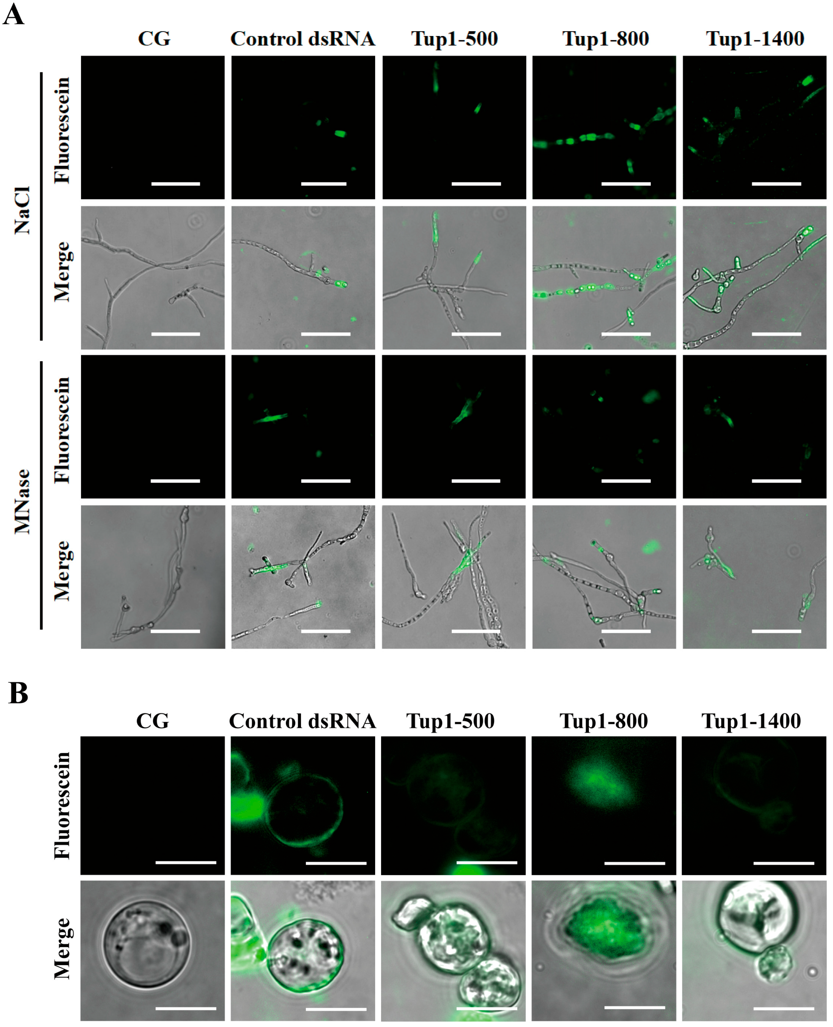
2.3. Tup1-dsRNA Was Effectively Taken Up and Absorbed by Fusarium oxysporum

To investigate whether dsRNA could be taken up by fungi from the environment, we applied fluorescently labeled Tup1-dsRNA to F. oxysporum spores. After 24 h, fluorescent signals were observed accumulating inside the fungal cells (Figure 3A). To exclude the possibility that Tup1-dsRNA was adhering to the exterior of the fungal cells, the fungi were treated with micrococcal nuclease (MNase), with NaCl used as a control. Fluorescent signals were still detectable, indicating that the dsRNA had been taken up by F. oxysporum. Additionally, the control dsRNA, which has no homology to F. oxysporum, was also effectively taken up into fungal cells, suggesting that dsRNA uptake is unlikely to be selective. Additionally, to confirm that the dsRNA had entered the cytoplasm of F. oxysporum rather than being embedded in the cell wall matrix, we treated F. oxysporum protoplasts with fluorescently labeled Tup1-dsRNA. The results showed that fluorescent signals were clearly detectable in the protoplasts of F. oxysporum (Figure 3B). These indicate that the uptake of environmental Tup1-dsRNA by F. oxysporum is non-selective.

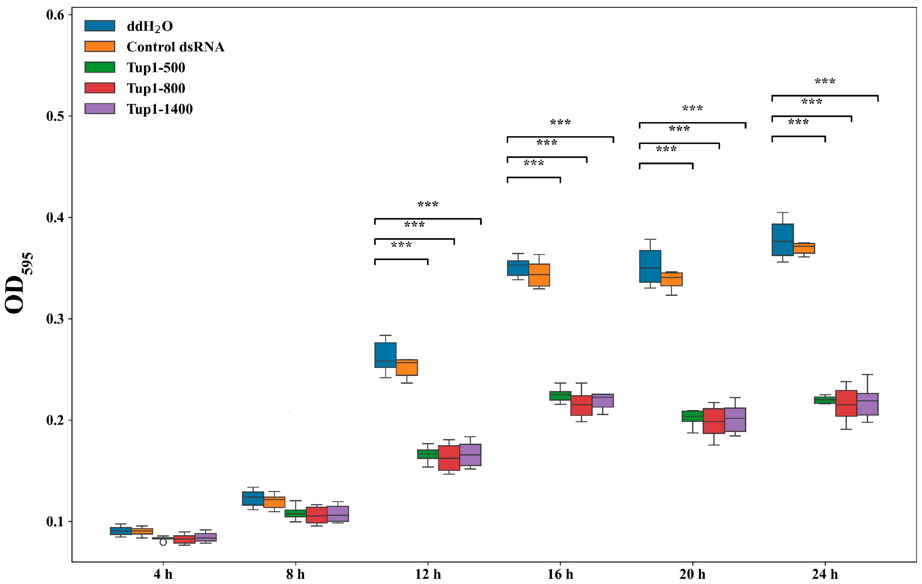
2.4. Effect of Exogenous Application of Tup1-dsRNA on the Growth and Germination of Fusarium oxysporum Spores and Fungal Transcript Analysis

To investigate the effect of the exogenous application of Tup1-dsRNA on the growth of F. oxysporum, the optical density changes at OD595 of fungal spore suspensions in a 96-well plate were measured over 24 h (n = 6). After co-culturing Tup1-500, Tup1-800, and Tup1-1400 (50 ng μL−1) with F. oxysporum spores (1 × 10⁶ cfu mL−1), no significant change in the growth of F. oxysporum spores treated with control dsRNA was observed compared to the control group over 24 h. After treatment with different lengths of Tup1-dsRNA, no significant difference in the growth of F. oxysporum spores was observed at 4 h and 8 h. However, after 12 h, the growth of F. oxysporum spores treated with Tup1-dsRNA began to slow down significantly, and the inhibitory effects of different lengths of Tup1-dsRNA on the growth of F. oxysporum spores were similar (Figure 4).
To further investigate the effect of co-culturing Tup1-dsRNA with F. oxysporum on spore growth morphology and the silencing efficiency of Tup1-500, Tup1-800, and Tup1-1400 on the Tup1 gene, F. oxysporum spore suspensions were collected at different incubation times for microscopic observation and Reverse Transcription Quantitative Polymerase Chain Reaction (RT-qPCR) analysis. The results showed that, in comparison to the control group, spore growth was inhibited and fewer hyphae were observed after treatment with Tup1-500, Tup1-800, and Tup1-1400, with continued inhibitory effects on F. oxysporum growth observed at 24 h (Figure 5A). To further evaluate the silencing capability of Tup1-dsRNA, RT-qPCR was employed to measure the expression of the Tup1 gene in F. oxysporum after 12 h of treatment with Tup1-dsRNA (n = 3). The results showed that Tup1-1400 and Tup1-800 significantly reduced the expression of the Tup1 gene, with Tup1-1400 exhibiting a stronger inhibitory effect than Tup1-800. Meanwhile, Tup1-500 also reduced the expression of the Tup1 gene, but the difference was not significant (Figure 5B). The control dsRNA had no effect on Tup1 gene expression, indicating that the Tup1-dsRNA has gene-targeting specificity.

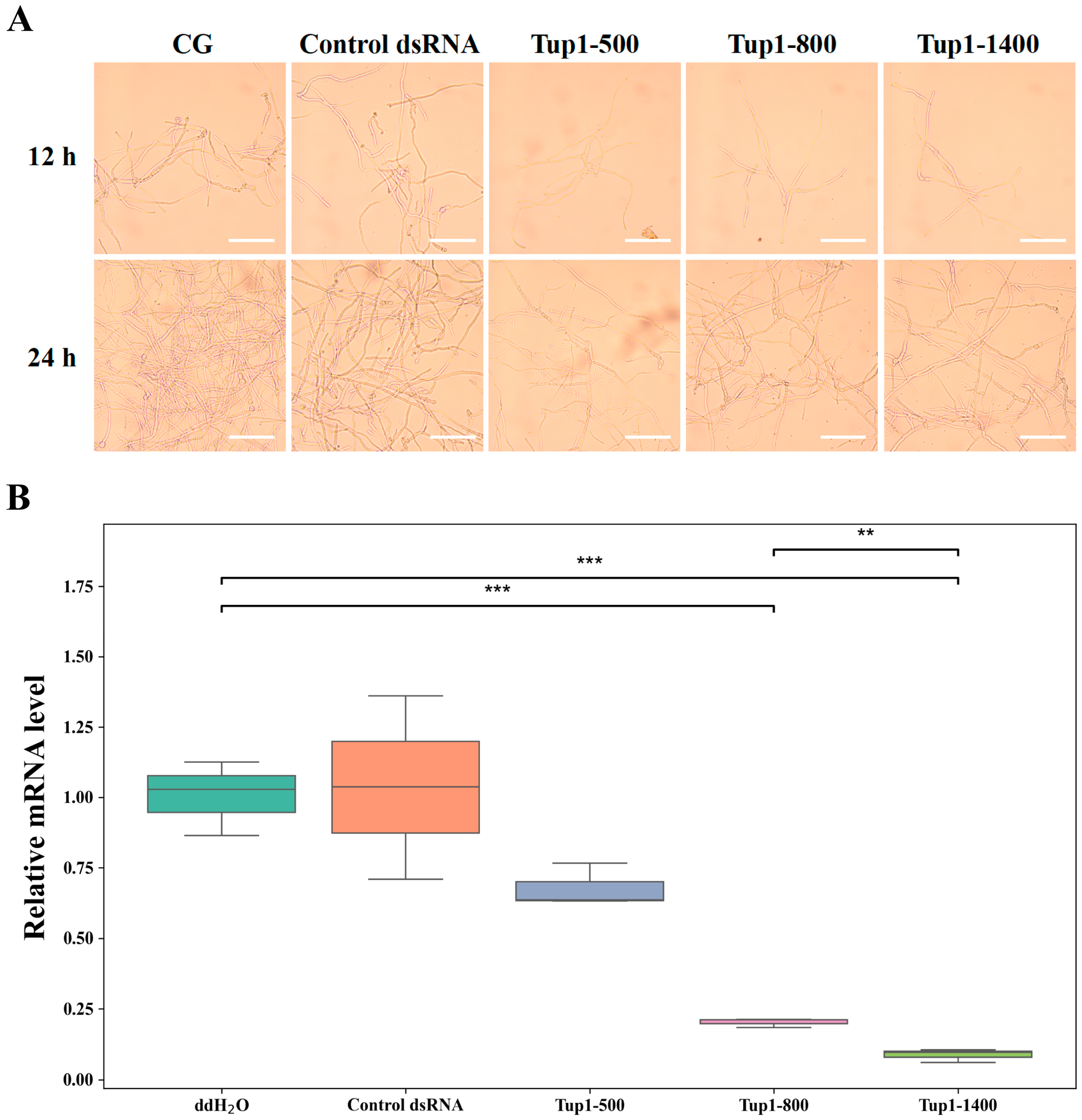
2.5. Effect of Exogenous Application of Tup1-dsRNA on the Growth and Morphology of Fusarium oxysporum Hyphae

To investigate the inhibitory effect of dsRNA on the growth of F. oxysporum hyphae, Tup1-500, Tup1-800, and Tup1-1400 were applied to F. oxysporum plates (n = 3). In our previous results, we found that the control dsRNA had no effect on the growth of F. oxysporum; therefore, the control dsRNA group was not included in this experiment. On the fourth day of F. oxysporum growth on the plates, ddH2O, Tup1-500, Tup1-800, and Tup1-1400 were separately applied to different areas at the growing edge of the hyphae (Figure 6A). On the fifth day, it was observed that hyphal growth was inhibited to varying degrees in all areas except where ddH2O had been applied (Figure 6B), and this inhibition persisted on the sixth day (Figure 6C).
To further observe the effect of Tup1-dsRNA on the morphology of F. oxysporum hyphae, the hyphae at the edges of the plates treated with ddH2O, Tup1-500, Tup1-800, and Tup1-1400 were examined under an electron microscope. Compared to the control group, varying degrees of shrinkage and a rougher surface were exhibited by the hyphae treated with the three different lengths of Tup1-dsRNA. The most severe shrinkage was observed in the hyphae treated with Tup1-1400 (Figure 7). These results indicate that Tup1-dsRNA can inhibit the growth of F. oxysporum hyphae and affect its morphology.

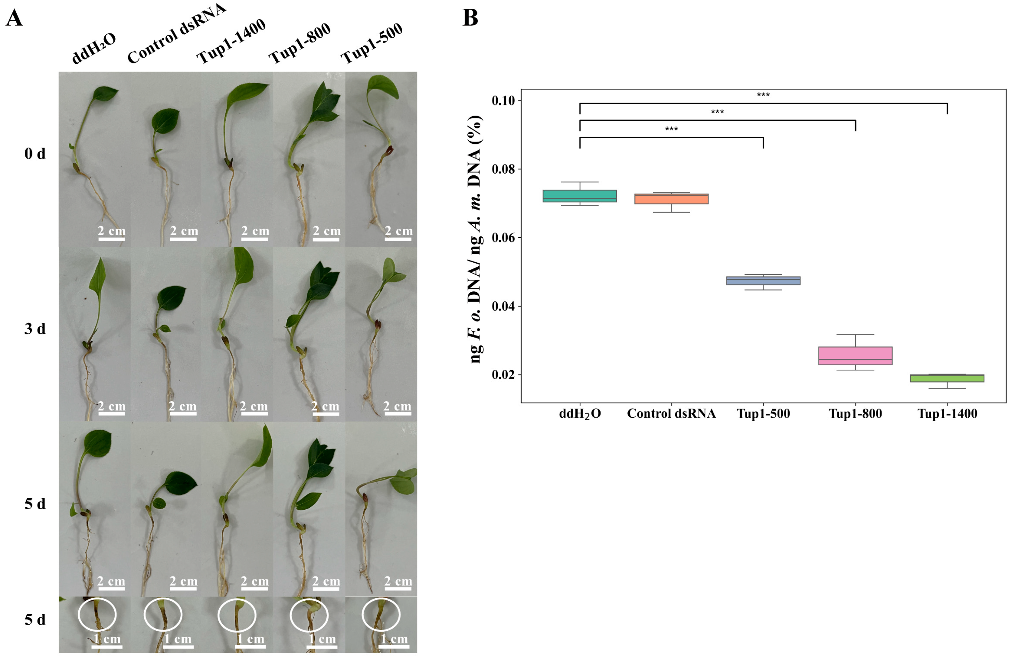
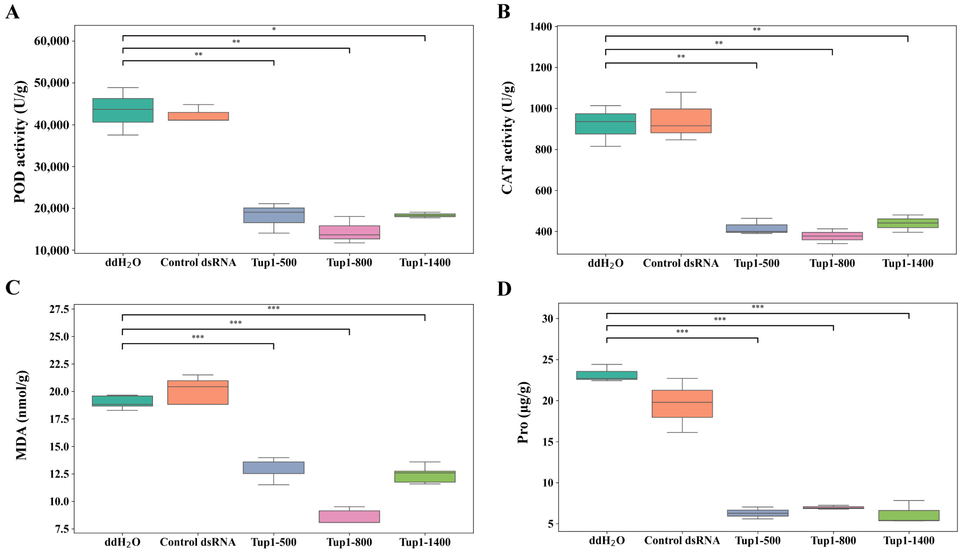
2.6. Exogenous Application of Tup1-dsRNA Alleviates the Occurrence of Atractylodes macrocephala Root Rot

We explored whether Tup1-dsRNA could be used as a new and potentially effective agent for controlling plant diseases, and Tup1-dsRNA was applied to the roots of A. macrocephala to evaluate its effect on controlling root rot (n = 6). To determine whether dsRNA itself affects the growth of A. macrocephala roots, we also included a control dsRNA group. First, a cut was made on the root of A. macrocephala at the two-leaf stage and ddH2O, control dsRNA, Tup1-500, Tup1-800, and Tup1-1400 were applied, respectively. After being placed in a light incubator for 24 h, the roots were inoculated with F. oxysporum spore suspension (1 × 10⁶ cfu mL−1). Then, 5 days after inoculation with F. oxysporum, symptoms of rot were observed on the roots treated with ddH2O and control dsRNA, while reduced symptoms of the disease were observed on the roots treated with Tup1-dsRNA (Figure 8A). Subsequently, we conducted RT-qPCR analysis on the roots of A. macrocephala 5 days after treatment to detect the colonization of F. oxysporum. The results showed that the colonization of F. oxysporum was significantly lower in all groups treated with Tup1-dsRNA compared to the ddH2O and control dsRNA groups (Figure 8B) (p < 0.001). These findings suggest that all three different lengths of Tup1-dsRNA effectively reduce the colonization of F. oxysporum in A. macrocephala roots, thereby significantly alleviating root rot symptoms.
Further analysis was conducted on the fifth day of treatment to examine the activities of peroxidase (POD) and catalase (CAT), as well as the levels of malondialdehyde (MDA) and proline (Pro) in the roots of A. macrocephala. The results showed that POD and CAT activities were higher in root tissues treated with ddH2O and control dsRNA, while these enzyme activities were significantly reduced following Tup1-dsRNA treatment. Since POD and CAT are key enzymes involved in scavenging reactive oxygen species as part of the plant’s defense response [27], this reduction suggests that Tup1-dsRNA may decrease the pathogenicity of F. oxysporum, thereby reducing the plant’s defensive response (Figure 9A,B). Moreover, MDA content, an indicator of lipid peroxidation and cell membrane oxidative damage [28], was higher in the ddH2O and control dsRNA groups but significantly decreased after Tup1-dsRNA treatment, indicating reduced oxidative stress due to decreased pathogen activity (Figure 9C). Similarly, Pro, which typically accumulates in response to stress, was also higher in the ddH2O and control dsRNA groups but significantly decreased following Tup1-dsRNA treatment (Figure 9D). Pro plays a key role in plant stress tolerance, and its levels can reflect the plant’s response to environmental stresses [29]. These findings suggest that Tup1-dsRNA reduces the pathogenicity of F. oxysporum, thereby modulating the defensive responses of A. macrocephala and effectively controlling root rot.

3. Discussion

Exogenous dsRNA-induced gene silencing represents a promising strategy for plant protection that has shown great potential and could serve as an environmentally friendly alternative to conventional fungicide treatments [30,31]. Our research demonstrated that exogenous dsRNA-induced gene-silencing technology significantly reduces the pathogenicity of F. oxysporum.
Different lengths of Tup1-dsRNA were first synthesized, and their inhibitory effect on the growth of F. oxysporum in vitro was validated through exogenous application. The results showed that Tup1-dsRNA was effectively taken up by F. oxysporum, inducing gene silencing and significantly reducing its growth and pathogenicity. Additionally, experiments with fluorescently labeled dsRNA confirmed that F. oxysporum could non-selectively uptake dsRNA from the environment. Furthermore, the application of Tup1-dsRNA in controlling A. macrocephala root rot was explored. The experimental results indicated that the symptoms of the disease were significantly alleviated in A. macrocephala roots treated with Tup1-dsRNA after inoculation with F. oxysporum. This finding provides a theoretical basis for developing new RNAi-based methods for plant disease control.
Although our dsRNA formulation demonstrated the ability to inhibit the virulence of F. oxysporum, many challenges remain for further field trials. The first challenge is production. This study was conducted in the laboratory using kits to synthesize dsRNA in vitro, with each reaction producing only microgram levels of dsRNA, which is insufficient for large-scale field application. The current research has focused on expressing dsRNA in Escherichia coli and directly extracting dsRNA from the bacteria, achieving milligram levels [32]. Another challenge is the instability of dsRNA in soil, which makes direct application ineffective for protecting plants from pathogen infection [33]. A promising approach to significantly enhance the stability of dsRNA involves encapsulating it in liposomes or adsorbing it onto carriers [34]. Additionally, mixing dsRNA with other compounds can enhance the functionality of dsRNA [35].
We aimed to explore the feasibility of using SIGS to control A. macrocephala root rot, and thus only Tup1 was selected as the model gene for the study. Tup1 was chosen for silencing because it acts as a virulence factor and regulates the transcriptional expression of other virulence genes [36]. Currently, there is limited research on the F. oxysporum Tup1 gene, which adds novelty to this study and advances research on Tup1 as a representative virulence gene of F. oxysporum. Both the phylogenetic analysis and amino acid sequence alignment indicated that the Tup1 protein is conserved within the Fusarium species, providing the potential for developing broad-spectrum dsRNA formulations to control root rot diseases in other medicinal plants. When designing dsRNA, it was found that Tup1-500, Tup1-800, and Tup1-1400 all inhibited the pathogenicity of F. oxysporum. During our investigations, we observed that although the inhibitory effect of Tup1-500 dsRNA on Tup1 gene expression was not as significant as that of Tup1-800 and Tup1-1400, its impact on fungal growth and virulence could still be observed. This may be due to the fact that partial silencing of the Tup1 gene is sufficient to affect the phenotype of F. oxysporum. Additionally, the partial expression of the Tup1 gene may lead to secondary effects by regulating other metabolic pathways, thereby influencing the virulence of the fungus. Therefore, even if the inhibition of Tup1 gene expression is not significant, it can still have a notable impact on the biological function of the fungus. However, in practical production, longer dsRNA requires a longer template, which increases the difficulty of synthesis. Therefore, the length of the dsRNA fragment is not necessarily better the longer it is. Genes such as Fub, which encodes fusaric acid [37], and Sge, which is essential for full virulence [38], are worthy targets for designing targeted silencing of virulence genes. More virulence genes are expected to be discovered in future research.
After targeting the Tup1 gene with Tup1-dsRNA, the growth and colonization ability of F. oxysporum were affected. Comparing the inhibitory effect of dsRNA in vitro with that on A. macrocephala roots, it was found that after 24 h of dsRNA pre-treatment at the wound site, the colonization ability of F. oxysporum was still inhibited at 5 days after inoculation. This may be partly due to the non-specific action of siRNA on similar fragments within A. macrocephala, mediating the production of secondary siRNA [39], thereby amplifying the inhibition of F. oxysporum.
In conclusion, exogenous dsRNA-induced gene-silencing technology demonstrated significant effectiveness in controlling A. macrocephala root rot caused by F. oxysporum, showcasing its potential as a novel biological fungicide. However, challenges related to dsRNA production and stability need to be addressed to achieve widespread field application. Additionally, further research on other virulence gene targets will help develop more efficient and broad-spectrum RNAi fungicides.

4. Materials and Methods

4.1. Fungal Strains and Culture Conditions

We obtained F. oxysporum from Beina Chuanglian Biotech Institute (Chaoyang District, Beijing, China) and cultured T7 on potato dextrose agar (PDA) plates at 25 °C. To prepare the medium, we used 0.7 g of potato dextrose broth (PDB) (Hangzhou Microbial Reagent, Hangzhou, China) and 0.375 g of agar (Sangon Biotech, Shanghai, China) per plate. Conidial suspensions were generated from 5- to 6-day-old PDB cultures. Using a sterile punch, we extracted fungal plugs and inoculated them into 100 mL of PDB liquid medium. We then filtered the conidial suspensions through sterile gauze (Solarbio, Beijing, China) and determined the concentration of conidia using a hemocytometer (Hirschmann, Eberstadt, Germany), adjusting it to the desired concentration [40].

4.2. Plant Materials

A. macrocephala seeds were obtained from the Traditional Chinese Medicine Research Institute (Jinhua, Zhejiang, China). The seeds underwent a pre-treatment process that included soaking for 12 h, followed by germination on moistened blotting paper at 25 ± 2 °C for 15 days. Seedlings, upon reaching the first leaf stage, were transplanted into sterile soil and grown in an artificial climate chamber (Boxun, Shanghai, China). Conditions within the chamber were maintained at 25 ± 2 °C, with a light intensity of 1600 lux, a 16/8 h light/dark cycle, and 75% humidity until the seedlings developed to the two-leaf stage.

4.3. Preparation of Fusarium oxysporum Protoplasm

We cultured F. oxysporum in 100 mL of Yeast Extract Peptone Dextrose (YPD) (Hangzhou Microbial Reagent, Hangzhou, China) liquid medium at 25 °C with shaking at 200 rpm for 3 days. The fungal suspension was filtered through sterile gauze to remove mycelia, followed by centrifugation at 3000 rpm for 5 min. After discarding the supernatant, we resuspended the collected spores in sterile distilled water and adjusted their concentration to 3 × 10⁸ cfu mL−1 using a hemocytometer. Subsequently, 1 mL of this spore suspension was inoculated into 100 mL of YPD medium containing 100 μg mL−1 ampicillin (Sigma-Aldrich, St. Louis, MO, USA) and cultured overnight under the same conditions. We then harvested and washed the mycelia sequentially with sterile distilled water and 1.2 M KCl (Sigma-Aldrich, St. Louis, MO, USA). The mycelia were then digested in an enzyme solution containing 500 mg Driselase (GlpBio, Montclair, CA, USA), 200 mg Lysing Enzymes (GlpBio, Montclair, CA, USA), and 200 mg Snailase (GlpBio, Montclair, CA, USA) in 20 mL of 1.2 M KCl [41], with shaking at 28 °C and 90 rpm for 2–3 h. Finally, we isolated the resulting protoplasts by filtration and centrifugation at 5000 rpm for 90 s at 4 °C, washing them three times with ice-cold Sucrose–tris–calcium (STC) buffer. A total of 100 mL of STC buffer contains of 1 mL 1.2 M Sorbitol (Aladdin, Shanghai, China), 1 mL 1 M Tris-HCl (pH 7.5) (Solarbio, Beijing, China), 2 mL 2.5 M CaCl2 (Aladdin, Shanghai, China), and 96 mL ddH2O.

4.4. DNA Extraction and Tup1 Gene Amplification

We extracted genomic DNA from 20 mg of F. oxysporum mycelium using the Rapid Fungi Genomic DNA Isolation Kit (Sangon Biotech, Shanghai, China). Specific primers were designed to amplify the Tup1 gene (Table 1), with PCR conditions set at 94 °C for 3 min for initial denaturation, followed by 35 cycles of 94 °C for 15 s, 55 °C for 20 s, and 72 °C for 2 min, with a final extension at 72 °C for 10 min. The amplified sequences were confirmed via agarose gel electrophoresis and analyzed using BLAST (blast.ncbi.nlm.nih.gov/Blast.cgi, Date of access: 16 March 2024), CD SERVER (www.ncbi.nlm.nih.gov/Structure/cdd/wrpsb.cgi, Date of access: 16 March 2024), MEGA 11 (www.megasoftware.net, Date of access: 17 March 2024), and Clustalw (http://www.clustal.org, Date of access: 17 March 2024) [42,43].

4.5. Total RNA Extraction and cDNA Synthesis

Total RNA was extracted from F. oxysporum using TRIzol Reagent (Thermo Fisher, Waltham, MA, USA), following the same grinding method used for DNA extraction. We then performed reverse transcription using the M-MuLV First Strand cDNA Synthesis Kit (Sangon Biotech, Shanghai, China) according to the manufacturer’s instructions, with 0.5 µg of total RNA serving as the template.

4.6. dsRNA Synthesis

Three different lengths of Tup1-dsRNA were designed: 500 bp, 800 bp, and 1400 bp (referred to as Tup1-500, Tup1-800, and Tup1-1400). Primer pairs for Tup1-dsRNA were designed with T7 promoter sequences at the ends of both the forward and reverse primers for the synthesis of dsRNA templates (Table 2) [44]. Using F. oxysporum cDNA and the designed primers, we synthesized dsRNA templates under the following PCR conditions: initial denaturation at 94 °C for 3 min, followed by 35 cycles of 94 °C for 45 s, 50 °C for 45 s, and 72 °C for 60 s, with a final extension at 72 °C for 5 min. The dsRNAs were generated using the T7 RNAi Transcription Kit (Vazyme, Nanjing, China). The control dsRNA was synthesized using the control template provided in the kit. The synthesized dsRNA was then purified using the magnetic bead purification method recommended by the kit.

4.7. Assessment of dsRNA Uptake by Fusarium oxysporum

During the in vitro synthesis of fluorescein-labeled dsRNA using the T7 RNAi Transcription Kit (Vazyme, Nanjing, China), we replaced the regular Nucleoside triphosphate (NTP) mixture in the kit with fluorescein-labeled NTP. The dsRNA was incubated with F. oxysporum spores for 24 h at 25 °C. We observed the uptake of dsRNA by F. oxysporum using a fluorescence-inverted microscope (Carl Zeiss AG, Oberkochen, Germany). Additionally, F. oxysporum protoplasts were observed under the same microscope to assess dsRNA uptake.

4.8. Effects of Tup1-dsRNA on the Growth of Fusarium oxysporum Conidia In Vitro

Conidial growth was studied using sterile 96-well plates (SAINING, Suzhou, China) to evaluate the effects of Tup1-dsRNA [18,19]. Equal volumes of a spore suspension at a concentration of 1 × 10⁶ cfu mL−1 and Tup1-dsRNA (final concentration 50 ng μL−1) were added to the wells (each treatment n = 6, total assay volume 200 μL). The controls were prepared in PDB and included the following components: (i) ddH2O + F. oxysporum conidia, (ii) control dsRNA + F. oxysporum conidia. The plates were incubated at 25 °C, and fungal growth was assessed by measuring the optical density at OD595 using a microplate reader spectrophotometer (Bio-Rad, Cressier, Switzerland) at various time points between 0 and 24 h. After incubation, we collected the F. oxysporum mycelium, observed its growth under an optical microscope (Sunny Optical Technology, Yuyao, China), and then washed it with ddH2O by centrifugation. Fungal transcript analysis was performed to evaluate the silencing of the Tup1 gene.

4.9. Effects of Tup1-dsRNA on Fusarium oxysporum Mycelial Morphology and Growth In Vitro

We used a sterile puncher to create holes in the F. oxysporum PDA plate and then inoculated the fungi into the center of a new PDA plate covered with cellophane (Acmec, Shanghai, China). After incubation at 25 °C for 4 days, ddH2O and Tup1-dsRNA (at a concentration of 100 ng μL−1) were added around the edges of the hyphal growth. Incubation continued until the sixth day. Then, the hyphal edges were cut, made into slices, and observed under an electron microscope to examine the hyphal morphology. During the observation, we performed a morphological analysis using a scanning electron microscope (SEM) (Carl Zeiss AG, Oberkochen, Germany) at magnifications of 1000×, 5000×, and 10,000× to conduct a detailed examination of the hyphal morphology, allowing us to evaluate the effects of Tup1-dsRNA treatment on the morphology of F. oxysporum hyphae.

4.10. Application of Tup1-dsRNA on the Roots of Atractylodes macrocephala

A. macrocephala seedlings at the two-leaf stage were transferred to trays with moistened blotting paper. A 5 mm wound was made on the root 5 mm below the stem using a dissecting needle. We applied Tup1-dsRNA (5 µL of 100 ng µL−1) to the wound, with control dsRNA and ddH2O serving as controls. The trays were placed in an artificial climate chamber (25 °C, 1600 lux light intensity, 16 h light/8 h dark) for 24 h. Following this, the wound was inoculated with 5 µL of F. oxysporum PDB culture homogenate. The seedlings were incubated for 5 days, and symptoms were observed. To detect the colonization of F. oxysporum in the roots of A. macrocephala, root tissues were collected on the fifth day after treatment. Total DNA was extracted from the plant roots using the Rapid Plant Genomic DNA Isolation Kit (Sangon Biotech, Shanghai, China) and used as a template. RT-qPCR analysis was performed using specific primer pairs (Table 3). The DNA content of A. macrocephala and F. oxysporum was quantified using standard curves to determine the colonization of F. oxysporum.

4.11. Biochemical Indicators and Enzyme Activity Assays

To measure the activities of POD and CAT, as well as the contents of MDA and Pro in the roots of A. macrocephala, we used the appropriate detection kits (POD: #BC0095; CAT: #BC0205; MDA: #BC0025; Pro: #BC0295) (Solarbio, Beijing, China) and followed the manufacturer’s instructions. For each indicator, 0.1 g of A. macrocephala root tissue was used for the measurements, with three biological replicates.

4.12. Gene Expression Analysis by Reverse Transcription Quantitative Polymerase Chain Reaction

The mRNA expression analysis was conducted using RT-qPCR to evaluate the silencing of pathogenic genes. A total of 1 μL of cDNA was used as a template for RT-qPCR in a real-time PCR system (Thermo Fisher, Waltham, MA, USA); the primers used are shown in Table 4. Amplification was performed in a 10 μL reaction mixture containing 5 µL SYBR® Green and 5 pmol oligonucleotides. Each sample had three biological replicates. We used primers specifically designed to study the expression of the Tup1 pathogenic gene, and the PCR conditions were set as follows: 95 °C for 5 min, followed by 35 cycles of 95 °C for 15 s and 60 °C for 60 s.

4.13. Statistical Analysis

Quantitative PCR data were transformed using the 2−ΔΔCT method [45]. Statistical analysis was performed using SPSS 20.0 (IBM Corp., Armonk, NY, USA). One-way ANOVA was used for comparisons between groups [46]. Significant differences between different treatment groups are marked in the figure. GraphPad 9.5.0 (GraphPad Software, San Diego, CA, USA) was used for plotting.

Author Contributions

Conceptualization, S.F., Y.Z. (Yanguang Zhou), N.Z., and Q.M.; data curation, S.D. and X.Y.; formal analysis, Y.Z. (Yujin Zhao), J.X. and Y.T.; funding acquisition, S.D. and X.Y.; investigation, Y.Z. (Yujin Zhao), J.X. and Y.T.; methodology, S.F., Y.Z. (Yanguang Zhou), N.Z. and Q.M.; project administration, S.D. and X.Y.; resources, S.D. and X.Y.; supervision, S.D. and X.Y.; validation, S.F., Y.Z. (Yanguang Zhou), N.Z. and Q.M.; visualization, Y.Z. (Yujin Zhao), J.X. and Y.T.; writing—original draft, S.F. and Y.Z. (Yanguang Zhou); writing—review and editing, S.F. and Y.Z. (Yanguang Zhou). All authors have read and agreed to the published version of the manuscript.

Funding

This study was supported by the National Natural Science Foundation of China (82173920).

Institutional Review Board Statement

Not applicable.

Informed Consent Statement

Not applicable.

Data Availability Statement

Data are contained within the article.

Acknowledgments

The authors appreciate the assistance from Zhejiang Chinese Medical University.

Conflicts of Interest

The authors declare no conflicts of interest.

Abbreviations

F. oxysporumFusarium oxysporum
A. macrocephalaAtractylodes macrocephala
dsRNADouble-stranded RNA
siRNASmall interfering RNA
ODOptical density
TCATricarboxylic acid (cycle)
PDBPotato dextrose broth
PDAPotato dextrose agar
YPDYeast peptone dextrose
SIGSSpray-induced gene silencing
SEMScanning electron microscope
NTPNucleoside triphosphate
STCSucrose–tris–calcium buffer
PODPeroxidase
CATCatalase
MDAMalondialdehyde
Proproline

References

  1. Gordon, T.R. Fusarium oxysporum and the Fusarium Wilt Syndrome. Annu. Rev. Phytopathol. 2017, 55, 23–39. [Google Scholar] [CrossRef] [PubMed]
  2. Michielse, C.B.; Rep, M. Pathogen profile update: Fusarium oxysporum. Mol. Plant Pathol. 2009, 10, 311–324. [Google Scholar] [CrossRef] [PubMed]
  3. Yang, L.; Yu, H.; Hou, A.; Man, W.; Wang, S.; Zhang, J.; Wang, X.; Zheng, S.; Jiang, H.; Kuang, H. A Review of the Ethnopharmacology, Phytochemistry, Pharmacology, Application, Quality Control, Processing, Toxicology, and Pharmacokinetics of the Dried Rhizome of Atractylodes macrocephala. Front. Pharmacol. 2021, 12, 727154. [Google Scholar] [CrossRef] [PubMed]
  4. Cheng, H.; Zhang, D.; Wu, J.; Liu, J.; Tan, Y.; Feng, W.; Peng, C. Atractylodes macrocephala Koidz. volatile oil relieves acute ulcerative colitis via regulating gut microbiota and gut microbiota metabolism. Front. Immunol. 2023, 14, 1127785. [Google Scholar] [CrossRef] [PubMed]
  5. Ruqiao, L.; Yueli, C.; Xuelan, Z.; Huifen, L.; Xin, Z.; Danjie, Z.; Le, S.; Yanxue, Z. Rhizoma Atractylodis macrocephalae: A review of photochemistry, pharmacokinetics and pharmacology. Pharmazie 2020, 75, 42–55. [Google Scholar] [CrossRef]
  6. Huang, X.G.; Li, M.Y.; Yan, X.N.; Yang, J.S.; Rao, M.C.; Yuan, X.F. The potential of Trichoderma brevicompactum for controlling root rot on Atractylodes macrocephala. Can. J. Plant Pathol. 2021, 43, 794–802. [Google Scholar] [CrossRef]
  7. Dzinyela, R.; Alhassan, A.R.; Kiani-Pouya, A.; Rasouli, F.; Yang, L.; Movahedi, A. Role of small RNAs in plant stress response and their potential to improve crops. Crop Pasture Sci. 2023, 74, 1116–1127. [Google Scholar] [CrossRef]
  8. Islam, W.; Noman, A.; Qasim, M.; Wang, L. Plant Responses to Pathogen Attack: Small RNAs in Focus. Int. J. Mol. Sci. 2018, 19, 515. [Google Scholar] [CrossRef]
  9. Huang, C.Y.; Wang, H.; Hu, P.; Hamby, R.; Jin, H. Small RNAs—Big Players in Plant-Microbe Interactions. Cell Host Microbe 2019, 26, 173–182. [Google Scholar] [CrossRef]
  10. Chen, C.; Zeng, Z.; Liu, Z.; Xia, R. Small RNAs, emerging regulators critical for the development of horticultural traits. Hortic. Res. 2018, 5, 63. [Google Scholar] [CrossRef]
  11. Niu, D.; Hamby, R.; Sanchez, J.N.; Cai, Q.; Yan, Q.; Jin, H. RNAs—A new frontier in crop protection. Curr. Opin. Biotech. 2021, 70, 204–212. [Google Scholar] [CrossRef]
  12. Cai, Q.; He, B.; Weiberg, A.; Buck, A.H.; Jin, H. Small RNAs and extracellular vesicles: New mechanisms of cross-species communication and innovative tools for disease control. PLoS Pathog. 2020, 15, e1008090. [Google Scholar] [CrossRef] [PubMed]
  13. Gualtieri, C.; Leonetti, P.; Macovei, A. Plant miRNA Cross-Kingdom Transfer Targeting Parasitic and Mutualistic Organisms as a Tool to Advance Modern Agriculture. Front. Plant Sci. 2020, 11, 930. [Google Scholar] [CrossRef] [PubMed]
  14. Cagliari, D.; Dias, N.P.; Galdeano, D.M.; Dos, S.E.; Smagghe, G.; Zotti, M.J. Management of Pest Insects and Plant Diseases by Non-Transformative RNAi. Front. Plant Sci. 2019, 10, 1319. [Google Scholar] [CrossRef] [PubMed]
  15. Werner, B.T.; Gaffar, F.Y.; Schuemann, J.; Biedenkopf, D.; Koch, A.M. RNA-Spray-Mediated Silencing of Fusarium graminearum AGO and DCL Genes Improve Barley Disease Resistance. Front. Plant Sci. 2020, 11, 476. [Google Scholar] [CrossRef]
  16. Ouyang, S.Q.; Ji, H.M.; Feng, T.; Luo, S.J.; Cheng, L.; Wang, N. Artificial trans-kingdom RNAi of FolRDR1 is a potential strategy to control tomato wilt disease. PLoS Pathog. 2023, 19, e1011463. [Google Scholar] [CrossRef]
  17. Sarkar, A.; Roy-Barman, S. Spray-Induced Silencing of Pathogenicity Gene MoDES1 via Exogenous Double-Stranded RNA Can Confer Partial Resistance Against Fungal Blast in Rice. Front. Plant Sci. 2021, 12, 733129. [Google Scholar] [CrossRef]
  18. Spada, M.; Pugliesi, C.; Fambrini, M.; Pecchia, S. Silencing of the Slt2-Type MAP Kinase Bmp3 in Botrytis cinerea by Application of Exogenous dsRNA Affects Fungal Growth and Virulence on Lactuca sativa. Int. J. Mol. Sci. 2021, 22, 5362. [Google Scholar] [CrossRef]
  19. Spada, M.; Pugliesi, C.; Fambrini, M.; Palpacelli, D.; Pecchia, S. Knockdown of Bmp1 and Pls1 Virulence Genes by Exogenous Application of RNAi-Inducing dsRNA in Botrytis cinerea. Int. J. Mol. Sci. 2023, 24, 4869. [Google Scholar] [CrossRef]
  20. Hannon, G.J. RNA interference. Nature 2002, 418, 244–251. [Google Scholar] [CrossRef]
  21. Das, P.R.; Sherif, S.M. Application of Exogenous dsRNAs-induced RNAi in Agriculture: Challenges and Triumphs. Front. Plant Sci. 2020, 11, 946. [Google Scholar] [CrossRef] [PubMed]
  22. Dalakouras, A.; Papadopoulou, K.K. Epigenetic Modifications: An Unexplored Facet of Exogenous RNA Application in Plants. Plants 2020, 9, 673. [Google Scholar] [CrossRef] [PubMed]
  23. Asada, R.; Umeda, M.; Adachi, A.; Senmatsu, S.; Abe, T.; Iwasaki, H.; Ohta, K.; Hoffman, C.S.; Hirota, K. Recruitment and delivery of the fission yeast Rst2 transcription factor via a local genome structure counteracts repression by Tup1-family corepressors. Nucleic Acids Res. 2017, 45, 9361–9371. [Google Scholar] [CrossRef] [PubMed]
  24. Bailey, T.B.; Whitty, P.A.; Selker, E.U.; McKnight, J.N.; McKnight, L.E. Tup1 is critical for transcriptional repression in Quiescence in S. cerevisiae. PLoS Genet. 2022, 18, e1010559. [Google Scholar] [CrossRef] [PubMed]
  25. Ruben, S.; Garbe, E.; Mogavero, S.; Albrecht-Eckardt, D.; Hellwig, D.; Häder, A.; Krüger, T.; Gerth, K.; Jacobsen, I.D.; Elshafee, O.; et al. Ahr1 and Tup1 Contribute to the Transcriptional Control of Virulence-Associated Genes in Candida albicans. mBio 2020, 11. [Google Scholar] [CrossRef]
  26. Huang, Z.; Lou, J.; Gao, Y.; Noman, M.; Li, D.; Song, F. FonTup1 functions in growth, conidiogenesis and pathogenicity of Fusarium oxysporum f. sp. niveum through modulating the expression of the tricarboxylic acid cycle genes. Microbiol. Res. 2023, 272, 127389. [Google Scholar] [CrossRef]
  27. Hasanuzzaman, M.; Bhuyan, M.B.; Zulfiqar, F.; Raza, A.; Mohsin, S.M.; Al Mahmud, J.; Fujita, M.; Fotopoulos, V. Reactive Oxygen Species and Antioxidant Defense in Plants under Abiotic Stress: Revisiting the Crucial Role of a Universal Defense Regulator. Antioxidants 2020, 9, 681. [Google Scholar] [CrossRef]
  28. Cao, P.; Yang, J.; Xia, L.; Zhang, Z.; Wu, Z.; Hao, Y.; Liu, P.; Wang, C.; Li, C.; Yang, J.; et al. Two gene clusters and their positive regulator SlMYB13 that have undergone domestication-associated negative selection control phenolamide accumulation and drought tolerance in tomato. Mol. Plant 2024, 17, 579–597. [Google Scholar] [CrossRef]
  29. Ingrisano, R.; Tosato, E.; Trost, P.; Gurrieri, L.; Sparla, F. Proline, Cysteine and Branched-Chain Amino Acids in Abiotic Stress Response of Land Plants and Microalgae. Plants 2023, 12, 3410. [Google Scholar] [CrossRef]
  30. Koch, A.; Biedenkopf, D.; Furch, A.; Weber, L.; Rossbach, O.; Abdellatef, E.; Linicus, L.; Johannsmeier, J.; Jelonek, L.; Goesmann, A.; et al. An RNAi-Based Control of Fusarium graminearum Infections through Spraying of Long dsRNAs Involves a Plant Passage and Is Controlled by the Fungal Silencing Machinery. PLoS Pathog. 2016, 12, e1005901. [Google Scholar] [CrossRef]
  31. Nerva, L.; Sandrini, M.; Gambino, G.; Chitarra, W. Double-Stranded RNAs (dsRNAs) as a Sustainable Tool against Gray Mold (Botrytis cinerea) in Grapevine: Effectiveness of Different Application Methods in an Open-Air Environment. Biomolecules 2020, 10, 200. [Google Scholar] [CrossRef] [PubMed]
  32. Cai, Q.; Qiao, L.; Wang, M.; He, B.; Lin, F.-M.; Palmquist, J.; Huang, S.-D.; Jin, H. Plants send small RNAs in extracellular vesicles to fungal pathogen to silence virulence genes. Science 2018, 360, 1126–1129. [Google Scholar] [CrossRef] [PubMed]
  33. Whitfield, R.; Anastasaki, A.; Truong, N.P.; Cook, A.B.; Omedes-Pujol, M.; Rose, V.L.; Nguyen, T.A.H.; Burns, J.A.; Perrier, S.; Davis, T.P.; et al. Efficient Binding, Protection, and Self-Release of dsRNA in Soil by Linear and Star Cationic Polymers. ACS Macro Lett. 2018, 7, 909–915. [Google Scholar] [CrossRef] [PubMed]
  34. Su, C.; Liu, S.; Sun, M.; Yu, Q.; Li, C.; Graham, R.I.; Wang, X.; Wang, X.; Xu, P.; Ren, G. Delivery of Methoprene-Tolerant dsRNA to Improve RNAi Efficiency by Modified Liposomes for Pest Control. ACS Appl. Mater. Interfaces 2023, 15, 13576–13588. [Google Scholar] [CrossRef] [PubMed]
  35. De Palma, M.; Ambrosone, A.; Leone, A.; Del Gaudio, P.; Ruocco, M.; Turiák, L.; Bokka, R.; Fiume, I.; Tucci, M.; Pocsfalvi, G. Plant Roots Release Small Extracellular Vesicles with Antifungal Activity. Plants 2020, 9, 1777. [Google Scholar] [CrossRef]
  36. Thomma, B.P.H.J.; Cook, D.E. Targeting microbial pathogens. Science 2018, 360, 1070–1071. [Google Scholar] [CrossRef]
  37. Shahid, S.; Kim, G.; Johnson, N.R.; Wafula, E.; Wang, F.; Coruh, C.; Bernal-Galeano, V.; Phifer, T.; Depamphilis, C.W.; Westwood, J.H.; et al. MicroRNAs from the parasitic plant Cuscuta campestris target host messenger RNAs. Nature 2018, 553, 82–85. [Google Scholar] [CrossRef]
  38. Dunker, F.; Trutzenberg, A.; Rothenpieler, J.S.; Kuhn, S.; Pröls, R.; Schreiber, T.; Tissier, A.; Kemen, A.; Kemen, E.; Hückelhoven, R.; et al. Oomycete small RNAs bind to the plant RNA-induced silencing complex for virulence. eLife 2020, 9, e56096. [Google Scholar] [CrossRef]
  39. Ji, H.; Mao, H.; Li, S.; Feng, T.; Zhang, Z.; Cheng, L.; Luo, S.; Borkovich, K.A.; Ouyang, S. Fol-milR1, a pathogenicity factor of Fusarium oxysporum, confers tomato wilt disease resistance by impairing host immune responses. New Phytol. 2021, 232, 705–718. [Google Scholar] [CrossRef]
  40. Caligiore-Gei, P.F.; Valdez, J.G. Adjustment of a rapid method for quantification of Fusarium spp. spore suspensions in plant pathology. Rev. Argent. Microbiol. 2015, 47, 152–154. [Google Scholar] [CrossRef]
  41. Ramamoorthy, V.; Govindaraj, L.; Dhanasekaran, M.; Vetrivel, S.; Kumar, K.K.; Ebenezar, E. Combination of driselase and lysing enzyme in one molar potassium chloride is effective for the production of protoplasts from germinated conidia of Fusarium verticillioides. J. Microbiol. Methods 2015, 111, 127–134. [Google Scholar] [CrossRef] [PubMed]
  42. Hall, B.G. Building Phylogenetic Trees from Molecular Data with MEGA. Mol. Biol. Evol. 2013, 30, 1229–1235. [Google Scholar] [CrossRef] [PubMed]
  43. Hung, J.H.; Weng, Z. Sequence Alignment and Homology Search with BLAST and ClustalW. Cold Spring Harb. Protoc. 2016, 2016, pdb-prot093088. [Google Scholar] [CrossRef] [PubMed]
  44. Li, C.; Zamore, P.D. Preparation of dsRNAs for RNAi by In Vitro Transcription. Cold Spring Harb. Protoc. 2019, 2019, pdb.prot097469. [Google Scholar] [CrossRef]
  45. Livak, K.J.; Schmittgen, T.D. Analysis of relative gene expression data using real-time quantitative PCR and the 2−ΔΔCT Method. Methods 2001, 25, 402–408. [Google Scholar] [CrossRef] [PubMed]
  46. Chatzi, A.; Doody, O. The one-way ANOVA test explained. Nurse Res. 2023, 31, 8–14. [Google Scholar] [CrossRef]
Figure 1. Identification of F. oxysporum Tup1 gene. (A) Tup1 DNA agarose gel electrophoresis (M: marker); (B) Phylogenetic analysis of Tup1 (the red line represents F. oxysporum in this study); (C) Amino acid sequence alignment of Tup1. Different grey backgrounds in the figure represent specific functional regions or conserved regions in the sequence alignment. Darker backgrounds indicate highly conserved regions, while lighter backgrounds indicate less conserved regions. Red box indicates the Tup_N domain. * marks fully conserved sites. The lowercase letters at the bottom of the figure represent key amino acid residues, “e” stands for glutamic acid, “q” stands for glutamine, “g” stands for glycine, “r” stands for arginine, “y” stands for tyrosine, “l” stands for leucine, and “h” stands for histidine.
Ijms 25 10286 g001
Figure 2. Design and detection of Tup1-dsRNA. (A) Design of different lengths of Tup1-dsRNA; (B) Agarose gel electrophoresis results of different lengths of Tup1-dsRNA.
Ijms 25 10286 g002
Figure 3. Uptake of Tup1-dsRNA by F. oxysporum. (A) Uptake of fluorescent dsRNA by F. oxysporum after co-incubation (MNase: micrococcal nuclease. Scale bar 100 μm); (B) Fluorescent dsRNA uptake into F. oxysporum protoplasts (Scale bar 10 μm).
Ijms 25 10286 g003
Figure 4. Effect of exogenous application of Tup1-dsRNA on the growth of F. oxysporum spores (n = 6). *** marks p < 0.001.
Ijms 25 10286 g004
Figure 5. Effect of Tup1-dsRNA on the growth morphology of F. oxysporum spores. (A) Inhibition of F. oxysporum growth after co-culturing with Tup1-dsRNA (Scale bar: 100 μm); (B) Inhibition of Tup1 gene expression by Tup1-dsRNA (n = 3). *** marks p < 0.001, ** marks p < 0.01.
Ijms 25 10286 g005
Figure 6. Effect of Tup1-dsRNA on the growth of F. oxysporum hyphae on plates (n = 3). (A) Application of ddH2O, Tup1-500, Tup1-800, and Tup1-1400 to different areas of F. oxysporum hyphae on the fourth day of plate growth; (B) Growth of F. oxysporum hyphae on plates on the fifth day; (C) Growth of F. oxysporum hyphae on plates on the sixth day.
Ijms 25 10286 g006
Figure 7. Electron micrographs of F. oxysporum hyphae after Tup1-dsRNA treatment (Magnifications: 1000×, 5000×, 10,000×, scale bars shown in the images).
Ijms 25 10286 g007
Figure 8. Tup1-dsRNA can reduce the infection of F. oxysporum. (A) After 24 h of Tup1-dsRNA treatment at the wound site on the root of A. macrocephala, the severity of lesions caused by F. oxysporum infection was reduced (indicated by white circles in the figure) (n = 6). (B) Tup1-dsRNA inhibits the colonization ability of F. oxysporum in the roots of A. macrocephala. *** marks p < 0.001.
Ijms 25 10286 g008
Figure 9. Analysis of POD and CAT enzyme activities, MDA, and Pro contents in the roots of A. macrocephala after treatment with ddH2O, control dsRNA, Tup1-500, Tup1-800, and Tup1-1400 (n = 3). (A) POD activity; (B) CAT activity; (C) MDA content; (D) Pro content. *** marks p < 0.001, ** marks p < 0.01, * marks p < 0.05.
Ijms 25 10286 g009
Table 1. Primers for amplifying the Tup1 gene.
Gene5′-3′ SequenceAnnealing
Temperature
Tup1ATGTCGATGTATCCGCATCGC63.1 °C
TGACTTCGACAGAACCGGAC58.0 °C
Table 2. Primers for amplifying the fragments of Tup1-dsRNA templates.
dsRNA5′-3′ SequenceAnnealing
Temperature
Tup1-500TAATACGACTCACTATAGGGCTCAAGTCAGCCATCCGACTCC80.8 °C
TAATACGACTCACTATAGGGGCGAGTAAATATCTTGCTCGTGACC79.6 °C
Tup1-800TAATACGACTCACTATAGGGCCTTACTCTCAGAACTATCCTCCGG78.8 °C
TAATACGACTCACTATAGGGGACGCCATCCTCTATTGTCAGAG79.0 °C
Tup1-1400TAATACGACTCACTATAGGGTCTCGTTCCTCCTCAGCCAGG79.9 °C
TAATACGACTCACTATAGGGGAGCCAGTGGCAAGTATCTTCC79.0 °C
Table 3. Primers used for RT-qPCR analysis of F. oxysporum colonization in the roots of A. macrocephala.
Primer5′-3′ SequenceAnnealing
Temperature
Q-Matk
(A. macrocephala)
TGCCCCAATGCGTTACAAAATTTCG70.7 °C
AAAGCCTTCAATGGTCCGCAGTC66.6 °C
Q-Prot
(F. oxysporum)
CGAGGTGTGGATTTAGGAGATG58.9 °C
GTGCTGATTCGTTGTACGTTATTC58.8 °C
Table 4. Primers Used for Reverse Transcription Quantitative Polymerase Chain Reaction (RT-qPCR) Analysis of Tup1 Gene Expression.
Primer5′-3′ SequenceAnnealing
Temperature
Q-FoTup1GCCTCAAGCAGACGTATGAAGATG63.1 °C
TTGCCAGGAGCGATAGAAGGAG63.5 °C
Q-ActinGAGGGACCGCTCTCGTCGT62.1 °C
GGAGATCCAGACTGCCGCTGAG65.8 °C
Disclaimer/Publisher’s Note: The statements, opinions and data contained in all publications are solely those of the individual author(s) and contributor(s) and not of MDPI and/or the editor(s). MDPI and/or the editor(s) disclaim responsibility for any injury to people or property resulting from any ideas, methods, instructions or products referred to in the content.

