Article

The Effects of swnN Gene Function of Endophytic Fungus Alternaria oxytropis OW 7.8 on Its Swainsonine Biosynthesis

1
College of Life Science and Technology, Inner Mongolia Normal University, Hohhot 010022, China
2
Key Laboratory of Biodiversity Conservation and Sustainable Utilization in Mongolian Plateau for College and University of Inner Mongolia Autonomous Region, Hohhot 010022, China
*
Author to whom correspondence should be addressed.
These authors contributed equally to this work.
Int. J. Mol. Sci. 2024, 25(19), 10310; https://doi.org/10.3390/ijms251910310
Submission received: 30 August 2024 / Revised: 20 September 2024 / Accepted: 22 September 2024 / Published: 25 September 2024
(This article belongs to the Section Molecular Genetics and Genomics)

Abstract

:
The swnN gene in the endophytic fungus Alternaria oxytropis OW 7.8 isolated from Oxytropis glabra was identified, and the gene knockout mutant ΔswnN was first constructed in this study. Compared with A. oxytropis OW 7.8, the ΔswnN mutant exhibited altered colony and mycelia morphology, slower growth rate, and no swainsonine (SW) in mycelia. SW was detected in the gene function complementation strain ΔswnN/swnN, indicating that the function of the swnN gene promoted SW biosynthesis. Six differentially expressed genes (DEGs) closely associated with SW synthesis were identified by transcriptomic analysis of A. oxytropis OW 7.8 and ΔswnN, with P5CR, swnR, swnK, swnH2, and swnH1 down-regulating, and sac up-regulating. The expression levels of the six genes were consistent with the transcriptomic analysis results. Five differential metabolites (DEMs) closely associated with SW synthesis were identified by metabolomic analysis, with L-glutamate, α-ketoglutaric acid, and L-proline up-regulating, and phosphatidic acid (PA) and 2-aminoadipic acid down-regulating. The SW biosynthetic pathways in A. oxytropis OW 7.8 were predicted and refined. The results lay the foundation for in-depth elucidation of molecular mechanisms and the SW synthesis pathway in fungi. They are also of importance for the prevention of locoism in livestock, the control and utilization of locoweeds, and the protection and sustainable development of grassland ecosystems.

1. Introduction

Locoweeds primarily refer to the toxic plants of the genera Astragalus and Oxytropis that contain swainsonine (SW) [1,2]. These plants are widely distributed across arid and semi-arid grasslands around the world, including countries in the Northern Hemisphere such as the United States, Russia, and China, as well as some countries in the Southern Hemisphere including Australia, New Zealand, and Brazil [3,4,5,6]. Consumption of locoweeds by livestock results in locoism, with symptoms including neurological disorders, dysgenesis, emaciation, addiction [7,8,9], and even death in severe cases, which cause considerable losses for grassland animal husbandry [10,11,12,13,14,15].
The SW-producing locoweed endophytic fungi were first isolated from O. sericea, O. lambertii, and A. mollisimus, and were identified as Embellisia 16] and revised to Alternaria later [16,17,18,19]. An SW-producing endophytic fungus was isolated from O. glabra by our research group, and SW was not detected in the plants without this endophytic fungus. The fungus was identified as Alternaria by analysis of morphological and DNA sequence. Therefore, we proposed that the toxicity of O. glabra was caused by its endophytic fungus Alternaria oxytropis [20,21]. Previous reports proposed that SW was synthesized by endophytic fungi in locoweeds [16,22].
SW is an indolizidine alkaloid [23] initially isolated and purified from Swainsona canescens and other plants including locoweeds and Ipomoea [24,25,26,27,28]. SW competitively inhibits the activity of lysosomal α-mannosidase I and Golgi α-mannosidase II, leading to inhibition of glycoprotein synthesis and the accumulation of mannose within cells, which causes cellular vacuolar degeneration and results in metabolic dysfunction in animals [9,10,11,23,24].
The saccharopine reductases (sac) gene (KY052048) was previously knocked out in A. oxytropis OW 7.8 isolated from O. glabra by our research group. Compared with A. oxytropis OW 7.8, sac gene knockout mutant M1 exhibited decreased levels of SW and saccharopine while the level of L-lysine did not change significantly [29]. The SW level was higher in the sac gene complementation strain C1 than in A. oxytropis OW 7.8 and M1 [30], suggesting that the function of the sac gene promoted SW synthesis in fungi.
A comparative genomic analysis was conducted on Metarhizium robertsii, Ipomoea carnea endophyte, Arthroderma otea, Trichophyton equinum, A. oxytropis, and Pseudogymnoascus sp., suggesting the presence of an “SWN gene cluster” closely related to SW synthesis in these fungi [31]. The SWN gene cluster consists of swnA, swnH1, swnH2, swnK, swnN, swnR, and swnT (Table 1). However, the swnA and swnT genes do not exist in all SW-producing fungi. For example, there are no swnA and swnT genes in A. oxytropis and Pyrenophora semeniperda. Slafractonia leguminicola lacks the swnA gene, and Chaetothyriaceae sp. does not have the swnT gene [32].
The swnN gene encodes one of the reductases in the Rossmann-fold family [31]. SW levels decreased significantly when the swnN was knocked out in M. robertsii with PA levels significantly increasing, and the 1-oxoindolizine levels were higher than that in the swnR gene knocked out strain ΔswnR and the swnN/swnR double knockout strain ΔswnNR. The SwnN protein was hypothesized to catalyze the reduction of 1-hydroxyindolizine from 1-oxoindolizine [33].
Research on SW biosynthesis pathway in fungi began with Slafractonia (formerly Rhizoctonia) leguminicola [34], followed by in M. robertsii and A. oxytropis [35,36,37]. The predicted SW biosynthesis pathway in S. leguminicola is as follows [38,39]: L-lysine → saccharopine → α-aminoadipic semialdehyde → P6C → L-PA → 1-oxoindolizidine → 1-hydroxyindolizine → 1,2-dihydroxyindolizine → SW (Figure 1A). The predicted SW biosynthesis pathway from L-lysine to L-PA has two branches in M. robertsii [31,33]: L-lysine → L-PA and L-lysine → P6C → L-PA. From L-PA to SW, there are also two branches: L-PA → 1-oxoindolizidine → 1-hydroxyindolizine → 1,2-dihydroxyindolizine → SW and L-PA → 1-hydroxyindolizine → 1,2-dihydroxyindolizine → SW (Figure 1B). The predicted SW biosynthesis pathway from L-lysine to L-PA has two branches in A. oxytropis [36]: L-lysine ↔ saccharopine → α-aminoadipic semialdehyde → P6C → L-PA and L-lysine → 6-amino-2-oxohexanoate → P2C → L-PA. The process from L-PA to SW is predicted to be L-PA → 1-hydroxyindolizine → SW.
In this study, the swnN gene was first cloned and knocked out in A. oxytropis OW 7.8, and the ΔswnN/swnN gene complementation test was performed. The SW levels in the mycelia of the three strains were determined. Additionally, the analyses of transcriptome and metabolome were conducted on A. oxytropis OW 7.8 and ΔswnN to predict the SW synthesis pathway. The results lay the foundation for in-depth analysis of the molecular mechanisms and metabolic pathways of SW synthesis in fungi, and provide reference for future control of SW in locoweed which will benefit the development of grassland animal husbandry and the sustainable use of grassland ecosystem.

2. Results

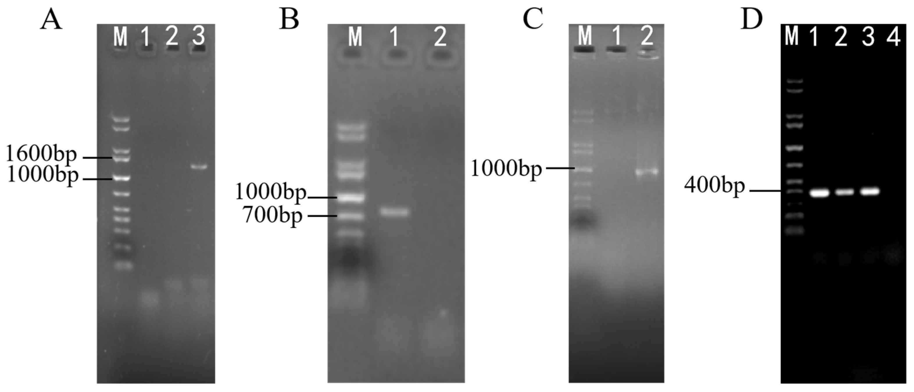
2.1. Cloning and Bioinformatics Analysis of the swnN Gene in A. oxytropis OW 7.8

The swnN gene (GeneBank: OR596336) in A. oxytropis OW 7.8 was cloned, with a length of 1164 bp (ATG-TGA) and three introns (57 bp, 107–163 bp; 56 bp, 402–457 bp; 55 bp, 815–869 bp). The length of swnN cDNA is 996 bp (ATG-TGA). The swnN gene is predicted to encode a protein with 331 amino acids. The molecular formula of this protein is C1646H2600N440O486S9, with a molecular weight of 36.62 kDa and a pI of 5.49. The predicted protein is hydrophilic, without transmembrane domains.
A phylogenetic tree of SwnN proteins from 13 SW-producing fungi was constructed using the neighbor-joining method (Figure 2B) with two branches: The upper branch consisted of three species of the genus Trichophyton and one species of the genus Nannizzia, and another consists of four Metarhizium species and a separated branch of Chaetothyriaceae fungi. In the lower branch, A. oxytropis Raft River and A. oxytropis OW 7.8 first clustered together, then joined with P. semeniperda, and finally clustered with the separately branched S. leguminicola. The amino acid sequence alignment of SwnN proteins showed the highest sequence identity of 100% between A. oxytropis OW 7.8 and A. oxytropis Raft River. The sequence identity is 87.39% between A. oxytropis OW 7.8 and S. leguminicola, while 85.71% between A. oxytropis OW 7.8 and M. acridum, Microsporum canis, 84.03% between A. oxytropis OW 7.8 and three Metarhizium species (M. brunneum ARSEF 3297, M. guizhouense ARSEF 977, M. robertsii ARSEF 2575), approximately.

2.2. Sensitivity Screening of A. oxytropis OW 7.8 to Hyg B

After incubation in darkness at 25 °C for 20 days, the growth of A. oxytropis OW 7.8 colonies on PDA media containing different concentrations of hygromycin B (Hyg B) indicated that the fungus is sensitive to ≥2 μg/mL of Hyg B.

2.3. Identification for Transformants of ΔswnN Colonies

The swnN gene knockout transformants gradually appeared on PDA media containing 2 μg/mL Hyg B one week after transformation. PCR results showed that bands of hygromycin phosphotransferase (hpt) gene, hpt gene and upstream homologous to the swnN, the hpt gene and downstream homologous to the swnN were amplified in swnN gene knockout transformants (Figure 3). The sequencing of the PCR products confirmed their accuracy, resulting in the identification of the ∆swnN.

2.4. Morphology of Colonies and Mycelia

The colonies of A. oxytropis OW 7.8 displayed soft white velvety patches, round, raised, with uniform margin and radial growth colonies which grew slowly (Figure 4A). Later a black brown pigment was secreted in each colony [21]. In contrast, the colonies of ∆swnN were loose creamy yellow and irregularly shaped, with no pigment accumulation and slower growth rate (Figure 4E).
Significant differences were observed in the mycelial morphology and structure between A. oxytropis OW 7.8 and ΔswnN with scanning electron microscopy. A. oxytropis OW 7.8 exhibited typical single, tubular, smooth-surfaced and unbranched fungal hyphae (Figure 4B–D). In contrast, ΔswnN showed abnormal jointed hyphae, with protrusions, depressions, and swollen branched tips (Figure 4F–H).

2.5. Screening of ΔswnN for Glufosinate Sensitivity

After incubation in darkness at 25 °C for 20 days, the growth of ΔswnN colonies on PDA media containing different concentrations of glufosinate (Gla) indicated that the fungus is sensitive to ≥500 μg/mL of Gla.

2.6. Screening and Identification of ΔswnN/swnN

PCR results showed that sequence of the glufosinate resistance (bar) gene and the swnN cDNA were amplified in the swnN gene function complement transformants (Figure 5). The sequencing of the PCR products confirmed their accuracy, resulting in the verification of the gene function complement strain ΔswnN/swnN.

2.7. SW Levels in A. oxytropis OW 7.8, ΔswnN, and ΔswnN/swnN Mycelia

The regression equation for the SW standard curve is Y = 1.006 × 10⁴X + 9.811 × 10² (R2 = 0.9910). SW was not detected in the mycelia of ΔswnN, whereas the SW levels in the mycelia of A. oxytropis OW 7.8 and ΔswnN/swnN were 24.799 ± 0.132 μg/g·DW and 25.656 ± 2.258 μg/g·DW after 20 days of cultivation (Figure 6), indicating that the gene function of swnN promotes SW synthesis.

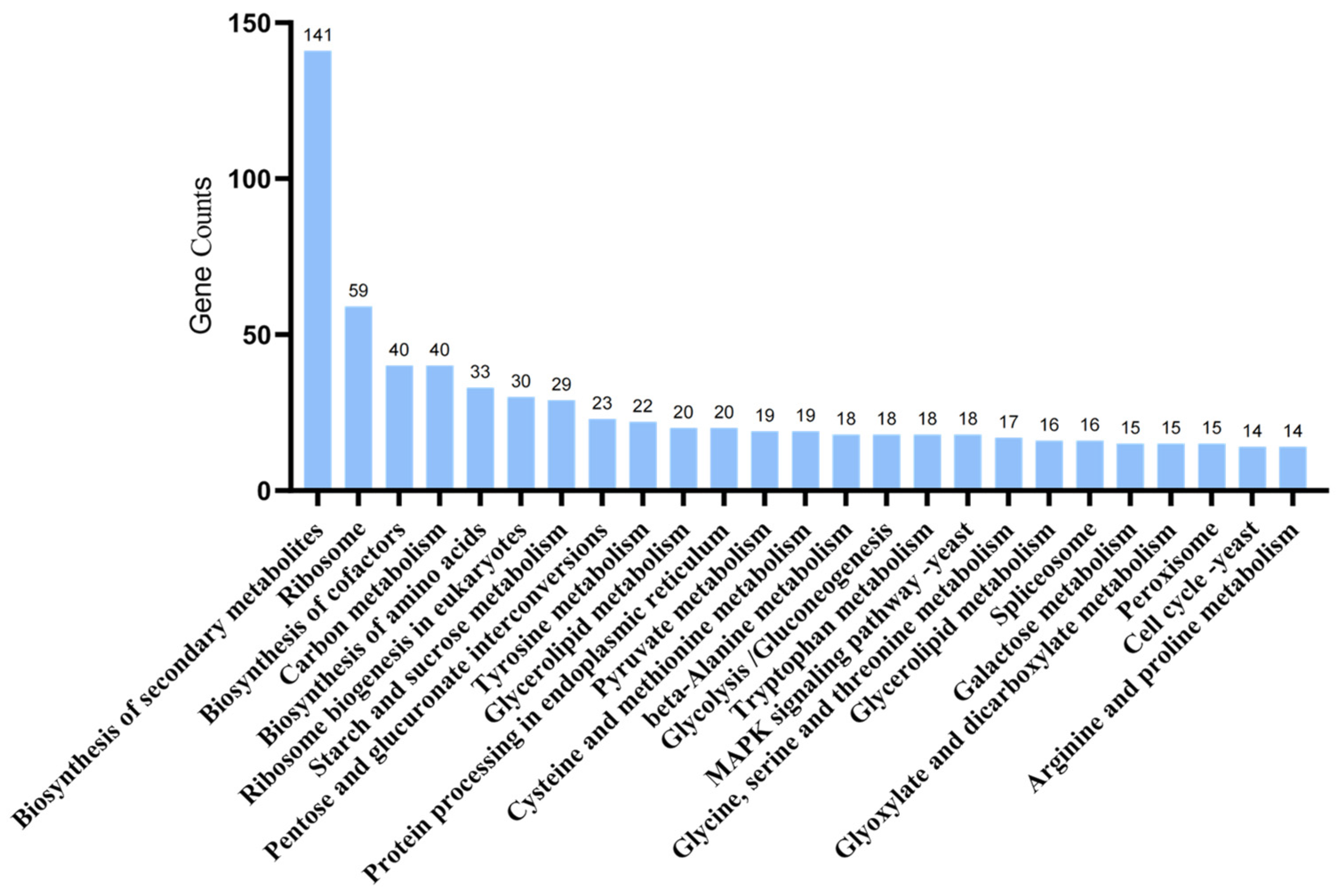
2.8. Transcriptome Sequencing Analysis of A. oxytropis OW 7.8 and ΔswnN

There was no swnN transcripts detected from ΔswnN transcriptomics. Transcriptome sequencing of A. oxytropis OW 7.8 and ΔswnN produced a total of 254,788,656 clean reads, amounting to 38.21 G. Gene differential expression was shown between A. oxytropis OW 7.8 and ΔswnN. A total of 3385 DEGs were identified, of which 1587 (46.89% of DEGs) were up-regulated and 1798 (53.11%) were down-regulated. Five DEGs closely related to SW synthesis were identified, among which the gene of sac was up-regulated, while the genes of swnR, swnK, swnH1, and swnH2 were down-regulated (Figure 7).
GO functional enrichment analysis of DEGs between A. oxytropis OW 7.8 and ΔswnN showed that 701 DEGs (277 up-regulated and 424 down-regulated) were assigned to 47 GO terms. In the Biological Process (BP) category, the top three enriched metabolic groups were oxidation–reduction process (30.67%), transmembrane transport (24.39%), and cellular amide metabolic process (8.70%). In the Cellular Component (CC) category, the top three groups were membrane component (19.54%), intrinsic component of membrane (19.54%), and non-membrane-bounded organelle (7.85%). In the Molecular Function (MF) category, the top three groups were oxidoreductase activity (28.53%), cofactor binding (22.11%), and transition metal ion binding (18.83%) (Figure 8).
KEGG enrichment analysis of DEGs between A. oxytropis OW 7.8 and ΔswnN annotated 553 DEGs (301 up-regulated and 252 down-regulated) to 97 pathways. The top three metabolic groups (Figure 9) included biosynthesis of secondary metabolites (25.50%), ribosome (10.04%), biosynthesis of cofactors (7.23%), and carbon metabolism (7.23%).

2.9. Metabolomic Analysis of A. oxytropis OW 7.8 and ΔswnN

The principal component analysis (PCA) of the metabolomic data for A. oxytropis OW 7.8 and ΔswnN is shown in Figure 10A,B, indicating differences in metabolites between ΔswnN and A. oxytropis OW 7.8. In positive ion mode, the most abundant metabolites were lipids and lipid-like molecules (30.04%), followed by organic acids and derivatives (23.99%), and organoheterocyclic compounds (14.48%). In negative ion mode, the most abundant metabolites were lipids and lipid-like molecules (40.84%), followed by organic acids and derivatives (20.30%), nucleosides, nucleotides, and analogues (10.89%), and alkaloids and derivatives (9.90%) (Figure 10C,D,G,H). In positive ion mode, 733 DEMs were identified, with 258 up-regulated and 204 down-regulated. In negative ion mode, 271 DEMs were identified, with 172 up-regulated and 99 down-regulated (Table 2).
KEGG enrichment analysis was performed on 733 differential metabolites, and enriched into 57 metabolic groups, including 50 DEMs for metabolic pathways, 29 DEMs for various antibiotic, 20 DEMs for secondary metabolites, 19 DEMs for amino acids, 11 DEMs for 2-oxocarboxylic acid metabolism (Figure 10F). Five DEMs involved in SW synthesis were identified with L-glutamate, α-ketoglutaric acid, and L-proline up-regulated, while PA and 2-aminoadipic acid were down-regulated (Figure 10E).

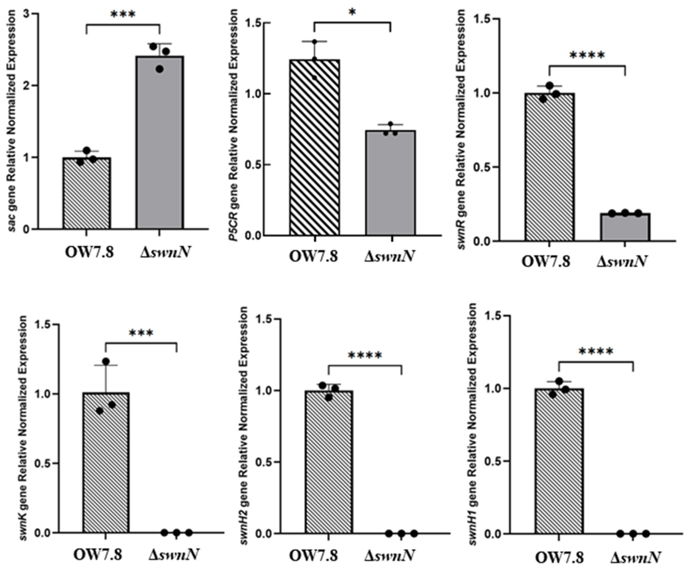
2.10. Expression of sac, P5CR, and SWN Cluster Genes in A. oxytropis OW 7.8 and ΔswnN

RT-qPCR results indicated that the relative expression levels of the sac gene in ΔswnN are significantly increased compared to A. oxytropis OW 7.8. Conversely, the relative expression levels of the P5CR, swnH1, swnH2, swnK, and swnR genes significantly decreased (Figure 11).

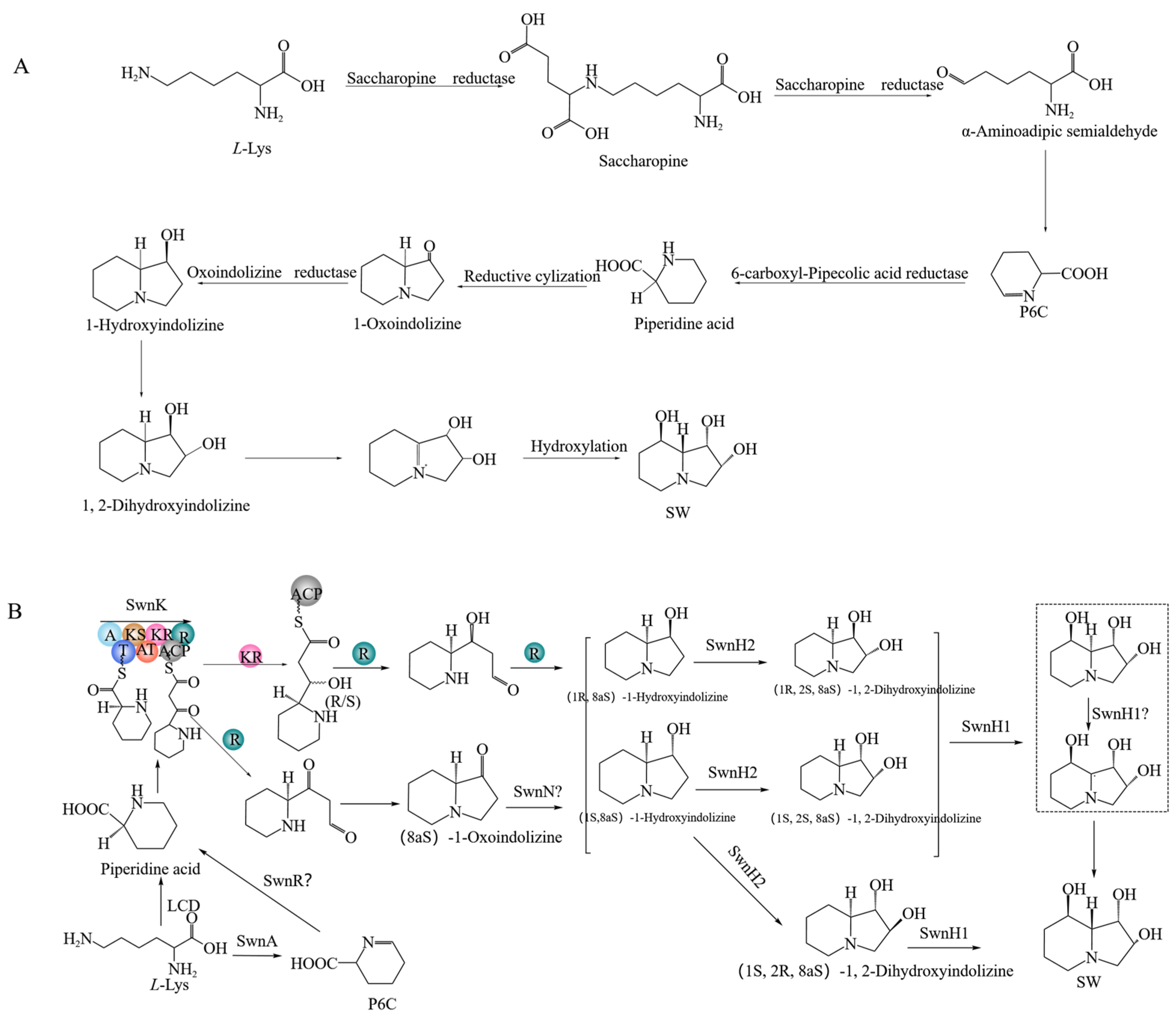
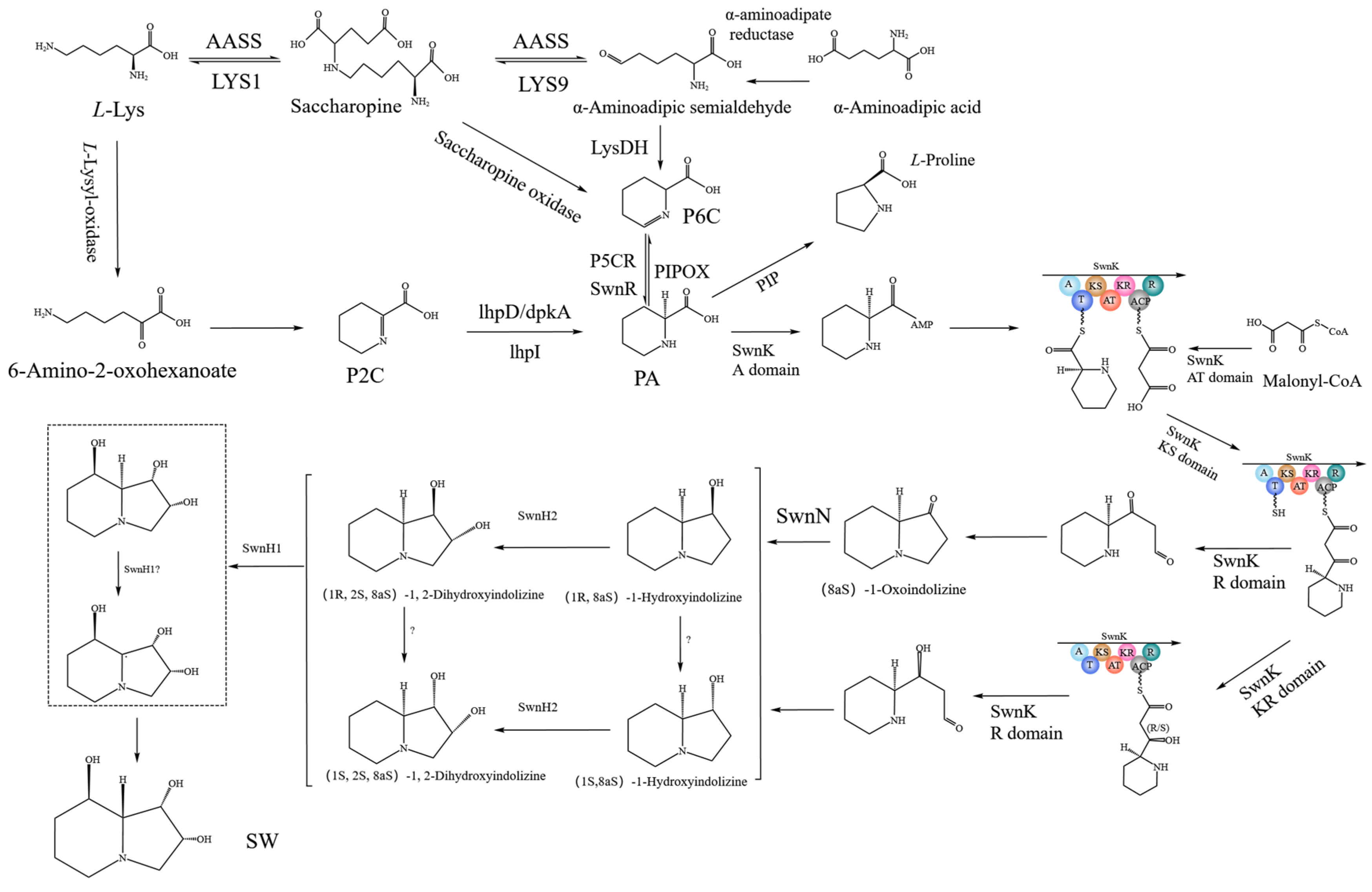
2.11. SW Synthesis Pathway in A. oxytropis OW 7.8

The predicted SW biosynthesis pathway in A. oxytropis OW 7.8 is shown in Figure 12, starting from L-lysine artificially. The Sac enzyme catalyzes the synthesis of saccharopine from L-Lysine. Saccharopine is reduced to α-aminoadipic semialdehyde (α-aminoadipic semialdehyde is also formed from α-aminoadipic acid catalyzed by α-aminoadipate reductase). α-Aminoadipic semialdehyde cyclizes to form P6C (P6C is also formed from saccharopine catalyzed by saccharopine oxidase). P6C is then catalyzed by the P5CR enzyme or SwnR enzyme to form L-PA. There might be an alternative pathway synthesizing L-PA, in which L-lysine is converted to 6-amino-2-oxohexanoate catalyzed by L-lysyl-alpha-oxidase. 6-Amino-2-oxohexanoate isomerizes to form P2C, which is subsequently catalyzed by the enzymes lhpD/dpkA/lhpI to produce L-PA.
L-PA was converted to (8aS)-1-oxoindolizine, (1R,8aS)-1-hydroxyindolizine, or (1S,8aS)-1-hydroxyindolizine by the action of multifunctional SwnK protein. Subsequently, (8aS)-1-oxoindolizine was synthesized form (8aS)-1-hydroxyindolizine by SwnN enzyme. (1R,2S,8aS)-1,2-dihydroxyindolizine was synthesized from (1R,8aS)-1-hydroxyindolizine by the action of the SwnH2 enzyme, (1S,2S,8aS)-1,2-dihydroxyindolizine was synthesized from (1S,8aS)-1-hydroxyindolizine by the action of the SwnH2 enzyme. Finally, (1S,2S,8R,8aR)-1,2,8-octahydroindolizinetriol (SW) was synthesized from (1R,2S,8aS)-1,2-dihydroxyindolizine or (1S,2S,8aS)-1,2-dihydroxyindolizine catalyzed by SwnH1 enzyme.

3. Discussion

The nucleotide sequence identity of swnN gene between A. oxytropis OW 7.8 and A. oxytropis Raft River (KY365741.1) was 99.91% indicating a close phylogenetic relationship, with one synonymous mutation (at 1053, C in OW 7.8 and G in Raft River, both encoding threonine). The identity of SwnN amino acid sequence was 100% between A. oxytropis OW 7.8 and A. oxytropis Raft River, while the identity between A. oxytropis OW 7.8 and M. robertsii ARSEF 2575 was 84.03%. The phylogenetic relationship of SwnN protein (Figure 2B) in SW-producing fungi is consistent with that in the fungi reported by Neyaz et al. [32].
The endophytic fungus A. oxytropis OW 7.8 exhibits a very slow growth rate and long growing cycle in vitro, with a very low homologous recombination rate, resulting in few transformants after long-term exploration. In this study, the swnN gene of A.oxytropis OW 7.8 was knocked out for the first time, and it was found that the SW levels in mycelia of ΔswnN were always lower than that in A. oxytropis OW 7.8 at different cultivation times. For example, SW was not detected in ΔswnN cultured for 20 days, and it was detected in trace amounts in the ΔswnN strain cultured for 35 d. SW was detected again in the gene function complementation strain ΔswnN/swnN, indicating that the function of the swnN gene promotes the synthesis of SW in fungi which is consistent with the conclusion in M. robertsii.
Compared with A. oxytropis OW7.8, the expressions of genes including P5CR, swnR, swnK, swnH2, and swnH1 were all down-regulated in the transcriptome of ΔswnN. RT-qPCR results for A. oxytropis OW 7.8 and ΔswnN were consistent with the data of transcriptome analysis. The levels of PA and α-aminoadipic acid were down-regulated, and L-proline was up-regulated in the metabolome of ΔswnN. The SW levels in mycelia of ΔswnN were always lower than that in A. oxytropis OW 7.8. The PA levels were down-regulated, consistent with down-regulated gene expressions of P5CR and swnR encoding enzymes involved in PA synthesis. Down-regulation of α-aminocaproic acid and up-regulation of L-proline might also contribute to decrease of PA levels. Knockout of swnN gene might result in the accumulation of substrate of SwnN enzyme, which could decrease the expressions of swnK gene due to negative feedback regulation. SW could not be synthesized through the 1-oxoindolizine branch after knocking out the swnN gene, but it could be synthesized through the 1-hydroxyindolizine branch. The synthesis of 1-hydroxyindolizine might be decreased due to down-regulation of the swnK gene. Since the expressions of downstream swnH2 gene and swnH1 gene were also determined showing down-regulation, the SW levels were greatly decreased, finally, in ΔswnN.
Significant gene expression changes occurred in ΔswnN in multiple metabolic groups through GO enrichment, including biological processes, cell components, and molecular functions. Transcriptome and metabolome joint analysis of A. oxytropis OW7.8 and ΔswnN showed significant changes in levels of gene expression and metabolites in some metabolic groups such as biosynthesis of secondary metabolites, biosynthesis of amino acids, 2-oxocarbonyl acid metabolism, lysine biosynthesis, and lysine degradation. We postulated that the function of SwnN protein was involved in both SW biosynthesis and other physiological and biochemical reactions in A. oxytropis OW7.8. Therefore, the colonies and mycelia morphology, and growth rate changed in ΔswnN.
Our previously predicted SW biosynthesis pathway in A. oxytropis OW 7.8 included P6C and P2C branches [36]. In this study, the P6C branch in A. oxytropis OW 7.8 was refined, predicting that α-aminoadipic semialdehyde can also be produced from α-aminoadipic acid catalyzed by α-aminoadipate reductase, and P6C can also be formed from saccharopine catalyzed by saccharopine oxidase. The pathway from L-PA to SW was also refined, including the conversion of L-PA to (8aS)-1-indolizidinone, (1R,8aS)-1-hydroxyindolizine, or (1S,8aS)-1-hydroxyindolizine by action of the SwnK protein, the conversion from (8aS)-1-oxoindolizine to (8aS)-1-hydroxyindolizine catalyzed by the SwnN enzyme, the conversion from (1R,8aS)-1-hydroxyindolizine and (1S,8aS)-1-hydroxyindolizine to (1R,2S,8aS)-1,2-dihydroxyindolizine, (1S,2S,8aS)-1,2-dihydroxyindolizine catalyzed by the SwnH2 enzyme, the conversion of (1R,2S,8aS)-1,2-dihydroxyindolizine and (1S,2S,8aS)-1,2-dihydroxyindolizine to SW catalyzed by the SwnH1 enzyme.

4. Materials and Methods

4.1. Strain

A. oxytropis OW 7.8 was isolated by our research group from O. glabra collected in Wushen Banner, Ordos city, Inner Mongolia, China (108°52′ E, 38°36′ N, elevation 1291 m) [17]. The mycelia were cultured on potato dextrose agar (PDA) media (fresh peeled potatoes 200 g, glucose 20 g, agarose 15 g, diluted to 1000 mL) at 25 °C.

4.2. Extraction of Genomic DNA and Identification of swnN Gene from A. oxytropis OW 7.8

The genomic DNA of A. oxytropis OW 7.8 was extracted (Plant Genomic DNA Kit, Tiangen, Beijing, China). The quality of the DNA was verified by 1% agarose gel electrophoresis. Primers NupF (5′-CTGGCTAGCTGCATATGCAGCAG-3′) and NdownR (5′-CGTGGCAGTTGATGACTGGG-3′) were designed based on the genomic data of A. oxytropis OW 7.8 to amplify the swnN gene. The PCR program was as follows: 94 °C for 3 min; 94 °C for 30 s, 58 °C for 30 s, 72 °C for 70 s, repeated for 29 cycles; and a final extension at 72 °C for 10 min. The PCR products were detected by 1% agarose gel electrophoresis, purified (SanPrep Column PCR Product Purification Kit, Sangon Biotech, Shanghai, China), and then sequenced (Sangon Biotech).

4.3. Total RNA Extraction and swnN cDNA Cloning of A. oxytropis OW 7.8

Total RNA of A. oxytropis OW 7.8 was extracted (the OminiPlant RNA Kit, CWBIO, Shanghai, China) and reverse-transcribed to synthesize cDNA. The swnN cDNA was amplified with primers NF/NR (5′-CCTCGACTCTAGAGGATCCATGGTTGTCGTTGCTGTCGCC-3′; 5′- CCTCGCCCTTGCTCACCATCACAAGCTCCCTGTGATCAAGAT-3′). The PCR program was as follows: 98 °C for 3 min; 98 °C for 30 s, 58 °C for 30 s, 72 °C for 60 s, repeated for 29 cycles; and a final extension at 72 °C for 10 min. The PCR products were detected by 1.0% agarose gel electrophoresis, purified (SanPrep Column PCR Product Purification Kit, Sangon Biotech, Shanghai, China), and then sequenced (Sangon Biotech, Shanghai, China).

4.4. Construction of Vectors

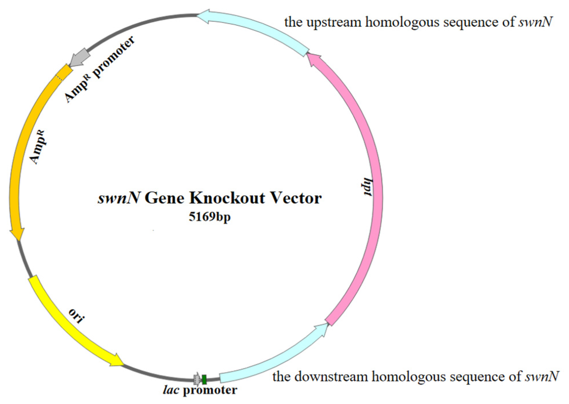
4.4.1. Construction of the swnN Gene Knockout Vector

The upstream and downstream homologous sequences of the swnN were amplified using the genomic DNA of A. oxytropis OW 7.8 as a template and primers upF/upR (5′-CTGGCTAGCTGCATATGCAGCAG-3′; 5′-CTGGCTAGCTGCATATGCAGCAG-3′) and downF/downR (5′-ATAGAGTAGATGCCGACCGCGGGTTCGCCATCCATGGAGGCCTCAT-3′; 5′-CGTGGCAGTTGATGACTGGG-3′). The hpt 5′ and 3′ end sequences were amplified using the pCB1003 plasmid as a template and primers HYGF/hygR (5′-GGCTTGGCTGGAGCTAGTGGAGGTCAA-3′; 5′-GTATTGACCGATTCCTTGCGGTCCGAA-3′) and hygF/HYGR (5′-GATGTAGGAGGGCGTGGATATGTCCT-3′; 5′-GAACCCGCGGTCGGCATCTACTCTAT-3′). The upstream and downstream homologous sequences of the swnN were linked to the hpt gene on both sides using the split-marker technique to construct the swnN gene knockout cassette, and this cassette was then ligated into the pMD-19T vector (Takara, Beijing, China) using TA cloning to make up the swnN gene knockout vector (Figure 13).

4.4.2. Construction of the swnN Gene Complementation Vector

The pBARGPE1-EGFP vector (MIAO LING BIOLOGY, Wuhan, China) was digested with EcoRI (Takara, Beijing, China) and BamHI (Takara, Beijing, China), reaction at 37 °C for 4 h, enzyme inactivation at 60 °C for 15 min. The swnN cDNA with homologous regions of the vector was ligated into the above linearized pBARGPE1-EGFP vector by seamless cloning. The bar was used as a selection marker, with PgpdA as the promoter and TtrpC as the terminator, to drive the expression of swnN cDNA.

4.5. Sensitivity Test of A. oxytropis OW 7.8 to Hygromycin B

The mycelia of A. oxytropis OW 7.8 were inoculated on PDA media containing 0 μg/mL, 0.8 μg/mL, 0.9 μg/mL, 1 μg/mL,2 μg/mL, and 3 μg/mL of Hyg B, respectively. The cultures were incubated in the dark at 25 °C for 20 days. The growth of colonies was observed, and an appropriate concentration of Hyg B was chosen to add to the media for swnN gene knockout transformant screening.

4.6. Preparation and Transformation of A. oxytropis OW 7.8 Protoplasts

Young mycelia of OW7.8 were inoculated in PDB media (50 μg/mL Amp) shaking at 25 °C, 200 rpm for 7 days. Protoplasts from mycelia of A. oxytropis OW 7.8 were prepared according to the method described by Hu et al. [40]. The swnN gene knockout vector was then transformed into protoplast of A. oxytropis OW 7.8 mediated by PEG8000 [40].

4.7. Screening and Identification of Gene Knockout Transformants

The swnN gene knockout transformants were cultured on TB3 (0.3% yeast extract, 0.3% acid hydrolyzed casein, 20% sucrose) regeneration media (bottom layer: 50 μg/mL Amp and 1 μg/mL Hyg B; top layer: 50 μg/mL Amp and 2 μg/mL Hyg B) at 25 °C to cultivate. Single colony was transferred to PDA media (2 μg/mL Hyg B) for culture. Transformants resistant to Hyg B were screened and subjected to PCR analysis. The sequence of upstream homologous and hpt was amplified with primers uphygF/hygR (5′-GCACAGGGCCATCGTACTATCC-3′/5′-GTATTGACCGATTCCTTGCGGTCCGAA-3′). The sequence of hpt and downstream homologous sequence was amplified with primers hygF/downhygR (5′-GATGTAGGAGGGCGTGGATATGTCCT-3′/5′-GCGCATGACTCAACATTGAGAG-3′). The sequence of hpt was amplified with primers HYGF/HYGR (5′-GGCTTGGCTGGAGCTAGTGGAGGTCAA-3′/5′-GAACCCGCGGTCGGCATCTACTCTAT-3′) (Figure 14). All PCR products were sequenced for verification (Sangon Biotech, Shanghai, China).

4.8. Sensitivity Test of ΔswnN to Glufosinate

The mycelia of ΔswnN were inoculated on PDA media containing 300 μg/mL, 400 μg/mL, and 500 μg/mL of Gla, respectively. The cultures were incubated in the dark at 25 °C for 20 days. The growth of colonies was observed, and an appropriate concentration of Gla was chosen to add to the media for the swnN gene complementation transformant screening.

4.9. Preparation and Transformation of ΔswnN Protoplasts

Young mycelia of ΔswnN were inoculated in PDB media (50 μg/mL Amp, 2 μg/mL Hyg B) shaking at 25 °C, 200 rpm for 14 days. Protoplasts were prepared according to the method described by Hu et al. [40]. The swnN gene complementation vector was then transformed into protoplast of ΔswnN mediated by PEG8000 [40].

4.10. Screening and Identification of Gene Complementation Transformants

The swnN gene complementation transformants were cultured on TB3 regeneration media (bottom layer: 50 μg/mL Amp and 1 μg/mL Hyg B; top layer: 50 μg/mL Amp, 2 μg/mL Hyg B, and 500 μg/mL Gla) at 25 °C. Single colony was transferred to PDA media (50 μg/mL Amp, 2 μg/mL Hyg B, and 500 μg/mL Gla) for culture. The swnN gene complementation transformants resistant to Gla were screened and subjected to PCR analysis. The sequence of bar gene was amplified with primers barF/barR (5′-ATTAGCAGACAGGAACGAGGACA-3′/5′-CATCGCAAGACCGGCAAC-3′), and the swnN cDNA was amplified with primers NF/NR (5′-GTCAGGGTGGTCACGAGGG-3′/5′-CAAGGTCGTTGCGTCAGTCC-3′). All PCR products were sequenced for verification (Sangon Biotech, Shanghai, China). The placement of primers and expected size of PCR products are shown in Figure 15.

4.11. Colony and Mycelia Morphology

The colony morphology of A. oxytropis OW 7.8 and ΔswnN after 20 days of cultivation was observed. The mycelia morphology of A. oxytropis OW 7.8 and ΔswnN was observed under a scanning electron microscope (SU81003.0 kV × 30, Wuhan Servicebio Technology, Wuhan, China).

4.12. Extraction and Detection of SW in Mycelia of A. oxytropis OW 7.8, ΔswnN, and ΔswnN/swnN

Mycelia of A. oxytropis OW 7.8, ΔswnN and ΔswnN/swnN cultured for 20 days were used for SW extraction by acetic acid–chloroform solution, purification by cation exchange resin, and elution with 1 mol/L ammonia solution. The SW levels in the mycelia were determined by HPLC-MS, with three replicates for each sample [41]. Data were analyzed by one-way ANOVA in GraphPad Prism 9.5 software. The chromatographic conditions were as follows: mobile phase, 5% methanol and 20 mmol/L ammonium acetate; flow rate, 0.4 mL/min; column temperature, 30 °C. MS conditions were as follows: positive ion 156; negative ion 70; IonSpray voltage (IS), 30 V.

4.13. Transcriptome Analysis of A. oxytropis OW 7.8 and ΔswnN

Mycelia of A. oxytropis OW 7.8 and ΔswnN strains (fresh weight 0.1–0.3 g) cultured for 20 days were rapidly frozen with liquid nitrogen in cryovials (1–3 min), and then delivered on dry ice with three replicates for each sample. Transcriptome sequencing was performed on the Illumina NovaSeq 6000 platform (Novogene, Beijing, China). The data were analyzed using DESeq2 (1.20.0) [42] and clusterProfiler (3.8.1) [43] package in R (4.1.0). The differentially expressed genes (DEGs|log2(Fold Change)|≥1 and p-value ≤ 0.05) [42,44] were subjected to functional enrichment analysis with GO and KEGG.

4.14. Metabolome Detection and Analysis of A. oxytropis OW 7.8 and ΔswnN

Mycelia of A. oxytropis OW 7.8 and ΔswnN (fresh weight 0.1–0.3 g) cultured for 20 days were rapidly frozen with liquid nitrogen in cryovials (1–3 min), and then delivered on dry ice with five replicates for each sample. Metabolome was performed using LC-MS (Novogene, Beijing, China). The data were analyzed using Compound Discoverer (3.1) package in R [45]. The differential expressed metabolites (DEMs VIP > 1, |log2(Fold Change)| ≥ 1 and p-value < 0.05) [46,47] were subjected to functional enrichment analysis with KEGG.

4.15. RT-qPCR Detection of Genes Closely Related to SW Synthesis in A. oxytropis OW 7.8 and ΔswnN

The cDNAs of A. oxytropis OW 7.8 and ΔswnN were used as templates with actin as internal reference gene [48] to conduct RT-qPCR to amplify six differentially expressed genes closely related to SW synthesis. The PCR program was as follows: 95 °C for 2 min; 95 °C for 5 s, 60 °C for 30 s, repeated for 29 cycles, 95 °C for 20 s, 65 °C for 1 min,95 °C for 20 s. The RT-qPCR for six genes of sac (QsacF/QsacR 5′-CTGCTGCTCGGTGCTGGATTC-3′; 5′-CTAGACTGATGGCGTTGGTGTTGG-3′), P5CR (QP5CRF/QP5CRR 5′-TAGCAATAATGGGCGGCGTGATG-3′; 5′-GGCGATGAAGTTGGAGAGGTTGG-3′), swnR (QswnRF/QswnRR 5′-TTCTACTTTGCCACACACGAACCC-3′; 5′-ATAGTCAGCCAACCAGCCAATGC-3′), swnK (QswnKF/QswnKR 5′-GACCGCTTGCTCGCCTGTG-3′; 5′-CTCGTCAACTCGTCCAACACTTCC-3′), swnH1 (QswnH1F/QswnH1R 5′-TTGCTTTGCGGAGATGGAACCAG-3′; 5′-CGGAGTGTGCCTGAGATGAAGAAG-3′), and swnH2 (QswnH2F/QswnH2R 5′-CATCTGCTCCTCGCTTGCTACC-3′; 5′-CAGGACAACGCCTCCATCTCTTTC-3′) were performed with the 2^(−ΔΔCt) method (ΔCt = Ct(gene) − Ct(actin), ΔΔCt = ΔCt(∆swnN) − ΔCt(OW 7.8)). All statistical analysis was conducted by GraphPad Prism 9.5.0 software with one-way ANOVA for each group of samples.

5. Conclusions

The swnN gene of endophytic fungus Alternaria oxytropis OW 7.8 isolated from Oxytropis glabra was cloned, and the gene knocked out mutant ∆swnN was first constructed. The colony morphology of ΔswnN differed from that of A. oxytropis OW 7.8, appearing creamy yellow, with irregular shapes and a slower growth rate. Compared to A. oxytropis OW 7.8, no SW was detected in the mycelia of ∆swnN cultured for 20 days, and SW was detected again in the gene function complementation strain ΔswnN/swnN, indicating that the function swnN gene promotes SW biosynthesis. Six DEGs and five DEMs closely associated with SW biosynthesis were identified by analyzing the data of the transcriptome and metabolome of A. oxytropis OW 7.8 and ΔswnN. The SW biosynthesis pathway in A. oxytropis OW 7.8 was hypothesized and refined.

Author Contributions

Conceptualization, P.L.; methodology, P.L., C.L. and N.D.; investigation, P.L., C.L., N.D., B.Y., Y.L. and K.J.; data curation, C.L., N.D. and P.L.; writing—original draft preparation, C.L. and N.D.; writing—review and editing, P.L., C.L. and N.D., visualization, C.L. and N.D.; supervision, P.L.; project administration, P.L. All authors have read and agreed to the published version of the manuscript.

Funding

This research was funded by the National Natural Science Foundation of China (31460235; 31960130; 30860049), Natural Science Foundation of Inner Mongolia Autonomous Region of China (2022MS03014), Fundamental Research Funds for the Inner Mongolia Normal University (2022JBTD010).

Institutional Review Board Statement

Not applicable.

Informed Consent Statement

Not applicable.

Data Availability Statement

The data presented in this study are available on reasonable request from the corresponding author.

Conflicts of Interest

The authors declare no conflicts of interest.

References

  1. James, L.F.; Foote, W. Estrogenic Properties of Locoweed (Astragalus lentiginosus). Can. J. Comp. Med. 1972, 36, 360–365. [Google Scholar] [PubMed]
  2. Molyneux, R.J.; McKenzie, R.A.; O’Sullivan, B.M.; Elbein, A.D. Identification of the Glycosidase Inhibitors Swainsonine and Calystegine B2 in Weir Vine (Ipomoea sp. Q6 {AFF. CALOBRA}) and Correlation with Toxicity. J. Nat. Prod. 1995, 58, 878–886. [Google Scholar] [CrossRef] [PubMed]
  3. Chen, X.; Peng, L.; Li, N.; Li, Q.; Li, P.; Fung, K.; Leung, P.; Gao, J. The Healing and Anti-Scar Effects of Astragaloside IV on the Wound Repair in Vitro and in Vivo. J. Ethnopharmacol. 2012, 139, 721–727. [Google Scholar] [CrossRef] [PubMed]
  4. Wu, C.; Wang, W.; Liu, X.; Ma, F.; Cao, D.; Yang, X.; Wang, S.; Geng, P.; Lu, H.; Zhao, B. Pathogenesis and Preventive Treatment for Animal Disease Due to Locoweed Poisoning. Environ. Toxicol. Pharmacol. 2014, 37, 336–347. [Google Scholar]
  5. Cook, D.; Gardner, D.R.; Lee, S.T.; Pfister, J.A.; Stonecipher, C.A.; Welsh, S.L. A Swainsonine Survey of North American Astragalus and Oxytropis Taxa Implicated as Locoweeds. Toxicon 2016, 118, 104–111. [Google Scholar] [CrossRef]
  6. Cook, D.; Ralphs, M.H.; Welch, K.D.; Stegelmeier, B.L. Locoweed Poisoning in Livestock. Rangelands 2009, 31, 16–21. [Google Scholar] [CrossRef]
  7. Fu, J.; Guo, Y.; Huang, W.; Guo, R.; Luo, H.; Wu, C.; Zhao, B. The Distribution of Locoweed in Natural Grassland in the United States and the Current Status and Prospects of Research on Animal Poisoning. Acta Agrestia Sin. 2019, 27, 519–530. [Google Scholar]
  8. Guo, C.; Zhang, L.; Zhao, Q.; Beckmann, M.; Phillips, H.; Meng, H.; Mo, C.; Mur, L.A.J.; He, W. Host-Species Variation and Environment Influence Endophyte Symbiosis and Mycotoxin Levels in Chinese Oxytropis Species. Toxins 2022, 14, 181. [Google Scholar] [CrossRef]
  9. Tulsiani, D.R.P.; Broquist, H.P.; James, L.F.; Touster, O. The Similar Effects of Swainsonine and Locoweed on Tissue Glycosidases and Oligosaccharides of the Pig Indicate That the Alkaloid Is the Principal Toxin Responsible for the Induction of Locoism. Arch. Biochem. Biophys. 1984, 232, 76–85. [Google Scholar] [CrossRef]
  10. Wang, S.; Tan, P.; Wang, H.; Wang, J.; Zhang, C.; Lu, H.; Zhao, B. Swainsonine Inhibits Autophagic Degradation and Causes Cytotoxicity by Reducing CTSD O-GlcNAcylation. Chem. Biol. Interact. 2023, 382, 110629. [Google Scholar] [CrossRef]
  11. Panter, K.E.; James, L.F.; Stegelmeier, B.L.; Ralphs, M.H.; Pfister, J.A. Locoweeds: Effects on Reproduction in Livestock. J. Nat. Toxins 1999, 8, 53–62. [Google Scholar] [PubMed]
  12. Dennis, J.W. Effects of Swainsonine and Polyinosinic:Polycytidylic Acid on Murine Tumor Cell Growth and Metastasis. Cancer Res. 1986, 46, 5131–5136. [Google Scholar] [PubMed]
  13. Stegelmeier, B.L.; James, L.F.; Panter, K.E.; Ralphs, M.H.; Gardner, D.R.; Molyneux, R.J.; Pfister, J.A. The Pathogenesis and Toxicokinetics of Locoweed (Astragalus and Oxytropis spp.) Poisoning in Livestock. J. Nat. Toxins 1999, 8, 35–45. [Google Scholar]
  14. Molyneux, R.J.; James, L.F. Loco Intoxication: Indolizidine Alkaloids of Spotted Locoweed (Astragalus lentiginosus). Science 1982, 216, 190–191. [Google Scholar] [CrossRef] [PubMed]
  15. Kino, T.; Inamura, N.; Nakahara, K.; Kiyoto, S.; Goto, T.; Terano, H.; Kohsaka, M.; Aoki, H.; Imanaka, H. Studies of an Immunomodulator, Swainsonine. II. Effect of Swainsonine on Mouse Immunodeficient System and Experimental Murine Tumor. J. Antibiot. 1985, 38, 936–940. [Google Scholar] [CrossRef] [PubMed]
  16. Braun, K.; Romero, J.; Liddell, C.; Creamer, R. Production of Swainsonine by Fungal Endophytes of Locoweed. Mycol. Res. 2003, 107, 980–988. [Google Scholar] [CrossRef]
  17. Woudenberg, J.H.C.; Groenewald, J.Z.; Binder, M.; Crous, P.W. Alternaria Redefined. Stud. Mycol. 2013, 75, 171–212. [Google Scholar] [CrossRef]
  18. Wang, Q.I.; Nagao, H.; Yu-Ling, L.I.; Wang, H.S.; Kakishima, M. Embellisia oxytropis, a New Species Isolated from Oxytropis kansuensis in China. Mycotaxon 2006, 95, 255–260. [Google Scholar]
  19. Pryor, B.M.; Creamer, R.; Shoemaker, R.A.; McLain-Romero, J.; Hambleton, S. Undifilum, a New Genus for Endophytic Embellisia oxytropis and Parasitic Helminthosporium bornmuelleri on Legumes. Botany 2009, 87, 178–194. [Google Scholar] [CrossRef]
  20. Wang, W.; Qian, Y.; Lu, P.; He, S.; Du, L.; Li, Y.; Gao, F. The Relationship between Swainsonine and Endophytic Fungi in Different Populations of Oxytropis glabra from Inner Mongolia Chin. J. Anim. Vet. Sci. 2022, 53, 304–314. [Google Scholar]
  21. Lu, P.; Child, D.; Zhao, M.; Gardener, D.R.; Lv, G.; Han, G. Culture and Identification of Endophytic Fungi from Oxytropis glabra DC. Acta Ecol. Sin. 2009, 29, 53–58. [Google Scholar]
  22. Cook, D.; Gardner, D.R.; Pfister, J.A. Swainsonine-Containing Plants and Their Relationship to Endophytic Fungi. J. Agric. Food Chem. 2014, 62, 7326–7334. [Google Scholar] [CrossRef] [PubMed]
  23. Dorling, P.R.; Huxtable, C.R.; Colegate, S.M. Inhibition of Lysosomal Alpha-Mannosidase by Swainsonine, an Indolizidine Alkaloid Isolated from Swainsona canescens. Biochem. J. 1980, 191, 649–651. [Google Scholar] [CrossRef] [PubMed]
  24. Colegate, S.M.; Dorling, P.R.; Huxtable, C.R. A Spectroscopic Investigation of Swainsonine: An a-Mannosidase Inhibitor Isolated from Swainsona canescens. Aust. J. Chem. 1979, 32, 2257–2264. [Google Scholar] [CrossRef]
  25. Davis, D.; Schwarz, P.; Hernandez, T.; Mitchell, M.; Warnock, B.; Elbein, A.D. Isolation and Characterization of Swainsonine from Texas Locoweed (Astragalus emoryanus). Plant Physiol. 1984, 76, 972–975. [Google Scholar] [CrossRef]
  26. Gardner, D.R.; Molyneux, R.J.; Ralphs, M.H. Analysis of Swainsonine: Extraction Methods, Detection, and Measurement in Populations of Locoweeds (Oxytropis spp.). J. Agric. Food Chem. 2001, 49, 4573–4580. [Google Scholar] [CrossRef]
  27. Colodel, E.M.; Gardner, D.R.; Zlotowski, P.; Driemeier, D. Identification of Swainsonine as a Glycoside Inhibitor Responsible for Sida Carpinifolia Poisoning. Vet. Hum. Toxicol. 2002, 44, 177–178. [Google Scholar]
  28. Haraguchi, M.; Gorniak, S.L.; Ikeda, K.; Minami, Y.; Kato, A.; Watson, A.A.; Nash, R.J.; Molyneux, R.J.; Asano, N. Alkaloidal Components in the Poisonous Plant, Ipomoea carnea (Convolvulaceae). J. Agric. Food Chem. 2003, 51, 4995–5000. [Google Scholar] [CrossRef]
  29. Lu, P.; Li, X.; Wang, S. Saccharopine Reductase Influences Production of Swainsonine in Alternaria oxytropis. Sydowia 2021, 73, 69–74. [Google Scholar]
  30. Wang, Y.; Lu, P.; Sarula; Xi, L.; Yang, B.; Du, L. Construction and Identification of sac Functional Complementary Strain in Alternaria oxytropis. Mol. Plant Breed. 2020, 18, 7777–7783. [Google Scholar]
  31. Cook, D.; Donzelli, B.G.G.; Creamer, R.; Baucom, D.L.; Gardner, D.R.; Pan, J.; Moore, N.; Krasnoff, S.B.; Jaromczyk, J.W.; Schardl, C.L. Swainsonine Biosynthesis Genes in Diverse Symbiotic and Pathogenic Fungi. G3 2017, 7, 1791–1797. [Google Scholar] [CrossRef] [PubMed]
  32. Neyaz, M.; Das, S.; Cook, D.; Creamer, R. Phylogenetic Comparison of Swainsonine Biosynthetic Gene Clusters among Fungi. J Fungi 2022, 8, 359. [Google Scholar] [CrossRef] [PubMed]
  33. Luo, F.; Hong, S.; Chen, B.; Yin, Y.; Tang, G.; Hu, F.; Zhang, H.; Wang, C. Unveiling of Swainsonine Biosynthesis via a Multibranched Pathway in Fungi. ACS Chem. Biol. 2020, 15, 2476–2484. [Google Scholar] [CrossRef] [PubMed]
  34. Wickwire, B.M.; Harris, C.M.; Harris, T.M.; Broquist, H.P. Pipecolic Acid Biosynthesis in Rhizoctonia leguminicola. I. The Lysine Saccharopine, Delta 1-Piperideine-6-Carboxylic Acid Pathway. J. Biol. Chem. 1990, 265, 14742–14747. [Google Scholar] [CrossRef] [PubMed]
  35. Sim, K.L.; Perry, D. Analysis of Swainsonine and Its Early Metabolic Precursors in Cultures of Metarhizium anisopliae. Glycoconj. J. 1997, 14, 661–668. [Google Scholar] [CrossRef]
  36. Li, X.; Lu, P. Transcriptome Profiles of Alternaria oxytropis Provides Insights into Swainsonine Biosynthesis. Sci. Rep. 2019, 9, 6021. [Google Scholar] [CrossRef]
  37. Mo, Y.; Yang, Z.; Hao, B.; Cheng, F.; Song, X.; Shang, X.; Zhao, H.; Shang, R.; Wang, X.; Liang, J.; et al. Screening of Endophytic Fungi in Locoweed Induced by Heavy-Ion Irradiation and Study on Swainsonine Biosynthesis Pathway. J Fungi 2022, 8, 951. [Google Scholar] [CrossRef]
  38. Harris, T.M.; Harris, C.M.; Hill, J.E.; Ungemach, F.S.; Broquist, H.P.; Wickwire, B.M. (1S,2R,8aS)-1,2-Dihydroxyindolizidine Formation by Rhizoctonia leguminicola, the Fungus That Produces Slaframine and Swainsonine. J. Org. Chem. 1987, 52, 3094–3098. [Google Scholar] [CrossRef]
  39. Harris, C.M.; Schneider, M.J.; Ungemach, F.S.; Hill, J.E.; Harris, T.M. Biosynthesis of the Toxic Indolizidine Alkaloids Slaframine and Swainsonine in Rhizoctonia leguminicola: Metabolism of 1-Hydroxyindolizidines. J. Am. Chem. Soc. 1988, 110, 940–949. [Google Scholar] [CrossRef]
  40. Hu, J.; Lu, P.; Niu, Y. Preparation and Regeneration of Protoplasts Isolated from Embellisia Fungal Endophyte of Oxytropis glabra. Biotechnol. Bull. 2015, 31, 134–139. [Google Scholar]
  41. Sarula; Xi, L.; Lu, P.; Gao, F.; Du, L. The Influence of Different Additives on the Swainsonine Biosynthesis in Saccharopine Reductase Gene Disruption Mutant M1 of an Endophytic Fungus from Oxytropis glabra. Life Sci. Res. 2018, 22, 298–304. [Google Scholar]
  42. Love, M.I.; Huber, W.; Anders, S. Moderated Estimation of Fold Change and Dispersion for RNA-Seq Data with DESeq2. Genome Biol. 2014, 15, 550. [Google Scholar] [CrossRef] [PubMed]
  43. Yu, G.; Wang, L.; Han, Y.; He, Q. clusterProfiler: An R Package for Comparing Biological Themes among Gene Clusters. OMICS 2012, 16, 284–287. [Google Scholar] [CrossRef] [PubMed]
  44. Young, M.D.; Wakefield, M.J.; Smyth, G.K.; Oshlack, A. Gene Ontology Analysis for RNA-Seq: Accounting for Selection Bias. Genome Biol. 2010, 11, R14. [Google Scholar] [CrossRef] [PubMed]
  45. Cooper, B.; Yang, R. An Assessment of AcquireX and Compound Discoverer Software 3.3 for Non-Targeted Metabolomics. Sci. Rep. 2024, 14, 4841. [Google Scholar] [CrossRef]
  46. Haspel, J.A.; Chettimada, S.; Shaik, R.S.; Chu, J.-H.; Raby, B.A.; Cernadas, M.; Carey, V.; Process, V.; Hunninghake, G.M.; Ifedigbo, E.; et al. Circadian Rhythm Reprogramming during Lung Inflammation. Nat. Commun. 2014, 5, 4753. [Google Scholar] [CrossRef]
  47. Sreekumar, A.; Poisson, L.M.; Rajendiran, T.M.; Khan, A.P.; Cao, Q.; Yu, J.; Laxman, B.; Mehra, R.; Lonigro, R.J.; Li, Y.; et al. Metabolomic Profiles Delineate Potential Role for Sarcosine in Prostate Cancer Progression. Nature 2009, 457, 910–914. [Google Scholar] [CrossRef]
  48. Zhao, L.; Lu, P.; Du, L.; Niu, Y. Selection of Reference Genes in Endophytic Fungi Alternaria oxytropis in Oxytropis glabra and Expression Analysis of Its. Sac. Genom. Appl. Biol. 2019, 38, 5091–5098. [Google Scholar]
Figure 1. SW synthesis pathways in two fungi (A): S. leguminicola, (B): M. robertsii.
Ijms 25 10310 g001
Figure 2. Bioinformatics analysis of the SwnN protein. (A): Predicted SwnN protein structure; (B): The phylogenetic tree of the SwnN protein.
Ijms 25 10310 g002
Figure 3. Electrophoresis analysis of PCR products of transformant DNA. Marker: 1 kb plus DNA Ladder. (A): Lanes 1, 2: negative control, Lane 3 shows a band of the hpt gene, with the expected product being 1388 bp. (B): Lane 1 shows a band of the upstream homologous sequence of swnN + hpt gene, with the expected product being 861 bp, Line 2: negative control. (C): Line 1: negative control, Lane 2 shows a band of the hpt gene + downstream homologous sequence of the swnN, with the expected product being 1055 bp. (D): Lane 1, 2, 3 show bands of the internal sequence of swnN with the expected product being 401 bp, Lanes 4: PCR results using swnN knockout transformant DNA as a template with no band.
Ijms 25 10310 g003
Figure 4. Morphology of colonies and mycelia from A. oxytropis OW 7.8 and ΔswnN. (A): A.oxytropis OW 7.8 colonies. (BD): A.oxytropis OW 7.8 mycelia magnified 2000×, 5000×, and 10,000×. (E): ΔswnN colonies. (FH): ΔswnN mycelia magnified 2000×, 5000×, and 10,000×.
Ijms 25 10310 g004
Figure 5. Electrophoresis analysis of PCR products of swnN gene function complement transformants DNA. Marker: 1 kb plus DNA Ladder. (A): Lanes 1, 2, 3, and 4 show bands of the bar gene, with the expected product being 816 bp, Line 5, 6: negative control. (B): Lane 2 shows a band of the swnN cDNA, with the expected product being 1356 bp, Line 1, 3: negative control.
Ijms 25 10310 g005
Figure 6. SW level on day 20 in A. oxytropis OW 7.8, ΔswnN and ΔswnN/swnN. Error bars represent the standard error of the mean (n = 3), (****) p < 0.0001. ns: not significant.
Ijms 25 10310 g006
Figure 7. Volcano plot of DEGs between A. oxytropis OW 7.8 and ΔswnN.
Ijms 25 10310 g007
Figure 8. GO annotation of DEGs between A. oxytropis OW 7.8 and ΔswnN.
Ijms 25 10310 g008
Figure 9. KEGG enrichment analysis of DEGs between A. oxytropis OW 7.8 and ΔswnN.
Ijms 25 10310 g009
Figure 10. Metabolomic analysis of A. oxytropis OW 7.8 and ΔswnN. (A): Principal component analysis in positive ion mode. (B): Principal component analysis in negative ion mode. (C): Pie chart of metabolite classification in positive ion mode. (D): Pie chart of metabolite classification in negative ion mode. (E): Volcano plot of DEMs between A. oxytropis OW 7.8 and ΔswnN. (F): KEGG enrichment analysis of DEMs. (G): Heatmap of clustering analysis of DEMs in positive ion mode. (H): Heatmap of clustering analysis of DEMs in negative ion mode.
Ijms 25 10310 g010aIjms 25 10310 g010b
Figure 11. RT-qPCR detection of SW synthesis-related gene expression levels in A. oxytropis OW 7.8 and ΔswnN. Note: The x-axis represents genes; the y-axis represents relative expression levels. Error bars indicate the standard error of the mean (n = 3), with (*) p < 0.05, (***) p < 0.001, and (****) p < 0.0001.
Ijms 25 10310 g011
Figure 12. SW synthesis pathway in A. oxytropis OW 7.8. Note: LYS1/LYS9: Saccharopine reductase; AASS: α-Aminoadipic semialdehyde synthase; lysDH: Lysine dehydrogenase; PIPOX: L-Pipecolic acid oxidase/proline oxidase; P5CR: Pyrroline-5-carboxylate reductase; AAR: α-Aminoadipic acid reductase; L-Lysine-oxidase: L-Lysine oxidase; dpkA/lhpD/lhpI: 1-Piperideine-2-carboxylate reductase; PIP: Proline iminopeptidase.
Ijms 25 10310 g012
Figure 13. Diagram of the swnN gene knockout vector structure.
Ijms 25 10310 g013
Figure 14. Identification figure for swnN gene knockout transformants.
Ijms 25 10310 g014
Figure 15. Diagram of the swnN gene complementation vector structure.
Ijms 25 10310 g015
Table 1. Members of “SWN” gene cluster and their function prediction [31].
GeneEncoding ProductFunction Prediction
swnAAminotransferaseCatalyzing the synthesis of Pyrroline-6-carboxylate (P6C) from L-Lysine
swnRDehydrogenase or reductaseCatalyzing the synthesis of L-PA from P6C
swnKMultifunctional proteinCatalyzing the synthesis of 1-Oxoindolizidine (or 1-Hydroxyindolizine) from L-PA
swnNDehydrogenase or reductaseCatalyzing the synthesis of 1-Hydroxyindolizine from 1-Oxoindolizidine
swnH1Fe (II)/α-Ketoglutarate-dependent dioxygenaseCatalyzing the synthesis of SW from 1,2-Dihydroxyindolizine
swnH2Fe (II)/α-Ketoglutarate-dependent dioxygenaseCatalyzing the synthesis of 1,2-Dihydroxyindolizine form 1-Hydroxyindolizine
swnTTransmembrane transporterTransport of SW
Table 2. Screening of DEMs.
Screening ModeTotal of MetabolitesTotal of DEMsUp-RegulatedDown-Regulated
Positive801462258204
Negative47227117299
Disclaimer/Publisher’s Note: The statements, opinions and data contained in all publications are solely those of the individual author(s) and contributor(s) and not of MDPI and/or the editor(s). MDPI and/or the editor(s) disclaim responsibility for any injury to people or property resulting from any ideas, methods, instructions or products referred to in the content.

