Abstract
Iprodione is a pesticide that belongs to the dicarboximide fungicide family. This pesticide was designed to combat various agronomical pests; however, its use has been restricted due to its environmental toxicity and risks to human health. In this study, we explored the proteomic changes in the Pseudomonas sp. C9 strain when exposed to iprodione, to gain insights into the affected metabolic pathways and enzymes involved in iprodione tolerance and biodegradation processes. As a result, we identified 1472 differentially expressed proteins in response to iprodione exposure, with 978 proteins showing significant variations. We observed that the C9 strain upregulated the expression of efflux pumps, enhancing its tolerance to iprodione and other harmful compounds. Peptidoglycan-binding proteins LysM, glutamine amidotransferase, and protein Ddl were similarly upregulated, indicating their potential role in altering and preserving bacterial cell wall structure, thereby enhancing tolerance. We also observed the presence of hydrolases and amidohydrolases, essential enzymes for iprodione biodegradation. Furthermore, the exclusive identification of ABC transporters and multidrug efflux complexes among proteins present only during iprodione exposure suggests potential counteraction against the inhibitory effects of iprodione on downregulated proteins. These findings provide new insights into iprodione tolerance and biodegradation by the Pseudomonas sp. C9 strain.
1. Introduction
The pesticide iprodione (IPR) (3-(3,5-dichlorophenyl) N-isopropyl-2,4-dioxoimidazoli-18 dine-1-carboxamide) is a member of the dicarboximide fungicide family, created to combat various agronomical pests such as grey mould disease and onion white rot caused by different types of fungi, particularly Botrytis [1,2]. Previous studies have investigated the toxic effects of IPR, revealing its impact on sexual development in mice, neurobehavioral alterations, and its potential carcinogenicity in humans [3,4,5].
Due to this established toxicity, effective management of IPR residues is necessary. One in-farm method for controlling pesticide water residues is through pesticide biopurification systems (BPSs), which rely on their biomixture components (wheat straw, peat, and soil) for the removal of pesticides via biodegradation processes by microorganisms [6]. Accordingly, various types of soil and BPS bacteria have been identified and reported as capable of degrading IPR. These microorganisms include genera such as Arthrobacter, Achromobacter, Microbacterium, and Pseudomonas, among others [7,8,9]. These bacterial strains possess the enzymatic machinery necessary to biodegrade IPR, making them of interest for deciphering the metabolic pathways of IPR biodegradation.
A metabolic pathway has been suggested for the degradation of IPR, beginning with its conversion to N-(3,5-dichlorophenyl)-2,4-dioxoimidazolidine (metabolite I), followed by the formation of 3,5-dichlorophenylurea acetic acid (metabolite II), and ultimately leading to the production of 3,5-dichloroaniline (3,5-DCA) [10,11]. Recent studies have identified a gene called ipaH, which has an affinity for aromatic amino compounds, as having potential for the initial degradation of IPR [12]. Furthermore, an Achromobacter sp. C1 strain has been shown through proteomic analysis to undergo significant changes in its protein expression during the biodegradation of IPR, principally amidohydrolases, enzymes reported to be capable of degrading IPR due to their C-N affinity.
This study aimed to gain insights into the metabolic pathways and enzymes involved in the bacterial biodegradation process. Understanding the metabolic processes associated with the degradation of pesticides facilitates the construction of biotechnological tools that detoxify these compounds, which are hazardous to human health and the environment. To achieve this, we performed a gel-free comparative proteomic analysis, comparing the proteomes of Pseudomonas sp. C9 grown in the presence and absence of the fungicide IPR. Pseudomonas sp. C9 is an IPR-degrading bacterium isolated from a BPS used to treat pesticides [7] and has been reported to achieve high degradation rates of IPR, with more than 91% removal within 48 h, and exhibited a twofold increase in growth under IPR treatment [7,13] making strain C9 a suitable candidate for an in-depth comparative proteomic analysis under IPR degradation. The results of this study could contribute to understanding resistance mechanisms and provide valuable information for enhancing bioremediation strategies, ultimately reducing the environmental impacts of pesticides.
2. Results
2.1. Overall Results of Pseudomonas sp. C9 Protein Expression in Response to IPR Exposure
The proteomic composition of Pseudomonas sp. C9 when exposed to a concentration of 50 mg L−1 of IPR was analysed using the gel-free method. The mass spectrometric data were then analysed to identify the differentially expressed proteins compared to the untreated control. Out of a total of 1472 identified proteins, 978 proteins exhibited variations in expression, and 489 proteins were upregulated. Nonetheless, only 223 exhibited significant expression variations (≥1.21 and ≤−1.21-fold change) in the IPR-treated sample compared to the untreated control (Figure 1). Additionally, 382 proteins were solely present upon treatment with IPR. These findings revealed distinct protein profiles of Pseudomonas sp. C9 exposed to IPR, indicating the activation of specific proteins and pathways that contribute to the degradation and tolerance of the pesticide IPR (Table S1).
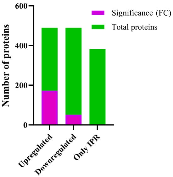
Figure 1.
Differential expression of proteins of strain Pseudomonas sp. C9 in response to IPR treatment. Total: total number of proteins. Significance (FC): the threshold for differential expression was set as follows: upregulated proteins (≥1.21-fold change) and downregulated proteins (≤−1.21-fold change). Only IPR: proteins identified only in IPR treatment.
2.2. Identification and Analysis of Differentially Expressed Proteins in Response to IPR Treatment
For the strain Pseudomonas sp. C9, analysis using Blast2GO software version 4.1 showed that the biological processes involved in IPR degradation and tolerance corresponded primarily to metabolic and cellular processes, representing 40% and 35%, respectively (Figure 2A). Additionally, based on molecular function, the proteins identified in strain C9 were mainly involved in transferase activity (21%) and hydrolase activity (18%), followed by other functions (Figure 2B).
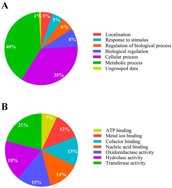
Figure 2.
Metabolic activity analysis of Pseudomonas sp. C9 in response to iprodione. (A) Bacterial biological process; (B) bacterial molecular function. Modified from Blast2GO software [14].
2.3. Upregulated Proteins
Functional analysis using OmicsBox software version 3.1 with the NCBI database showed that 60% of the upregulated proteins possess catalytic activities, and 51% possess binding activities. Of those proteins with catalytic activities, 22% have hydrolase activities, 14% transferase, and 13% oxidoreductase (Figure 3A). Of those with binding activities, 38% have organic cyclic compound binding, 30% ion binding, 16% small molecule binding, and 13% carbohydrate derivative binding. These proteins were located primarily in the cytoplasm of the cell, representing 74% of the identified proteins (Figure 4).
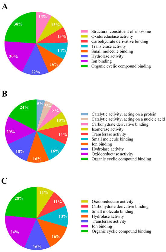
Figure 3.
Molecular functional activity analysis of Pseudomonas sp. C9 in response to IPR. (A) Upregulated proteins; (B) downregulated proteins; (C) proteins expressed only in IPR treatment. Modified from OmicsBox software version 3.1 [15].
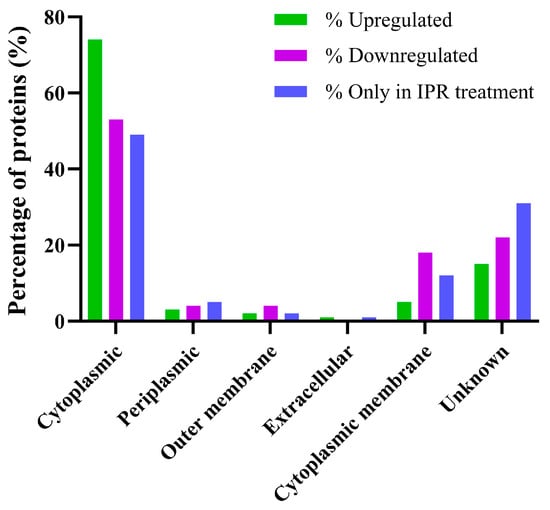
Figure 4.
Cellular localisation of proteins that are differentially regulated in response to iprodione in Pseudomonas sp. C9.
The significantly upregulated proteins analysed using the KEGG® database identified 76 pathways that might aid the organism in the degradation and tolerance of the pesticide IPR (Table S2). The proteins in these upregulated pathways are mainly involved in the biosynthesis of secondary metabolites, ribosomes, microbial metabolism in diverse environments, carbon metabolism, and biosynthesis of amino acids.
Among the upregulated proteins, the list of those significantly upregulated is provided in Table S1. In Table 1, we identified the upregulated proteins with a fold change ≥4.0, including two catalase enzymes [EC 1.11.1.6], peptidoglycan-binding protein LysM, guanylate kinase [EC:2.7.4.8], acetyl-CoA carboxylase biotin carboxyl carrier protein, Type 1 glutamine amidotransferase, 2,3,4,5-tetrahydropyridine-2,6-dicarboxylate N-succinyltransferase [EC:2.3.1.117], 30S ribosomal protein S6, Electron transfer flavoprotein subunit beta/FixA family protein, Grx4 family monothiol glutaredoxin, OsmC family protein, and efflux resistance–nodulation–division (RND) transporter periplasmic adaptor. These proteins are expressed in response to the stress caused by IPR. Additionally, among the upregulated proteins, we identified those involved in chemical degradation, such as formaldehyde dehydrogenase (fdhA), dhcA, 3-oxo adipate enol-lactonase, and ddlB. These proteins are involved in xenobiotic degradation and could be associated with IPR degradation. Among the hydrolase enzymes, we found an amidohydrolase [EC:3.5.1.-] which is likely involved in IPR degradation (Table 1).

Table 1.
Pseudomonas sp. C9 identified proteins differentially expressed under 50 mg L−1 IPR exposure.
2.4. Downregulated Proteins
For downregulated proteins, the significantly expressed ones were analysed according to their molecular function. We found that 63% of the proteins possess catalytic activities, while 27% have binding activities. Of the catalytic activities, 20% are oxidoreductases, 18% are hydrolases, and 14% are transferases, among others (Figure 3B). Additionally, we found that binding activities were present. Among these, 24% of the proteins bind to organic cyclic compounds, and 16% bind to small molecules and ions, respectively. These proteins were primarily located in the cytoplasm of the cell (Figure 4).
The proteins were also analysed using the KEGG® database, identifying 32 metabolic pathways affected by IPR treatment (Table S2). The proteins most affected were involved in the following metabolic pathways: biosynthesis of secondary metabolites, biosynthesis of cofactors, glutathione metabolism, bacterial secretion system, butanoate metabolism, biosynthesis of nucleotide sugars, oxidative phosphorylation, two-component system, and biofilm formation.
Of the 51 downregulated proteins, the most affected protein, maleylacetoacetate isomerase [EC:5.2.1.2], has a fold change of ≤−2.0 and participates in the styrene degradation metabolic pathway with catalytic activity. Among the downregulated proteins, we found those that act on DNA damage, such as class II aldolase/adducin family protein and cold-shock protein with actin filament binding and nucleic acid binding activities, respectively. Furthermore, a variety of proteins possess oxidoreductase and hydrolase activities.
2.5. Proteins Expressed Only in IPR Treatment
The 382 proteins expressed only in response to IPR treatment were analysed for their molecular function. We found that 47% of these proteins possess catalytic activity, with 11% having oxidoreductase activities, 16% having transferase activities, and 16% having hydrolase activities. Additionally, 39% possess binding activities, with 28% of these proteins binding to organic cyclic compounds (Figure 3C), including nucleic acids (DNA and RNA) and nucleotides. Like the up- and downregulated proteins, they are mainly located in the cytoplasm (Figure 4).
According to the KEGG® database, these proteins participate in 81 metabolic pathways, principally in the biosynthesis of secondary metabolites, two-component systems, microbial metabolism in diverse environments, biosynthesis of amino acids, and biosynthesis of cofactors (Table S2).
3. Discussion
Soil bacteria are microorganisms capable of degrading pesticides through enzymatic reactions, thereby potentially reducing their environmental impact. Among soil bacteria, the genus Pseudomonas has been reported to degrade various pesticides, including endosulfan, chlorpyrifos, diuron, atrazine, and IPR [7,21,22,23]. In this study, an IPR concentration of 50 mg L−1 and an exposure time of 24 h were used, as previous studies have shown that the C9 strain requires nearly 10 h to degrade 50% of the pesticide, with this percentage increasing to 80% at 24 h [7,13]. Therefore, the entire enzymatic machinery should be actively engaged in the degradation of the pesticide during this period. Transformation of 50 mg L−1 IPR to N-(3,5-dichlorophenyl)-2,4-dioxoimidazolidine (II) and under restrictive conditions to 3,5-dichlorophenylurea acetic acid (III) was reported for Pseudomonas fluorescens, Pseudomonas sp., while Pseudomonas paucimobilis was responsible for degrading II to III and III to 3,5-dichloroaniline [11].
The degradation of pesticides provides these bacteria with a new source of carbon and can occur through hydrolytic pathways, as observed by Zaffar et al. [24]. In this study, we identified 1472 proteins that were differentially expressed in response to IPR exposure in the strain Pseudomonas sp. C9. Similar to Aswathi et al. [25], where proteomic analysis of Pseudomonas nitroreducens revealed 1316 proteins in response to chlorpyrifos pesticide exposure, our findings suggest that exposure to IPR triggers significant changes in the protein expression of Pseudomonas sp. C9. These changes in protein expression may be associated with the enzymatic pathways involved in the degradation of IPR or other adaptive responses to tolerate the pesticide.
It was previously shown that exposure to IPR significantly influences metabolic and cellular processes, as observed in Achromobacter sp. C1 in response to IPR treatment [13]. This influence is also reflected in their catabolic activities, where transferase and hydrolase activities are the most affected in strain C9. Overall, the proteomic analysis of Pseudomonas sp. C9 suggests that exposure to IPR can cause notable alterations in bacterial metabolic and cellular processes, potentially leading to changes in enzymatic pathways involved in pesticide degradation. Furthermore, as observed in the studies by Aswathi et al. [25] and Donoso-Piñol et al. [13], our research on Pseudomonas sp. C9 showed that among upregulated proteins, the most affected metabolic pathways are the biosynthesis of secondary metabolites and microbial metabolism in diverse environments (Table S1). Moreover, among the upregulated proteins, the most significant catalytic activities are hydrolase and transferase activities, which are linked to detoxification, molecular transport, modification and maintenance of the bacterial cell wall, cell viability and growth, and degradation of aromatic compounds.
For detoxification, we identified catalase as the most upregulated enzyme [EC:1.11.1.6], which is involved in the detoxification of reactive oxygen species (ROS) through the decomposition of hydrogen peroxide to release oxygen, a part of tryptophan and glyoxylate metabolism (Figure 5). Bacterial strains such as Escherichia coli K12, Bacillus subtilis B19, and Pseudomonas sp. CMA 6.9 have demonstrated increased catalase activity when exposed to atrazine or the herbicide Heat® (active ingredient saflufenacil), respectively, indicating a response to oxidative stress [26,27]. The upregulation of catalase activity in strain C9 due to IPR treatment suggests that the strain is employing a similar adaptive response to oxidative stress caused by IPR, enhancing strain C9 capability to neutralise ROS. Additionally, among the upregulated proteins associated with detoxification, we identified guanylate kinase [EC:2.7.4.8], an enzyme involved in nucleotide metabolism for DNA and RNA synthesis, which reduces errors in RNA synthesis during oxidative stress [28]. The OsmC family protein, which is involved in the response to osmotic stress via peroxiredoxin function, reduces peroxide generated during stress and removes ROS [29,30]. Additionally, the upregulated protein Grx4 is a monothiol glutaredoxin, which is part of purine and nucleotide metabolism (Figure 5).
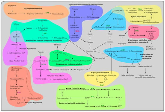
Figure 5.
Scheme representing the metabolic changes caused by iprodione exposure in the strain Pseudomonas sp. C9. In green letters upregulated proteins, in red letters downregulated proteins, and in purple letters proteins solely expressed on IPR exposure are represented. CAT, catalase; GK, guanylate kinase; TDS, 2,3,4,5-tetrahydropyridine-2,6-dicarboxylate-N-succinyltransferase; ACOA, acetyl-CoA carboxylate biotin carboxyl carrier protein; DDL, D-alanine–D-alanine ligase; (3OL) 3-oxiadipate enol-lactonase; AMD, amidohydrolase; 3HD, 3-hydroxyacyl-CoA dehydrogenase; MAL, maleyacetoacetate isomerase.
Previously, in Pseudomonas aeruginosa, GrxD, a monothiol glutaredoxin, was found to be essential for oxidative stress protection, acting as an electron donor for the organic hydroperoxide resistance enzyme during cumene hydroperoxide degradation [31]. The differential expression of catalase, guanylate kinase, OsmC family proteins, and Grx4 in Pseudomonas sp. C9 exposed to IPR suggests that the strain employs a range of adaptive responses to combat oxidative stress and enhance its ability to detoxify ROS. For molecular transport, efflux RND transporter periplasmic adaptors, such as AcrA and AcrE, play a role in the assembly and function of RND efflux pumps in bacteria and contribute to antibiotic resistance by exporting various classes of antibiotics out of the bacterial cell [32]. For strain C9, the differential expression of these proteins indicates that upregulated efflux pumps actively export IPR and other potentially toxic compounds out of the cell, enabling the strain to enhance its resistance to the pesticide.
Among the significantly upregulated proteins associated with the modification and maintenance of the bacterial cell wall, the peptidoglycan-binding protein LysM helps bacteria retain proteins within their cell envelopes by attaching to peptidoglycan, the primary component of bacterial cell walls [33], and glutamine amidotransferase catalyses the transfer of amide nitrogen from glutamine to specific acceptor substrates and is involved in the modification of cell wall peptidoglycan [34]. Moreover, D-alanine–D-alanine ligase (Ddl) [EC:6.3.2.4] is an enzyme involved in the biosynthesis of peptidoglycan and D-amino acid metabolism (Figure 5). Inhibition of Ddl activity has been explored as a potential strategy for developing novel antibacterial agents [35,36,37]. Furthermore, 2,3,4,5-tetrahydropyridine-2,6-dicarboxylate N-succinyltransferase [EC:2.3.1.117], also known as tetrahydrodipicolinate N-succinyltransferase, is involved in the biosynthesis of L-lysine (Figure 5), a component of peptidoglycan [38]. The differential expression of these proteins in strain C9 suggests they potentially contribute to the strain’s ability to withstand the toxic effects of IPR treatment. In terms of cell viability and growth, the upregulation of acetyl-CoA carboxylase biotin carboxyl carrier protein indicates a heightened requirement for fatty acid synthesis [39] (Figure 5). This is associated with cellular damage, as fatty acids are vital components of bacterial cell membranes and require significant energy for production, making their regulation crucial for bacterial viability [40]. This finding aligns with what was observed by Donoso-Piñol et al. [13], where Pseudomonas sp. C9 was exposed to IPR, and the biomass was duplicated in comparison to the control condition. Additionally, ribosomal protein S6, a component of the 30S ribosomal subunit in bacteria, plays a role in ribosome assembly and is involved in the binding of other ribosomal proteins and RNA molecules [41]. Electron transfer flavoprotein (ETF) also plays a crucial role in electron bifurcation and energy conservation in bacteria [42,43]. Depletion of ETF in Burkholderia cenocepacia leads to a loss of redox potential and cell viability [44]. In the context of strain Pseudomonas sp. C9 exposed to IPR, these proteins are essential for the sustainability of the bacterial cell.
Regarding the degradation of aromatic compounds, 3-oxo adipate enol-lactonase [EC:3.1.1.24] is an enzyme involved in the bioprocessing of lactones [45] and is part of the benzoate and aromatic compound degradation pathways (Figure 5). It also participates in the biodegradation of methyl aromatics in Pseudomonas reinekei MT1 [46]. Amidohydrolases [EC:3.5.1.-], also known for their activities as amidase enzymes, play a role in the degradation of pesticides and the detoxification of pesticide residues and are part of the alanine, aspartate, and glutamate metabolism pathways in Pseudomonas (Figure 5). A broad-spectrum amidohydrolase gene has been identified in bacteria that can completely degrade substituted urea herbicides within 24 h [47]. In Pseudomonas, the amidohydrolase AtzH is suspected to be an enzyme that converts 1,3-dicarboxyurea to allophanate in cyanuric acid catabolism, a common metabolic intermediate in the catabolism of s-triazine compounds, including atrazine and other herbicides [48]. Additionally, the bacterial strain Ochrobactrum sp. PP-2, with a unique arylamidase enzyme, effectively transforms the herbicide propanil into a less harmful compound, showing high efficiency and specificity [49]. Overall, our findings indicate that exposure to IPR leads to changes in proteomic expression, suggesting that strain Pseudomonas sp. C9 has developed adaptive mechanisms to efficiently degrade IPR and mitigate its potentially harmful effects on the environment.
Among the downregulated proteins, which are proteins whose expression levels decrease in response to a particular condition or treatment, in Pseudomonas sp. C9 exposed to IPR, those associated with the degradation of aromatic compounds, glycolysis and gluconeogenesis, and stress adaptations were identified. For the degradation of aromatic compounds such as amino acids, we report maleylacetoacetate isomerase [EC:5.2.1.2], an enzyme involved in the metabolic degradation of phenylalanine and tyrosine (Figure 5) [50]. This decrease could be impacting the ability of the C9 strain to metabolise aromatic compounds effectively.
Additionally, class II aldolases [EC:1.1.2.13] are enzymes that catalyse the reversible cleavage of fructose 1,6-bisphosphate. These zinc-containing metalloproteins play a crucial role in glycolysis and gluconeogenesis, converting three-carbon molecules into six-carbon sugars, and vice versa, thereby supporting energy production and carbon metabolism; in addition, they are involved in the degradation of the hydrocarbon tetralin [51].
Furthermore, we report cold-shock proteins (CS), a group of proteins found in bacteria that are important for cold adaptation and survival. CS proteins are essential for bacterial growth under unfavourable conditions [52,53]. In Bacillus subtilis, the loss of CS proteins affects the expression of about 20% of all genes and can lead to growth defects and loss of genetic competence [54].
Overall, our findings suggest that exposure to IPR inhibits key proteins. However, we also observed that the strain Pseudomonas sp. C9 has developed adaptive mechanisms to efficiently degrade IPR and mitigate its potentially harmful effects through upregulated proteins and those proteins that are only present with IPR treatment. Moreover, among the proteins only present with IPR treatment, we found those associated with transport, multidrug efflux complexes, chemical degradation, β-lactam resistance, and CAMP resistance. For transport, we identified ABC transporters; these proteins are primary active transporters that move substrates across biological membranes using ATP as an energy source (Figure 6).
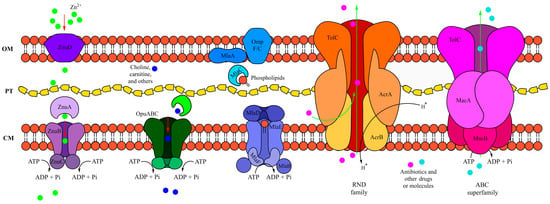
Figure 6.
Schematic representation of transporters in Pseudomonas sp. C9. From left to right: Znu ABC import system, osmoprotectant uptake (Opu) system, Mla ABC transport system, RND family TolC AcrAB system, and ABC superfamily TolC MacAB outer membrane protein system [16,17,18,19,20].
In bacteria, they are involved in processes such as drug resistance (Mac system), LPS transport (Lpt system), phospholipid transport (Mla system), and lipoprotein transport to the outer membrane (Lol system). For instance, the Mac system is an ABC transporter-based nanomachinery that contributes to bacterial drug resistance and is part of antibiotic resistance mechanisms [55,56]. In strain C9, we identified six proteins belonging to ABC transporters. Among them, the protein ZnuA (Figure 6) is an essential component for transporting zinc across the cell membrane, with ZnuB assisting in zinc uptake. This transport system is crucial for bacterial survival in zinc-limiting environments and is involved in various cellular processes, including virulence [57]. Additionally, we report a choline ABC transporter substrate-binding protein, such as Opu-family proteins, which have been studied in Bacillus subtilis and are part of the ABC transporters responsible for acquiring compatible solutes under osmotic stress [58]. The Mla system (Figure 6) is involved in maintaining lipid asymmetry in the outer membrane of Gram-negative bacteria and includes proteins such as MlaA and MlaD [16,59]. The Mla system is also implicated in antibiotic resistance in certain pathogens, including Acinetobacter baumannii and Pseudomonas aeruginosa [60]. Additionally, the AdeC/AdeK/OprM family belongs to a multidrug efflux complex associated with β-lactamase resistance (Figure 6). These proteins are involved in the transport of various antibiotics and xenobiotics in Gram-negative bacteria. For example, the AdeABC efflux pump in Acinetobacter baumannii is a notable RND efflux system that expels antibiotics, similar to the outer membrane factor (OprM) in Pseudomonas aeruginosa, leading to multidrug resistance [17,61]. For chemical degradation, 3-hydroxyacyl-CoA dehydrogenase is part of the benzoate degradation pathway, where benzoate, a common aromatic compound found in various environmental sources, is converted to acetyl-CoA via several enzymatic steps. This enzyme was reported to be significantly upregulated in Enterobacter sp. Z1 during the degradation of triazophos, methamidophos, and carbofuran pesticides [62]. In our results, this protein was found only when exposed to IPR. Additionally, we report upregulated proteins related to CAMP resistance, similar to those observed in a Pseudomonas strain exposed to chlorpyrifos by Aswathi et al. [25]. These proteins may assist Pseudomonas strains in pesticide degradation and resistance.
4. Materials and Methods
4.1. Chemicals and Medium
An analytical standard of IPR with a purity of 99% was procured from Sigma-Aldrich (St. Louis, MO, USA). For the pesticide exposure assay, a formulated commercial IPR (Rovral 50 WP) was obtained from Agan Chemical Manufacturers Ltd. (Ashdod, Israel). A stock solution containing 10,000 mg L−1 of IPR was prepared by dissolving the pesticide in dimethyl sulfoxide, then filtering it through a 0.22 µm polytetrafluoroethylene filter before storing it at 4 °C until required. All other chemicals and solvents used were of analytical reagent grade (Merck-Sigma, St. Louis, MO, USA).
The microbiological procedures utilised Luria Bertani modified broth with the following components per litre—2.5 g NaCl, 2.5 g yeast extract, and 5.0 g tryptone—adjusted to pH 6.5. The broth was autoclaved at 121 °C.
4.2. Proteome Preparation, Digestion, and Mass Spectrometry Analysis
The strain used in this study was Pseudomonas sp. C9 (Accession No. MK110046), an IPR-degrading strain that was isolated from an organic biomixture sample obtained from a BPS used for the treatment of pesticide residues, including IPR [7]. The partial sequence of the 16S ribosomal RNA gene of the strain is available in GenBank with the accession number MK110046. This strain has a genome of 6.9 Mb with 6296 protein-coding genes. The strain was exposed to 50 mg L−1 of IPR for 24 h, after which the biomass was collected by centrifugation at 8000× g for 20 min at 4 °C and rinsed twice with phosphate-buffered saline (PBS) adjusted to pH 7.0. Following centrifugation, the biomass pellet was suspended in 5 mL of PBS buffer with the addition of a protease inhibitor solution at a concentration of 1 mM. Subsequently, the cells were disrupted using ultrasonication with an amplitude of 40% in 8 cycles of 30 s on ice and subjected to another round of centrifugation at 8000× g for 15 min at 4 °C. The resulting supernatant was collected and lyophilised to preserve its integrity [13,63].
For desalting, the sample was suspended in 200 μL of MilliQ© water (Merck, Darmstadt, Germany) and 800 μL of cold acetone, then stored at −30 °C. The sample underwent centrifugation twice at 10,000× g for 30 min at 4 °C; the supernatant was discarded, and the extracts were air-dried. The protein extracts were reconstituted with a mix of 500 μL ammonium bicarbonate (25 mM) and 250 μL of MilliQ© water before the protein concentration was verified using the Qubit™ 2.0 Fluorometer (Invitrogen, Carlsbad, CA, USA) according to the manufacturer’s instructions. The protein concentration was then established at 100 µg. An aliquot of dithiothreitol was added to the sample to obtain a final concentration of 10 mM. Next, the samples were homogenised and incubated at 800 rpm and 60 °C for 1 h. After incubation, iodoacetamide was added to obtain a final concentration of 40 mM. Samples were then incubated for 30 min in the dark [13].
Trypsin digestion was performed using a 1:100 ratio of protein to 50 mM ammonium bicarbonate buffer. The sample was treated with 75 μL of trypsin (equivalent to 0.02 µg) and incubated at 800 rpm and 37 °C for 20 h. The trypsinisation process was stopped by adding trifluoroacetic acid (TFA, 1%) to lower the pH value. Subsequently, stage tip columns made with Applied Biosystems™ POROS™ R2 resin (Thermo Fisher Scientific, Waltham, MA, USA) were used for clean-up; elution was carried out using an ACN gradient (50% and 70%). Finally, the peptides were dried at 45 °C for 1 h using a SpeedVac concentrator (Thermo Fisher Scientific, Waltham, MA, USA), then suspended in 100 µL of 0.1% formic acid following standard procedures before quantification with the Qubit™ 2.0 Fluorometer protein assay kit [64].
The samples were analysed in technical triplicates using nano-liquid chromatography (Easy-nLC 1000, Thermo Fisher Scientific, Waltham, MA, USA) coupled to a hybrid quadrupole Orbitrap mass spectrometer (Q Exactive Plus, Thermo Fisher Scientific, Waltham, MA, USA). Peptides from the samples were loaded onto a home-made C18 trap column (Dr. Maisch GmbH, Ammerbuch, Germany) and then separated using a home-made C18 New Objective PicoFRIT column (Dr. Maisch GmbH, Ammerbuch, Germany). The chromatographic flow rate was 0.3 mL min™1 with the application of a linear gradient starting at 100% mobile phase A and increasing to 40% mobile phase B over 180 min. Ionisation and transfer of peptides occurred via a nanoelectrospray source with positive polarity, at a potential of 3.0 kV, and heating at 250 °C [13].
The mass spectrometer was operated in data-dependent analysis mode with dynamic exclusion of 45 ms and full-scan MS1 spectra with a resolution of 70,000 at m/z 200. This was followed by fragmentation of the top 15 most intense ions using high-collision dissociation (HCD), a normalised collision energy (NCE) of 30, and a resolution of 17,500 at m/z Acce200 in MS/MS scans. Additionally, species with a charge of +1 or greater than +4 were excluded from the MS/MS analysis.
4.3. Proteomic Data Analysis
The mass spectrometric data were analysed using Proteome Discoverer 2.1 software. Peptide identification was carried out with the Sequest HT algorithm against a protein database, including common contaminants in mass spectrometry-based proteomics analysis, concatenated with the Pseudomonas (Taxon ID 76760) database available from UniProt at http://www.uniprot.org/ (accessed on 15 November 2019). The searches utilised parameters such as peptide mass tolerance, MS/MS range, trypsin cleavage, missed cleavage limit, and fixed and variable modifications. False discovery rates were obtained, and protein quantification was performed using an extracted ion chromatogram (XIC) via the Precursor Ions Area Detector tool. The top 3 methods were used for protein quantification, selecting peptides that were considered unique [13].
4.4. Pathway Analysis
Identified proteins were then analysed using BLAST2Go software version 4.1 [14] and OmicsBox software version (3.1.11) [15] with the NCBI databank [65] to identify their molecular functions. To map the proteins in various possible pathways, the KEGG© database [66] was used to visualise the proteins in the metabolic pathway of Pseudomonas sp. C9 under IPR exposure, using KEGG© Mapper to visualise the upregulated and downregulated proteins, and proteins present only in the presence of IPR. To determine the cellular location, we used PSORTb version 3.0 [67] for subcellular localisation prediction.
4.5. Statistical Analysis
All experiments were performed in triplicate, and the standard deviation was calculated for protein expression. The ratio between quantitative values from IPR exposure (B) and control (A) was determined in triplicate for each protein, and the mean was used to calculate the fold change (FC, B/A). To normalise the data, the fold change value was calculated with log2. To determine the significant values, the log2FC—median was calculated, and values within the [−SD–+SD] range of the median standard deviation were considered significant.
5. Conclusions
In conclusion, the BPS bacterium Pseudomonas sp. C9, an IPR-degrading strain, was studied through comparative proteomics, revealing several differentially expressed proteins and proteins expressed only under IPR treatment. These proteins may play roles in IPR tolerance and degradation and are primarily involved in metabolic and cellular processes, with predominant molecular functions related to transferase and hydrolase activities. The upregulation of certain proteins and the presence of proteins unique to IPR treatment indicate the bacterium’s capacity to adapt and respond to the pesticide, demonstrating its ability to degrade and detoxify IPR. Additionally, these proteins are involved in ROS detoxification, growth, and transport, which may enhance the C9 strain’s resistance to IPR exposure. These results suggest that Pseudomonas sp. C9 has developed adaptive strategies for effective IPR degradation and tolerance, despite the downregulation of certain proteins. The BPS provides a valuable source of adapted microorganisms that can be used in bioremediation strategies. Finally, proteomic analysis of pesticide-degrading bacteria is essential for identifying key proteins involved in the breakdown and detoxification of these compounds. This technique reveals how the bacterium adapts its metabolism and responds to stress caused by pesticides. These findings provide crucial insights for improving bioremediation strategies, as they inform the development of microbial strains with enhanced environmental detoxification capacity.
Supplementary Materials
The following supporting information can be downloaded at https://www.mdpi.com/article/10.3390/ijms251910471/s1.
Author Contributions
Conceptualisation, P.D.-P. and M.C.D.; methodology, J.A.M.E. and F.C.S.N.; software, J.A.M.E. and F.C.S.N.; formal analysis, P.D.-P.; investigation, P.D.-P.; resources, P.D.-P. and M.C.D.; writing—original draft preparation, P.D.-P.; writing—review and editing, G.B. and H.S.; funding acquisition, P.D.-P. and M.C.D. All authors have read and agreed to the published version of the manuscript.
Funding
The authors acknowledge the funding received from the grants of projects ANID-FONDECYT 1211738, ANID/FONDAP/15130015, and ANID/FONDAP/1523A0001.
Institutional Review Board Statement
Not applicable.
Informed Consent Statement
Not applicable.
Data Availability Statement
Data is contained within the article and supplementary material.
Acknowledgments
We thank the Environmental Biotechnology Laboratory CIBAMA-UFRO and the Laboratory of Proteomics/LADETEC, UFRJ.
Conflicts of Interest
The authors declare no conflicts of interest.
References
- Berg, C.; Hill, M.; Bonetti, C.; Mitchell, G.C.; Sharma, B. The effects of iprodione fungicide on survival, behavior, and brood development of honeybees (Apis mellifera L.) after one foliar application during flowering on mustard. Environ. Toxicol. Chem. 2018, 37, 3086–3094. [Google Scholar] [CrossRef] [PubMed]
- Correia, M.; Rodrigues, M.; Paíga, P.; Delerue-Matos, C. Fungicides. In Encyclopedia of Food and Health; Elsevier: Amsterdam, The Netherlands, 2016; pp. 169–176. [Google Scholar]
- Abd-Elhakim, Y.M.; El Sharkawy, N.I.; El Bohy, K.M.; Hassan, M.A.; Gharib, H.S.A.; El-Metwally, A.E.; Arisha, A.H.; Imam, T.S. Iprodione and/or chlorpyrifos exposure induced testicular toxicity in adult rats by suppression of steroidogenic genes and SIRT1/TERT/PGC-1alpha pathway. Environ. Sci. Pollut. Res. Int. 2021, 28, 56491–56506. [Google Scholar] [CrossRef] [PubMed]
- Abd-Elhakim, Y.M.; El Sharkawy, N.I.; Gharib, H.S.A.; Hassan, M.A.; Metwally, M.M.M.; Elbohi, K.M.; Hassan, B.A.; Mohammed, A.T. Neurobehavioral responses and toxic brain reactions of juvenile rats exposed to iprodione and chlorpyrifos, alone and in a mixture. Toxics 2023, 11, 431. [Google Scholar] [CrossRef] [PubMed]
- Hassan, M.A.; El Bohy, K.M.; El Sharkawy, N.I.; Imam, T.S.; El-Metwally, A.E.; Hamed Arisha, A.; Mohammed, H.A.; Abd-Elhakim, Y.M. Iprodione and chlorpyrifos induce testicular damage, oxidative stress, apoptosis and suppression of steroidogenic- and spermatogenic-related genes in immature male albino rats. Andrologia 2021, 53, e13978. [Google Scholar] [CrossRef]
- Castillo, M.D.P.; Torstensson, L.; Stenstrom, J. Biobeds for environmental protection from pesticide use—A review. J. Agric. Food Chem. 2008, 56, 6206–6219. [Google Scholar] [CrossRef]
- Briceño, G.; Lamilla, C.; Leiva, B.; Levio, M.; Donoso-Piñol, P.; Schalchli, H.; Gallardo, F.; Diez, M.C. Pesticide-tolerant bacteria isolated from a biopurification system to remove commonly used pesticides to protect water resources. PLoS ONE 2020, 15, e0234865. [Google Scholar] [CrossRef]
- Campos, M.; Perruchon, C.; Vasilieiadis, S.; Menkissoglu-Spiroudi, U.; Karpouzas, D.G.; Diez, M.C. Isolation and characterization of bacteria from acidic pristine soil environment able to transform iprodione and 3,5-dichloraniline. Int. Biodeterior. Biodegrad. 2015, 104, 201–211. [Google Scholar] [CrossRef]
- Cao, L.; Shi, W.; Shu, R.; Pang, J.; Liu, Y.; Zhang, X.; Lei, Y. Isolation and characterization of a bacterium able to degrade high concentrations of iprodione. Can. J. Microbiol. 2018, 64, 49–56. [Google Scholar] [CrossRef]
- Campos, M.; Karas, P.S.; Perruchon, C.; Papadopoulou, E.S.; Christou, V.; Menkissoglou-Spiroudi, U.; Diez, M.C.; Karpouzas, D.G. Novel insights into the metabolic pathway of iprodione by soil bacteria. Environ. Sci. Pollut. Res. Int. 2017, 24, 152–163. [Google Scholar] [CrossRef]
- Mercadier, C.; Vega, D.; Bastide, J. Iprodione degradation by isolated soil microorganisms. FEMS Microbiol. Ecol. 1997, 23, 207–215. [Google Scholar] [CrossRef]
- Yang, Z.; Jiang, W.; Wang, X.; Cheng, T.; Zhang, D.; Wang, H.; Qiu, J.; Cao, L.; Wang, X.; Hong, Q. An amidase gene, ipaH, is responsible for the initial step in the iprodione degradation pathway of Paenarthrobacter sp. strain YJN-5. Appl. Environ. Microbiol. 2018, 84, e01150-18. [Google Scholar] [CrossRef] [PubMed]
- Donoso-Piñol, P.; Briceno, G.; Evaristo, J.A.M.; Nogueira, F.C.S.; Leiva, B.; Lamilla, C.; Schalchli, H.; Diez, M.C. Metabolic profiling and comparative proteomic insight in respect of amidases during iprodione biodegradation. Microorganisms 2023, 11, 2367. [Google Scholar] [CrossRef] [PubMed]
- Gotz, S.; Garcia-Gomez, J.M.; Terol, J.; Williams, T.D.; Nagaraj, S.H.; Nueda, M.J.; Robles, M.; Talon, M.; Dopazo, J.; Conesa, A. High-throughput functional annotation and data mining with the Blast2GO suite. Nucleic Acids Res. 2008, 36, 3420–3435. [Google Scholar] [CrossRef] [PubMed]
- OmicsBox—Bioinformatics Made Easy, version 3.1.11 ed.; Biobam Bioinformatics: Valencia, Spain, 2019.
- Low, W.Y.; Chng, S.S. Current mechanistic understanding of intermembrane lipid trafficking important for maintenance of bacterial outer membrane lipid asymmetry. Curr. Opin. Chem. Biol. 2021, 65, 163–171. [Google Scholar] [CrossRef]
- Phan, G.; Picard, M.; Broutin, I. Focus on the outer membrane factor OprM, the forgotten player from efflux pumps assemblies. Antibiotics 2015, 4, 544–566. [Google Scholar] [CrossRef]
- Greene, N.P.; Kaplan, E.; Crow, A.; Koronakis, V. Antibiotic resistance mediated by the MacB ABC transporter family: A structural and functional perspective. Front. Microbiol. 2018, 9, 950. [Google Scholar] [CrossRef]
- Teichmann, L.; Chen, C.; Hoffmann, T.; Smits, S.H.J.; Schmitt, L.; Bremer, E. From substrate specificity to promiscuity: Hybrid ABC transporters for osmoprotectants. Mol. Microbiol. 2017, 104, 761–780. [Google Scholar] [CrossRef]
- Wang, S.; Cheng, J.; Niu, Y.; Li, P.; Zhang, X.; Lin, J. Strategies for zinc uptake in Pseudomonas aeruginosa at the host-pathogen interface. Front. Microbiol. 2021, 12, 741873. [Google Scholar] [CrossRef]
- Gilani, R.A.; Rafique, M.; Rehman, A.; Munis, M.F.; Rehman, S.U.; Chaudhary, H.J. Biodegradation of chlorpyrifos by bacterial genus Pseudomonas. J. Basic. Microbiol. 2016, 56, 105–119. [Google Scholar] [CrossRef]
- Mirenga, E.O.; Korir, J.; Kimosop, S.; Orata, F.; Getenga, Z. Isolation and molecular characterization of soil bacteria capable of degrading chlorpyrifos and diuron pesticides. Appl. Environ. Microbiol. 2018, 6, 18–24. [Google Scholar]
- Zaffar, H.; Ahmad, R.; Pervez, A.; Naqvi, T.A. A newly isolated Pseudomonas sp. can degrade endosulfan via hydrolytic pathway. Pestic. Biochem. Physiol. 2018, 152, 69–75. [Google Scholar] [CrossRef] [PubMed]
- Zaffar, H.; Sabir, S.R.; Pervez, A.; Naqvi, T.A. Kinetics of endosulfan biodegradation by Stenotrophomonas maltophilia EN-1 isolated from pesticide-contaminated soil. Soil Sediment Contam. 2018, 27, 267–279. [Google Scholar] [CrossRef]
- Aswathi, A.; Pandey, A.; Sukumaran, R.K. Rapid degradation of the organophosphate pesticide—Chlorpyrifos by a novel strain of Pseudomonas nitroreducens AR-3. Bioresour. Technol. 2019, 292, 122025. [Google Scholar] [CrossRef] [PubMed]
- Rovida, A.; Costa, G.; Santos, M.I.; Silva, C.R.; Freitas, P.N.N.; Oliveira, E.P.; Pileggi, S.A.V.; Olchanheski, R.L.; Pileggi, M. Herbicides tolerance in a Pseudomonas strain is associated with metabolic plasticity of antioxidative enzymes regardless of selection. Front. Microbiol. 2021, 12, 673211. [Google Scholar] [CrossRef]
- Zhang, Y.; Meng, D.; Wang, Z.; Guo, H.; Wang, Y. Oxidative stress response in two representative bacteria exposed to atrazine. FEMS Microbiol. Lett. 2012, 334, 95–101. [Google Scholar] [CrossRef]
- Inokuchi, H.; Ito, R.; Sekiguchi, T.; Sekiguchi, M. Search for proteins required for accurate gene expression under oxidative stress: Roles of guanylate kinase and RNA polymerase. J. Biol. Chem. 2013, 288, 32952–32962. [Google Scholar] [CrossRef]
- Shin, D.H.; Choi, I.G.; Busso, D.; Jancarik, J.; Yokota, H.; Kim, R.; Kim, S.H. Structure of OsmC from Escherichia coli: A salt-shock-induced protein. Acta Crystallogr. D Biol. Crystallogr. 2004, 60, 903–911. [Google Scholar] [CrossRef]
- Svenningsen, N.B.; Damgaard, M.; Rasmussen, M.; Perez-Pantoja, D.; Nybroe, O.; Nicolaisen, M.H. Cupriavidus pinatubonensis AEO106 deals with copper-induced oxidative stress before engaging in biodegradation of the herbicide 4-chloro-2-methylphenoxyacetic acid. BMC Microbiol. 2017, 17, 211. [Google Scholar] [CrossRef]
- Saninjuk, K.; Romsang, A.; Duang-Nkern, J.; Wongsaroj, L.; Leesukon, P.; Dubbs, J.M.; Vattanaviboon, P.; Mongkolsuk, S. Monothiol glutaredoxin is essential for oxidative stress protection and virulence in Pseudomonas aeruginosa. Appl. Environ. Microbiol. 2023, 89, e0171422. [Google Scholar] [CrossRef]
- Alav, I.; Bavro, V.N.; Blair, J.M.A. A role for the periplasmic adaptor protein AcrA in vetting substrate access to the RND efflux transporter AcrB. Sci. Rep. 2022, 12, 4752. [Google Scholar] [CrossRef]
- Sexton, D.L.; Herlihey, F.A.; Brott, A.S.; Crisante, D.A.; Shepherdson, E.; Clarke, A.J.; Elliot, M.A. Roles of LysM and LytM domains in resuscitation-promoting factor (Rpf) activity and Rpf-mediated peptidoglycan cleavage and dormant spore reactivation. J. Biol. Chem. 2020, 295, 9171–9182. [Google Scholar] [CrossRef] [PubMed]
- Levefaudes, M.; Patin, D.; De Sousa-D’auria, C.; Chami, M.; Blanot, D.; Herve, M.; Arthur, M.; Houssin, C.; Mengin-Lecreulx, D. Diaminopimelic acid amidation in corynebacteriales: New insights into the role of LtsA in peptidoglycan modification. J. Biol. Chem. 2015, 290, 13079–13094. [Google Scholar] [CrossRef] [PubMed]
- Ameryckx, A.; Thabault, L.; Pochet, L.; Leimanis, S.; Poupaert, J.H.; Wouters, J.; Joris, B.; Van Bambeke, F.; Frederick, R. 1-(2-Hydroxybenzoyl)-thiosemicarbazides are promising antimicrobial agents targeting d-alanine-d-alanine ligase in bacterio. Eur. J. Med. Chem. 2018, 159, 324–338. [Google Scholar] [CrossRef] [PubMed]
- Diaz-Saez, L.; Torrie, L.S.; Mcelroy, S.P.; Gray, D.; Hunter, W.N. Burkholderia pseudomallei d-alanine-d-alanine ligase; detailed characterization and assessment of a potential antibiotic drug target. FEBS J. 2019, 286, 4509–4524. [Google Scholar] [CrossRef] [PubMed]
- Vu, H.; Gilari, K.; Sathiyamoorthy, V.; Beckham, J. Discovery of novel inhibitors for Mycobacterium tuberculosis D-alanine: D-alanine ligase through virtual screening. FASEB J. 2022, 36. [Google Scholar] [CrossRef]
- Pavelka, M.S., Jr.; Jacobs, W.R., Jr. Biosynthesis of diaminopimelate, the precursor of lysine and a component of peptidoglycan, is an essential function of Mycobacterium smegmatis. J. Bacteriol. 1996, 178, 6496–6507. [Google Scholar] [CrossRef]
- Evans, A.; Ribble, W.; Schexnaydre, E.; Waldrop, G.L. Acetyl-CoA carboxylase from Escherichia coli exhibits a pronounced hysteresis when inhibited by palmitoyl-acyl carrier protein. Arch. Biochem. Biophys. 2017, 636, 100–109. [Google Scholar] [CrossRef]
- López-Lara, I.M.; Soto, M.J. Fatty acid synthesis and regulation. In Biogenesis of Fatty Acids, Lipids and Membranes; Geiger, O., Ed.; Springer International Publishing: Berlin/Heidelberg, Germany, 2018. [Google Scholar]
- Holmqvist, E.; Vogel, J. RNA-binding proteins in bacteria. Nat. Rev. Microbiol. 2018, 16, 601–615. [Google Scholar] [CrossRef]
- Duan, H.D.; Mohamed-Raseek, N.; Miller, A.F. Spectroscopic evidence for direct flavin-flavin contact in a bifurcating electron transfer flavoprotein. J. Biol. Chem. 2020, 295, 12618–12634. [Google Scholar] [CrossRef]
- Vogt, M.S.; Schuhle, K.; Kolzer, S.; Peschke, P.; Chowdhury, N.P.; Kleinsorge, D.; Buckel, W.; Essen, L.O.; Heider, J. Structural and functional characterization of an electron transfer flavoprotein involved in toluene degradation in strictly anaerobic bacteria. J. Bacteriol. 2019, 201, 10-1128. [Google Scholar] [CrossRef]
- Bloodworth, R.A.M.; Zlitni, S.; Brown, E.D.; Cardona, S.T. An electron transfer flavoprotein is essential for viability and its depletion causes a rod-to-sphere change in Burkholderia cenocepacia. Microbiology 2015, 161, 1909–1920. [Google Scholar] [CrossRef] [PubMed]
- Bains, J.; Kaufman, L.; Farnell, B.; Boulanger, M.J. A product analog bound form of 3-oxoadipate-enol-lactonase (PcaD) reveals a multifunctional role for the divergent cap domain. J. Mol. Biol. 2011, 406, 649–658. [Google Scholar] [CrossRef] [PubMed]
- Marin, M.; Perez-Pantoja, D.; Donoso, R.; Wray, V.; Gonzalez, B.; Pieper, D.H. Modified 3-oxoadipate pathway for the biodegradation of methylaromatics in Pseudomonas reinekei MT1. J. Bacteriol. 2010, 192, 1543–1552. [Google Scholar] [CrossRef] [PubMed]
- Jiang, J.; Zhang, L.; Chen, K. Broad-Spectrum Substituted Urea Herbicide Degradation Bacterium and Amidohydrolase Gene and Application. China Patent Application. CN105779477A, 14 May 2019. [Google Scholar]
- Esquirol, L.; Peat, T.S.; Wilding, M.; Hartley, C.J.; Newman, J.; Scott, C. A novel decarboxylating amidohydrolase involved in avoiding metabolic dead ends during cyanuric acid catabolism in Pseudomonas sp. strain ADP. PLoS ONE 2018, 13, e0206949. [Google Scholar] [CrossRef] [PubMed]
- Zhang, L.; Hu, Q.; Hang, P.; Zhou, X.; Jiang, J. Characterization of an arylamidase from a newly isolated propanil-transforming strain of Ochrobactrum sp. PP-2. Ecotoxicol. Environ. Saf. 2019, 167, 122–129. [Google Scholar] [CrossRef]
- Izmalkova, T.Y.; Sazonova, O.I.; Nagornih, M.O.; Sokolov, S.L.; Kosheleva, I.A.; Boronin, A.M. The organization of naphthalene degradation genes in Pseudomonas putida strain AK5. Res. Microbiol. 2013, 164, 244–253. [Google Scholar] [CrossRef]
- Hernaez, M.J.; Floriano, B.; Rios, J.J.; Santero, E. Identification of a hydratase and a class II aldolase involved in biodegradation of the organic solvent tetralin. Appl. Environ. Microbiol. 2002, 68, 4841–4846. [Google Scholar] [CrossRef]
- Yu, T.; Keto-Timonen, R.; Jiang, X.; Virtanen, J.P.; Korkeala, H. Insights into the phylogeny and evolution of cold shock proteins: From Enteropathogenic Yersinia and Escherichia coli to Eubacteria. Int. J. Mol. Sci. 2019, 20, 4059. [Google Scholar] [CrossRef]
- Zhou, Z.; Tang, H.; Wang, W.; Zhang, L.; Su, F.; Wu, Y.; Bai, L.; Li, S.; Sun, Y.; Tao, F.; et al. A cold shock protein promotes high-temperature microbial growth through binding to diverse RNA species. Cell Discov. 2021, 7, 15. [Google Scholar] [CrossRef]
- Kim, M.J.; Lee, Y.K.; Lee, H.K.; Im, H. Characterization of cold-shock protein A of Antarctic Streptomyces sp. AA8321. Protein J. 2007, 26, 51–59. [Google Scholar] [CrossRef]
- Bilsing, F.L.; Anlauf, M.T.; Hachani, E.; Khosa, S.; Schmitt, L. ABC transporters in bacterial nanomachineries. Int. J. Mol. Sci. 2023, 24, 6227. [Google Scholar] [CrossRef] [PubMed]
- Orelle, C.; Mathieu, K.; Jault, J.M. Multidrug ABC transporters in bacteria. Res. Microbiol. 2019, 170, 381–391. [Google Scholar] [CrossRef] [PubMed]
- Alquethamy, S.; Ganio, K.; Luo, Z.; Hossain, S.I.; Hayes, A.J.; Ve, T.; Davies, M.R.; Deplazes, E.; Kobe, B.; Mcdevitt, C.A. Structural and biochemical characterization of Acinetobacter baumannii ZnuA. J. Inorg. Biochem. 2022, 231, 111787. [Google Scholar] [CrossRef] [PubMed]
- Hoffmann, T.; Bremer, E. Guardians in a stressful world: The Opu family of compatible solute transporters from Bacillus subtilis. Biol. Chem. 2017, 398, 193–214. [Google Scholar] [CrossRef]
- Mann, D.; Fan, J.; Somboon, K.; Farrell, D.P.; Muenks, A.; Tzokov, S.B.; Dimaio, F.; Khalid, S.; Miller, S.I.; Bergeron, J.R.C. Structure and lipid dynamics in the maintenance of lipid asymmetry inner membrane complex of A. baumannii. Commun. Biol. 2021, 4, 817. [Google Scholar] [CrossRef]
- Guest, R.L.; Lee, M.J.; Wang, W.; Silhavy, T.J. A periplasmic phospholipase that maintains outer membrane lipid asymmetry in Pseudomonas aeruginosa. Proc. Natl. Acad. Sci. USA 2023, 120, e2302546120. [Google Scholar] [CrossRef]
- Verma, P.; Tiwari, V. Targeting outer membrane protein component AdeC for the discovery of efflux pump inhibitor against AdeABC efflux pump of multidrug resistant Acinetobacter baumannii. Cell Biochem. Biophys. 2018, 76, 391–400. [Google Scholar] [CrossRef]
- Zhang, Y.; Xu, Z.; Chen, Z.; Wang, G. Simultaneous degradation of triazophos, methamidophos and carbofuran pesticides in wastewater using an Enterobacter bacterial bioreactor and analysis of toxicity and biosafety. Chemosphere 2020, 261, 128054. [Google Scholar] [CrossRef]
- Madhavan Nampoothiri, K.; Roopesh, K.; Chacko, S.; Pandey, A. Comparative study of amidase production by free and immobilized Escherichia coli cells. Appl. Biochem. Biotechnol. 2005, 120, 97–108. [Google Scholar] [CrossRef]
- Andrade, M.T.; Neto, D.F.M.; Nascimento, J.R.S.; Soares, E.L.; Coutinho, I.C.; Velasquez, E.; Domont, G.B.; Nogueira, F.C.S.; Campos, F.A.P. Proteome dynamics of the developing acai berry pericarp (Euterpe oleracea Mart.). J. Proteome. Res. 2020, 19, 437–445. [Google Scholar] [CrossRef]
- Sayers, E.W.; Bolton, E.E.; Brister, J.R.; Canese, K.; Chan, J.; Comeau, D.C.; Connor, R.; Funk, K.; Kelly, C.; Kim, S.; et al. Database resources of the national center for biotechnology information. Nucleic Acids Res. 2022, 50, D20–D26. [Google Scholar] [CrossRef] [PubMed]
- Kanehisa, M.; Furumichi, M.; Sato, Y.; Kawashima, M.; Ishiguro-Watanabe, M. KEGG for taxonomy-based analysis of pathways and genomes. Nucleic Acids Res. 2023, 51, D587–D592. [Google Scholar] [CrossRef] [PubMed]
- Yu, N.Y.; Wagner, J.R.; Laird, M.R.; Melli, G.; Rey, S.; Lo, R.; Dao, P.; Sahinalp, S.C.; Ester, M.; Foster, L.J.; et al. PSORTb 3.0: Improved protein subcellular localization prediction with refined localization subcategories and predictive capabilities for all prokaryotes. Bioinformatics 2010, 26, 1608–1615. [Google Scholar] [CrossRef] [PubMed]
Disclaimer/Publisher’s Note: The statements, opinions and data contained in all publications are solely those of the individual author(s) and contributor(s) and not of MDPI and/or the editor(s). MDPI and/or the editor(s) disclaim responsibility for any injury to people or property resulting from any ideas, methods, instructions or products referred to in the content. |
© 2024 by the authors. Licensee MDPI, Basel, Switzerland. This article is an open access article distributed under the terms and conditions of the Creative Commons Attribution (CC BY) license (https://creativecommons.org/licenses/by/4.0/).