Article

The Modification of H3K4me3 Enhanced the Expression of CgTLR3 in Hemocytes to Increase CgIL17-1 Production in the Immune Priming of Crassostrea gigas

1
School of Life Science, Liaoning Normal University, Dalian 116029, China
2
Liaoning Key Laboratory of Marine Animal Immunology and Disease Control, Dalian Ocean University, Dalian 116023, China
3
Southern Marine Science and Engineering Guangdong Laboratory (Zhuhai), Zhuhai 519000, China
4
Laboratory of Marine Fisheries Science and Food Production Process, Qingdao National Laboratory for Marine Science and Technology, Qingdao 266235, China
5
Dalian Key Laboratory of Aquatic Animal Disease Prevention and Control, Dalian Ocean University, Dalian 116023, China
*
Authors to whom correspondence should be addressed.
Int. J. Mol. Sci. 2024, 25(2), 1036; https://doi.org/10.3390/ijms25021036
Submission received: 5 December 2023 / Revised: 4 January 2024 / Accepted: 10 January 2024 / Published: 15 January 2024
(This article belongs to the Section Molecular Biology)

Abstract

:
Increasing evidence confirms that histone modification plays a critical role in preserving long-term immunological memory. Immune priming is a novel form of immunological memory recently verified in invertebrates. Toll-like receptor (TLR) signaling and cytokines have been reported to be involved in the immune priming of the Pacific oyster Crassostrea gigas. In the present study, the expression of Toll-like receptor 3 (CgTLR3), myeloid differentiation factor 88-2 (CgMyd88-2) and interleukin 17-1 (CgIL17-1) was found to be elevated in the hemocytes of C. gigas at 6 h after the secondary stimulation with Vibrio splendidus, which was significantly higher than that at 6 h after the primary stimulation (p < 0.05). A significant increase in histone H3 lysine 4 trimethylation (H3K4me3) enrichment was detected in the promoter region of the CgTLR3 gene at 7 d after the primary stimulation with inactivated V. splendidus (p < 0.05). After the treatment with a histone methyltransferase inhibitor (5′-methylthioadenosine, MTA), the level of H3K4me3 at the promoter of the CgTLR3 gene decreased significantly at 7 d after the primary stimulation with inactivated V. splendidus (p < 0.05), and the expression of CgTLR3, CgMyD88-2 and CgIL17-1 was significantly repressed at 6 h after the secondary stimulation with V. splendidus (p < 0.05). Conversely, the treatment with monomethyl fumarate (MEF, an inhibitor of histone demethylases) resulted in a significant increase in H3K4me3 enrichment levels at the CgTLR3 promoter at 7 d after the primary stimulation (p < 0.05), and the expression of CgTLR3, CgMyD88-2 and CgIL17-1 was observed to increase significantly at 6 h after the secondary stimulation (p < 0.05). These results suggested that H3K4me3 regulated MyD88-dependent TLR signaling in the hemocytes of C. gigas, which defined the role of histone modifications in invertebrate immune priming.

Graphical Abstract

1. Introduction

The immune systems of invertebrates can mount an immune memory response to subsequent reinfections with the same or different pathogens, which is defined as immune priming [1,2]. In contrast to the adaptive immunity in vertebrates that primarily depends on the rearrangement of immunoglobulin family genes and clonal expansion, the immune priming in invertebrates may be mainly governed by epigenetic modification [3,4,5,6]. Epigenetic programming usually occurs as a result of viral and bacterial infections, the stimulation of pathogen-associated molecular patterns (PAMPs), and the induction of various cytokines. Epigenetic modification plays a crucial role in tightly regulating pathogen recognition, intracellular signal transduction, and downstream effector activation, which serves as a form of cellular memory in immune cells. It bestows distinct functions and specialized phenotypes upon immune cells through establishing cell-specific gene expression patterns. These epigenetic reprogramming events contribute to the biological functions of primed cells and the enhancement of their immune responses [7,8].
Epigenetic modifications are heritable changes in chromatin structure and gene expression without alterations in the DNA sequence. Major epigenetic modifications include histone methylation, acetylation and phosphorylation, DNA methylation, and microRNA modifications. Trimethylation of histone H3 lysine 4 (H3K4me3) is a common and evolutionarily conserved epigenetic modification, which is known to be associated with transcriptionally active chromatin [9]. It is widely recognized that the transcriptional activity of a gene positively correlates with the degree of H3K4me3 at its promoters. The presence of H3K4me3 maintains the chromatin in a conformation that facilitates accessibility of the promoter region for specific genes, thereby promoting transcriptional activity [10]. In mammals, several investigations have highlighted the pivotal significance of the H3K4me3 modification in innate immunity as well as the “trained immunity” [11,12,13,14]. Trained immunity is a functional adaptation of mammalian innate immune cells induced by epigenetic reprogramming, resulting in an enhanced immunologic response [15]. The H3K4me3 modification has been implicated in various forms of trained immunity of immune cells, including lipopolysaccharide-trained dendritic cells, oxidized low-density lipoprotein-trained monocytes, catecholamines-trained monocytes, and interleukin-trained natural killer cells [16,17,18,19]. Systematic chromatin reprogramming has been reported in β-Glucan-trained human monocytes, which is characterized by elevated levels of H3K4me3 at the gene promoters [20]. The H3K4me3 modification is necessary for the enhanced release of cytokines in the trained monocytes after Bacillus Calmette–Guerin (BCG) vaccination in human [12]. It is believed that H3K4me3 modification is of utmost importance in preserving the delicate balance of the innate immune response by increasing the expression of specific pattern recognition receptors (PRRs) and some immune effectors. However, H3K4me3 modification and its role in the immune priming of invertebrate are still not well understood.
Immune recognition is the first step in the immune response to foreign invaders, which is initiated by binding and interaction between host PRRs and PAMPs from invasive pathogens. The Toll-like receptor (TLR) is a type of PRR that is widely expressed by immune cells to mediate immune signaling. Among the various pathways, TLR signaling has been extensively investigated and the mechanism of signal transduction is well documented. After binding with a PAMP, the TLR initiates signaling transduction by recruiting its canonical adaptor such as myeloid differentiation factor 88 (MyD88) to activate various transcription factors such as nuclear factor nuclear factor kappa-B (NF-κB), activating protein-1 and interferon regulatory factors, and eventually induce immune responses [21,22,23]. An increase in H3K4me3 levels was observed on the promoters of many TLR signaling molecules and cytokines such as TLR4, scavenger receptor A, interleukin (IL) 6 and tumor necrosis factor (TNF) α in trained immunity [17,24,25,26]. Histone methyltransferases, such as KMT2B and ASH1L, were reported to modulate the TLR4-mediated signaling in macrophages by increasing the H3K4me3 levels of TLR ligands or regulators [14,27]. For invertebrates, more research is needed to clarify the histone modifications and other epigenetic modifications on PRRs and downstream signaling pathways during immune responses.
Cytokines are a diverse group of soluble proteins that regulate the development, differentiation, and activation of immune cells to modulate both innate and adaptive immune responses. Several proinflammatory cytokines, such as IL1, IL6, IL1β, and TNFα have been extensively investigated in the context of trained immunity [12,20,28]. To date, most vertebrate proinflammatory cytokines, including IL1, IL2, and IL6, as well as their corresponding receptors, have not been identified in invertebrates [29]. Recently, IL17 signaling components were annotated and identified in bivalves [30], and they were reported to play important roles in the initiation of the proinflammatory response and other immune responses in the Pacific oyster Crassostrea gigas [31,32,33,34,35]. Considering that proinflammatory cytokines have been extensively investigated in the trained immunity of vertebrate, IL17 signaling components are also suspected to play a crucial role in oyster immune priming.
The Pacific oyster C. gigas is a worldwide aquaculture animal, which belongs to the second largest animal phylum Mollusca. As a sessile filter-feeder exposed to numerous microorganisms, the oyster represents an attractive model for studying immune mechanisms in invertebrates. The oyster mainly relies on the innate immune system to defend against infection by pathogens [36,37]. Increasing evidence demonstrates the presence of immune priming in C. gigas, and many key components of PRR signaling pathways are suspected to be involved in immune priming [35,38,39,40,41]. At the same time, H3K4me3 has also reported to be crucially involved in regulating the signaling pathways in trained immunity [7,12,20,25]. The TLR-MyD88-NF-κB signaling has been reported to govern the expression of inflammatory cytokines [32,42,43,44] and play an indispensable role in the enhanced immunity against re-infection of Vibrio splendidus [35]. TLR3 from C. gigas was upregulated after both the first and second immune stimulations, indicating that it was involved in immune priming [35]. It was reported that CgTLR3 was able to interact with CgMyD88-2 [45], and CgRel1 functioned as an important transcription factor, regulating the expression of CgIL17s in the immune responses of oysters [46]. In the present study, the effect of H3K4me3 on TLR expression in oysters was investigated with the objectives of (1) examining the responses of CgIL17-1 and TLR signaling in hemocytes after the secondary immune stimulation with V. splendidus, (2) measuring the H3K4me3 modification at the CgTLR3 promoter after the stimulations with of V. splendidus, 5′-methylthioadenosine (MTA, a methyltransferase inhibitor) and Monoethyl fumarate (MEF, a demethylase inhibitor), respectively, and (3) determining the changes in TLR signaling and CgIL17-1 mRNA expression after the treatment with MTA.

2. Results

2.1. The mRNA Expression Levels of CgTLR3, CgMyD88-2, CgRel1, and CgIL17-1 at 6 h after the Secondary Stimulation with Live V. splendidus

Based on the transcriptome analysis and annotation of TLRs and ILs in oyster hemocytes after the secondary immune stimulation with live V. splendidus, the mRNA transcripts of CgTLR3, CgMyD88-2, CgRel1, and CgIL17-1 were selected and examined in the four treatment groups (PBS + PBS, Vs + PBS, PBS + Vs, Vs + Vs) by RT-qPCR (Figure 1). The mRNA transcripts of CgIL17-1 were detected in all four groups with the highest expression level in the Vs + Vs group, which was 2.77-fold (p < 0.05) of that in the PBS + Vs group (Figure 1A). The CgTLR3 mRNA expression level in the Vs + Vs group was significantly increased (2.64-fold, p < 0.05) compared to that in the PBS + Vs group (Figure 1B). The relative mRNA expression level of the CgMyD88-2 in Vs + Vs group was 1.03-fold of that in PBS + Vs group, with no significant differences (Figure 1C). There was no significant difference in the CgRel1 mRNA expression levels among the four treatment groups (Figure 1D).

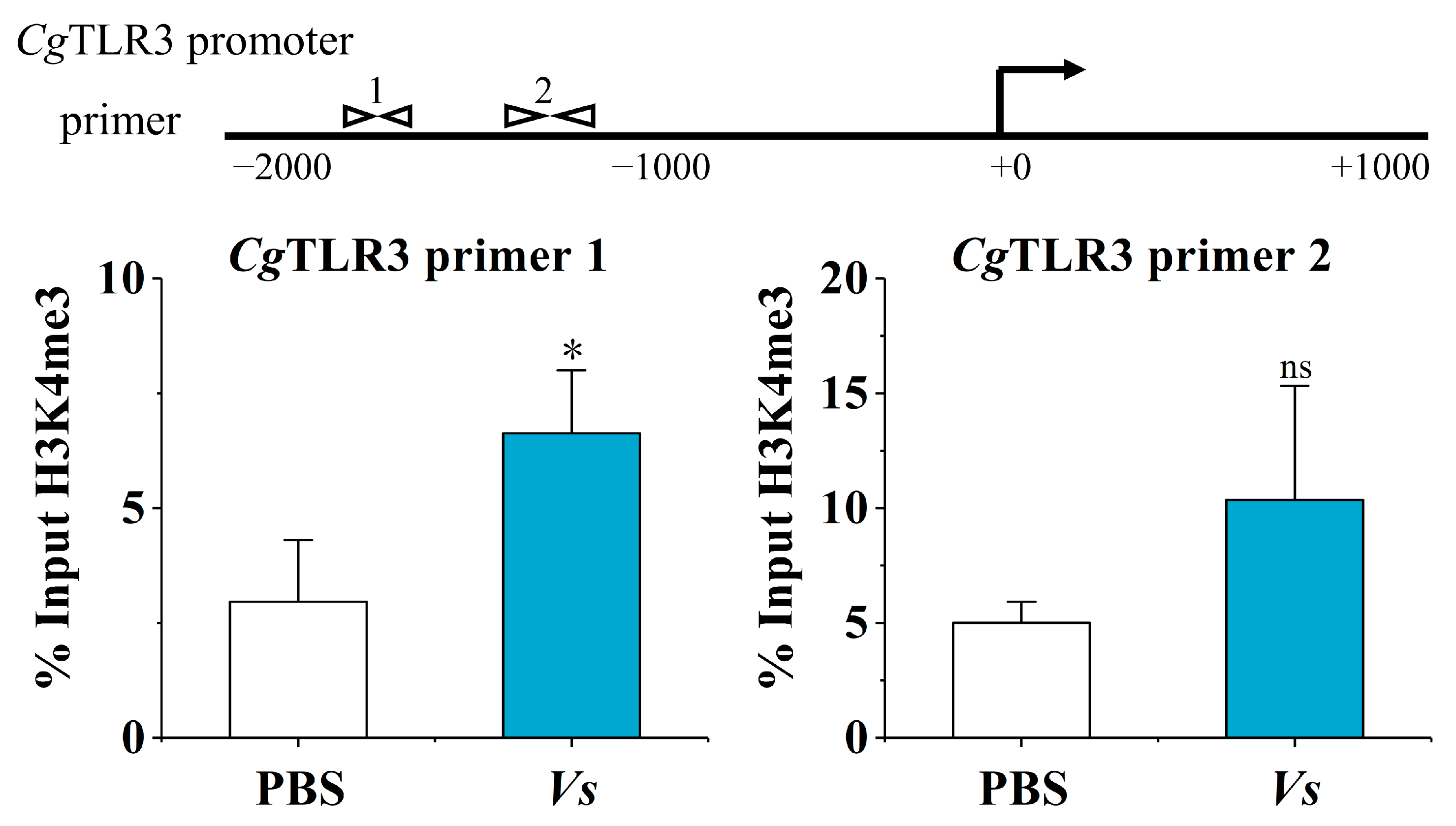
2.2. The H3K4me3 Modification of the CgTLR3 Gene Promoter at 7 d after the Stimulation with Inactivated V. splendidus

The H3K4me3 modification levels of the CgTLR3 promoter at 7 d after the stimulation with inactivated V. splendidus were examined using chromatin immunoprecipitation followed by qPCR (ChIP-qPCR). The primers were designed using Primer Premier 5 to cover the CgTLR3 promoter region (Figure 2). ‘% input H3K4me3’ indicates the ratio of the DNA fragments of each promoter region bound by H3K4me3 to the total amount of input DNA fragments without H3K4me3 antibody pull-down. At 7 d after the stimulation with inactivated V. splendidus, anti-H3K4me3 antibodies markedly enriched CgTLR3 promoter DNA. Compared to the PBS group, the H3K4me3 modification level of CgTLR3 distal promoter regions (spanning from −1879 bp to −1735 bp) was significantly higher in the Vs group (2.24-fold, p < 0.05), and the proximal promoter regions −1307 bp to −1164 of CgTLR3 also showed a slightly higher H3K4me3 modification level in the Vs group, but this difference did not reach significance (2.07-fold, p = 0.14).

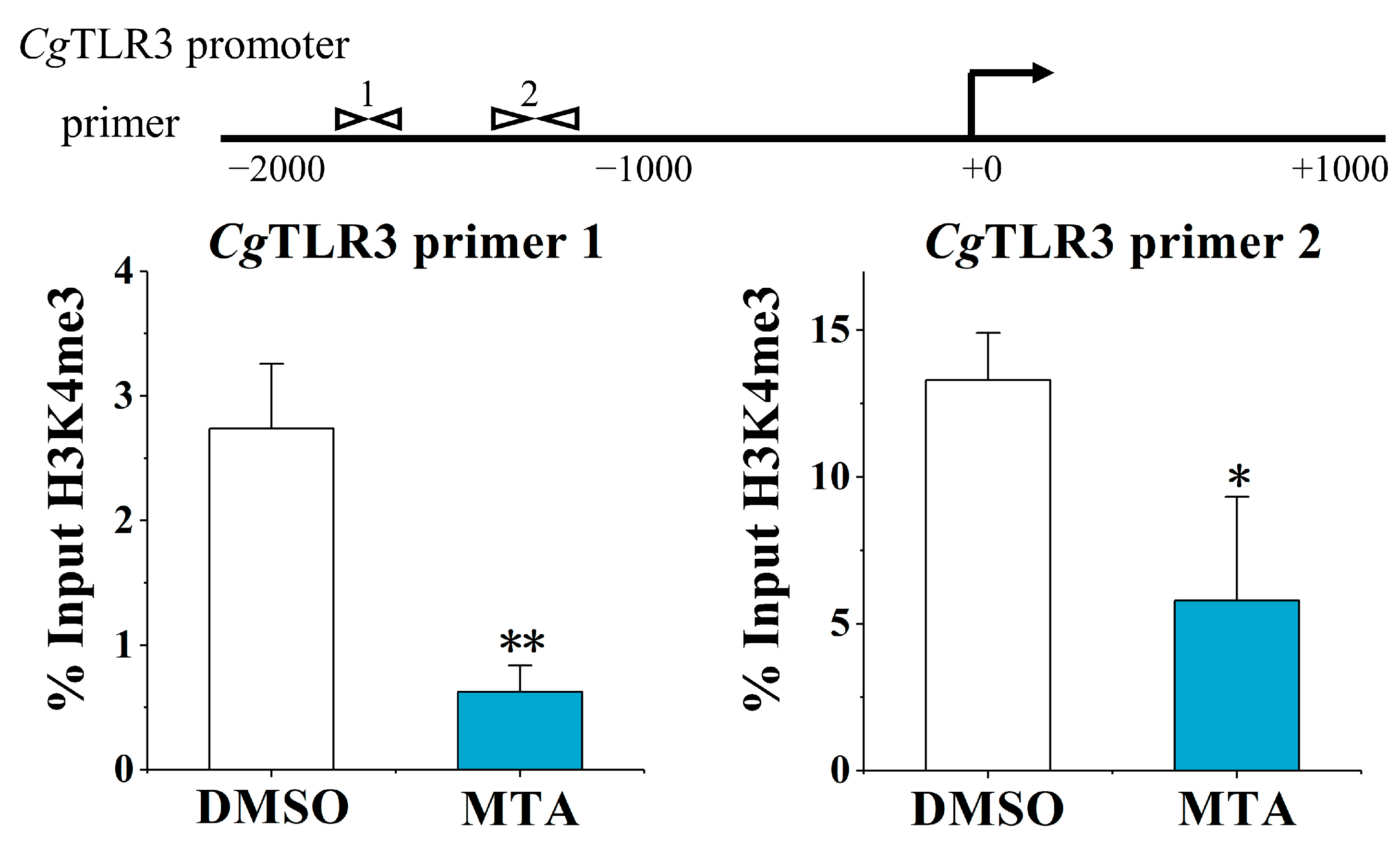
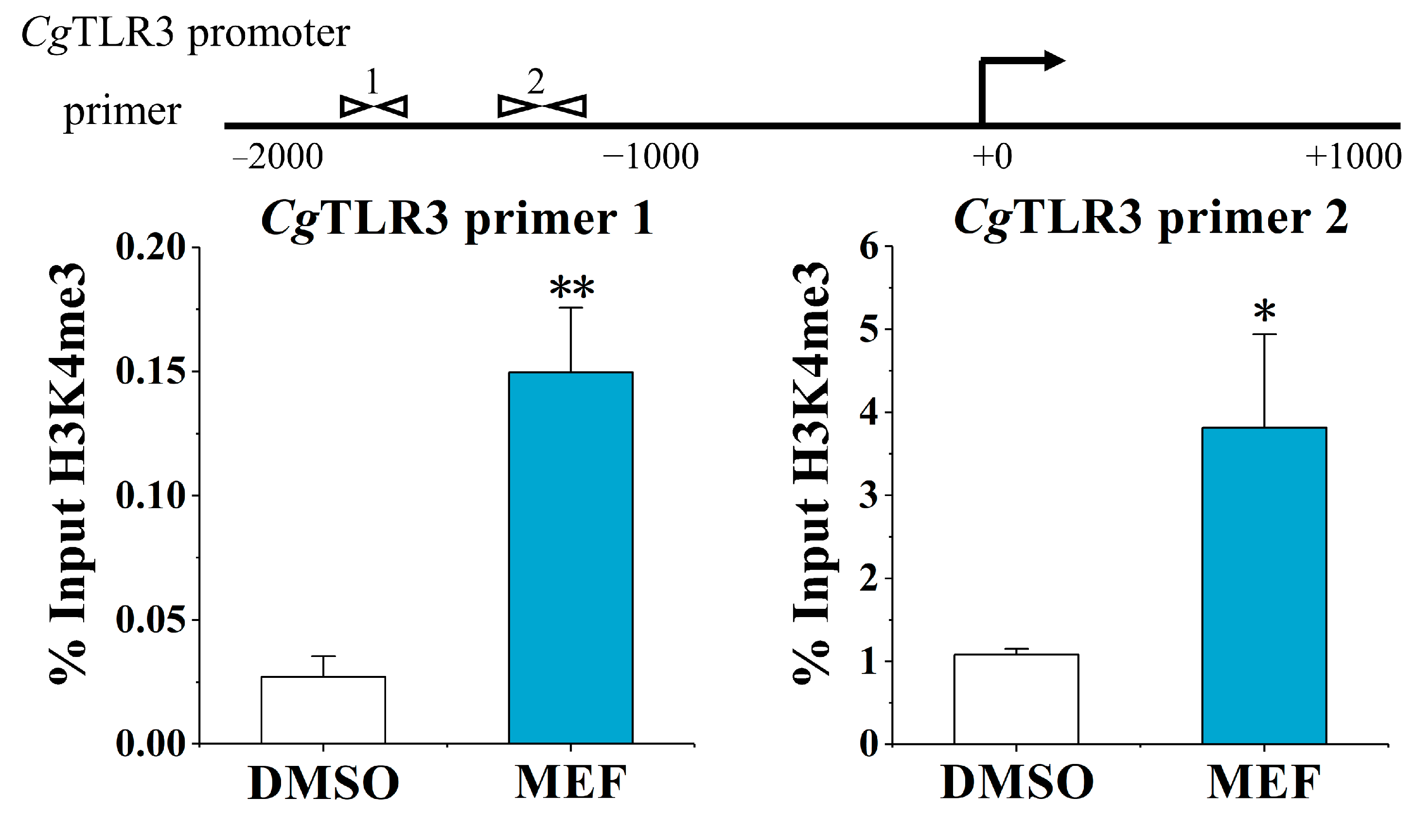
2.3. The Alternation of H3K4me3 Modification Levels at the CgTLR3 Gene Promoter at 7 d after the Treatments with MTA and MEF

The H3K4me3 modification levels of the CgTLR3 promoter were examined at 7 d after the stimulation with the methylation inhibitor MTA and histone demethylases inhibitor MEF. Upon the MTA treatment, the H3K4me3 modification levels at the CgTLR3 gene promoter were significantly lower: between primer pair −1879 and −1735 (0.23-fold, p < 0.01, primer 1) and primer pair −1307 and −1164 (0.44-fold, p < 0.05, primer 2), compared with the DMSO group, respectively (Figure 3). The MEF treatment also resulted in a 6.68-fold enrichment of CgTLR3 distal promoter DNA using primer 1 (p < 0.01) and a 3.54-fold enrichment of CgTLR3 proximal promoter DNA using primer 2 (p < 0.05), compared with the DMSO group, respectively (Figure 4).

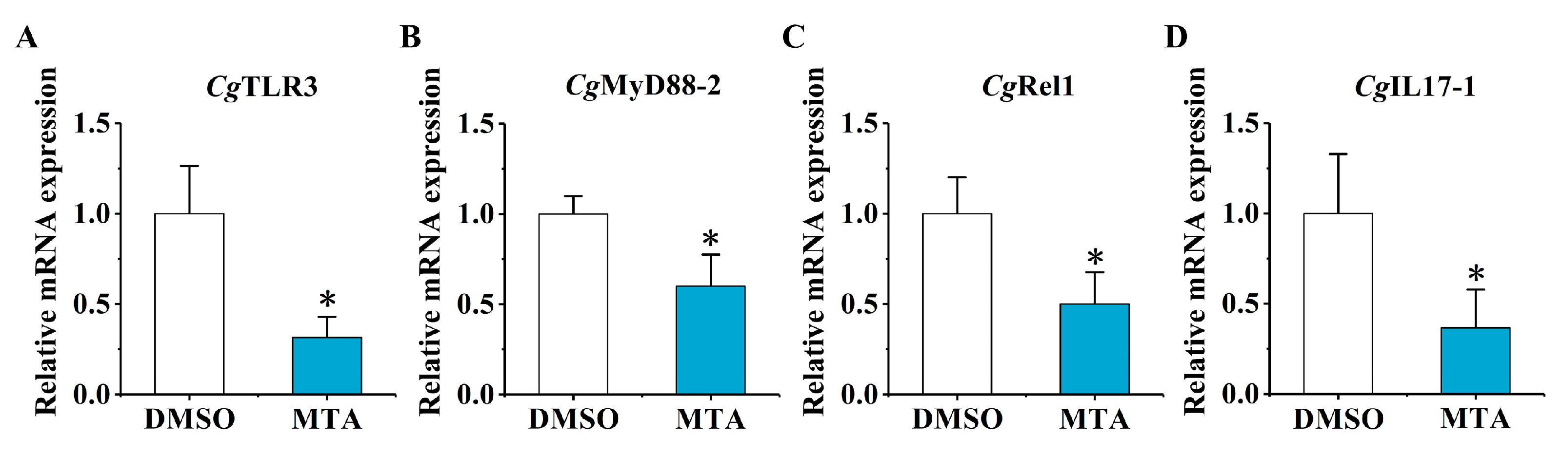
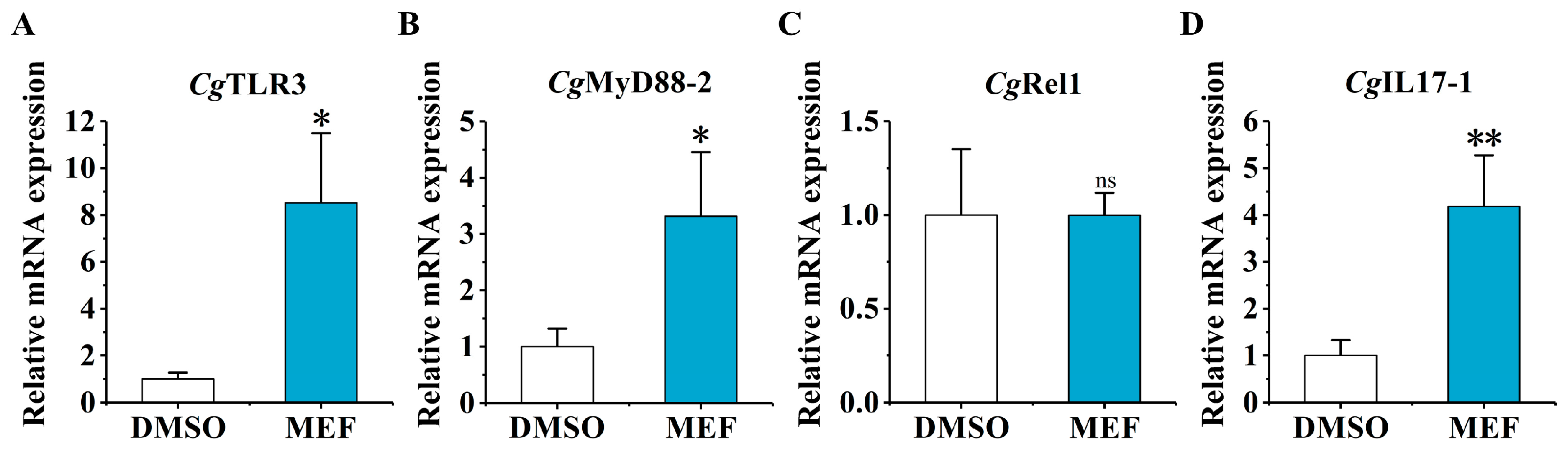
2.4. The mRNA Expression of CgTLR3, CgMyD88-2, CgRel1, and CgIL17-1 at 6 h after the Secondary Stimulation upon the MTA and MEF Treatment

The relative mRNA expression levels of CgTLR3, CgMyD88-2, CgRel1, and CgIL17-1 were examined at 6 h after the secondary stimulation in the MTA group and the MEF group by RT-qPCR. Upon the MTA treatment, the mRNA expression levels of CgTLR3, CgMyD88-2, CgRel1, and CgIL17-1 were significantly repressed by 31.45% (p < 0.05), 60.06% (p < 0.05), 50.06% (p < 0.05), and 36.60% (p < 0.05), compared with the DMSO group, respectively (Figure 5). In the MEF group, the relative mRNA expression levels of CgTLR3, CgMyD88-2, and CgIL17-1 increased significantly (8.52-fold, p < 0.05, 3.32-fold, p < 0.05, and 4.18-fold, p < 0.01, compared with the DMSO group, respectively), while the mRNA transcripts of CgRel1 showed no significant change (Figure 6).

3. Discussion

Epigenetic modifications play an important role in the maintenance of immunological memory. To date, knowledge of the epigenetic regulation mechanism in invertebrate immune priming has remained very limited. The circulatory hemocytes in invertebrates are considered counterparts of vertebrate monocytes, which are responsible for the immune response, as well as immune priming [36,47]. In our previous study, enhanced phagocytosis and the promoted regeneration of circulating hemocytes were found in primed oysters when they encountered the secondary challenge with V. splendidus, which indicated that hemocytes play important roles in oyster immune priming [41]. Oyster hemocytes are important immune effector cells that are thought to participate in phagocytosis and the secretion of cytokines [36]. In the present study, the prestimulation of C. gigas with V. splendidus induces an H3K4me3 modification of the CgTLR3 promoter that modulates the mRNA expression levels of CgTLR3, CgMyD88-2, CgRel1, and CgIL17-1 in hemocytes after the secondary immune stimulation with V. splendidus. The epigenetic treatment with MTA and MEF could affect the H3K4me3 modification of the CgTLR3 promoter as well as the CgTLR3 and CgIL17-1 mRNA expression in oyster hemocytes.
A number of pro-inflammatory cytokines have been proposed to play critical roles in trained immunity. The increased production of IL6, IL18, IL1β, or TNFs was used as key indicator for the detection of trained immunity in vertebrates [12,48,49]. Some cytokines such as IL17s [31], TNFs [50], and interferons have been identified in C. gigas [29,51,52]. There are ten IL17 genes annotated from the genome of C. gigas, which are considered to play diverse roles in immune defense [30]. Several reports have shown that CgIL17-1 is involved in inflammatory mobilization and hemocyte proliferation in the innate immune response of C. gigas [33,34,53]. In the present study, in order to find the cytokines with enhanced expression in immune priming, we analyzed the previously reported transcriptome data [35] and identified CgIL17-1 as a key cytokine for further investigation. Increased transcripts of CgIL17-1 upon restimulation were observed (Figure 1A), which confirms the role of CgIL17-1 in oyster immune priming. These results are in agreement with the available evidence that the IL family is associated with immunological memory. For instance, a review article suggested that IL1 family members, especially IL1β, are crucial components of trained immunity [28]. Studies in mice demonstrated that the training of monocytes led to enhanced production of pro-inflammatory cytokines TNF-α and IL6 [20]. In teleost fish, Scophthalmus maximus, trained neutrophils exhibited a significant elevation of the IL1R signaling pathway after Edwardsiella piscicida infection [54]. Hence, it could conceivably be hypothesized that CgIL17-1 is an important component of oyster immune priming, which warrants further study.
The recognition of PAMPs through TLRs induces a series of signaling events that result in an acute inflammatory response. The binding of the TLR to ligands is known to trigger the MyD88-dependent NF-κB pathway, which activates the expression of proinflammatory genes such as TNFs and ILs in the innate response of vertebrates [55,56]. In mollusks, TLRs also shown to activate MyD88 and induce IL17 expression [57,58,59]. Studies have reported the existence of the TLR-MyD88-NF-κB signaling axis, which regulates the expression of inflammatory cytokines such as IL17s and TNFs in oysters [32,42,43,44]. Of note, compared to the finding that mammals typically possess 13 TLRs, 83 TLRs were identified in the genome of C. gigas and shown to be strain-specific, possibly because of oyster-specific immune adaptations [41]. Recently, CgTLR3 was reported to be involved in oyster immune defense by a MyD88-dependent NF-κB pathway [45], and to regulate CgIL17-1 expression [60]. In the present study, CgTLR3 was identified as a key PRR for further investigating its role in immune priming. The enhanced expression of CgTLR3 in the second immune response of oysters was validated, which could further promote the enhanced expression of CgIL17-1. Studies have demonstrated that TLRs play a crucial role in the initiation of immunological memory [23,61]. Various microbial components induce the activation of dendritic cells through TLR signals, which leads to T-cell priming and an acquired immune response [62,63,64]. In Drosophila melanogaster, the Toll pathway is required for enhanced protection against Streptococcus pneumonia re-infection [65]. In crab Eriocheir sinensis, the EsTLR1-mediated productions of EsALF-1 and EsALF-3 in hemolymph played an indispensable role in the month-long humoral immune protection induced by Aeromonas hydrophila. In mud crab Scylla paramamosain, the TLR pathway played an important role in enhanced immune protection against reinfection by Vibrio parahaemolyticus [66]. In Pacific abalone Haliotis discus hannai, the IL17 and TLR signaling pathway were likely dominant in the immune enhancement process in response to the re-infection of V. parahaemolyticus [67]. Our previous transcriptomic data also suggested higher expression levels of CgMyD88 and CgTIMP after the second stimulation with V. splendidus, indicating the key roles of CgTLR3 signaling in oyster immune priming [35]. According to these data, we can infer that CgTLR3 was involved in immune priming and contributes to inducing MyD88-dependent NF-κB pathway activation and IL17-1 expression in oyster hemocytes, implying the indispensable role of the TLR signaling pathway in enhanced immune protection. Though it is conserved for TLR3 in activating the MyD88-dependent NF-κB pathway, the exact roles of this pathway in the immune priming of oysters needs further investigation.
The epigenetic modifications that facilitate gene transcription have been demonstrated as an important mechanism that influences the trained immune response to secondary stimulation in vertebrates [8]. For invertebrates, the possible epigenetic mechanisms of gene expression related to innate immune memory were recently proposed [68,69], in which histone modifications have been recognized as pivotal players. It was found that Anopheles gambiae and Aedes aegypti possess an innate immune memory maintained by histone acetyltransferase [70,71]. H3K4me3 is a histone modification exclusively found at active promoters and is therefore often enriched in promoter regions regulating TLRs [72]. It was found that TLR4 expression is regulated by H3K4me3 on its promoter in diabetic macrophages and wound myeloid cells [24,73]. In C. gigas, H3K4 methylation modifications have been reported to be crucial for the hematopoiesis and the embryonic development process [74,75,76]. In this study, the inactivated bacteria enriched H3K4me3 modification at the CgTLR3 promoter regions. Moreover, the epigenetic drugs MTA and MEF can change the H3K4me3 enrichment of the CgTLR3 promoter, and further affect the mRNA expression of CgMyD88-2 and CgIL17-1. These results suggested that H3K4me3 was involved in the regulation of TLR3 expression and IL17-1 production in hemocyte-mediated immune priming in oysters. It was found that the oysters primed with the stimulation of V. splendidus exhibited enhanced phagocytosis and the regeneration of circulating hemocytes when they encountered the second stimulation with V. splendidus [41]. Studies have suggested that different PRRs and associated molecules were over-represented after the secondary challenge. Several PRRs, such as Down syndrome cell adhesion molecules in crustaceans and insects [77], the variable lymphocyte receptor in crab E. sinensis [78], fibrinogen-related proteins in vector snail Biomphalaria glabrata [79], C-lectins in scallop Chlamys farreri [80], have been implicated in the innate immune memory of invertebrates. Speculative priming in the invertebrate immune system may be caused by synergistic interactions and dosage effects within the immune system, which is more complicated than expected [81]. It is generally assumed that the duration of immune priming can range from 7 to 30 d in different species with different pathogens [82]. The antiviral immune priming phenomenon in oysters was reported to be long-lasting, persisting for at least 5 months [83]. Besides TLRs and H3K4me3, there is abundant room for further progress in determining the detailed mechanism in the immune priming of oysters.
In conclusion, the regulatory mechanism of H3K4me3 in the immune priming of oyster hemocytes was investigated. The TLR expression can be regulated by H3K4me3 methylation in its promoter, which may further affect the enhanced MyD88-dependent TLR signaling transduction and IL expression against the second stimulation with V. splendidus. All the results suggested that H3K4me3 was involved in the immune priming of C. gigas by regulating the mRNA expression of TLR signaling molecules, which offer valuable insights for the regulatory mechanisms of immune memory in invertebrates.

4. Materials and Methods

4.1. Experimental Animals and Bacteria

The oysters C. gigas used in the present study were about two years old, and their shell length was of 12–16 cm. All the oysters were collected from an aquaculture farm in Dalian, Liaoning Province, China, and temporarily cultivated in filtered seawater at ambient temperature. The seawater was aerated using an air pump during the laboratory cultivation. The oysters were fed with concentrated algal powder daily and the water was completely replaced once a day. All oyster experiments were performed in accordance with the approval and guidelines of the Ethics Review Committee of Dalian Ocean University.
V. splendidus, isolated from lesion-like niduses of the moribund scallop Patinopecten yessoensis, was employed to stimulate oysters as previously described [84]. It was cultured in 2116E media at 18 °C for 24 h, harvested by centrifugation at 4000× g for 10 min, resuspended in phosphate-buffered saline (PBS), and adjusted to the final concentration of 2 × 10⁸ CFU mL−1.

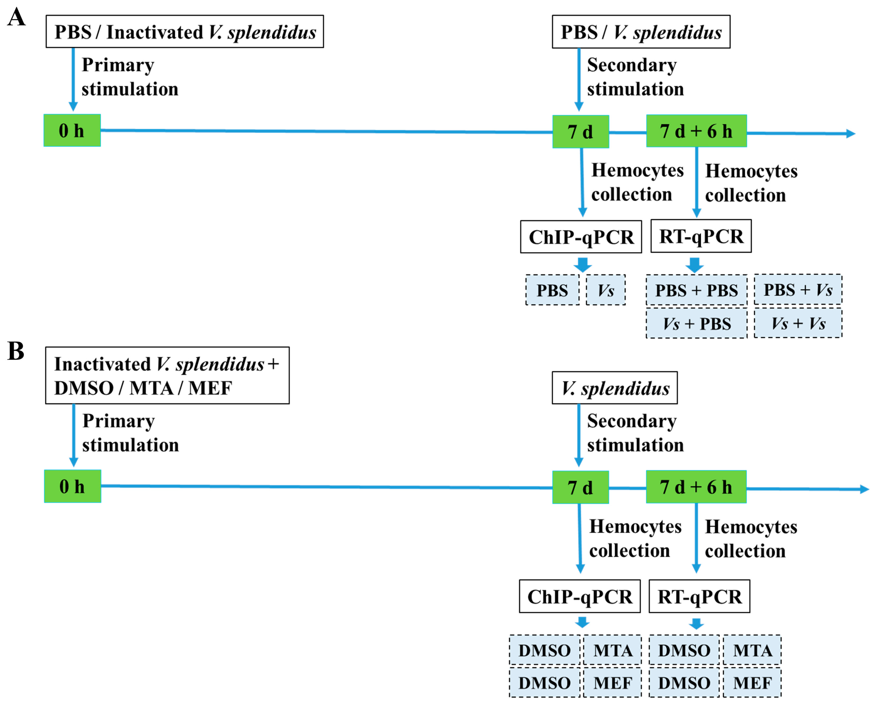
4.2. Immune Priming Induction and Hemocyte Collection

The bacterial stimulation experiment was conducted as previously described [85]. Fifty-four oysters were employed and divided equally into six groups designated as the PBS, Vs, PBS + PBS, PBS + Vs, Vs + PBS, and Vs + Vs groups (Figure 7A). In the PBS and Vs groups, the oysters individually received an injection of 100 μL of PBS or 100 μL of heat-killed V. splendidus (2 × 10⁸ CFU mL−1). At 7 d after the bacterial injection, nine oysters were randomly selected from each group and hemolymph samples were collected to examine the H3K4me3 modification levels of the CgTLR3 gene promoter. In the PBS + PBS and PBS + Vs groups, the oysters received a first injection with 100 μL of PBS and a second injection with 100 μL of PBS or 100 μL of live V. splendidus (2 × 10⁸ CFU mL−1) at 7 d after the first injection, respectively. In the Vs + PBS and Vs + Vs groups, the oysters were first stimulated with 100 μL of heat-killed V. splendidus, and then treated with 100 μL of PBS and 100 μL of live V. splendidus as the second stimulation at 7 d after the first injection, respectively. The hemolymph samples from the PBS + PBS, PBS + Vs, Vs + PBS, and Vs + Vs groups were collected at 6 h after the second immune stimulation to examine the mRNA transcripts of CgIL17-1, CgTLR3, CgMyD88-2, and CgRel1 (Figure 7A).
The hemolymph (about 0.5 mL from each oyster) was aseptically withdrawn from the posterior adductor muscle sinus, and the hemolymph collected from three oysters was mixed together as one sample. There were three replicates for each group. The hemocytes were harvested by centrifugation immediately at 800× g, 4 °C for 10 min.

4.3. The Treatments of MTA and MEF

Seventy-two oysters were employed for the treatment with MTA (the nonselective methyltransferase inhibitor, Sigma) and MEF (the histone demethylases inhibitor, Sigma). The oysters in the MTA group (18 oysters) and the MEF group (18 oysters) received injection of MTA and MEF at a dose of 96 μmol kg−1 body weight [86] and 50 mg kg−1 body weight [13], respectively. Thirty-six oysters treated with 100 μL of dimethyl sulfoxide (DMSO, 5% in PBS) were used as a control. After the treatments with MTA and MEF, 100 μL of heat-killed V. splendidus was injected immediately into each oyster. Hemolymph samples were collected from 36 oysters to examine the H3K4me3 modification level of the CgTLR3 gene promoter at 7 d after the epigenetic treatment. At the same time, the secondary stimulation with 100 μL of live V. splendidus was conducted with the remaining 36 oysters. The mRNA transcripts of CgIL17-1, CgTLR3, CgMyD88-2, and CgRel1 were examined at 6 h after the secondary stimulation.

4.4. RNA Isolation and cDNA Synthesis

Total RNA from oyster hemocytes was extracted using Trizol reagent (Invitrogen, Carlsbad, CA, USA) according to the manufacturer’s instructions. Nanodrop 2000 (Thermo Fisher Scientific, Waltham, MA, USA) and the Agilent 2100 Bioanalyzer (Agilent Technologies, Santa Clara, CA, USA) were used to determine the quantity and purity of the extracted RNA. The cDNA synthesis was performed using PrimeScript™ RT reagent Kit with gDNA Eraser (Takara, Dalian, China) according to the manufacturer’s instruction, and the cDNA mix was diluted 1:40 and stored at −80 °C for the subsequent fluorescent real-time quantitative PCR.

4.5. Reverse Transcription-Quantitative PCR (RT-qPCR) Analysis

The mRNA transcripts of CgIL17-1, CgTLR3, CgMyD88-2, and CgRel1 were measured using specific primers (Table 1) with the elongation factor of C. gigas (CgEF, CGI_10012474) as an internal control. The promoter sequences of the target genes were obtained from the NCBI database (http://www.ncbi.nlm.nih.gov, accessed on 9 September 2021), and Primer premier 6.0 software was used to design the primers for ChIP-qPCR (Table 1).
The PCR reactions were performed with the SYBR premix ExTap (RR420, Takara, Dalian, China) using the ABI PRISM 7500 Sequence Detection System (Thermo Fisher Scientific, Waltham, MA, USA) according to the manual. The relative expression levels were analyzed by comparative Ct method (2−ΔΔCt method) [87].

4.6. ChIP-qPCR Assays

Chromatin immunoprecipitation in oyster hemocytes was conducted as previously described [85]. ChIP-qPCR assay was performed using a ChIP Assay Kit (Beyotime, Shanghai, China) according to the manufacturer’s instructions. The collected hemocytes were resuspended in the modified Leibovitz L-15 medium (supplemented with 20.2 g L−1 NaCl, 0.54 g L−1 KCl, 0.6 g L−1 CaCl2, 1.0 g L−1 MgSO4, 3.9 g L−1 MgCl2, 20.8 g L−1 glucose, 10% FCS, 100 mg mL−1 penicillin G, 100 mg mL−1 streptomycin, 40 mg mL−1 gentamicin and 0.1 mg mL−1 amphotericin B, pH 7.0) and formaldehyde (1% final concentration) was added into the hemocyte suspension to crosslink the DNA and protein. Hemocytes were lysed in SDS lysis buffer, and the cross-linked DNA was sonicated for 10 min to obtain DNA fragments around 250 bp. The cross-linked fragmented DNA was precleared with protein A agarose/salmon sperm DNA and the precleared DNA was incubated with H3K4me3 antibodies (Beyotime, Shanghai, China) at 4 °C overnight. The immunoprecipitates were then incubated with protein A/G agarose/salmon sperm DNA, and the DNA-histone complexes with salmon sperm DNA/protein G agarose beads were collected. The resultant immune complexes were successively washed once with low-salt buffer (150 mM NaCl, 0.1% SDS, 1% Triton X-100, 2 mM EDTA, 20 mM TRIS), once with high-salt buffer (500 mM NaCl, 0.1% SDS, 1% Triton X-100, 2 mM EDTA, 20 mM TRIS), once with LiCl buffer (0.25 M LiCl, 1% NP-40, 1% Na-deoxycholate, 1 mM EDTA, 10 mM TRIS), and twice with TE buffer. The cross-linked fragmented DNA was eluted by using elution buffer (0.1 M NaHCO3, 1% SDS). The same amount of cross-linked fragmented DNA without antibody precipitation was processed in the same manner and served as an input control. The cross-linked DNA was de-crosslinked with 200 mM sodium chloride at 65 °C for 4 h and the proteins were removed by treatment with proteinase K. The resultant DNA was extracted using the phenol/chloroform/isoamyl alcohol method. The primer sets used for the ChIP-qPCR were listed in Table 1.

4.7. Statistical Analysis

All data were given as means ± standard deviation (N = 3) and processed using SPSS version 20.0 software using a two-way ANOVA analysis of variance with Students t-test. Differences were considered significant at p < 0.05 and extremely significant at p < 0.01.

Author Contributions

X.L., W.W. and L.W. designed the study; X.L., Y.L. and J.Z. performed the experiments; X.L., J.Z. and T.Y. managed oyster and bacteria materials; X.L., W.W. and L.W. analyzed data; X.L., L.W. and L.S. wrote and edited the manuscript. All authors have read and agreed to the published version of the manuscript.

Funding

This research was supported by grants from the National Natural Science Foundation of China (Nos. 32230110, 41961124009), the Fund for CARS-49 and for Outstanding Talents and Innovative Team of Agricultural Scientific Research in MARA, the innovation team of Aquaculture Environment Safety from Liaoning Province (LT202009), the Program for Innovative Talents in Higher Education of Liaoning Province (LR2016036), and the Dalian High Level Talent Innovation Support Program (2022RG14).

Institutional Review Board Statement

Not applicable.

Informed Consent Statement

Not applicable.

Data Availability Statement

Data are contained within the article.

Acknowledgments

We are grateful to all the laboratory members for their technical advice and helpful discussions.

Conflicts of Interest

The authors declare they have no competing conflict of interest.

Abbreviations

CgIL17-1: Crassostrea gigas interleukin 17-1; CgMyD88-2, Crassostrea gigas myeloid differentiation factor 88-2; CgRel1, Crassostrea gigas Rel1; CgTLR3, Crassostrea gigas Toll-like receptor 3; ChIP-qPCR, chromatin immunoprecipitation followed by qPCR; DMSO, dimethyl sulfoxide; H3K4me3, histone H3 lysine 4 trimethylation; IL, interleukin; MEF, monomethyl fumarate; MTA, 5′-methylthioadenosine; MyD88, myeloid differentiation factor 88; NF-κB, nuclear factor nuclear factor kappa-B; PAMPs, pathogen-associated molecular patterns; PBS, phosphate-buffered saline; PRRs, pattern recognition receptors; TLR, Toll-like receptor; TNF, tumor necrosis factor; RT-qPCR, reverse transcription-quantitative PCR.

References

  1. Little, T.J.; Kraaijeveld, A.R. Ecological and evolutionary implications of immunological priming in invertebrates. Trends Ecol. Evol. 2004, 19, 58–60. [Google Scholar] [CrossRef] [PubMed]
  2. Yang, W.; Tran, N.T.; Zhu, C.-H.; Yao, D.-F.; Aweya, J.J.; Gong, Y.; Ma, H.-Y.; Zhang, Y.-L.; Li, G.-L.; Li, S.-K. Immune priming in shellfish: A review and an updating mechanistic insight focused on cellular and humoral responses. Aquaculture 2021, 530, 735831. [Google Scholar] [CrossRef]
  3. Divangahi, M.; Aaby, P.; Khader, S.A.; Barreiro, L.B.; Bekkering, S.; Chavakis, T.; van Crevel, R.; Curtis, N.; DiNardo, A.R.; Dominguez-Andres, J.; et al. Trained immunity, tolerance, priming and differentiation: Distinct immunological processes. Nat. Immunol. 2021, 22, 2–6. [Google Scholar] [CrossRef] [PubMed]
  4. Ottaviani, E. Invertebrate immunological memory: Could the epigenetic changes play the part of lymphocytes? Invertebr. Surviv. J. 2015, 12, 1–4. [Google Scholar]
  5. Melillo, D.; Marino, R.; Italiani, P.; Boraschi, D. Innate immune memory in invertebrate metazoans: A critical appraisal. Front. Immunol. 2018, 9, 1915. [Google Scholar] [CrossRef]
  6. Gourbal, B.; Pinaud, S.; Beckers, G.J.M.; Van Der Meer, J.W.M.; Conrath, U.; Netea, M.G. Innate immune memory: An evolutionary perspective. Immunol. Rev. 2018, 283, 21–40. [Google Scholar] [CrossRef]
  7. Zhang, Q.; Cao, X. Epigenetic regulation of the innate immune response to infection. Nat. Rev. Immunol. 2019, 19, 417–432. [Google Scholar] [CrossRef]
  8. Dominguez-Andres, J.; Fanucchi, S.; Joosten, L.A.B.; Mhlanga, M.M.; Netea, M.G. Advances in understanding molecular regulation of innate immune memory. Curr. Opin. Cell Biol. 2020, 63, 68–75. [Google Scholar] [CrossRef]
  9. Barski, A.; Cuddapah, S.; Cui, K.; Roh, T.Y.; Schones, D.E.; Wang, Z.; Wei, G.; Chepelev, I.; Zhao, K. High-resolution profiling of histone methylations in the human genome. Cell 2007, 129, 823–837. [Google Scholar] [CrossRef]
  10. Bernstein, B.E.; Kamal, M.; Lindblad-Toh, K.; Bekiranov, S.; Bailey, D.K.; Huebert, D.J.; McMahon, S.; Karlsson, E.K.; Kulbokas, E.J., 3rd; Gingeras, T.R.; et al. Genomic maps and comparative analysis of histone modifications in human and mouse. Cell 2005, 120, 169–181. [Google Scholar] [CrossRef]
  11. Foster, S.L.; Hargreaves, D.C.; Medzhitov, R. Gene-specific control of inflammation by TLR-induced chromatin modifications. Nature 2007, 447, 972–978. [Google Scholar] [CrossRef]
  12. Kleinnijenhuis, J.; Quintin, J.; Preijers, F.; Joosten, L.A.; Ifrim, D.C.; Saeed, S.; Jacobs, C.; van Loenhout, J.; de Jong, D.; Stunnenberg, H.G.; et al. Bacille Calmette-Guerin induces NOD2-dependent nonspecific protection from reinfection via epigenetic reprogramming of monocytes. Proc. Natl. Acad. Sci. USA 2012, 109, 17537–17542. [Google Scholar] [CrossRef]
  13. Arts, R.J.; Novakovic, B.; Ter Horst, R.; Carvalho, A.; Bekkering, S.; Lachmandas, E.; Rodrigues, F.; Silvestre, R.; Cheng, S.C.; Wang, S.Y.; et al. Glutaminolysis and fumarate accumulation integrate immunometabolic and epigenetic programs in trained immunity. Cell Metab. 2016, 24, 807–819. [Google Scholar] [CrossRef]
  14. Austenaa, L.; Barozzi, I.; Chronowska, A.; Termanini, A.; Ostuni, R.; Prosperini, E.; Stewart, A.F.; Testa, G.; Natoli, G. The histone methyltransferase Wbp7 controls macrophage function through GPI glycolipid anchor synthesis. Immunity 2012, 36, 572–585. [Google Scholar] [CrossRef]
  15. Netea, M.G.; Quintin, J.; van der Meer, J.W. Trained immunity: A memory for innate host defense. Cell Host Microbe 2011, 9, 355–361. [Google Scholar] [CrossRef]
  16. Vandenbon, A.; Kumagai, Y.; Lin, M.; Suzuki, Y.; Nakai, K. Waves of chromatin modifications in mouse dendritic cells in response to LPS stimulation. Genome Biol. 2018, 19, 138. [Google Scholar] [CrossRef]
  17. Bekkering, S.; Quintin, J.; Joosten, L.A.; van der Meer, J.W.; Netea, M.G.; Riksen, N.P. Oxidized low-density lipoprotein induces long-term proinflammatory cytokine production and foam cell formation via epigenetic reprogramming of monocytes. Arterioscler. Thromb. Vasc. Biol. 2014, 34, 1731–1738. [Google Scholar] [CrossRef]
  18. van der Heijden, C.; Groh, L.; Keating, S.T.; Kaffa, C.; Noz, M.P.; Kersten, S.; van Herwaarden, A.E.; Hoischen, A.; Joosten, L.A.B.; Timmers, H.; et al. Catecholamines induce trained immunity in monocytes in vitro and in vivo. Circ Res. 2020, 127, 269–283. [Google Scholar] [CrossRef]
  19. Zhao, D.; Zhang, Q.; Liu, Y.; Li, X.; Zhao, K.; Ding, Y.; Li, Z.; Shen, Q.; Wang, C.; Li, N.; et al. H3K4me3 demethylase Kdm5a is required for NK cell activation by associating with p50 to suppress SOCS1. Cell Rep. 2016, 15, 288–299. [Google Scholar] [CrossRef]
  20. Quintin, J.; Saeed, S.; Martens, J.H.A.; Giamarellos-Bourboulis, E.J.; Ifrim, D.C.; Logie, C.; Jacobs, L.; Jansen, T.; Kullberg, B.J.; Wijmenga, C.; et al. Candida albicans infection affords protection against reinfection via functional reprogramming of monocytes. Cell Host Microbe 2012, 12, 223–232. [Google Scholar] [CrossRef] [PubMed]
  21. Yamamoto, M.; Takeda, K.; Akira, S. TIR domain-containing adaptors define the specificity of TLR signaling. Mol. Immunol. 2004, 40, 861–868. [Google Scholar] [CrossRef] [PubMed]
  22. Verstrepen, L.; Bekaert, T.; Chau, T.L.; Tavernier, J.; Chariot, A.; Beyaert, R. TLR-4, IL-1R and TNF-R signaling to NF-kappaB: Variations on a common theme. Cell Mol. Life Sci. 2008, 65, 2964–2978. [Google Scholar] [CrossRef]
  23. Pasare, C.; Medzhitov, R. Toll-like receptors and acquired immunity. Semin. Immunol. 2004, 16, 23–26. [Google Scholar] [CrossRef]
  24. Davis, F.M.; Kimball, A.; denDekker, A.; Joshi, A.D.; Boniakowski, A.E.; Nysz, D.; Allen, R.M.; Obi, A.; Singer, K.; Henke, P.K.; et al. Histone methylation directs myeloid TLR4 expression and regulates wound healing following cutaneous tissue injury. J. Immunol. 2019, 202, 1777–1785. [Google Scholar] [CrossRef] [PubMed]
  25. Piatek, P.; Tarkowski, M.; Namiecinska, M.; Riva, A.; Wieczorek, M.; Michlewska, S.; Dulska, J.; Domowicz, M.; Kulinska-Michalska, M.; Lewkowicz, N.; et al. H3K4me3 histone ChIP-Seq analysis reveals molecular mechanisms responsible for neutrophil dysfunction in HIV-infected individuals. Front. Immunol. 2021, 12, 682094. [Google Scholar] [CrossRef]
  26. Escoubet-Lozach, L.; Benner, C.; Kaikkonen, M.U.; Lozach, J.; Heinz, S.; Spann, N.J.; Crotti, A.; Stender, J.; Ghisletti, S.; Reichart, D.; et al. Mechanisms establishing TLR4-responsive activation states of inflammatory response genes. PLoS Genet. 2011, 7, e1002401. [Google Scholar] [CrossRef] [PubMed]
  27. Jeljeli, M.M.; Adamopoulos, I.E. Innate immune memory in inflammatory arthritis. Nat. Rev. Rheumatol. 2023, 19, 627–639. [Google Scholar] [CrossRef]
  28. Moorlag, S.; Roring, R.J.; Joosten, L.A.B.; Netea, M.G. The role of the interleukin-1 family in trained immunity. Immunol. Rev. 2018, 281, 28–39. [Google Scholar] [CrossRef]
  29. Huang, X.D.; Zhang, H.; He, M.X. Comparative and evolutionary analysis of the interleukin 17 gene family in invertebrates. PLoS ONE 2015, 10, e0132802. [Google Scholar] [CrossRef]
  30. Rosani, U.; Varotto, L.; Gerdol, M.; Pallavicini, A.; Venier, P. IL-17 signaling components in bivalves: Comparative sequence analysis and involvement in the immune responses. Dev. Comp. Immunol. 2015, 52, 255–268. [Google Scholar] [CrossRef]
  31. Li, J.; Zhang, Y.; Zhang, Y.; Xiang, Z.; Tong, Y.; Qu, F.; Yu, Z. Genomic characterization and expression analysis of five novel IL-17 genes in the Pacific oyster, Crassostrea gigas. Fish Shellfish Immunol. 2014, 40, 455–465. [Google Scholar] [CrossRef]
  32. Roberts, S.; Gueguen, Y.; de Lorgeril, J.; Goetz, F. Rapid accumulation of an interleukin 17 homolog transcript in Crassostrea gigas hemocytes following bacterial exposure. Dev. Comp. Immunol. 2008, 32, 1099–1104. [Google Scholar] [CrossRef]
  33. Xin, L.; Zhang, H.; Du, X.; Li, Y.; Li, M.; Wang, L.; Wang, H.; Qiu, L.; Song, L. The systematic regulation of oyster CgIL17-1 and CgIL17-5 in response to air exposure. Dev. Comp. Immunol. 2016, 63, 144–155. [Google Scholar] [CrossRef]
  34. Cao, W.; Wang, W.; Fan, S.; Li, J.; Li, Q.; Wu, S.; Wang, L.; Song, L. The receptor CgIL-17R1 expressed in granulocytes mediates the CgIL-17 induced haemocytes proliferation in Crassostrea gigas. Dev. Comp. Immunol. 2022, 131, 104376. [Google Scholar] [CrossRef]
  35. Wang, W.; Wang, L.; Liu, Z.; Song, X.; Yi, Q.; Yang, C.; Song, L. The involvement of TLR signaling and anti-bacterial effectors in enhanced immune protection of oysters after Vibrio splendidus pre-exposure. Dev. Comp. Immunol. 2020, 103, 103498. [Google Scholar] [CrossRef]
  36. Bachère, E.; Rosa, R.D.; Schmitt, P.; Poirier, A.C.; Merou, N.; Charrière, G.M.; Destoumieux-Garzón, D. The new insights into the oyster antimicrobial defense: Cellular, molecular and genetic view. Fish Shellfish Immunol. 2015, 46, 50–64. [Google Scholar] [CrossRef]
  37. Allam, B.; Raftos, D. Immune responses to infectious diseases in bivalves. J. Invertebr. Pathol. 2015, 131, 121–136. [Google Scholar] [CrossRef]
  38. Green, T.J.; Helbig, K.; Speck, P.; Raftos, D.A. Primed for success: Oyster parents treated with poly(I:C) produce offspring with enhanced protection against Ostreid herpesvirus type I infection. Mol. Immunol. 2016, 78, 113–120. [Google Scholar] [CrossRef]
  39. Green, T.J.; Speck, P. Antiviral defense and innate immune memory in the oyster. Viruses 2018, 10, 133. [Google Scholar] [CrossRef]
  40. Lafont, M.; Vergnes, A.; Vidal-Dupiol, J.; de Lorgeril, J.; Gueguen, Y.; Haffner, P.; Petton, B.; Chaparro, C.; Barrachina, C.; Destoumieux-Garzon, D.; et al. A sustained immune response supports long-term antiviral immune priming in the Pacific oyster, Crassostrea gigas. mBio 2020, 11, e02777-19. [Google Scholar] [CrossRef]
  41. Zhang, T.; Qiu, L.; Sun, Z.; Wang, L.; Zhou, Z.; Liu, R.; Yue, F.; Sun, R.; Song, L. The specifically enhanced cellular immune responses in Pacific oyster (Crassostrea gigas) against secondary challenge with Vibrio splendidus. Dev. Comp. Immunol. 2014, 45, 141–150. [Google Scholar] [CrossRef] [PubMed]
  42. Zhang, Y.; He, X.; Yu, F.; Xiang, Z.; Li, J.; Thorpe, K.L.; Yu, Z. Characteristic and functional analysis of toll-like receptors (TLRs) in the lophotrocozoan, Crassostrea gigas, reveals ancient origin of TLR-mediated innate immunity. PLoS ONE 2013, 8, e76464. [Google Scholar]
  43. Gerdol, M.; Venier, P.; Edomi, P.; Pallavicini, A. Diversity and evolution of TIR-domain-containing proteins in bivalves and Metazoa: New insights from comparative genomics. Dev. Comp. Immunol. 2017, 70, 145–164. [Google Scholar] [CrossRef] [PubMed]
  44. Sang, X.; Dong, J.; Chen, F.; Wei, L.; Liu, Y.; Zhang, M.; Huang, B.; Wang, X. Molecular cloning and immune function study of an oyster IκB gene in the NF-κB signaling pathway. Aquaculture 2020, 525, 735322. [Google Scholar] [CrossRef]
  45. Chen, H.; Cai, X.; Li, R.; Wu, Y.; Qiu, H.; Zheng, J.; Zhou, D.; Fang, J.; Wu, X. A novel toll-like receptor from Crassostrea gigas is involved in innate immune response to Vibrio alginolyticus. Infect. Genet. Evol. 2022, 97, 105159. [Google Scholar] [CrossRef]
  46. Li, Y.; Sun, J.; Zhang, Y.; Wang, M.; Wang, L.; Song, L. CgRel involved in antibacterial immunity by regulating the production of CgIL17s and CgBigDef1 in the Pacific oyster Crassostrea gigas. Fish Shellfish Immunol. 2020, 97, 474–482. [Google Scholar] [CrossRef]
  47. Buchmann; Kurt, Evolution of innate immunity: Clues from invertebrates via fish to mammals. Front. Immunol. 2014, 5, 459.
  48. Hellinga, A.H.; Tsallis, T.; Eshuis, T.; Triantis, V.; Ulfman, L.H.; Neerven, R. In vitro induction of trained innate immunity by bIgG and whey protein extracts. Int. J. Mol. Sci. 2020, 21, 9077. [Google Scholar] [CrossRef]
  49. Saeed, S.; Quintin, J.; Kerstens, H.H.; Rao, N.A.; Aghajanirefah, A.; Matarese, F.; Cheng, S.C.; Ratter, J.; Berentsen, K.; van der Ent, M.A.; et al. Epigenetic programming of monocyte-to-macrophage differentiation and trained innate immunity. Science 2014, 345, 1251086. [Google Scholar] [CrossRef]
  50. Gao, D.; Qiu, L.; Gao, Q.; Hou, Z.; Wang, L.; Song, L. Repertoire and evolution of TNF superfamily in Crassostrea gigas: Implications for expansion and diversification of this superfamily in Mollusca. Dev. Comp. Immunol. 2015, 51, 251–260. [Google Scholar] [CrossRef]
  51. Wang, P.H.; He, J.G. Nucleic Acid Sensing in Invertebrate Antiviral Immunity. Int. Rev. Cell Mol. Biol. 2019, 345, 287–360. [Google Scholar]
  52. Huang, S.; Yuan, S.; Guo, L.; Yu, Y.; Li, J.; Wu, T.; Liu, T.; Yang, M.; Wu, K.; Liu, H.; et al. Genomic analysis of the immune gene repertoire of amphioxus reveals extraordinary innate complexity and diversity. Genome Res. 2008, 18, 1112–1126. [Google Scholar] [CrossRef]
  53. Thomas, C. Hemocytes: Forms and functions. East. Oyster Crassostrea Virginica 1996, 1, 75–93. [Google Scholar]
  54. Mu, D.; Yang, J.; Jiang, Y.; Wang, Z.; Chen, W.; Huang, J.; Zhang, Y.; Liu, Q.; Yang, D. Single-cell transcriptomic analysis reveals neutrophil as orchestrator during β-glucan-induced trained immunity in a teleost fish. J. Immunol. 2022, 209, 783–795. [Google Scholar] [CrossRef]
  55. Mandraju, R.; Troutman, T.D.; Pasare, C. Toll-like receptor function and signaling. In Reference Module in Biomedical Sciences; Elsevier: Amsterdam, The Netherlands, 2014. [Google Scholar]
  56. Blumenthal, A.; Feng, H.; Zhang, Y.-B.; Gui, J.-F.; Lemon, S.M.; Yamane, D. Interferon regulatory factor 1 (IRF1) and anti-pathogen innate immune responses. PLoS Pathog. 2021, 17, e1009220. [Google Scholar]
  57. Ren, Y.; Ding, D.; Pan, B.; Bu, W. The TLR13-MyD88-NF-κB signalling pathway of Cyclina sinensis plays vital roles in innate immune responses. Fish Shellfish. Immunol. 2017, 70, 720–730. [Google Scholar] [CrossRef]
  58. Yu, F.; Chen, J.; Lin, J.; Zhong, Z.; Lu, Y.; Zeng, X.; Lei, X. TLR4 involved in immune response against Vibrio Parahaemolyticus by MyD88-dependent pathway in Crassostrea hongkongensis. Fish Shellfish Immunol. 2023, 134, 108591. [Google Scholar] [CrossRef]
  59. Fan, S.; Wang, W.; Li, J.; Cao, W.; Li, Q.; Wu, S.; Wang, L.; Song, L. The truncated MyD88s negatively regulates TLR2 signal on expression of IL17-1 in oyster Crassostrea gigas. Dev. Comp. Immunol. 2022, 133, 104446. [Google Scholar] [CrossRef]
  60. Liu, J.; Wang, W.; Kong, N.; Yu, S.; Dong, M.; Yang, W.; Li, Y.; Zhou, X.; Wang, L.; Song, L. A pattern recognition receptor CgTLR3 involves in regulating the proliferation of haemocytes in oyster Crassostrea gigas. Dev. Comp. Immunol. 2023, 147, 104762. [Google Scholar] [CrossRef]
  61. Hua, Z.; Hou, B. TLR signaling in B-cell development and activation. Cell Mol. Immunol. 2013, 10, 103–106. [Google Scholar] [CrossRef]
  62. Reis e Sousa, C. Toll-like receptors and dendritic cells: For whom the bug tolls. Semin. Immunol. 2004, 16, 27–34. [Google Scholar] [CrossRef]
  63. Mills, K.H. TLR-dependent T cell activation in autoimmunity. Nat. Rev. Immunol. 2011, 11, 807–822. [Google Scholar] [CrossRef]
  64. Lahiri, A.; Das, P.; Chakravortty, D. Engagement of TLR signaling as adjuvant: Towards smarter vaccine and beyond. Vaccine 2008, 26, 6777–6783. [Google Scholar] [CrossRef] [PubMed]
  65. Pham, L.N.; Dionne, M.S.; Shirasu-Hiza, M.; Schneider, D.S. A specific primed immune response in Drosophila is dependent on phagocytes. PLoS Pathog. 2007, 3, e26. [Google Scholar] [CrossRef]
  66. Zhang, X.; Zeng, X.; Sun, Y.; Wang, Y.; Zhang, Z. Enhanced immune protection of mud crab Scylla paramamosain in response to the secondary challenge by Vibrio parahaemolyticus. Front. Immunol. 2020, 11, 565958. [Google Scholar] [CrossRef]
  67. Zhang, X.; Guo, M.; Sun, Y.; Wang, Y.; Zhang, Z. Transcriptomic analysis and discovery of genes involving in enhanced immune protection of Pacific abalone (Haliotis discus hannai) in response to the re-infection of Vibrio parahaemolyticus. Fish Shellfish Immunol. 2022, 125, 128–140. [Google Scholar] [CrossRef]
  68. Willis, A.R.; Sukhdeo, R.; Reinke, A.W. Remembering your enemies: Mechanisms of within-generation and multigenerational immune priming in Caenorhabditis elegans. FEBS J. 2020, 288, 1759–1770. [Google Scholar] [CrossRef]
  69. Low, C.F.; Chong, C.M. Peculiarities of innate immune memory in crustaceans. Fish Shellfish Immunol. 2020, 104, 605–612. [Google Scholar] [CrossRef]
  70. Gomes, F.M.; Tyner, M.D.W.; Barletta, A.B.F.; Saha, B.; Yenkoidiok-Douti, L.; Canepa, G.E.; Molina-Cruz, A.; Barillas-Mury, C. Double peroxidase and histone acetyltransferase AgTip60 maintain innate immune memory in primed mosquitoes. Proc. Natl. Acad. Sci. USA 2021, 118, e2114242118. [Google Scholar] [CrossRef]
  71. Amarante, A.M.; da Silva, I.C.A.; Carneiro, V.C.; Vicentino, A.R.R.; Pinto, M.A.; Higa, L.M.; Moharana, K.C.; Talyuli, O.A.C.; Venancio, T.M.; de Oliveira, P.L.; et al. Zika virus infection drives epigenetic modulation of immunity by the histone acetyltransferase CBP of Aedes aegypti. PLoS Negl. Trop. Dis. 2022, 16, e0010559. [Google Scholar] [CrossRef] [PubMed]
  72. Schafer, A.; Baric, R.S. Epigenetic landscape during coronavirus infection. Pathogens 2017, 6, 8. [Google Scholar] [CrossRef]
  73. Davis, F.M.; denDekker, A.; Kimball, A.; Joshi, A.D.; El Azzouny, M.; Wolf, S.J.; Obi, A.T.; Lipinski, J.; Gudjonsson, J.E.; Xing, X.; et al. Epigenetic regulation of TLR4 in diabetic macrophages modulates immunometabolism and wound repair. J. Immunol. 2020, 204, 2503–2513. [Google Scholar] [CrossRef]
  74. Fellous, A.; Favrel, P.; Guo, X.; Riviere, G. The Jumonji gene family in Crassostrea gigas suggests evolutionary conservation of Jmj-C histone demethylases orthologues in the oyster gametogenesis and development. Gene 2014, 538, 164–175. [Google Scholar] [CrossRef]
  75. Gu, X.; Qiao, X.; Yu, S.; Song, X.; Wang, L.; Song, L. Histone lysine-specific demethylase 1 regulates the proliferation of hemocytes in the oyster Crassostrea gigas. Front. Immunol. 2022, 13, 1088149. [Google Scholar] [CrossRef] [PubMed]
  76. Fellous, A.; Le Franc, L.; Jouaux, A.; Goux, D.; Favrel, P.; Rivière, G. Histone methylation participates in gene expression control during the early development of the Pacific oyster Crassostrea gigas. Genes 2019, 10, 695. [Google Scholar] [CrossRef] [PubMed]
  77. Armitage, S.A.; Peuss, R.; Kurtz, J. Dscam and pancrustacean immune memory—A review of the evidence. Dev. Comp. Immunol. 2015, 48, 315–323. [Google Scholar] [CrossRef] [PubMed]
  78. Liu, H.; Song, C.; Ning, J.; Liu, Y.; Cui, Z. Identification, functional characterization and the potential role of variable lymphocyte receptor EsVLRA from Eriocheir sinensis in response to secondary challenge after Vibrio parahaemolyticus vaccine. Fish Shellfish Immunol. 2020, 98, 201–209. [Google Scholar] [CrossRef]
  79. Pinaud, S.; Portela, J.; Duval, D.; Nowacki, F.C.; Olive, M.A.; Allienne, J.F.; Galinier, R.; Dheilly, N.M.; Kieffer-Jaquinod, S.; Mitta, G.; et al. A shift from cellular to humoral responses contributes to innate immune memory in the vector snail Biomphalaria glabrata. PLoS Pathog. 2016, 12, e1005361. [Google Scholar] [CrossRef]
  80. Wang, J.; Wang, L.; Yang, C.; Jiang, Q.; Zhang, H.; Yue, F.; Huang, M.; Sun, Z.; Song, L. The response of mRNA expression upon secondary challenge with Vibrio anguillarum suggests the involvement of C-lectins in the immune priming of scallop Chlamys farreri. Dev. Comp. Immunol. 2013, 40, 142–147. [Google Scholar] [CrossRef]
  81. Schulenburg, H.; Boehnisch, C.; Michiels, N.K. How do invertebrates generate a highly specific innate immune response? Mol. Immunol. 2007, 44, 3338–3344. [Google Scholar] [CrossRef]
  82. Chang, Y.-H.; Kumar, R.; Ng, T.H.; Wang, H.-C. What vaccination studies tell us about immunological memory within the innate immune system of cultured shrimp and crayfish. Dev. Comp. Immunol. 2018, 80, 53–66. [Google Scholar] [CrossRef] [PubMed]
  83. Lafont, M.; Petton, B.; Vergnes, A.; Pauletto, M.; Segarra, A.; Gourbal, B.; Montagnani, C. Long-lasting antiviral innate immune priming in the Lophotrochozoan Pacific oyster, Crassostrea gigas. Sci. Rep. 2017, 7, 13143. [Google Scholar] [CrossRef] [PubMed]
  84. Liu, R.; Qiu, L.; Yu, Z.; Zi, J.; Yue, F.; Wang, L.; Zhang, H.; Teng, W.; Liu, X.; Song, L. Identification and characterisation of pathogenic Vibrio splendidus from Yesso scallop (Patinopecten yessoensis) cultured in a low temperature environment. J. Invertebr. Pathol. 2013, 114, 144–150. [Google Scholar] [CrossRef] [PubMed]
  85. Liu, Y.; Zhu, Q.; Li, L.; Wang, W.; Zhang, G. Identification of HSF1 Target Genes Involved in Thermal Stress in the Pacific Oyster Crassostrea gigas by ChIP-seq. Mar. Biotechnol. 2020, 22, 167–179. [Google Scholar] [CrossRef]
  86. Moreno, B.; Hevia, H.; Santamaria, M.; Sepulcre, J.; Munoz, J.; Garcia-Trevijano, E.R.; Berasain, C.; Corrales, F.J.; Avila, M.A.; Villoslada, P. Methylthioadenosine reverses brain autoimmune disease. Ann. Neurol. 2006, 60, 323–334. [Google Scholar] [CrossRef]
  87. Livak, K.J.; Schmittgen, T.D. Analysis of relative gene expression data using real-time quantitative PCR and the 2(-Delta Delta C(T)) Method. Methods 2001, 25, 402–408. [Google Scholar] [CrossRef]
Figure 1. The mRNA expression levels of CgTLR3, CgMyD88-2, CgRel1, and CgIL17-1 at 6 h after the secondary stimulation with live V. splendidus (Vs). The relative mRNA expression levels of (A) CgIL17-1, (B) CgTLR3, (C) CgMyD88-2, and (D) CgRel1 in the four groups (PBS + PBS, Vs + PBS, PBS + Vs, Vs + Vs) were determined by qPCR. Vertical bars represent the mean ± SD (N = 3). *: p < 0.05, ns: no significant difference (t-test).
Ijms 25 01036 g001
Figure 2. The H3K4me3 modification levels of the CgTLR3 gene promoter at 7 d after the stimulation with inactivated V. splendidus (Vs). The H3K4me3 modification levels of the CgTLR3 gene promoter were determined by ChIP-qPCR. The positions of the primers used to examine H3K4me3 modification at the promoter regions of CgTLR3 are shown in the top panel of the figure. Vertical bars represent the mean ± SD (N = 3). *: p < 0.05, ns: no significant difference (t-test).
Ijms 25 01036 g002
Figure 3. The H3K4me3 modification levels of the CgTLR3 gene promoter at 7 d after the histone methyltransferase inhibitor (MTA) treatment. The H3K4me3 modification levels of the CgTLR3 gene promoter were determined by ChIP-qPCR. The positions of the primers used to examine H3K4me3 modification at the promoter regions of CgTLR3 are shown in the top panel of the figure. Vertical bars represent the mean ± SD (N = 3). *: p < 0.05, **: p < 0.01 (t-test).
Ijms 25 01036 g003
Figure 4. The H3K4me3 modification levels of the CgTLR3 gene promoter at 7 d after the histone demethylase inhibitor (MEF) treatment. The H3K4me3 modification levels of the CgTLR3 gene promoter were determined by ChIP-qPCR. The positions of the primers used to examine H3K4me3 modification at the promoter regions of CgTLR3 are shown in the top panel of the figure. Vertical bars represent the mean ± SD (N = 3). *: p < 0.05, **: p < 0.01 (t-test).
Ijms 25 01036 g004
Figure 5. The mRNA expression levels of CgTLR3, CgMyD88-2, CgRel1, and CgIL17-1 at 6 h after the secondary stimulation upon the MTA treatment. The relative mRNA expression levels of (A) CgIL17-1, (B) CgTLR3, (C) CgMyD88-2, and (D) CgRel1 in the MTA group and the DMSO group were determined by qPCR. Vertical bars represent the mean ± SD (N = 3). *: p < 0.05 (t-test).
Ijms 25 01036 g005
Figure 6. The mRNA expression levels of CgTLR3, CgMyD88-2, CgRel1, and CgIL17-1 at 6 h after the secondary stimulation upon the MEF treatment. The relative mRNA expression levels of (A) CgIL17-1, (B) CgTLR3, (C) CgMyD88-2, and (D) CgRel1 in the MEF group and the DMSO group were determined by qPCR. Vertical bars represent the mean ± SD (N = 3). *: p < 0.05, **: p < 0.01, ns: no significant difference (t-test).
Ijms 25 01036 g006
Figure 7. Schematic representation of the experimental design used to measure H3K4me3 modification levels of genes promoter and mRNA expression levels in oyster hemocytes. (A) Detailed information about the experimental design pertaining to primary and secondary stimulation for immune priming study. The same batch of oysters was injected with inactivated V. splendidus (Vs) or PBS, and at 7 d after the first immune stimulation, the oysters were restimulated with Vs or PBS for 6 h. Hemolymph samples were collected at 7 d after the first immune stimulation and at 6 h after the secondary stimulation for the following experiment. (B) Experimental design for epigenetic treatment. MTA, MEF, or DMSO was injected together with inactivated V. splendidus, and at 7 d after the first stimulation, the oysters were restimulated with live V. splendidus for 6 h. Hemolymph samples were collected at 7 d after the first immune stimulation and at 6 h after the secondary stimulation for the following experiment.
Ijms 25 01036 g007
Table 1. Sequences of the primers used in this study.
PrimerSequence (5′-3′)
RT-qPCR primers
CgTLR3-RT-FTGCCAAAAGCAAATGGTGTGAAT
CgTLR3-RT-RTTTCCCCCAAAACAAACTTCGTC
CgMyD88-2-RT-FCAGATAAACCGCTACGACGCCTA
CgMyD88-2-RT-RATTTCCGATTCCTTTTGGTGGTC
CgRel1-RT-FTCCGCACACCACCTTACAA
CgRel1-RT-RCGCCTTTATCTTCAGCCTCT
CgIL17-1-RT-FGCGAACGCCACAGTGTCAAA
CgIL17-1-RT-RGACGCTACGAGGAAATACGGAC
CgEF-RT-FAGTCACCAAGGCTGCACAGAAAG
CgEF-RT-RTCCGACGTATTTCTTTGCGATGT
ChIP-qPCR primers
CgTLR3-Pro-F1CAACATGAATCTCAGCAGACG
CgTLR3-Pro-R1TTCTTCCCAAACTGCCACA
CgTLR3-Pro-F2AAGAAGGGGGAGGAGTGCT
CgTLR3-Pro-R2ATGTGTCTTTAAAAGCCGGTG
Disclaimer/Publisher’s Note: The statements, opinions and data contained in all publications are solely those of the individual author(s) and contributor(s) and not of MDPI and/or the editor(s). MDPI and/or the editor(s) disclaim responsibility for any injury to people or property resulting from any ideas, methods, instructions or products referred to in the content.

