Article

Anticancer Activity of Vitamin D, Lumisterol and Selected Derivatives against Human Malignant Melanoma Cell Lines

by
Paweł Domżalski
1,
Anna Piotrowska
1,
Robert C. Tuckey
2 and
Michał A. Zmijewski
1,*
1
Department of Histology, Medical University of Gdansk, 1a Debinki, 80-211 Gdansk, Poland
2
School of Molecular Sciences, University of Western Australia, Perth, WA 6009, Australia
*
Author to whom correspondence should be addressed.
Int. J. Mol. Sci. 2024, 25(20), 10914; https://doi.org/10.3390/ijms252010914
Submission received: 11 September 2024 / Revised: 30 September 2024 / Accepted: 7 October 2024 / Published: 10 October 2024
(This article belongs to the Special Issue The Role of Vitamin D in Human Health and Diseases 4.0)

Abstract

:
Despite the recent development of improved methods of treating melanoma such as targeted therapy, immunotherapy or combined treatment, the number of new cases worldwide is increasing. It is well known that active metabolites of vitamin D3 and lumisterol (L3) exert photoprotective and antiproliferative effects on the skin, while UV radiation is a major environmental risk factor for melanoma. Thus, many natural metabolites and synthetic analogs of steroidal and secosteroidal molecules have been tested on various cancer cells and in animal models. In this study, we tested the anti-melanoma properties of several natural derivatives of vitamin D3 and L3 in comparison to 1,25-dihydroxyvitamin D3 (1,25(OH)2D3). A significant decrease in melanoma cell proliferation and cell mobility was observed for selected derivatives, with (25R)-27-hydroxyL3 showing the highest potency (lowest IC50) in A375 cells but lower potency in SK-MEL-28 cells, whereas the parent L3 failed to inhibit proliferation. The efficacy (% inhibition) by 1,24,25(OH)3D3 and 1,25(OH)2D3 were similar in both cell types. 1,25(OH)2D3 showed higher potency than 1,24,25(OH)3D3 in SK-MEL-28 cells, but lower potency in A375 cells for the inhibition of proliferation. As for 1,25(OH)2D3, but not the other derivatives tested, treatment of melanoma cells with 1,24,25(OH)3D3 markedly increased the expression of CYP24A1, enhanced translocation of the vitamin D receptor (VDR) from the cytoplasm to the nucleus and also decreased the expression of the proliferation marker Ki67. The effects of the other compounds tested were weaker and occurred only under certain conditions. Our data indicate that 1,24,25(OH)3D3, which has undergone the first step in 1,25(OH)2D3 inactivation by being hydroxylated at C24, still shows anti-melanoma properties, displaying higher potency than 1,25(OH)2D3 in SK-MEL-28 cells. Furthermore, hydroxylation increases the potency of some of the lumisterol hydroxy-derivatives, as in contrast to L3, (25R)-27(OH)L3 effectively inhibits proliferation and migration of the human malignant melanoma cell line A375.

1. Introduction

7-Dehydroholesterol (7DHC) is a key precursor of cholesterol; however, its exposure to ultraviolet B radiation (UVB) in the skin results in opening of the B-ring and formation of pre-vitamin D3 (pre-D3). Pre-D3 spontaneously isomerizes to vitamin D3 in a thermal, non-catalytic process. Exposure of pre-D3 to further high doses of UVB can result in resealing of the B-ring to form lumisterol (L3), or additional rearrangement of its double bounds to give tachysterol [1,2,3]. The classical pathway of vitamin D metabolism requires subsequent hydroxylation of vitamin D at C25 by CYP2R1 and at C1 by CYP27B1. The level of the major hormonally active form of vitamin D, 1,25(OH)2D3, is tightly regulated by its hydroxylation and inactivation by CYP24A1, whose gene expression is up-regulated by 1,25(OH)2D3 [4,5]. CYP24A1 is a mitochondrial cytochrome P450 that catalyzes the catabolism of 1,25(OH)2D3 predominantly by the C24 oxidations pathway with some contribution from the C23 pathway [6,7,8]. The initial product of the C24-oxidation pathway is 1,24,25(OH)3D3 and this accumulates due to its less efficient metabolism by CYP24A1 compared to 1,25(OH)2D3 [9]. Its subsequent metabolism by CYP24A1 eventually produces calcitroic acid which is excreted [5,7,8,10]. The C23-oxidation pathway through a series of hydroxylations and lactonization produces 1,25(OH)2D3-26,23-lactone [7,8]. Importantly, it is now clear that the key enzyme initiating steroidogenesis, CYP11A1 (also known as cytochrome P450scc), can also utilize vitamin D, lumisterol and tachysterol as substrates, producing series of biologically active hydroxy derivatives [8,11]. Key metabolites from vitamin D3 include 20(OH)D3 and 20,23(OH)2D3, and unlike cholesterol there is no cleavage of the side chain [12]. 20(OH)D3 can undergo hydroxylation at C24 by CYP24A1 producing 20,24(R)-dihydroxycholesterol (20,24(OH)2D3) [8]. CYP11A1 acts on L3 producing 22(OH)L3, 24(OH)L3 and 20,22(OH)2L3 while another CYP that acts on sterols, CYP27A1, produces 25(OH)L3 and the two C25 stereoisomers of 27(OH)L3, namely (25R)-27(OH)L3 and (25S)-27(OH)L3. CYP11A1-generated hydroxymetabolites of vitamin D and L3 and CYP27A-derived metabolites of L3 have been detected in human and animal sera and in some cases in skin and adrenal glands [13,14,15,16], with 20(OH)D3 and 1,20(OH)2D3 being present in human serum at substantially higher concentrations than 1,25(OH)2D3 [17].
In spite of its known anticancer properties, the use of 1,25(OH)2D3 in cancer therapy has been limited by its potential hypercalcemic effects [18]. Thus, for several decades many synthetic analogs of vitamin D and natural derivatives of vitamin D3, 7-DHC and lumisterol have been investigated and their anticancer properties tested on melanoma in vivo and in vitro [14,19,20,21,22,23,24]. Some of the resulting analogs displayed anti-proliferative, pro-differentiation and anti-inflammatory effects similar to or even better than 1,25(OH)2D3 [16,25,26]. Furthermore, vitamin D and its derivatives increase the anticancer properties of several antimelanoma drugs including doxorubicin, genistein, carboplatine [27,28,29,30], tamoxifen [31] and paclitaxel [32], both in vitro and using mouse models. Furthermore, the antitumor synergism between temozolomide and vitamin D on a model of glioblastoma was observed. The authors suggest that the combination of calcitriol, all-trans retinoic acid (ATRA) and temozolomide (known as CAT combination) can be a safe approach to benefit from vitamin D treatment of high-grade glial tumors [33]. It turns out that vitamin D also improves the anticancer properties of cisplatin and dacarbazine on melanoma cells [34]. This hormone not only improves the results of chemotherapy but also augments targeted therapy such as with cediranib where it was observed that 1,25(OH)2D3 enhances the anticancer properties of the Vascular Endothelial growth factor receptor (VEGFR) inhibitor [35]. Moreover, the same team discovered that vitamin D modulates the response of patient-derived metastatic melanoma cells to cediranib or vemurafenib therapy [36]. Another study showed that vitamin D sensitizes melanoma cells to a low dose of proton beam radiation, which suggests that it can be an adjuvant for this type of treatment of melanoma [37]. Interestingly, vitamin D and its analogs were shown to protect against UVB-induced damage in epidermal human keratinocytes and melanocytes [38]. Considering that UVB radiation is an important factor in the development of melanoma, vitamin D analogs may not only have application to anticancer therapy, but also to cancer prevention due to their protective effects against UVB. Additionally, low levels of 25(OH)D3 are associated with reduced survival in melanoma patients. A single nucleotide polymorphism in the vitamin D receptor affects development or progression of cancer and disease outcome further confirming the role of vitamin D in cancer [39].
In the current study, the effects of a range of natural hydroxy-derivatives of vitamin D3 and lumisterol were tested on melanoma cells and compared to those of 1,25(OH)2D3. Derivatives of vitamin D3 selected were 1α,24R,25(OH)3D3, which is the first major intermediate in 1,25(OH)2D3 inactivation produced by CYP24A1, and 20S,24R(OH)2D3, which is a product of the consecutive actions of CYP11A1 and CYP24A1 on vitamin D3, which has previously been shown to have biological activity, including on melanoma cells [40,41]. For L3, 22(OH)L3 which results from the action of CYP11A1 on L3 was selected to study as was (25R)-27(OH)L3 as a representative product of the action of CYP27A1 on L3. Both of these have previously been shown to exhibit biological activity on skin cells [11,14,16,19,38]. Two human malignant melanoma cell lines, A375 and SK-MEL-28, were selected to analyze the effects of these hydroxy-derivatives on cell proliferation and migration, cell cycle modulation, changes in the expression of selected genes and VDR translocation to the nucleus.

2. Results

2.1. The Hydroxy-Derivatives Were Synthesized Using Extracted and Purified CYP Enzymes Expressed in E.coli

The hydroxy-derivatives of vitamin D3 and L3 tested in this study, 1,24,25(OH)3D3, 20,24(OH)2D3, 22(OH)L3 and (25R)-27(OH)L3 (Figure S1), have all been detected in human serum [8,42], but their chemical synthesis is difficult [40] and they are not commercially available. However, CYP enzymes expressed in E. coli, extracted and purified, enabled us to utilize enzymatic synthesis to produce sufficient amounts (>300 nmol) of these sterols for testing their anti-melanoma activities on A375 and SK-MEL-28 cells.

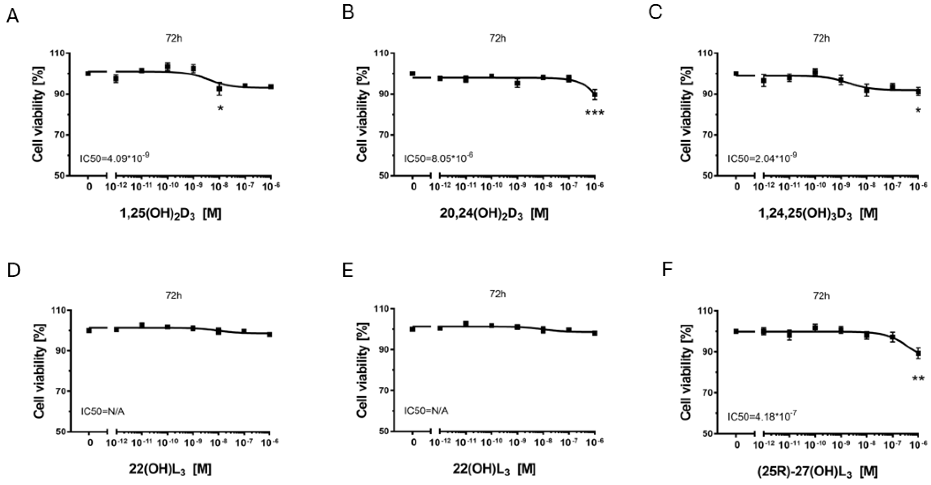
2.2. The Hydroxy-Derivatives of Vitamin D3 and L3 Affects on Proliferation of Melanoma Cells A375 and SK-MEL-28

To assess the effect of these vitamin D and lumisterol derivatives in comparison to the well-characterized, hormonally active form of vitamin D, 1,25(OH)2D3, on the proliferation of melanoma A375 (Figure 1) and SK-MEL 28 cells (Figure 2), SRB assays were performed. All three vitamin D hydroxy-derivatives tested inhibited proliferation of melanoma A375 cells (Figure 1A–C). The IC50 value was the lowest (highest potency) for 1,25(OH)2D3 (1.15 nM), followed by 1,24,25(OH)3D3 (17.8 nM) and 20,24(OH)2D3 (280 nM). The highest inhibitory effect (efficacy), measured at the highest concentration of secosteroid tested (1 μM), was comparable for the 3 secosteroids (20–25%). The efficacy of the vitamin D hydroxy-derivatives for inhibition of proliferation was weaker in SK-MEL-28 melanoma cells, with only approximately 10% growth inhibition at the highest concentration (1 µM) of each derivative tested (Figure 2A–C). 1,24,25(OH)3D3 showed the highest potency with an IC50 half that of 1,25(OH)2D3.
For the lumisterols, neither L3 nor 22(OH)L3 inhibited proliferation of either cell type, with L3 actually causing a 10% increase in proliferation in SK-MEL-28 melanoma cells. In contrast, (25R)-27(OH)L3 (Figure 1F and Figure 2F) inhibited proliferation in both melanoma cell lines, with approximately 20% inhibition at the highest concentration tested (1 µM). High potency for this inhibition was seen with A375 cells (IC50 = 1 pM), but not for SK-MEL-28 cells.

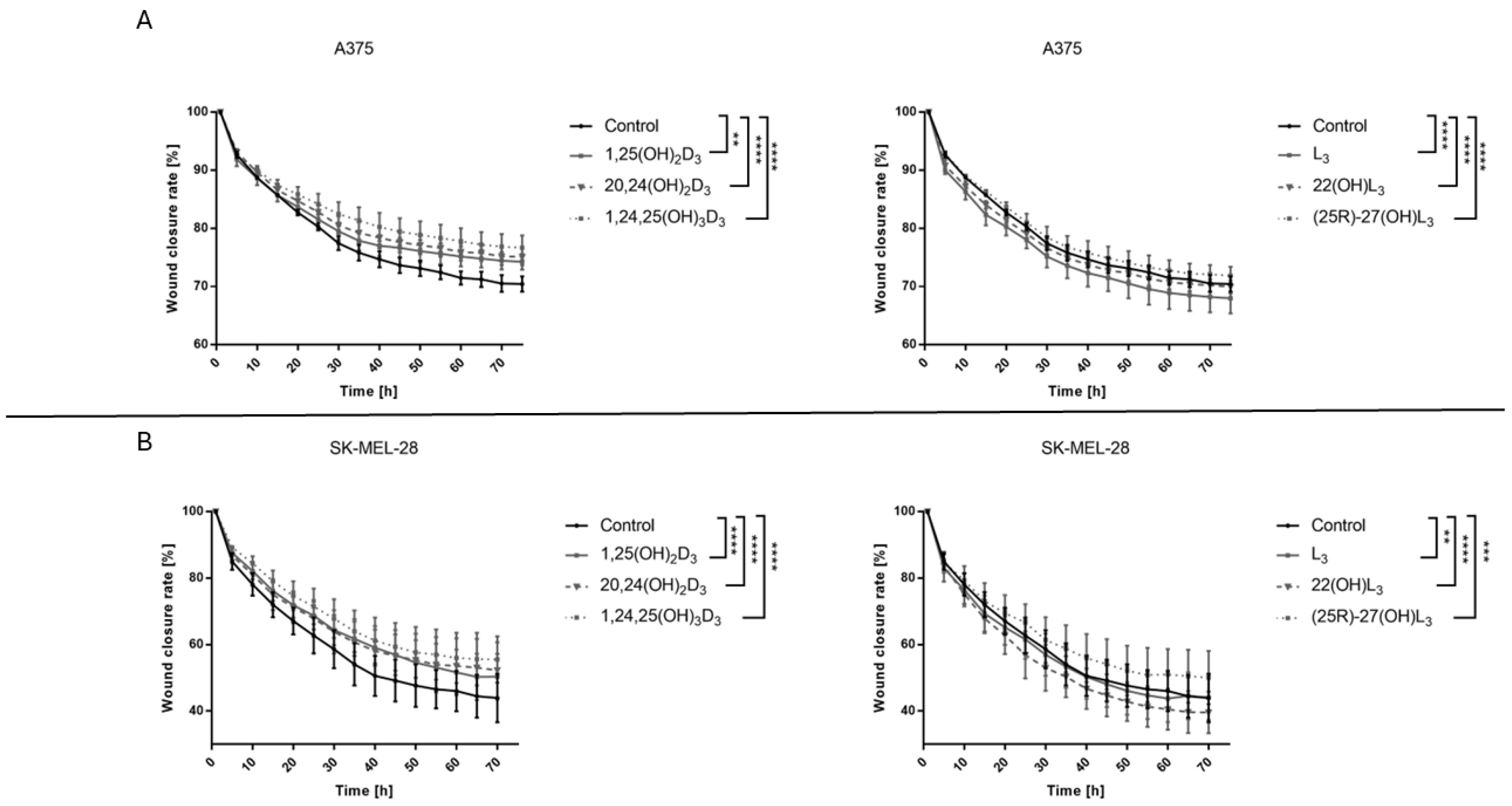
2.3. The Hydroxy-Derivatives of Vitamin D3 and L3 Affects on the Mobility of Melanoma Cells A375 and SK-MEL-28

The effects of the hydroxy-derivatives (100 nM) under study on the migration of A375 and SK-MEL-28 melanoma cells was examined. Interestingly, both melanoma cell lines differed in their migration efficiency with wound closure reaching ~30% for A375 and 50% for SK-MEL-28 cells; however, the effects of vitamin D and lumisterol derivatives were similar for both cell types (Figure 3). Treatment with 1,25(OH)2D3, 20,24(OH)2D3, 1,24,25(OH)3D3 or (25R)-27(OH)L3 resulted in slower migration of melanoma cells when compared to untreated cells, with statistical significance for both cells lines. 1,24,25(OH)3D3 decreased cell migration the most, leaving the wound at 75% of its original size for A375 cells and 40% for SK-MEL-28 cells. In contrast, L3 and 22(OH)L3 enhanced migration of A375 cells with the strongest effect being for L3 in comparison to untreated cells, while for SK-MEL-28 cells 22(OH)L3 increased migration more than L3. (Figure 3A,B). The representative pictures are shown in Figures S2 and S3.

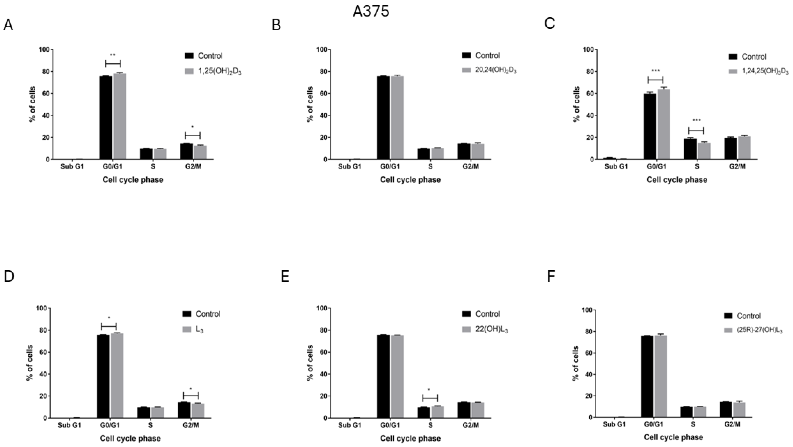
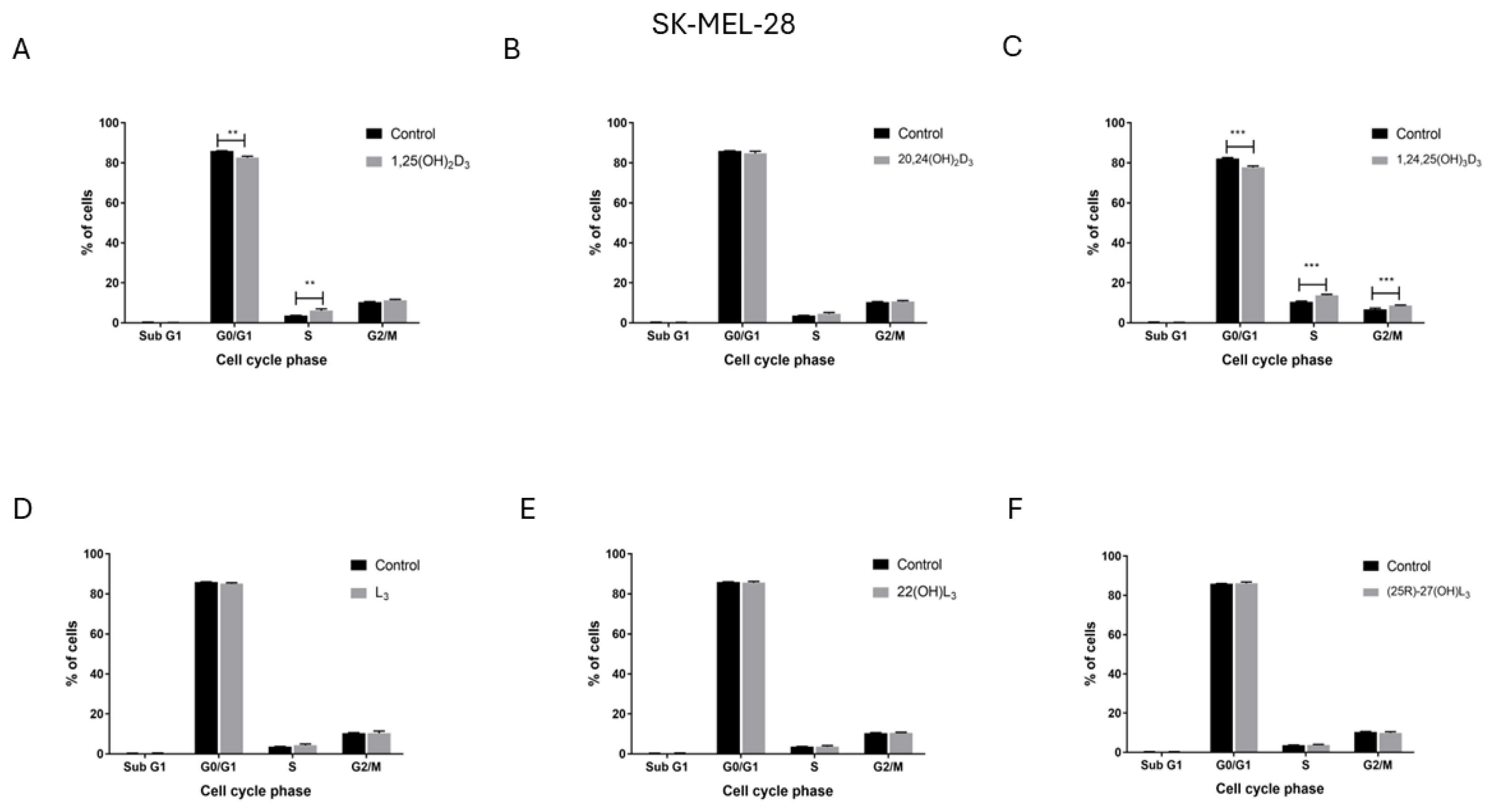
2.4. The Hydroxy-Derivatives of Vitamin D3 and L3 Influence the Distribution of Cells in the Various Phases of the Cell Cycle

To explore the potential mechnisms of inhibition of A375 and SK-MEL-28 melanoma cell proliferation by the compounds under study, their influence on the distribution of melanoma cells in the various phases of the cell cycle was measured by flow cytometry. Treatment of the A375 melanoma cell line with 1,25(OH)2D3 (Figure 4A) or 1,24,25(OH)3D3 (Figure 4C) increased the percentage of cells in the G0/G1 phase of the cell cycle. The percentage of cells in the G2/M phase decreased after treatment with 1,25(OH)2D3 and in the S phase after treatment with 1,24,25(OH)3D3. For these cells L3 showed a similar but weaker effect to 1,25(OH)2D3 (Figure 4D) even though it did not inhibit cell proliferation (Figure 1 and Figure 2). In contrast, opposite effects to those seen on A375 cells were observed for SK-MEL-28 human melanoma cells following treatment. Both 1,25(OH)2D3 (Figure 5A) and 1,24,25(OH)3D3 (Figure 5C) increased the percentage of cells in G0/G1 phase and increased it in the S (1,25(OH)2D3) or G2/M and S (1,24,25(OH)3D3) phases. Neither 24,25(OH)2D3 nor L3 and its hydroxy-derivatives affected the cell cycle of SK-MEL-28 melanoma cells (Figure 5B,D–F). The representative flow fitting curve are shown in Figures S4 and S5.

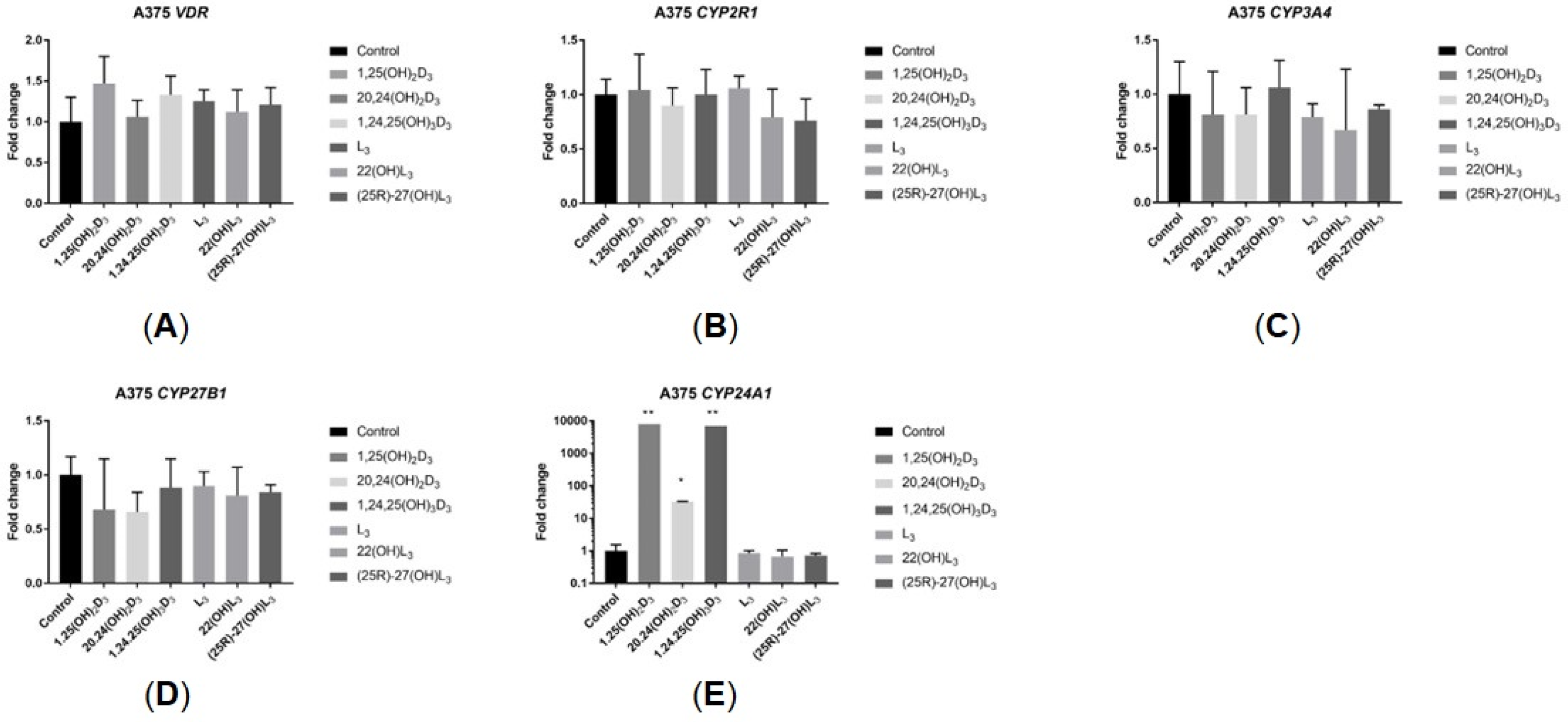
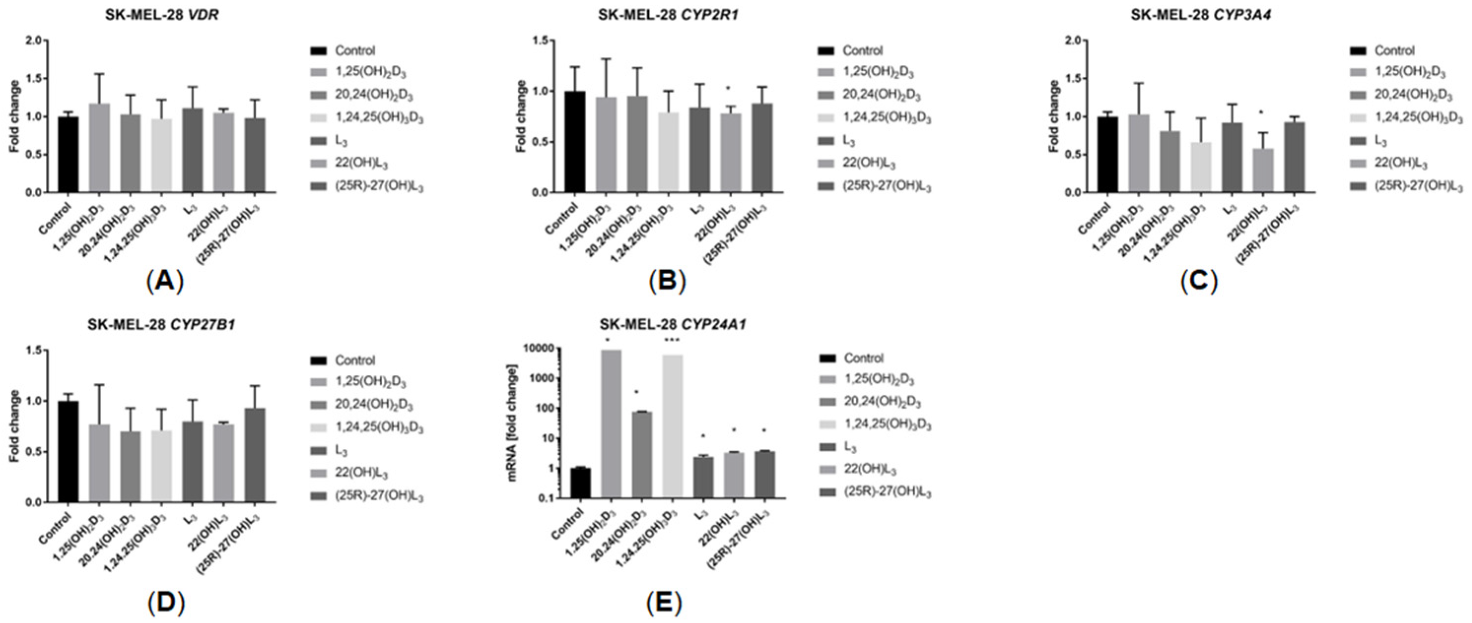
2.5. The Hydroxy-Derivatives of Vitamin D3 and L3 Impact the Expression of Genes Involved in Vitamin D Metabolism

The impact of the sterols under study on the expression of selected genes involved in vitamin D metabolism was investigated. As expected, 1,25(OH)2D3 dramatically increased the CYP24A1 mRNA level in both cell lines. 1,24,25(OH)3D3 showed a similar effect, while 20,24(OH)2D3 caused a lower level of induction. L3 and its derivatives caused a small but significant increase in CYP24A1 mRNA levels in SK-MEL-28 cells but not in A375 cells (Figure 6E and Figure 7E). The expression of other genes encoding CYPs that metabolize vitamin D, including CYP2R1, CYP27B1 and CYP3A4, as well as the VDR, was not altered by the sterols under study (Figure 6A–D and Figure 7A–D).

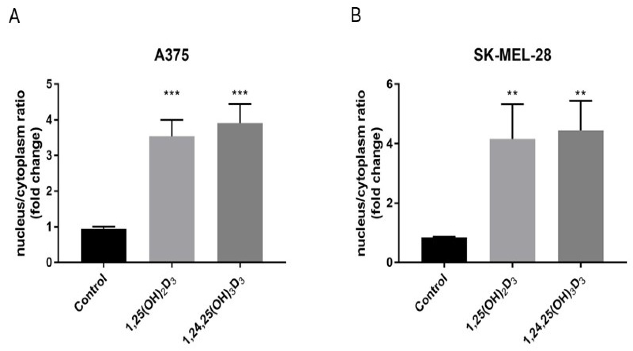
2.6. 1,25(OH)2D3 and 1,24,25(OH)3D3 Influence VDR Translocation from the Cytoplasm to the Nucleus

Since at a concentration of 100 nM, 1,24,25(OH)3D3 showed comparable ability to 1,25(OH)2D3 to stimulate the expression of CYP24A1, we compared the ability of these two secosteroids to stimulate translocation of the VDR from the cytoplasm to the nucleus. The two sterols caused a similar 3- to 4-fold stimulation in the nuclear/cytoplasmic VDR ratio in both A375 and SK-MEL-28 cells (Figure 8A,B). The representative pictures are shown in Figures S6 and S7.

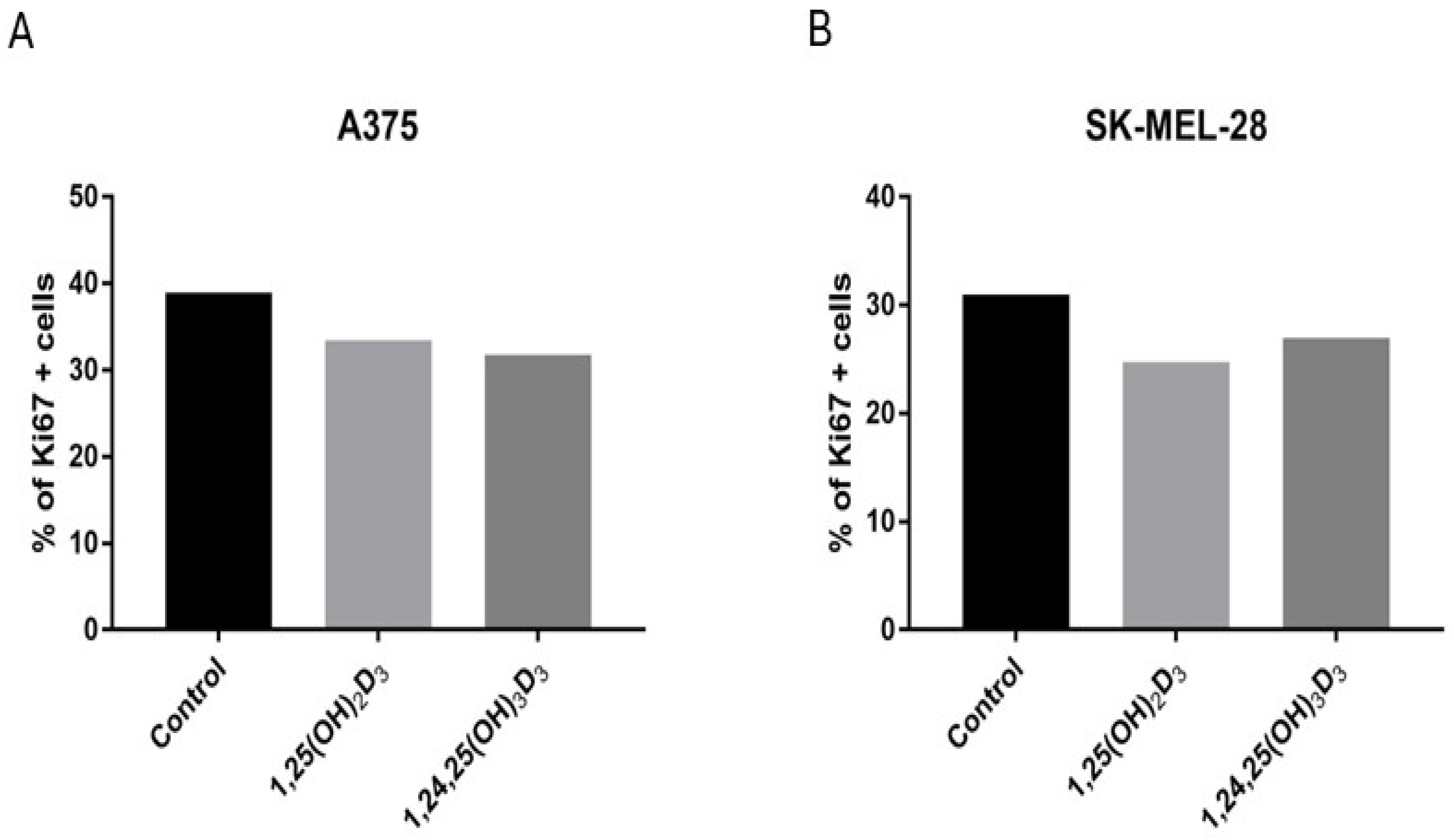
2.7. 1,25(OH)2D3 and 1,24,25(OH)3D3 Influence the Expression of the Ki67 Protein

To complement the proliferation and cell cycle tests, the effects of 1,25(OH)2D3 and 1,24,25(OH)3D3 on the expression of the tumour proliferation marker Ki67 [43] were examined. These secosteroids were selected since they caused statistically significant effects on the inhibition of cell proliferation at a concentration of 100 nM in at least one cell line and caused the largest stimulation of CYP24A1 expression. Treatment of both cell lines with 1,25(OH)2D3 or 1,24,25(OH)3D3 (Figure 9A,B) decreased the percentage of cells expressing Ki67. The effect was similar for both cell lines despite a much weaker response of SK-MEL-28 in the SRB test, with a stronger effect of 1,24,25(OH)3D3 for A375 and 1,25(OH)2D3 for SK-MEL-28 cells. The representative pictures are shown in Figure S8.

3. Discussion

Melanoma is the most common malignant skin cancer with a growing incident rate [44,45]. Several observational studies have demonstrated an association between higher serum vitamin D levels at the stage of diagnosis and better prognosis for melanoma patients [46,47,48,49]. It is well-established that the major hormonal form of vitamin D, 1,25(OH)2D3, activates the VDR, a nuclear steroid hormone receptor [50]. This receptor is found in many tissues, including the skin, where it is involved in photoprotection, which contributes to melanoma prevention [51,52,53]. 1,24,25(OH)3D3, the CYP24A1-derived metabolite of 1,25(OH)2D3, is considered as largely inactive being the initial product of 1,25(OH)2D3 oxidation and removal [8,54]. In contrast, the primary metabolite of CYP24 action on 20(OH)D3, namely 20,24(OH)2D3, has been reported to show increased anti-melanoma activity compared to the parent 20(OH)D3 [41]. The current study shows that both CYP24A1-derived metabolites, 20,24(OH)2D3 and 1,24,25(OH)3D3, inhibit the proliferation of human melanoma A375 cells maximally by about 25%, while for SK-MEL-28 cells this effect was markedly weaker. Interestingly, SK-MEL-28 cells are reported to have lower expression of the VDR than A375 cells [55]. We also found that the hydroxy-derivatives of vitamin D, including 1,25(OH)2D3, decreased the migration of cells in comparison to untreated cells and this effect was similar for both A375 and SK-MEL-28 cells. Furthermore, 1,24,25(OH)3D3 increased CYP24A1 expression, a known VDR-driven genomic response [21], by the same degree as 1,25(OH)2D3. The effect was approximately 10-fold higher for A375 cells than SK-MEL-28 cells (fold change 10,000 vs 1000) which may be associated with the lower VDR expression in SK-MEL-28 cells, mentioned above. Stimulation of CYP24A1 expression is by 1,24,25(OH)3 D3 is consistent with our results showing that it enhances VDR translocation to the nucleus to a similar degree to 1,24,25(OH)3D3.
1,24,25(OH)3D3, like 1,25(OH)2D3, also exerted anti-melanoma effects through increasing the proportion of cells in G0/G1 arrest and decreased the proportion in the S phase for A375 human malignant melanoma cells, with no effect on subG1 (an indicator of apoptosis/necrosis). Interestingly, the effect of 1,25(OH)2D3 on SK-MEL-28 was the opposite. This suggests the importance of the VDR for the anti-cancer activity of vitamin D hydroxy-derivatives since the level of tumor malignancy inversely correlates with VDR expression [56]. Thus, for the first time we have demonstrated the anti-cancer activity of 1,24,25(OH)3D3 using melanoma cell lines. In respect to the relative potency of 1,24,25(OH)3D3 compared 1,25(OH)2D3, for proliferation of A375 cells it was lower than for 1,25(OH)2D3 but for SK-MEL-28 cells it as two-fold higher, indicating a cell-type dependance. Previously 1,24,25(OH)3D3 was shown to stimulate HL-60 cells (a human leukemia cell line) to differentiate with a comparable potency to 1,25(OH)2D3 [8,57] despite showing a 2 to 3-fold lower affinity for the chick intestinal VDR [58].
Interestingly, although the expression of the proliferation marker Ki67 is dependent on the stage of the cell cycle [59], both 1,25(OH)2D3 and 1,24,25(OH)3D3 reduced the percentage of Ki67 positive cells in both cell lines. This is despite some differences in the effect of these two secosteroids on the distribution of cells at specific stages of the cell cycle. The anti-melanoma effects of 20,24(OH)2D3, which, unlike the other two secosteroids, tested lacks a 1α-hydroxyl group, was relatively weak, confirming the requirement for a 1α-hydroxyl group for strong interaction with the VDR [60].
In general, L3 and its hydroxy-derivatives, like vitamin D hydroxy-derivatives, demonstrate biological activity, including anticancer properties. This was further confirmed with the sterols tested in this work. In past studies, CYP11A1-derived 20(OH)L3, 22(OH)L3, 24(OH)L3 and 20,22(OH)L3 were shown inhibit the proliferation of human melanoma cells (SKMel-188) [16]. The effect was similar to that for the CYP11A1-derived hydroxyvitamin D3 analogs, but strong effects were confined to melanoma cells with no effect on normal melanocytes [16]. In addition, fibroblasts have been identified as a target for regulation by the hydroxylumisterols which act to inhibit basal and TNFα-induced NFκB transcriptional activity [16]. Hydroxylumisterols also stimulated translocation of the VDR from the cytoplasm to the nucleus, but the effect was weaker in comparison to 1,25(OH)2D3 and other hydroxyderivatives of vitamin D [16]. Despite this, CYP11A1-derived hydroxylumisterols do not appear to be involved in the regulation of genomic VDR activity [60]. The current study confirms the anti-cancer properties of hydroxylumisterols with CYP27A1-derived (25R)-27(OH)L3 inhibiting proliferation of A375 and SK-MEL-28 cells. Furthermore, in the case of A375 cells this effect occurred with an IC50 in the pM range. Surprisingly, the parent compound, L3, increased the proliferation of SK-MEL-28 cells by about 10%. We also show that L3 has similar properties to 1,25(OH)2D3 in terms of acting on the cell cycle, at least for A375 cells. It increased the proportion of cells in G1/G0 arrest while the proportion in G2/M arrest was decreased. One of the hydroxylumisterols tested, (25R)-27(OH)L3, slowed down cell migration for both A375 and SK-Mel-28 cell lines. It has been suggested recently that L3 derivatives may act through alternative receptors such as Liver X receptor (LXR), Retinoic Acid-related Orphan Receptor (ROR), and Aryl Hydrocarbon Receptor (AHR) [53,61].
In conclusion, despite recent advances in the treatment of melanoma and in light of emerging data of concern on the occurrence of resistance during treatment, it is prudent to continue to look for new treatment strategies. Vitamin D deficiency was shown to be one of the factors contributing to melanoma development [51,62] and might also have a negative effect on further therapy [63]. Despite the proven carcinogenetic effects of UV radiation, including on melanogenesis, it is also necessary for the synthesis of vitamin D3 in the skin [64,65]. Here, we show for the first time that 1,24,25(OH)3D3, the first metabolite in the catabolism of 1,25(OH)2D3, shows anticancer properties against melanoma cell lines generally similar to 1,25(OH)2D3, with cell-type dependent potency. In general, as shown for both D3 and L3 derivatives, hydroxylation increases their anticancer activity. However, the detailed mechanism of action of L3 derivatives remains to be established because they do not appear to work through the classical VDR-genomic pathway. Nevertheless, the naturally produced hydroxylated derivatives of vitamin D and lumisterol tested in this study, especially 1,24,25(OH)3D3 and (25R)-27(OH)L3, effectively inhibit proliferation and invasiveness of melanoma cell lines. This strongly supports the importance of vitamin D-related compounds in melanoma prevention, and the possibility of using them for melanoma treatment, especially as an adjuvant.

4. Materials and Methods

4.1. Synthesis of Hydroxy-Derivatives of Vitamin D3 and L3

1,24,25(OH)3D3 was produced by incubating 1,25(OH)2D3 with pure recombinant rat CYP24A1 and purifying the product by reverse-phase HPLC using both methanol in water and acetonitrile in water solvent systems, as described before [9]. It was produced in approximately 5% yield under the conditions employed with other intermediates of the C24-oxidation pathway also being products of the incubation. 20(OH)D3 was synthesized by incubating vitamin D3 with recombinant bovine CYP11A1. The HPLC-purified product was then incubated with recombinant rat CYP24A1 producing 20S,24R(OH)2D3. This was purified by HPLC using two solvent systems also as described before [66]. 22(OH)L3 was produced by the action of bovine CYP11A1 on L3 [15] while (25R)-27(OH)L3 was produced by the action of human CYP27A1 on L3 [14]. The structures of these products have previously been determined by mass spectrometry and NMR [14,40,41,66].

4.2. Cell Culture

Human malignant melanoma A375 and SK-MEL-28 cell lines were obtained from the American Type Culture Collection. The A375 cell line was cultured in DMEM supplemented with 10% fetal bovine serum and 1% penicillin/streptomycin while the SK-MEL-28 cells were cultured in MEM with 10% fetal bovine serum, 2 mM glutamine and 1% penicillin/streptomycin.

4.3. SRB Proliferation Assay

The sulforhodamine B (SRB) assay which measures protein content was performed as described previously [20,22]. Briefly, A375 and SK-MEL-28 cells were seeded in 96-well plates (3000 cells per well). Cells were cultured overnight and then subjected to treatment with a serial dilution of the hydroxy-derivatives being examined, for 72 h. After incubation, cells were fixed with 10% trichloroacetic acid (TCA) for 1 h at 4 °C. Medium was removed, plates washed with distilled water 5 times and left to dry overnight. Cells were stained with 0.4% sulphoroamide solution in acetic acid for 15 min. After staining, plates were washed with 1% acetic acid and air-dried. Finally, the SRB dye was solubilized with 10 mM Tris Base. Absorbance was measured at 570 nm using an Epoch spectrophotometer (BioTek, Wniooski, VT, USA).

4.4. Wound Healing Assay

Cells (A375 and SK-MEL-28) were seeded on an 8-well chamber slide at a density of 300,000 cells per well. After 24 h, the cells had formed a confluent monolayer. A mechanical wound was created by handmade scraping using a pipette tip. The hydroxy-derivatives to be tested were diluted in fresh medium at 100 nM concentration and then added to the chambers. The compounds being studied were used at a concentration of 100 nM, which corresponds to the optimal serum 25(OH)D3 level [67] to guarantee the maximum possible effect, although the plasma concentration of 1,25(OH)2D3 in humans is typically below 1 nM [48]. The migration of cells was observed for 72 h using a live imaging system with an Olympus cellVivo IX 83. The cell-free area was calculated as a percentage closure relative to the original size with cellSense Dimension Deskop 4.3 software (Olympus, Tokyo, Japan), as described before [35].

4.5. RT-PCR Assay of Gene Expression

Cells were seeded in 6-well plates at a destiny 150,000 per well. After 24 h, cells were treated with 100 nM of the hydroxy-derivatives to be tested. In another 24 h, cells were collected by using scrapers. Total RNA was isolated utilizing the ExtractME Total RNA Kit (Blirt, Gdańsk, Poland) following the protocol provided by the manufacturer. The concentration and purity of isolated RNA were determined using an Epoch Microplate Spectrophotometer (BioTek, Wniooski, VT, USA). Subsequently the RNA was reverse transcribed, and cDNA synthesized using a RevertAid First Strand cDNA Synthesis Kit (Thermo Fisher Scientific Inc., Waltham, MA, USA). The expression of selected genes was measured by Real-Time PCR and the primers used were described previously [34]. This was done using a StepOnePlus Real-Time PCR System (LifeTechnologies-Applied Biosystems, Grand Island, NE, USA) with RealTime AMPLIFYME SYBR Green No-ROX Mix (Blirt, Poland). RPL37A was used as a housekeeping gene to normalize expression of the genes under study [34].

4.6. Cell Cycle Analysis of Cells Treated with Hydroxy-Derivatives of Sterols and Secosteroids

Melanoma cells were seeded in 6-well plates at 150,000 per well. After 24 h of incubation, the medium was replaced with fresh medium containing 100 nM of the steroid hydroxy-derivative to be tested and the plates incubated for an additional 48 h. Cells were trypsynized and collected together with culture medium. Then, cells were fixed in 70% ethanol for 48 h and treated with ribonuclease in order to remove RNA. DNA was stained with propidium iodide for 30 min. The fluorescence of cells was measured by flow cytometry (FACSCalibur, Becton, Dickinson and Company, Franklin, Lakes, NJ, USA). The results were analyzed by CellQuest Pro software version 6.0 provided by Becton Dickinson. Data are presented as a percentage of cells with DNA content corresponding to the subG1 (apoptotic/necrotic cells) fraction or cells in G1, S and G2/M phases of the cycle.

4.7. VDR Translocation to the Nucleus

A375 and SK-MEL-28 cells were seeded on an 8-well chamber slide at a density of 10,000 per well. After 24 h 100 nM 1,25(OH)2D3 or 1,24,25(OH)3D3, diluted in a fresh medium, was added. Following a 1 h incubation, slides were washed 3 times for 5 min in PBS and fixed in 4% paraformaldehyde in PBS for 10 min. The cells were washed again (3 times for 5 min) and permeabilized for 5 min in 0.2% Triton X100 in PBS. Following another washing, slides were incubated for 30 min at RT with 1% BSA in PBS. After blocking, primary antibodies (anti-VDR, Santa Cruz Biotechnology, Dallas, TX, USA) were applied and cells were incubated at 4˚C overnight. The next day the primary antibody was decanted, and the slides were washed 3 times for 5 min in PBS, followed by incubation with secondary antibody (Alexa Fluor 488 anti-rabbit, Thermo Fisher Scientific Inc., Waltham, MA, USA) solution in PBS for 1 h at RT in the dark. After washing (3 times for 5 min in PBS), the slides were counterstained with 4′,6′-diamidinio-2-phenylindole (DAPI). Images were collected with an Olympus cellVivo IX83 and VDR translocation from cytoplasm to nucleus was determined using Olympus cellSens Dimension Deskop 4.3 software.

4.8. Ki67 Expression

A375 and SK-MEL-28 cells were seeded on an 8-well chamber slide at a density of 30,000 per well, incubated overnight, and then exposed to 100 nM 1,25(OH)2D3 or 1,24,25(OH)3D3, diluted in a fresh medium. Following a 1 h incubation, slides were washed 3 times for 5 min in PBS and fixed in 4% paraformaldehyde in PBS for 10 min. The cells were washed again (3 times for 5 min) and permeabilized for 5 min in 0.2% Triton X100 in PBS. Following another washing, slides were incubated for 30 min at RT with 1% BSA in PBS. After blocking, primary antibodies (anti-Ki67, Santa Cruz Biotechnology, Dallas, TX, USA) were applied and cells were incubated at 4 °C overnight. Following incubation, the cells were rinsed three times for 5 min in PBS, incubated with secondary antibody (Alexa Fluor 549 anti-mouse, Invitrogen, Carlsbad, CA, USA) solution in PBS for 1 h at RT in the dark, then rinsed three times for 5 min in PBS and incubated with DAPI. Images were collected with an Olympus cellVivo IX83 and the expression of Ki67 was determined using Olympus cellSens software.

Supplementary Materials

The following supporting information can be downloaded at: https://www.mdpi.com/article/10.3390/ijms252010914/s1.

Author Contributions

Conceptualization, P.D. and M.A.Z.; methodology, P.D. and A.P.; software, P.D.; validation, P.D.; formal analysis, P.D. and M.A.Z.; investigation, P.D. and A.P.; resources, P.D. and R.C.T.; data curation, P.D.; writing—original draft preparation, P.D.; writing—review and editing, M.A.Z., A.P. and R.C.T.; visualization, P.D.; supervision, P.D., M.A.Z. and A.P.; project administration, P.D.; funding acquisition, P.D. and R.C.T. All authors have read and agreed to the published version of the manuscript.

Funding

The study was supported by Medical University of Gdańsk.

Institutional Review Board Statement

Not applicable.

Informed Consent Statement

Not applicable.

Data Availability Statement

The data presented in this study are available on request from the corresponding author.

Conflicts of Interest

The authors declare no conflicts of interest.

References

  1. Wacker, M.; Holick, M.F. Sunlight and Vitamin D: A global perspective for health. Dermatoendocrinol 2013, 5, 51–108. [Google Scholar] [CrossRef] [PubMed]
  2. Holick, M.F.; MacLaughlin, J.A.; Clark, M.B.; Holick, S.A.; Potts, J.T.; Anderson, R.R.; Blank, I.H.; Parrish, J.A.; Elias, P. Photosynthesis of previtamin D3 in human skin and the physiologic consequences. Science 1980, 210, 203–205. [Google Scholar] [CrossRef]
  3. MacLaughlin, J.A.; Anderson, R.R.; Holick, M.F. Spectral character of sunlight modulates photosynthesis of previtamin D3 and its photoisomers in human skin. Science 1982, 216, 1001–1003. [Google Scholar] [CrossRef]
  4. Bikle, D.; Christakos, S. New aspects of vitamin D metabolism and action—Addressing the skin as source and target. Nat. Rev. Endocrinol. 2020, 16, 234–252. [Google Scholar] [CrossRef]
  5. Makin, G.; Lohnes, D.; Byford, V.; Ray, R.; Jones, G. Target cell metabolism of 1,25-dihydroxyvitamin D3 to calcitroic acid. Evidence for a pathway in kidney and bone involving 24-oxidation. Biochem. J. 1989, 262, 173–180. [Google Scholar] [CrossRef]
  6. Wierzbicka, J.; Piotrowska, A.; Żmijewski, M.A. The renaissance of vitamin D. Acta Biochim. Pol. 2014, 61, 679–686. [Google Scholar]
  7. Jones, G.; Prosser, D.E.; Kaufmann, M. Cytochrome P450-mediated metabolism of vitamin D. J. Lipid Res. 2014, 55, 13–31. [Google Scholar] [CrossRef] [PubMed]
  8. Tuckey, R.C.; Cheng, C.Y.S.; Slominski, A.T. The serum vitamin D metabolome: What we know and what is still to discover. J. Steroid Biochem. Mol. Biol. 2019, 186, 4–21. [Google Scholar] [CrossRef]
  9. Tieu, E.W.; Tang, E.K.; Tuckey, R.C. Kinetic analysis of human CYP24A1 metabolism of vitamin D via the C24-oxidation pathway. FEBS J. 2014, 281, 3280–3296. [Google Scholar] [CrossRef]
  10. Sakaki, T.; Sawada, N.; Nonaka, Y.; Ohyama, Y.; Inouye, K. Metabolic studies using recombinant escherichia coli cells producing rat mitochondrial CYP24 CYP24 can convert 1alpha,25-dihydroxyvitamin D3 to calcitroic acid. Eur. J. Biochem. 1999, 262, 43–48. [Google Scholar] [CrossRef]
  11. Slominski, A.T.; Kim, T.K.; Janjetovic, Z.; Slominski, R.M.; Li, W.; Jetten, A.M.; Indra, A.K.; Mason, R.S.; Tuckey, R.C. Biological Effects of CYP11A1-Derived Vitamin D and Lumisterol Metabolites in the Skin. J. Investig. Dermatol. 2024, 144, 2145–2161. [Google Scholar] [CrossRef] [PubMed]
  12. Guryev, O.; Carvalho, R.A.; Usanov, S.; Gilep, A.; Estabrook, R.W. A pathway for the metabolism of vitamin D3: Unique hydroxylated metabolites formed during catalysis with cytochrome P450scc (CYP11A1). Proc. Natl. Acad. Sci. USA 2003, 100, 14754–14759. [Google Scholar] [CrossRef] [PubMed]
  13. Slominski, A.T.; Kim, T.K.; Li, W.; Postlethwaite, A.; Tieu, E.W.; Tang, E.K.Y.; Tuckey, R.C. Detection of novel CYP11A1-derived secosteroids in the human epidermis and serum and pig adrenal gland. Sci. Rep. 2015, 5, 14875. [Google Scholar] [CrossRef]
  14. Tuckey, R.C.; Li, W.; Ma, D.; Cheng, C.Y.S.; Wang, K.M.; Kim, T.K.; Jeayeng, S.; Slominski, A.T. CYP27A1 acts on the pre-vitamin D3 photoproduct, lumisterol, producing biologically active hydroxy-metabolites. J. Steroid Biochem. Mol. Biol. 2018, 181, 1–10. [Google Scholar] [CrossRef]
  15. Tuckey, R.C.; Slominski, A.T.; Cheng, C.Y.; Chen, J.; Kim, T.K.; Xiao, M.; Li, W. Lumisterol is metabolized by CYP11A1: Discovery of a new pathway. Int. J. Biochem. Cell Biol. 2014, 55, 24–34. [Google Scholar] [CrossRef] [PubMed]
  16. Slominski, A.T.; Kim, T.K.; Hobrath, J.V.; Janjetovic, Z.; Oak, A.S.W.; Postlethwaite, A.; Lin, Z.; Li, W.; Takeda, Y.; Jetten, A.M.; et al. Characterization of a new pathway that activates lumisterol in vivo to biologically active hydroxylumisterols. Sci. Rep. 2017, 7, 11434. [Google Scholar] [CrossRef]
  17. Jenkinson, C.; Desai, R.; Slominski, A.T.; Tuckey, R.C.; Hewison, M.; Handelsman, D.J. Simultaneous measurement of 13 circulating vitamin D3 and D2 mono and dihydroxy metabolites using liquid chromatography mass spectrometry. Clin. Chem. Lab. Med. 2021, 59, 1642–1652. [Google Scholar] [CrossRef]
  18. Trump, D.L. Calcitriol and cancer therapy: A missed opportunity. Bone Rep. 2018, 9, 110–119. [Google Scholar] [CrossRef]
  19. Slominski, A.T.; Chaiprasongsuk, A.; Janjetovic, Z.; Kim, T.K.; Stefan, J.; Slominski, R.M.; Hanumanthu, V.S.; Raman, C.; Qayyum, S.; Song, Y.; et al. Photoprotective Properties of Vitamin D and Lumisterol Hydroxyderivatives. Cell Biochem. Biophys. 2020, 78, 165–180. [Google Scholar] [CrossRef]
  20. Piotrowska, A.; Wierzbicka, J.; Kwiatkowska, K.; Chodyński, M.; Kutner, A.; Żmijewski, M.A. Antiproliferative activity of side-chain truncated vitamin D analogs (PRI-1203 and PRI-1204) against human malignant melanoma cell lines. Eur. J. Pharmacol. 2020, 881, 173170. [Google Scholar] [CrossRef]
  21. Wasiewicz, T.; Piotrowska, A.; Wierzbicka, J.; Slominski, A.T.; Zmijewski, M.A. Antiproliferative Activity of Non-Calcemic Vitamin D Analogs on Human Melanoma Lines in Relation to VDR and PDIA3 Receptors. Int. J. Mol. Sci. 2018, 19, 2583. [Google Scholar] [CrossRef] [PubMed]
  22. Piotrowska, A.; Wierzbicka, J.; Nadkarni, S.; Brown, G.; Kutner, A.; Żmijewski, M.A. Antiproliferative Activity of Double Point Modified Analogs of 1,25-Dihydroxyvitamin D₂ Against Human Malignant Melanoma Cell Lines. Int. J. Mol. Sci. 2016, 17, 76. [Google Scholar] [CrossRef]
  23. Zmijewski, M.A.; Li, W.; Chen, J.; Kim, T.K.; Zjawiony, J.K.; Sweatman, T.W.; Miller, D.D.; Slominski, A.T. Synthesis and photochemical transformation of 3β,21-dihydroxypregna-5,7-dien-20-one to novel secosteroids that show anti-melanoma activity. Steroids 2011, 76, 193–203. [Google Scholar] [CrossRef] [PubMed]
  24. Zmijewski, M.A.; Li, W.; Zjawiony, J.K.; Sweatman, T.W.; Chen, J.; Miller, D.D.; Slominski, A.T. Photo-conversion of two epimers (20R and 20S) of pregna-5,7-diene-3beta, 17alpha, 20-triol and their bioactivity in melanoma cells. Steroids 2009, 74, 218–228. [Google Scholar] [CrossRef] [PubMed]
  25. Li, W.; Chen, J.; Janjetovic, Z.; Kim, T.K.; Sweatman, T.; Lu, Y.; Zjawiony, J.; Tuckey, R.C.; Miller, D.; Slominski, A. Chemical synthesis of 20S-hydroxyvitamin D3, which shows antiproliferative activity. Steroids 2010, 75, 926–935. [Google Scholar] [CrossRef] [PubMed]
  26. Slominski, A.T.; Janjetovic, Z.; Fuller, B.E.; Zmijewski, M.A.; Tuckey, R.C.; Nguyen, M.N.; Sweatman, T.; Li, W.; Zjawiony, J.; Miller, D.; et al. Products of vitamin D3 or 7-dehydrocholesterol metabolism by cytochrome P450scc show anti-leukemia effects, having low or absent calcemic activity. PLoS ONE 2010, 5, e9907. [Google Scholar] [CrossRef]
  27. Cho, Y.L.; Christensen, C.; Saunders, D.E.; Lawrence, W.D.; Deppe, G.; Malviya, V.K.; Malone, J.M. Combined effects of 1,25-dihydroxyvitamin D3 and platinum drugs on the growth of MCF-7 cells. Cancer Res. 1991, 51, 2848–2853. [Google Scholar] [PubMed]
  28. Siwińska, A.; Opolski, A.; Chrobak, A.; Wietrzyk, J.; Wojdat, E.; Kutner, A.; Szelejewski, W.; Radzikowski, C. Potentiation of the antiproliferative effect in vitro of doxorubicin, cisplatin and genistein by new analogues of vitamin D. Anticancer. Res. 2001, 21, 1925–1929. [Google Scholar]
  29. Wietrzyk, J.; Pełczyńska, M.; Madej, J.; Dzimira, S.; Kuśnierczyk, H.; Kutner, A.; Szelejewski, W.; Opolski, A. Toxicity and antineoplastic effect of (24R)-1,24-dihydroxyvitamin D3 (PRI-2191). Steroids 2004, 69, 629–635. [Google Scholar] [CrossRef]
  30. Pelczynska, M.; Switalska, M.; Maciejewska, M.; Jaroszewicz, I.; Kutner, A.; Opolski, A. Antiproliferative activity of vitamin D compounds in combination with cytostatics. Anticancer. Res. 2006, 26, 2701–2705. [Google Scholar]
  31. Abe-Hashimoto, J.; Kikuchi, T.; Matsumoto, T.; Nishii, Y.; Ogata, E.; Ikeda, K. Antitumor effect of 22-oxa-calcitriol, a noncalcemic analogue of calcitriol, in athymic mice implanted with human breast carcinoma and its synergism with tamoxifen. Cancer Res. 1993, 53, 2534–2537. [Google Scholar] [PubMed]
  32. Hershberger, P.A.; Yu, W.D.; Modzelewski, R.A.; Rueger, R.M.; Johnson, C.S.; Trump, D.L. Calcitriol (1,25-dihydroxycholecalciferol) enhances paclitaxel antitumor activity in vitro and in vivo and accelerates paclitaxel-induced apoptosis. Clin. Cancer Res. 2001, 7, 1043–1051. [Google Scholar] [PubMed]
  33. Elmaci, I.; Ozpinar, A.; Perez, J.L.; Altinoz, M.A. From epidemiology and neurometabolism to treatment: Vitamin D in pathogenesis of glioblastoma Multiforme (GBM) and a proposal for Vitamin D+ all-trans retinoic acid + Temozolomide combination in treatment of GBM. Metab. Brain Dis. 2019, 34, 687–704. [Google Scholar] [CrossRef] [PubMed]
  34. Piotrowska, A.; Wierzbicka, J.; Rybarczyk, A.; Tuckey, R.C.; Slominski, A.T.; Żmijewski, M.A. Vitamin D and its low calcemic analogs modulate the anticancer properties of cisplatin and dacarbazine in the human melanoma A375 cell line. Int. J. Oncol. 2019, 54, 1481–1495. [Google Scholar] [CrossRef]
  35. Piotrowska, A.; Beserra, F.P.; Wierzbicka, J.M.; Nowak, J.I.; Żmijewski, M.A. Vitamin D Enhances Anticancer Properties of Cediranib, a VEGFR Inhibitor, by Modulation of VEGFR2 Expression in Melanoma Cells. Front. Oncol. 2021, 11, 763895. [Google Scholar] [CrossRef]
  36. Piotrowska, A.; Zaucha, R.; Król, O.; Żmijewski, M.A. Vitamin D Modulates the Response of Patient-Derived Metastatic Melanoma Cells to Anticancer Drugs. Int. J. Mol. Sci. 2023, 24, 8037. [Google Scholar] [CrossRef]
  37. Podgorska, E.; Drzal, A.; Matuszak, Z.; Swakon, J.; Slominski, A.; Elas, M.; Urbanska, K. Calcitriol and Calcidiol Can Sensitize Melanoma Cells to Low⁻LET Proton Beam Irradiation. Int. J. Mol. Sci. 2018, 19, 2236. [Google Scholar] [CrossRef]
  38. Chaiprasongsuk, A.; Janjetovic, Z.; Kim, T.K.; Jarrett, S.G.; D’Orazio, J.A.; Holick, M.F.; Tang, E.K.Y.; Tuckey, R.C.; Panich, U.; Li, W.; et al. Protective effects of novel derivatives of vitamin D. Redox Biol. 2019, 24, 101206. [Google Scholar] [CrossRef]
  39. Slominski, A.T.; Brożyna, A.A.; Skobowiat, C.; Zmijewski, M.A.; Kim, T.K.; Janjetovic, Z.; Oak, A.S.; Jozwicki, W.; Jetten, A.M.; Mason, R.S.; et al. On the role of classical and novel forms of vitamin D in melanoma progression and management. J. Steroid Biochem. Mol. Biol. 2018, 177, 159–170. [Google Scholar] [CrossRef]
  40. Lin, Z.; Marepally, S.R.; Ma, D.; Myers, L.K.; Postlethwaite, A.E.; Tuckey, R.C.; Cheng, C.Y.; Kim, T.K.; Yue, J.; Slominski, A.T.; et al. Chemical Synthesis and Biological Activities of 20S,24S/R-Dihydroxyvitamin D3 Epimers and Their 1α-Hydroxyl Derivatives. J. Med. Chem. 2015, 58, 7881–7887. [Google Scholar] [CrossRef]
  41. Tieu, E.W.; Tang, E.K.; Chen, J.; Li, W.; Nguyen, M.N.; Janjetovic, Z.; Slominski, A.; Tuckey, R.C. Rat CYP24A1 acts on 20-hydroxyvitamin D(3) producing hydroxylated products with increased biological activity. Biochem. Pharmacol. 2012, 84, 1696–1704. [Google Scholar] [CrossRef] [PubMed]
  42. Clemens, T.L.; Fraher, L.J.; Sandler, L.M.; O’Riordan, J.L. Demonstration of circulating 1,24,25-trihydroxyvitamin D3 in man by radioimmunoassay. Clin. Endocrinol. 1982, 16, 337–343. [Google Scholar] [CrossRef]
  43. Cuylen, S.; Blaukopf, C.; Politi, A.Z.; Müller-Reichert, T.; Neumann, B.; Poser, I.; Ellenberg, J.; Hyman, A.A.; Gerlich, D.W. Ki-67 acts as a biological surfactant to disperse mitotic chromosomes. Nature 2016, 535, 308–312. [Google Scholar] [CrossRef] [PubMed]
  44. Eggermont, A.M.; Spatz, A.; Robert, C. Cutaneous melanoma. Lancet 2014, 383, 816–827. [Google Scholar] [CrossRef]
  45. Zanrè, V.; Campagnari, R.; Cerulli, A.; Masullo, M.; Cardile, A.; Piacente, S.; Menegazzi, M. Salviolone from Salvia miltiorrhiza Roots Impairs Cell Cycle Progression, Colony Formation, and Metalloproteinase-2 Activity in A375 Melanoma Cells: Involvement of P21(Cip1/Waf1) Expression and STAT3 Phosphorylation. Int. J. Mol. Sci. 2022, 23, 1121. [Google Scholar] [CrossRef]
  46. Gambichler, T.; Bindsteiner, M.; Höxtermann, S.; Kreuter, A. Serum 25-hydroxyvitamin D serum levels in a large German cohort of patients with melanoma. Br. J. Dermatol. 2013, 168, 625–628. [Google Scholar] [CrossRef]
  47. Saiag, P.; Aegerter, P.; Vitoux, D.; Lebbé, C.; Wolkenstein, P.; Dupin, N.; Descamps, V.; Aractingi, S.; Funck-Brentano, E.; Autier, P.; et al. Prognostic Value of 25-hydroxyvitamin D3 Levels at Diagnosis and During Follow-up in Melanoma Patients. J. Natl. Cancer Inst. 2015, 107, djv264. [Google Scholar] [CrossRef]
  48. Fang, S.; Sui, D.; Wang, Y.; Liu, H.; Chiang, Y.J.; Ross, M.I.; Gershenwald, J.E.; Cormier, J.N.; Royal, R.E.; Lucci, A.; et al. Association of Vitamin D Levels With Outcome in Patients With Melanoma After Adjustment For C-Reactive Protein. J. Clin. Oncol. 2016, 34, 1741–1747. [Google Scholar] [CrossRef]
  49. Muralidhar, S.; Filia, A.; Nsengimana, J.; Poźniak, J.; O’Shea, S.J.; Diaz, J.M.; Harland, M.; Randerson-Moor, J.A.; Reichrath, J.; Laye, J.P.; et al. Vitamin D-VDR Signaling Inhibits Wnt/β-Catenin-Mediated Melanoma Progression and Promotes Antitumor Immunity. Cancer Res. 2019, 79, 5986–5998. [Google Scholar] [CrossRef]
  50. Feldman, D.; Krishnan, A.V.; Swami, S.; Giovannucci, E.; Feldman, B.J. The role of vitamin D in reducing cancer risk and progression. Nat. Rev. Cancer 2014, 14, 342–357. [Google Scholar] [CrossRef]
  51. Brożyna, A.A.; Hoffman, R.M.; Slominski, A.T. Relevance of Vitamin D in Melanoma Development, Progression and Therapy. Anticancer. Res. 2020, 40, 473–489. [Google Scholar] [CrossRef] [PubMed]
  52. La Marra, F.; Stinco, G.; Buligan, C.; Chiriacò, G.; Serraino, D.; Di Loreto, C.; Cauci, S. Immunohistochemical evaluation of vitamin D receptor (VDR) expression in cutaneous melanoma tissues and four VDR gene polymorphisms. Cancer Biol. Med. 2017, 14, 162–175. [Google Scholar] [CrossRef]
  53. Becker, A.L.; Carpenter, E.L.; Slominski, A.T.; Indra, A.K. The Role of the Vitamin D Receptor in the Pathogenesis, Prognosis, and Treatment of Cutaneous Melanoma. Front. Oncol. 2021, 11, 743667. [Google Scholar] [CrossRef]
  54. Jones, G.; Prosser, D.E.; Kaufmann, M. 25-Hydroxyvitamin D-24-hydroxylase (CYP24A1): Its important role in the degradation of vitamin D. Arch. Biochem. Biophys. 2012, 523, 9–18. [Google Scholar] [CrossRef]
  55. Evans, S.R.; Houghton, A.M.; Schumaker, L.; Brenner, R.V.; Buras, R.R.; Davoodi, F.; Nauta, R.J.; Shabahang, M. Vitamin D receptor and growth inhibition by 1,25-dihydroxyvitamin D3 in human malignant melanoma cell lines. J. Surg. Res. 1996, 61, 127–133. [Google Scholar] [CrossRef]
  56. Brożyna, A.A.; Jozwicki, W.; Janjetovic, Z.; Slominski, A.T. Expression of vitamin D receptor decreases during progression of pigmented skin lesions. Hum. Pathol. 2011, 42, 618–631. [Google Scholar] [CrossRef]
  57. Rao, D.S.; Campbell, M.J.; Koeffler, H.P.; Ishizuka, S.; Uskokovic, M.R.; Spagnuolo, P.; Reddy, G.S. Metabolism of 1alpha,25-dihydroxyvitamin D(3) in human promyelocytic leukemia (HL-60) cells: In vitro biological activities of the natural metabolites of 1alpha,25-dihydroxyvitamin D(3) produced in HL-60 cells. Steroids 2001, 66, 423–431. [Google Scholar] [CrossRef]
  58. Mayer, E.; Bishop, J.E.; Ohnuma, N.; Norman, A.W. Biological activity assessment of the vitamin D metabolites 1,25-dihydroxy-24-oxo-vitamin D3 and 1,23,25-trihydroxy-24-oxo-vitamin D3. Arch. Biochem. Biophys. 1983, 224, 671–676. [Google Scholar] [CrossRef]
  59. Li, L.T.; Jiang, G.; Chen, Q.; Zheng, J.N. Ki67 is a promising molecular target in the diagnosis of cancer (review). Mol. Med. Rep. 2015, 11, 1566–1572. [Google Scholar] [CrossRef]
  60. Slominski, A.T.; Kim, T.K.; Hobrath, J.V.; Oak, A.S.W.; Tang, E.K.Y.; Tieu, E.W.; Li, W.; Tuckey, R.C.; Jetten, A.M. Endogenously produced nonclassical vitamin D hydroxy-metabolites act as “biased” agonists on VDR and inverse agonists on RORα and RORγ. J. Steroid Biochem. Mol. Biol. 2017, 173, 42–56. [Google Scholar] [CrossRef]
  61. Scott, J.F.; Das, L.M.; Ahsanuddin, S.; Qiu, Y.; Binko, A.M.; Traylor, Z.P.; Debanne, S.M.; Cooper, K.D.; Boxer, R.; Lu, K.Q. Oral Vitamin D Rapidly Attenuates Inflammation from Sunburn: An Interventional Study. J. Investig. Dermatol. 2017, 137, 2078–2086. [Google Scholar] [CrossRef]
  62. Wyatt, C.; Lucas, R.M.; Hurst, C.; Kimlin, M.G. Vitamin D deficiency at melanoma diagnosis is associated with higher Breslow thickness. PLoS ONE 2015, 10, e0126394. [Google Scholar] [CrossRef] [PubMed]
  63. Slominski, R.M.; Kim, T.K.; Janjetovic, Z.; Brożyna, A.A.; Podgorska, E.; Dixon, K.M.; Mason, R.S.; Tuckey, R.C.; Sharma, R.; Crossman, D.K.; et al. Malignant Melanoma: An Overview, New Perspectives, and Vitamin D Signaling. Cancers 2024, 16, 2262. [Google Scholar] [CrossRef] [PubMed]
  64. Holick, M.F. Sunlight, UV-radiation, vitamin D and skin cancer: How much sunlight do we need? Adv. Exp. Med. Biol. 2008, 624, 1–15. [Google Scholar] [CrossRef] [PubMed]
  65. Bikle, D.D. Vitamin D and the skin: Physiology and pathophysiology. Rev. Endocr. Metab. Disord. 2012, 13, 3–19. [Google Scholar] [CrossRef] [PubMed]
  66. Tieu, E.W.; Li, W.; Chen, J.; Kim, T.K.; Ma, D.; Slominski, A.T.; Tuckey, R.C. Metabolism of 20-hydroxyvitamin D3 and 20,23-dihydroxyvitamin D3 by rat and human CYP24A1. J. Steroid Biochem. Mol. Biol. 2015, 149, 153–165. [Google Scholar] [CrossRef]
  67. Płudowski, P.; Kos-Kudła, B.; Walczak, M.; Fal, A.; Zozulińska-Ziółkiewicz, D.; Sieroszewski, P.; Peregud-Pogorzelski, J.; Lauterbach, R.; Targowski, T.; Lewiński, A.; et al. Guidelines for Preventing and Treating Vitamin D Deficiency: A 2023 Update in Poland. Nutrients 2023, 15, 695. [Google Scholar] [CrossRef]
Figure 1. The effect of 1,25(OH)2D3, lumisterol and selected hydroxy-derivatives on the proliferation of human malignant melanoma A375 cells. Cells were treated with serial dilutions (10−12–10−6 M) of 1,25(OH)2D3 (A), 20,24(OH)2D3 (B), 1,24,25(OH)3D3 (C), L3 (D), 22(OH)L3 (E) or (25R)-27(OH)L3 (F) for 72 h and proliferation measured using the SRB assay. Data are shown as the mean from three independent experiments (n = 3) ±SD. Statistical significance was determined using One-Way Anova and presented as * p < 0.05, ** p < 0.005, *** p < 0.0005, **** p < 0.00005 vs. control.
Ijms 25 10914 g001
Figure 2. The effect of 1,25(OH)2D3, lumisterol and selected hydroxy-derivatives on the proliferation of human malignant melanoma SK-MEL-28 cells. The cells were treated with serial dilutions (10−12–10−6 M) of 1,25(OH)2D3 (A), 20,24(OH)2D3 (B), 1,24,25(OH)3D3 (C), L3 (D), 22(OH)L3 (E) or (25R)-27(OH)L3 (F) for 72 h and proliferation measured using the SRB assay. Data are shown as the mean from three independent experiments (n = 3) ±SD. Statistical significance was determined using OneWay ANOVA and presented as * p < 0.05, ** p < 0.005, *** p < 0.0005 vs. control; N/A—not available.
Ijms 25 10914 g002
Figure 3. Effects of 1,25(OH)2D3, lumisterol and selected hydroxy-derivatives on the rate of a wound closure in A375 (A) and SK-MEL-28 (B) human malignant melanoma cells. After wounding, cells were treated with the hydroxy-derivatives (100 nM) and the cell-free area was measured every 30 min for 72 h using live imaging with the help of Olympus cell Vivo IX 83 software. The results were calculated as % wound closure. Statistical values (n = 3) were calculated with one-way analysis of variance and Tukey’s post hoc and presented as ** p < 0.01, *** p < 0.001, **** p < 0.0001.
Ijms 25 10914 g003
Figure 4. The effect of treatment with 1,25(OH)2D3, lumisterol and selected hydroxy-derivatives on the cell-cycle distribution of human malignant melanoma A375 cells. Cells were treated with sterols and secosteroids (100 nM) for 48 h, as indicated (AF). Cells were harvested, stained with propidium iodide and the phases of the cell-cycle determined by Flow Cytometry (SubG1—apoptotic/necrotic cells, G1—growth, S—DNA synthesis, G2/M—preparation for mitosis). Data are presented as the mean ± SD (n = 3). Statistical significance was estimated using two-way Anova followed by Sidak’s multiple comparison test and presented as * p < 0.05, ** p < 0.01, *** p < 0.001. The Results are representative of three experiments.
Ijms 25 10914 g004
Figure 5. The effect of treatment with vitamin D, lumisterol and selected hydroxy-derivatives on the cell-cycle distribution of human malignant melanoma SK-MEL-28 cells. Cells were treated with sterols and secosteroids (100 nM) for 48 h, as indicated (AF). Cells were harvested, stained with propidium iodide and the phases of the cell-cycle determined by Flow Cytometry (SubG1—apoptotic/necrotic cells, G1—growth, S—DNA synthesis, G2/M—preparation for mitosis). Data are presented as the mean ±SD (n = 3). Statistical significance was estimated using two-way Anova followed by Sidak’s multiple comparison test and presented as ** p < 0.01, *** p < 0.001. Results are representative of three experiments.
Ijms 25 10914 g005
Figure 6. Effects of 1,25(OH)2D3, L3 and hydroxyderivatives on the expression of genes involved in vitamin D activation or metabolism in A375 human malignant melanoma cells. Cells were treated with the sterol or secosteroid (100 nM) for 24 h. mRNA levels were measured by qPCR. Data are shown as means ± S.D. of one representative experiment carried out in duplicate. * p < 0.05, ** p < 0.01 vs. control.
Ijms 25 10914 g006
Figure 7. Effects of 1,25(OH)2D3, L3 and hydroxyderivatives on the expression of genes involved in vitamin D activation or metabolism in SK-MEL-28 human malignant melanoma cells. Cells were treated with sterol or secosteroid (100 nM) for 24 h. mRNA levels were measured by qPCR. Data are shown as means ± S.D. of one representative experiment carried out in duplicate. * p < 0.05, *** p < 0.001 vs. control.
Ijms 25 10914 g007
Figure 8. Effects of 1,25(OH)2D3 and 1,24,25(OH)3D3 on VDR translocation to the nucleus. A375 cells (A) or SK-MEL-28 cells (B) were incubated with the secosteroid (100 nM) for 1 h. The localization of the VDR was determined by immunostaining. Data are presented as the nucleus/cytoplasm ratio of the VDR (mean of fold change ± SD) calculated from the fluorescence intensities. The statistical significance was evaluated by the student t-test, ** p < 0.01, *** p < 0.001 vs. control.
Ijms 25 10914 g008
Figure 9. Effects of 1,25(OH)2D3 and 1,24,25(OH)3D3 on the expression of Ki67. A375 cells (A) or SK-MEL-28 cells (B) were incubated with the secosteroid (100 nM) for 1 h. Ki67 positive cells were determined by immunostaining. The fluorescence intensity threshold was set at the same level for each analyzed photo with yellow as an artificial color being applied to each cell above this cut-off. The number of these points was then counted using Olympus cellSens Dimension Deskop 4.3 software.
Ijms 25 10914 g009
Disclaimer/Publisher’s Note: The statements, opinions and data contained in all publications are solely those of the individual author(s) and contributor(s) and not of MDPI and/or the editor(s). MDPI and/or the editor(s) disclaim responsibility for any injury to people or property resulting from any ideas, methods, instructions or products referred to in the content.

