Review

The Genus Cladosporium: A Prospective Producer of Natural Products

by
Yanjing Li
1,
Yifei Wang
1,
Han Wang
1,
Ting Shi
1,2,* and
Bo Wang
1,*
1
College of Chemical and Biological Engineering, Shandong University of Science and Technology, Qingdao 266590, China
2
State Key Laboratory of Microbial Technology, Institute of Microbial Technology, Shandong University, Qingdao 266200, China
*
Authors to whom correspondence should be addressed.
Int. J. Mol. Sci. 2024, 25(3), 1652; https://doi.org/10.3390/ijms25031652
Submission received: 19 December 2023 / Revised: 24 January 2024 / Accepted: 25 January 2024 / Published: 29 January 2024
(This article belongs to the Special Issue Natural Products and Synthetic Compounds for Drug Development)

Abstract

:
Cladosporium, a genus of ascomycete fungi in the Dematiaceae family, is primarily recognized as a widespread environmental saprotrophic fungus or plant endophyte. Further research has shown that the genus is distributed in various environments, particularly in marine ecosystems, such as coral reefs, mangroves and the polar region. Cladosporium, especially the marine-derived Cladosporium, is a highly resourceful group of fungi whose natural products have garnered attention due to their diverse chemical structures and biological activities, as well as their potential as sources of novel leads to compounds for drug production. This review covers the sources, distribution, bioactivities, biosynthesis and structural characteristics of compounds isolated from Cladosporium in the period between January 2000 and December 2022, and conducts a comparative analysis of the Cladosporium isolated compounds derived from marine and terrestrial sources. Our results reveal that 34% of Cladosporium-derived natural products are reported for the first time. And 71.79% of the first reported compounds were isolated from marine-derived Cladosporium. Cladosporium-derived compounds exhibit diverse skeletal chemical structures, concentrating in the categories of polyketides (48.47%), alkaloids (19.21%), steroids and terpenoids (17.03%). Over half of the natural products isolated from Cladosporium have been found to have various biological activities, including cytotoxic, antibacterial, antiviral, antifungal and enzyme-inhibitory activities. These findings testify to the tremendous potential of Cladosporium, especially the marine-derived Cladosporium, to yield novel bioactive natural products, providing a structural foundation for the development of new drugs.

1. Introduction

The fungus Cladosporium sp. belongs to the Dematiaceae family in the Dothideomycetes order of Ascomycota [1]. It was initially classified by Link in 1815 [2]. The mycelium of the colonies is well-developed, branched and colored white at the edges. It appears floccose, with a dark center and a white mycelium coating, gradually fading to white margins. In the center, the mycelium is dark and raised, containing conidia in varying shades of olive, along with clear spore scars and umbilicals [3,4,5]. The three primary species of the fungi are C. herbarum, C. cladosporioides and C. sphaerospermum [6]. It includes many important phytopathogenic bacteria that cause stem rot and leaf spot diseases, such as yellow rot, which is the causal agent of leaf mold in tomato [7]. Several species are commonly found as contaminants in clinical laboratories and can prompt allergic lung diseases [8,9]. For instance, C. sphaerospermum, a predominantly indoor fungus, has been reported in cases of meningitis, subcutaneous and intra-bronchial infections [8]. Besides, C. cladosporioides is a dark mold that can be found on outdoor and indoor materials worldwide. It is one of the most common fungi in outdoor air, and its spores are important in seasonal allergies. While it rarely causes invasive diseases in animals, it is a significant pathogen for plant diseases, attacking the leaves and fruits of many plants [9]. However, some plants have been found to benefit from Cladosporium. For example, C. sphaerospermum, isolated from glycine roots, promotes plant growth [10]. Some species of genus Cladosporium are also associated with allergic rhinitis, respiratory arrest in asthmatics and brown filariasis in humans and animals [11,12].
In 2000, cladosporide A was found as a characteristic antifungal agent against the human pathogenic filamentous fungus Aspergillus fumigatus from Cladosporium sp. IFM49189 by Tomoo Hosoe et al. [13], which is the first natural product discovered from the genus Cladosporium. Although the first research on the secondary metabolites of the genus Cladosporium in 2000 occurred more than one century after the first identification of the genus Cladosporium in 1815, the discovery of this characteristic antifungal agent opened the door to the study of natural products from Cladosporium. Some species produce compounds with insecticidal activity that may have potential as biological controls in agriculture and forestry [14,15,16]. Cladosporium are abundantly parasitized in soil, sea and plant bodies, and in addition to synthesizing compounds consistent with or similar to those of their hosts, they also synthesize structurally diverse compounds such as polyketides and alkaloids, macrocyclic endolipids, steroids and terpenoids, which have potent or moderate biological activities [17,18], making them potentially useful in the treatment of diseases and agricultural applications.
Marine organisms, particularly fungi-derived compounds display a variety of biological activities, including cytotoxicity [19], antibacterial [20], antiviral [21], antifungal [22] and enzyme-inhibitory activities [23], which can produce a wide range of bioactive secondary metabolites with possible pharmaceutical applications. Consequently, these compounds have emerged as promising candidates for addressing the treatment of challenging diseases such as human colon cancer, Hepatitis B Virus (HBV) and inflammation [24]. Based on the investigation’s findings, we discovered that Cladosporium from the ocean exhibited a greater abundance of secondary metabolites. Marine-derived Cladosporium are a valuable source of diverse compounds that have been extensively studied for their potential biomedical applications. They show a diversity of structures, including polyketides [25], alkaloids [26], steroids and terpenoids [21], benzene derivatives [27] as well as cyclic peptides [28]. The rich variety of secondary metabolite chemicals found in marine organisms can be attributed to several factors. The marine environment is unique in terms of pH, temperature, pressure, oxygen, light and salinity. Moreover, it also occupies most of the earth’s surface and nearly 87% of the world’s biosphere, the complexity and diversity of the marine environment offer abundant living space and resources for microorganisms, leading to increased competition and interaction among them. Additionally, interactions between microorganisms in the ocean and other organisms, such as symbiotic relationships with marine plants or animals, can also influence the diversity of secondary metabolites as they may lead to the production of specific compounds by microorganisms [27].
Although the secondary metabolites of Cladosporium have been reviewed [29,30,31], the analysis of possible biosynthetic pathways, multi-dimensional comparative analysis of natural products from marine and terrestrial sources, structure–activity relationships, and activity-based molecular network analysis have not been conducted. This review, through these analysis methods, aimed to explore the research on the secondary metabolites, especially the differences between marine- and terrestrial-derived compounds, of genus Cladosporium, including their chemical structures and biological activities, as well as their biosynthetic pathways. Herein, this work surveyed all the studies about the natural products isolated from Cladosporium published between January 2000 and December 2022, providing a theoretical reference for the investigation of natural products from genus Cladosporium and providing a good support for the discovery and development of new drugs.

2. Polyketides

2.1. Lactones

Four lactone derivatives, 7-hydroxy-3-(2,3-dihydroxybutyl)-1(3H)-isobenzofuranone (1), isomeric 1-(1,3-dihydro-4-hydroxy-1-isobenzofuranyl) butan-2,3-diols (2), 7-hydroxy-1(3H)-isobenzo furanone (3) and isoochracinic acid (4) (Figure 1), were isolated from the solid ferment of the fungus Cladosporium NRRL 29097. None of the compounds showed any antifungal activity [32]. (R)-Mevalonolactone (5) (Figure 1) was isolated from the endophytic fungus Cladosporium sp. N5 derived from red alga Porphyra yezoensis. No cytotoxicity was found in the brine shrimp lethality test. The result indicated that the crude extract of Cladosporium sp. N5 has no toxicity to the aquatic ecosystem. Thus, Cladosporium sp. could be used as a potential biocontrol agent to protect the alga from pathogens [33].
(S)-5-((1S,7S)-1,7-dihydroxyoctyl)furan-2(5H)-one (6) (Figure 1) was isolated from the Cladosporium sp. isolated from the Red Sea sponge Niphates rowi [34]. (S)-5-((1R,7R)-1,7-dihydroxyoctyl)furan-2(5H)-one (7) (Figure 1) was isolated from a gorgonian-derived Cladosporium RA07-1 collected from the South China Sea [35]. Cladospolide F (8),11-hydroxy-γ-dodecalactone (9) and (S)-5-((1S,7R)-1,7-dihydroxyoctyl) furan-2(5H)-one (10) (Figure 1), were isolated from a soft coral-derived fungus Cladosporium sp. TZP-29. Compound 8 exhibited moderate inhibitory activity against human pathogenic bacterium Staphylococcus aureus (MRSA) with a MIC value of 8.0 μg/mL and acetylcholinesterase inhibitory activity with an IC50 value of 40.26 μM [36]. Compound 9 showed potent lipid-lowering activity in HepG2 hepatocytes with an IC50 value of 8.3 μM indicating a promising antihyperlipidemic application [25].
Two new cytotoxic 12-membered macrolides, sporiolide A (11) and sporiolide B (12) (Figure 1), were isolated from the cultured broth of Cladosporium sp. L037, which was separated from an Okinawan marine brown alga Actinotrichia fragilis. Compounds 11 and 12 exhibited cytotoxicity against murine lymphoma L1210 cells with IC50 values of 0.13 and 0.81 μg/mL, respectively. Compound 11 showed antifungal activity against Candida albicans, Cryptococcus neoformans, Aspergillus niger and Neurospora crassa with IC50 values of 16.7, 8.4, 16.7 and 8.4 μg/mL, respectively, and compounds 11 and 12 exhibited antibacterial activities against Micrococcus luteus with IC50 values of 16.7 and 16.7 μg/mL, respectively [37]. Additionally, Gesner et al. [34] found one new hexaketide lactone pandangolide 1a (13) and its diastereomer pandangolide 1 (14) (Figure 1) from the ethyl acetate extract of a Cladosporium sp. that was isolated from the Red Sea sponge Niphates rowi. Additionally, cladospolide D (15), cladospolide A (16) and cladospolide B (17) (Figure 1), were isolated from the endophytic fungus Cladosporium sp. FT-0012. Cladospolide D is a new 12-membered macrolide antibiotic that exhibited excellent antifungal activities against Pyricularia oryzae and Muco rracemosus with IC50 values of 0.15 and 29 μg/mL, respectively [22]. On the basis of biosynthetic considerations (Figure 2), a plausible suggestion for compounds 13 and 14 is that they share a common trihydroxydodecanoic acid-polyketide precursor. The absolute structures of C-11 in 13 and 14 are suggested to be S, similar to that of iso-cladospolide B (6), considering the function of type I modular PKS in generating a polyketide chain [34].
Two new 12-membered macrolides (18 and 19) (Figure 1) were isolated from the plant endophytic fungus Cladosporium sp. IFB3lp-2, neither of which exhibited antitumor, antiviral, or acetylcholinesterase inhibitory activities [38]. Five 12-membered macrolides, cladospolide B (20), dendrodolide A (21), dendrodolide C (22), dendrodolide M (23) and dendrodolide L (24) (Figure 1) were isolated from the gorgonian Anthogorgia ochracea (GXWZ-07) derived fungus Cladosporium sp. RA07-1 collected from the South China Sea [35]. All the isolated compounds 2024 were evaluated for their antibacterial activities against a panel of pathogenic bacteria, including Bacillus cereus, Tetrag enococcus halophilus, S. epidermidis, S. aureus, Escherichia coli, Pseudomonas putida, Nocardia brasiliensis and Vibrio parahaemolyticus. Compounds 2123 showed antibacterial activities against all of the tested pathogenic bacteria, with MIC values ranging from 3.13 to 25.0 μM. Among the active compounds, 21 and 22 exhibited the most significant antibacterial activities against T. halophilus (MIC = 3.13 μM).
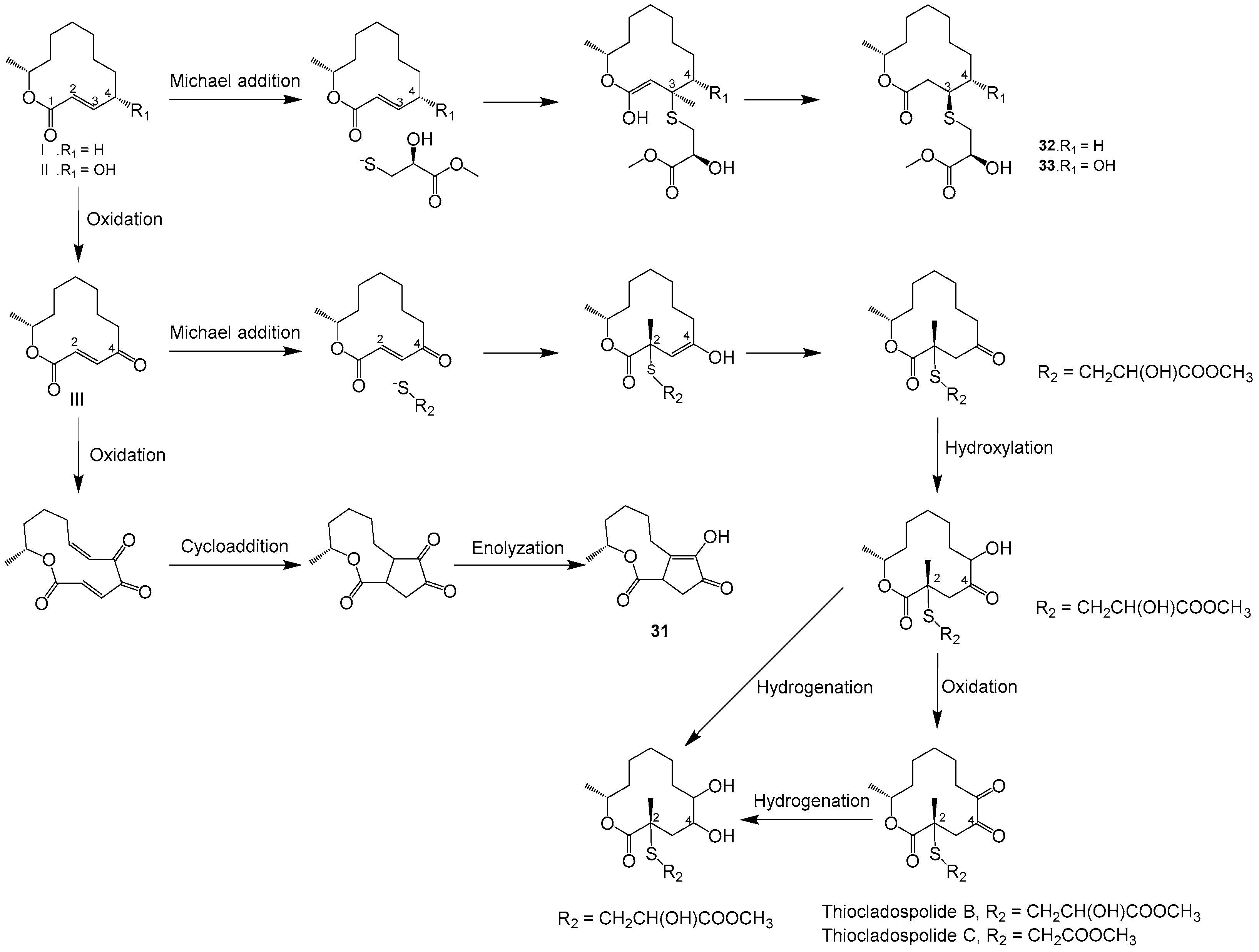
Two new polyketide metabolites, the 12-membered macrolide 4-hydroxy-12-methyloxacyclododecane-2,5,6-trione (25) and 12-methyloxacyclododecane-2,5,6-trione (26) (Figure 1), were isolated from the endophytic fungal Cladosprium colocasiae A801 of the plant Callistemon viminalis. Neither of which exhibited cytotoxic activity, antibacterial activity or α-glucosidase inhibition [39]. One new macrolide compound named thiocladospolide E (27) and a novel macrolide lactam named cladospamide A (28), along with cladospolide B (29) (Figure 1), were isolated from mangrove endophytic fungus Cladosporium sp. SCNU-F0001. None of the three compounds showed antitumor cell proliferation activity or antibacterial activity [40]. Cladospolide B (30) (Figure 1) was isolated from endophytic fungus Cladosporium sp. IS384. Compound 30 exhibited antibacterial activity against Enterococcus faecalis ATCC 29212 with a MIC value of 0.31 μg/mL [41]. Three new macrolides, cladocladosin A (31), thiocladospolide F (32) and thiocladospolide G (33) (Figure 1), were isolated from the marine mangrove-derived endophytic fungus, Cladosporium cladosporioides MA-299 [42]. Compounds 3133 displayed activities against the aquatic pathogenic bacteria Edwardsiella tarda and Vibrio anguillarum with MIC values ranging from 1.0 to 4.0 μg/mL [43]. Moreover, compound 31 demonstrated activity against aquatic pathogenic bacterium Pseudomonas aeruginosa, while compound 32 exhibited activity against plant-pathogenic fungus Helminthosporium maydis, both with MIC values of 4.0 μg/mL. A plausible biosynthetic pathway for compounds 3133 as well as the related congeners such as thiocladospolides A–D and pandangolide 3 was proposed, as shown in Figure 3. Briefly, compounds 32 and 33, thiocladospolides A–D and pandangolide 3 might be derived from the possible precursors dehydroxylated-patulolide C (I), patulolide C (II) or patulolide A (III) through a Michael addition, followed by further oxidation or reduction. The difference was the adding position and attack orientation of the nucleophile (sulfide group) upon the types of the substituent groups at C-4. Generally, when there is no substituent group or the substituent group is only a hydroxyl at C-4, the sulfide group prefers to be added at C-3 to generate compounds 32 and 33, resulting in the sulfide group at C-3 and methyl group at C-11 located on the opposite faces of the molecule. However, when the substituent group at C-4 is a ketone carbonyl at C-4, the adding position of the sulfide group would be at C-2 to yield thiocladospolide A, resulting in the sulfide group at C-2 and methyl group at C-11 located on the same face of the molecule. Adding different sulfide groups such as methyl 2-hydroxy-3-sulfanylpropanoate and methyl 2-sulfanylethanoate to C-2 of the precursor III, would generate thiocladospolides B and C, respectively. Moreover, compound 31 might be derived via oxidation, cyclization and rearrangement from precursor III.
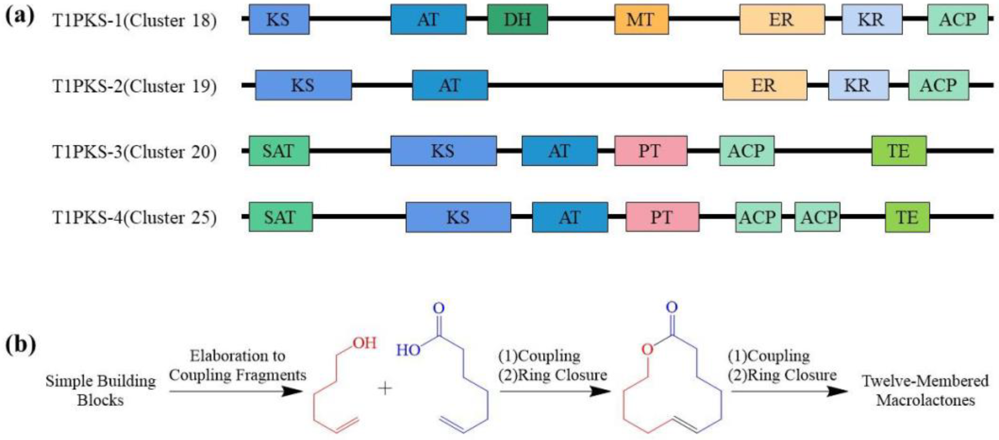
The strategy of diverted total synthesis (DTS) (Figure 4) has gained popularity as a method of obtaining compounds otherwise unavailable from nature or through the manipulation of the parent compounds [44,45]. The 12-membered macrolactone class of compounds is uniquely well-positioned for exploitation via DTS. These compounds typically have modular synthetic routes, where fragments are synthesized convergently, coupled and cyclized [46,47,48,49]. Side-chain decorations can either be carried through as part of a coupling fragment or synthesized concurrently and coupled to the macrocycle at a later stage. The ability to synthesize these molecules in such a way offers several advantages. Firstly, individual reactions can be modulated to alter stereochemistry and substitution, thus providing flexibility in the synthesis process. Secondly, by using different coupling partners to generate the core structure, it becomes possible to create structural libraries of analogs. Finally, late-stage manipulation through oxidation and substitution reactions allows for the alteration of specific functional group moieties.
Brefeldin A (34) (Figure 5) was a new macrolide isolated from the liquid fermentation broth of the cork oak endophytic fungus Cladosporium sp. I(R)9-2. Compound 34 was tested for antifungal activity against Aspergillus niger, Candida albicans and Trichophyton rubrum, demonstrating significant antifungal activities with MIC values of 0.97 μg/mL, 1.9 μg/mL and 1.9 μg/mL, respectively [50]. Brefeldin A has been touted as a promising lead molecule in the world of drug development because of its potent biological activity in the antitumor [51] and antiviral [52] fields. Two macrolide compounds, 5Z-7-oxozeaenol (35) and zeaenol (36) (Figure 5), were isolated from the fermentation broth of the fungus Cladosporium oxysporum DH14, a fungus residing in the gut of the Chinese rice locust Oxya chinensis. Both compounds exhibited potent phytotoxic activities against the radicle growth of Amaranthus retroflexus L. with IC50 values of 4.80 and 8.16 μg/mL, respectively, which are comparable to those of the positive control 2,4-dichlorophenoxyacetic acid (IC50 = 1.95 μg/mL) [53].
The polyketides cladosporin (37), isocladosporin (38), 5′-hydroxyasperentin (39) and cladosporin-8-methyl ether (40) (Figure 5) were isolated from the endophytic fungus Cladosporium cladosporioides [54]. Compounds 39 and 40 were first discovered in C. cladosporioides. Additionally, 5′,6-diacetylcladosporin (41) (Figure 5) was synthesized by acetylating compound 39. These compounds, 3741, were evaluated for their antifungal activities against plant pathogens. Compound 37 inhibited the growth of Colletotrichum acutatum, Colletotrichum fragariae, Colletotrichum gloeosporioides and Phomopsis viticola by 92.7%, 90.1%, 95.4% and 79.9%, respectively, when tested at 30 µM. Compound 38 showed 50.4%, 60.2% and 83.0% growth inhibition against C. fragariae, C. gloeosporioides and P. viticola, respectively, at the same concentration. Liu Huanhuan and colleagues [55] extracted two compounds, 6,8-dihydroxy-3-methyl-1H-isochroman-1-one (42) and 6-methoxy-8-hydroxy-3-methylisocoumarin (43) (Figure 5), from the solid fermentation of Cladosporium cladosporioides. Five isocoumarins (4448), 6,8-dihydroxy-4-(I-hydroxyethyl)-isocoumarin (44), sescandelin B (45), 6-hydroxy-8-methoxy-3-methylisocoumarin (46), 6-hydroxy-8methoxy-3,4-dimethylisocoumarin (47) and aspergillumarin A (48) (Figure 5), were isolated from the culture extract of Cladosporium sp. JS1-2, an endophytic fungus obtained from the mangrove plant Ceriops tagal [56]. Among these, compounds 47 and 48 exhibited inhibitory effects on the growth of cotton bollworm larvae Helicoverpa armigera Hubner with IC50 values of 100 μg/mL.

2.2. Quinone

Five cladosporols, cladosporol (49), cladosporol B (50), cladosporol C (51), cladosporol D (52) and cladosporol E (53) (Figure 6), were isolated from the fermentation broth of Cladosporium tenuissimum, a known hyperparasite of several rust fungi [57]. Compounds 4953 were active in inhibiting the urediniospore germination of the bean rust agent Uromyces appendiculatus. Compound 50 was the most active, since germination was completely inhibited at 50 ppm, significantly reduced to more than 90% at 25 ppm, and still low at 12.5 ppm. Compound 49 was more active than compounds 5153, reaching an inhibition value higher than 80% at the highest concentration. These compounds, by contributing to the reduction of rust survival structures (number and longevity of spores), are expected to play major roles in the multiple aspects of C. tenuissimum biocontrol.
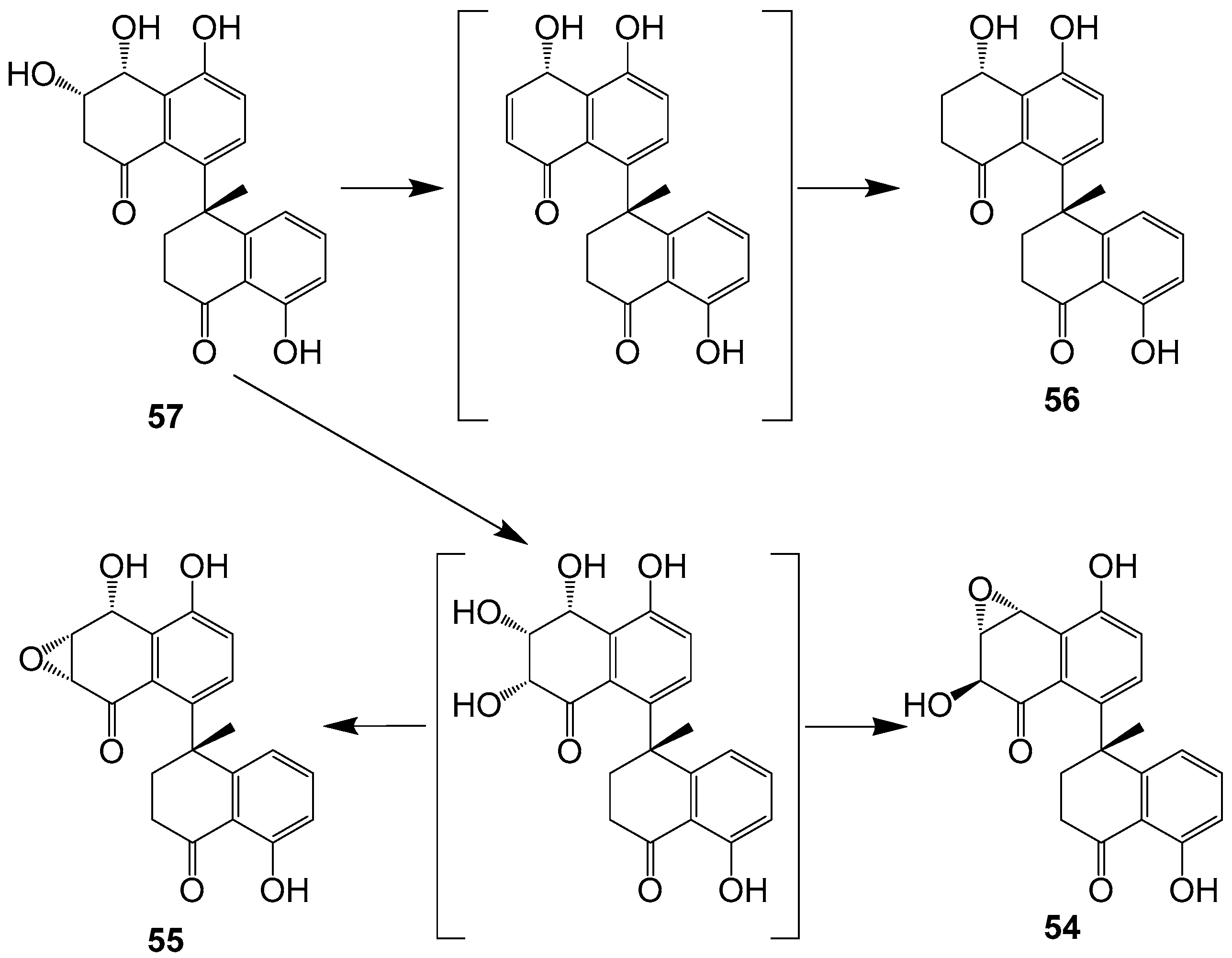
One new dimeric tetralone cladosporone A (54) and three known analogues cladosporones B–D (5557) (Figure 6) were isolated from fungus Cladosporium sp. KcFL6′ derived from mangrove plant Kandelia candel [19]. Cladosporone A (54) inhibits colon cancer cell proliferation by modulating the p21waf1/cip1 expression [62]. None of the compounds showed antifungal activity. From a biosynthetic aspect, compounds 5456 could be generated from hexaketide or pentaketide to form the key monomer tetralone, and then the tetralone coupled to yield the key intermediate 56 (Figure 7).
Seven quinones, cladosporol (58), cladosporols B–D (5961), cladosporol F (62), one new cladosporol derivative, 2-chloro-cladosporol D (63) and one new brominated derivative 2-bromo-cladosporol D (64) (Figure 6), were isolated from the fermentation of the plant-associated fungus Cladosporium sp. TMPU1621 [58]. The chlorinated derivative 63 did not exhibit anti-MRSA activity, whereas the bromine congener 64 inhibited the growth of MRSA ATCC43300 and MRSA ATCC700698 with the same MIC values of 25 µg/mL, suggesting that the presence of a bromine atom affects antimicrobial activity against MRSA. Compound 59 displayed potent anti-MRSA activities against both strains with MIC values of 3.13 and 12.5 μg/mL, respectively, whereas compounds 58 and 60 showed weaker activities. Five new perylenequinone derivatives, altertoxins VIII–XII (6569), as well as one known compound cladosporol I (70) (Figure 6), were isolated from the fermentation broth of the marine-derived fungus Cladosporium sp. KFD33 from a blood cockle from Haikou Bay, China. Compounds 6570 exhibited quorum sensing inhibitory activities against Chromobacterium violaceum CV026 with MIC values of 30, 30, 20, 30, 20 and 30 μg/well, respectively [59].
Altersolanol J (71), altersolanol A (72) and macrosporin (73) (Figure 6) were isolated from the solid-substrate fermentation culture of Cladosporium sp. NRRL 29097 [27]. Compounds 7273 inhibited the growth of B. subtilis, producing zones of inhibition of 33 and 23 mm, respectively, and compounds 7273 inhibited S. aureus, causing inhibition zones of 31 and 20 mm. While the antibacterial activity of 72 and its analogues is well established, no activity appears to have been reported for 73. Two naphthoquinones, anhydrofusarubin (74) and methyl ether of fusarubin (75) (Figure 6), were isolated from Cladosporium sp. RSBE-3. Compounds 74 and 75 showed potential cytotoxicity against human leukemia cells (K-562) with IC50 values of 3.97 μg/mL and 3.58 μg/mL, respectively. Compound 75 (40 μg/disc) showed prominent activities against S. aureus, Escherichia coli, Pseudomonas aeruginosa and Bacillus megaterium with an average zone of inhibition of 27 mm, 25 mm, 24 mm and 22 mm, respectively, and the activities were compared with kanamycin (30 μg/disc) [60]. Compounds 74 and 75 might be useful lead compounds for developing potential cytotoxic and antimicrobial drugs. Anthraquinone (76) (Figure 6) was isolated from the rice medium culture of mangrove-derived fungus Cladosporium sp. HNWSW-1, isolated from the healthy root of Ceriops tagal collected at the Dong Zhai Gang Mangrove Reserve in Hainan. Compound 76 displayed inhibitory activity against α-glycosidase with a IC50 value of 49.3 ± 10.6 μΜ [61].

2.3. Linear Alkane Compounds

One new polyketide, compound 77 (Figure 8), was isolated from the plant endophytic fungus Cladosporium sp. IFB3lp-2. Compound 77 showed no cytotoxicity against human colon cancer cell lines SW1116 and HCT116, breast adenocarcinoma cell line MD-MBA-231, lung adenocarcinoma epithelial cell line A549, hepatocellular carcinoma cell line HepG2 and melanoma cell line A375; no antiviral activity against human enterovirus 71 and Coxsachievirus A16 cell lines; and no acetylcholinesterase inhibition, at the concentration of 20 mM [38]. One new compound, cladospolide E (78), and two known derivatives, secopatulolides A and C (7980) (Figure 8), were isolated from an unidentified soft coral-derived fungus Cladosporium sp. TZP-29. Compounds 7880 were non-cytotoxic, and both showed potent lipid-lowering activities in HepG2 hepatocytes with IC50 values of 12.1, 8.4 and 13.1 μM [25]. Two new polyketides (8182) (Figure 8) were isolated from the mangrove plant Excoecaria agallocha-derived fungus Cladosporium sp. OUCMDZ-302 [20]. Mannitol (83) (Figure 8) was isolated from the endophytic fungi Cladosporium cladosporioides [55].

2.4. Other Classes of Polyketides

Four new polyketide-derived metabolites, cladoacetal A (84), cladoacetal B (85), 3-deoxyisoochracinic acid (86) and (+)-cyclosordariolone (87) (Figure 9), were isolated from the solid-substrate fermentation culture of Cladosporium sp. NRRL 29097. Compound 84 inhibited S. aureus, displaying inhibition circles of 13 mm. In addition, compound 86 inhibited the growth of B. subtilis, producing zones of inhibition of 8 mm [27]. Alternariol (88) and alternariol 5-O-methyl ether (89) (Figure 9) were isolated from endophytic Cladosporium sp. J6 from endangered Chrysosplenium carnosum from Tibet [63]. Compounds 88 and 89 were found to inhibit the photosynthetic electron transport chain in isolated spinach chloroplasts at the same concentrations at which its presence reduced the growth constant of a cyanobacterial (Synechococcus elongatus, strain PCO6301) model. These compounds may represent a novel lead for the development of new active principles targeting photosynthesis [64].
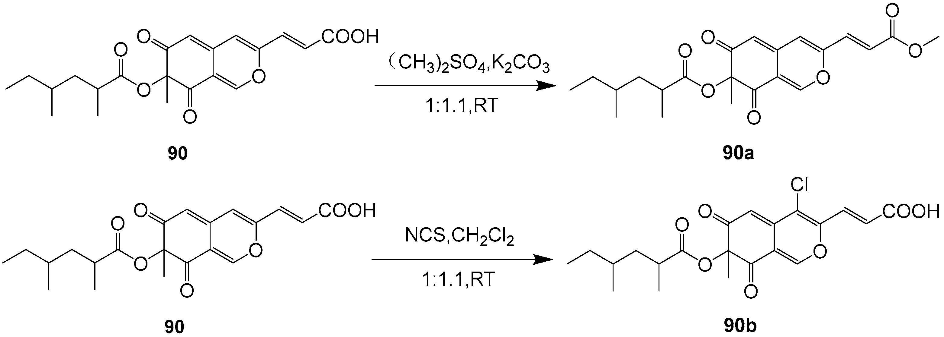
Lunatoic acid A (90) (Figure 9) was isolated from the endophytic fungus Cladosporium oxysporum DH14, a locust-associated fungus. Compound 90 exhibited significantly phytotoxic activity against the radicle growth of Amaranthus retroflexus L. with an IC50 value of 4.51 μg/mL, which is comparable to that of the positive control 2,4-dichlorophenoxyacetic acid (IC50 = 1.95 μg/mL). Furthermore, compound 90 showed selective phytotoxic activity with an inhibition rate of less than 22% against the crops of Brassica rapa L., Sorghum durra, Brassica campestris L., Capsicum annucm and Raphanus sativus L. under a concentration of 100 μg/mL. The synthesis pathways of derivative compounds 90a and 90b on the basis of compound 90 are shown in Figure 10. Both derivatives of compound 90 had moderate phytotoxic activity against the radicle growth of A. retroflexus L with inhibition rates of 53.17 and 56.14%, respectively, comparable to that of 2,4-D (87.09%), co-assayed as a positive control under a concentration of 100 μg mL−1. A comparison of the phytotoxic activities of compounds 90, 90a and 90b may provide useful hints for the understanding of the ability of compound 90 to inhibit the radicle growth of A. retroflexus L. These findings suggest that compound 90 has some potential as a new agent for weed control [53].
From the endophytic fungus Cladosporium cladosporioides JG-12, five compounds with different structural types were identified [65]. Of these compounds, (5S)-5-hydroxy-7-(4″-hydroxy-3″-methoxy-phenyl)-1-phenyl-3-heptanone (91) (Figure 9) showed antibacterial activity against Ralstonia solanacearum and S. aureus. Additionally, this compound exhibited acetylcholinesterase inhibitory activity with an inhibitory rate of 23.54%.
The fungal strains Cladosporium sp. NJF4 and NJF6 were collected from marine sediments in the Gulf of Prydz, and their secondary metabolites were isolated and characterized comprehensively. The results of this study yielded a total of 20 compounds, with two of them, 7-deoxy-7,8-didehydrosydonic acid (92) and sydonic acid (93) (Figure 9), being isolated for the first time from the genus Cladosporium [66]. Compound 93 was found to be weakly cytotoxic against HL-60 human promyelocytic leukemia and A-549 human lung carcinoma cell lines. Compound 93 exhibited significant inhibiting activities to four pathogenic bacteria, Bacillus subtilis, Sarcina lutea, Escherichia coli and Micrococcus tetragenus, and uniquely against two marine bacterial strains Vibrio Parahaemolyticus and Vibrio anguillarum [67]. These findings suggest that the marine environment in the Gulf of Prydz may harbor diverse fungal species with the potential to produce unique secondary metabolites. The identification of new compounds from these strains could have significant implications for drug discovery and development.
One new abscisic acid analogue, cladosacid (94) (Figure 9), was isolated from the marine-derived fungus, Cladosporium sp. OUCMDZ-1635 [68]. Compound 94 did not demonstrate antibacterial activities against Bacillus subtilis CGMCC 1.3376, Clostridium perfringens CGMCC 1.0876, Escherichia coli ATCC 11775, Pseudomonas aeruginosa ATCC10145, S. aureus ATCC 6538 or Candida albicans ATCC 10231, even when tested at concentrations of 100 μg/mL. One prenylated flavanone derivative (95) (Figure 9) was isolated from the culture broth of Indonesian marine sponge-derived Cladosporium sp. TPU1507 [69]. Compound 95 exhibited moderate inhibitory activity against PTP1B with an IC50 value of 11 μM. Therefore, compound 95 shows promise as a potential drug target for treating diseases associated with PTP1B inhibition, such as obesity, diabetes mellitus or cancer.
Three new polyketides (9799) and one known compound (96) (Figure 9) were obtained from the fermentation products of the endophytic fungus Cladosporium sp. OUCMDZ-302, which was derived from the mangrove plant Excoecaria agallocha (Euphorbiaceae) [20]. Notably, compound 99 exhibited significant radical scavenging activity against DPPH with an IC50 value of 2.65 μM, indicating its promising potential as a natural antioxidant agent. Compounds 96 and 97 were postulated to be biosynthesized by the polyketide pathway from acetyl coenzyme A (Figure 11). The pathway involves the condensation, cyclization, dehydration and hydrogenation of acetyl-CoA units. The oxidation and reduction of (S)-12 results in the formation of compound 98. Furthermore, (S)-12 underwent Baeyer–Villiger oxidation, followed by methanolysis and hydrolysis, to yield compound 99. The formations of compounds 75 and 76, on the other hand, are the results of the condensation, reduction, dehydration and decarboxylation of acetyl-CoA units of different lengths.
Four new citrinin derivatives, cladosporins A–D (100103) (Figure 12) were isolated from a culture broth of the deep-sea-derived fungus Cladosporium sp. SCSIO z015. Compounds 100103 showed weak toxicity toward brine shrimp naupalii with IC50 values of 72.0, 81.7, 49.9 and 81.4 μM, respectively. These values were compared to a positive control, toosendanin, with an IC50 value of 21.2 μM. And 103 also showed significant antioxidant activity against DPPH radicals with an IC50 value of 16.4 μM. This promising compound holds potential for further development as a natural antioxidant agent [70].
One benzofuran derivative (105), one isochroman derivative (106) and two other compounds 104 and 107 (Figure 12) were isolated from the culture extract of Cladosporium sp. JS1-2, an endophytic fungus obtained from the mangrove plant Ceriops tagal. Compounds 105107 displayed antibacterial activities against S. aureus with the same MIC values of 12.5 μg/mL; the positive control was ciprofloxacin, with a MIC value of 3.12 μg/mL. Compound 107 displayed growth inhibition activity against newly hatched larvae of Helicoverpa armigera Hubner, with the same IC50 values of 100 μg/mL; the positive control was azadirachtin, with an IC50 value of 50 μg/mL [56].
7,4′-dihydroxyisoflavone (108) (Figure 12) was isolated from the Antarctic fungus Cladosporium sp. NJF6 [66]. Compound 108 displayed inhibitory activities on inflammatory markers like NF-kB, TGF-β, TNF-α, IL-6, IL-8 and COX-2. It also had an effect on various apoptotic markers like caspase-3, caspase-8, Bcl-2 and Bax, which indicates that compound 108 may have significant effects on breast cancer, cardiovascular diseases, neurodegenerative diseases, diabetes and its complications, osteoporosis, eczema and skin inflammation [72].
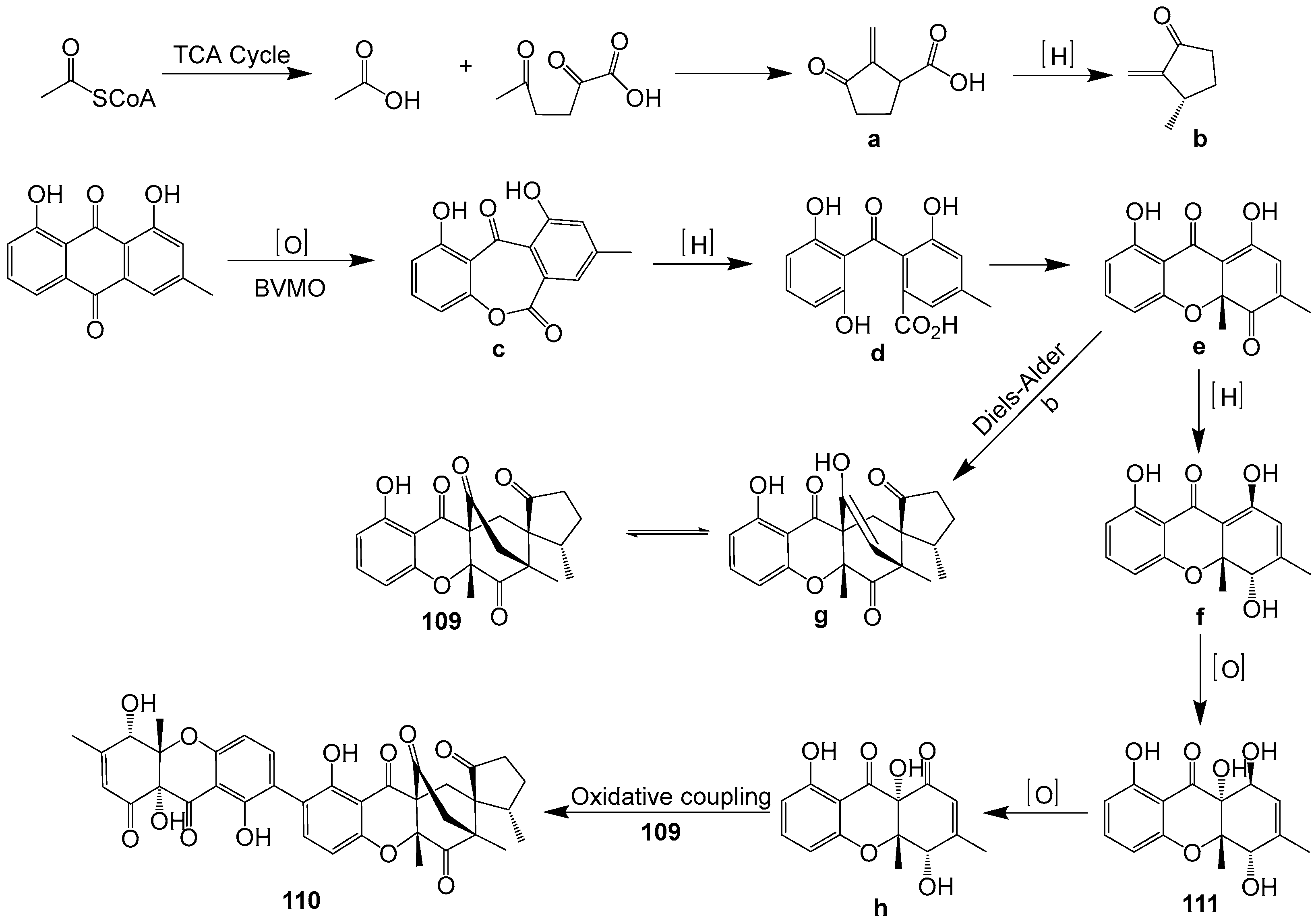
Two novel xanthone-derived metabolites, cladoxanthones A (109) and B (110), along with one known mangrovamide J (111) (Figure 12) were discovered in Cladosporium sp. QH07-10-13 [71]. The new metabolites 109 and 110 exhibited an intriguing spiro [cyclopentane-1,2′-[3,9a] ethanoxanthene]-2,4′,9′,11′(4a′H)-tetraone framework. Compound 109 could be produced from α-methylene ketone and dihydro-xanthone, through a Diels–Alder reaction. Compound 110 could result from the oxidative coupling products of compounds 109 and 111 (Figure 13). Compounds 109111 demonstrated low cytotoxic activities.
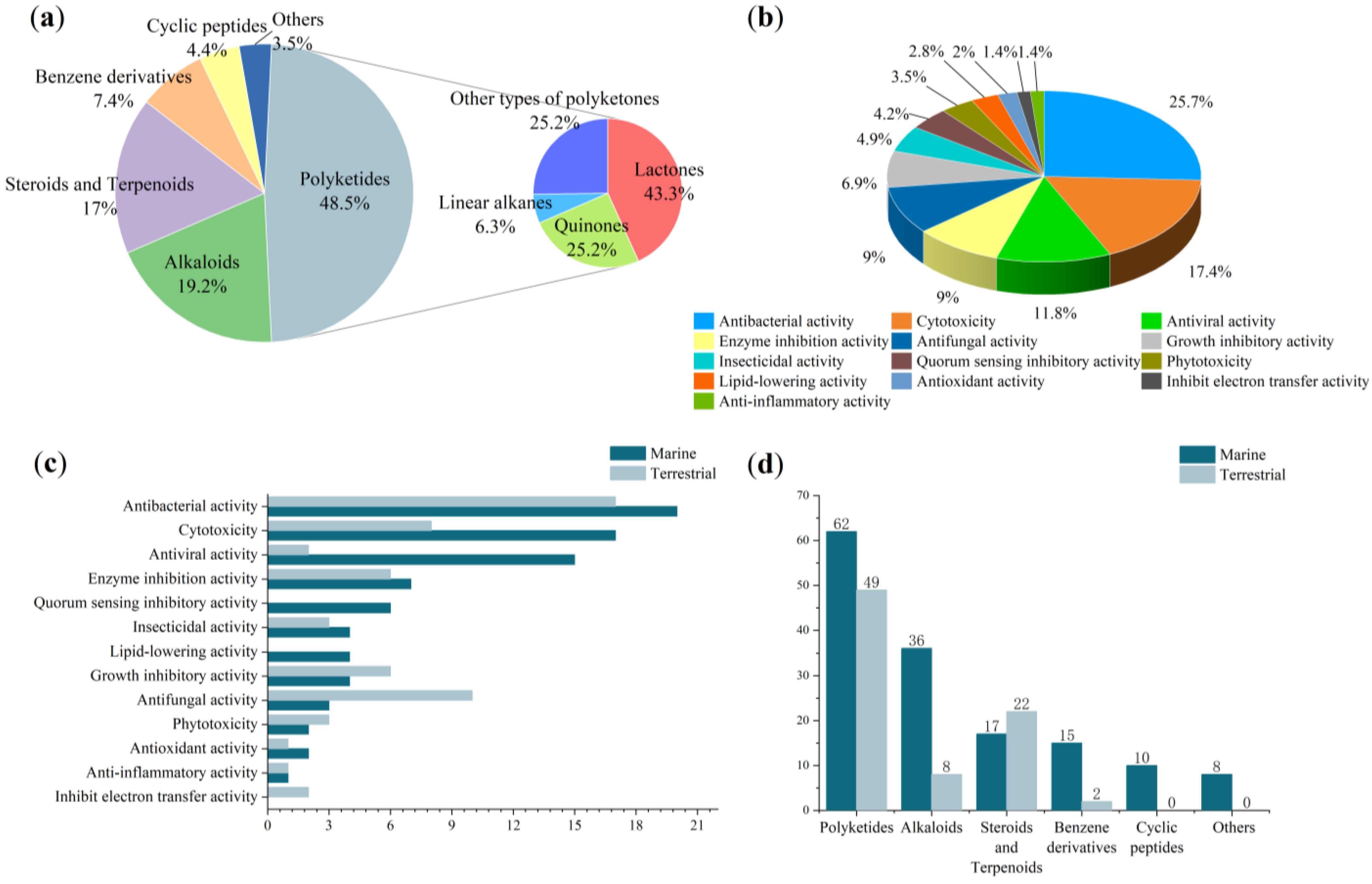
There are a total of 111 polyketides that have been isolated from Cladosporium. These polyketides are classified as lactones (43.24%), quinones (25.23%), linear alkanes (6.31%) and others (25.23%). Out of the total isolated polyketides, 62 originated from marine sources, while 39 compounds are newly identified compounds. Hence, marine sources make up 66.7% of the newly discovered substances. Additionally, there are 66 bioactive compounds in total, with 36 of them coming from the ocean, accounting for 54.5% of the bioactive substances. Polyketides are significant secondary metabolites of Cladosporium, with some natural products exhibiting biological activities concentrating on cytotoxicity, antibacterial activity and antifungal activity.

3. Alkaloids

N-β-acetyltryptamine (112) (Figure 14), was isolated from the endophytic fungus Cladosporium sp. N5 associated with red alga Porphyra yezoensis. No cytotoxicity was found in the brine shrimp lethality test, which indicated that the crude extract of Cladosporium sp. has no toxicity to the aquatic ecosystem. Thus, Cladosporium sp. can be applied as a biocontrol agent [33]. Six new indole alkaloids including five new glyantrypine derivatives, 3-hydroxyglyantrypine (113), 14R-2-oxoglyantrypine (114), 14S-2-oxoglyantrypine (115), cladoquinazoline (116) and epi-cladoquinazoline (117), and one new pyrazinoquinazoline derivative, norquinadoline A (118), together with eight known alkaloids, quinadoline A (119), deoxynortryptoquivaline (120), deoxytryptoquivaline (121), tryptoquivaline (122), CS-C (123), quinadoline B (124), prelapatin B (125) and glyantrypine (126) (Figure 14), were isolated from the culture of the mangrove-derived fungus Cladosporium sp. PJX-41. Anti-H1N1 activities were measured for these compounds, with compounds 115, 118, 120122 and 124 showing noteworthy antiviral activities with IC50 values of 85, 82, 87, 85, 89 and 82 μM, comparable to that of the positive control ribavirin (IC50 = 87 μM). However, the other alkaloids, 113, 114, 116, 117, 119, 123 and 126, showed weakly antiviral activities (IC50 values ranging between 100 and 150 μM), and compound 125 displayed no activity (IC50 > 200 μM) [26].
Ma Yanhong’s group [63] isolated β-carboline (127) and uracil nucleoside (128) (Figure 14) from the fermentation of endophytic Cladosporium sp. J6 from endangered Chrysosplenium carnosum from Tibet. Compound 127 displayed a wide range of biological activities including antitumor [73], antiviral [74] and antimicrobial activity [75]. Three sulfur-containing compounds, cladosporin A (129), cladosporin B (130) and haematocin (131) (Figure 14), were isolated from the marine fungus Cladosporium sp. The compounds belong to the class of cyclic di-acid alkaloids, with 129 and 130 being new members. Compounds 129131 displayed moderate cytotoxic activities against the HepG2 cell line, with IC50 values of 21, 42 and 48 μg/mL, respectively [76]. Uracil (132) (Figure 14) was isolated from the fermentation of endophytic Cladosporium sp. J6 from endangered Chrysosplenium carnosum from Tibet [63]. Cladosporilactam A (133) (Figure 14), one new bicyclic lactam, was isolated from the fungus Cladosporium sp. RA07-1, which originated from the gorgonian Anthogorgia ochracea (GXWZ-07) derived from the South China Sea [35]. Compound 133 was the first example of a 7-oxabicyclic [6.3.0] lactam obtained from a natural source. Research indicates that compound 133 exhibits potent cytotoxicity against a series of cancer cell lines, including cervical cancer HeLa, mouse lymphocytic leukemia P388, human colon adenocarcinoma HT-29 and human lung carcinoma A549 with IC50 values of 0.76, 1.35, 2.48 and 3.11 μM, respectively, which suggests that it might have potential to be developed as an antitumor agent. Three alkaloids, ilicicolin H (134), (7R)-methoxypurpuride (135) and (5aS,9S,9aS)-1,3,4,5,5a,6,7,8,9,9,9a-decahydro-6,6,9a-trimethyl-3-oxonaphtho [1,2-c] furan-9-yl N-acetyl-L-valinate (136) (Figure 14), were isolated from the endophytic fungus Cladosporium cladosporioides JG-12. Compounds 135 and 136 showed inhibitory activities against Panagrellus redivivus, and compounds 134136 exhibited acetylcholinesterase inhibitory activities [65]. 2′-deoxythymidine (137) and 3-carboxylic acid (138) (Figure 14) were isolated from the extracts of the culture of sponge Callyspongia sp. derived fungus Cladosporium sp. SCSIO41007 [28].
Figure 14. Structures of compounds 112138 [26,28,33,35,63,65,76].
Ijms 25 01652 g014
One new alkaloid with a 3-(2H-pyran-2-ylidene) pyrrolidine-2,4-dione nucleus, cladodionen (139) (Figure 15), was isolated from the marine-derived fungus Cladosporium sp. OUCMDZ-1635. Cladodionen (139) showed cytotoxic activities against MCF-7, HeLa, HCT-116 and HL-60 human cancer cell lines with IC50 values of 18.7, 19.1, 17.9 and 9.1 μM [68]. Cladosporamide A (140) (Figure 15), one new protein tyrosine phosphatase (PTP) 1B inhibitor, was isolated from the culture broth of an unidentified Indonesian marine sponge-derived Cladosporium sp. TPU1507. Compound 140 modestly inhibited PTP1B and T-cell PTP (TCPTP) activities with IC50 values of 48 and 54 μM, respectively [69]. One alkaloid adenine nucleoside (141) (Figure 15) was isolated from the solid fermentation of Cladosporium cladosporioides [55]. Two new succinimide-containing derivatives, cladosporitin A (142) and B (143), along with the previously reported talaroconvolutin A (144) (Figure 15), were isolated from the fermentation culture of the tree (Ceriops tagal) root-derived fungus Cladosporium sp. HNWSW-1. Compound 143 exhibited cytotoxicity against BEL-7042, K562 and SGC-7901 cell lines, with IC50 values of 29.4 ± 0.35, 25.6 ± 0.47 and 41.7 ± 0.71 μM, respectively. Compound 144 showed cytotoxicity against Hela and BEL-7042 cell lines with IC50 values of 14.9 ± 0.21 and 26.7 ± 1.1 μM, respectively. Moreover, compound 144 was found to display enzyme inhibitory activity against α-glycosidase, with an IC50 value of 78.2 ± 2.1 μM [61]. 3-indoleacetic acid (145) and 3-formylindole (146) (Figure 15) were isolated from the fungal strains Cladosporium sp. NJF6 derived from marine sediments in the Gulf of Priz, Antarctica [66]. Compound 146 caused inhibition on the growth of Trypanosoma cruzi, with an IC50 value of 26.9 μM, with moderate cytotoxicity against Vero cells. And compound 146 was found to be inactive when tested against Plasmodium falciparum and Leishmania donovani, therefore showing selectivity against T. cruzi parasites [77]. Eight new tetramic acid derivatives, cladosporiumins A–H (147154) (Figure 15), were isolated from a culture broth of Cladosporium sp. SCSIO z0025 derived from deep-sea sediment collected from Okinawa Trough. Despite their novelty, assays for antitumor cytotoxic, antibacterial and antiacetylcholinesterase activities demonstrated that these compounds exhibited negligible inhibitory effects [78]. One indole diterpenoid alkaloid, cladosporine A (155) (Figure 15), has been identified and isolated from the fungus Cladosporium sp. JNU17DTH12-9-01 [79], marking the first discovery of such an alkaloid within the genus Cladosporium. This new alkaloid has demonstrated antimicrobial activities against S. aureus 209P with a MIC of 4 μg/mL, and a MIC of 16 μg/mL against Candida albicans FIM709.
A total of 44 alkaloids have been identified from the genus Cladosporium. The structural types of these alkaloids include amines, indoles, pyrrolizidines and quinazolines. Out of the total isolated alkaloids, 36 originated from marine sources. And 21 compounds are newly identified compounds, with marine sources accounting for 66.7% of these newly discovered substances. Furthermore, all of this new material comes from the ocean. In total, there are 27 bioactive compounds, with 22 of them derived from marine sources, making up 81.5% of the bioactive substances. These alkaloids exhibit biological activities, focusing on antiviral, cytotoxicity and enzyme inhibition activities.

4. Steroids and Terpenoids

One new pentanorlanostane derivative, cladosporide A (156), along with 23,24,25,26,27-pentanorlanost-8-ene-3β,22-diol (157), 18,22-cyclosterols (158), tetranorditerpenoids (159), ketodialdehyde derivative (160), ketoaldehyde carboxylic acid (161), lanosterol (162), hexanorlanosterols (163) and (164), pentanorlanostanecarboxylic acid derivatives, 3β-hy-droxy-4,4,14α-trimethyl-5α-pregna-7,9(11)-diene-20S-car-boxylic acid (165), 3β,12β-dihydroxy-4,4,14α-trimethyl-5α-pregna-7,9(11)-diene-20S-carboxylic acid (166) and 4,4,14α-trimethyl-3-oxo-5α-pregna-7,9(11)-diene-20S-car-boxylic acid (167) (Figure 16) were isolated from Cladosporium sp. IFM 49189 [13]. Cladosporide A (156) showed the characteristic inhibition against a human pathogenic filamentous fungus, Aspergillus. fumigatus, at 6.25 μg/disc, whereas no inhibition was observed against a pathogenic filamentous fungus, A. niger, or pathogenic yeasts, C. albicans and C. neoformans. Compound 156 did not completely inhibit the growth of A. fumigatus, but 156 apparently reduced the growth of this fungus. Thus, compound 156 has been identified as a characteristic antifungal agent against the human pathogenic filamentous fungus A. fumigatus. Hosoe et al. [80] extended their research to isolate an additional set of several triterpenoids, including cladosporide A (156), new pentanorlanostane derivatives, cladosporides B–D (168170), 23,24,25,26,27-pentanorlanost-8-ene-3β,22-diol (171), dihydrocladosporide A (172) and 3,30-dioxo-23,24,25,26,27-pentanorlanost-8-en-22-0ic acid (173) (Figure 16) from Cladosporium sp. IFM 49189. Compounds 156 and 168 strongly inhibited the growth of A. fumigatus (1.5 and 3.0 ug/disc, respectively), whereas compounds 169 and 170 showed no antifungal activity. Compounds 172 and 173 retained weak inhibitory activity against A. fumigatus. Zou et al. [81] identified three steroids, ergosta-5,7,22-triene-3β-ol (174), eburicol (175) and β-sitosterol (176) (Figure 16), from Cladosporium cladosporioides, an endophytic fungus derived from an unidentified mangrove. One steroidal compound, ergosterol peroxide (3β-hydroxy-5,8-epidioxy-ergosta-6,22-diene) (177) (Figure 16), was isolated from the endophytic fungus Cladosporium cladosporioides JG-12 derived from Ceriops tagal [65]. Compound 177 exhibited potent anti-inflammatory activity [82]. It has been reported that ergosterol peroxide has the potential for inhibiting cancer cell proliferation as well as inducing apoptosis in cancer cells [83,84,85].
Three new highly oxygenated sterols, cladosporisteroids A–C (178180) (Figure 17), together with three known compounds, pregn-7-dien-3,6,20-trione (181), 3β,5α,9α-trihydroxy-(22E,24R) ergosta-7,22-diene-6-one (182) and cerevisterol (183) (Figure 17), were isolated from the extracts of the culture of the sponge Callyspongia sp. derived fungus Cladosporium sp. SCSIO41007 [28]. Antiviral and cytotoxic activities of compounds 178183 were tested against H3N2 and EV71 viruses, as well as the K562, MCF-7 and SGC7901 cancer cell lines. None of the tested compounds showed any cytotoxic effects on the cancer cell lines. However, compound 178 exhibited weak inhibitory activity against H3N2 with an IC50 value of 16.2 μM, while the IC50 value for the positive control oseltamivir was 34.0 μM.
Cladosporium sp. WZ-2008-0042, a fungus obtained from a gorgonian Dichotella gemmacea collected from the South China Sea, produced one new pregnane, 3α-hydroxy-7-ene-6,20-dione (184), along with five known steroids, including 5α,8α-epidioxy-ergosta-6,9,22E-triene3β-ol (185), 5α,8α-epidioxy-ergosta-6,22E-dien-3β-ol (186), ergosta-7,22E-diene-3β,5α,6β-triol (187), 3β,5α-dihydroxy-6β-methoxyergosta7,22-diene (188) and ergosterol (189), and one known steroidal glycoside (190) (Figure 17) [21]. Compounds 184186 and 188 exhibited antiviral activities against respiratory syncytial virus (RSV). Specifically, compound 184 exhibited potential antiviral activity against RSV, with an IC50 value of 0.12 mM. Additionally, compound 189 demonstrated moderate antibacterial activity against Shigella dysenteriae, with a MIC value of 3.13 μM. This discovery presents a promising avenue for further research on the antiviral and antibacterial properties of these compounds, as well as their potential therapeutic applications. Ma Chuan et al. [41] obtained ergosterol (191) (Figure 17) from the fermentation broth of an endophytic fungus Cladosporium sp. IS384. Compound 191 showed weak cytotoxic and antioxidant activities [86]. The dose effect relationship of ergosterol to nerve cell SH-SY5Y neurotoxicity was not significant at 6.25–25 μg/mL, but it had better effect on the oxidative damage protection of nerve cell SH-SY5Y at 6.25 μg/mL. And ergosterol can inhibit the activity of apoptosis protein caspase 3 to achieve the antioxidative protection effect of nerve cell SHSY5Y [87]. Additionally, two steroids, myristate-4-en-3-one (192) and 3β-hydroxy-5α,8α-peroxidized ergot-6,22-diene (193) (Figure 17), were isolated from the endophytic fungi Cladosporium cladosporioides [55]. The fungus Cladosporium sp. NJF4 was found to produce 5,22-diene-ergosta-3β,7β,8β-triol (194) (Figure 17) [66]. Compound 194 was found to induce cytotoxicity with a MIC value of 14.1 μM in HeLa cells in vitro [88].
A total of 39 steroids and terpenoids have been reported in the genus Cladosporium, with steroids being more common and having keratosteroid skeletons, while terpenoids are more commonly tetracyclic triterpenoids with lanolin steroid skeletons. Out of the total isolated steroids and terpenoids, 17 are derived from marine sources. Additionally, 11 compounds are newly identified, with marine sources accounting for 36.4% of these newly discovered substances. These steroids and terpenoids exhibit various biological activities, including antifungal, antiviral, cytotoxicity, and enzyme inhibition activities. In total, there are 10 bioactive compounds, with marine sources contributing to 40% of these bioactive substances.

5. Benzene Derivatives

One new polyketide-derived metabolite, 3-(2-formyl-3-hydroxyphenyl)-propionic acid (195) (Figure 18), was isolated from solid-substrate fermentation cultures of Cladosporium sp. NRRL 29097. Compound 195 showed growth inhibition activity against Bacillus subtilis with an inhibition circle of 22 mm [27]. Seven benzene derivatives, L-β-phenyllactic acid (196), α-resorcylic acid (197), p-hydroxy benzoic acid methyl ester (198), phenyllactic acid (199), 4-hydroxyphenyl alcohol (200), p-hydroxyphenylacetic acid (201) and p-hydroxybenzyl alcohol (202) (Figure 18), were derived from the endophytic fungus Cladosporium sp. N5 associated with red alga Porphyra yezoensis. It is significant to note that none of the identified compounds show any toxic effects on brine shrimps, which indicates that the environment-friendly Cladosporium sp. could be used as a potential biocontrol agent to protect the alga from pathogens [33]. Compound 199 was reported to be active against various bacteria and fungi [89,90], and compound 196 was reported to be a strong fungicide [91]. They may play important roles in developing the symbiotic relationship between plants and microbes. Compounds 199 and 201 were reported to inhibit the growth of red alga Porphyra tenera conchocelis [92]. Compound 203 (Figure 18), 2-chloro-3,5-dimethoxybenzyl alcohol, was isolated from the endophytic fungus Cladosporium cladosporioides JG-12. It displayed significant inhibitory effects against Candida albicans, and also showed inhibitory activities against Panagrellus redivivus and acetylcholinesterase. This finding suggests that compound 203 may have potential therapeutic applications for the treatment of fungal infections, such as Staphylococcus aureus, Canidia albicans, Ralstonia solanacearum and nematode Panagrellus redivivus infestations, and neurodegenerative diseases [65]. Citrinin H2 (204) and N-(4-hydroxy-2-methoxyphenyl) acetamide (205) (Figure 18) were isolated from the culture extract of Cladosporium sp. JS1-2, an endophytic fungus obtained from the mangrove plant Ceriops tagal. Compound 204 displayed antibacterial activity against S. aureus with the same MIC values of 12.5 μg/mL. Compounds 204 and 205 displayed growth inhibition activities against newly hatched larvae of Helicoverpa armigera Hubner, with the same IC50 values of 100 μg/mL, and the positive control was azadirachtin, with a IC50 value of 50 μg/mL [56]. The Antarctic fungus Cladosporium sp. derived NJF6 and NJF4 were found to produce six benzene derivative analogues, N-acetyl phenethylamine (206), phenethylamine (207), and p-hydroxyphenethylamine (208), p-hydroxyphenyl propionic acid (209), 1,2-benzenedicarboxylic acid (210) and benzoic acid (211) (Figure 18) [66].
A total of 17 benzene derivatives were identified from the ferments of Cladosporium sp., with 15 of these compounds originating from marine sources. Among the total isolated benzene derivatives, there are a total of eight bioactive compounds, with seven of them derived from oceanic sources. Notably, the oceanic compounds account for 87.5% of the bioactive substances. These benzene derivatives exhibit biological activities such as antibacterial, phytotoxicity and insecticidal activities.

6. Cyclic Peptides

Cyclo (Trp-Pro) (212) and cyclo (Trp-Val) (213) (Figure 19) were derived from the endophytic fungus Cladosporium sp. N5 associated with red alga Porphyra yezoensis [33]. Cyclo (Gly-Leu) (214) (Figure 19) was isolated from the extracts of the culture of the sponge Callyspongia sp. derived fungus Cladosporium sp. SCSIO41007 [28]. An antimicrobial study indicated that compound 214 has therapeutic potential as an antibacterial and antifungal agent. In an anticancer study, cyclo (Gly-Leu) exhibited moderate activities in inhibiting various cancer cell lines including HT-29, MCF-7 and HeLa cells [93]. Cyclic (phenylalanine-aspartic acid) (215), cyclic (tryptophan-aspartic acid) (216), cyclic (tryptophan-aspartic acid) (217), 4-hydroxyphenylalanine-leucine (218), cyclic (proline-tyrosine) (219), cyclic (proline-tyrosine) (220) and cyclic (valine-proline) (221) (Figure 19) were isolated from the fungal Cladosporium sp. NJF4 and NJF6, derived from marine sediments in the Gulf of Priz, Antarctica [66].
Ten cyclic peptides were identified from the ferments of Cladosporium sp. These compounds originate from the ocean. However, only compound 214 demonstrates growth inhibitory activity, while the biological activities of the remaining compounds require further investigation.

7. Others

Nicotinic acid (222) and acetyl-tyramine (223) (Figure 20) were obtained from the endophytic fungus Cladosporium sp. N5 associated with red alga Porphyra yezoensis [33]. 1,8-dimethoxynaphthalene (224) (Figure 20) was isolated from the culture extract of Cladosporium sp. JS1-2, an endophytic fungus obtained from the mangrove plant Ceriops tagal. Compound 224 displayed antibacterial activity against S. aureus with a MIC value of 12.5 μg/mL; the positive control was ciprofloxacin, with a MIC value of 3.12 μg/mL. It also displayed growth inhibition activity with an IC50 value of 100 μg/mL against newly hatched larvae of Helicoverpa armigera Hubner; the positive control was azadirachtin, with an IC50 value of 50 μg/mL [56,94]. Four new cyclohexene derivatives, cladoscyclitols A–D (225228), and one new ribofuranose phenol derivative, 4-O-α-D-ribofuranose-2-pentyl-3-phemethylol (229) (Figure 20), were obtained from the mangrove-derived endophytic fungus Cladosporium sp. JJM22. Compounds 226 and 229 displayed potent inhibitory activities against α-glucosidase with IC50 values of 2.95 and 2.05 μM, respectively [23].
Eight bioactive compounds with antibacterial, insecticidal, and enzyme inhibitory activities were discovered in the ferments of Cladosporium sp., all originating from the ocean. This further highlights the significance of the ocean as an invaluable resource.

8. Conclusions

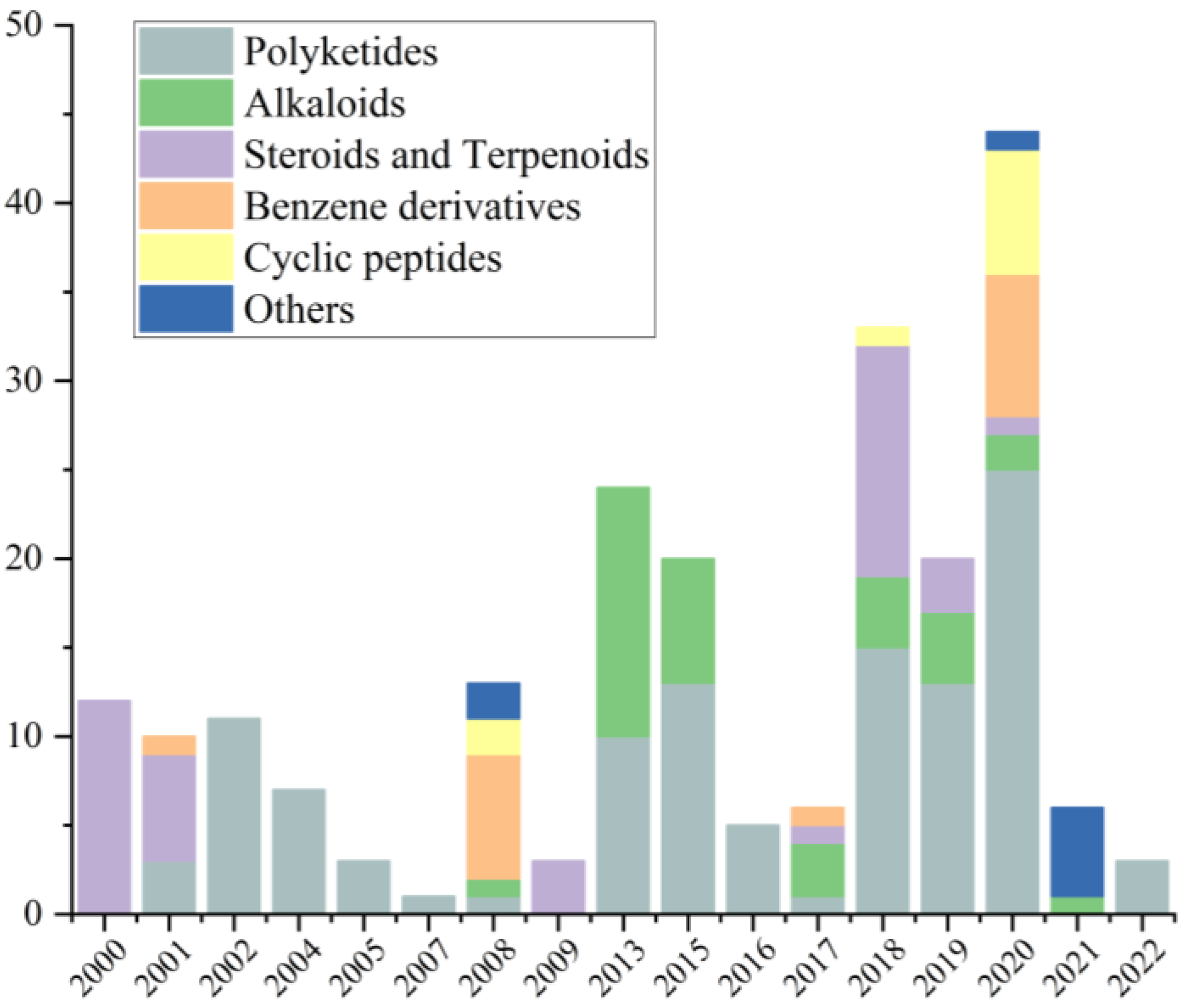
From January 2000 to December 2022, a total of 229 natural products were isolated from the genus Cladosporium (Table 1), of which 64.63% of the compounds were isolated from the ocean and 34% of the compounds were found for the first time. These findings strongly suggest that marine-derived Cladosporium has great potential to produce abundant compounds with new structures. Before 2007, there were few studies on the secondary metabolites produced by Cladosporium, and their structural types were mainly concentrated on terpenoids and polyketides. After 2008, studies on the natural products isolated from Cladosporium gradually increased, with the number of compounds isolated each year on overall upward trend, and the structural types of the isolated compounds were gradually diversified, including cyclic peptides, alkaloids and some other types of compounds, indicating that the genus Cladosporium has the potential to produce compounds of multiple structural types (Figure 21). The structures of the isolated compounds with diverse skeletons are mainly concentrated in the classes of polyketides, alkaloids, steroids, terpenoids, benzene derivatives and cyclic peptides (Figure 22). Polyketides, which make up 48% of the natural products derived from this genus, are notably significant among these compounds.
The sources of Cladosporium are distributed in different ecosystems, including the Antarctic, forests and oceans. About 65% of the isolated natural products were separated from marine organism-derived Cladosporium, including sponge, mangrove and gorgonian. The first marine-derived natural product was isolated from an unidentified sponge-derived Cladosporium in 2001 [22]. A larger number of compounds, excluding steroids and terpenoids, have been isolated from Cladosporium in the ocean compared to those obtained from land (Figure 22), which indicates that marine fungi Cladosporium have great potential to produce compounds.
The genus Cladosporium has the potential to produce a great diversity of bioactive secondary metabolites, including antibacterial, cytotoxic, growth inhibitory, enzyme inhibitory, antifungal activity, and quorum sensing inhibitory activity (Figure 22, Table 2, Table 3 and Table 4). The bioactive compounds isolated from the genus Cladosporium mainly focus on antibacterial activity (26%), cytotoxicity (16%) and antiviral activity (12%), indicating considerable potential for the development of new antibiotics and anticancer compounds from Cladosporium. In addition, many compounds with diverse bioactivities, especially cytotoxic, antibacterial, antiviral and quorum sensing inhibitory compounds are predominantly found in the ocean (Figure 22). These findings emphasize the ocean as a valuable resource and propose that the marine genus Cladosporium has the capacity to generate numerous secondary metabolites with various bioactivities.
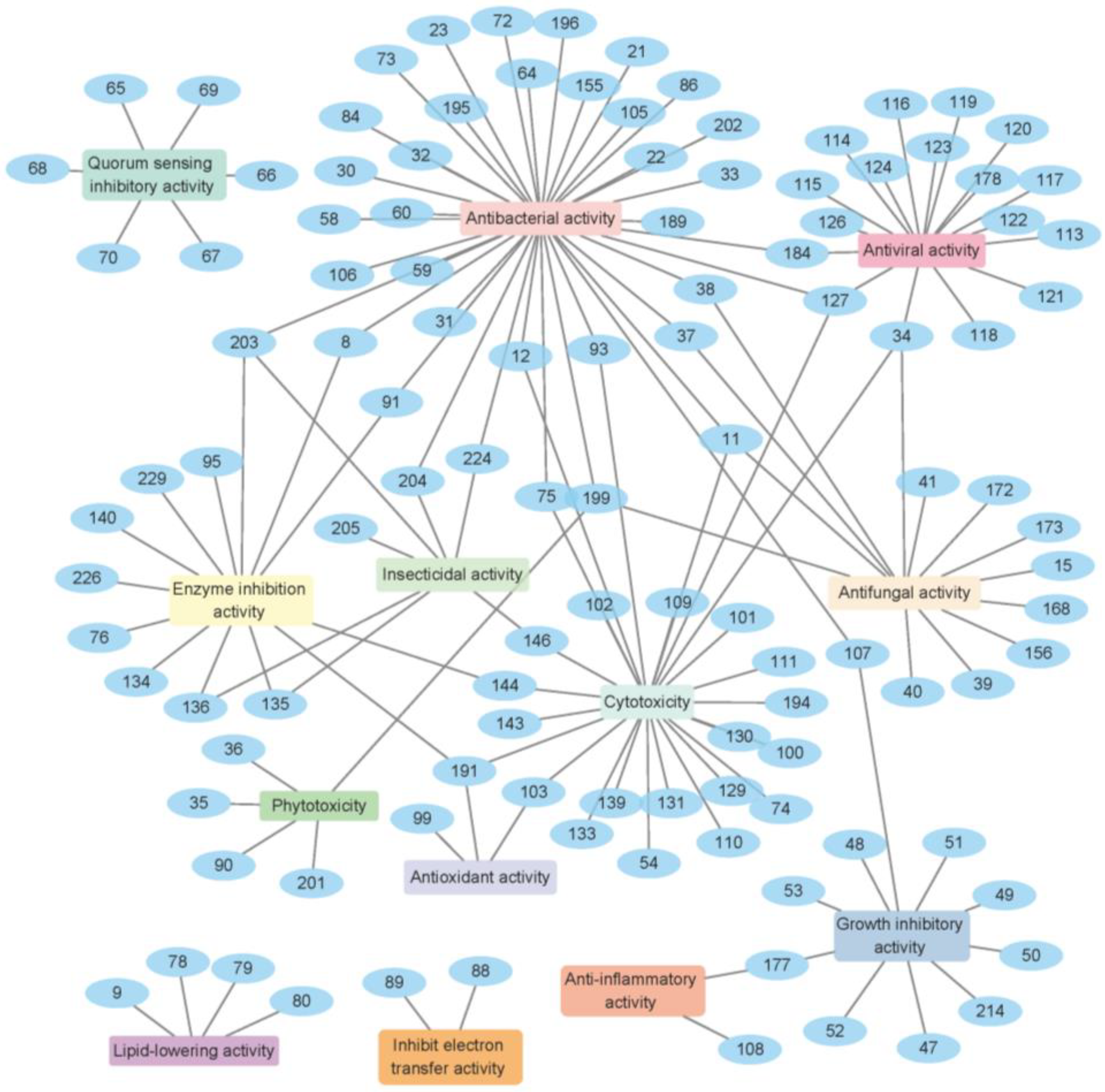
The genus Cladosporium is capable of producing various secondary metabolites with diverse bioactivities, including antibacterial activity, cytotoxicity, antifungal activity, enzyme inhibition activity, antiviral activity, quorum sensing inhibitory activity and antioxidant activity (Figure 22). Research shows that 63% of the natural products derived from Cladosporium exhibit bioactive activities. Among these compounds, 11, 34, 127, 191, 199 and 203 have demonstrated more than three types of activities (Figure 22, Table 2, Table 3 and Table 4), highlighting the potential of this genus to produce bioactive natural products. Additionally, it is noteworthy that 59% of these active compounds are isolated from marine-derived fungi, further supporting the development prospects of marine fungi.
Structure activity relationships (SARs) can be used to predict biological activity from molecular structure. Wang et al. [54] reported an evaluation of the relationships between structure and bioactivity for cladosporin (37) and its analogues (3841). After an overall evaluation of the relationship between the structures and antifungal activity of the compounds at 30 μM, several essential positions were identified as potential determinants of their antifungal activity. The absolute configuration of C-6′ in the structures of compounds 37 and 38 was found to have a significant impact on the antifungal activity of the parent compound. Specifically, the R configuration of C-6′ in structure 38 led to a marked decrease in antifungal activity against Colletotrichum species, while slightly increasing the antifungal activity against Phomopsis species. Comparing the structures of compounds 37 and 39 revealed that he introduction of a hydroxyl group at the C-5′ position results in a complete loss of antifungal activity against Colletotrichum species and decreased selectivity against Phomopsis species, highlighting the importance of maintaining an unsubstituted C-5′ for antifungal activity. Furthermore, by comparing the structures of compounds 37 and 40, it was observed that the replacement of the hydroxyl group with a methoxy group at C-8 caused a broad loss of antifungal activity against all tested fungi, suggesting that this position might be the active site where hydrogen bonds are formed. Additionally, when compounds 39 and 41 were compared, the replacement of the hydrogen of the hydroxyl group at C-6 and the hydrogen at C-5′ with acetyl groups greatly increased the selectivity toward the two Phomopsis species. Therefore, the differences in activity indicated that the S configuration of C-6′, the openness of C-5′, the presence of a hydroxyl group at C-8 and the introduction of functional groups at C-6 influence the antifungal properties of these compounds [54].
Compounds 9, 78, 79 and 80 exhibit potent lipid-lowering activities in HepG2 hepatocytes (Figure 23, Table 4), suggesting that they can be developed into hypoglycemic agents (Figure 23). Compound 34 displays significant antifungal activity (Figure 23, Table 4), declaring the potential of 34 to be applied in agricultural fungicide. Compounds 35, 36 and 90 demonstrate potent phytotoxic activities against the radicle growth of Amaranthus retroflexus L (Figure 23, Table 4). This indicates the potential for developing compounds 35, 36 and 90 as new herbicides. Compound 59 demonstrates a stronger antibacterial activity against MRSA than the positive control (Figure 23, Table 2), highlighting the challenge of bacterial drug resistance. Compounds 75, 93, 156 and 168 display potent antibacterial activities compared to the positive control (Figure 23, Table 2), which means they could be valuable starting points for the development of new antibiotics. Compounds 115, 118, 120122 and 124 show noteworthy antiviral activities (Figure 23, Table 4), which support their potential use as antibiotics. Compound 133 exhibits potent cytotoxicity against a series of cancer cell lines (Figure 23, Table 4), including cervical cancer HeLa, mouse lymphocytic leukemia P388, human colon adenocarcinoma HT-29 and human lung carcinoma A549 with IC50 values of 0.76, 1.35, 2.48 and 3.11 μM, respectively, which suggests that it might have potential to be developed as an antitumor agent. Compound 177 exhibits potent anti-inflammatory activity (Figure 23, Table 4), declaring the potential of 177 to be applied in adjuvant drugs for anticancer therapy. Compound 184 displays potential antiviral activity against RSV (Figure 23, Table 4), which demonstrates that 184 could be employed in developing vaccines and antiviral drugs. Compound 196 exhibits noteworthy antibacterial activity (Figure 23, Table 2), making it a promising candidate for developing a strong fungicide. Compound 203 displays significant inhibitory effects against Candida albicans, and also showed inhibitory activities against Panagrellus redivivus and acetylcholinesterase (Figure 23, Table 2, Table 3 and Table 4). This finding suggests that compound 203 may have potential therapeutic applications for the treatment of fungal infections, nematode infestations and neurodegenerative diseases. Compounds 226 and 229 exhibit strong inhibition against α-glucosidase (Figure 23, Table 4), indicating their potential use in antidiabetic therapy. These results further suggest that the genus Cladosporium holds promise as a source of bioactive compounds.
The secondary metabolites of Cladosporium may play crucial roles in the ecosystem and have specific ecological effects. Some secondary metabolites, such as compounds 115, 120122, 124 and 156, have antifungal and antiviral effects, which can be utilized for biological control, managing the growth and reproduction of pests and pathogenic microorganisms and safeguarding crops and forest vegetation [13,21,26,44]. Some compounds, like compounds 35, 36 and 90, exhibit potent phytotoxicity and show promise as new herbicides (Figure 23) [53]. Volatile organic compounds can influence plant communication, aid in defense against pathogens and enhance plant growth and development. They can also bolster plant immunity and stress resistance, improve soil quality, increase soil fertility and contribute to vegetation recovery and ecosystem stability [95].
In this review, we comprehensively summarized the chemical structure types, biosyntheses, bioactivities, sources, and distributions of secondary metabolites isolated from Cladosporium in the period from January 2000 to December 2022. The literature survey indicates that the genus Cladosporium, especially marine-derived Cladosporium, has great potential as a producer to generate abundant and diverse new bioactive natural products. Some potent antibacterial and cytotoxic compounds isolated from Cladosporium have the potential to be developed into new drugs. Additionally, all the natural products isolated from Cladosporium provide a structural foundation for drug design.

Author Contributions

Y.L. collected the literature regarding natural products isolated from Cladosporium and wrote the paper; Y.W. and H.W. revised the manuscript; T.S. and B.W. organized and guided the writing of the manuscript. All authors have read and agreed to the published version of the manuscript.

Funding

This research was funded by the National Natural Science Foundation of China (No. 82104029; 21868011) and the Talent Support Program of Shandong University of Science and Technology in 2019–2023.

Institutional Review Board Statement

Not applicable.

Informed Consent Statement

Not applicable.

Data Availability Statement

Not applicable.

Conflicts of Interest

The authors declare no conflicts of interest.

References

  1. Li, X. Review of morphology classification of Cladosporium. J. Agric. Sci. 2013, 41, 6254–6255. [Google Scholar]
  2. De Hoog, G.S.; Guého, E.; Masclaux, F.; Gerrits Van Den Ende, A.H.G.; Kwon-Chung, K.J.; McGinnis, M.R. Nutritional physiology and taxonomy of human-pathogenic CladosporiumXylohypha species. Med. Mycol. 1995, 33, 339–347. [Google Scholar] [CrossRef]
  3. Bensch, K.; Braun, U.; Groenewald, J.; Crous, P. The genus Cladosporium. Stud. Mycol. 2012, 72, 1–401. [Google Scholar] [CrossRef]
  4. Levetin, E.; Dorsey, K. Contribution of leaf surface fungi to the air spora. Aerobiologia. 2006, 22, 3–12. [Google Scholar] [CrossRef]
  5. Bensch, K.; Groenewald, J.Z.; Meijer, M.; Dijksterhuis, J.; Jurjević, Ž.; Andersen, B.; Houbraken, J.; Crous, P.; Samson, R. Cladosporium species in indoor environments. Stud. Mycol. 2018, 89, 177–301. [Google Scholar] [CrossRef]
  6. El-Dawy, E.G.A.E.M.; Gherbawy, Y.A.; Hussein, M.A. Morphological, molecular characterization, plant pathogenicity and biocontrol of Cladosporium complex groups associated with faba beans. Sci. Rep. 2021, 11, 14183. [Google Scholar] [CrossRef]
  7. Denning, D.W.; O’Driscoll, B.R.; Hogaboam, C.M.; Bowyer, P.; Niven, R.M. The link between fungi and severe asthma: A summary of the evidence. Eur. Respir. J. 2006, 27, 615–626. [Google Scholar] [CrossRef] [PubMed]
  8. Batra, N.; Kaur, H.; Mohindra, S.; Singh, S.; Shamanth, A.; Rudramurthy, S. Cladosporium sphaerospermum causing brain abscess, a saprophyte turning pathogen: Case and review of published reports. J. Med. Mycol. 2019, 29, 180–184. [Google Scholar] [CrossRef] [PubMed]
  9. Sandoval-Denis, M.; Gené, J.; Sutton, D.; Wiederhold, N.; Cano-Lira, J.; Guarro, J. New species of Cladosporium associated with human and animal infections. Persoonia—Mol. Phylogeny Evol. Fungi 2016, 36, 281–298. [Google Scholar] [CrossRef] [PubMed]
  10. Hamayun, M.; Khan, S.A.; Ahmad, N.; Tang, D.-S.; Kang, S.-M.; Na, C.-I.; Sohn, E.-Y.; Hwang, Y.-H.; Shin, D.-H.; Lee, B.-H.; et al. Cladosporium sphaerospermum as a new plant growth-promoting endophyte from the roots of Glycine max (L.) Merr. World J. Microbiol. Biotechnol. 2009, 25, 627–632. [Google Scholar] [CrossRef]
  11. Sandoval-Denis, M.; Sutton, D.A.; Martin-Vicente, A.; Cano-Lira, J.F.; Wiederhold, N.; Guarro, J.; Gené, J. Cladosporium species recovered from clinical samples in the United States. J. Clin. Microbiol. 2015, 53, 2990–3000. [Google Scholar] [CrossRef]
  12. Sellart-Altisent, M.; Torres-Rodríguez, J.M.; de Ana, S.G.; Alvarado-Ramírez, E. Microbiota fúngica nasal en sujetos alérgicos y sanos. Rev. Iberoam. Micol. 2007, 24, 125–130. [Google Scholar] [CrossRef]
  13. Hosoe, T.; Okada, H.; Itabashi, T.; Nozawa, K.; Okada, K.; Takaki, G.M.d.C.; Fukushima, K.; Miyaji, M.; Kawai, K.-I. A new pentanorlanostane derivative, cladosporide A, as a characteristic antifungal agent against Aspergillus fumigatus, isolated from Cladosporium sp. Chem. Pharm. Bull. 2000, 48, 1422–1426. [Google Scholar] [CrossRef] [PubMed]
  14. Sun, J.-Z.; Liu, X.-Z.; McKenzie, E.H.C.; Jeewon, R.; Liu, J.-K.J.; Zhang, X.-L.; Zhao, Q.; Hyde, K.D. Fungicolous fungi: Terminology, diversity, distribution, evolution, and species checklist. Fungal Divers. 2019, 95, 337–430. [Google Scholar] [CrossRef]
  15. Torres, D.E.; Rojas-Martínez, R.I.; Zavaleta-Mejía, E.; Guevara-Fefer, P.; Márquez-Guzmán, G.J.; Pérez-Martínez, C. Cladosporium cladosporioides and Cladosporium pseudocladosporioides as potential new fungal antagonists of Puccinia horiana Henn., the causal agent of chrysanthemum white rust. PLoS ONE 2017, 12, e0170782. [Google Scholar] [CrossRef] [PubMed]
  16. Jashni, M.K.; van der Burgt, A.; Battaglia, E.; Mehrabi, R.; Collemare, J.; de Wit, P.J.G.M. Transcriptome and proteome analyses of proteases in biotroph fungal pathogen Cladosporium fulvum. J. Plant Pathol. 2020, 102, 377–386. [Google Scholar] [CrossRef]
  17. Li, H.-L.; Li, X.-M.; Mándi, A.; Antus, S.; Li, X.; Zhang, P.; Liu, Y.; Kurtán, T.; Wang, B.-G. Characterization of cladosporols from the marine algal-derived endophytic fungus Cladosporium cladosporioides EN-399 and configurational revision of the previously reported cladosporol derivatives. J. Org. Chem. 2017, 82, 9946–9954. [Google Scholar] [CrossRef]
  18. Wu, J.-T.; Zheng, C.-J.; Zhang, B.; Zhou, X.-M.; Zhou, Q.; Chen, G.-Y.; Zeng, Z.-E.; Xie, J.-L.; Han, C.-R.; Lyu, J.-X. Two new secondary metabolites from a mangrove-derived fungus Cladosporium sp. JJM22. Nat. Prod. Res. 2019, 33, 34–40. [Google Scholar] [CrossRef] [PubMed]
  19. Ai, W.; Lin, X.; Wang, Z.; Lu, X.; Mangaladoss, F.; Yang, X.; Zhou, X.; Tu, Z.; Liu, Y. Cladosporone A, a new dimeric tetralone from fungus Cladosporium sp. KcFL6′ derived of mangrove plant Kandelia candel. J. Antibiot. 2015, 68, 213–215. [Google Scholar] [CrossRef]
  20. Wang, L.; Han, X.; Zhu, G.; Wang, Y.; Chairoungdua, A.; Piyachaturawat, P.; Zhu, W. Polyketides from the endophytic fungus Cladosporium sp. isolated from the mangrove plant Excoecaria agallocha. Front. Chem. 2018, 6, 344. [Google Scholar] [CrossRef]
  21. Yu, M.-L.; Guan, F.-F.; Cao, F.; Jia, Y.-L.; Wang, C.-Y. A new antiviral pregnane from a gorgonian-derived Cladosporium sp. fungus. Nat. Prod. Res. 2018, 32, 1260–1266. [Google Scholar] [CrossRef] [PubMed]
  22. Zhang, H.; Tomodai, H.; Tabata, N.; Miura, H.; Namikoshi, M.; Yamaguchi, Y.; Masuma, R.; Omura, S. Cladospolide D, a new 12-membered macrolide antibiotic produced by Cladosporium sp. FT-0012. J. Antibiot. 2001, 54, 635–641. [Google Scholar] [CrossRef] [PubMed]
  23. Zhang, B.; Wu, J.-T.; Zheng, C.-J.; Zhou, X.-M.; Yu, Z.-X.; Li, W.-S.; Chen, G.-Y.; Zhu, G.-Y. Bioactive cyclohexene derivatives from a mangrove-derived fungus Cladosporium sp. JJM22. Fitoterapia 2021, 149, 104823. [Google Scholar] [CrossRef] [PubMed]
  24. Zahran, E.M.; Albohy, A.; Khalil, A.; Ibrahim, A.H.; Ahmed, H.A.; El-Hossary, E.M.; Bringmann, G.; Abdelmohsen, U.R. Bioactivity potential of marine natural products from scleractinia-associated microbes and in silico anti-SARS-COV-2 evaluation. Mar. Drugs 2020, 18, 645. [Google Scholar] [CrossRef] [PubMed]
  25. Zhu, M.; Gao, H.; Wu, C.; Zhu, T.; Che, Q.; Gu, Q.; Guo, P.; Li, D. Lipid-lowering polyketides from a soft coral-derived fungus Cladosporium sp. TZP29. Bioorg. Med. Chem. Lett. 2015, 25, 3606–3609. [Google Scholar] [CrossRef] [PubMed]
  26. Peng, J.; Lin, T.; Wang, W.; Xin, Z.; Zhu, T.; Gu, Q.; Li, D. Antiviral alkaloids produced by the mangrove-derived fungus Cladosporium sp. PJX-41. J. Nat. Prod. 2013, 76, 1133–1140. [Google Scholar] [CrossRef]
  27. Moghaddam, J.A.; Jautzus, T.; Alanjary, M.; Beemelmanns, C. Recent highlights of biosynthetic studies on marine natural products. Org. Biomol. Chem. 2021, 19, 123–140. [Google Scholar] [CrossRef]
  28. Pang, X.; Lin, X.; Wang, J.; Liang, R.; Tian, Y.; Salendra, L.; Luo, X.; Zhou, X.; Yang, B.; Tu, Z.; et al. Three new highly oxygenated sterols and one new dihydroisocoumarin from the marine sponge-derived fungus Cladosporium sp. SCSIO41007. Steroids 2018, 129, 41–46. [Google Scholar] [CrossRef]
  29. AlMatar, M.; Makky, E.A. Cladosporium cladosporioides from the perspectives of medical and biotechnological approaches. 3 Biotech 2016, 6, 4. [Google Scholar] [CrossRef] [PubMed]
  30. Salvatore, M.M.; Andolfi, A.; Nicoletti, R. The genus Cladosporium: A rich source of diverse and bioactive natural compounds. Molecules 2021, 13, 3959. [Google Scholar] [CrossRef] [PubMed]
  31. Mohamed, G.A.; Ibrahim, S.R.M. Untapped potential of marine-associated Cladosporium species: An overview on secondary metabolites, biotechnological relevance, and biological activities. Mar. Drugs 2021, 11, 645. [Google Scholar] [CrossRef]
  32. Höller, U.; Gloer, J.B.; Wicklow, D.T. Biologically active polyketide metabolites from an undetermined fungicolous hyphomycete resembling Cladosporium. J. Nat. Prod. 2002, 65, 876–882. [Google Scholar] [CrossRef] [PubMed]
  33. Ding, L.; Qin, S.; Li, F.; Chi, X.; Laatsch, H. Isolation, antimicrobial activity, and metabolites of fungus Cladosporium sp. associated with red alga Porphyra yezoensis. Curr. Microbiol. 2008, 56, 229–235. [Google Scholar] [CrossRef] [PubMed]
  34. Gesner, S.; Cohen, N.; Ilan, M.; Yarden, O.; Carmeli, S. Pandangolide 1a, a metabolite of the sponge-associated fungus Cladosporium sp., and the absolute stereochemistry of pandangolide 1 and iso-cladospolide B. J. Nat. Prod. 2005, 68, 1350–1353. [Google Scholar] [CrossRef] [PubMed]
  35. Cao, F.; Yang, Q.; Shao, C.-L.; Kong, C.-J.; Zheng, J.-J.; Liu, Y.-F.; Wang, C.-Y. Bioactive 7-oxabicyclic [6.3.0] lactam and 12-membered macrolides from a gorgonian-derived Cladosporium sp. fungus. Mar. Drugs 2015, 13, 4171–4178. [Google Scholar] [CrossRef] [PubMed]
  36. Zhang, F.-Z.; Li, X.-M.; Li, X.; Yang, S.-Q.; Meng, L.-H.; Wang, B.-G. Polyketides from the mangrove-derived endophytic fungus Cladosporium cladosporioides. Mar. Drugs 2019, 17, 296. [Google Scholar] [CrossRef] [PubMed]
  37. Shigemori, H.; Kasai, Y.; Komatsu, K.; Tsuda, M.; Mikami, Y.; Kobayashi, J. Sporiolides A and B, new cytotoxic twelve-membered macrolides from a marine-derived fungus Cladosporium species. Mar. Drugs 2004, 2, 164–169. [Google Scholar] [CrossRef]
  38. Wuringege; Guo, Z.-K.; Wei, W.; Jiao, R.-H.; Yan, T.; Zang, L.-Y.; Jiang, R.; Tan, R.-X.; Ge, H.-M. Polyketides from the plant endophytic fungus Cladosporium sp. IFB3lp-2. J. Asian Nat. Prod. Res. 2013, 15, 928–933. [Google Scholar] [CrossRef]
  39. Liu, H.-X.; Tan, H.-B.; Li, S.-N.; Chen, Y.-C.; Li, H.-H.; Qiu, S.-X.; Zhang, W.-M. Two new 12-membered macrolides from the endophytic fungal strain Cladosprium colocasiae A801 of Callistemon viminalis. J. Asian Nat. Prod. Res. 2019, 21, 696–701. [Google Scholar] [CrossRef]
  40. Huang, C.; Chen, T.; Yan, Z.; Guo, H.; Hou, X.; Jiang, L.; Long, Y. Thiocladospolide E and cladospamide A, novel 12-membered macrolide and macrolide lactam from mangrove endophytic fungus Cladosporium sp. SCNU-F0001. Fitoterapia 2019, 137, 104246. [Google Scholar] [CrossRef]
  41. Ma, C.; Meng, C.; Peng, C.; Xiong, L.; Zhou, Q. Isolation and identification of secondary metabolites of endophytic fungus Cladosporium sp. and study of antimicrobial activity. Nat. Prod. Res. 2019, 31, 69–74. [Google Scholar]
  42. Zhang, F.-Z.; Li, X.-M.; Meng, L.-H.; Wang, B.-G. Cladocladosin A, an unusual macrolide with bicyclo 5/9 ring system, and two thiomacrolides from the marine mangrove-derived endophytic fungus, Cladosporium cladosporioides MA-299. Bioorg. Chem. 2020, 101, 103950. [Google Scholar] [CrossRef] [PubMed]
  43. Wang, W.; Feng, H.; Sun, C.; Che, Q.; Zhang, G.; Zhu, T.; Li, D. Thiocladospolides F-J, antibacterial sulfur containing 12-membered macrolides from the mangrove endophytic fungus Cladosporium oxysporum HDN13-314. Phytochemistry 2020, 178, 112462. [Google Scholar] [CrossRef] [PubMed]
  44. Fürstner, A. From Total Synthesis to Diverted Total Synthesis: Case Studies in the Amphidinolide Series. Isr. J. Chem. 2011, 51, 329–345. [Google Scholar] [CrossRef]
  45. Szpilman, A.M.; Carreira, E.M. Probing the biology of natural products: Molecular editing by diverted total synthesis. Angew. Chem. Int. Ed. 2010, 49, 9592–9628. [Google Scholar] [CrossRef] [PubMed]
  46. Driggers, E.M.; Hale, S.P.; Lee, J.; Terrett, N.K. The exploration of macrocycles for drug discovery an underexploited structural class. Nat. Rev. Drug Discov. 2008, 7, 608–624. [Google Scholar] [CrossRef] [PubMed]
  47. Mallinson, J.; Collins, I. Macrocycles in new drug discovery. Futur. Med. Chem. 2012, 4, 1409–1438. [Google Scholar] [CrossRef] [PubMed]
  48. Marsault, E.; Peterson, M.L. Macrocycles are great cycles: Applications, opportunities, and challenges of synthetic macrocycles in drug discovery. J. Med. Chem. 2011, 54, 1961–2004. [Google Scholar] [CrossRef]
  49. Kopp, F.; Stratton, C.F.; Akella, L.B.; Tan, D.S. A diversity-oriented synthesis approach to macrocycles via oxidative ring expansion. Nat. Chem. Biol. 2012, 8, 358–365. [Google Scholar] [CrossRef]
  50. Wang, F.W.; Jiao, R.H.; Cheng, A.B.; Tan, S.H.; Song, Y.C. Antimicrobial potentials of endophytic fungi residing in Quercus variabilis and brefeldin A obtained from Cladosporium sp. World J. Microbiol. Biotechnol. 2007, 23, 79–83. [Google Scholar] [CrossRef]
  51. Betina, V. Effects of the macrolide antibiotic cyanein on HeLa cells growth and metabolism. Neoplasma 1969, 16, 23–32. [Google Scholar]
  52. Tamura, G.; Ando, K.; Suzuki, S.; Takatsuki, A.; Arima, K. Antiviral activity of brefeldin A and verrucarin A. J. Antibiot. 1968, 21, 160–161. [Google Scholar] [CrossRef] [PubMed]
  53. Lu, Y.-H.; Li, S.; Shao, M.-W.; Xiao, X.-H.; Kong, L.-C.; Jiang, D.-H.; Zhang, Y.-L. Isolation, identification, derivatization and phytotoxic activity of secondary metabolites produced by Cladosporium oxysporum DH14, a locust-associated fungus. J. Integr. Agric. 2016, 15, 832–839. [Google Scholar] [CrossRef]
  54. Wang, X.; Radwan, M.M.; Taráwneh, A.H.; Gao, J.; Wedge, D.E.; Rosa, L.H.; Cutler, H.G.; Cutler, S.J. Antifungal activity against plant pathogens of metabolites from the endophytic fungus Cladosporium cladosporioides. J. Agric. Food Chem. 2013, 61, 4551–4555. [Google Scholar] [CrossRef]
  55. Liu, H.; Luo, D. Study on secondary metabolites of Cladosporium. Chem. Eng. Commun. 2019, 45, 2. [Google Scholar]
  56. Bai, M.; Wang, H.; Lian, Y.; Liu, Y.; Liu, T.; Zheng, C.; Chen, G. Studies on secondary metabolites from the endophytic fungus Cladosporium sp. JS1-2 from mangrove plant Ceriops tagal. Zhongguo Kangshengsu Zazhi 2020, 45, 1166–1169. [Google Scholar] [CrossRef]
  57. Nasini, G.; Arnone, A.; Assante, G.; Bava, A.; Moricca, S.; Ragazzi, A. Secondary mould metabolites of Cladosporium tenuissimum, a hyperparasite of rust fungi. Phytochemistry 2004, 65, 2107–2111. [Google Scholar] [CrossRef]
  58. Yamazaki, H.; Yagi, A.; Akaishi, M.; Kirikoshi, R.; Takahashi, O.; Abe, T.; Chiba, S.; Takahashi, K.; Iwakura, N.; Namikoshi, M.; et al. Halogenated cladosporols produced by the sodium halide-supplemented fermentation of the plant-associated fungus Cladosporium sp. TMPU1621. Tetrahedron Lett. 2018, 59, 1913–1915. [Google Scholar] [CrossRef]
  59. Zhang, F.; Zhou, L.; Kong, F.; Ma, Q.; Xie, Q.; Li, J.; Dai, H.; Guo, L.; Zhao, Y. Altertoxins with quorum sensing inhibitory activities from the marine-derived fungus Cladosporium sp. KFD33. Mar. Drugs 2020, 18, 67. [Google Scholar] [CrossRef]
  60. Khan, I.H.; Sohrab, H.; Rony, S.R.; Tareq, F.S.; Hasan, C.M.; Mazid, A. Cytotoxic and antibacterial naphthoquinones from an endophytic fungus, Cladosporium sp. Toxicol. Rep. 2016, 3, 861–865. [Google Scholar] [CrossRef]
  61. Wang, P.; Cui, Y.; Cai, C.; Chen, H.; Dai, Y.; Chen, P.; Kong, F.; Yuan, J.; Song, X.; Mei, W.; et al. Two new succinimide derivatives cladosporitins A and B from the mangrove-derived fungus Cladosporium sp. HNWSW-1. Mar. Drugs 2019, 17, 4. [Google Scholar] [CrossRef]
  62. Zurlo, D.; Leone, C.; Assante, G.; Salzano, S.; Renzone, G.; Scaloni, A.; Foresta, C.; Colantuoni, V.; Lupo, A. Cladosporol a stimulates G1-phase arrest of the cell cycle by up-regulation of p21waf1/cip1 expression in human colon carcinoma HT-29 cells. Mol. Carcinog. 2013, 52, 1–17. [Google Scholar] [CrossRef]
  63. Ma, Y.; Jiang, S.; Xu, A.; Pubu, D.; Chen, B.; Wang, J. Secondary metabolites of endophytic Cladosporium sp. J6 from endangered Chrysosplenium carnosum. Zhongshan Da Xue Xue Bao Zi Ran Ke Xue Ban 2015, 54, 84–86. [Google Scholar]
  64. Demuner, A.J.; Barbosa, L.C.A.; Miranda, A.C.M.; Geraldo, G.C.; da Silva, C.M.; Giberti, S.; Bertazzini, M.; Forlani, G. The fungal phytotoxin alternariol 9-methyl ether and some of its synthetic analogues inhibit the photosynthetic electron transport chain. J. Nat. Prod. 2013, 76, 2234–2245. [Google Scholar] [CrossRef] [PubMed]
  65. Cui, Y.; Wang, P.; Kong, F.; Mei, W.; Guo, Z.; Chen, H.; Dai, H. Secondary metabolites from the endophytic fungus Cladosporium cladosporioides JG-12 of Ceriops tagal and their biological activity. J. Trop. Ecol. 2017, 8, 29–36. [Google Scholar]
  66. Chang, J.; Tian, X.; Fan, C.; Huang, J.; Lu, Y.; Han, Q. Secondary metabolites from the antarctic fungi Cladosporium sp. NJF4 and NJF6. Chin. J. Polar Res. 2020, 32, 8. [Google Scholar] [CrossRef]
  67. Li, D.; Xu, Y.; Shao, C.-L.; Yang, R.-Y.; Zheng, C.-J.; Chen, Y.-Y.; Fu, X.-M.; Qian, P.-Y.; She, Z.-G.; de Voogd, N.J.; et al. Antibacterial bisabolane-type sesquiterpenoids from the sponge-derived fungus Aspergillus sp. Mar. Drugs 2012, 10, 234–241. [Google Scholar] [CrossRef] [PubMed]
  68. Zhu, G.; Kong, F.; Wang, Y.; Fu, P.; Zhu, W. Cladodionen, a cytotoxic hybrid polyketide from the marine-derived Cladosporium sp. OUCMDZ-1635. Mar. Drugs 2018, 16, 71. [Google Scholar] [CrossRef] [PubMed]
  69. Rotinsulu, H.; Yamazaki, H.; Sugai, S.; Iwakura, N.; Wewengkang, D.S.; Sumilat, D.A.; Namikoshi, M. Cladosporamide A, a new protein tyrosine phosphatase 1B inhibitor, produced by an Indonesian marine sponge-derived Cladosporium sp. J. Nat. Med. 2018, 72, 779–783. [Google Scholar] [CrossRef] [PubMed]
  70. Amin, M.; Zhang, X.-Y.; Xu, X.-Y.; Qi, S.-H. New citrinin derivatives from the deep-sea-derived fungus Cladosporium sp. SCSIO z015. Nat. Prod. Res. 2020, 34, 1219–1226. [Google Scholar] [CrossRef]
  71. Zhang, Y.; Fu, P.; Zhang, Y.; Xu, Y.; Zhang, C.; Liu, X.; Che, Y. Cladoxanthones A and B, xanthone-derived metabolites with a spiro[cyclopentane-1,2′-[3,9a] ethanoxanthene]-2,4′,9′,11′-tetraone skeleton from a Cladosporium sp. J. Nat. Prod. 2022, 85, 2541–2546. [Google Scholar] [CrossRef]
  72. Laddha, A.P.; Kulkarni, Y.A. Pharmacokinetics, pharmacodynamics, toxicity, and formulations of daidzein: An important isoflavone. Phytother. Res. 2023, 37, 2578–2604. [Google Scholar] [CrossRef]
  73. Zheng, C.; Fang, Y.; Tong, W.; Li, G.; Wu, H.; Zhou, W.; Lin, Q.; Yang, F.; Yang, Z.; Wang, P.; et al. Synthesis and biological evaluation of novel tetrahydro-β-carboline derivatives as antitumor growth and metastasis agents through inhibiting the transforming growth factor-β Signaling Pathway. J. Med. Chem. 2014, 57, 600–612. [Google Scholar] [CrossRef] [PubMed]
  74. Formagio, A.S.N.; Santos, P.R.; Zanoli, K.; Ueda-Nakamura, T.; Tonin, L.T.D.; Nakamura, C.V.; Sarragiotto, M.H. Synthesis and antiviral activity of β-carboline derivatives bearing a substituted carbohydrazide at C-3 against poliovirus and herpes simplex virus (HSV-1). Eur. J. Med. Chem. 2009, 44, 4695–4701. [Google Scholar] [CrossRef] [PubMed]
  75. Zhang, J.; Li, L.; Dan, W.; Li, J.; Zhang, Q.; Bai, H.; Wang, J. Synthesis and antimicrobial activities of 3-methyl-β-carboline deriva-tives. Nat. Prod. Commun. 2015, 10, 899–902. [Google Scholar] [PubMed]
  76. Gu, B.; Zhang, Y.; Ding, L.; He, S.; Wu, B.; Dong, J.; Zhu, P.; Chen, J.; Zhang, J.; Yan, X. Preparative separation of sulfur-containing diketopiperazines from marine fungus Cladosporium sp. using high-speed counter-current chromatography in stepwise elution mode. Mar. Drugs 2015, 13, 354–365. [Google Scholar] [CrossRef] [PubMed]
  77. Martínez-Luis, S.; Gómez, J.F.; Spadafora, C.; Guzmán, H.M.; Gutiérrez, M. Antitrypanosomal alkaloids from the marine bacterium Bacillus pumilus. Molecules 2012, 17, 11146–11155. [Google Scholar] [CrossRef] [PubMed]
  78. Huang, Z.-H.; Nong, X.-H.; Liang, X.; Qi, S.-H. New tetramic acid derivatives from the deep-sea-derived fungus Cladosporium sp. SCSIO z0025. Tetrahedron 2018, 74, 2620–2626. [Google Scholar] [CrossRef]
  79. Han, X.; Bao, X.-F.; Wang, C.-X.; Xie, J.; Song, X.-J.; Dai, P.; Chen, G.-D.; Hu, D.; Yao, X.-S.; Gao, H. Cladosporine A, a new indole diterpenoid alkaloid with antimicrobial activities from Cladosporium sp. Nat. Prod. Res. 2021, 35, 1115–1121. [Google Scholar] [CrossRef]
  80. Hosoe, T.; Okamoto, S.; Nozawa, K.; Kawai, K.-I.; Okada, K.; Takaki, G.M.D.C.; Fukushima, K.; Miyaji, M. New pentanorlanostane derivatives, cladosporide B-D, as characteristic antifungal agents against Aspergillus fumigatus, isolated from Cladosporium sp. J. Antibiot. 2001, 54, 747–750. [Google Scholar] [CrossRef]
  81. Zou, J.; Dai, J. Study on chemical constituents in marine fungus of Cladosporium cladosporioides. Chin. Pharm. J. 2009, 44, 418–421. [Google Scholar]
  82. Shen, G.; Oh, S.-R.; Min, B.-S.; Lee, J.; Ahn, K.S.; Kim, Y.H.; Lee, H.-K. Phytochemical investigation of Tiarella polyphylla. Arch. Pharmacal Res. 2008, 31, 10–16. [Google Scholar] [CrossRef]
  83. Li, X.; Wu, Q.; Bu, M.; Hu, L.; Du, W.W.; Jiao, C.; Pan, H.; Sdiri, M.; Wu, N.; Xie, Y.; et al. Ergosterol peroxide activates Foxo3-mediated cell death signaling by inhibiting AKT and c-Myc in human hepatocellular carcinoma cells. Oncotarget 2016, 7, 33948–33959. [Google Scholar] [CrossRef] [PubMed]
  84. He, L.; Shi, W.; Liu, X.; Zhao, X.; Zhang, Z. Anticancer action and mechanism of ergosterol peroxide from paecilomyces cicadae fermentation broth. Int. J. Mol. Sci. 2018, 19, 3935. [Google Scholar] [CrossRef] [PubMed]
  85. Jeong, Y.-U.; Park, Y.-J. Ergosterol peroxide from the medicinal mushroom ganoderma lucidum inhibits differentiation and lipid accumulation of 3T3-L1 adipocytes. Int. J. Mol. Sci. 2020, 21, 460. [Google Scholar] [CrossRef] [PubMed]
  86. Cheng, C.; Yang, Y.; Ding, J.; Guan, S.; Guo, D. Chemical constituents from the fruiting body of Ganoderma lucidum with cytotoxicity investigations. J. S. Pharm. Univ. 2014, 31, 102–106. [Google Scholar]
  87. Qin, W.; Jiang, W.; Li, H.; Xue, Y.; Liu, C.; Liu, S. Isolation and identification of main substance ergosterol from the en-dophytic fungus MG-9 and its antioxidant activity analysis. J of China Three Gorges Univ. (Natl Sci.) 2017, 39, 108–112. [Google Scholar]
  88. Mitome, H.; Shirato, N.; Hoshino, A.; Miyaoka, H.; Yamada, Y.; Van Soest, R.W. New polyhydroxylated sterols stylisterols A–C and a novel 5,19-cyclosterol hatomasterol from the Okinawan marine sponge Stylissa sp. Steroids 2005, 70, 63–70. [Google Scholar] [CrossRef]
  89. Hwang, B.K.; Lim, S.W.; Kim, B.S.; Lee, J.Y.; Moon, S.S. Isolation and in vivo and in vitro antifungal activity of phenylacetic acid and sodium phenylacetate from Streptomyces humidus. Appl. Environ. Microbiol. 2001, 67, 3739–3745. [Google Scholar] [CrossRef]
  90. Kim, Y.; Cho, J.-Y.; Kuk, J.-H.; Moon, J.-H.; Kim, Y.-C.; Park, K.-H. Identification and antimicrobial activity of phenylacetic acid produced by Bacillus licheniformis isolated from fermented soybean, Chungkook-Jang. Curr. Microbiol. 2004, 48, 312–317. [Google Scholar] [CrossRef]
  91. Lavermicocca, P.; Valerio, F.; Visconti, A. Antifungal activity of phenyllactic acid against molds isolated from bakery products. Appl. Environ. Microbiol. 2003, 69, 634–640. [Google Scholar] [CrossRef] [PubMed]
  92. Fries, L.; Iwasaki, H. p-Hydroxyphenylacetic acid and other phenolic compounds as growth stimulators of the red alga Porphyra tenera. Plant Sci. Lett. 1976, 6, 299–307. [Google Scholar] [CrossRef]
  93. Celik, S.; Ozel, A.E.; Akyuz, S. Comparative study of antitumor active cyclo(Gly-Leu) dipeptide: A computational and molecular modeling study. Vib. Spectrosc. 2016, 83, 57–69. [Google Scholar] [CrossRef]
  94. Wen, L.; Wei, Q.; Chen, G.; Cai, J.; She, Z. Chemical constituents from the mangrove endophytic fungus Sporothrix sp. Chem. Nat. Compd. 2013, 49, 137–140. [Google Scholar] [CrossRef]
  95. Pang, Z.; Chen, J.; Wang, T.; Gao, C.; Li, Z.; Guo, L.; Xu, J.; Cheng, Y. Linking plant secondary metabolites and plant microbiomes: A review. Front. Plant Sci. 2021, 12, 621276. [Google Scholar] [CrossRef]
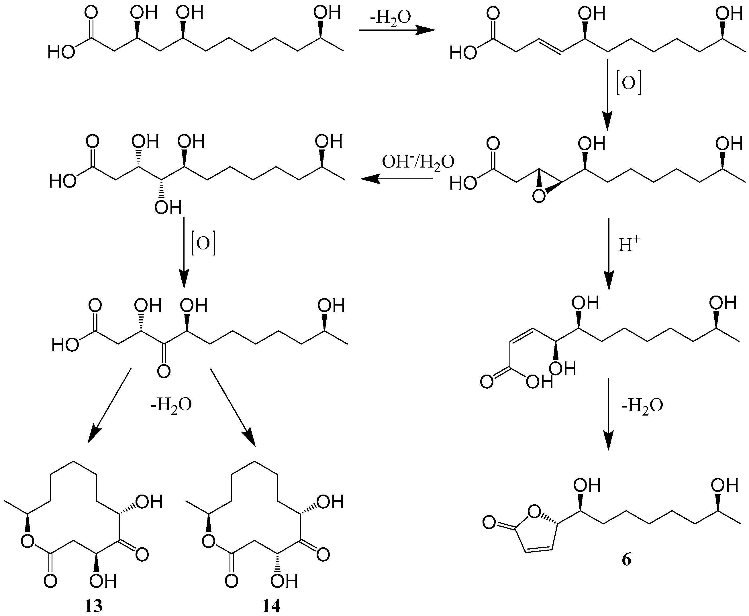
Figure 1. Structures of compounds 133 [22,32,33,34,35,36,37,38,39,40,41,42].
Ijms 25 01652 g001
Figure 2. Proposed biosynthesis pathway for compounds 6, 13 and 14 [34].
Ijms 25 01652 g002
Figure 3. Proposed biosynthesis pathway for compounds 3133 [32].
Ijms 25 01652 g003
Figure 4. (a) Gene clusters of secondary metabolic synthesis genes of Cladosporium. KS: Ketoacyl synthase; AT: Acyl transferase; DH: Dehydratase; MT: Methyltransferase; ER: Enoyl reductase; KR: Ketolreductase; ACP: Acyl carrier protein; SAT: Starting unit acyltransferase; PT: Product template; TE: Thioesterase [46,47,48,49]. (b) Proposed biosynthesis of the 12-membered macrolactones [44,45].
Ijms 25 01652 g004
Figure 5. Structures of compounds 3448 [50,53,54,55,56].
Ijms 25 01652 g005
Figure 6. Structures of compounds 4976 [19,32,57,58,59,60,61].
Ijms 25 01652 g006
Figure 7. Proposed biosynthesis mechanism for compounds 5457 [19].
Ijms 25 01652 g007
Figure 8. Structures of compounds 7783 [20,25,38,55].
Ijms 25 01652 g008
Figure 9. Structures of compounds 8499 [20,32,53,64,65,66,67].
Ijms 25 01652 g009
Figure 10. Chemical synthetic pathways of compound 90 derivatives [53].
Ijms 25 01652 g010
Figure 11. Proposed biosynthesis pathway for compounds 7576 and 9699 [20].
Ijms 25 01652 g011
Figure 12. Structures of compounds 100111 [56,66,70,71].
Ijms 25 01652 g012
Figure 13. Proposed biosynthesis pathway for compounds 109111 [71].
Ijms 25 01652 g013
Figure 15. Structures of compounds 139155 [55,61,66,68,69,77,78,79].
Ijms 25 01652 g015
Figure 16. Structures of compounds 156177 [13,65,80,81].
Ijms 25 01652 g016
Figure 17. Structures of compounds 178194 [21,28,41,55,66].
Ijms 25 01652 g017
Figure 18. Structures of compounds 195211 [32,33,56,65,66].
Ijms 25 01652 g018
Figure 19. Structures of compounds 212221 [28,33,66].
Ijms 25 01652 g019
Figure 20. Structures of compounds 222229 [23,33,56].
Ijms 25 01652 g020
Figure 21. Structural types of compounds isolated from Cladosporium over different years.
Ijms 25 01652 g021
Figure 22. (a) Structural types of compounds isolated from Cladosporium from January 2000 to December 2022; (b) Bioactivities of natural products isolated from Cladosporium discovered from January 2000 to December 2022; (c) Activities of compounds isolated from marine and terrestrial Cladosporium from January 2000 to December 2022; (d) Structural types of compounds of marine and terrestrial isolated from Cladosporium from January 2000 to December 2022.
Ijms 25 01652 g022
Figure 23. Bioactive molecular network of natural products isolated from Cladosporium from January 2000 to December 2022.
Ijms 25 01652 g023
Table 1. Compounds isolated from Cladosporium from January 2000 to December 2022.
TypesComps.SourcesDistributionYearsRefs.
Polyketides14Cladosporium sp. NRRL 29097Malette2002[32]
5Cladosporium sp. N5Jiangsu
China
2008[33]
6Sponge Niphatesrowi sp. derived Cladosporium sp. DQ100370the Red Sea2005[34]
7Gorgonian Anthogorgia ochracea (GXWZ-07)-derived Cladosporium sp. RA07-1 (GenBank No. KP720581)the South China Sea2015[35]
810Soft coral-derived fungus Cladosporium sp. TZP-29 (GenBank No. KR817674) 2019[36]
1112Marine-derived fungus Cladosporium sp. L037Okinawa Island2004[37]
1314Sponge Niphatesrowi sp. derived Cladosporium sp. DQ100370the Red Sea2005[34]
1517Sponge-derived Cladosporium sp. FT-0012Pohnpei island2001[22]
1819Plant Rhizophora stylosa-derived endophytic fungus Cladosporium sp. IFB3lp-2Hainan China2013[38]
2024Gorgonian Anthogorgia ochracea (GXWZ-07)-derived Cladosporium sp. RA07-1 (GenBank No. KP720581)the South China Sea2015[35]
2526plant Callistemon viminalis-derived fungus Cladosporium sp. A801 (GenBank No.MF138133)Guangzhou China2019[39]
2729Mangrove endophytic fungus Cladosporium sp. SCNU-F0001 (GenBank NO. NG062723.1)Guangzhou China2019[40]
30Endophytic fungus Cladosporium sp. IS384 (GenBank NO. KU158172)Sichuan China2019[41]
3133Marine mangrove-derived endophytic fungus, Cladosporium cladosporioides MA-299 (GenBank No. MH822624)Hainan China2020[42]
34Cladosporium sp. I(R)9-2Nanjing China2007[50]
3536Cladosporium oxysporum DH14 (GenBank No. JN887339.1)Jiangsu
China
2016[53]
3741Endophytic fungus Cladosporium cladosporioidesTifton2013[54]
4243Plant Ammopiptanthus mongolicus-derived fungus Cladosporium cladosporioides 2019[55]
4448Plant Ceriops tagal-derived fungus Cladosporium sp. JS1-2Hainan China2020[56]
4953Cladosporium tenuissimum ITT21Tuscany Italy2004[57]
5457Mangrove plant Kandelia candel-derived Cladosporium sp. KcFL6′Guangzhou China2015[19]
5864Cladosporium sp. TMPU1621Okinawa Japan2018[58]
6570Cladosporium sp. KFD33 (GenBank No.MN737504)Hainan China2020[59]
7173Cladosporium sp. NRRL 29097Red River2002[32]
7475Cladosporium sp. RSBE-3 2016[60]
76Mangrove-derived fungus Cladosporium sp. HNWSW-1 (GenBank No. MH 535968)Hainan China2019[61]
77Plant Rhizophora stylosa-derived endophytic fungus Cladosporium sp. IFB3lp-2Hainan China2013[38]
7880Soft coral-derived fungus Cladosporium sp. TZP-29 (GenBank NO.KR817674) 2015[25]
8182Cladosporium sp. OUCMDZ-302Hainan China2018[20]
83Plant Ammopiptanthus mongolicus-derived fungus Cladosporium cladosporioides 2019[55]
8487Cladosporium sp. NRRL 29097Red River2002[31]
8889Plant endangered Chrysosplenium carnosum-derived fungus Cladosporium sp. J6 (GenBank NO.KR492688)Tibet China2013[64]
90Cladosporium oxysporum DH14 (GenBank NO. JN887339.1) 2016[53]
91Cladosporium cladosporioides JG-12Hainan China2017[65]
9293Marine sediment-derived Cladosporium sp. NJF6Antarctica2020[66]
94Marine-derived Cladosporium sp. OUCMDZ-1635 (GenBank No. KT336457)Xisha Islands of China2018[68]
95Marine sponge-derived Cladosporium sp. TPU1507Manado, Indonesia2018[69]
9699Cladosporium sp. OUCMDZ-302Hainan, China2018[20]
100103Deep sea-derived fungus Cladosporium sp. SCSIO z015 (GenBank No. KX258800)Okinawa Trough2020[70]
104107Cladosporium sp. JS1-2Hainan China2020[56]
108Marine sediment-derived Cladosporium sp. NJF6Antarctica2020[66]
109111Cladosporium sp. (GenBank No. QH071013)Qinghai China2022[71]
Alkaloids112Cladosporium sp. N5(GenBank No. EF424419)Lianyungang China2008[33]
113126Mangrove-derived fungus Cladosporium sp. PJX-41 (GenBank No. KC589122)Guangzhou China2013[26]
127128Plant endangered Chrysosplenium carnosum-derived fungus Cladosporium sp. J6 (GenBank NO. KR492688)Tibet China2015[63]
129131Marine sediment-derived Cladosporium sp.Zhejiang China2015[76]
132Plant endangered Chrysosplenium carnosum-derived fungus Cladosporium sp. J6 (GenBank NO. KR492688)Tibet China2015[63]
133Gorgonian Anthogorgia ochracea (GXWZ-07)-derived Cladosporium sp.RA07-1 (GenBank NO. KP720581)the South China Sea2015[35]
134136Cladosporium cladosporioides JG-12Hainan China2017[65]
137138Marine sponge Callyspongia sp. derived fungus Cladosporium sp. scsio41007 (GenBank NO.MF188197)Guangzhou China2018[28]
139Marine-derived fungus Cladosporium sp. OUCMDZ-1635 (GenBank No. KT336457)Xisha Islands of China2018[68]
140Marine sponge-derived Cladosporium sp. TPU1507Indonesian2018[69]
141Plant Ammopiptanthus mongolicus-derived fungus Cladosporium cladosporioides 2019[55]
142144Mangrove-derived fungus Cladosporium sp. HNWSW-1 (GenBank access No. MH 535968)Hainan China2019[61]
145146Marine sediment-derived Cladosporium sp. NJF6Antarctica2020[66]
147154Deep sea sediment-derived Cladosporium sp. SCSIO z0025Okinawa2018[78]
155Cladosporium sp. JNU17DTH12-9-01 (GenBank no. MK994007) 2021[79]
Steroids and Terpenoids156167Cladosporium sp. IFM49189 2000[13]
168173Cladosporium sp. IFM49189 2001[80]
174176Marine mangrove-derived fungus Cladosporium cladosporioidesGuangzhou China2009[81]
177Cladosporium cladosporioides JG-12Hainan China2017[65]
178183Marine sponge-derived fungus Cladosporium sp. SCSIO41007Guangzhou China2018[28]
184190Gorgonian-derived fungus Cladosporium sp. WZ-2008-0042the South China Sea2018[21]
191Endophytic fungus Cladosporium sp. IS384Sichuan China2019[41]
192193Plant Ammopiptanthus mongolicus-derived fungus Cladosporium cladosporioides 2019[55]
194Marine sediment-derived Cladosporium sp. NJF4 2020[66]
Benzene derivatives195Cladosporium sp. NRRL 29097Red River2001[32]
196202Cladosporium sp. N5 (GenBank No. EF424419)Lianyungang China2008[33]
203Cladosporium cladosporioides JG-12Hainan China2017[65]
204205Cladosporium sp. JS1-2Hainan China2020[56]
206211Marine sediment-derived Cladosporium sp. NJF6Antarctica2020[66]
Cyclic peptides212213Cladosporium sp. N5 (GenBank No. EF424419)Lianyungang China2008[33]
214Marine sponge Callyspongia sp. derived fungus Cladosporium sp. scsio41007 (GenBank NO.MF188197)Guangzhou China2018[28]
215221Marine sediment-derived Cladosporium sp. NJF4Antarctica2020[66]
Others222223Cladosporium sp. N5Lianyungang China2008[33]
224Cladosporium sp. JS1-2Hainan China2020[56]
225229Mangrove-derived fungus Cladosporium sp. JJM22 (GenBank No.MF593626)Hainan China2021[23]
Table 2. Antibacterial activities of compounds isolated from Cladosporium during 2000–2022.
StrainsComps.Values (MIC)Values of Positive Controls (MIC)ProsCons
Staphylococcus aureus (MRSA)8 (μg/mL) [36]8.01.0 Moderate inhibitory activity
Micrococcus luteus1112 (μg/mL) [37]16.7 Moderate activities
Bacillus cereus2123 (μM) [35]12.5/25.0/6.251.56Broad-spectrum antibacterial activity; compounds 21 and 22 exhibited the strongest activities against T. halophilusModerate activity
Tetrag enococcus halophilus3.13/3.13/25.01.56
S. epidermidis6.25/25.0/25.00.78
MRSA6.25/25.0/12.50.39
Escherichia coli12.5/12.5/25.01.56
Pseudomonas putida12.5/25.0/6.250.39
Nocardia brasiliensis6.25/12.5/25.00.78
Vibrio parahaemolyticus12.5/25.0/25.01.56
Enterococcus faecalis ATCC 2921230 (μg/mL) [41]0.3120.83Strong activity
Edwardsiella tarda3133 (μg/mL) [42]1.0/2.0/2.00.5Strong activities
V. anguillarum2.0/2.0/4.01.0
MRSA ATCC4330058/59/60/64 (μg/mL) [58]25/3.13/25/250.78Compound 59 displayed strong activity of MRSACompounds 58 and 60 showed weak activities
MRSA ATCC70069825/12.5/50/251.56
B. subtilis7273 (inhibition zone) (mm) [32]33/23 Moderate activities
MRSA31/20
MRSA75 (inhibition zone) (mm) [60]2732Strong and broad-spectrum activity
E. coli2530
Pseudomonas aeruginosa2430
B. megaterium2232
MRSA84 (inhibition zone) (mm) [32]13 Moderate activity
B. subtilis86 (inhibition zone) (mm) [32]8 Weak activity
Ralstonia solanacearum91 (inhibition zone) (mm) [65]6.29 ± 0.1025.16 ± 0.06 Weak activity
MRSA6.45 ± 0.1117.62 ± 0.08
B. subtilis93 (μM) [66]2.501.25Broad-spectrum activity
Sarcina lutea2.502.50
E. coli5.000.625
M. tetragenus20.00.312
V. parahaemolyticus10.00.160
V. anguillarum5.00.160
MRSA105107 (μg/mL) [56]12.53.12 Moderate activities
MRSA 209P155 (μg/mL) [79]40.13 Weak activity
Candida albicans FIM709160.06
Shigella dysenteriae189 (μM) [21]3.13 Moderate activity
B. subtilis195 (inhibition zone) (mm) [32]22 Weak activity
C. albicans203 (inhibition zone) (mm) [65]7.43 ± 0.1212.00 ± 0.09Strong activity
MRSA204/224 (μg/mL) [56]12.5/12.53.12 Moderate activities
Table 3. Cytotoxicity of compounds isolated from Cladosporium during 2000–2022.
CellsComps.Values (IC50)Values of Positive Controls (IC50)ProsCons
L12101112 (μg/mL) [37]0.13/0.81 Strong activities
MCF–734 (μM) [50]0.2 Strong and broad-spectrum cytotoxicity
A5490.3
HCT1160.5
786-O0.7
PC31.5
K56254 (μM) [19]14.30.24 Weak activity
A54915.70.05
Huh-729.90.08
H197540.60.09
MCF-721.30.78
U93710.50.06
BGC82317.00.09
HL6010.10.09
Hela53.70.11
MOLT-414.60.03
K-5627475 (μg/mL) [60]3.97/3.5812.0Potential cytotoxicities against human leukemia cells (K-562)
HL-6093 (μM) [66]>50 Weak activity
A-549>50
Brine shrimp naupalii100103 (μM) [70]72.0/81.7/49.9/81.421.2 Moderate activities
MB49109110 (μM) [71]13.9 ± 2.5/41.7 ± 7.52.7 ± 0.6Broad-spectrum cytotoxicityWeak activities
J8225.0 ± 6.1/24.7 ± 4.40.6 ± 0.1
4T138.7 ± 4.2/27.5 ± 2.84.5 ± 1.6
Huh724.3 ± 3.5/46.4 ± 9.35.1 ± 1.2
MCF-7127 (μM) [63]20 Broad-spectrum cytotoxicityModerate activity
A54915
HT-2910
HepG210
HepG2129131 (μg/mL) [76]21/42/48 Moderate activities
HeLa133 (μM) [35]0.76 Potent cytotoxicity against a series of cancer cell lines and broad-spectrum cytotoxicity
P3881.35
HT-292.48
A5493.11
MCF-7139 (μM) [68]18.70.67Broad-spectrum cytotoxicityModerate activity
HeLa19.10.32
HCT-11617.90.21
HL-609.10.02
BEL-7042143144 (μM) [60]29.4 ± 0.35/26.7 ± 1.111.9 ± 0.37Broad-spectrum cytotoxicitiesModerate activities
K56225.6 ± 0.47/-14.2 ± 0.66
SGC-790141.7 ± 0.71/-6.66 ± 0.2
Hela-/14.9 ± 0.2111.5 ± 0.18
Vero146 (μM) [66]87 Weak activity
HeLa191 (μM) [41]22 Weak activity
HeLa194 (μM) [66]14.1 Moderate activity
Table 4. Other activities of compounds isolated from Cladosporium during 2000–2022.
BioactivitiesCells/Stains/EnzymeComps.ValuesValues of Positive ControlsProsCons
Antiviral activityHBV34 (μM) [50]0.5 Strong and broad-spectrum antiviral activity
HIV-11.0
HCMV1.5
IAV0.5
H1N1113126 (μM) [26]80–15087Compounds 115, 118, 120122 and 124 showed noteworthy antiviral activitiesCompounds 113, 114, 116, 117, 119, 123 and 126 showed weak antiviral activities
HBV127 (μM) [63]5 Broad-spectrum antiviral activity
HIV-15
HCMV5
IAV5
H3N2178 (μM) [28]16.234.0 Weak activity
RSV184 (mM) [21]0.120.08Potent antiviral activity
Antifungal activityCandida albicans11 (μg/mL) [37]16.7 Strong and broad-spectrum antifungal activity
Cryptococcus neoformans8.4
Aspergillus niger16.7
Neurospora crassa8.4
Pyricularia oryzae15 (μg/mL) [22]0.15 Strong antifungal activity
Muco rracemosus29
A. niger34 (μg/mL) [50]0.97 Strong activity
C. albicans1.9
Trichophyton rubrum1.9
Colletotrichum acutatum3741 (tested at 30 µM) (%) [54]92.7/38.3/-/-/-99.7Compound 37 showed broad-spectrum antifungal activity, and the activity is significant
Co. fragariae90.1/50.4/-/-/-99.6
Co. gloeosporioides95.4/60.2/-/-/-96.1
Phomopsis viticola79.9/83.0/53.9/35.1/79.494.2
P. obscurans22.1/22.5/25.6/-/10.396.2
A. Fumigatus IFM 4942/40849/41375/41382/46075/47064/47078/47031/47032156 (μg/disc) [13]0.5–4 Strong and exhibited specific antifungal activity toward A. fumigatus
A. fumigatus IFM 4942/40819/41375/46075/47064/47078168, 172173 (μg/disc) [80]9–17 Compounds 172 and 173 retained weak antifungal activities against A. fumigatus
C. albicans199 (mM) [33]1.5 Broad-spectrum antifungal activityWeak activity
Pseudomonas aeruginosa3.1
Staphylococcus aureus0.78
Enzyme inhibition activityAcetylcholinesterase8 (μM) [36]40.26 Potent inhibitory activity against acetylcholinesterase
α-glycosidase76 (μM) [61]49.3 ± 10.6275.7 ± 2.7Strong activity
Acetylcholinesterase91 (tested at 1 g/L) (%) [65]23.54 ± 0.7077.43 ± 1.47 Weak activity
PTP1B95 (μM) [69]110.9 Moderate inhibitory activity
Acetylcholinesterase134136 (tested at 1 g/L) (%) [65]37.20 ± 1.31/26.94 ± 5.64/26.35 ± 1.5577.43 ± 1.47 Weak activity
PTP1B140 (μM) [69]480.9Strong activity
TCPTP540.8
α-glycosidase144 (μM) [61]78.2 ± 2.1275.7 ± 2.7Strong activity
Acetylcholinesterase203 (tested at 1 g/L) (%) [65]25.43 ± 1.0877.43 ± 1.47 Weak activity
α-glucosidase226, 229 (μM) [23]2.95/2.052.35Potent inhibitory activities against α-glucosidase
Growth inhibitory activityGrowth inhibition activity against newly hatched larvae of Helicoverpa armigera Hubner4748, 107, 204205, 224 (μg/mL) [56]100/100/100/100/100/10050 Moderate inhibitory activities
Inhibition of Uromyces appendiculatus urediniospore germination4953 (tested at 100 ppm) (%) [57]84.2/100/77.6/69.4/74.820.9Strong inhibitory activities
Anti-inflammatory activity177 (μM) [65]27.961.7Potent anti-inflammatory activity
Insecticidal activityPanagrellus redivivus135136, 203 (tested at 2.5 g/L) (%) [65]78.2 ± 0.7/80.7 ± 0.4/89.6 ± 0.937.2 ± 0.3Significant and comparable to that of the positive control
Trypanosoma cruzi146 (μM) [66]26.9 Moderate activity
Quorum sensing inhibitory activityChromobacterium violaceum CV0266570 (μg/well) [59]30/30/20/30/20/30 Strong activities
PhytotoxicityAmaranthus
retroflexus L.
3536, 90 (μg/mL) [53]4.80/8.16/4.511.95Potent phytotoxic activities against the radicle growth of Amaranthus retroflexus L.
Porphyra tenera199, 201 (mm) [33]1.57 ± 0.06/0.95 ± 0.040.98 ± 0.05Strong activities
Lipid-lowering activityHepG2 hepatocytes9, 7880 (μM) [36] 8.3/12.1/8.4/13.18.3Potent lipid-lowering activities in HepG2 hepatocytes
Antioxidant activity DPPH free radical scavenging99 (μM) [71]2.65 Significant radical scavenging activity against DPPH
DPPH free radical scavenging103 (μM) [70]16.44.9Significant antioxidant activity against DPPH radicals
SH-SY5Y191 (μg/mL) [41]6.25 Weak activity
Inhibit electron transfer activityPhotosynthetic electron transport in spinach8889 (μM) [64]29.1 ± 6.5/22.8 ± 8.8 Strong activities
Anti-inflammatory activityNF-kB, TGF-β, TNF-α, IL-6, IL-8 and COX-2; caspase-3, caspase-8, Bcl-2 and Bax108 [66] Potential candidate for the therapy of different vascular inflammatory diseases
Breast cancer cells, colon cancer cells177 [65] Potential candidate for inhibiting cancer cell proliferation
Disclaimer/Publisher’s Note: The statements, opinions and data contained in all publications are solely those of the individual author(s) and contributor(s) and not of MDPI and/or the editor(s). MDPI and/or the editor(s) disclaim responsibility for any injury to people or property resulting from any ideas, methods, instructions or products referred to in the content.