Review

Iron–Imine Cocktail in Drug Development: A Contemporary Update

by
Judith Anane
1,
Esther Owusu
1,
Gildardo Rivera
2 and
Debasish Bandyopadhyay
1,3,*
1
School of Integrative Biological and Chemical Sciences (SIBCS), University of Texas Rio Grande Valley, Edinburg, TX 78539, USA
2
Laboratorio de Biotecnología Farmacéutica, Centro de Biotecnología Genómica, Instituto Politécnico Nacional, Reynosa 88710, Mexico
3
School of Earth, Environmental, and Marine Sciences (SEEMS), University of Texas Rio Grande Valley, Edinburg, TX 78539, USA
*
Author to whom correspondence should be addressed.
Int. J. Mol. Sci. 2024, 25(4), 2263; https://doi.org/10.3390/ijms25042263
Submission received: 28 December 2023 / Revised: 4 February 2024 / Accepted: 7 February 2024 / Published: 13 February 2024
(This article belongs to the Special Issue Latest Review Papers in Molecular Pharmacology 2023)

Abstract

:
Organometallic drug development is still in its early stage, but recent studies show that organometallics having iron as the central atom have the possibility of becoming good drug candidates because iron is an important micro-nutrient, and it is compatible with many biological systems, including the human body. Being an eco-friendly Lewis acid, iron can accept the lone pair of electrons from imino(sp2)-nitrogen, and the resultant iron–imine complexes with iron as a central atom have the possibility of interacting with several proteins and enzymes in humans. Iron–imine complexes have demonstrated significant potential with anticancer, bactericidal, fungicidal, and other medicinal activities in recent years. This article systematically discusses major synthetic methods and pharmacological potentials of iron–imine complexes having in vitro activity to significant clinical performance from 2016 to date. In a nutshell, this manuscript offers a simplistic view of iron complexes in medicinal inorganic chemistry: for instance, iron is presented as an “eco-friendly non-toxic” metal (as opposed to platinum) that will lead to non-toxic pharmaceuticals. The abundant literature on iron chelators shows that many iron complexes, particularly if redox-active in cells, can be quite cytotoxic, which can be beneficial for future targeted therapies. While we made every effort to include all the related papers, any omission is purely unintentional.

1. Introduction

The terms ‘imine’ and ‘Schiff base’ were invented by Albert Ladenburg and Hugo Schiff, respectively, and refer to the condensation products of carbonyl compounds (aldehydes and ketone) and amines. They are formed by the condensation of a primary amine with a carbonyl (aldehyde or ketone) compound [1] and an azomethine (-RC=N-) linkage [2]. Imines have wide applicability in many fields, especially in drug development research, because of their versatile characteristics that enable them to form a wide range of stable products. Imines can be polarized to generate an electrophilic carbon center that makes the nitrogen more nucleophilic. In mild acidic conditions, the nitrogen is protonated, making the carbon significantly electrophilic. Since their discovery by Hugo Schiff in 1864, Schiff bases have become the most important ligands in transition metal coordination chemistry due to their ease of synthesis, electronic features, solubility in many solvents, structural diversity, and abundance in biological systems [3,4,5].
The coordination of metals to organic ligands (organometallics) was not widely employed until the discovery of cisplatin and other metal-derived drugs. Many organometallic drugs have effectively been used to treat several diseases, including cancer, diabetes, and ulcers, and in imaging studies, etc. Metal–Schiff base complexes have improved antimicrobial, antioxidant, anti-inflammatory, antibacterial, and anticancer activity relative to their free Schiff base ligands [6,7]. Schiff base ligands coordinate with metal ions and stabilize them in various oxidation states. Depending on their dipole moment, solubility, enzymatic action, and cell permeability, they can increase biological activity [5]. The challenge, however, is that some of these therapeutics have raised concerns due to the fatal side effects they confer on patients. The need for new, less toxic, and more potent organometallic drugs has led to extensive research on iron–imine complex formation. So far, iron–imine complexes have been found to exhibit effective biological activities [8]. For instance, Sarkar et al. found a significant photocytotoxicity of an iron(III)–Schiff base complex (obtained from thiosemicarbazide and vitamin B6) against cervical cancer cells (HeLa) through the intracellular generation of ROS [9]. Also, some iron(III)–Schiff base complexes derived from aminophenol/aminobenzene and salicylaldehyde have shown better antibacterial and antifungal activity when compared to antibacterial and antifungal standard drugs, chloramphenicol and terbinafine, respectively [10].
It is worth mentioning that iron is the fourth most abundant (5.6%) element in the Earth’s crust [11]. It is important for the normal functioning of mammalian cells because iron plays essential roles in many dynamic biological processes that occur in the human body, like DNA synthesis, metabolism, respiration, electron transport, and erythropoiesis, among others [12,13], making their participation in mammalian cells vital for appropriate cellular function [14,15]. It is, therefore, a safer alternative for developing organometallic drugs. This review outlines the syntheses and pharmacological potential of iron–imine complexes.

2. Bioactivity of Imine–Iron Complexes

Imine–iron complexes provide an intriguing insight into the future of organometallic chemistry. Limited attention was given to organometallic drugs until recently when some metal-containing drugs were discovered to be useful in the battle against various diseases like cancer, antimicrobial resistance diseases, oxidative stress [3], HIV [16], bacterial (malaria), fungal, and viral infections [17], tuberculosis [18], diabetes, rheumatoid arthritis, and cardiovascular diseases [19]. Among the exciting tapestry of organometallic compounds, imine–iron complexes stand out with their bright threads of fascination and ability. These unique compounds attach iron atoms to ligands with the intriguing imine functional group, which is a nitrogen–carbon double bond generated by an amine and a carbonyl compound. Imine–iron complexes are adaptable building blocks with high promise in catalysis, biomimicry, magnetic materials, and beyond. This distinguishing trait endows them with an enticing combination of properties, making them the focus of significant research and interest in the sector.

2.1. Imine–Iron Complexes as Anticancer Agents

The success of cisplatin as a potent anticancer drug led to researchers exploring and discovering platinum-based drugs like carboplatin and oxaliplatin. The downside of these platinum-based drugs is their adverse side effects and drug resistance. This shifted attention to other metal-based anticancer drugs, particularly iron-based complexes, after the prolific activity of naturally occurring iron–bleomycin and ferrocenium salts like trichloroacetate and ferrocenium picrate was discovered. Their effectiveness was attributed to the oxidative DNA damage they caused by upsetting the oxidative homeostasis in cancer cells [11]. Iron–imine complexes provide a unique and potential avenue for cancer therapy due to their tailored delivery and different mechanisms of action, which include altering iron metabolism, producing reactive oxygen species (ROS), and blocking key enzymes. They additionally enhanced tumor penetration and retention and have the ability to overcome drug resistance.
Iron–imine complexes and other organometallic complexes have been found to have potent anticancer activity [20].
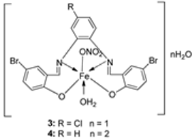
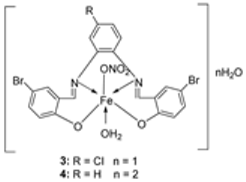
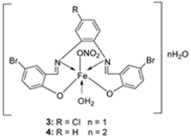
El-Lateef et al. synthesized two tetradentate dibasic chelating imine–iron complexes (3 and 4, Scheme 1) from the reaction of 1 and 2 with Fe3+ salt. The free ligand (1 and 2) and its synthesized complexes (3 and 4) were investigated for their in vitro cytotoxic effect against MCF-7, HepG-2, and HCT-116 cancer cell lines at different concentrations. These tested compounds had activity on breast carcinoma cells, with the cytotoxicity of the complexes being higher than that of their free ligands. Compound 4 showed the highest cytotoxicity activity against MCF-7, HepG-2, and HCT-116 (5.14, 6.75, and 4.45 µM, respectively), comparable to the standard drug doxorubicin, which had the activity of 4.10, 5.15, and 4.35 µM, respectively, and could be used as a tumor drug candidate (Table 1). The cytotoxicity of metal complexes is assumed to be due to their ability to bind DNA, hence disrupting its structure, causing replication and transcription processes to be inhibited, and eventually damaging the cancer cells (Table 1) [21].
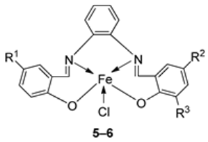
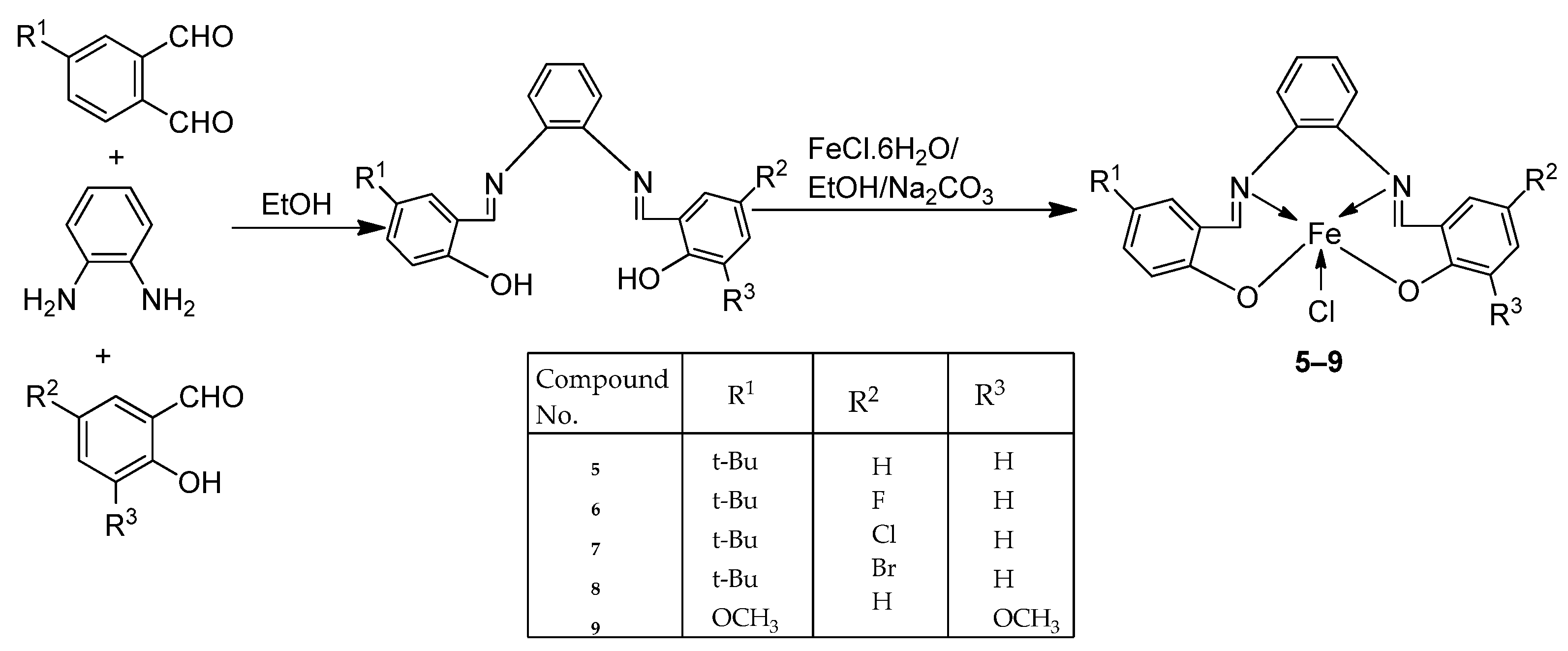
Nguyen et al. [22] synthesized unsymmetrical tetradentate imine–Fe(III) complexes (59, Scheme 2) by coordinating the imine ligands with FeCl3·6H2O and tested them on KB and Hep-G2 human cancer cell lines. The iron–imine complexes showed excellent cytotoxicity for KB and Hep-G2 (IC50 < 20 µM). The presence of substituted groups in the salicyl rings affects the electrical properties and bulk of the complexes. Complex 5, which did not have the substituted group in the second salicyl ring, exhibited the best cytotoxic activity for KB and Hep-G2 (0.68 and 0.83 µM, respectively), even better than the standard compound ellipticine, which showed an activity of 1.14 and 2.11 µM, respectively (Table 1) [22].
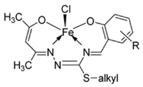
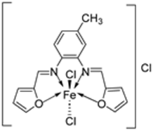
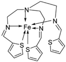
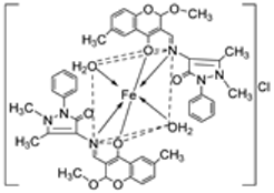
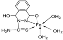
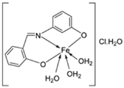
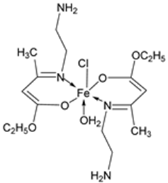
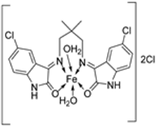
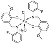
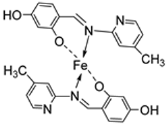
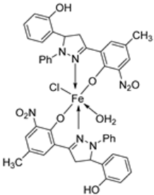
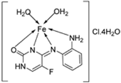
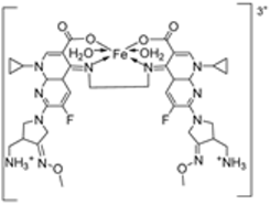
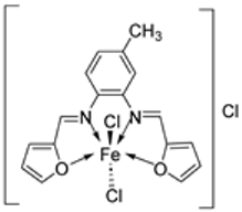
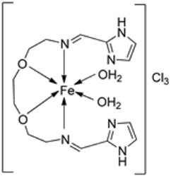
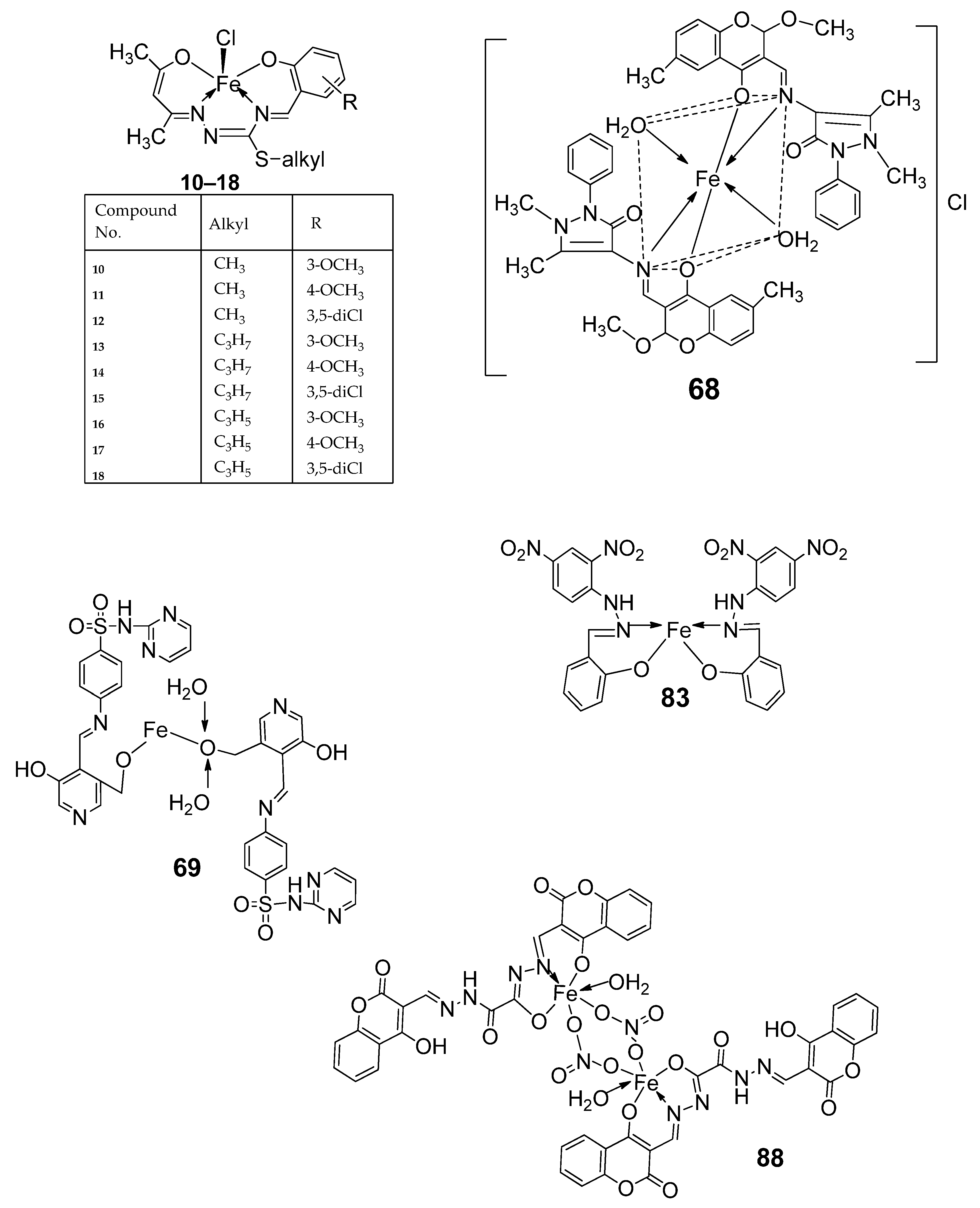
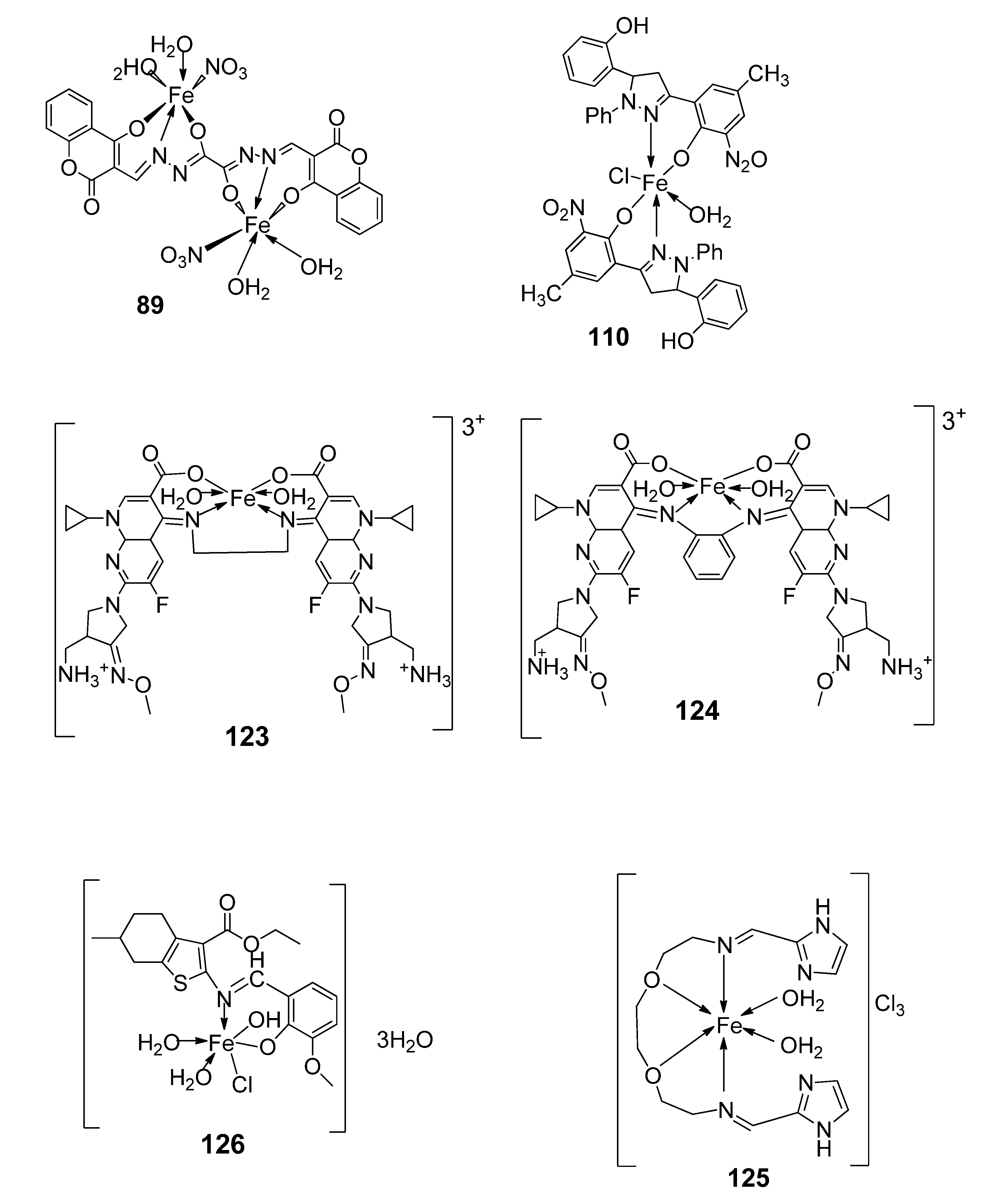
Nine iron(III) complexes (1018, Figure 1) were synthesized by Kalındemirtaş et al. The in vitro cytotoxicity activity of the iron complexes was investigated on P3HR1, K562, JURKAT, HUVEC, and 3T3 cell lines. The complexes 11, 14, 16, and 17 showed a better cytotoxicity effect (in the range of 4.81–14.05 μM) on the K562 cell line than the standard imatinib, which had an activity of 9.67 µM. Five complexes had significantly lower IC50 values than the positive control (imatinib) for P3HR1 cells (Table 1). Complexes 12, 15, and 18, which had a 3,5-dichloro substituent, could not compete with imatinib. All the synthesized complexes were ineffective against the JURKAT cell line in the studied concentrations. Different cells may die in different ways, and cancer cells of different types might respond very differently to the same treatment. P3HR1 and JURKAT are lymphoid cells with T- and B-lymphocytes of origin, respectively, whereas K562 is myeloid. T-cell lineage-derived leukemia includes a diverse range of neoplasms. They are typically more aggressive than their B-cell counterpart, differing in clinicopathological characteristics and biological function, and are marked by resistance to conventional chemotherapy and a bad prognosis for the patients [23]. Studies have also shown miRNAs to be critical regulators in tumorigenesis [24,25]. When exposed to chemotherapeutic drugs that are commonly used in T-cell leukemia/lymphoma treatment, like cisplatin, cytarabine, doxorubicin, and cyclophosphamide, JURKAT cells’ expression of miR181a increased along with AKT activation [26]. The different results obtained in the JURKAT cells may be due to these differences [27].
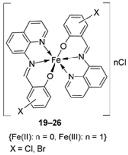
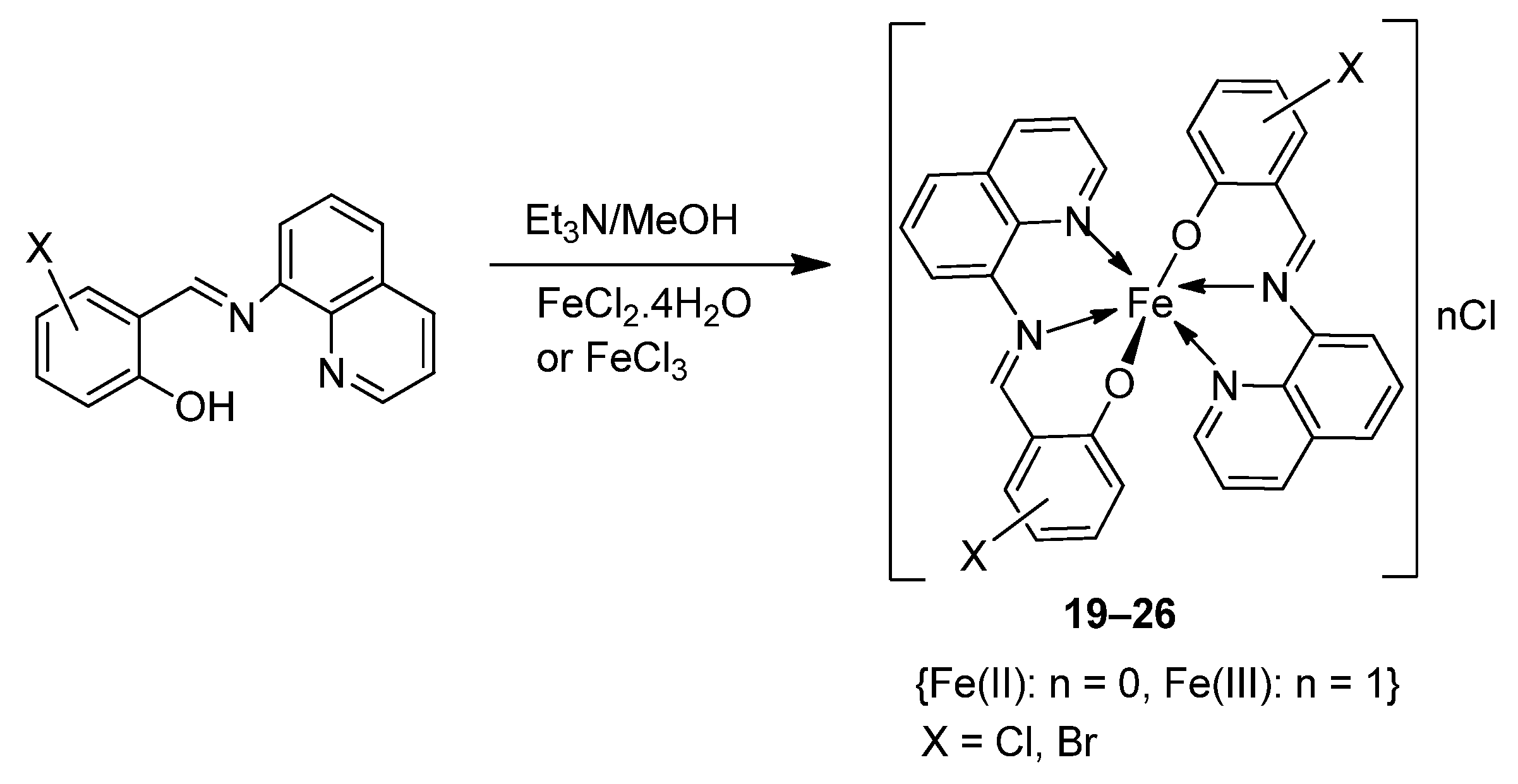
Wongsuwan et al. synthesized a series of Fe(II) complexes (1922, Scheme 3) and Fe(III) complexes (2326, Scheme 3) by coordinating imine derived from 8-aminoquinoline and salicylaldehyde with Fe(II)/(III) chloride (Scheme 3). Imine complexes were screened against the A549 human lung adenocarcinoma cell line. The imine ligand showed no anticancer activity, but the complexes showed moderate-to-high anticancer activity against A549 cells with IC50 values ranging from 10 to 34 µM. Complex 22 showed the highest antiproliferative activity of 10 µM, which is higher than that of two well-known commercial drugs, etoposide (19 µM) and cisplatin (16 µM) (Table 1). Transition metal complexes can bind to DNA through both covalent and non-covalent interactions. Complex 6 showed very high DNA affinity and induced high levels of ROS (hydroxyl and peroxyl radicals) in A549 cancer cells. These two factors together contributed to the antiproliferative activity of complex 6. Therefore, DNA binding and intracellular ROS that cause macromolecular or DNA damage and cell death are potential mechanisms by which the complexes enter A549 cells [28].
An iron(III) complex (30, Scheme 4) of novel imine ligand 29 was synthesized by Ismail et al., and its cytotoxicity activity against the Hep-G2 cell line was evaluated. The Fe(III) complex (30, Scheme 4) showed an enhanced antitumor activity (7.31 μg/mL) compared to that of the solo Schiff base (IC50 = 27 μg/mL). Still, compared to the standard drug vinblastine, which showed a value of 2.93 μg/mL, its antitumor activity was moderate (Table 1) [29].
The ligand (33) and its metal complex (34, Scheme 5) were synthesized by Kavitha et al. and studied over three cancer cell lines: human pancreatic carcinoma (MiaPaCa-2), human cervical adenocarcinoma (HeLa), and murine melanoma cancer cells (B16F10), and one normal cell N1H/3T3 (fibroblast cells). The IC50 value for the complex, 106.26 μg/mL, was beyond 100 µg/mL, signifying very low anticancer activity against the selected cancer cell lines (Table 1). DNA binding studies showed that the complex had a low binding affinity for the DNA, which could have been responsible for its low antitumor potency [30].
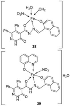
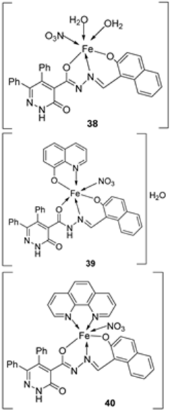
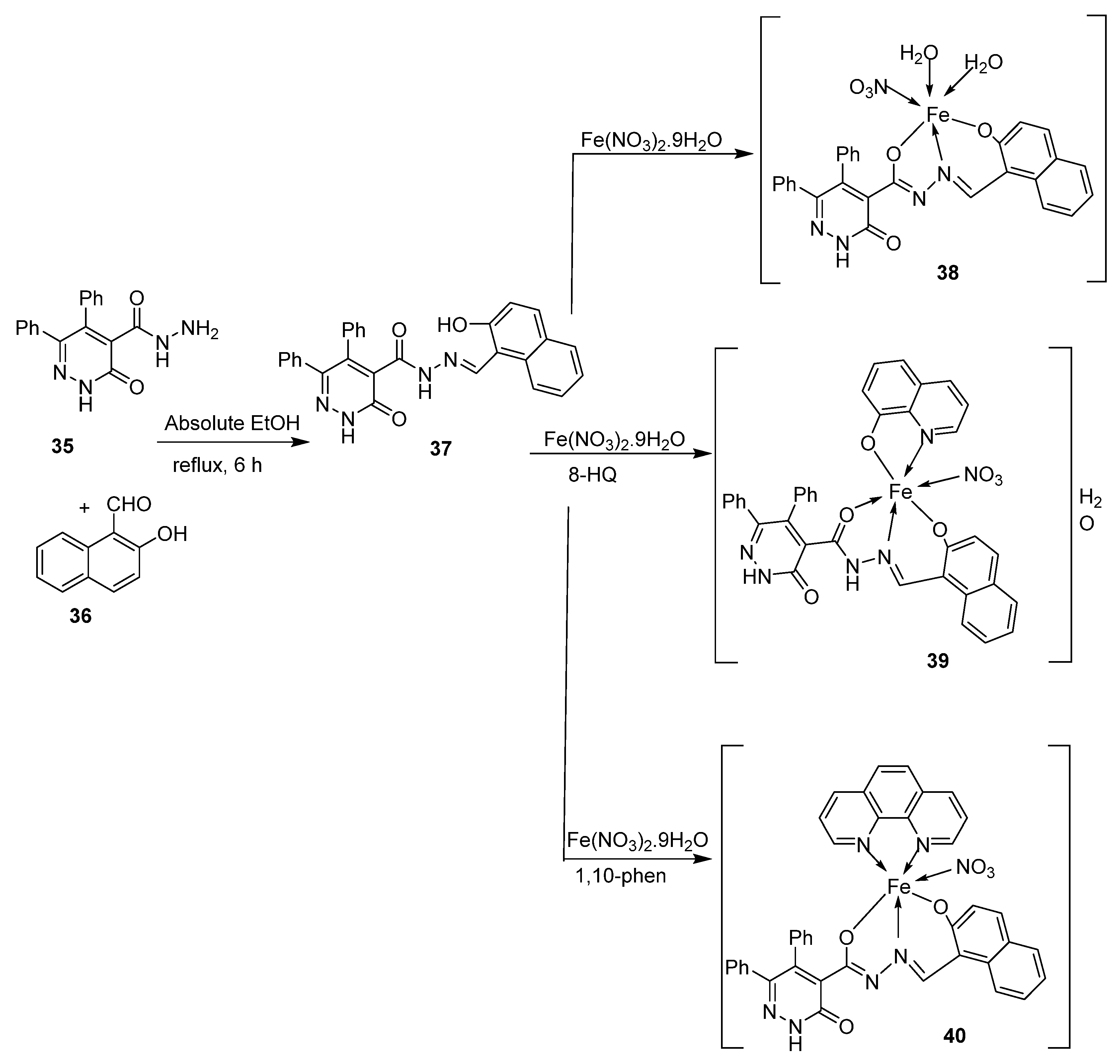
Abdelrahman et al. synthesized new nano-Fe(III) complexes (3840) of pyridazinone-acid hydrazone ligand 37 and new mixed-ligand complexes using 8-hydroxyquinoline or 1, 10-phenanthroline (Scheme 5) as an auxiliary ligand. The complexes and the imine ligand were tested against hepatocellular carcinoma cell lines (HepG-2 cells) for their antitumor activity in vitro. The imine ligand showed strong antitumor activity against the HepG-2 cells, but the activity of the synthesized iron complexes (3840) was insignificant (Scheme 6, Table 1). Complex 37 showed an antitumor activity of 3.80 μg/mL against HepG-2, whilst the standard drug, Cisplatin, showed an activity of 3.27 μg/mL (Table 1) [31].
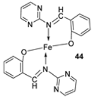
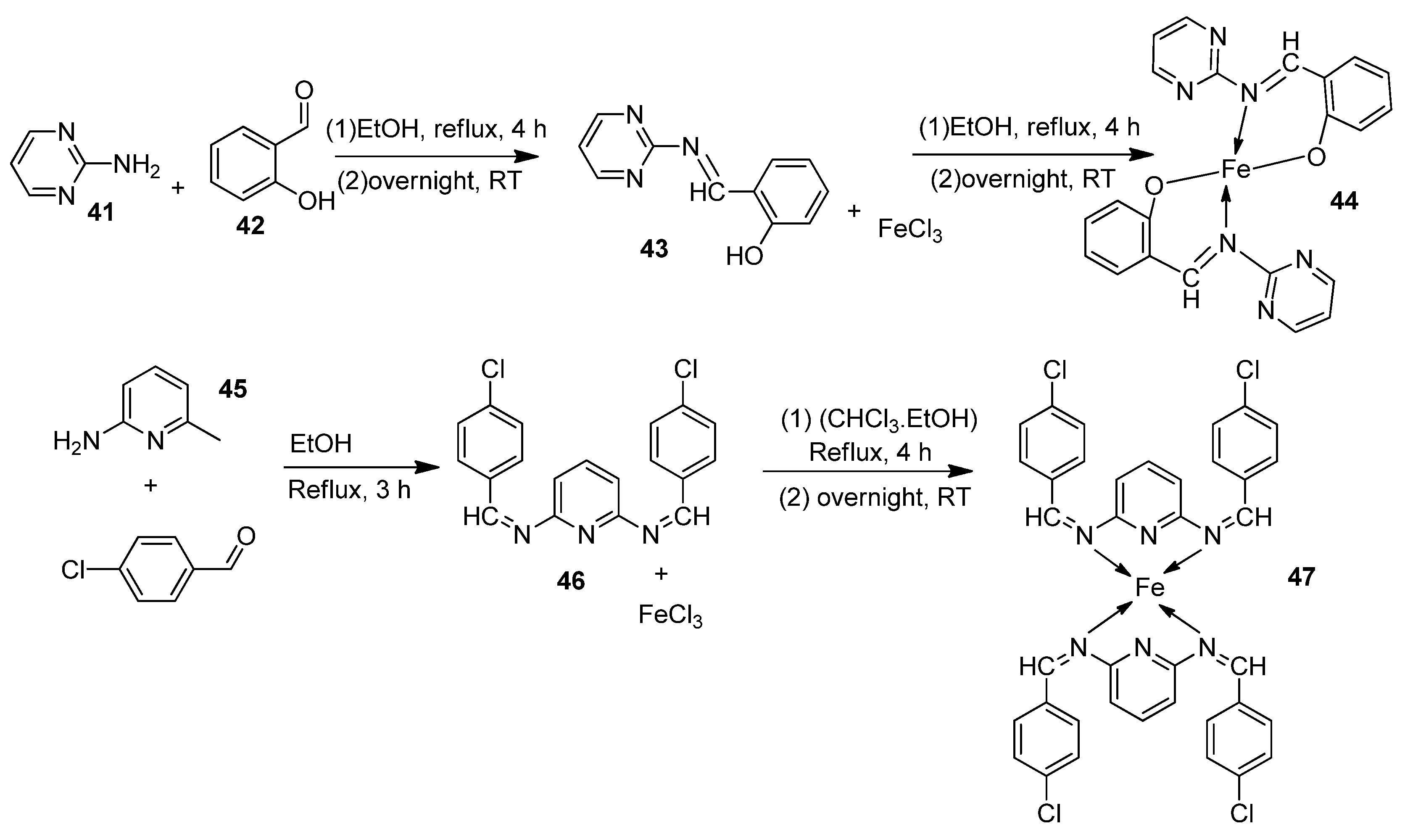
Farhan et al. synthesized two heterocyclic imine ligands (43 and 46) and prepared complexes (44 and 47, Scheme 7) from the fusion of the imine ligands with Fe(III), resulting in an octahedral geometry and paramagnetic complex (44 and 47). The ligands and imine complexes were investigated for their anticancer potency against the L20B cell line at a 4000 μg/mL concentration. The iron complex 44 demonstrated a high anticancer activity of 8.7 μg/mL against the (L20B) cell line. The anticancer activity of 47 was comparatively low, 22.9μg/mL (Table 1). The results were not compared with a standard anticancer agent [32].
Table 1. Product, synthesis conditions, and in vitro anticancer activity (IC50 in µM) of selected iron–imine complexes compared to the respective positive controls .
Entry No.Complex No.StructuresSynthesis ConditionComplex
and
Positive Control
Cancer Cell LinesRef.
1.3, 4Ijms 25 02263 i001EtOH
Reflux, 2 h
Stirring
MCF-7HepG-2HCT-116[21]
321.35 ± 0.1227.70 ± 0.1115.75 ± 0.07
45.14 ± 0.056.75 ± 0.124.45 ± 0.14
Doxorubicin4.10 ± 0.135.15 ± 0.074.35 ± 0.15
2.59Ijms 25 02263 i002EtOH
Reflux, 3 h
KBHepG-2 [22]
50.68 ± 0.050.83 ± 0.05
63.25 ± 0.167.05 ± 0.25
71.84 ± 0.106.07 ± 0.22
82.76 ± 0.1719.78 ± 1.07
91.95 ± 0.132.38 ± 0.17
Ellipticine1.14 ± 0.062.11 ± 0.12
3.1018Ijms 25 02263 i003Stirring, 30 min K562P3HR1JURKAT[27]
10>25>25>25
119.25 ± 0.425.61 ± 0.19>25
1222.24 ± 0.068.09 ± 0.62>25
13>25>25>25
144.81 ± 0.1511.98 ± 0.6922.79 ± 0.54
15>2522.4 ± 0.47>25
1614.05 ± 0.315.72 ± 0.28>25
175.04 ± 0.1811.47 ± 0.4222.0 ± 0.39
18>2521.03 ± 0.39>25
Imatinib9.67 ± 0.4923.74 ± 1.023.73 ± 0.21
4.1926Ijms 25 02263 i0040 °C, 7 days A549 [28]
1930 ± 1.1
2030 ± 7.7
2128 ± 2.0
2228 ± 2.0
2328 ± 2.0
2410 ± 2.1
2534 ± 4.7
2632 ± 1.5
Etoposide19 ± 1.3
Cisplatin16 ± 1.9
5.30Ijms 25 02263 i005Reflux, 3 h
Stirring, 2 h
Hep-G2 [29]
307.31
Vinblastine2.93
6.34Ijms 25 02263 i006Reflux, 8–9 h HelaMiaPaCa-2B16F10[30]
34106.26 ± 0.5112.13 ± 0.6104.15 ± 1.2
7.3840Ijms 25 02263 i007Stir, 2 h
Reflux, 12–15 h
Hep-G2[31]
373.8058.00
38R
39R
403.27
Cisplatin
8. Ijms 25 02263 i008Reflux, 3–4 h L20B[32]
44448.70
454513.20
464618.4
474722.9
C50 values written as these have been reported in the literature.

2.2. Imine–Iron Complexes as Antimicrobial Agents

Antimicrobial agents are compounds that can inhibit (stop or reduce) the growth of microorganisms such as bacteria, fungi, protozoa, etc. Microbial resistance to antibiotics and other antimicrobial drugs has become one of the major health concerns globally. Due to their distinct characteristics and action methods, research has focused on imine–iron complexes as promising agents [33]. Imine–iron complexes appear as possible game changers in the fight against microorganisms, outperforming many traditional antimicrobials. They are considered possible game changers because of the following:
  • Novel mechanism of action: Traditional antibiotics typically target specific bacterial functions like cell wall synthesis or protein translation, which can lead to resistance development as bacteria mutate those targets, imine–iron complexes employ diverse mechanisms, including iron starvation, DNA cleavage, and reactive oxygen species (ROS) generation, making it harder for bacteria to develop resistance.
  • Broad-spectrum activity: Traditional antibiotics often have specific targets, limiting their effectiveness against different types of bacteria, whilst imine–iron complexes can exhibit activity against a wider range of bacteria, including multi-drug-resistant strains, due to their multiple attacking mechanisms.
  • Biofilm disruption: Traditional antibiotics may struggle to penetrate bacterial biofilms, protective communities are resistant to many drugs, whilst imine–iron complexes show potential to disrupt biofilms, exposing bacteria within to attack further.
  • Reduced side effects: Traditional antibiotics can harm beneficial gut bacteria and other healthy cells due to their broad targeting, whilst imine–iron complexes can be designed to be more selective for bacterial targets, potentially reducing the side effects on human cells.
In recent years, metal complex-based antibiotic compounds have become a promising avenue in drug development. According to research, 21% of the metal compounds examined exhibited antibacterial action against typical strains of Candida and Cryptococcus strains [34]. Therefore, there is an urgent need to develop next-generation antimicrobial agents, and imine-iron complexes can be the right avenue to move forward because these complexes are known for their antimicrobial activity. The observed microbial activity can also be traced to (i) the concept of cell permeability and the chelation process which reduces the polarity of a metal ion; (ii) the chelation process which increases electron delocalization on the chelate ring and enhances the lipophilicity of the complex, granting it easy penetration through microbial cells; (iii) the toxicity of metal ions [35]; (iv) the introduction of an azomethine linkage improves the hydrophobicity and liposolubility of the molecules; and additional factors that contribute to the improved biological activity are the solubility, conductivity, and dipole moment of the metal ion [36,37,38]. The antimicrobial activities of Schiff bases and their metal complexes have been studied against different bacterial and fungal strains [39].
Rahmatabadi et al. synthesized the iron metal complex (51, Scheme 8) of imine ligand (50), prepared by condensing 48 with 49. Imine–iron complex (51) was tested for its in vitro antibacterial potency against Gram-negative Escherichia coli (E. coli) and Pseudomonas aeruginosa (P. aeruginosa) bacteria and Gram-positive bacteria Bacillus cereus (B. cereus) and Staphylococcus aureus (S. aureus) with tetracycline, gentamicin, chloramphenicol, and cephradine as a standard control. Complex 51 showed enhanced activity compared to the free ligand (50). It had the highest antibacterial activity against B. cereus (29 mm) and S. aureus (14 mm), which was higher than the activity of the standard drug tetracycline against B. cereus (11 mm) and S. aureus (9 mm), but it showed moderate activity against E. coli (14 mm) and P. aeruginosa (14 mm), which was for both bacteria (Table 2). These recorded activities of the complexes are due to the more pronounced lipophilic nature of the metal centers in the complexes [40,41].
Shukla et al. synthesized imine ligand 54 by condensing 52 with 53 in a 1:2 molar ratio. Imine ligand 54, 1,10-phenanthroline, and FeCl3 were combined to form a mixed-ligand iron complex (55, Scheme 9) and analyzed for their antibacterial activity against Gram-negative bacteria E. coli in comparison to amoxicillin and chloramphenicol standard drugs. Complex 55 exhibited enhanced activity against E. coli (29 mm) compared to 54 (23 mm). Still, it showed moderate antibacterial activity compared with standards chloramphenicol and amoxicillin, which showed inhibition zones of 39 mm and 41 mm, respectively (Table 2). The action of metal ions on the normal cell membrane may cause the metal complex’s increased activity. Either the microbes’ cells’ impermeability or variations in the ribosomes of microbial cells determine the complex’s ability to combat E. coli. The outcome could be explained by considering the chelation theory, which suggests that chelation could facilitate a complex’s capacity to pass across a cell membrane [42,43].
El-Lateef et al. explored 3 and 4 (Scheme 1) for their antibacterial potency against three selected bacterial strains: S. marcescence, E. coli, and M. Luteus. Both complexes showed high antibacterial activity against the selected bacteria, with 4 showing the highest antibacterial activity against M. luteus (2.50 μg/mL) (Table 2). The values of the activity of standard drugs were not provided. The activity of the complexes was high compared to that of the free ligands (1 and 2) due to the chelation theory. The polarity of the metal ion is greatly reduced during chelation due to electron delocalization throughout the entire chelate ring system and partial sharing of its positive charge with the hetero-donor atoms of the ligand [44,45]. The different activities displayed by the complexes against the various microbes are due to the differences in the chemical makeup of the microorganisms’ cell walls [21].
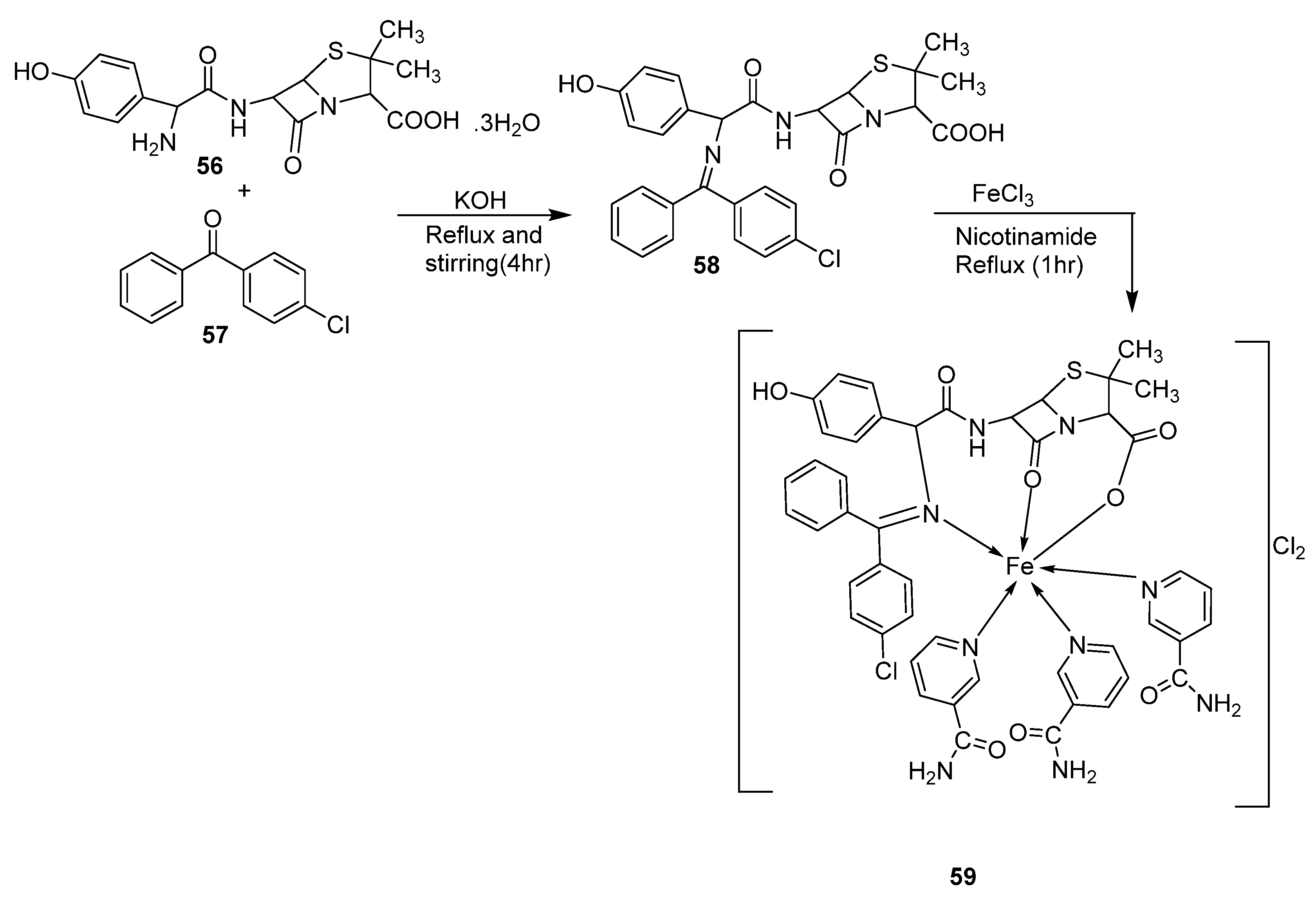
The iron complex 59 (Scheme 10) was synthesized by Karem et al., and its antibacterial potency was evaluated against P. aeruginosa, E. coli, S. aureus, and B. subtilis. The iron complex showed no activity for all the bacterial strains except for E. coli, against which it showed an activity of 25 μg/mL. This value was higher than that of the free ligand, which showed an inhibition of 2.5 μg/mL (Table 2). The observed increase in activity against E. coli can be explained by Tweedy’s theory [46]. The results obtained were not compared to a standard drug [47].
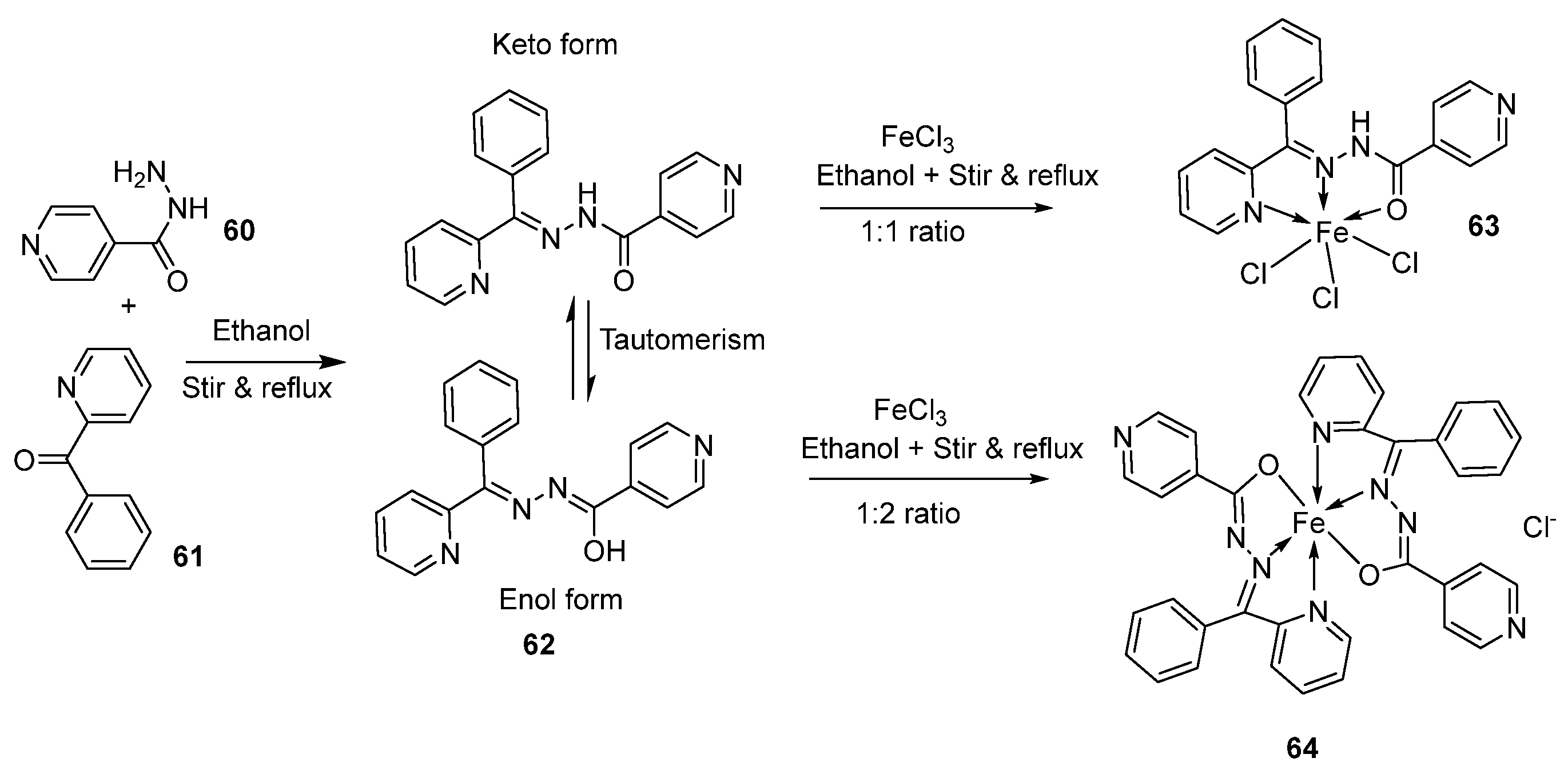
The imine ligand 62 synthesized by the condensation of 60 and 61 (Scheme 11) was complexed with Fe(III) by Shukla et al. to form two imine–iron complexes, 63 and 64. The complexes were tested against Gram-positive bacteria, B. subtilis, and Gram-negative bacteria, E. coli, with amoxicillin as a standard. The complex 64 (14 mm and 18 mm, respectively) showed enhanced activity compared to the free ligand (11 mm and 15 mm, respectively) against B. subtilis and E. coli, and this activity of 64 was similar to that of the standard amoxicillin (16 mm and 20 mm, respectively) against the same microbes. The antimicrobial activity of complex 63 is similar to that of ligand 62 against the selected microbes (Table 2). The result shows that chelation makes it easier for these complexes to traverse the cell membrane, which is consistent with Tweedy’s chelation theory. Due to the partial sharing of the metal ion’s positive charge with donor groups during chelation, the metal ion’s polarity will be lowered, and the delocalization of π-electrons over the entire chelate ring will be increased. This improves the complex’s lipophilicity, favoring its passage through the lipid membrane, and interferes with the metal binding sites in the microbes’ enzymes [48].
The imine ligand 66, prepared by the condensation of 65 with 1,2-diaminobenzene and its iron complex (67, Scheme 12), was synthesized by Anacona et al. and analyzed for its antibacterial activity against pathogenic bacteria Gram-positive Enterococcus faecalis (E. faecalis) ATCC 29212 and S. aureus ATCC 25923 and clinical isolates of Streptococcus viridans (S. viridans), Enterococcus Sp., and methicillin-resistant S. aureus (MRSA). The iron complex (67) showed enhanced activity against all the selected microbes compared to the ligand 66. It exhibited very good antibacterial activity against methicillin-resistant S. aureus (15 mm), whereas the standard drug and free ligand showed no activity at all. The complex under study [49] showed moderate activity against the other bacterial strains (Table 2). The moderate-to-high activity of the complex is attributed to not only the chelation theory but also other factors like the nature of the metal ion, the type and quantity of donor atoms, stereochemistry, chelate stability, and pharmacokinetic factors [50].
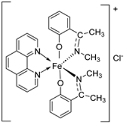
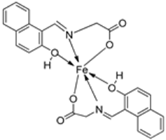
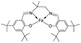
Pahontu et al. synthesized an Fe(III) complex (68, Figure 1) and tested its antimicrobial activity against Gram-positive bacteria S. aureus, B. cereus, and E. faecalis and Gram-negative bacteria E. coli and A. baumannii, as well as fungal strains Candida albicans (C. albicans), Candida krusei (C. krusei), and Cryptococcus neoformans (C. neoformans). The MIC values of the iron–Schiff base complex obtained correlated with very low antibacterial activity against all the bacterial strains selected compared to the standards used (furacilin, ciprofloxacin, and amikacin). The complex showed improved antifungal activity against C. albicans and C. neoformans, with values of 0.0156 and 0.0078μg/mL, respectively, compared to the standard drugs nystatin (C. albicans = 0.032 μg/mL, C. neoformans = 0.032 μg/mL) and miconazole (C. albicans = 0.016 μg/mL, C. neoformans = 0.0162 μg/mL) used in studies (Table 2) [6]. The lack of activity of the synthesized complex against the bacterial strain is unclear. Still, its impressive antifungal activity against C. albicans can be attributed to the metal ion’s ability to reduce binding energy while increasing the binding affinity of the microbe protein, hence interrupting its biological processes [10].
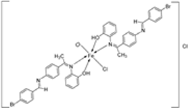
Mumtaz et al. complexed iron(II) with an imine ligand to form the iron(II) metal complex 69 (Figure 1), which was investigated for its antimicrobial activity against E. coli, Enterobacter aerogenes (E. aerogenes), S. aureus, B. pumilus, K. oxytoca, and C. butyrium. The iron complex’s zone of inhibition of the various bacterial strains was quite small, demonstrating low antibacterial activity towards the bacteria. Still, these values were higher than those of the free imine ligand. Complex 69 showed an activity of 12, 10, and 9 (mm) against E. coli, E. aerogenes, and C. barium, respectively, and the ligand showed an activity of 14, 12, and 12 (mm), respectively (Table 2). The complex’s enhanced activity compared to the ligand can be explained by chelation theory. [46,51].
Al-Wasidi et al. synthesized an iron–Schiff base complex by complexing imine ligand 72 with Fe(III) to form an octahedral iron complex (73, Scheme 13) which was investigated for its antibacterial and antifungal activity against Gram-positive B. subtilis, S. pneumonia, and S. aureus, Gram-negative E. coli Sp. and Pseudomonas Sp., and fungal strains Aspergillus niger (A. niger) and Penicillium Sp. The iron–imine complex 73 showed enhanced antibacterial activity relative to the free ligand 72 with a great zone of inhibition against S. pneumonia (7–10 mm) and S. aureus (7–10 mm). It demonstrated low inhibition against the selected fungal strains (Table 2). The results obtained were not compared to any standard drug [52].
El-Sonbati et al. synthesized imine–iron complex 76 with ligand 75 (Scheme 14) and evaluated its antimicrobial activity against Gram-positive bacteria B. subtilis and S. aureus; Gram-negative bacteria such as Salmonella sp, P. aeruginosa, and E. coli; and fungal strains A. fumigatus and C. albicans. For all the bacterial strains selected, complex 76 showed similar antibacterial activity to the free imine ligand and low antibacterial activity when compared with the selected standard drugs ampicillin and gentamycin (Table 2). Against the fungal strains C. albicans and A. fumigatus, the complex showed improved inhibition of 16 mm and 18 mm, respectively, compared to the free imine ligand (13 mm and 15 mm, respectively). This inhibition was low compared to the standard antifungal drug amphotericin, which had an activity of 25 mm and 23 mm zones of inhibition, respectively. The improved antifungal activity of complex 76 in relation to the free ligand (75) can be explained by the chelation theory, where the chelation of the ligand causes an increase in the lipophilicity properties of the metal chelate, enhancing its ability to permeate the lipoid layers of the microbe membrane blocking the metal binding site [4,46].
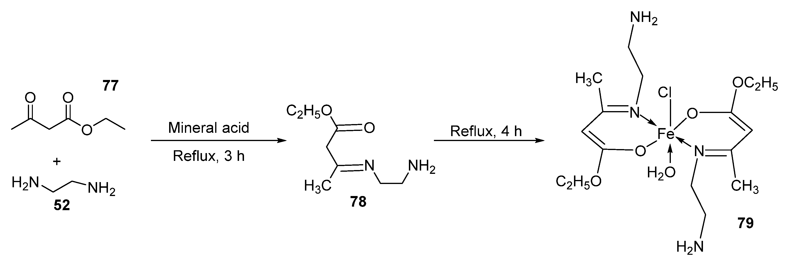
Kumar et al. synthesized the imine–iron complex 79 (Scheme 15) of imine ligand 78 and evaluated its antibacterial activity against Gram-positive P. aeruginosa and Gram-negative S. aureus bacteria. The complex (79) showed an improved antibacterial activity compared to the free ligand (78) against S. aureus and P. aeruginosa with a zone of inhibition of 14 mm and 11 mm, respectively, whereas the ligand showed an inhibition of 8 mm and 6 mm, respectively. Complex 79 had a comparable zone of inhibition to that of the standard drugs ampicillin against S. aureus (14 mm) and chloramphenicol against P. aeruginosa (8 mm) (Table 2) and can be further investigated as an antibacterial drug candidate. The improved antibacterial activity of the complex can be attributed to the chelation theory [46,53].
Fe(II) complex 82 (Scheme 16) of compound 81 was synthesized by Shinde et al., and upon investigating its antimicrobial activity against Gram-positive bacteria S. aureus (ATCC 29737), Gram-negative bacteria E. coli (ATCC 25922), and fungal strains C. albicans (MTCC 277) and A. niger (MCIM 545), it was found to possess high activity against E. coli (ATCC25922) and S. aureus with an MIC value of 10 μg/mL against both bacteria. This value is the same for the standard drug gentamicin, which also showed an activity of 10 μg/mL. Complex 82 also showed improved activity against both fungal strains C. albicans (MTCC 277) and A. niger (MCIM 545) with an MIC value of 10 μg/mL when compared with the standard drug fluconazole, which showed an activity of 20 μg/mL against both strains (Table 2) [2]. The reason for the exceptional antimicrobial potency of the synthesized complex (82) was not stated, but it could be due to chelation theory [46] and the good binding interaction of 82 with the proteins of the selected strains.
Mukhtar et al. synthesized an imine–iron metal complex (83, Figure 1), and its antimicrobial activity was investigated against five bacterial isolates, E. coli, S. aureus, P. aureginosa, K. Pneumoniae, and S. aureus, and three fungal species, F. solani, A. fumigate, and C. albicans. The results of these studies revealed that the complex showed the highest antibacterial activity against E. coli (14 mm) at a concentration of 1000 μg/mL but moderate activity against the other bacterial isolates (Table 2). Its antifungal activity was quite low. It inhibited the growth of C. albicans and F. solani by 7 mm at a concentration of 2000 μg/mL and 12 mm at a concentration of 4000 μg/mL, respectively. It showed no antifungal activity against A. fumigate at the studied concentrations. The ligand showed no zone of inhibition against E. coli and P. aeruginosa at the given concentrations. It, however, showed similar activity to the complex against S. aureus (12 mm) at a concentration of 1000 μg/mL. It also showed no activity against all the selected fungal strains (Table 2). The results obtained in this study were not compared to any standard drug [54]. The reason for the improved antimicrobial activity of the synthesized complex can be attributed to the chelation theory [46].
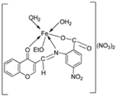
The synthesis of a chromone imine nano-complex of Fe(III) (87, Scheme 17) was conducted by Shebl et al., and its antimicrobial activity was tested against microorganisms such as E. coli, P. vulgaris, K. pneumonia, S. aureus, and C. albicans. The results showed that the iron complex (87) has moderate activity against fungal species C. albicans (8 μg/mL) when compared to free ligand 86 (4 μg/mL) and a standard (2 μg/mL); it, however, exhibited very low activity (>50 µg/mL) toward all the selected bacterial stains in comparison to the standard drug doximycin which showed activity in the range of 2–4 μg/mL (Table 2) [3].
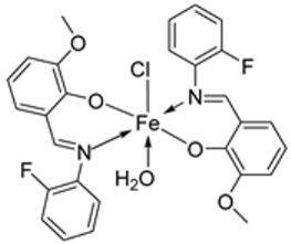
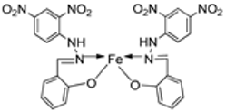
Knittl et al. synthesized two different iron–imine complexes (88 and 89, Figure 1) and evaluated them for their antimicrobial activity against Gram-positive bacteria S. aureus (ATCC25923), Gram-negative P. phaseolicol (S97), and fungal species F. oxysporium using cephalothin, chloramphenicol, and cycloheximide, respectively, as standard antibiotics. The results indicate that 88 exhibits higher antibacterial and antifungal activity against the selected microbes, S. aureus (37 mm), P. phaseolicol (26 mm), and F. oxysporium (31 mm), in comparison to 89, which showed an inhibition of 32 mm, 23 mm, and 30 mm against S. aureus, P. phaseolicol, and F. oxysporium, respectively. These values suggest moderate antibacterial and antifungal activities of the complexes compared to the standard antibiotic and antifungal drugs cephalothin, chloramphenicol, and cycloheximide. Both synthesized complexes showed improved antimicrobial activity against the selected microbes compared to the free ligand (Table 2). Chelation tends to increase the ligand’s effectiveness as a potent antibacterial agent. From the results obtained, there is evidence for the relationship between the structure of the complexes and their activity. Antimicrobial activity is enhanced by binuclear complexes rather than acyclic complexes, revealing that these complexes are biologically more efficient and, therefore, can be useful as new drugs. It is also discussed that the chemical geometry of compounds is important in explaining the biological activity of the complexes [55].
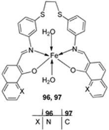
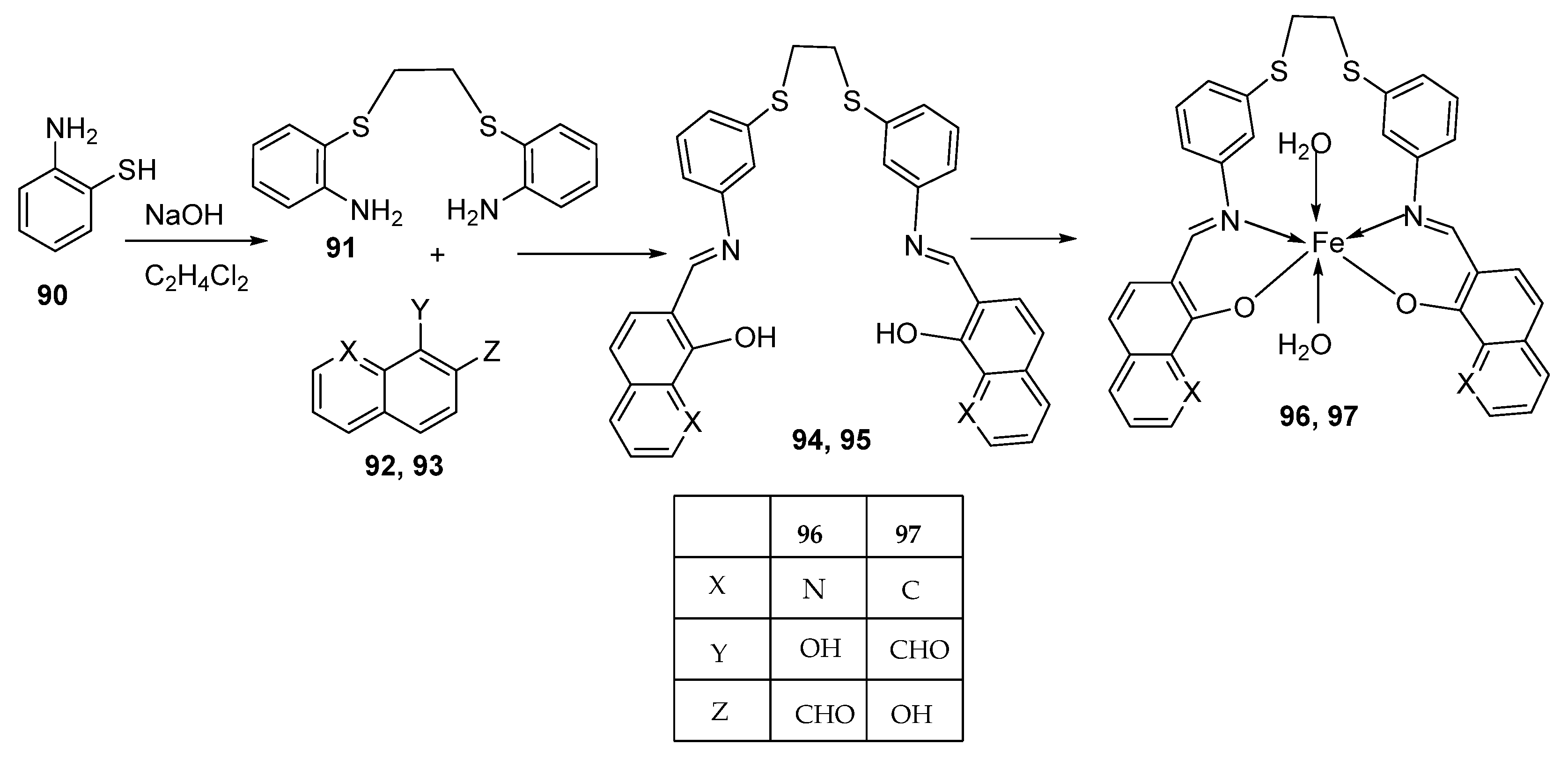
Alosaimi et al. synthesized two symmetrical imine ligands (94 and 95) and reacted each with FeCl3·6H2O to form mononuclear octahedral Fe(III) complexes 96 and 97 (Scheme 18). The complexes were screened for their antibacterial activity against Gram-positive bacterial strains S. epidermidis, S. aureus, and E. faecalis and Gram-negative bacterial strains P. aeruginosa, E. coli, and P. mirabilis. Antifungal activity was also determined against the common pathogenic fungal strain C. albicans. The tested Schiff base ligands (94 and 95) exhibited negligible antibacterial action against Gram-positive bacterial species with growth-limiting diameters of 15 mm. They also showed no antifungal activity against C. albicans. Iron complex 96 showed higher antibacterial activity against the Gram-positive bacterial strain S. epidermidis (14 mm) than iron complex 97 (with a zone of inhibition of 12 mm). The Gram-negative bacterial strain P. mirabilis was slightly inhibited by both iron complexes, 96 (8 mm) and 97 (22 mm), but all the other strains were resistant to both complexes. The complexes exhibited low antibacterial activity compared to the standard antibiotic agent, amoxicillin. The antibiotic agent inhibited S. epidermidis and P. mirabilis with zones of inhibition of 28 mm and 44 mm, respectively. The fungus C. albicans was resistant to both iron complexes and showed no significant antifungal activity (Table 2). Overtone’s permeability concept and Tweedy’s chelation theory can both be used to explain why coordination compounds have more activity than their parent ligands [46,56]. The complexes become more permeable when a metal ion is present because they dissolve in lipids and enter the cell more readily, causing negative changes in the cell environment and its enzymes, further hindering the microbe’s growth. Additionally, the metal complexes impede the production of proteins by impeding the cell’s respiration process, further inhibiting the organism’s growth. Additionally, the probability of hydrogen bonds forming between the azomethine linkage and the cell components will negatively impact the cell’s normal functions [57,58].
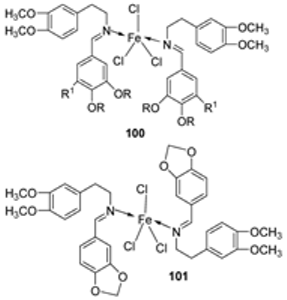
Iron(III) was complexed with two imine ligands (98 and 99, Scheme 19) by Naureen et al. to form iron complexes 100 and 101. The ligands and their complexes were evaluated for their antibacterial activity against Gram-positive P. aeruginosa and Gram-negative E. coli and S. aureus using tetracycline as the standard drug. Their antifungal activity was also evaluated against C. albicans and C. glabrata with nystatin as the standard drug. The antimicrobial activity of the synthesized complexes was enhanced when compared to their free Schiff ligands. Both complexes showed similar inhibition against all the bacterial strains used in this research, but 101 showed better activity against S. aureus (20 mm) and C. albicans (24 mm) compared to 100, which showed a zone of inhibition of 16 mm and 20 mm, respectively. The complexes showed low antibacterial activity when compared with the standard drug tetracycline. Both complexes showed higher antifungal activity against C. albicans than the standard drug nystatin (19 mm) and could be investigated as promising antifungal drug candidates (Table 2) [59]. The chelation theory can explain the improved activity of the complexes compared to their free Schiff ligands [46].
Singh et al. synthesized an imine ligand by the condensation of compounds 102 and 103 in the molar ratio of 2:1, respectively. The synthesized ligand (104) was complexed with iron to form an octahedral 105 (Scheme 20) and tested against S. epidermidis, E. coli, A. flavus, A. niger, and C. lunata to validate its antibacterial and antifungal potentials. The complex showed better antibacterial activity against the selected bacterial and fungal strains than the Schiff base ligand. The complex showed the highest activity against A. niger (16 mm) and low activity against E. coli fungal strains (15 mm) (Table 2) [60]. The improved activity of the complexes in relation to their ligands can be explained based on Overtone’s concept and Tweedy’s chelation theory [46,56].
Kavitha et al. evaluated 34 (Scheme 5) for its antibacterial and antifungal activity against Gram-positive Staphylococcus Sp. and Bacillus Sp. as well as Gram-negative E. coli and Pseudomonas bacterial strains and fungal strains Macrophamina phaseolina (M. phaseolina) and Sclerotium rolfsii (S. rolfsii). The iron complex 34 showed enhanced biological activity against the bacterial and fungal strains, B. subtilis (4 mm), E. coli (4 mm), and M. phaseolina (14 mm) compared to the ligand (33), which showed an activity of 1, 1, and 8 mm, respectively. However, these activities of the complex are low when compared to the standard antibiotic streptomycin and the standard antifungal agent mancozeb (Table 2). The concept of Overtone explains the increased activity of complexes [51].
Borase et al. synthesized a pyridine imine transition metal complex of Fe(III) (109, Scheme 21) by reacting metal salts (FeCl3) with compound 108. The complex was evaluated for its antibacterial and antifungal potency against Gram-positive bacteria S. aureus and Gram-negative bacteria E. coli, as well as three fungal strains, C. albicans, A. niger, and F. moniliforme. The iron complex (109) showed potent antifungal activity against A. niger (15.80 mm) when compared to the standard amphotericin-B (15.78 mm). Complex 109 showed low antifungal and antibacterial activity against C. albicans (7.44 mm) and S. aureus (3.02 mm). The complex was resisted by E. coli and F. moniliforme (Table 2). The antimicrobial activities of the ligand were not provided in this study, so a comparison could not be made [61].
Deshmukh et al. reported an imine ligand and used it to synthesize the Fe(III) complex 110 (Figure 1) and analyzed its antimicrobial activity against Gram-positive S. aureus and S. pyrogenes and Gram-negative E. coli and S. typhi pathogens. The complex showed the highest activity against S. aureus (22 mm) and the least activity against E. coli (16 mm) (Table 2). The results of antibacterial activity were not compared with ligands or standard drugs, and hence a comparison could not be made [62].
Savcı et al. synthesized a Schiff base ligand (112) and complexed it with FeCl2⋅4H2O to form the transition metal imine–iron complex 113 (Scheme 22). Compounds 111, 112, and 113 were evaluated for their antimicrobial activity against Gram-positive B. subtilis, S. aureus, and B. megaterium and Gram-negative E. aerogenes, E. coli, P. aeroginosa, and K. pneumonia bacterial strains and fungal strains C. albicans, Y. lipolytica, and S. cerevisiae. The results indicate that both 111 and 112 show better antibacterial activity against B. subtilis ATCC 6633 (zone of inhibition of 40 mm and 30 mm, respectively) than the synthesized iron complex 113 (21 mm) at a concentration of 0.2 mg/mL. The complex showed antibacterial activity against E. aerogenes (30 mm) and P. aeruginosa (36 mm) only at an elevated concentration of 1 mg/mL but did not show significant antifungal activity against the selected fungal strains (Table 2). Only 111 showed activities against K. pneumonia(36 mm) at a 0.2 mg/mL concentration. Compounds 111 and 112 were found to have superior antibacterial activity compared to all the standard antibiotic drugs against B. subtilis, B. megaterium, E. aerogenes, and P. aeruginosa. The sizes and load distributions of the metal ions, the shape of the metal chelate, and the potential for redox, as well as the increased lipophobicity of the molecules, may all affect the impact of the metal complexes on microbes [63]. However, it does not appear to be possible to simply attribute the bactericidal activity to the metal complex structure [64,65].
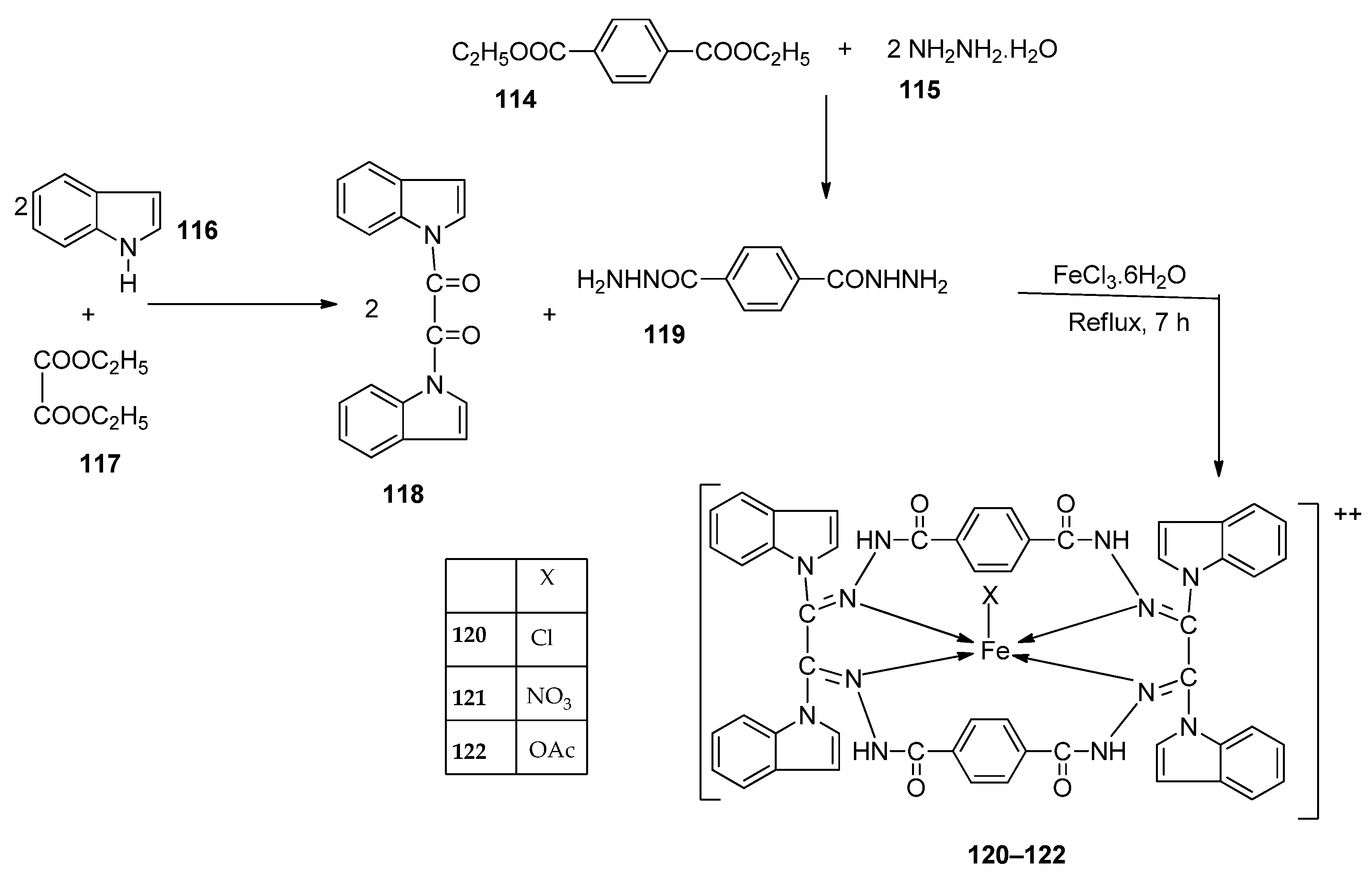
Kumar et al. synthesized an imine ligand and complexed it with FeCl3·6H2O, Fe(NO3)3·9H2O, and Fe(OAc)3·2H2O to form iron complexes 120, 121, and 122 (Scheme 23), respectively. The synthesized complexes were screened for their antimicrobial activity against S. aureus and B. subtilis (as Gram-positive bacteria), P. aeruginosa, E. coli, and Salmonella typhi (as Gram-negative bacteria), and fungi Rizoctonia sp., Aspergillus sp., and Penicillium Sp. Complex 122 demonstrated the highest antibacterial activity against S. aureus (62 mm) and P. aeruginosa (65 mm). Complex 120 showed the highest activity against E. coli (41 mm) and S. typhi (42 mm). The antibacterial activity of complexes 121 and 122 was higher against the Gram-positive bacteria than against the Gram-negative bacteria (Table 2), and this is due to the difference in the structure of the cell walls. Gram-negative cells have more complex cell walls than Gram-positive ones (Table 2). The results for antifungal screening show that 122 has high antifungal potency against Aspergillus sp (80 mm) and Penicillium Sp. (66 mm), even better than that of the standard drug miconazole with an inhibition of 57 mm and 65 mm, respectively, at a concentration of 1.0 mg/mL. Complexes 120 and 121 showed moderate antifungal activity toward the selected strains (Table 2). Generally, the ligand demonstrated moderate activity and the complexes displayed moderate-to-high activity toward all the organisms compared to standard drugs. This could be due to the presence of the -NH group, which is believed to impart the biological system’s transformation reaction and plays a significant role in biological activity. Chelation theory also explains the enhanced activity of the complexes compared to the ligand [46,66].
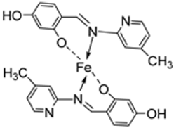
Mohamed et al. synthesized the novel octahedral iron–imine complex 123 (Figure 1) and evaluated its antimicrobial potential on the bacterial strains Clavibacter michiganensis, Xanthomonas campestris, and Bacillus megaterium and fungal strains Monilinia fructicola, Penicillium digitatum, and Colletotrichum acutatum. The free Schiff base ligand showed better antibacterial activity against all the selected bacterial strains than its iron complex. The ligand exhibited higher antibacterial activity against C. michiganensis (32 mm) than the standard drug tetracycline (30 mm). It also showed similar activity to tetracycline against B. megaterium and X. campestris (Table 2). Also, complex 123 (Figure 1) showed enhanced antifungal activity against M. fructicola, (62.5 mm) and P. digitatum (62.5 mm) compared to both the free Schiff base ligand (36.0 and 28.0 mm, respectively) and the standard antifungal agent azoxystrobin (45.3 and 58.1 mm, respectively) and can be considered as an antifungal drug candidate. The microbicide impact of the investigated compounds may result from the chemical structure of the free ligand as well as the toxicity of the investigated metal ions [67,68]. The increased antimicrobial activity of freshly synthesized metal chelates was explained by the principle of cell permeability of the microbes [35].
The imine–iron complex 124 (Figure 1) was synthesized by Elshafie et al., and its biological activity was evaluated against both human and phytopathogens. Antimicrobial analysis was conducted on pathogenic bacterial strains E. coli, B. cereus, Pseudomonas fluorescens, and P. aeruginosa and phytopathogenic fungi Monilinia fructicola, Aspergillus flavus, Penicillium italicum, and Botrytis cinerea. The antibacterial activity of 124 was dose-dependent. It showed the highest antibacterial activity against B. cereus with a measured zone of inhibition of 14 mm at a concentration of 100 µg/mL, higher than that of both the ligand (12 mm) and tetracycline (12 mm). Complex 124 inhibited the growth of P. aeruginosa (8 mm) and P. fluorescens (12 mm) only at a higher concentration of 200 µg/mL. Generally, the free imine showed better antibacterial activity than the metal complex 124. Complex 124 exhibited no antifungal activity against M. fructicola. Still, it showed enhanced activity against B. cinerea (6.7 mm) at a concentration of 400 µg/mL, whereas at the same concentration, it was resisted by the free ligand. The activity of both the Schiff base ligand and the complex was low when compared to the standard natural antifungal drug cycloheximide (Table 2). The acquired antimicrobial test findings demonstrated that the tested ligands and their metal complexes have the capacity to suppress the growth of all strains under study in a dose-dependent manner. In particular, the chemical structure of the free ligand itself and the toxicity of the metal ions under study could both contribute to the fungicidal effects of the compounds under study [63,64]. Chelation theory can also explain the enhanced activity of the complex. Also, the investigated gemifloxacin ligand and its metal complexes’ capacity to block the DNA gyrase and DNA topoisomerase IV enzymes may potentially be related to their antifungal and antibacterial action [69,70].
Ismail et al. synthesized an imine–iron complex (30, Scheme 4) and evaluated it for its in vitro antibacterial activity against Gram-positive bacteria S. aureus and B. subtilis, Gram-negative bacteria P. vulgaris and E. coli, and fungi A. flavus and C. albicans. The imine ligand exhibited better antibacterial and antifungal activity against all the microbial strains studied than 30. Complex 30 showed lower antibacterial and antifungal activity against S. aureus, E. coli, and C. albicans with a zone of inhibition of 17, 19, and 15 mm, respectively, compared to the selected antibacterial standard drugs gentamycin (S. aureus = 24 mm and E. coli = 30 mm) and ketoconazole (C. albicans = 20 mm). The ligand had high action against C. albicans, displaying antimicrobial activity (25 mm) superior to that of the ketoconazole standard (20 mm). Additionally, the ligand’s inhibition zone value against B. subtilis is 25 mm, which is comparable to the standard gentamycin (26 mm) (Table 2). The reason for the reduced antimicrobial efficiency of the complex was not stated [29].
Abdelrahman et al. evaluated complexes 38, 39, and 40 (Scheme 6) for their antimicrobial activity against Gram-positive bacteria S. aureus and B. subtilis, Gram-negative S. typhimurium and E. coli bacteria, and unicellular C. albicans and multicellular A. fumigatus fungi. The free ligand was ineffective against all the studied microbes except for C. albicans, which had an inhibition zone of 8 mm. Iron complexes 38 and 40 showed no activity against the selected bacterial strains. Complexes 38 and 39 showed moderate antifungal activity against C. albicans with an inhibition zone of 14 mm and 22 mm, respectively; these values were high when compared to the free ligand, which showed an inhibition of 8 mm. The lipophilicity of compounds significantly influences the antimicrobial activity. The enhanced antimicrobial activity of the complexes in relation to the ligand is due to chelation theory. Chelation results in an increase in the lipophilicity of the metal complexes, causing the concentration of complexes in the lipid membrane to increase and reducing microorganism multiplicity. It is hypothesized that the complexes’ antifungal effects result from either killing the bacteria or preventing their growth by obstructing their active sites [31,71].
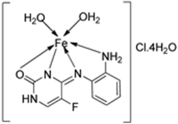
Ahmed et al. synthesized the imine–iron complex 125 (Figure 1) in a 1:1 ratio with the ligand. The synthesized compounds were tested for their antimicrobial activity against the Gram-positive bacteria S. aureus and Gram-negative bacteria E. coli, as well as fungal strains C. albicans and A. flavus. All the selected microbes resisted the ligand except E. coli, against which it showed an inhibition zone of 9 mm, a value higher than that of the antibacterial drug amikacin (6 mm). Complex 125 showed the same zone of inhibition (10 mm) as the standard drug amikacin against S. aureus and enhanced activity against E. coli (10 mm) compared to the same standard drug. Several factors could be responsible for the remarkable antibacterial activity of the complex, including interference with the creation of the cell wall, harm because of which the permeability of the cell may be changed, or disorganization of the lipoprotein, resulting in cell death. Also, different cellular enzymes, essential in the metabolic pathways of microbes, could be deactivated. Another factor could be the formation of a hydrogen bond between the azomethine group and the active center of the cell’s components, interfering with proper cell function [72].
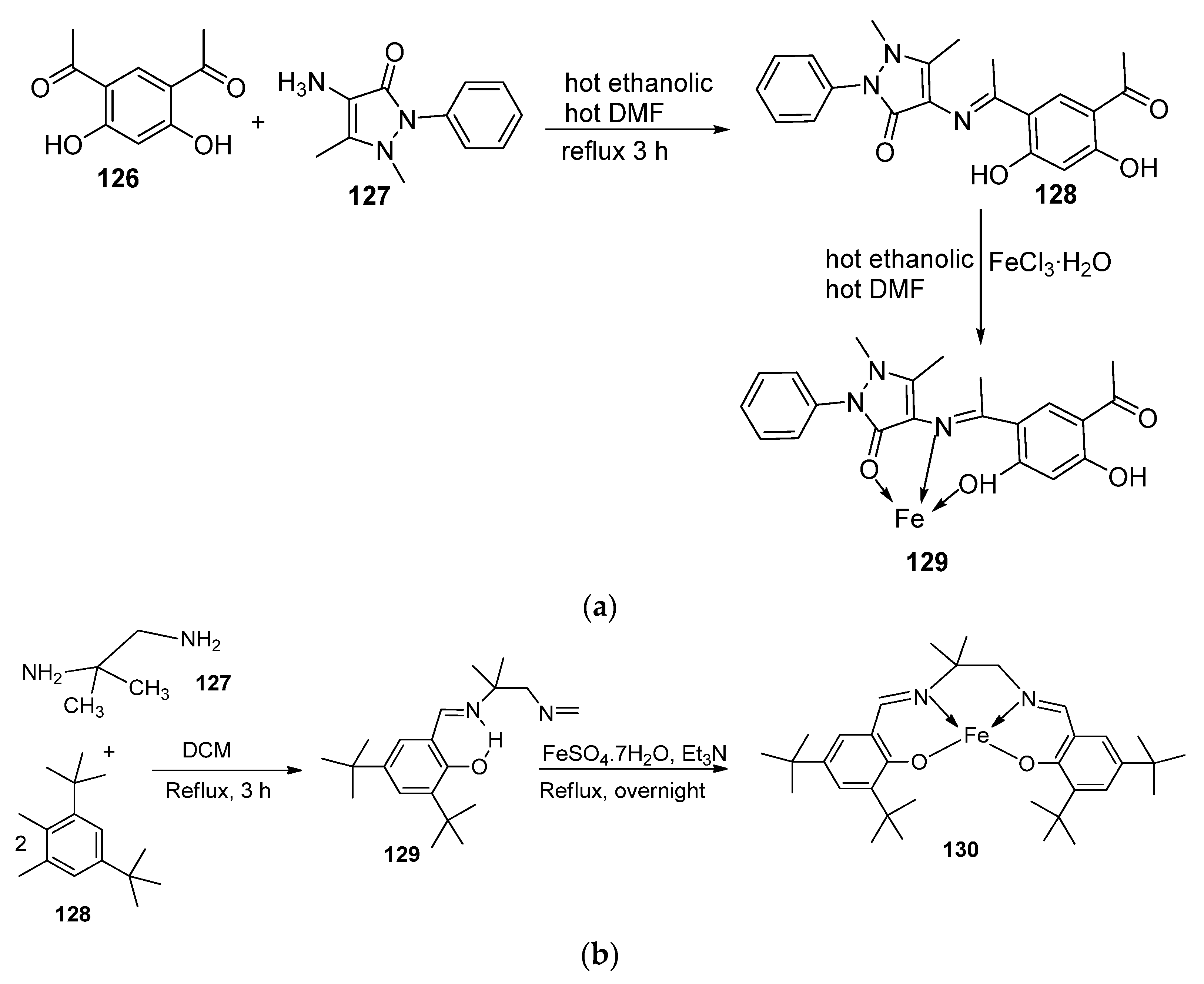
A mononuclear chelate of iron(III) was synthesized by Mohamed et al. by condensing a new tridentate Schiff base ligand (128) with iron chloride (FeCl3·H2O) in a 1:1 ratio. The complex formed (129) had an octahedral geometry. The in vitro antimicrobial potency of the synthesized complex (129) was evaluated against Gram-negative bacteria E. coli and Gram-positive bacteria S. aureus and fungal strains C. albicans and A. flavus. Complex 129 showed a broad zone of inhibition (14 mm/mg sample) against A. flavus, whereas the free Schiff base ligand demonstrated zero activity. This activity was much higher than that of ketoconazole (8 mm/mg sample), the selected standard antifungal agent. The enhanced microbial activity of the complex can be attributed to the increased lipophilicity of the metal complex upon coordination with the free ligand. This ensures the easy movement of the metal chelate into the fungal cell membrane, inhibiting microbial growth or distorting its active site [73,74]. For the other microbial strains, the Schiff base ligand showed activity similar to its free ligand (Table 2). The reason for the reduced activity of the Schiff base against E. coli, S. aureus, and C. albicans was not stated [75].
Hidayati et al. synthesized an N-(2-hydroxybenzylidene) chitosan Schiff base and its iron(II) complex and evaluated them for their antibacterial potency. Chitosan (poly-β-(1→4)-glucosamine) is a very abundant non-toxic natural biopolymer, and its metal complexes are known to exhibit very good biological activities. Hidayati et al. evaluated chitosan, the synthesized chitosan Schiff base ligand, and its imine complex for their ability to inhibit the growth of E. coli and S. aureus and found—at a concentration of 1000 ppm—the complex being most active against both bacterial strains (9.86 mm and 10.16 mm, respectively), followed by the chitosan Schiff base (9.50 mm and 9.33 mm, respectively) and lastly the chitosan itself (8.75 mm and 9.25 mm, respectively). The observed improvement in the antibacterial activity of the chitosan Schiff base–iron complex can be explained by chelation, which enhanced the lipophilic nature of the complex, ensuring its faster diffusion across bacterial cell membranes [9,76].
Table 2. Product, synthesis conditions, and in vitro antimicrobial activity of imine–iron complexes compared to the respective positive controls .
No.Complex No.Structures of Synthesized ComplexesReaction ConditionsAntimicrobial Biological Activity Ref.
1.51Ijms 25 02263 i009Stirring, 30 min
Reflux, 7 h
Zone of inhibition, mm[40]
S. aureusE. coliP. aeruginosaB. cereus
5011101112
5114141429
Tetracycline9101211
2.55Ijms 25 02263 i010Stirring, 1–2 h
Reflux, 2–11 h
Zone of inhibition, mm[42]
E. coli
5423
5529
Amoxicillin41
Chloramphenicol39
3.34Ijms 25 02263 i011Reflux, 2 hMinimum Inhibitory Concentration (MIC)/µg/mL[21]
BacteriaFungi
S. marcescenceE. coliM. luteusG. candidumA. flavusF. oxysporum
17.257.256.256.758.007.50
25.506.254.755.256.756.25
33.754.253.004.004.504.25
43.253.502.503.003.753.50
4.59Ijms 25 02263 i012Stirring
Reflux, 1 h
Minimum Inhibitory Concentration (MIC)/µg/mL[47]
E. coliPseudomonasS. aureusBacillus
582.581517
5925RR** R
5.63
64
Ijms 25 02263 i013Stirring, 1 h
Reflux, 9 h
Zone of inhibition, mm[48]
Bacillus subtilisE. coli
621115
631211
641418
Amoxicillin1620
6.67Ijms 25 02263 i014Reflux, 5 hZone of inhibition, mm[65]
S. vE. spS. aE. fMRSA
6515241617R **
672030252215
Standard19364536R
7.68Ijms 25 02263 i015Reflux, 4 hMinimum Inhibitory Concentration (MIC)/µg/mL[10]
C. albicansC. neoformansS. aureusB. cereusE. coli
680.01560.00780.06250.03120.0625
Nystatin0.0320.032
Miconazole0.0160.0162
Furacillinum
Ciprofloxacin0.00460.00460.0046
Amikacin0.0010.0003 0.008
8.69Ijms 25 02263 i016Reflux, 2 hZone of inhibition, mm[51]
E. coliE. aerogenesC. butyrium
Ligand141212
6912109
Standard1179
9.73Ijms 25 02263 i017Stir and reflux, 1 hZone of inhibition, mm[52]
S. pneumoniaS. aureus
727–101–3
737–107–10
10.76Ijms 25 02263 i018Stir and reflux, 2 hZone of inhibition, mm[4]
S. aureusE. coliP. aeruginosaC. albicansA. fumigatus
751514161315
761614151618
Ampicillin23 16
Gentamycin
Amphotericin192523
11.79Ijms 25 02263 i019Reflux, 4 hZone of inhibition, mm[53]
S. aureusP. aeruginosa
7886
791411
Ampicillin14
Choloramphenicol8
12.82Ijms 25 02263 i020Stir (overnight)Minimum Inhibitory Concentration (MIC)/µg/mL[2]
E. coliS. aureusC. albicansA. niger
8210101010
Gentamicin1010
Fluconazole2020
13.83Ijms 25 02263 i021Reflux and stirring, 3 hZone of inhibition, mm[54]
E. coliP. aeruginosaS. aureusC. albicansF. solani
LigandRR12** R** R
8314812712
14.87Ijms 25 02263 i022Stirring, 6 hMinimum Inhibitory Concentration (MIC)/µg/mL[3]
E. coliC. albicansP. vulgarisK. pneumoniaS. aureus
8612.54>501>50
87˃508>50>50>50
Doxymycin
Fluconazole
2 244
2
15.88
89
Ijms 25 02263 i023Stir, 30 min
Reflux, 6 h
Zone of inhibition, mm[55]
S. aureusP. phaseolicolF. oxysporium
Ligand22 ± 0.213 ± 0.117 ± 0.2
8837 ± 0.426 ± 0.131 ± 0.2
8932 ± 0.223 ± 0.130 ± 0.2
Cephalothin42
Chloramphenicol36
Cycloheximide 40
16.96
97
Ijms 25 02263 i024Reflux and stirring, 4–5 hZone of inhibition, mm[57]
S. epidermidisE. faecalisS. aureusP. mirabilisC. albicans
94597** R** R
95689** R** R
961415128** R
97128722** R
Amoxicillin28262744
17.100
101
Ijms 25 02263 i025Reflux and stirring, 50 minZone of inhibition, mm[59]
E. coliP. aeruginosaC. albicansS. aureusC. glabrata
981115151911
991212121812
10022192016<10
1012016132012
Tetracycline2520 23
Nystatin19 16
18.105Ijms 25 02263 i026Reflux, 15 hZone of inhibition, mm[60]
E. coliS. epidermidisA. nigerA. flavusC. lunata
104** R611910
1051515161415
19.34Ijms 25 02263 i027Reflux, 8–9 hZone of inhibition, mm[30]
BacillusStaphylococcusE. coliS. rolfsiiM. phaseolina
3311128
34434614
Streptomycin9115
Mancozeb1824
20.109Ijms 25 02263 i028Reflux, 4–5 hZone of inhibition, mm[61]
S. aureusE. coliA. nigerC. albicansF. moniliforme
1093.02** R15.807.44** R
Chloramphenicol15.1125.44 23.23
Amphotericin15.7812.58
21.110Ijms 25 02263 i029Reflux, 15–16 hZone of inhibition, mm[62]
S. pyrogenesE. coliS. typhi
110251619
22.113Ijms 25 02263 i030Reflux, 6 hZone of inhibition, mm (concentration, mg/mL)[64]
B. subtilisB. megateriumP. aeroginosaK. pneumoniaE. aerogenes
11140±0.47(0.2)34±0.81(0.2)42±1.24(1)36±0.47(0.2)45 ± 0.00
11230 ± 0.81(0.2)22±0.81(0.5)33±0.81(0.2)** R28 ± 0.00
11321 ± 0.00(0.2)** R36±1.24(1)** R** R
Erythromycin20 ± 0.0025±0.4719±0.4719±0.00 27 ± 1.24
23.120
121
122
Ijms 25 02263 i031Reflux, 8 hZone of inhibition, mm[66]
S. aureusP. aureginosaE. coliS. typhiiAspergillus sp.Penicillium sp.
119360810104829
120303641426861
121242522285154
122626533358066
Imipenem1001001001005765
Miconazole
24.123Ijms 25 02263 i032Reflux, 3 hZone of inhibition, mm[35]
X. campestrisB. megateriumC. michiganensisM. fructicolaP. digitatum
Ligand30283236.0 ± 3.128.0±3.5
12326192062.5 ± 6.262.5 ± 8.2
Tetracycline342830
Azoxystrobin45.3 ± 2.158.1 ± 1.2
25.124Ijms 25 02263 i033Reflux, 3 hZone of inhibition, mm (concentration, µg/mL)[69]
E. coliB. cereusP. fluorescensB. cinereaA. flavus
Ligand2012110.0 ± 0.00.00 ±0.0
1241212186.7 ± 2.36.7±2.6
Tetracycline14108
Cycloheximide42.2±2.69.7±3.0
26.30Ijms 25 02263 i034Stir and reflux, 4 hZone of inhibition, mm[29]
S. aureusB. subtilisE. coliC. albicans
Ligand19252425
3017161915
Gentamycin242630
Ketoconazole20
27.125Ijms 25 02263 i035Stirring and reflux, 1 hZone of inhibition, mm[72]
S. aureusE. coli
Ligand0.009
1251010
Amikacin106
28.38
39
40
Ijms 25 02263 i036Stirring, 2 h
Reflux, 12–15 h
Zone of inhibition, mm[31]
S. typhimuriumC. albicans
Ligand** R8
38** R14
39 1522
40** RR
Cephalothin 36
Cycloheximide 35
29.129Ijms 25 02263 i037Stirring and reflux, 1 h Zone of inhibition, mm/mg[75]
E. coliS. aureusC. albicansA. flavus
1281412100
12913111214
Amikacin610--
Ketoconazole--98
Values written as these have been reported in the literature. ** R = 0 = Resistant.

2.3. Imine–Iron Complexes as Antioxidants

Oxidative metabolism is one of the crucial factors for cell survival. Free radicals and other reactive oxygen species (ROS) are produced because of this reliance, which leads to oxidative alterations. When too many free radicals are produced, the ROS concentration becomes above average, which can overwhelm protective enzymes and have detrimental and fatal effects on cells by oxidizing membrane lipids, cellular proteins, DNA, and enzymes, which stops cellular respiration [18]. The way to counter the reaction of these free radicals is to introduce an antioxidant, which can be experimentally carried out using assays such as DPPH [77]. Compounds of metal chelates like iron–imine complexes offer advantages over conventional synthetic antioxidants because of the different geometry, oxidation states, and coordination numbers of metal chelates that support and promote the redox processes linked to antioxidant action. In its high oxidation state, the metal ion’s ability to extract electrons influences the antioxidant activity of the resultant complex by moving the ligand’s electron density to the metal center, where it functions as a modulator agent. When the metal is in its low oxidation state, it behaves in the opposite way. These actions significantly alter the ligand’s electrical charge distribution, facilitating the loss of electrons and raising the complex’s capacity to scavenge radicals. A metal ion like iron (Fe), which is found in many biologically privileged structures and essential to an organism’s ability to function, is useful in constructing novel chelate antioxidants because it lowers toxicity [78].
Turan et al. synthesized an imine ligand and its octahedral imine–iron(II) complex 126 (Figure 1) and evaluated their in vitro antioxidant activity using 2,2-diphenyl-1-picryl-hydrazyl-hydrate (DPPH) radical scavenging, 2,2’-azino-bis(3-ethylbenzothiazoline-6-sulfonic acid) (ABTS) cation radical scavenging, and the ferric reducing ability of plasma (FRAP). In the ABTS assay, a compound’s antioxidant ability is measured based on the reduction of ABTS•+ cation radicals [79]. Complex 126 (0.6) demonstrated weak ABTS•+ radical scavenging activity, while the parent ligand exhibited no discernible ABTS•+ radical scavenging activity. The molecule’s structure and single electron transfer potential influence the results. The complex showed a more enhanced DPPH radical scavenging ability (1.25) than the ligand (1.35) itself, but this activity was moderate when compared with the standard drugs (0.10–0.31). The FRAP method measures a compound’s ability to cause the reduction of ferric ions (Fe3+) to ferrous ions (Fe2+). The ligand (0.5) showed an antioxidant ability similar to that of 126 (0.4) in this assay (Table 3). The antioxidant potency of a series of compounds has been studied for the potential that they can be influenced by the aromatic ring and the number of hydroxyl groups present in a compound [80,81].
El-Lateef et al. explored imine–iron complexes 3 and 4 (Scheme 1) for their antioxidant activity using the DPPH assay. The results revealed that the free ligand and its metal complexes have better antioxidant activity than the standard antioxidant agent vitamin C (55 μg/mL). The complexes showed enhanced activity compared to the free ligands (1 = 45µg/mL, 2 = 32 µg/mL), with 3 possessing the highest DPPH free radical scavenging ability with an IC50 value of 22μg/mL (Table 3). The results indicate that the complexes had greater antioxidant effects against the DPPH free radical than standard vitamin C and can be considered antioxidant drug candidates. This study did not state the reason for the exceptional activities of the ligands and complexes [21].
Naureen et al. explored the antioxidant activity of 100 and 101 (Scheme 19) using the DPPH assay. The free imine ligands 98 and 99 demonstrated better antioxidant activity (1.23 and 1.02 μg/mL, respectively) than their iron complexes, 100 (1.70 μg/mL) and 101 (1.41 μg/mL). The free ligand 99 showed better antioxidant activity (1.02 μg/mL) than the standard vitamin C (1.14 μg/mL). Generally, both the free ligand and iron complexes exhibited good free radical scavenging abilities (Table 3). The modes of action of the ligands and their complexes were not outlined [59].
The tetradentate Schiff base 129 was synthesized along with its Fe complex 130 (Scheme 24) by Said et al. An in vitro antioxidant activity was determined using the DPPH radical scavenging, ferric thiocyanate (FTC), hydroxyl radical scavenging activity (HRSA), and hydrogen peroxide scavenging activity methods. Complex 129 demonstrated a better free radical scavenging ability than the synthesized complex 130 in the DPPH radical scavenging, FTC, and HRSA methods with IC50 values of 53.55, 48.81, and 63.43, respectively, whereas those of 130 were 44.65, 9.47, and 30.29. The complex showed moderate activity compared to the standard Trolox and BHA in the DPPH radical scavenging, FTC, and HRSA methods (Table 3). It, however, demonstrated a better ability (93.74 μg/mL) to remove H2O2 from the reaction mixture than 129 (92.52 μg/mL) and the standards Trolox (91.80 μg/mL) and BHA (92.97 μg/mL) when the hydrogen peroxide scavenging activity method was employed (Table 3). Due to the presence of the hydroxyl group on the ligand, its antioxidant activity was expected to be higher than that observed in this study, and this may be due to the steric hindrance or the presence of bulky donating groups (or both), making it challenging for the ligand to supply the hydrogen atom (H) to the DPPH radical [82].
Hayder et al. synthesized the new imine ligand 135 and its octahedral imine–iron complex 136 (Scheme 25). The antioxidant activity of 135 and 136 was evaluated using the DPPH radical scavenging activity method. The iron complex showed an enhanced ability to scavenge DPPHꞏ radicals (49% scavenging) than the free ligand (24% scavenging). Compared to the standard ascorbic acid (82% scavenging), the complex showed a moderate ability to scavenge the free radicals in the reaction mixture (Table 3) [16].
Elshafie et al. evaluated complex 124 (Figure 1) for its in vitro antioxidant activity. The free imine ligand and complex 124 both showed a high antioxidant activity (164.6%), with the iron complex being slightly higher than the ligand (169.7%). Complex 124 can donate hydrogen to scavenge the free radicals, hence reducing the oxidation process (Table 3) [69].
Borase et al. conducted an antioxidant assay on the metal complex 109 (Scheme 21) to determine its free radical scavenging ability, and it proved to have moderate antioxidant activity (1615.22 μg/mL) (Table 3). The results of the antioxidant activity of ligands were not given, and subsequent comparisons could not be made [61].
Savcı et al. investigated 111, imine ligand 112, and its imine–iron complex 113 (Scheme 22) for their antioxidant activity using DPPH radical scavenging, total antioxidant activity, FRAP, and CUPRAC activity. The results obtained revealed that the iron complex 113 (0.7) had a high ability to remove DPPH from the reaction mixture when compared to 111 (1.9), 112 (0.8), and the standard BHT (1.1). For the total antioxidant activity assay, both 112 (0.62)) and 113 (0.61) showed a similar potential to the standard BHA (0.60) in eliminating lipid peroxide from the reaction mixture and an enhanced potential compared to the standard BHT (0.40). In the FRAP assay, 111 (0.06) showed the lowest reduction capacity in reducing the Fe3+ ions, followed by the standard BHT (0.08), complex (0.11), and BHA (0.2), and the ligand 112 (0.38) showed the highest activity. Finally, the CUPRAC method confirmed the results of the other assays, with 111 indicating the lowest antioxidant activity (Table 3). Most of the inhibitor’s antioxidant effect comes from its ability to donate one electron or hydrogen to the radical centers formed in biological systems, thus neutralizing them. The inhibitor’s structure and characteristics are critical factors in demonstrating activity [49]. Potential sites for biochemically active substances connected to the balance of molecular proton transfer and hydrogen bonds can be found in the Schiff bases. The biological activity of the Schiff base [83] is typically increased by complexes formed with transition metals. Hence, the good antioxidant activity of both the ligand and complex was achieved in this study [84].
Table 3. Products, synthesis conditions, and antioxidant activity of selected imine–iron complexes using DPPH, H2O2SA (hydrogen peroxide scavenging activity assay), %RSA (radical scavenging activity), and total antioxidant assay (TAC) .
Entry No.Complex No.StructuresReaction ConditionAntioxidant Activity (IC50/µg/mL)Ref.
1.3
4
Ijms 25 02263 i038Reflux (2 h) DPPH[21]
145
322
253
432
Vit C55
2.100
101
Ijms 25 02263 i039Reflux and stirring (50 min) DPPH [59]
981.23
1001.70
991.02
1011.41
Vit C1.14
3.126Ijms 25 02263 i040Reflux (10 min) and
stirring (24 h)
ABTS
(734 nm)
DPPH
(517 nm)
FRAP
(700 nm)
[81]
Ligand1.901.350.50
1260.601.250.40
Ascorbic acid0.000.102.10
BHA0.000.182.90
BHT0.000.312.30
4.130Ijms 25 02263 i041Reflux and stir (overnight) DPPHH2O2 SAFTCHRSA[82]
12953.55 ± 2.9592.52 ± 0.0748.81 ± 5.0463.43 ± 5.66
13044.65 ± 1.1093.74 ± 0.439.47 ± 2.19130.29 ± 0.81
Trolox85.42 ± 0.0491.80 ± 1.7790.45 ± 6.7057.72 ± 1.62
BHA75.69 ± 0.1192.97 ± 0.9850.57 ± 5.4210.00 ± 3.64
5.136Ijms 25 02263 i042Reflux (3 h) DPPH (% scavenging)[16]
13524
13649
Ascorbic acid82
6.124Ijms 25 02263 i043Reflux (3 h) %RSA[69]
Ligand169.7
124164.6
7.109Ijms 25 02263 i044Reflux (4–5 h) DPPH[61]
1091615.22
8.113Ijms 25 02263 i045Reflux (6 h) DPPHTotal antioxidantFRAPCUPRAC[64]
1111.90.640.060.30
1120.80.620.383.50
1130.700.610.111.20
BHT0.600.600.083.20
BHA1.200.440.203.10
Values are written as reported in the literature. DPPH: 2,2-diphenylpicrylhydrazyl, FTC: ferric thiocyanate, FRAP: Ferric Reducing Antioxidant Power, CUPRAC: CUPric Reducing Antioxidant Capacity, BHA: beta hydroxy acid, BHT: butylated hydroxytoluene, HRSA: hydroxyl radical scavenging activity.

2.4. Other Pharmacological Activities of the Imine–Iron Complexes

Iron–imine complexes have been found to possess anti-inflammatory properties, and their mechanism of action has been studied extensively. One of the primary mechanisms by which these complexes exhibit anti-inflammatory activity is inhibiting the production of pro-inflammatory cytokines such as TNF-α, IL-1β, and IL-6. These cytokines play a crucial role in the inflammatory response by recruiting immune cells to the site of inflammation and activating them. By inhibiting the production of these cytokines, iron–imine complexes can reduce the activity of immune cells and thus reduce inflammation. Additionally, iron–imine complexes have been found to inhibit the activity of cyclooxygenase-2 (COX-2), an enzyme that plays a key role in producing prostaglandins. Prostaglandins are lipid mediators that are involved in the inflammatory response, and their production is increased during inflammation. By inhibiting COX-2 activity, iron–imine complexes can reduce the production of prostaglandins and thus reduce inflammation. Iron–imine complexes have also been found to inhibit the activity of NF-κβ, a transcription factor that plays a crucial role in regulating immune and inflammatory responses. NF-κβ is activated in response to various stimuli, including pro-inflammatory cytokines, and its activation results in the transcription of genes involved in the inflammatory response. By inhibiting NF-κβ activity, iron–imine complexes can reduce the expression of inflammatory genes and thus reduce inflammation.
Kumar et al. screened imine–iron complexes 120, 121, and 122 (Scheme 23) for their in vivo anti-inflammatory activity using albino rats. All the complexes showed anti-inflammatory activity higher than that of the standard drug phenyl butazone (18.2% anti-inflammatory activity), with 122 (31.1%) exhibiting the highest activity at the same concentration of 25 mg/kg. Complex 121 showed the least anti-inflammatory activity (27.2%). Compared to the complexes, the ligand (9.0%) showed very low anti-inflammatory activity (Table 4). Complex 122 can be explored further as an anti-inflammatory drug candidate. The increased anti-inflammatory activity of the complex in relation to the ligand can be explained by the chelation theory, which describes the increase in polarity and the lipophilic nature of the complex due to chelation and how this causes it to efficiently cross the lipid layer, affecting the desired anti-inflammatory action [66].
Imine–iron complexes have shown a few other medicinal activities. Ahmed et al. screened complex 125 (Figure 1) on coronavirus (SARS-CoV-2) using molecular docking. The molecular docking studies investigated the interaction that exists between the complex and the crystal structure of the virus’s (SARS-CoV-2) main protease with unliganded active site (2019-nCoV, coronavirus disease 2019, or COVID-19) (PDB ID: 6Y84) proteins. The imine–iron complex 125 (Figure 1) had low energy, (−8.5 kcal/mol), which means it has a strong binding affinity and can inhibit the biochemical processes of the proteins, inhibiting viral capability (Table 4) [72].
Elkanzi et al. synthesized imine–iron complex 139 (Scheme 26) and screened its in vitro anti-inflammatory activity using the anti-denaturation method of egg albumin. Heat applied to the egg denatures the egg albumin, and the denatured protein produces certain antigens. These antigens are linked to type-III hypersensitivity reactions, which cause several diseases. The anti-inflammatory assay analyses test an agent’s ability to limit the denaturation process. The result obtained from this study showed that 139 exhibited a moderate percentage of inhibition (0.70) as compared to the ligand (0.13) and standard anti-inflammatory drug ibuprofen (2.9) at a concentration of 100 µg/mL (Table 4). The inverse relationship between the dipole moment of the complex and its activity explains the reduced activity of the complex. The dipole moment of the complex (10.11) is higher than that of the ligand (5.91), and this increases the polarity and decreases the lipophilic nature of the complex, lowering its efficiency to passing through the lipid layer, hence making it less efficient as an anti-inflammatory agent [83,84].
Table 4. Products, synthesis conditions, and biological activities of selected imine–iron complexes .
Entry No.Complex No.StructuresReaction ConditionsBiological ActivitiesRef
1.120
121
122
Ijms 25 02263 i046Reflux, 8 h Anti-inflammatory activity (%)[66]
1199.00
12028.50
12127.20
12231.10
Phenyl butazone18.20
2.125Ijms 25 02263 i047Stirring and reflux, 1 h Binding energy (kcal/mol)[72]
Ligand−2.1
125−8.5
3.139Ijms 25 02263 i048Reflux, 12 h % Inhibition of heat-induced denaturation of proteins[84]
1380.13
1390.70
Ibuprofen2.9
Values are written as reported in the literature.

3. Conclusions

While nitrogen is a vital macro-nutrient, iron is a significant micro-nutrient in the human body. Accordingly, the biocompatibility (possibility of being bioavailable) of iron–imine complexes in the human body is higher than that of other organometallic complexes. This article discusses the recent development of organo-iron compounds as medicinally privileged compounds. As discussed herein, some of the iron–imine complexes demonstrated good-to-excellent pharmacological activity in several dreadful diseases like different types of cancers and microbial, oxidation, and inflammation-related diseases. The observed anticancer activity of iron–imine complexes is believed to be due to their tailored delivery and different mechanisms of action, which include altering iron metabolism, producing reactive oxygen species (ROS), and blocking key enzymes. As antimicrobial agents, they outperform many conventional antimicrobial agents due to novel mechanisms of action, broad-spectrum activity, and biofilm disruption, which can be traced to the chelation process, the toxicity of metal ions against bacteria, and the improvement in the hydrophobicity and liposolubility of the molecules due to the presence of an azomethine linkage in the complex. The different geometry, oxidation states, and coordination numbers of metal chelates like iron complexes support and promote the redox processes linked to antioxidant action. One of the primary mechanisms by which these complexes exhibit anti-inflammatory activity is inhibiting the production of pro-inflammatory cytokines such as TNF-α, IL-1β, and IL-6. They also inhibit the activity of cyclooxygenase-2 (COX-2). This enzyme plays a key role in the production of prostaglandins (lipid mediators that are involved in the inflammatory response, and their production is increased during inflammation).
Iron (Fe), complex in many biological structures and essential to an organism’s ability to function, can be useful in constructing novel chelate drugs because it can possibly lower toxicity. The chemistry and biology of iron is still under investigation. Quite recently, a unique version of an iron-dependent non-apoptotic cell death procedure was reported [85,86]. Iron–imine complexes can be developed as a valuable probe for antimicrobial, antifungal, anti-inflammatory, and antioxidant drug development. Accordingly, iron–imine complexes can play a crucial role in future drug development research. In a nutshell, iron, the fourth most abundant element in the Earth’s crust, can form various non-toxic complexes with imines. Iron–imine complexes have demonstrated diverse medicinal activities, and some of these derivatives have shown promise in becoming commercial drugs in the future.

Author Contributions

D.B. conceived the idea and designed this study. J.A. collected the literature. J.A. and E.O. wrote the manuscript, and G.R. organized the manuscript. D.B. and G.R. contributed to editorial changes in the manuscript. All authors have read and agreed to the published version of the manuscript.

Funding

This research was partially funded by the core facilities of CPRIT Integrated Cancer Research Core (ICRC) (RP210180) and UT-System and part of CPRIT South Texas Center of Excellence in Cancer Research (RP230419). DB is thankful to the Agricultural and Food Research Initiative grant no. 2022-68018-36606 from the USDA National Institute of Food and Agriculture. We acknowledge the funding support from the University of Texas Rio Grande Valley Faculty SEED Grant (to D.B.) and the School of Integrative Biological and Chemical Sciences for the Departmental Grant from the Robert A. Welch Foundation (Grant No. BX-0048).

Institutional Review Board Statement

Not applicable.

Informed Consent Statement

Not applicable.

Data Availability Statement

All data are available online. No unpublished data have been used in this paper.

Acknowledgments

We thank the School of Integrative Biological and Chemical Sciences (University of Texas Rio Grande Valley) for providing infrastructural support.

Conflicts of Interest

The authors declare no conflicts of interest.

References

  1. Vija, K.J.; Abhishek, P.; Mohit, P.; Shweta, C.T.; Om, P.; Akansha, A.; Viveka, N. Schiff base metal complexes as a versatile catalyst: A review. J. Organomet. Chem. 2023, 999, 122825. [Google Scholar] [CrossRef]
  2. Manvatkar, V.D.; Patle, R.Y.; Meshram, P.H.; Dongre, R.S. Azomethine-functionalized organic–inorganic framework: An overview. Chem. Pap. 2023, 77, 5641–5662. [Google Scholar] [CrossRef]
  3. Ying, K.L.; Mohand, M.; Dominik, M.; Guy, B. An Air-Stable “Masked” Bis (imino) carbene: A Carbon-Based Dual Ambiphile. J. Am. Chem. Soc. 2023, 145, 2064–2069. [Google Scholar] [CrossRef]
  4. Chérifa, B.; Hana, F.; Amel, D.; Amel, D.; Abdesalem, K.; Abir, B.; Ahmad, S.D.; Tarek, R.; Rajesh, V.; Yacine, B. Schiff bases and their metal Complexes: A review on the history, synthesis, and applications. Inorg. Chem. Commun. 2023, 150, 110451. [Google Scholar] [CrossRef]
  5. El-Attar, M.S.; Elshafie, H.S.; Sadeek, S.A.; El-Farargy, A.F.; El-Desoky, S.I.; El-Shwiniy, W.H.; Camele, I. Biochemical Characterization and Antimicrobial Activity against Some Human or Phyto-Pathogens of New Diazonium Heterocyclic Metal Complexes. Chem. Biodivers. 2022, 19, 2. [Google Scholar] [CrossRef]
  6. Anjali Krishna, G.T.; Dhanya, M.; Shanty, A.A.; Raghu, K.G.; Mohanan, P.V. Transition metal complexes of imidazole derived Schiff bases: Antioxidant/anti-inflammatory/antimicrobial/enzyme inhibition and cytotoxicity properties. J. Mol. Struct. 2023, 1274, 134384. [Google Scholar] [CrossRef]
  7. Zhang, Z.; Song, Q.; Jin, Y.; Feng, Y.; Li, J.; Zhang, K. Advances in Schiff Base and Its Coating on Metal Biomaterials—A Review. Metals 2023, 13, 386. [Google Scholar] [CrossRef]
  8. Sohtun, W.P.; Khamrang, T.; Kannan, A.; Balakrishnan, G.; Saravanan, D.; Akhbarsha, M.A.; Velusamy, M.; Palaniandavar, M. Iron(III) bis-complexes of Schiff bases of S -methyldithiocarbazates: Synthesis, structure, spectral and redox properties and cytotoxicity. Appl. Organomet. Chem. 2020, 34, e5593. [Google Scholar] [CrossRef]
  9. Tukki, S.; Samya, B.; Akhtar, H. Significant photocytotoxic effect of an iron (iii) complex of a Schiff base ligand derived from vitamin B6 and thiosemicarbazide in visible light. RSC Adv. 2015, 5, 29276–29284. [Google Scholar] [CrossRef]
  10. Dubey, R.K.; Mariya, A.; Mishra, S.K. Synthesis and spectral (ir, nmr, fab-ms and xrd) characterization of lanthanide complexes containing bidentate schiff base derived from sulphadiazine and ovanillin. Int. J. Basic Appl. Sci. 2011, 1, 70–78. [Google Scholar]
  11. Basu, U.; Roy, M.; Chakravarty, A.R. Recent advances in the chemistry of iron-based chemotherapeutic agents. Coord. Chem. Rev. 2020, 417, 213339. [Google Scholar] [CrossRef]
  12. Satya, D.P.; Logesh, R.; Dhanabal, P.; Suresh, M.K. Importance of Iron Absorption in Human Health: An Overview. Curr. Nutr. Food Sci. 2021, 17, 293–301. [Google Scholar]
  13. Kargar, H.; Fallah-Mehrjardi, M.; Behjatmanesh-Ardakani, R.; Munawar, K.S.; Ashfaq, M.; Tahir, M.N. Diverse coordination of isoniazid hydrazone Schiff base ligand towards iron (III): Synthesis, characterization, SC-XRD, HSA, QTAIM, MEP, NCI, NBO and DFT study. J. Mol. Struct. 2022, 1250, 131691. [Google Scholar] [CrossRef]
  14. Lee, H.W.; Jeong, G.-U.; Kim, M.-C.; Kim, D.; Kim, S.; Han, S.S. Atomistic origin of mechanochemical NH3 synthesis on Fe catalysts. International Journal of Hydrogen Energy. Int. J. Hydrogen Energy 2023, 48, 3931–3941. [Google Scholar] [CrossRef]
  15. Peterson, P.O.; Joannou, M.V.; Simmons, E.M.; Wisniewski, S.R.; Kim, J.; Chirik, P.J. Iron-Catalyzed C(sp2)-C(sp3) Suzuki-Miyaura Cross-Coupling Using an Alkoxide Base. ACS Catal. 2023, 13, 2443–2448. [Google Scholar] [CrossRef]
  16. Hayder, M.; Hayder, A.M. In vitro antioxidant activity of new Schiff base ligand and its metal ion complexes. J. Pharm. Sci. Res. 2019, 11, 2051–2061. [Google Scholar]
  17. Yahaya, N.P.; Mukhtar, M.S. Synthesis, Characterization and Antibacterial Activity of Mixed Ligands of Schiff Base and Its Metal (II) Complexes Derived from Ampicilin, 3-Aminophenol and Benzaldehyde. Sci. J. Chem. 2021, 9, 9–13. [Google Scholar] [CrossRef]
  18. Antolovich, M.; Prenzler, P.D.; Patsalides, E.; McDonald, S.; Robards, K. Methods for testing antioxidant activity. Anal. R. Soc. Chem. 2002, 27, 183–198. [Google Scholar] [CrossRef]
  19. Bharti, S.; Singh, S. Metal Based Drugs: Current Use and Future Potential. Der Pharm. Lett. 2009, 1, 39–51. [Google Scholar]
  20. Li, Y.; Qian, C.; Li, Y.; Yang, Y.; Lin, D.; Liu, X.; Chen, C. Syntheses, crystal structures of two Fe(III) Schiff base complexes with chelating o-vanillin aroylhydrazone and exploration of their bio-relevant activities. J. Inorg. Biochem. 2021, 218, 111405. [Google Scholar] [CrossRef]
  21. El-Lateef, H.M.A.; Khalaf, M.M.; Shehata, M.R.; Abu-Dief, A.M. Fabrication, DFT Calculation, and Molecular Docking of Two Fe(III) Imine Chelates as Anti-COVID-19 and Pharmaceutical Drug Candidate. Int. J. Mol. Sci. 2022, 23, 3994. [Google Scholar] [CrossRef] [PubMed]
  22. Bednarski, P.; Nguyen, Q.T.; Pham, T.P.N.; Nguyen, V.T. Synthesis, Characterization, and In Vitro Cytotoxicity of Unsymmetrical Tetradentate Schiff Base Cu(II) and Fe(III) Complexes. Bioinorg. Chem. Appl. 2021, 2021, 6696344. [Google Scholar]
  23. Zhao, W.L. Targeted therapy in T-cell malignancies: Dysregulation of the cellular signaling pathways. Leukemia 2010, 24, 13–21. [Google Scholar] [CrossRef] [PubMed]
  24. Sadeghi, M.S.; Lotfi, M.; Soltani, N.; Farmani, E.; Fernandez, J.H.O.; Akhlaghitehrani, S.; Mohammed, S.H.; Yasamineh, S.; Kalajahi, H.G.; Gholizadeh, O. Recent advances on high-efficiency of microRNAs in different types of lung cancer: A comprehensive review. Cancer Cell Int. 2023, 23, 284. [Google Scholar] [CrossRef] [PubMed]
  25. Gujrati, H.; Ha, S.; Wang, B.D. Deregulated microRNAs Involved in Prostate Cancer Aggressiveness and Treatment Resistance Mechanisms. Cancers 2023, 15, 3140. [Google Scholar] [CrossRef] [PubMed]
  26. Yan, Z.X.; Zheng, Z.; Xue, W.; Zhao, M.Z.; Fei, X.C.; Wu, L.L.; Huang, L.M.; Leboeuf, C.; Janin, A.; Wang, L.; et al. Is Overexpressed in T-Cell Leukemia/Lymphoma and Related to Chemoresistance. BioMed Res. Int. 2015, 2015, 197241. [Google Scholar] [CrossRef] [PubMed]
  27. Kalındemirtaş, F.D.; Kaya, B.; Bener, M.; Şahin, O.; Kuruca, S.; Demirci, T.B.; Ülküseven, B. Iron(III) complexes based on tetradentate thiosemicarbazones: Synthesis, characterization, radical scavenging activity and in vitro cytotoxicity on K562, P3HR1 and JURKAT cells. Appl. Organomet. Chem. 2021, 35, e6157. [Google Scholar] [CrossRef]
  28. Wongsuwan, S.; Chatwichien, J.; Pinchaipat, B. Synthesis, characterization and anticancer activity of Fe(II) and Fe(III) complexes containing N-(8-quinolyl)salicylaldimine Schiff base ligands. J. Biol. Inorg. Chem. 2021, 26, 327–339. [Google Scholar] [CrossRef]
  29. Ismail, B.A.; Nassar, D.A.; El–Wahab Abd, Z.H.; Ali, A.M.O. Synthesis, characterization, thermal, DFT computational studies and anticancer activity of furfural-type schiff base complexes. J. Mol. Struct. 2021, 1227, 129393. [Google Scholar] [CrossRef]
  30. Kavitha, B.; Sravanthi, M.; Reddy, P.S. Studies on DNA binding, cleavage, molecular docking, antimicrobial and anticancer activities of Cr(III), Fe(III), Co(II) and Cu(II) complexes of o-vanillin and fluorobenzamine Schiff base ligand. Appl. Organomet. Chem. 2022, 36, e6531. [Google Scholar] [CrossRef]
  31. Abdelrahman, M.S.A.; Omar, F.M.; Saleh, A.A.; El-ghamry, M.A. Synthesis, molecular modeling, and docking studies of a new pyridazinone-acid hydrazone ligand, and its nano metal complexes. Spectroscopy, thermal analysis, electrical properties, DNA cleavage, antitumor, and antimicrobial activities. J. Mol. Struct. 2022, 1251, 131947. [Google Scholar] [CrossRef]
  32. Farhan, L.K.; Awad, M.A.; Kshash, A.H. Synthesis, Characterization and Evaluation Anti-cancer Activity of Fe(III), Co(II), Ni(II) and Cu(II) Complexes Derived from Heterocyclic Schiff bases Ligands. J. Pharm. Sci. Res. 2019, 11, 1577–1581. [Google Scholar]
  33. Claudel, M.; Schwarte, J.V.; Fromm, K.M. New Antimicrobial Strategies Based on Metal Complexes. Chemistry 2020, 2, 849–899. [Google Scholar] [CrossRef]
  34. Angelo, F.; Alysha, G.E.; Alex, K.; Hue, D.; Stefan, B.; Alice, E.B.; Mitchell, R.B.; Feng, C.; Dhirgam, H.; Nicole Jung, A.P.K.; et al. Metal complexes as antifungals? From a crowd-sourced compound library to the first in vivo experiments. JACS Au 2022, 2, 2277–2294. [Google Scholar] [CrossRef]
  35. Mohamed, A.A.; Elshafie, H.S.; Sadeek, S.A.; Camele, I. Biochemical Characterization, Phytotoxic Effect and Antimicrobial Activity against Some Phytopathogens of New Gemifloxacin Schiff Base Metal Complexes. Chem. Biodivers. 2021, 18, 9. [Google Scholar] [CrossRef]
  36. Raman, S.R.N.; Johnson, A.S. Transition metal complexes with Schiff-base ligands: 4-aminoantipyrine based derivatives—A review. J. Coord. Chem. 2009, 62, 691–709. [Google Scholar] [CrossRef]
  37. Chohan, Z.H.; Shaikh, A.U.; Naseer, M.M.; Supran, C.T. In-vitro antibacterial, antifungal and cytotoxic properties of metal-based furanyl derived sulfonamides. J. Enzym. Inhib. Med. Chem. 2006, 21, 771–781. [Google Scholar] [CrossRef] [PubMed]
  38. Chohan, Z.H.; Arif, M.; Akhtar, M.A.; Supuran, C.T. Metal-Based Antibacterial and Antifungal Agents: Synthesis, Characterization, and In Vitro Biological Evaluation of Co(II), Cu(II), Ni(II), and Zn(II) Complexes with Amino Acid-Derived Compounds. Bioinorg. Chem. Appl. 2006, 2006, 83131. [Google Scholar] [CrossRef]
  39. Tsacheva, I.; Todorova, Z.; Momekova, D.; Momekov, G.; Koseva, N. Pharmacological Activities of Schiff Bases and Their Derivatives with Low and High Molecular Phosphonates. Pharmaceuticals 2023, 16, 938. [Google Scholar] [CrossRef]
  40. Rahmatabadi, F.D.; Khojasteh, R.R.; Fard, H.K. New Cr, Mo, W, and Fe Metal Complexes with Potentially Heptadentate (S3N4) Tripodal Schiff Base Ligand: Synthesis, Characterization, and Antibacterial Activity. Russ. J. Gen. Chem. 2020, 90, 1317–1321. [Google Scholar] [CrossRef]
  41. Parekh, J.; Inamdhar, P.; Nair, R.; Baluja, S.; Chanda, S. Synthesis and antibacterial activity of some Schiff bases derived from 4-aminobenzoic acid. J. Serbian Chem. Soc. 2005, 70, 1155–1162. [Google Scholar] [CrossRef]
  42. Shukla, S.N.; Gaur, P.; Vaidya, P.; Chaurasia, B.; Jhariya, S. Biomimetic complexes of Mn(II), Fe(III), Co(II), and Ni(II) with 1,10-phenanthroline and a salen type ligand: Tailored synthesis, characterization, DFT, enzyme kinetics, and antibacterial screening. J. Coord. Chem. 2018, 71, 3912–3933. [Google Scholar] [CrossRef]
  43. Gehad, G.M.; Carmen, M.S. Metal complexes of Schiff base derived from sulphametrole and o-vanilin: Synthesis, spectral, thermal characterization and biological activity. Spectrochim. Acta Part A Mol. Biomol. Spectrosc. 2007, 66, 949–958. [Google Scholar] [CrossRef]
  44. Gowda, K.R.S.; Naik, H.S.B.; Kumar, B.V.; Sudhamani, C.N.; Sudeep, H.V.; Naik, T.R.R.; Krishnamurthy, G. Synthesis, antimicrobial, DNA-binding and photonuclease studies of Cobalt(III) and Nickel(II) Schiff base complexes. Spectrochim. Acta Part A 2013, 105, 229–237. [Google Scholar] [CrossRef] [PubMed]
  45. Abu-Dief, A.M.; Nassr, L.A.M.E. Tailoring, physicochemical characterization, anti-bacterial and DNA binding mode studies of Cu (II) Schiff bases amino acid bioactive agents incorporating 5-bromo-2-hydroxybenzaldehyde. J. Iran. Chem. Soc. 2015, 12, 943–955. [Google Scholar] [CrossRef]
  46. Tweedy, B.G. Plant extracts with metal ions as potential antimicrobial agents. Phytopathology 1964, 55, 910–914. [Google Scholar]
  47. Karem, L.K.A.; Al-Noor, T.H. Mixed Ligand Complexes of Schiff Base and Nicotinamide: Synthesis, Characterization and Antimicrobial Activities. J. Phys. Conf. 2020, 1660, 012094. [Google Scholar] [CrossRef]
  48. Shukla, S.N.; Gaur, P.; Raidas, M.L.; Chaurasia, B.; Bagri, S.S. Novel NNO pincer type Schiff base ligand and its complexes of Fe(IIl), Co(II) and Ni(II): Synthesis, spectroscopic characterization, DFT, antibacterial and anticorrosion study. J. Mol. Struct. 2021, 1240, 130582. [Google Scholar] [CrossRef]
  49. Ibrahim, M.; Khan, A.; Ikram, M.; Rehman, S.; Shah, M.; Un Nabi, H.; Ahuchaogu, A.A. Ahuchaogu, in vitro Antioxidant Properties of Novel Schiff Base Complexes. Asian J. Chem. Sci. 2017, 2, 1–12. [Google Scholar] [CrossRef]
  50. Anacona, J.R.; Ruiz, K.; Loroño, M.; Celis, F. Antibacterial activity of transition metal complexes containing a tridentate NNO phenoxymethylpenicillin-based Schiff base. An anti-MRSA iron (II) complex. Appl. Organomet. Chem. 2019, 33, e4744. [Google Scholar] [CrossRef]
  51. Mumtaz, A.; Mahmud, T.; Elsegood, M.; Weaver, G.W. Synthesis, Characterization and in vitro Biological Evaluation of a New Schiff Base Derived from Drug and its Complexes with Transition Metal Ions. Rev. Chim. 2019, 69, 1678–1681. [Google Scholar] [CrossRef]
  52. Al-Wasidi, A.S.; Naglah, A.M.; Al-Omar, M.A.; Al-Obaid, A.-R.M.; Alosaimi, E.H.; El-Metwaly, N.M.; Refat, M.S.; Ahmed, A.S.; El-Deen, I.M.; Soliman, A.H.; et al. Manganese (II), ferric (III), cobalt (II) and copper (II) thiosemicarbazone Schiff base complexes: Synthesis, spectroscopic, molecular docking and biological discussions. Mater. Express 2020, 10, 290–300. [Google Scholar] [CrossRef]
  53. Kumar, K.S.; Aravindakshan, K. Synthesis, Characterization Antimicrobial and Antioxidant Studies of Complexes of Fe (III), Ni (II) and Cu (II) with Novel Schiff Base Ligand (E)-Ethyl 3-((2-Aminoethyl) Imino) Butanoate. J. Pharm. Chem. Biol. Sci. 2017, 5, 177–186. [Google Scholar]
  54. Mukhtar, H.; Sani, U.M.; Shettima, U.A. Synthesis, Physico-chemical and Antimicrobial Studies on Metal (II) Complexes with Schiff Base Derived from Salicylaldehyde and 2,4-Dinitrophenylhydrazine. Int. Res. J. Pure Appl. Chem. 2019, 19, 1–8. [Google Scholar] [CrossRef]
  55. Knittl, E.T.; Abou-Hussein, A.A.; Linert, W. Syntheses, characterization, and biological activity of novel mono- and binuclear transition metal complexes with a hydrazone Schiff base derived from a coumarin derivative and oxalyldihydrazine. Monatshefte Für Chem.-Chem. Mon. 2018, 149, 431–443. [Google Scholar] [CrossRef]
  56. Abdullah, A.A.A. Biomedical applications of selective metal complexes of indole, benzimidazole, benzothiazole and benzoxazole: A review (From 2015 to 2022). Saudi Pharm J. 2023, 31, 101698. [Google Scholar] [CrossRef]
  57. Alosaimi, A.M.; Mannoubi, I.E.l.; Zabin, S.A. In Vitro Antimicrobial and In Vivo Molluscicidal Potentialities of Fe(III), Co(II) and Ni(II) Complexes Incorporating Symmetrical Tetradentate Schiff Bases (N2O2). Orient. J. Chem. 2020, 36, 373. [Google Scholar] [CrossRef]
  58. Priteshkumar, M.T.; Rajesh, J.P.; Ranjan, K.G.; Sunil, H.C.; Ankurkumar, J.K.; Yati, H.V.; Parth, T.; Anjali, B.T.; Jatin, D.P. Synthesis, Spectral Characterization, Thermal Investigation, Computational Studies, Molecular Docking, and In Vitro Biological Activities of a New Schiff Base Derived from 2-Chloro Benzaldehyde and 3,3′-Dimethyl-[1,1′-biphenyl]-4,4′-diamine. ACS Omega 2023, 8, 33069–33082. [Google Scholar] [CrossRef]
  59. Naureen, B.; Miana, G.A.; Shahid, K.; Asghar, M.; Tanveer, S.; Sarwar, A. Iron (III) and zinc (II) monodentate Schiff base metal complexes: Synthesis, characterisation and biological activities. J. Mol. Struct. 2021, 1231, 129946. [Google Scholar] [CrossRef]
  60. Singh, N.P.; Kumar, K.; Kumar, A.; Agarwal, U. Synthesis, characterization and antimicrobial activity of Mn(II),Fe(II), Ni(II),Co(II) and Zn(II) complexes of schiff base derived from 2,2-Dimethylpropane 1, 3-Diamine and 5-Chloro isatin. Rasayan J. Chem. 2020, 13, 215–221. [Google Scholar] [CrossRef]
  61. Borase, J.N.; Mahale, R.G.; Rajput, S.S.; Shirsath, D.S. Design, synthesis and biological evaluation of heterocyclic methyl substituted pyridine Schiff base transition metal complexes. SN Appl. Sci. 2021, 3, 197. [Google Scholar] [CrossRef]
  62. Deshmukh, S.Y.; Padole, N.S.; Wadekar, M.P.; Chaudhari, M.A. Synthesis, Spectroscopic and Antimicrobial Studies of Cu (II) and Fe (III) Complexes of Heterocyclic Schiff Base Ligand. J. Chem. Pharm. Res. 2021, 13, 1–5. [Google Scholar]
  63. Sutha, S.; Perumal, S.; Liviu, M.; Jeyaprakash, D.; Sundaram, A.N. Synthesis, structural elucidation, biological, antioxidant and nuclease activities of some 5-Fluorouracil–amino acid mixed ligand complexes. Spectrochim. Acta Part A Mol. Biomol. Spectrosc. 2015, 134, 333–344. [Google Scholar] [CrossRef]
  64. Savcı, A.; Buldurun, K.; Kırkpantur, G. A new Schiff base containing 5-FU and its metal Complexes: Synthesis, Characterization, and biological activities. Inorg. Chem. Commun. 2021, 134, 109060. [Google Scholar] [CrossRef]
  65. Anacona, J.R.; Rodriguez, H. Metalloantibiotics: Synthesis and antibacterial activity of cefepime metal complexes. J. Coord. Chem. 2009, 62, 2212–2219. [Google Scholar] [CrossRef]
  66. Kumar, G.; Devi, S.; Kumar, D. Synthesis of Schiff base 24-membered trivalent transition metal derivatives with their anti-inflammation and antimicrobial evaluation. J. Mol. Struct. 2016, 1108, 680–688. [Google Scholar] [CrossRef]
  67. Elshafie, H.S.; Sadeek, S.A.; Camele, I.; Mohamed, A.A. Biological and Spectroscopic Investigations of New Tenoxicam and 1. 10-Phenthroline Metal Complexes. Molecules 2020, 25, 1027. [Google Scholar] [CrossRef] [PubMed]
  68. Sakr, S.H.; Elshafie, H.S.; Camele, I.; Sadeek, S.A. Synthesis, spectroscopic, and biological studies of mixed ligand complexes of emifloxacin and glycine with Zn (II), Sn (II), and Ce (III). Molecules 2018, 23, 1182. [Google Scholar] [CrossRef]
  69. Elshafie, H.S.; Sadeek, S.A.; Camele, I.; Mohamed, A.A. Biochemical Characterization of New Gemifloxacin Schiff Base (GMFX-o-phdn) Metal Complexes and Evaluation of Their Antimicrobial Activity against Some Phyto- or Human Pathogens. Int. J. Mol. Sci. 2022, 23, 2110. [Google Scholar] [CrossRef]
  70. Heaton, V.J.; Ambler, J.E.; Fisher, L.M. Potent Antipneumococcal Activity of Gemifloxacin Is Associated with Dual Targeting of Gyrase and Topoisomerase IV, an In Vivo Target Preference for Gyrase, and Enhanced Stabilization of Cleavable Complexes In Vitro. Antimicrob. Agents Chemother. 2000, 44, 3112–3117. [Google Scholar] [CrossRef]
  71. Sultana, N.; Naz, A.; Arayne, M.S.; Mesaik, M.A. Synthesis, characterization, antibacterial, antifungal and immunomodulating activities of gatifloxacin–metal complexes. J. Mol. Struct. 2010, 969, 17–24. [Google Scholar] [CrossRef]
  72. Ahmed, Y.M.; Omar, M.M.; Mohamed, G.G. Synthesis, spectroscopic characterization, and thermal studies of novel Schiff base complexes: Theoretical simulation studies on coronavirus (COVID-19) using molecular docking. J. Iran. Chem. Soc. 2022, 19, 901–919. [Google Scholar] [CrossRef]
  73. Sivaprakash, G.P.; Tharmaraj, M.; Jothibasu, A. Arun, antimicrobial analysis of schiff base ligands pyrazole and diketone metal complex against pathogenic organisms. Int. J. Adv. Res. 2017, 5, 2656–2663. [Google Scholar] [CrossRef] [PubMed]
  74. Malik, M.A.; Dar, O.A.; Gull, P.; Wani, M.Y.; Hashmi, A.A. Heterocyclic Schiff base transition metal complexes in antimicrobial and anticancer chemotherapy. MedChemComm 2018, 9, 409. [Google Scholar] [CrossRef] [PubMed]
  75. Gehad, G.; Omar, M.M.; Yasmin, M.A. Metal complexes of Tridentate Schiff base: Synthesis, Characterization, Biological Activity and Molecular Docking Studies with COVID-19 Protein Receptor. Z. Anorg. Allg. Chem. 2021, 647, 2201–2218. [Google Scholar]
  76. Hidayati, N.F.; Purwaningrum, W. Synthesis and characterization schiff base and complexes with Copper (II) and Iron (II) and their application as antibacterial agents. J. Phys. Conf. Ser. 2019, 1282, 012074. [Google Scholar] [CrossRef]
  77. Kitouni, S.; Chafai, N.; Chafaa, S.; Houas, N.; Ghedjati, S.; Djenane, M. Antioxidant activity of new synthesized imine and its corresponding α-aminophosphonic acid: Experimental and theoretical evaluation. J. Mol. Struct. 2023, 1281, 135083. [Google Scholar] [CrossRef]
  78. El-Lateef, H.M.A.; El-Dabea, T.; Khalaf, M.M.; Abu-Dief, A.M. Recent Overview of Potent Antioxidant Activity of Coordination Compounds. Antioxidants 2023, 12, 213. [Google Scholar] [CrossRef]
  79. Nevin, T.; Memet, Ş. Synthesis and Spectral Studies of Novel Co(II), Ni(II), Cu(II), Cd(II), and Fe(II).Metal Complexes with N-[5′-Amino-2,2′-bis(1,3,4-thiadiazole)-5-yl]-2-hydroxybenzaldehyde Imine (HL). Spectrosc. Lett. 2009, 5, 258–267. [Google Scholar] [CrossRef]
  80. Ercan, B. Kinetic Properties of Peroxidase Enzyme from Chard (Beta vulgaris Subspecies cicla) Leaves. Int. J. Food Prop. 2013, 16, 1293–1303. [Google Scholar] [CrossRef]
  81. Turan, N.; Buldurun, K. Synthesis, characterization and antioxidant activity of Schiff base and its metal complexes with Fe(II), Mn(II), Zn(II), and Ru(II) ions: Catalytic activity of ruthenium(II) complex. Eur. J. Chem. 2018, 9, 22–29. [Google Scholar] [CrossRef]
  82. Said, M.A.; Al-unizi, A.; Al-Mamary, M.; Alzahrani, S.; Lentz, D. Easy coordinate geometry indexes, τ4 and τ5 and HSA study for unsymmetrical Pd(II), Fe(II), Zn(II), Mn(II), Cu(II) and VO(IV) complexes of a tetradentate ligand: Synthesis, characterization, properties, and antioxidant activities. Inorg. Chim. Acta 2020, 505, 119434. [Google Scholar] [CrossRef]
  83. Preeti, S.; Preeti, Y.; Kushneet, K.S.; Anurag, T.; Shilpika, B.M. Advancement in the synthesis of metal complexes with special emphasis on Schiff base ligands and their important biological aspects. Results Chem. 2024, 7, 101222. [Google Scholar] [CrossRef]
  84. Elkanzi, N.A.A.; Ali, A.M.; Hrichi1, H.; Abdou, A. New mononuclear Fe(III), Co(II), Ni(II), Cu(II), and Zn(II) complexes incorporating 4-{[(2 hydroxyphenyl) imino]methyl}phenyl-4-methylbenzenesulfonate (HL): Synthesis, characterization, theoretical, anti-inflammatory, and molecular docking investigation. Appl. Organomet. Chem. 2022, 36, e6665. [Google Scholar] [CrossRef]
  85. Dixon, S.J.; Lemberg, K.M.; Lamprecht, M.R.; Skouta, R.; Zaitsev, E.M.; Gleason, C.E.; Patel, D.N.; Bauer, A.J.; Cantley, A.M.; Yang, W.S.; et al. Ferroptosis: An Iron-Dependent Form of Nonapoptotic Cell Death. Cell 2012, 149, 1060–1072. [Google Scholar] [CrossRef]
  86. Chen, X.; Kang, R.; Kroemer, G.; Tang, D. Targeting ferroptosis in pancreatic cancer: A double-edged sword. Trends Cancer 2021, 7, 891–901. [Google Scholar] [CrossRef]
Scheme 1. Synthesis of imine–iron complexes 3 and 4 with anticancer activity.
Ijms 25 02263 sch001
Scheme 2. Synthesis of unsymmetrical tetradentate imine–Fe(III) complexes 59.
Ijms 25 02263 sch002
Figure 1. Some medicinally privileged iron–imine complexes.
Ijms 25 02263 g001aIjms 25 02263 g001b
Scheme 3. Synthesis of Fe(II) and Fe(III) complexes 1926 with antitumor activity.
Ijms 25 02263 sch003
Scheme 4. Synthesis of iron(III)–imine complex 30.
Ijms 25 02263 sch004
Scheme 5. Synthesis of iron complex 34.
Ijms 25 02263 sch005
Scheme 6. Synthesis of iron complexes 3840.
Ijms 25 02263 sch006
Scheme 7. Synthesis of complexes 44 and 47.
Ijms 25 02263 sch007
Scheme 8. Synthesis of complex 51.
Ijms 25 02263 sch008
Scheme 9. Synthesis of complex 55.
Ijms 25 02263 sch009
Scheme 10. Synthesis of complex 59.
Ijms 25 02263 sch010
Scheme 11. Synthesis of complexes 63 and 64.
Ijms 25 02263 sch011
Scheme 12. Synthesis of iron complex 67.
Ijms 25 02263 sch012
Scheme 13. Synthesis of complex 73.
Ijms 25 02263 sch013
Scheme 14. Synthesis of imine–iron complex 76.
Ijms 25 02263 sch014
Scheme 15. Synthesis of imine–iron complex 79.
Ijms 25 02263 sch015
Scheme 16. Synthesis of iron complex 82.
Ijms 25 02263 sch016
Scheme 17. Synthesis of complex 87.
Ijms 25 02263 sch017
Scheme 18. Synthesis of complexes 96 and 97.
Ijms 25 02263 sch018
Scheme 19. Synthesis of complexes 100 and 101.
Ijms 25 02263 sch019
Scheme 20. Synthesis of complex 105.
Ijms 25 02263 sch020
Scheme 21. Synthesis of complex 109.
Ijms 25 02263 sch021
Scheme 22. Synthesis of complex 113.
Ijms 25 02263 sch022
Scheme 23. Synthesis of complexes 120122.
Ijms 25 02263 sch023
Scheme 24. (a) Synthesis of complex 129. (b) Synthesis of complex 130.
Ijms 25 02263 sch024
Scheme 25. Synthesis of complex 136.
Ijms 25 02263 sch025
Scheme 26. Synthesis of complex 139.
Ijms 25 02263 sch026
Disclaimer/Publisher’s Note: The statements, opinions and data contained in all publications are solely those of the individual author(s) and contributor(s) and not of MDPI and/or the editor(s). MDPI and/or the editor(s) disclaim responsibility for any injury to people or property resulting from any ideas, methods, instructions or products referred to in the content.