Article

Integrated Physiologic and Proteomic Analyses Reveal the Molecular Mechanism of Navicula sp. in Response to Ultraviolet Irradiation Stress

1
College of Life Sciences and Bioengineering, School of Physical Science and Engineering, Beijing Jiaotong University, Beijing 100044, China
2
College of Materials Science and Engineering, Chongqing University, Chongqing 400045, China
*
Authors to whom correspondence should be addressed.
These authors contributed equally to this work.
Int. J. Mol. Sci. 2024, 25(5), 2747; https://doi.org/10.3390/ijms25052747
Submission received: 20 January 2024 / Revised: 20 February 2024 / Accepted: 25 February 2024 / Published: 27 February 2024
(This article belongs to the Section Biochemistry)

Abstract

:
With the continuous development of space station construction, space ecosystem research has attracted increasing attention. However, the complicated responses of different candidate plants and algae to radiation stress remain unclear. The present study, using integrated physiologic and proteomic analyses, was carried out to reveal the molecular mechanism of Navicula sp. in response to ultraviolet (UV) irradiation stress. Under 12~24 h of high-dose UV irradiation conditions, the contents of chlorophyll and soluble proteins in Navicula sp. cells were significantly higher than those in the control and 4~8 h of low-dose UV irradiation groups. The activity of catalase (CAT) increased with the extension of irradiation time, and the activity of superoxide dismutase (SOD) decreased first and then increased. Furthermore, differential volcano plot analysis of the proteomic data of Navicula sp. samples found only one protein with a significant difference. Differential protein GO analysis unveiled that UV irradiation can activate the antioxidant system of Navicula sp. and further impact photosynthesis by affecting the photoreaction and chlorophyll synthesis of Navicula sp. The most significant differences in KEGG pathway analysis were also associated with photosynthesis. The above results indicate that Navicula sp. has good UV radiation resistance ability by regulating its photosynthetic pigment content, photosynthetic activity, and antioxidant system, making it a potential candidate for the future development of space ecosystems.

1. Introduction

Diatoms, which have played an important role in ecosystems as primary producers, are attracting increasing attention [1]. They have made significant contributions to the early development of life on Earth. The siliceous shells of diatoms are widely used as a bioactive substance and biomass energy in industries such as environmental monitoring, energy development, metallurgy, chemical industry, electric power, agriculture, fertilizers, and building materials [2]. Navicula sp. is a benthic diatom that is not only an ideal opening bait for valuable aquatic animals such as the abalone and sea cucumber, but also a marine organism with potential pharmaceutical value and a potential oil-rich microalgae, hence deserving further research and development.
With the rapid development of space station construction and deep space exploration, the focus has shifted from exploiting diatoms on Earth to exploring their potential in the vastness of space [3,4,5]. Environmental control and life support systems (ECLSSs) are essential for human survival in the harsh environment of space. Biological systems can be incorporated into spacecrafts to fundamentally mimic the balance of photosynthesis and respiration in the Earth’s ecosystem, with an increased reuse of biomass throughout the food chain [4,6]. Algae (e.g., Navicula sp.), which can survive even after prolonged exposure to certain harsh conditions (e.g., irradiation, vacuum, and extreme temperature), are considered potential candidates for the development of future ECLSSs. Therefore, it is of great importance to investigate their responses to different stress conditions [3].
UV irradiation is short-wave radiation in space; it is destructive to life, and it is of significant importance in biology. The effects of UV irradiation on diatoms are mainly demonstrated in changes in cellular structure and metabolic functions, including damage to chloroplast membranes, changes in photosynthetic pigment content, decreased photosynthetic efficiency, and alterations in antioxidant systems [7,8,9,10,11]. These changes significantly affect diatom growth and physiological metabolism [12]. Many researchers have studied the effects of UV irradiation on algae, mostly focusing on the impact of UV-B radiation on microalgae. Some studies about the effect of UV irradiation on diatom proteins mainly focus on protein content, and some results are inconsistent. Some studies indicate that UV irradiation disrupts the normal physiological activities of algae and inhibits protein synthesis and reduces protein content [13,14,15,16,17,18,19]. However, some other studies claim that certain doses of UV irradiation can enhance protein synthesis and increase protein content [15]. Algal chlorophyll content decreases under stronger UV irradiation and chlorophyll a (Chla) is more susceptible to damage than chlorophyll b (Chlb) [13,14,15,16,17]. Besides affecting photosynthetic pigments, UV irradiation can also influence the metabolic processes of other plant pigments, particularly the synthesis of ultraviolet barrier pigments [18,19]. Analyses of differentially expressed proteins (DEPs) could provide a molecular-level explanation for the physiological response of algae to environmental stress. However, there are relatively few studies on the radiation response of Navicula sp. based on proteomics, and its regulatory metabolic networks in response to radiation stress remain unclear.
In this work, integrated physiologic and proteomic analyses were carried out to provide a comprehensive overview of the molecular mechanism of irradiation stress response in Navicula sp., a promising candidate for space ecosystems [6,20]. Firstly, chlorophyll and soluble protein levels as well as the expression of antioxidative enzymes were recorded to investigate the physiologic responses of Navicula sp. under ultraviolet irradiation. Furthermore, proteomic analysis was performed on Navicula sp. before and after irradiation to identify DEPs in this response process. Afterwards, the metabolic responses of Navicula sp. under UV irradiation were illustrated using GO and KEGG analysis. The findings of this study present a better understanding of the molecular mechanism underlying the irradiation stress response in Navicula sp., hence providing valuable information to the future development of space ecosystem.

2. Results

2.1. Effect of UV Irradiation Dose on Chlorophyll Content in Navicula sp.

The changes in the relative chlorophyll content of Navicula sp. after UV irradiation treatment are shown in Figure 1. Compared to the control group, under 4~8 h of low-dose UV radiation conditions, there was only a slight change in chlorophyll a, chlorophyll b, and total chlorophyll contents (i.e., total chlorophyll content, 0.00089~0.00101 mg/mL vs. 0.00110 mg/mL) (Figure 1). Under 12~24 h of high-dose UV irradiation conditions, the contents of the chlorophyll a, chlorophyll b, and total chlorophyll in Navicula sp. cells were significantly higher than those in the control and 4~8 h of low-dose UV irradiation groups (i.e., total chlorophyll content, 0.00148~0.00151 mg/mL vs. 0.00089~0.00110 mg/mL).

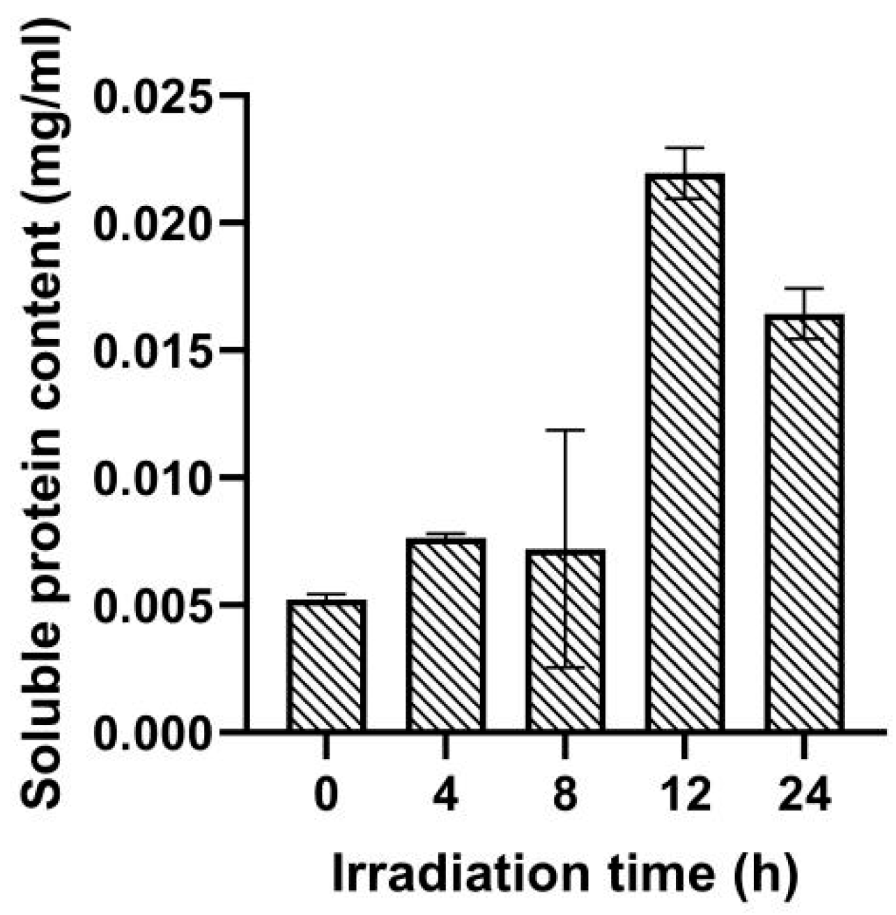
2.2. Effect of UV Irradiation Dose on Soluble Protein Content

The content of soluble protein in Navicula sp. after UV irradiation is shown in Figure 2. Compared to the control group, under 4~8 h of low-dose UV radiation conditions, a slight change in soluble protein content was recorded (0.0072~0.0076 mg/mL vs. 0.0052 mg/mL) (Figure 2). Under 12~24 h of high-dose UV irradiation conditions, soluble protein content in Navicula sp. cells was significantly higher than that in the control and 4~8 h of low-dose UV irradiation groups (0.016~0.021 mg/mL vs. 0.0052~0.0076 mg/mL). Intracellular soluble protein content increased with increasing irradiation time and showed obvious fluctuations. It rose sharply to 0.021 mg/mL in the case of 12 h of UV radiation and decreased slightly to 0.016 mg/mL in the 24 h of UV radiation group.

2.3. Effect of UV Irradiation Dose on CAT Activity

Reactive oxygen free radicals are closely associated with plant stress resistance. In a high-intensity radiation environment, more free radicals are produced in Navicula sp. cells, requiring more active antioxidant enzymes to remove them. The CAT activity of Navicula sp. cells after UV irradiation is shown in Figure 3. Under 4~24 h of UV radiation conditions, CAT activity increased with prolonged UV irradiation. Under 4~12 h of UV radiation conditions, CAT activity reached 2596~2917 U/mL, against 2188 U/mL observed in the control group (Figure 3). After 24 h of irradiation, CAT activity further increased to 3633 U/mL, an approximate increase of 66%.

2.4. Effect of UV Irradiation Dose on SOD Activity

As shown in Figure 4, after UV irradiation, the activity of SOD in Navicula sp. displayed a trend of initial decrease followed by an increase and then a subsequent decrease with the extension of irradiation time. SOD activity was 6.19 U/mL at 0 h. It decreased to 5.13 U/mL at 4 h, increased to 7.64 U/mL at 12 h, and then decreased sharply to 4.7 U/mL at 24 h.

2.5. Effect of UV Irradiation on Proteomics

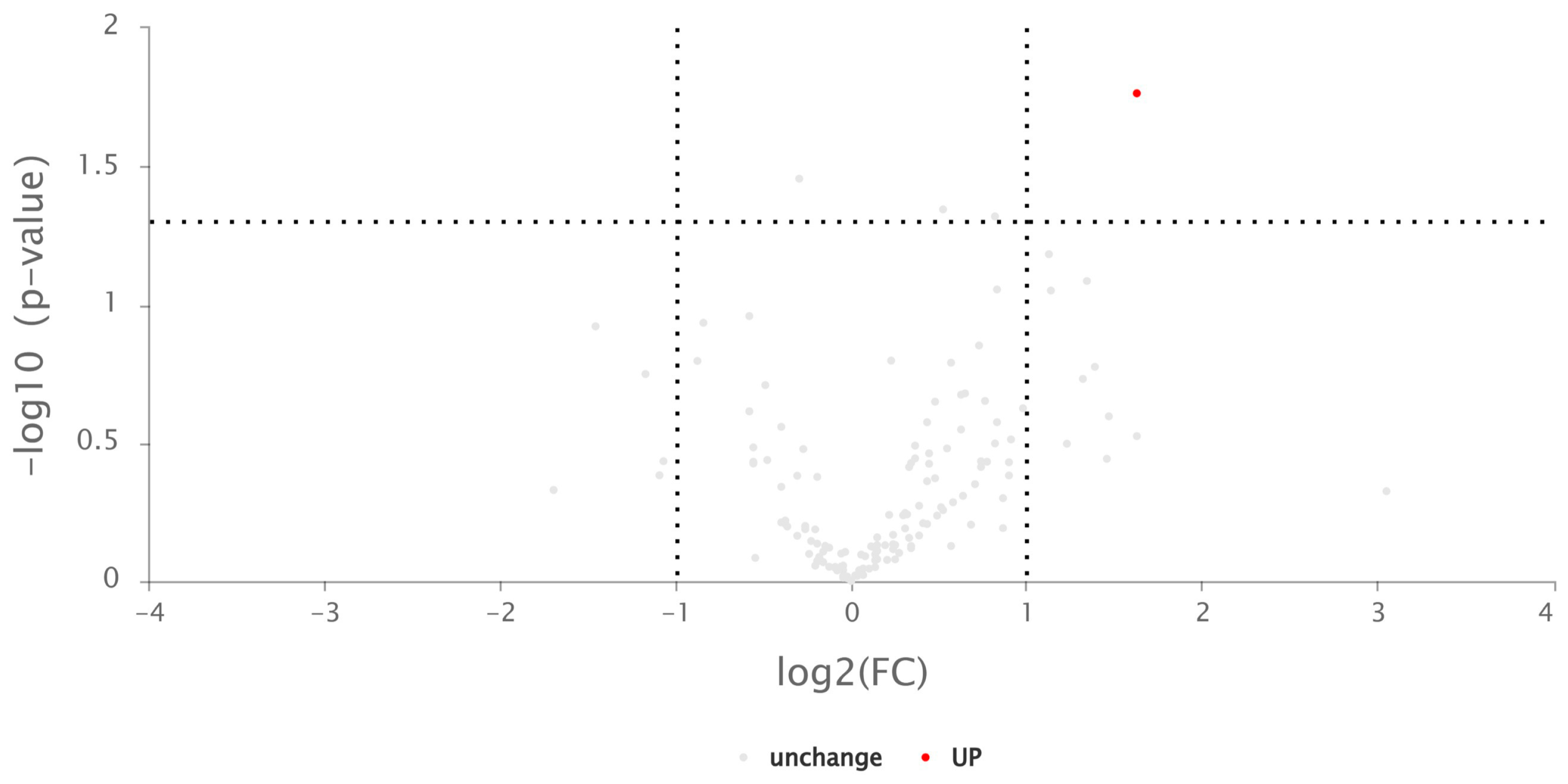
2.5.1. Differentially Expressed Proteins

The mass spectrum data obtained from three repeated biological samples represent the whole proteome of the plants. In this sequencing, a total of 975,990 secondary spectra were obtained, including 5071 effective spectra. A total of 555 peptides and 189 proteins were identified. One identified protein whose content significantly increased after ultraviolet irradiation, namely α/β hydrolase, was screened. A differential volcano plot is shown below (Figure 5).

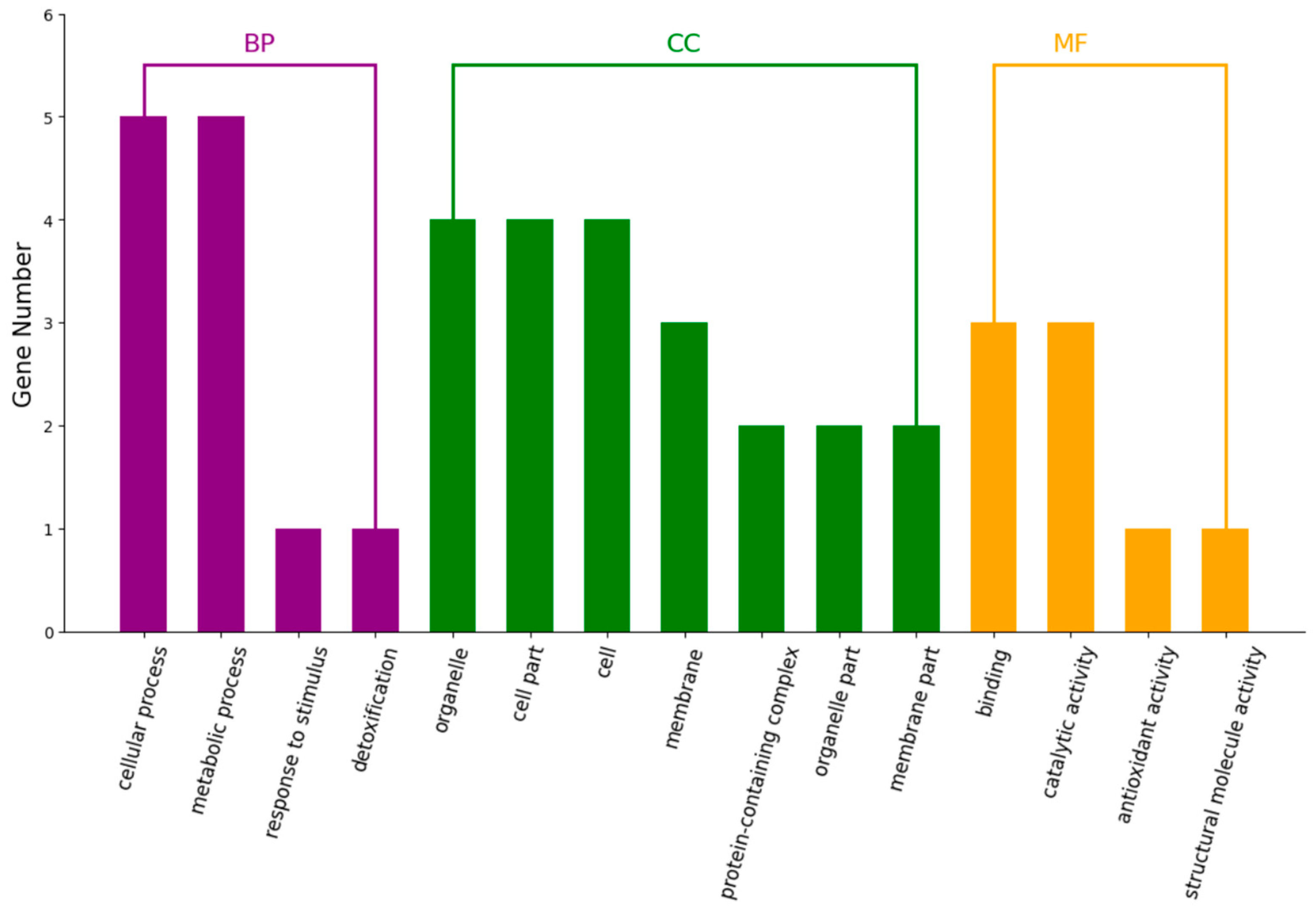
2.5.2. Differential Proteins in GO Analysis

Blast2Go 6.0 software was used to perform the GO functional annotation of all differentially expressed proteins. At the same time, the number of differential proteins was statistically analyzed at the GO secondary functional annotation level. According to functional characteristics, these differential proteins were divided into three categories: biological processes, cellular components, and molecular functions (Figure 6).
Proteins play an important role in Navicula sp.’s resistance to UV radiation. For biological processes, differential proteins in Navicula sp. after UV irradiation were mainly concentrated in cellular processes and metabolic processes. These related processes are mainly associated with oxidative stress. For cellular components, differential proteins in Navicula sp. were mainly concentrated in the organelles (cell parts) and in entire cells. In the case of molecular functions, they were mainly binding and catalytic activity (Figure 6).

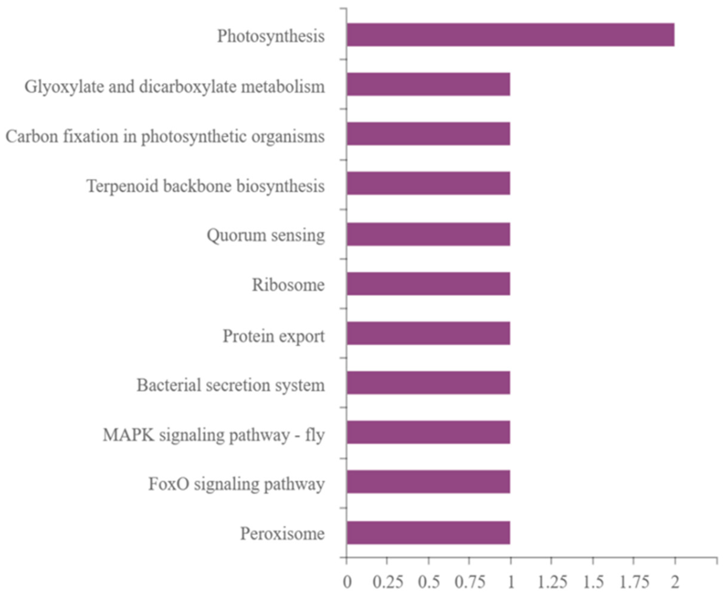
2.5.3. Differential Protein KEGG Pathway Analysis

Using KAAS software (https://www.genome.jp/tools/kaas/, accessed on 3 April 2023), KEGG pathway annotation was performed on the set of differentially expressed proteins and the results are shown in Figure 7.
Navicula sp. has strong UV resistance with only a few differentially expressed proteins. Among them, the photosynthesis pathway had the most differentially expressed proteins, i.e., the pathway which was most affected by UV irradiation was the photosynthesis system. At the same time, the expression of some photosynthesis-related proteins decreased, and the expression of other proteins increased, indicating that the effect of UV irradiation on photosynthesis is not only inhibition. The proteins with increased content were dicarboxylic acid metabolism and peroxisome-related proteins. The proteins with decreased content included ribosomal proteins.

3. Discussion

UV irradiation, as an abiotic stress factor, can significantly influence various aspects of cellular growth and metabolism. Plants and algae, when subjected to stress, can self-adjust to counteract the adversity posed by UV irradiation stress. The present study was carried out to reveal the molecular mechanism of Navicula sp. in response to UV irradiation stress based on integrated physiologic and proteomic analyses.
The synthesis of chlorophyll plays an important role in the UV radiation response process. Under low-dose ultraviolet radiation conditions, there is only a slight change in chlorophyll content. Under 12~24 h of UV irradiation, the contents of both chlorophyll a and b significantly increased (Figure 1). Chlorophyll a is responsible for absorbing and transmitting light energy. The reduction in chlorophyll content under short durations of UV radiation could be attributed to varying degrees of damage caused by UV irradiation to the photosynthetic system and chlorophyll lamella structure within Navicula sp. cells. These changes led to dysregulation of photosynthetic pigments and impaired photosynthesis, decreased efficiency of light energy utilization, and diminished chlorophyll synthesis and repair capabilities [21,22]. UV irradiation may damage the phototransduction chain and weaken the ability of Navicula sp. cells to capture and transmit light energy. When exposed to longer periods with higher doses of UV irradiation, Navicula sp. cells may enhance chlorophyll synthesis through metabolic network regulation to withstand UV irradiation pressure.
During the UV radiation response process, the regulation of diverse protein synthesis plays an important role. Under 12~24 h of high-dose UV irradiation conditions, the soluble protein contents in Navicula sp. cells were significantly higher than that in the control group (Figure 2). Aromatic amino acids are precursors in the synthesis of flavonoids, and flavonoids have the ability to absorb UV radiation and protect plants from partial damage under UV exposure [23,24]. The increase in protein content in Navicula sp. could be partly attributed to the enhanced synthesis of aromatic amino acids. UV stress could activate the antioxidation system in algae, leading to the expression of more proteins to mitigate the cellular damage caused by UV irradiation. The longer the irradiation time lasted, the higher the CAT activity, while the dynamic value of SOD showed a trend of first increasing and then decreasing. Research on a wide range of algae indicates that UV irradiation can disrupt algal cellular physiological processes to varying degrees, leading to oxidative stress responses in cells [25]. UV irradiation stimulates the production of reactive oxygen species (ROS) in Navicula sp. In response, the Navicula sp. antioxidation system (e.g., CAT and SOD) is activated to scavenge these ROS, thereby protecting the cells and maintaining the photosynthetic activity of the algal cells [17]. However, as the intensity of UV irradiation increases, excessive UV light leads to damage to the molecular structure of SOD, and as a result, the induction of SOD vitality can be weakened. Additionally, UV irradiation may induce more responses from other antioxidant systems besides CAT and SOD. It was found that Synechococcus sp. PCC7942, a UV-B cyanobacteria, can rapidly synthesize anti-UV-B proteins [26].
Proteomic analysis further elucidates the response of Navicula sp. to UV irradiation stress. Navicula sp. shows high adaptability to harsh environments, as evidenced by the presence of only one significantly differentially expressed protein (i.e., α/β hydrolase) after 24 h of UV irradiation (Figure 5). α/β hydrolases typically engage in fundamental cellular metabolic processes, such as the breakdown and recycling of cellular metabolites, processing of external nutrients, and detoxification of xenobiotics. They play a pivotal role in metabolic regulation [27]. GO analysis of differentially expressed proteins indicates that UV irradiation could activate the antioxidation system of Navicula sp., which is consistent with experimental data. UV irradiation has the most significant impact on proteins related to biological processes in Navicula sp. Combining GO and KEGG analysis, it can be concluded that the antioxidation system, and proteins related to the light reactions and chlorophyll synthesis in the photosynthetic system, are most affected by UV irradiation.
When Navicula sp. cells are exposed to UV irradiation, it can trigger oxidative stress and disrupt normal physiological activities and protein synthesis. Specifically, this leads to the generation of large amounts of ROS, which can damage the cellular structure and function of algal cells [26,27,28,29]. In order to counteract these deleterious effects, algal cells have evolved a complex antioxidant defense system consisting of antioxidant enzymes and non-enzymatic antioxidants [30,31]. In antioxidant defense, most differential proteins are implicated in a number of biological processes, including the cellular response to superoxide, neutralization of oxygen radicals, cellular detoxification, reactive oxygen metabolism, the action of oxidative detoxifiers, and response to oxidative stress. The activation of these defensive processes is not only a response of algal cells to the toxicity of UV irradiation and part of their stress response strategies, but is also involved in the regulation of reactive oxygen species signaling [32]. After UV irradiation, free radical scavenging systems within Navicula sp. cells, such as peroxidases, can actively eliminate these free radicals through redox reactions to establish an effective defense mechanism [30,31,32,33]. These antioxidative responses and defense mechanisms within Navicula sp. cells constitute a complex biological process, a key strategy of algae like Navicula sp. against UV radiation.
The differential proteins of cellular components include those found in chloroplast thylakoids, Photosystem II, complex catalysts, and redox enzyme complexes. Chloroplast thylakoids are the internal structure of chloroplasts, containing the chlorophyll and proteins necessary for photosynthesis [34]. The differential expression of thylakoid proteins indicates the impact of UV on the photosynthesis of Navicula sp. The core complex of Photosystem II, which is crucial for photosynthesis, uses energy absorbed from light to split water and transfers the released electrons to plastoquinone [35,36]. UV light affects the photosynthetic rate of algal cells by impacting the light reactions in photosynthesis. This further confirms that Photosystem II is the most sensitive to UV radiation and suffers the most severe photoinhibition [37,38]. The cytochrome b6/f complex plays a central role in oxygen photosynthesis, linking electron transfer between photosystems I and II and converting light energy into a transmembrane proton gradient for ATP synthesis [39]. The B6/f complex is an intrinsic protein of the stroma-thylakoid membrane. The photoreactions of photosynthesis take place in the chloroplast’s thylakoid membrane, further suggesting that UV radiation affects photoreactions in photosynthesis in algal cells, which in turn affects the algal photosynthetic rate. Magnesium is an important element in the formation of chlorophyll structure. If magnesium ions cannot be bound, the porphyrin ring and its surrounding organic chain cannot be assembled, and so chlorophyll molecules cannot be formed [40,41]. The differential expression of magnesium ion-binding proteins in algal cells indicates that UV radiation affects the synthesis of photosynthetic pigments by influencing the magnesium ion-binding function of algal cells. The effects of UV radiation on photosynthesis are mainly reflected in the photoreaction and synthesis of photosynthetic pigments.
In summary, the antioxidation system and proteins related to light reactions and chlorophyll synthesis in the photosynthetic system are most affected in Navicula sp. under UV irradiation. It should be noted that 30% of the world’s animal protein comes from the ocean, and many algae are primary producers in aquatic ecosystems. Considering the excellent photosynthetic carbon sequestration capacity and stress tolerance of these algae (e.g., Navicula sp.), they may play an equally important role in future aerospace life support systems.

4. Materials and Methods

4.1. Materials

The experimental alga was purchased from the Freshwater Algae Culture Collection of the Chinese Academy of Sciences (Catalog No.: FACHB-1950), species name: Navicula sp.
The culture medium was CSI culture medium (composition detailed in Table 1) acquired from Freshwater Algae Culture Collection at the Institute of Hydrobiology (FACHB), National Aquatic Biological Resource Center (Wuhan, China).
The main reagents were as follows: chlorophyll assay kit; soluble protein assay kit; catalase (CAT) assay kit; superoxide dismutase (SOD) assay kit. All were purchased from the Nanjing Jiancheng Bioengineering Institute (Nanjing, China).
Method of soil extract preparation: A total of 200 g of garden soil which had not been fertilized was placed in a beaker or Erlenmeyer flask (Product number: I015452, Sichuan Shubo (Group) Co., Ltd, Sichuan, China). Then, 1000 mL of distilled water was added. The flask was sealed with an air-permeable stopper and heated in a water bath at boiling temperature for 3 h. After heating, it was allowed to cool and settled for 24 h. This process was repeated 3 times. Then, the mixture was filtered and the supernatant was collected. The clear liquid was sterilized in an autoclave and stored in a refrigerator at 4 °C for future use.

4.2. Experimental Methods

4.2.1. Pre-Treatment before Irradiation

Navicula sp., which was in the logarithmic growth phase, was inoculated at a specified seeding density. The absorbance of the algae solution was measured every two days. After 8 days of incubation in an Erlenmeyer flask, the UV irradiation treatment was performed.

4.2.2. Irradiation Treatment

A 300 VA ultra-clean workbench UV lamp tube (Harbin Donglian Electronic Technology Development Co., Ltd, Beijing, China) was used as the radiation source. The Erlenmeyer flasks were placed 20 cm below the UV lamp. Treatment durations were set at 4 h, 8 h, 12 h, and 24 h, with each duration having three independent repetitions.

4.2.3. Treatment and Analysis after Irradiation Process

After irradiation, Navicula sp. samples continued to be cultured under standard conditions. Cultivation was carried out in 250 mL Erlenmeyer flasks, each containing 40 mL of CSI medium. The mouths of the flasks were sealed with a breathable but water-impermeable sealing film. The flasks were then placed under normal lighting conditions at room temperature, with a light/dark cycle of 12/12. The samples were harvested 96 h post-irradiation. After centrifugation at 3500 rpm for 10 min, the supernatant was reduced to 6 mL. The remaining Navicula sp. liquid was mixed and the cells were broken to obtain the homogenate. Subsequently, various physiological and biochemical indicators were measured, including the contents of chlorophyll a, chlorophyll b, total chlorophyll, soluble protein, and the activities of CAT and SOD. Additionally, proteomic analysis was conducted.

4.3. Assay Methods

4.3.1. Chlorophyll a, Chlorophyll b, and Total Chlorophyll Content

First, 0.1 mL of homogenate was added to 1 mL of distilled water. This was mixed and the mixture was transferred to a 5 mL centrifuge tube. Then, the extraction solution was prepared (anhydrous ethanol/acetone = 1/2, v/v) and made up to 5 mL. The extraction solution was used for the extraction process in dark conditions for over 3 h until the solution turned completely white. After centrifuging at 3000 rpm for 10 min, the supernatant was diluted with the extraction solution to an appropriate concentration with an OD value of less than 1. The spectrophotometer with the extraction solution was zeroed and absorbance values were recorded at 645 nm and 663 nm. The following formulas were used for calculation:
Chlorophyll a content (mg/mL) = (12.7 × A663 − 2.69 × A645) × V × N/(v × 1000);
Chlorophyll b content (mg/mL) = (22.9 × A645 − 4.68 × A663) × V × N/(v × 1000);
Total chlorophyll content (mg/mL) = (20.2 × A645 + 8.02 × A663) × V × N/(v × 1000).
Note: V is the volume of absolute alcohol (5 mL);
N is the colorimeter determination multiple;
v is the volume of the sample (mL).

4.3.2. Soluble Protein Content

Soluble protein content was determined using the Coomassie brilliant blue method. The homogenate was centrifuged at 3500 rpm for 10 min and the supernatant was collected. For the experimental group, 0.05 mL of supernatant was added to 3.0 mL of Coomassie brilliant blue staining solution. The blank control and standard groups were treated with 0.05 mL of distilled water and 0.05 mL of protein standard solution, respectively. After mixing and standing for 10 min, the spectrophotometer was zeroed with distilled water and absorbance was read at 595 nm. The following formula was used for calculation:
Soluble protein content(g/L) = (ASample − ABlank)/(AStandard − ABlank) × Standard concentration × N
Note: N is the dilution factor used during the assay.

4.3.3. CAT Activity

For the measurement of CAT activity, the ammonium molybdate method was used. After centrifuging the homogenate at 3500 rpm for 10 min, 0.05 mL of the supernatant was added to 1 mL of reagent 1 (at 37 °C) and 0.1 mL of reagent 2 (at 37 °C), mixed immediately, and reacted precisely for 1 min at 37 °C. Then, 1 mL of reagent 3 and 0.1 mL of reagent 4 were added and mixed. The spectrophotometer was zeroed with distilled water and absorbance was read at a wavelength of 405 nm. In the control group, the homogenate was replaced with distilled water and a similar procedure was followed. The following formula was used for calculation:
Activity of CAT (U/mL) = (ΔA × 271)/(Vs × T)
Note: ΔA = Acontrol − Asample;
271 is the molar extinction coefficient, in L/(mol·cm);
Vs: sample volume, 0.05 mL;
T: reaction time, 60 s.

4.3.4. SOD Activity

Here, the hydroxylamine method was employed. After centrifugation of the homogenate at 3500 rpm for 10 min, 1.0 mL of reagent 1, 0.05 mL of algal liquid, 0.1 mL of reagent 2, 0.1 mL of reagent 3, and 0.1 mL of reagent 4 were added to the experimental tube and mixed thoroughly. After a 40-min water bath at 37 °C, 2 mL of color reagent was added, mixed, and left to stand for 10 min. The spectrophotometer was zeroed with distilled water, and absorbance was read at 550 nm. In the control group, the homogenate was replaced with distilled water and a similar procedure was followed. The following formula was used for calculation:
SOD activity (U/mL) = (Acontrol − Asample)/(AControl × 50%) × Vt/Vs
Note: Vt = Total reaction volume (mL);
Vs = Sample volume (mL).

4.3.5. Proteomics

After UV irradiation, Navicula sp. liquid was mixed and centrifuged until 3 mL sediment remained for proteomic analysis. Label-free quantitative proteomics technology was used to obtain differential proteomic expression analysis.

5. Conclusions

In the present work, integrated physiologic and proteomic analyses were carried out to illustrate the molecular mechanism of Navicula sp. in response to UV irradiation stress. We found that ultraviolet radiation can alter the contents of soluble proteins and chlorophyll and increase the activity of antioxidation enzymes such as CAT in Navicula sp. cells. Differential protein GO analysis found that UV irradiation can activate the antioxidant system of Navicula sp. and further impact photosynthesis by affecting the photoreaction and chlorophyll synthesis of Navicula sp. The most significant differences in KEGG pathway analysis were also associated with those related to photosynthesis. The above results indicate that Navicula sp. has good UV radiation resistance ability by regulating its photosynthetic pigment content, photosynthetic activity, and antioxidant system, making it a potential candidate for the future development of space ecosystems.

Author Contributions

Q.Y. and Y.Z. planned and designed the research. S.G. and X.M. contributed to sample collection and performed the experiments and data collection. S.G., X.M. and H.X. analyzed the data and interpreted the results. S.G., P.P. and X.M. wrote the manuscript. X.C., Q.Y. and H.H. reviewed and revised the manuscript. All authors have read and agreed to the published version of the manuscript.

Funding

This study was financially funded by the Beijing Natural Science Foundation (7232101; 7242091), the Research Foundation of the Ministry of Education of China (8091B012103; 8091B022138), and the Fundamental Research Funds for the Central Universities (2014JBM118; 2017JBM073).

Institutional Review Board Statement

Not applicable.

Informed Consent Statement

Not applicable.

Data Availability Statement

The data that support the findings of this study are available from the corresponding author upon reasonable request.

Conflicts of Interest

The authors declare that this research was conducted in the absence of any commercial or financial relationships that could be construed as a potential conflict of interest.

References

  1. Kock, A.; Glanville, H.; Law, A.; Stanton, T.; Carter, L.; Taylor, J.C. Emerging challenges of the impacts of pharmaceuticals on aquatic ecosystems: A diatom perspective. Sci. Total Environ. 2023, 878, 162939. [Google Scholar] [CrossRef]
  2. Marella, T.K.; López-Pacheco, I.Y.; Parra-Saldívar, R.; Dixit, S.; Tiwari, A. Wealth from waste: Diatoms as tools for phycoremediation of wastewater and for obtaining value from the biomass. Sci. Total Environ. 2020, 724, 137960. [Google Scholar] [CrossRef]
  3. Alexandrov, S. Algal research in space: History, current status and future prospects. J. Life Sci. 2016, 4, 1–4. [Google Scholar]
  4. Sharma, G.C.; Nandan, R. Reliability modeling and analysis of environmental control and life support systems of space stations: A literature survey. Acta Astronaut. 2019, 155, 238–246. [Google Scholar] [CrossRef]
  5. Han, B.P.; Han, Z.G.; Fu, X. Mechanism and Model of Algal Photosynthesis; Science Press: Beijing, China, 2003. [Google Scholar]
  6. Niederwieser, T.; Kociolek, P.; Klaus, D. Spacecraft cabin environment effects on the growth and behavior of Chlorella vulgaris for life support applications. Life Sci. Space Res. 2018, 16, 8–17. [Google Scholar] [CrossRef] [PubMed]
  7. Nan, G.N.; Zhang, Q.S.; Sheng, Z.T.; Zhang, D. Coordination between xanthophyll cycle and antioxidant system in Sargassum thunbergii (Sargassaceae, Phaeophyta) in response to high light and dehydration stresses. J. Appl. Phycol. 2016, 28, 2587–2596. [Google Scholar] [CrossRef]
  8. Chen, S.W.; Wu, B.X. Algal responses to enhanced UV-B and its mechanism on molecular level. J. Jinan Univ. Nat. Sci. Med. Ed. 2000, 5, 88–94. [Google Scholar]
  9. Wu, H.; Cockshutt, A.M.; McCarthy, A.; Campbell, D.A. Distinctive photosystem II photoinactivation and protein dynamics in marine diatoms. Plant Physiol. 2011, 156, 2184–2195. [Google Scholar] [CrossRef] [PubMed]
  10. Nishiyama, Y.; Yamamoto, H.; Allakhverdiev, S.I.; Inaba, M.; Yokota, A.; Murata, N. Oxidative stress inhibits the repair of photodamage to the photosynthetic machinery. EMBO J. 2001, 20, 5587–5594. [Google Scholar] [CrossRef] [PubMed]
  11. Amin, N.; Sinha, R.P.; Kannaujiya, V.K. Effects of ultraviolet and photosynthetically active radiation on morphogenesis, antioxidants and photoprotective defense mechanism in a hot-spring cyanobacterium Nostoc sp. strain VKB02. Res. Microbiol. 2024, in press. [Google Scholar] [CrossRef] [PubMed]
  12. Callaghan, T.V.; Björn, L.O.; Chernov, Y.; Chapin, T.; Christensen, T.R.; Huntley, B.; Ims, R.A.; Johansson, M.; Jolly, D.; Jonasson, S.; et al. Responses to projected changes in climate and UV-B at the species level. Ambio 2004, 33, 418–435. [Google Scholar] [CrossRef] [PubMed]
  13. Schneider, G.; Figueroa, F.L.; Vega, J.; Avilés, A.; Horta, P.A.; Korbee, N.; Bonomi-Barufi, J. Effects of UV–visible radiation on growth, photosynthesis, pigment accumulation and UV-absorbing compounds in the red macroalga Gracilaria cornea (Gracilariales, Rhodophyta). Algal Res. 2022, 64, 102702. [Google Scholar] [CrossRef]
  14. Zeeshan, M.; Prasad, S.M. Differential response of growth, photosynthesis, antioxidant enzymes and lipid peroxidation to UV-B radiation in three cyanobacteria. S. Afr. J. Bot. 2009, 75, 466–474. [Google Scholar] [CrossRef]
  15. Lu, P.Y.; Zang, Y.; Xue, S.; Sun, Y.; Zhu, M.L.; Liang, S.; Tang, X.X. Gender-specific responses of Mougeotia’s reactive oxygen species scavenging system to enhanced UV-B radiation. J. Ocean Univ. China 2022, 52, 52–59. (In Chinese) [Google Scholar]
  16. Pfendler, S.; Munch, T.; Bousta, F.; Alaoui-Sosse, L.; Aleya, L.; Alaoui-Sossé, B. Bleaching of biofilm-forming algae induced by UV-C treatment: A preliminary study on chlorophyll degradation and its optimization for an application on cultural heritage. Environ. Sci. Pollut. Res. 2018, 25, 14097–14105. [Google Scholar] [CrossRef] [PubMed]
  17. Wu, Y.; Li, Z.; Du, W.; Gao, K. Physiological response of marine centric diatoms to ultraviolet radiation, with special reference to cell size. J. Photochem. Photobiol. B. 2015, 153, 1–6. [Google Scholar] [CrossRef] [PubMed]
  18. Korkaric, M.; Behra, R.; Fischer, B.B.; Junghans, M.; Eggen, R.I. Multiple stressor effects in Chlamydomonas reinhardtii—Toward understanding mechanisms of interaction between effects of ultraviolet radiation and chemical pollutants. Aquat. Toxicol. 2015, 162, 18–28. [Google Scholar] [CrossRef] [PubMed]
  19. Dwivedi, S.; Ahmad, I.Z. Evaluation of the effect of UV-B radiation on growth, photosynthetic pigment, and antioxidant enzymes of some cyanobacteria. Environ. Res. 2023, 218, 114943. [Google Scholar] [CrossRef]
  20. Arena, C.; Graham, T.; Legué, V.; Paradiso, R. Editorial: Higher plants, algae and cyanobacteria in space environments. Front. Plant Sci. 2021, 12, 629014. [Google Scholar] [CrossRef]
  21. Borderie, F.; Tête, N.; Cailhol, D.; Alaoui-Sehmer, L.; Bousta, F.; Rieffel, D.; Aleya, L.; Alaoui-Sossé, B. Factors driving epilithic algal colonization in show caves and new insights into combating biofilm development with UV-C treatments. Sci. Total Environ. 2014, 484, 43–52. [Google Scholar] [CrossRef]
  22. Marangoni, R.; Messina, N.; Gioffré, D.; Colombetti, G. Effects of UV-B irradiation on a marine icroecosystem. Photochem. Photobiol. 2004, 80, 78. [Google Scholar]
  23. Zhong, Z.; Liu, S.; Zhu, W.; Ou, Y.; Yamaguchi, H.; Hitachi, K.; Tsuchida, K.; Tian, J.; Komatsu, S. Phosphoproteomics Reveals the Biosynthesis of Secondary Metabolites in Catharanthus roseus under Ultraviolet-B Radiation. J. Proteome Res. 2019, 18, 3328–3341. [Google Scholar] [CrossRef] [PubMed]
  24. Ferreyra, M.L.F.; Serra, P.; Casati, P. Recent advances on the roles of flavonoids as plant protective molecules after UV and high light exposure. Physiol. Plant 2021, 173, 736–749. [Google Scholar] [CrossRef] [PubMed]
  25. Wang, G.; Chen, K.; Chen, L.; Hu, C.; Zhang, D.; Liu, Y. The involvement of the antioxidant system in protection of desert cyanobacterium Nostoc sp. against UV-B radiation and the effects of exogenous antioxidants. Ecotoxicol. Environ. Saf. 2008, 69, 150–157. [Google Scholar] [CrossRef] [PubMed]
  26. Campbell, D.; Eriksson, M.J.; Öquist, G.; Gustafsson, P.; Clarke, A.K. The cyanobacterium Synechococcus resists UV-B by exchanging photosystem II reaction-center D1 proteins. Proc. Natl. Acad. Sci. USA 1998, 95, 364–369. [Google Scholar] [CrossRef] [PubMed]
  27. Mindrebo, J.T.; Nartey, C.M.; Seto, Y.; Burkart, M.D.; Noel, J.P. Unveiling the functional diversity of the alpha/beta hydrolase superfamily in the plant kingdom. Curr. Opin. Struct. Biol. 2016, 41, 233–246. [Google Scholar] [CrossRef] [PubMed]
  28. Huarancca Reyes, T.; Mariotti, L.; Chiellini, C.; Guglielminetti, L.; Fonseca, G.G. UV-B irradiation effect on microalgae performance in the remediation of effluent derived from the cigarette butt cleaning process. Plants 2022, 11, 2356. [Google Scholar] [CrossRef]
  29. Forrester, S.J.; Kikuchi, D.S.; Hernandes, M.S.; Xu, Q.; Griendling, K.K. Reactive oxygen species in metabolic and inflammatory signaling. Circ. Res. 2018, 122, 877–902. [Google Scholar] [CrossRef]
  30. Gill, S.S.; Tuteja, N. Reactive oxygen species and antioxidant machinery in abiotic stress tolerance in crop plants. Plant Physiol. Biochem. 2010, 48, 909–930. [Google Scholar] [CrossRef]
  31. Khalid, M.F.; Vincent, C.; Morillon, R.; Anjum, M.A.; Ahmad, S.; Hussain, S. Different strategies lead to a common outcome: Different water-deficit scenarios highlight physiological and biochemical strategies of water-deficit tolerance in diploid versus tetraploid Volkamer lemon. Tree Physiol. 2021, 41, 2359–2374. [Google Scholar] [CrossRef]
  32. Malea, P.; Kokkinidi, D.; Kevrekidou, A.; Adamakis, I.S. The Enzymatic and non-enzymatic antioxidant system response of the seagrass Cymodocea nodosa to bisphenol-A Toxicity. Int. J. Mol. Sci. 2022, 23, 1348. [Google Scholar] [CrossRef]
  33. Saber, H.; El-Sheekh, M.M.; Ibrahim, A.; Alwaleed, E.A. Effect of UV-B radiation on amino acids profile, antioxidant enzymes and lipid peroxidation of some cyanobacteria and green algae. Int. J. Radiat. Biol. 2020, 96, 1192–1206. [Google Scholar] [CrossRef]
  34. Koochak, H.; Puthiyaveetil, S.; Mullendore, D.L.; Li, M.; Kirchhoff, H. The structural and functional domains of plant thylakoid membranes. Plant J. 2019, 97, 412–429. [Google Scholar] [CrossRef] [PubMed]
  35. Gisriel, C.J.; Shen, G.; Ho, M.; Kurashov, V.; Flesher, D.A.; Wang, J.; Armstrong, W.H.; Golbeck, J.H.; Gunner, M.R.; Vinyard, D.J.; et al. Structure of a monomeric photosystem II core complex from a cyanobacterium acclimated to far-red light reveals the functions of chlorophylls d and f. J. Biol. Chem. 2022, 298, 101424. [Google Scholar] [CrossRef] [PubMed]
  36. Boekema, E.J.; Hankamer, B.; Bald, D.; Kruip, J.; Nield, J.; Boonstra, A.F.; Barber, J.; Rögner, M. Supramolecular structure of the photosystem II complex from green plants and cyanobacteria. Proc. Natl. Acad. Sci. USA 1995, 92, 175–179. [Google Scholar] [CrossRef]
  37. Singh, R.D.; Sethy, S.; Ghosh, S.; Srivastava, A.K. UV and γ-radiation induced molecular changes for rapid lipid accumulation in Chlorella sorokiniana. Biomass Bioenergy 2022, 163, 106493. [Google Scholar] [CrossRef]
  38. Wang, B.; Ye, T.; Li, C.; Li, X.; Chen, L.; Wang, G. Cell damage repair mechanism in a desert green algae Chlorella sp. against UV-B radiation. Ecotoxicol. Environ. Saf. 2022, 242, 113916. [Google Scholar] [CrossRef]
  39. Yadav, K.N.; Semchonok, D.A.; Nosek, L.; Kouřil, R.; Fucile, G.; Boekema, E.J.; Eichacker, L.A. Supercomplexes of plant photosystem I with cytochrome b6f, light-harvesting complex II and NDH. Biochim. Biophys. Acta Bioenerg. 2017, 1858, 12–20. [Google Scholar] [CrossRef]
  40. Wang, Y.; Zhang, X.; Zhang, W.; Peng, M.; Tan, G.; Qaseem, M.F.; Li, H.; Wu, A. Physiological and transcriptomic responses to magnesium deficiency in Neolamarckia cadamba. Plant Physiol. Biochem. 2023, 197, 107645. [Google Scholar] [CrossRef] [PubMed]
  41. Vonlanthen, M.; Cuétara-Guadarrama, F.; Sorroza-Martínez, K.; González-Méndez, I.; Estrada-Montaño, A.S.; Rivera, E. A review of porphyrin dendrimers as light-harvesting versatile platforms. Dyes Pigm. 2024, 222, 111873. [Google Scholar] [CrossRef]
Figure 1. Effect of different doses of ultraviolet light on chlorophyll content of Navicula sp. (A) Effects on chlorophyll a; (B) effects on chlorophyll b; (C) effects on total chlorophyll.
Ijms 25 02747 g001
Figure 2. Effect of different doses of ultraviolet light on soluble protein content in Navicula sp.
Ijms 25 02747 g002
Figure 3. Effect of different doses of ultraviolet light on catalase activity in Navicula sp.
Ijms 25 02747 g003
Figure 4. Effect of different doses of ultraviolet light on superoxide dismutase activity of Navicula sp.
Ijms 25 02747 g004
Figure 5. Volcanic maps of differential proteins between irradiated and unirradiated Navicula sp. The horizontal axis represents the fold change of differential proteins (log2 value), and the vertical axis represents the p value (−log10 value). Grey indicates proteins with no significant difference, and red represents upregulated proteins.
Ijms 25 02747 g005
Figure 6. GO enrichment bar chart. The horizontal coordinate represents GO Level2 function annotation information. The purple group is biological processes, the green group is cellular components, and the orange group is molecular functions. The vertical coordinate represents the number of differentially expressed proteins under each functional classification.
Ijms 25 02747 g006
Figure 7. Column diagram of KEGG enrichment.
Ijms 25 02747 g007
Table 1. Formula of CSI culture medium.
NumberComponentConcentrationContent
1Ca(NO3)2·4H2O15 g/100 mL dH2O1 mL/L
2KNO310 g/100 mL dH2O1 mL/L
3MgSO4·7H2O4 g/100 mL dH2O1 mL/L
4β-Na2 glycerophosphate·5H2O2.5 g/100 mL dH2O1 mL/L
5Vitamin B12 0.1 µg/L
6Biotin 0.1 µg/L
7Thiamine HCl 10 µg/L
8HEPES 0.5 g/L
9Na2SiO3·9H2O 0.1 g/L
10Soil extract * 30 mL/L
11 (PIV)Na2EDTA0.75 g/L dH2O6 mL/L
MnCl2·4H2O0.041 g/L dH2O
ZnCl2·7H2O0.005 g/L dH2O
Na2MoO4·2H2O0.004 g/L dH2O
FeCl3·6H2O0.097 g/L dH2O
CoCl2·6H2O0.002 g/L dH2O
* Soil extract.
Disclaimer/Publisher’s Note: The statements, opinions and data contained in all publications are solely those of the individual author(s) and contributor(s) and not of MDPI and/or the editor(s). MDPI and/or the editor(s) disclaim responsibility for any injury to people or property resulting from any ideas, methods, instructions or products referred to in the content.

