Article

Acute and Chronic Exposure to Linagliptin, a Selective Inhibitor of Dipeptidyl Peptidase-4 (DPP-4), Has an Effect on Dopamine, Serotonin and Noradrenaline Level in the Striatum and Hippocampus of Rats

by
Małgorzata Łupina
1,*,
Agnieszka Wąsik
2,
Irena Baranowska-Bosiacka
3,
Maciej Tarnowski
4,
Tymoteusz Słowik
5,
Piotr Listos
6,
Jolanta Kotlińska
7,
Danuta Kosik-Bogacka
8,
Izabela Gutowska
9 and
Joanna Listos
7
1
Department of Experimental and Clinical Pharmacology, Medical University of Lublin, Jaczewskiego 8b St., 20-090 Lublin, Poland
2
Department of Neurochemistry, Polish Academy of Sciences, Smętna 12, 31-343 Kraków, Poland
3
Department of Biochemistry and Medical Chemistry, Pomeranian Medical University, Powstańców Wlkp. 72 Av., 70-111 Szczecin, Poland
4
Department of Physiology in Health Sciences, Pomeranian Medical University, Żołnierska 54, 70-210 Szczecin, Poland
5
Experimental Medicine Center, Medical University of Lublin, Jaczewskiego 8d St., 20-090 Lublin, Poland
6
Department of Pathological Anatomy, Faculty of Veterinary Medicine, University of Life Sciences, Głęboka 30, 20-612 Lublin, Poland
7
Department of Pharmacology and Pharmacodynamics, Medical University of Lublin, Chodźki 4a St., 20-093 Lublin, Poland
8
Independent Laboratory of Pharmaceutical Botany, Department of Biology and Medical Parasitology, Pomeranian Medical University in Szczecin, Powstańców Wlkp. 72, 70-111 Szczecin, Poland
9
Department of Biochemistry and Human Nutrition, Pomeranian Medical University, Broniewskiego 24, 71-460 Szczecin, Poland
*
Author to whom correspondence should be addressed.
Int. J. Mol. Sci. 2024, 25(5), 3008; https://doi.org/10.3390/ijms25053008
Submission received: 22 January 2024 / Revised: 29 February 2024 / Accepted: 1 March 2024 / Published: 5 March 2024
(This article belongs to the Section Molecular Pharmacology)

Abstract

:
Linagliptin is a selective dipeptidyl peptidase-4 (DPP-4) inhibitor that indirectly elevates the glucagon-like peptide-1 (GLP-1) level. The aim of the present study was to check whether linagliptin has an influence on neurotransmission in rat brain. Rats were acutely and chronically exposed to linagliptin (10 and 20 mg/kg, intraperitoneally (i.p.)). Twenty-four hours later, the striatum and hippocampus were selected for further studies. In neurochemical experiments, using high-performance liquid chromatography with electrochemical detection (HPLC-ED), the concentrations of three major neurotransmitters—dopamine, serotonin and noradrenaline—and their metabolites were measured. The analysis of mRNA expression of dopamine (D1 and D2), serotonin (5-HT-1 and 5-HT-2) and noradrenaline (α1 and α2a) receptors was also investigated using real-time quantitative reverse transcription polymerase chain reaction (RQ-PCR) in the same brain areas. Linagliptin has the ability to influence the dopaminergic system. In the striatum, the elevation of dopamine and its metabolites was observed after repeated administration of that linagliptin, and in the hippocampus, a reduction in dopamine metabolism was demonstrated. Acute linagliptin exposure increases the serotonin level in both areas, while after chronic linagliptin administration a tendency for the mRNA expression of serotoninergic receptors (5-HT1A and 5-HT2A) to increase was observed. A single instance of exposure to linagliptin significantly modified the noradrenaline level in the striatum and intensified noradrenaline turnover in the hippocampus. The recognition of the interactions in the brain between DPP-4 inhibitors and neurotransmitters and/or receptors is a crucial step for finding novel discoveries in the pharmacology of DPP-4 inhibitors and raises hope for further applications of DPP-4 inhibitors in clinical practices.

1. Introduction

Glucagon-like peptide-1 (GLP-1), an incretin hormone, is released to reduce glycemia after food intake. It stimulates the receptors for the GLP-1 located in the β cells in the pancreas [1]. These receptors are coupled to G-protein, and their stimulation activates adenylate cyclase [2]. GLP-1 is rapidly degraded by the enzyme dipeptidyl peptidase-4 (DPP-4) [3,4]; therefore, the pharmacological effect of GLP-1 is extremely short (about 2–3 min). Currently, two groups of incretin drugs have been approved for therapy of type 2 diabetes [5]. These include GLP-1 peptide analogs and DPP-4 enzyme inhibitors. The most important advantage of these drugs is their antihyperglycemic effect without risk of hypoglycemia.
GLP-1 is able to cross the blood–brain barrier [6], producing effects in the central nervous system (CNS). Moreover, the preproglucagon neurons, in which GLP-1 peptide is synthesized, occur in the nucleus of the solitary tract in the brainstem [7,8,9]. From here, GLP-1 neurons project widely throughout the brain and spinal cord [10,11,12], inducing a neuromodulatory effect [13]. The GLP-1 receptors occur in brain structures strongly associated with food intake regulation and rewarding and motivational behaviors, including the hypothalamus, ventral tegmental area (VTA), nucleus accumbens, amygdala, and others [12,13,14,15,16,17]. Many experiments have been performed to recognize the neuromodulatory activity of drugs affecting the GLP-1 level.
Firstly, it was noticed that GLP-1 analogs reduce appetite. This was observed after interventricular injections of GLP-1 in fasted rats [18] and after intraperitoneal injections with GLP-1 peptide in fasted mice [19]. In obese patients, the use of GLP-1 analog promoted insulin secretion [20]; reduced glycated hemoglobin, fasting blood glucose and body weight; and improved postprandial blood glucose control and β cell function [21]. Consequently, semaglutide was recently approved for use in the treatment of obesity. It is also known that GLP-1 is a mediator of the inflammatory response [22] as well as that it has an influence on neurotransmission [23], but detailed effects have not been fully recognized. In parallel, it was also evidenced that the administration of a GLP-1 analogue inhibited the rewarding effect of various addictive substances, i.e., amphetamine [24], cocaine [24,25], nicotine [26] and ethanol [27,28], in experimental animals. Thus, it was confirmed that GLP-1 receptors have an important role in rewarding effects of addictive drugs. Our previous experiments showed that administration of a DPP-4 inhibitor—linagliptin—reduced the rewarding effects of morphine in rats [29] and attenuated naloxone-induced morphine withdrawal in mice [30]. Our results suggest that GLP-1 peptide participates in morphine dependence while other mechanisms closely associated with DPP-4 inhibition may not be excluded.
An increasing amount of evidence documents that DPP-4 inhibitors may be an important pharmacological tool. They have pleiotropic effects. In addition to their antihyperglycemic effect, DPP-4 inhibitors have a beneficial impact on the circulatory system. They also produce anti-inflammatory and antiapoptotic effects [31]. Literature data demonstrate the effect of DPP-4 inhibitors on the CNS. Pipatpiboon et al. [32] proved that vildagliptin, a DPP-4 inhibitor, improved neuron function, in rats. Additionally, a decrease in β-amyloid accumulation in the mouse model of Alzheimer’s disease was also demonstrated [33]. Moreover, the significant increase in GLP-1 level in the hippocampus and improvement of cognitive functions were observed after administration of the DPP-4 inhibitors saxagliptin and vilda-gliptin in rats [34,35]. The beneficial effects of DPP-4 inhibitors on cognition function were also observed in people suffering from diabetes mellitus type 2 and Alzheimer’s disease [36]. Literature data demonstrated that sitagliptin, an inhibitor of DPP-4, induced an increase in the level of dopamine in the striatum and tyrosine hydroxylase protein. Furthermore, decreased neuroinflammation and the reversal of neuronal loss in rats were observed [37]. The role of linagliptin in a rat model of Parkinson’s disease was also investigated. Results confirmed that linagliptin prevented motor deficits and reduced the level of inflammatory cytokines in these rats [38]. Thus, taking into consideration the similarity in pharmacological action of GLP-1 analogs and DPP-4 inhibitors, it can be assumed that an increase in GLP-1 peptide activity has an important role in the mechanism of action of DPP-4 inhibitors.
The involvement of rewarding effects in food intake regulation and three major neurotransmitters—dopamine, serotonin and noradrenaline— in neurodegeneration is well known. Therefore, the aim of the present study was to check whether linagliptin, a xanthine-based molecule that acts selectively and reversibly on the DPP-4 enzyme, has an influence on neurotransmission in the brain. To obtain it, rats received linagliptin intraperitioneally (i.p.) in two schemes: an acute dose (one dose in the morning) and chronic doses (once a day for 8 consecutive days in the morning). Twenty-four hours after linagliptin exposure, rats were decapitated, and the striatum and hippocampus were selected for further studies. In a neurochemical study, using high-performance liquid chromatography with electrochemical detection (HPLC-ED), the concentrations of three major neurotransmitters—dopamine, serotonin and noradrenaline—and their metabolites were measured. Dopamine is catabolized in two ways: via monoamine oxidase B (monoaminooxidase-dependent oxidative pathway) to the intraneuronal metabolite 3,4-dihydroxyphenylacetic acid (DOPAC) or via catechol-O-methyltransferase (COMT-dependent o-methylation pathway) to the extraneuronal metabolite 3-methoxytyramine (3-MT). The final metabolite of dopamine is homovanillic acid (HVA). The concentrations of all three dopamine metabolites were measured in that study. Serotonin is metabolized to 5-hydroxyindoleacetic acid (5-HIAA) via monoamine oxidase A, while noradrenaline, via COMT, is metabolized to normetanephrine (NMN). The levels of both 5-HIAA and NMN were also examined. Additionally, the mRNA expression of dopamine (D1 and D2), serotonin (5-HT1 and 5-HT2), and noradrenaline (α1 and α2a) receptors was also investigated using real-time quantitative reverse transcription polymerase chain reaction (RQ-PCR) in the same brain areas. The striatum and hippocampus were chosen in this study as fundamental areas governing the reward system and memory processing. The present study significantly extends current knowledge on the impact of DPP-4 inhibitors on dopamine, serotonin and noradrenaline transmission in the brain. It provides information on the direct effect of DPP-4 inhibitors on the levels of major neurotransmitters in the brain. It is important proof that should be considered in the assessment of potential mechanisms of action of DPP-4 inhibitors.

2. Results

2.1. Ex Vivo Neurochemical Studies—The Analysis of the Concentration of Dopamine, Serotonin and Noradrenaline and Their Metabolites Using HPLC-ED

2.1.1. The Effect of Acute and Chronic Exposure to Linagliptin (10 and 20 mg/kg, i.p.) on the Concentration of Dopamine and Its Metabolites in the Striatum in Rats

One-way ANOVA revealed the significant effects of a single dose of linagliptin in the concentration of 3-MT (F(2,14) = 8.742, p = 0.0034) and in the ratio of 3-MT/DA (F(2,14) = 9.362, p = 0.0026). Statistically significant changes in DA (F(2,31) = 9.944, p = 0.0005), DOPAC (F(2,29) = 7.931, p = 0.0018), 3-MT (F(2,31) = 7.193, p = 0.0027) and HVA (F(2,31) = 7.322, p = 0.0025) concentrations were also documented as a result of chronic linagliptin administration to rats. There was no effect of chronic linagliptin administration on the analyzed metabolite/dopamine ratios; see Table 1.
The post hoc analysis (Tukey’s test) showed that a single dose of linagliptin (10 and 20 mg/kg) induced a significant increase in the 3-MT concentration (p < 0.05 and p < 0.01, respectively) and increase in the 3-MT/DA ratio (both doses p < 0.01) in the striatum of the rats. Chronic administration of linagliptin (10 and 20 mg kg) increased the concentration of DA (p < 0.01 and p < 0.001, respectively), DOPAC (both doses p < 0.01), 3-MT (p < 0.05 and p < 0.01, respectively) and HVA (p < 0.01 and p < 0.05, respectively) in the rat striatum; see Table 1.

2.1.2. The Effect of Acute and Chronic Exposure to Linagliptin (10 and 20 mg/kg, i.p.) on the Concentration of Dopamine and Its Metabolites in the Hippocampus in Rats

One-way ANOVA showed significant differences in the DA (F(2,31) = 6.051, p = 0.0060), DOPAC (F(2,28) = 4.159, p = 0.0262), and 3-MT (F(2,27) = 7.441, p = 0.0027) concentrations as well as in the DOPAC/DA (F(2,29) = 7.095, p = 0.0031), 3-MT/DA (F(2,24) = 5.619, p = 0.0100) and HVA/DA (F(2,25) = 3.526, p = 0.0448) ratios in the hippocampus of tested rats as a consequence of a single dose of linagliptin; see Table 2.
The post hoc analysis (the Tukey test) revealed that only the higher single dose of linagliptin (20 mg/kg) produced an increase in the DA (p < 0.05) concentration and decrease in the DOPAC (p < 0.05) and 3-MT (p < 0.01) concentration in the hippocampus in rats. Only in the rats administered the higher single dose of linagliptin were the DOPAC/DA (p < 0.01), 3-MT/DA (p < 0.05) and HVA/DA (p < 0.05) ratios statistically significantly reduced; see Table 2.
Chronic administration of linagliptin did not cause statistically significant changes in the concentration of dopamine and its metabolites or in the analyzed metabolite/DA ratios in the hippocampus of the studied rodents; see Table 2.

2.1.3. The Effect of Acute and Chronic Exposure to Linagliptin (10 and 20 mg/kg, i.p.) on the Concentration of Serotonin and Its Metabolite in the Striatum in Rats

One-way ANOVA demonstrated statistically significant effects of a single dose of linagliptin in Ser (F(2,14) = 21.13, p < 0.0001) and 5-HIAA (F(2,33) = 14.73, p < 0.0001) concentrations and of chronic administration of linagliptin in Ser (F(2,13) = 6.694, p = 0.0100) and 5-HIAA (F(2,13) = 9.126, p = 0.0033) concentrations in the striatum of tested rodents; see Table 3.
The post hoc analysis showed that the administration of a single dose of linagliptin (10 and 20 mg/kg) statistically significantly increased the Ser (p < 0.01 and p < 0.0001, respectively) and 5-HIAA (p < 0.05 and p < 0.0001) concentrations, but chronic administration of linagliptin, only at a lower dose (10 mg/kg), statistically significantly decreased the Ser (p < 0.05) and 5-HIAA (p < 0.05) concentrations in the rat striatum. No change in the 5-HIAA/Ser ratio was observed in the rat striatum due to the administration of linagliptin; see Table 3.

2.1.4. The Effect of Acute and Chronic Exposure to Linagliptin (10 and 20 mg/kg, i.p.) on the Concentration of Serotonin and Its Metabolite in the Hippocampus in Rats

One-way ANOVA revealed statistically significant effects of a single dose of linagliptin in Ser (F(2,14) = 5.253, p = 0.0199) and 5-HIAA (F(2,14) = 12.47, p = 0.0008) concentrations and of chronic administration of linagliptin in the 5-HIAA (F(2,29) = 11.17, p = 0.0003) concentration in the striatum of tested rodents; see Table 4.
According to the Tukey test, administration of a single dose of linagliptin (10 and 20 mg/kg) caused a statistically significant increase in Ser (both doses p < 0.05) and 5-HIAA (both doses p < 0.01) concentrations. The post hoc analysis also showed that chronic administration of linagliptin, only at a higher dose (20 mg/kg), statistically significantly increased the 5-HIAA (p < 0.05) concentration and 5-HIAA/Ser (p < 0.05) ratio; see Table 4.

2.1.5. The Effect of Acute and Chronic Exposure to Linagliptin (10 and 20 mg/kg, i.p.) on the Concentration of Noradrenaline and Its Metabolite in the Striatum in Rats

One-way ANOVA showed statistically significant effects of a single dose of linagliptin in NA (F(2,16) = 47.20, p < 0.0001) and NMN (F(2,25) = 4.632, p = 0.0194) concentration in the rat striatum and in the NMN/NA (F(2,27) = 8.183, p = 0.0017) ratio. No changes were observed in the rat striatum due to chronic administration of linagliptin; see Table 5.
The post hoc analysis demonstrated that a single dose of linagliptin (10 and 20 mg/kg) statistically significantly increased the NA (both doses p < 0.0001) concentration and that a single, higher dose linagliptin (20 mg/kg) statistically significantly decreased the NMN (p < 0.05) concentration in the striatum of rats. According to the Tukey test, a single dose of linagliptin (10 and 20 mg/kg) statistically significantly decreased the NMN/NA (both doses p < 0.01) ratio; see Table 5.

2.1.6. The Effect of Acute and Chronic Exposure to Linagliptin (10 and 20 mg/kg, i.p.) on the Concentration of Noradrenaline and Its Metabolite in the Hippocampus in Rats

One-way ANOVA demonstrated statistically significant effects of a single dose of linagliptin in NMN (F(2,13) = 25.06, p < 0.0001) concentrations and in the NMN/NA (F(2,23) = 23.52, p < 0.0001) ratio. No changes were observed in the hippocampus of rats as a result of chronic administration of linagliptin; see Table 6.
According to the Tukey test, administration of a single dose of linagliptin (10 and 20 mg/kg) induced a statistically significant decrease in NMN (p < 0.001 and p < 0.0001, respectively) concentration and statistically significant decrease in NMN/NA (both doses p < 0.0001); see Table 6.

2.2. The Analysis of mRNA Expression of Dopamine, Serotonin and Noradrenaline Receptors Using RQ-PCR in the Striatum and Hippocampus of Rats

2.2.1. The Effect of Acute Exposure to Linagliptin (10 and 20 mg/kg, i.p.) on mRNA Expression of D1, D2, 5-HT1A, 5-HT2A, α1A and α2A Receptors in the Striatum and Hippocampus in Rats

One-way ANOVA showed that a single dose of linagliptin (10 and 20 mg/kg) induces no statistically significant changes in the mRNA expression of D1, D2, 5-HT1A, 5-HT2A, α1A and α2A receptors in the striatum and hippocampus of the tested animals; see Table 7.

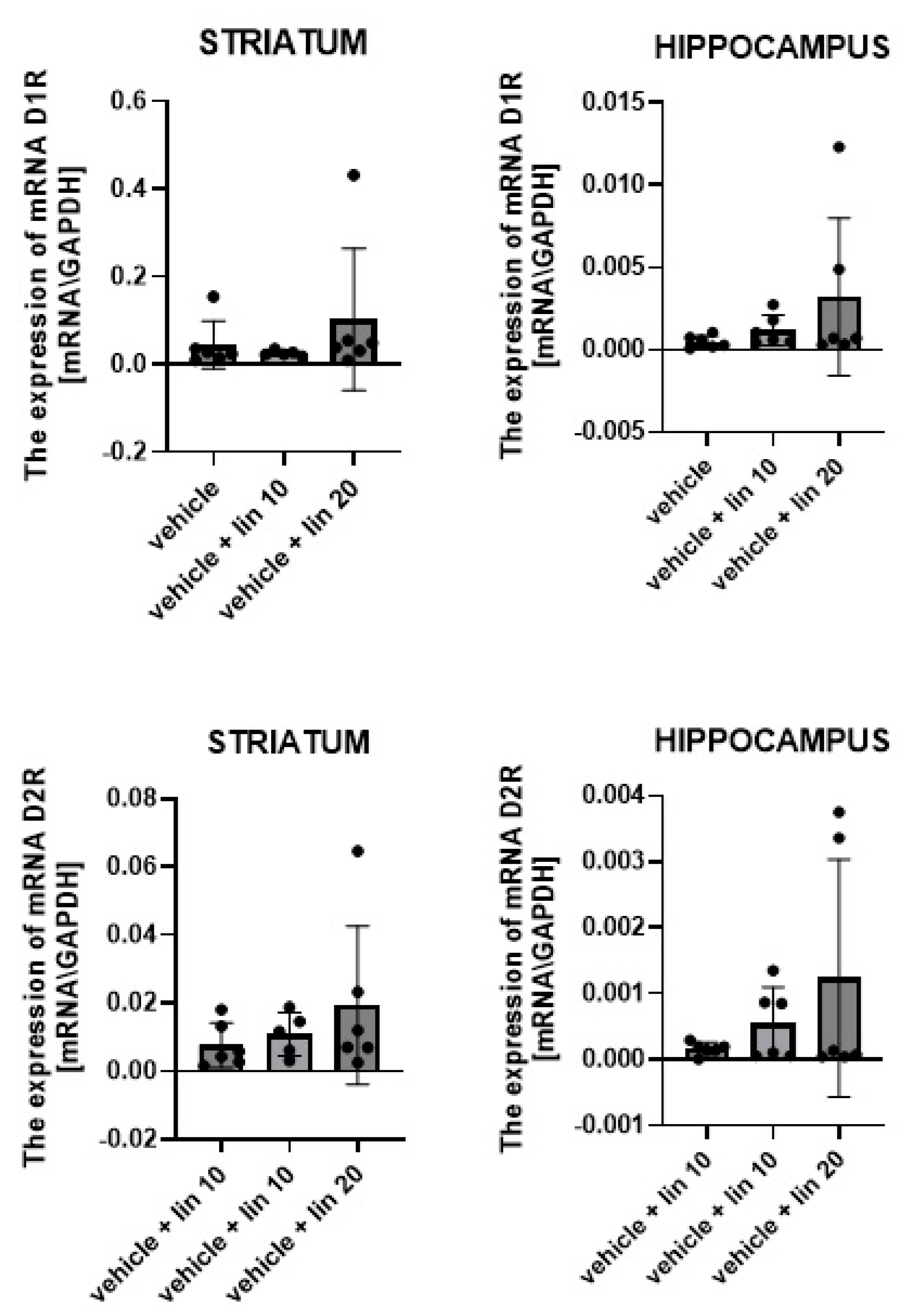
2.2.2. The Effect of Chronic Exposure to Linagliptin (10 and 20 mg/kg, i.p.) on mRNA Expression of D1, D2, 5-HT1A, 5-HT2A, α1A and α2A Receptors in the Striatum and the Hippocampus in Rats

One-way ANOVA showed that chronic administration of linagliptin (10 and 20 mg/kg) induces no statistically significant changes in the mRNA expression of D1 and D2 receptors in the striatum and hippocampus of the tested animals; see Figure 1.
One-way ANOVA showed statistically significant effects of chronic administration of linagliptin in mRNA expression of 5-HT1A receptors in the striatum (F(2,14) = 5.023, p = 0.0227) and hippocampus (F(2,15) = 6.810, p = 0.0033) of tested rats; see Figure 2.
The post hoc analysis demonstrated that chronic administration of linagliptin (10 and 20 mg/kg) statistically significantly increased mRNA expression of 5-HT1A receptors in the striatum (lower dose: p < 0.05) and hippocampus (higher dose: p < 0.01) in the studied rats. No more statistically significant changes were observed in the mRNA expression of 5-HT1A and 5-HT2A receptors; see Figure 2.
One-way ANOVA showed that chronic administration of linagliptin (10 and 20 mg/kg) produces no statistically significant changes in the mRNA expression of α1A and α2A receptors in the striatum and hippocampus of the tested animals; see Figure 3.

3. Discussion

The balance between different neurotransmission systems in the brain is extremely important for proper functioning of the body and well-being. It is well known that three major neurotransmitters—dopamine, serotonin and noradrenaline—regulate cerebral balance. They are the basis for stabilizing mood and seeking behavior. However, it is well documented that changes in the functioning of one neurotransmitter have an impact on the activity of the other [39]. It is a result of close interconnection between these neurotransmitters. Accordingly, dopaminergic structures like the VTA, substantia nigra, nucleus accumbens, prefrontal cortex and others have an innervation from serotoninergic fibers arising from the serotoninergic raphe nuclei [40] and also receive noradrenergic projections from the nucleus tractus solitaries [41]. It is known that many parameters may modify the functioning of these major neurotransmitters and that the interpretation of obtained results may not always be clear. Therefore, in the present study, we undertook the assessment of linagliptin effects on the dopamine, serotonin and noradrenaline activity to discover its significance in the brain. However, the particular interactions between these neurotransmitters cannot be excluded.
The major result of the reported experiments was that linagliptin, the selective DPP-4 inhibitor, influences the dopamine, serotonin, noradrenaline level in the striatum and hippocampus. Additionally, some changes in mRNA expression of selected receptors were observed.
In the neurochemical analysis, the concentrations of dopamine, serotonin and noradrenaline and their metabolites were measured using HPLC-ED. Moreover, the intensity of metabolic changes, assessed by determining the metabolite/neurotransmitter ratio, was calculated. In experiments assessing the mRNA expression of dopamine D1 and D2, serotonin 5-HT1A and 5-HT2A and noradrenergic α1A and α2A receptors were studied using RQ-PCR. Linagliptin was given to rats in two schedules. In the first one, rats were treated with a single dose of linagliptin; in the second one, linagliptin was given chronically—once a day for 8 consecutive days.
As it was documented, linagliptin increased the level of endogenous GLP-1 peptide 2–3 times [42]. In humans, oral application of linagliptin exhibits long-term pharmacological activity (T0.5 = 12 h). At present, linagliptin is approved for use in the treatment of type 2 diabetes mellitus in adults both in monotherapy or when metformin or other antidiabetics are ineffective. In the presented study, linagliptin was used in two doses (10 and 20 mg/kg), which were selected based on preliminary experiments. In that study, it was confirmed that intragastric and intraperitoneal administration of both linagliptin doses reduced glucose-induced hyperglycemia in rats but did not induce hypoglycemia (data unpublished). We decided to use intraperitoneal injections of linagliptin because we aimed for a decrease in repeated stressful situations associated with intragastric applications in rats. Intraperitoneal applications of DPP-4 inhibitors were also used by other scientists [43,44].
Moreover, the intraperitoneal administration of both doses of linagliptin inhibited the effect of morphine dependence [29,30] but did not produce any changes in the locomotor activity of mice or rats (data unpublished). Therefore, these two doses were chosen for neurochemical experiments and analysis of mRNA expression of receptors presented in that paper.
In the first step of that study, it was documented that linagliptin had an effect on the dopamine concentration both in the striatum and in the hippocampus. In the striatum, the major effect was observed after chronic administration of both linagliptin doses, and significant increases in the dopamine level and all of its metabolites were measured. In the case of a single dose of linagliptin, in the striatum, only the increases in the 3-MT concentration and 3-MT/dopamine ratio were observed. It is not clear what the reason for this was; it was possibly caused by an intensified conversion of dopamine to 3-MT or a slowdown conversion of 3-MT to HVA. It should be underlined, however, that the concentration of 3-MT is not currently considered as a reliable parameter of the activity of the dopaminergic pathway [45]. In fact, the first data on the role of 3-MT in the brain indicated its important role as an indicator of dopaminergic activity [46,47]. At present, however, it is known that in some conditions (e.g., depending on the method of animal euthanasia), 3-MT can be synthesized independently of dopamine [45]. Moreover, under certain conditions, 3-MT may act as a neuromodulator in the brain [48], playing a role as an inhibitory factor for the activity of catecholamines in the CNS [49]. It has also been documented that only about 20–30% of the amount of dopamine in the striatum is metabolized to 3-MT [50]. Therefore, in the interpretation of the results of this study, changes in 3-MT concentration were not considered as a crucial effect.
In the hippocampus, changes in the secretion of dopamine and its metabolites were also observed in the studied rats. A single dose of linagliptin significantly increased dopamine secretion and reduced dopamine metabolites. The HVA/DA ratio was reduced compared to the control group, showing a significant slowdown of dopamine metabolism in the hippocampus. In the case of the hippocampus of rats chronically treated with linagliptin, a similar tendency was observed; however, the results were not statistically significant.
In the case of acute linagliptin exposure, the changes in dopamine activity were probably too short-lived to induce receptor adaptation because, in both structures, no changes in mRNA expression of dopamine D1 and D2 receptors were observed. In the case of chronic administration of a higher dose of linagliptin, a trend towards higher mRNA expression of D1 and D2 receptors in the striatum was observed, but this effect was statistically insignificant, probably because of too high SD values. Taken together, linagliptin has the ability to influence the dopaminergic system because the elevation of dopamine and its metabolites in the striatum was observed after repeated administration of that drug, and in the hippocampus, a decrease in dopamine metabolism was demonstrated.
The interactions between GLP-1 peptide receptors and dopamine receptors have already been examined, but with varying results. Badawi et al. [51] demonstrated that both sitagliptin (DPP-4 inhibitor) and liraglutide (GLP-1 agonist) elevated the striatal dopamine level, which could be helpful in the treatment of Parkinson’s disease [51]. On the other hand, Egecioglu et al. [24], using microdialysis, demonstrated that the administration of exendin-4, a GLP-1 peptide receptor agonist, had no effect on the dopamine level in the nucleus accumbens but reduced the amphetamine- and cocaine-induced increase in dopamine release in that structure. Fortin and Roitman [52] used fast-scan cyclic voltammetry (FSCV) to study the pharmacological effects of exendin-4 on cocaine-evoked phasic dopamine signaling in the core and shell of nucleus accumbens of rats. Chronic exposure for GLP-1 receptor agonist suppressed dopamine signaling induced by cocaine in the core of nucleus accumbens but not in the shell.
The exact mechanisms between GLP-1 receptors and the dopamine level are not fully recognized. It is assumed that stimulation of GLP-1 peptide receptors in the VTA may weaken the transmission between VTA and nucleus accumbens neurons [53] in mice. In rats, it was confirmed that the stimulation of receptors for the GLP-1 peptide in the VTA may increase dopaminergic transmission through presynaptic, glutamatergic AMPA/kainate but not N-methyl-D-aspartate (NMDA) receptor signaling [54]. It was also confirmed that activation of GLP-1 receptors in the solitary nucleus changed the expression of D2 receptors in the VTA, indicating a direct link between the solitary nucleus and the VTA via GLP-1 neurons [55]. However, given the relatively low expression of GLP-1 receptors in the VTA and nucleus accumbens [13,56], other interactions cannot be excluded. It was confirmed that the stimulation of receptors for the GLP-1 peptide could weaken the effects of addictive substances by increasing the expression of the dopamine transporter (DAT) on the surface of cells, leading to a decrease in the concentration of dopamine in the synaptic space [57]. The effect of GLP-1 peptide receptor stimulation on DAT expression has also been confirmed by other authors in in vitro studies on striatal cells [58], but not in the mouse model and not in the GLP-1 knock-out model [58]. As a growing amount of evidence suggests a correlation between diabetes mellitus and Parkinson’s disease [59], many studies have been conducted on this in animal models. Parkinson’s disease and glucose intolerance are associated with nigrostriatal dopaminergic neurons loss [60,61]. Literature data report that GLP-1 analogs improve the striatal dopamine level in patients with Parkinson’s disease and diabetes [62], protect the dopaminergic neurons in the substantia nigra and prevent dopamine loss in basal ganglia retaining control of motor functions [63,64,65]. Similar results have also been obtained using the dual GLP-1/GIP receptor agonist [66,67]. DPP-4 inhibitors also reduced symptoms of Parkinson’s disease possibly by decreasing dopamine depletion via intensification of tyrosine hydroxylase and vesicular monoamine transporter 2 (VMAT2) [51] or via stimulation of antioxidant, anti-inflammatory, antiapoptotic, neuroprotective and neurorestorative mechanisms [68,69].
In the second step of the presented study, the effect of linagliptin on serotonin was also investigated in the same brain areas. It was evidenced that acute exposure to linagliptin significantly increased the level of serotonin and its metabolites both in the striatum and in the hippocampus, but no changes in mRNA expression of serotonin receptors 5-HT1 and 5-HT2 were observed. In the case of chronic linagliptin exposure, the effect on the serotoninergic system was poorer. In the striatum, only a lower dose of linagliptin decreased serotonin and its metabolite levels and increased mRNA expression of 5-HT1 receptors. On the other hand, in the hippocampus, after a higher dose of linagliptin, an increase in serotonin metabolism and higher mRNA expression of 5-HT1 receptors were evidenced. Some modifications were also observed in the mRNA expression of 5-HT2 receptors in both structures, but these changes were statistically insignificant, probably because of too high SD values and/or relatively low expression of serotonin receptors in both areas. Moreover, the expression of other subtypes of serotonin receptor that were not the subject of these experiments may have been altered.
Some interactions between GLP-1 receptors and the serotonin system have already been described. There are reports showing the existence of interactions between GLP-1 peptide and serotonin receptors in the peripheral nervous system and in the gastrointestinal tract [70,71]. It was shown in in vitro study that the GLP-1 peptide analog (exendin-4) increased serotonin secretion in the hypothalamus in rats [72]. In another study, it was shown that chronic pharmacological blocking of 5HT2A receptors, but not 5HT2C, attenuated the anorectic effect of the GLP-1 peptide [73]. Vestlund and Jerlhag [74] showed in mice that the administration of a GLP-1 receptor analogue into the solitary nucleus increased the concentration of serotonin in the nucleus accumbens but did not affect the rate of serotonin breakdown. The obtained results suggest the existence of complex interactions in the brain between the receptors for the GLP-1 peptide and the serotoninergic system.
Thus, that study indicates that acute linagliptin exposure elevates the serotonin level in both structures. Chronic linagliptin administration produces more complex effects—a decrease in serotonin level in the striatum and an increase in serotonin turnover in the hippocampus. Moreover, a tendency towards increased mRNA expression of serotoninergic receptors 5-HT1A and 5-HT2A after chronic linagliptin treatment was observed.
It is known that stimulation of GLP-1 receptors increases the activity of the hypothalamic–pituitary–adrenal axis [75] and that GLP-1 receptors, similarly to noradrenaline receptors, are present in the solitary tract nucleus [75] in the brain. Therefore, in the last part of the presented study, the changes in the noradrenergic system were also assessed. It was documented that a single dose of linagliptin significantly increased the striatal noradrenaline level and reduced its metabolism. In the hippocampus, the effect of a single dose of linagliptin was weaker—a decrease in noradrenaline metabolism was observed. In both structures, the chronic administration of linagliptin had no effect on the concentration of noradrenaline and its rate of metabolism. Although some modifications in mRNA expression of noradrenergic receptors α1A and α2A in the striatum and hippocampus were noticed after acute and chronic linagliptin administration, these changes were statistically insignificant. The lack of statistically significant changes was probably associated with too low expression of these receptors in both structures and too high SD values.
Taken together, although GLP-1 neurons do not colocalize with catecholamine neurons [76], in the presented experiments, the effect of acute linagliptin exposure on noradrenaline activity in the striatum and hippocampus was clearly observed. It should be emphasized that the current knowledge on the interactions between GLP-1 receptors and the noradrenaline system are poorly described, and further research in this area is necessary. Thus, the present study is the first to document the existing interactions between linagliptin and the noradrenaline system.
A weak point of this study is the lack of protein level analysis. Undoubtedly, it would significantly extend a view on the influence of linagliptin on the changes in the brain, at the molecular level. Unfortunately, during the performance of the experiments, there was no financial possibility to realize this.
Summing up, our study provides evidence for the existence of interactions in the brain. The peripheral administration of an acute dose of linagliptin elevates both the serotonin and noradrenaline levels, and chronic linagliptin exposure has an influence on the dopaminergic system. All of these neurotransmitters are strongly involved in mental disorders, including depression, schizophrenia, abuse disorder, and others. Currently, drugs affecting DPP-4 inhibitors have been approved for use in the treatment of type 2 diabetes mellitus. Conducting further experiments is necessary to recognize their full significance in behavior such as depressive effects, anxiety and schizophrenia. The recognition of the interactions in the brain between GLP-1 drugs and neurotransmitters and/or receptors is a crucial step for finding novel discoveries in pharmacology of GLP-1 drugs. Our results raise hope for further applications of GLP-1 drugs in clinical practices.

4. Materials and Methods

4.1. Animals

The experiments were performed on male Wistar rats (160–200 g). The animals were fed a standard pelleted diet of Murigran (Agropol, Motycz, Poland) and provided with water ad libitum. During the experiments, six to eight animals were kept per cage at room temperature (22 ± 1 °C) and exposed to a normal day/night cycle. All of the experiment procedures were carried out between 8:00 a.m. and 11:00 a.m. The study was performed according to the National Institute of Health Guidelines for the Care and Use of Laboratory Animals and the European Community Council Directive for Care and Use of Laboratory Animals and was approved (14/2017) by the Local Ethics Committee (The Medical University of Lublin Committee on the Use and Care of Animals).

4.2. Drug

Linagliptin (MedChem Express, Monmouth Junction, NJ, USA)—a selective DPP-4 inhibitor—was used in the experiments. Linagliptin was first suspended in 5 drops (about 0.095 mL) of ethanol 96° and then subsequently diluted in 100 mL of warm (80 °C) 0.9% saline. The final concentration of ethanol was below 0.1% (precisely 0.095%). The dose of linagliptin stock solution was 25 mg/kg. To obtain linagliptin solution at doses of 10 mg/kg and 20 mg/kg, the linagliptin stock solution was diluted with 0.9% saline in ratios of 1.5:1 and 4:1, respectively. The solution for the vehicle group was prepared analogically and was given at respective timepoints. All the substances were delivered i.p. in volumes of 5 mL/kg. Linagliptin was injected at the dose of 10 and 20 mg/kg, i.p.

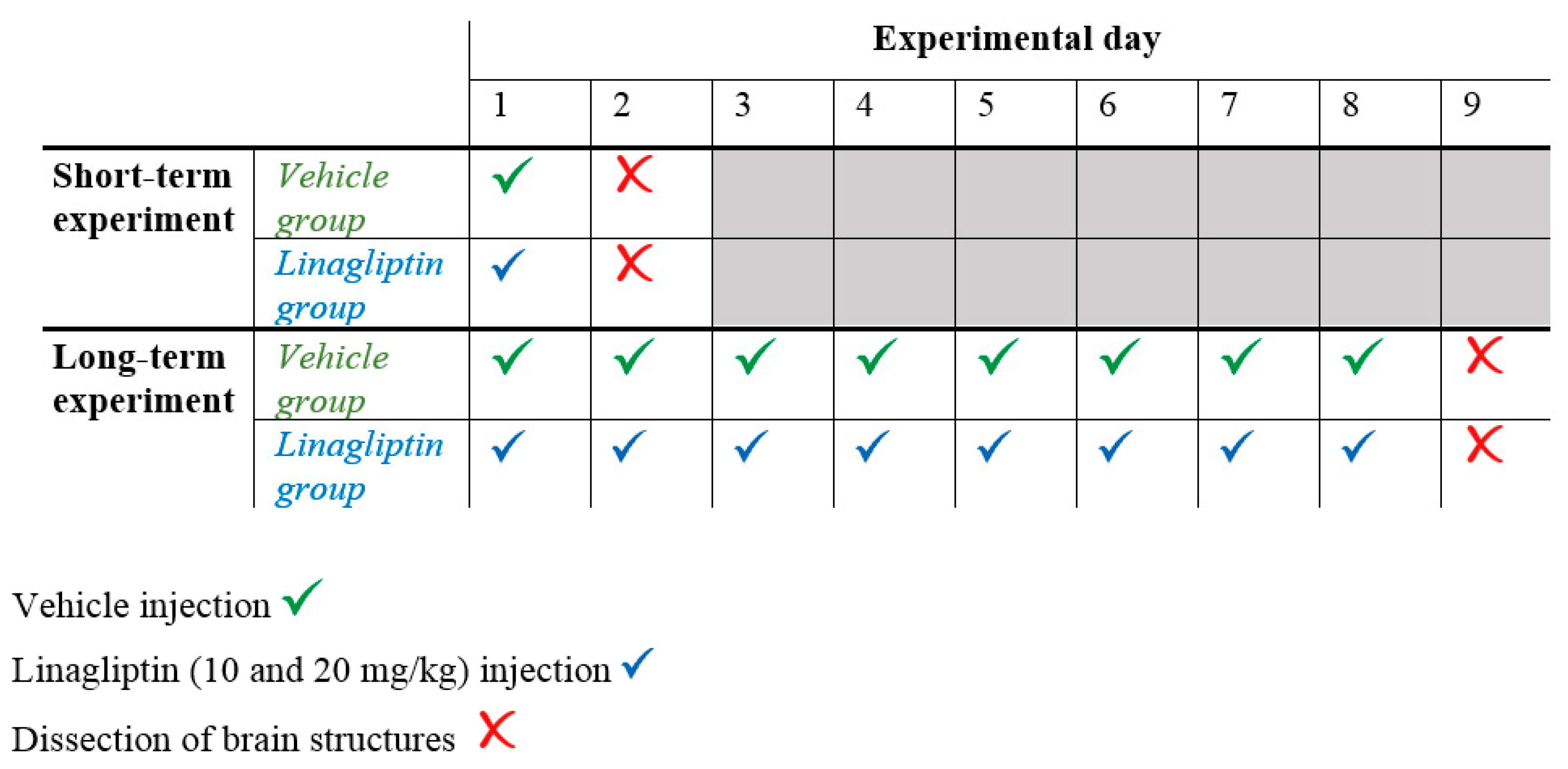
4.3. Experimental Procedure

To assess the effects of short- and long-term administration of linagliptin on the neurotransmitter levels (dopamine, serotonin and noradrenaline) and their metabolites and on the expression of mRNA receptors (dopamine receptors—D1, D2; serotonin receptors—5-HT1A and 5-HT2A; and noradrenaline receptors—α1A and α2A) in the striatum and hippocampus of rats, linagliptin was administered in two schedules. In the first, rats were treated with a single dose of linagliptin. In the second, rats received 1 dose of linagliptin per day for 8 consecutive days (8 injections in total). In both schedules, 24 h after the last linagliptin exposure, the rats were decapitated and their brain structures (the striatum and hippocampus) were dissected. Control animals received injections of the vehicle.
The experimental protocol is graphically depicted in Scheme 1.

4.4. Neurochemical Analysis—Ex Vivo Biochemical Studies Assessed Using HPLC-ED in the Striatum and Hippocampus of Rats

Twenty-four hours after the last vehicle/linagliptin injection, the rats were killed by decapitation, and their brain structures, including the striatum and hippocampus, were immediately dissected. The obtained tissues were frozen on liquid nitrogen (−80 °C) and stored until biochemical assay. Dopamine and its metabolites, DOPAC, 3-MT, and HVA; serotonin and its metabolite, 5-HIAA; and noradrenaline and its metabolite, NMN, were assayed using HPLC-ED. An HP 1050 chromatograph (Golden, CO, USA) was equipped with C18 columns. Tissue samples were weighed and homogenized in ice-cold 0.1 M perchloroacetic acid containing 0.05 mM of ascorbic acid. After centrifugation (10,000× g, 5 min), the supernatants were filtered through RC 58 0.2 µm cellulose membranes (Bioanalytical Systems, West Lafayette, IN, USA). The mobile phase consisted of 0.05 M citrate-phosphate buffer, pH 3.5, 0.1 mM of EDTA, 1 mM of sodium octyl sulfonate, and 3.5% methanol. The flow rate was maintained at 1 mL/min. dopamine, Ser, NA and their metabolites were quantified by peak height comparisons with standards run on the day of analysis. The temperature of the column and the voltage of the detector were set at 30 °C and +750 mV, respectively.

4.5. Analysis of mRNA Expression of Dopamine, Serotonin and Noradrenaline Receptors Using RQ-PCR in the Striatum and Hippocampus of Rats

Total RNA was extracted from 50 to 100 mg brain samples using an RNeasy Lipid Tissue Mini Kit (Qiagen, Hong Kong, China) and qualitative and quantitative analysis of isolated RNA were performed using a NanoDrop ND-1000 spectrophotometer (NanoDrop Technologies, Wilmington, NC, USA). Next, cDNA was obtained from 1 μg of total cellular RNA in 20 μL of reaction volume, using a First Strand cDNA synthesis kit and oligo-dT primers (Fermentas, Waltham, MA, USA). A quantitative assessment of mRNA levels was performed using RQ-PCR on an ABI 7500Fast instrument with Power SYBR Green PCR Master Mix reagent. The real-time conditions, i.e., 95 °C (15 s), 40 cycles at 95 °C (15 s), and 60 °C (1 min), were maintained. In compliance with melting point analysis, only one PCR product was amplified under those conditions. Each sample was analyzed in two technical replicates, and the mean Ct values were used for further analysis. The relative quantity of the target, normalized to the endogenous control Gapdh gene and relative to a calibrator, is expressed as 2−∆∆Ct (−fold difference), where Ct is the threshold cycle, ∆Ct = (Ct of target genes) − (Ct of endogenous control gene, β-2 microglobulin), and ∆∆Ct = (∆Ct of samples for the target gene) − (∆Ct of calibrator for the target gene). The following primer pairs were used:
D1R F: CGC GTA GAC TCT GAG ATT CTG AAT T,
D1R R: GAG TTA AGG AGC CAC CAC ATC AGT;
D2R F: TGA CAG TCC TGC CAA ACC AGA GAA,
D2R R: TGG GCA TGG TCT GGA TCT CAA AGA;
HT51AF: CGT GCA CCA TCA GCA AGG A;
HT51AR: CTG AAG ATG CGC CCG TAG AGA;
HT52AF: ACC GCT ATG TCG CCA TCC A;
HT52AR: GAC CTT CGA ATC ATC CTG TAG TCC A;
ADRA1AF: CGA ATC CAG TGT CTT CGC AG;
ADRA1AR: ACC ATG TCT CTG TGC TGT CCC;
ADRA2AF: TCC CGC CAC TCA TCT CCA TA;
ADRA2AR: CGT TAA TCT TGC AGC TCG GC;
Gapdh F: ATG ACT CTA CCC ACG GCA AG;
Gapdh R: CTG GAA GAT GGT GAT GGG TT.
The neurochemical results are presented in the table as the arithmetical mean concentration values  ±  standard error of the mean (SEM). The dopamine oxidation rate, the dopamine oxomethylation rate and the total dopamine catabolism rate were calculated using the formula ([DOPAC]/[DA)] × 100, ([3 − MT]/] [DA]) × 100, ([HVA]/[DA]) × 100, respectively. The rate of total serotonin catabolism was calculated using the formula ([5-HIAA]/[Ser]) × 100, and the rate of total noradrenaline was calculated using the formula ([NMN])/[NA]) × 100. These results were compared to a vehicle group. After confirming a normal distribution, analysis of variance (one-way ANOVA) was applied, using the Graph-Pad Prism Software package (version 8.02). Tukey’s test was applied to perform the post hoc comparisons. A probability value at p < 0.05 was considered as statistically significant. Every group was composed of 5–7 rats.
The results of mRNA expression analysis are presented in the figures as the arithmetical mean value of mRNA receptor expression  ±  standard deviation (SD). After determining a normal distribution, analysis of variance (one-way ANOVA) was applied using the Graph-Pad Prism Software package (version 8.02). The post hoc comparisons were conducted using Tukey’s test. A probability value at p < 0.05 was considered as statistically significant. Each group consisted of 5–7 rats.

Author Contributions

Conceptualization, M.Ł. and J.L.; Methodology (material preparation and data collection), M.Ł., A.W., I.B.-B., M.T., T.S., D.K.-B., I.G. and J.L.; Formal Analysis, P.L. and J.K.; Investigation, M.Ł. and J.L.; Resources, J.L.; Data Curation, M.Ł. and J.L.; Writing—Original Draft Preparation, M.Ł.; Writing—Review and Editing, J.L.; Visualization, M.Ł. and J.L.; Supervision, J.L.; Funding Acquisition, J.L. All authors have read and agreed to the published version of the manuscript.

Funding

This work was supported by funds from the Medical University of Lublin (DS 20/2018).

Institutional Review Board Statement

The study was performed according to the National Institute of Health Guidelines for the Care and Use of Laboratory Animals and to the European Community Council Directive for Care and Use of Laboratory Animals and was approved (14/2017 on 24 April 2017) by the Local Ethics Committee (The Medical University of Lublin Committee on the Use and Care of Animals).

Informed Consent Statement

Not applicable.

Data Availability Statement

The datasets generated and analyzed during the current study are available from the corresponding author on request.

Conflicts of Interest

The funders had no role in the design of the study; in the collection, analyses, or interpretation of data; in the writing of the manuscript; or in the decision to publish the results. The authors declare no conflicts of interest.

Abbreviations

3-MT3-methoxytyramine
5-HIAA5-hydroxyindoleacetic acid
CNSthe central nervous system
COMTcatechol-O-methyltransferase
DATdopamine transporter
DOPAC3,4-dihydroxyphenylacetic acid
DPP-4 enzymethe dipeptidyl peptidase-4 enzyme
FSCVfast-scan cyclic voltammetry
GLP-1the glucagon-like peptide-1
HVAhomovanillic acid
i.p.intraperitoneally
HPLC-EDhigh-performance liquid chromatography with electrochemical detection
NAnoradrenaline
NMDAN-methyl-D-aspartate
NMNnormetanephrine
RQ-PCRreal-time quantitative reverse transcription polymerase chain reaction
SDstandard deviation
SEMstandard error of the mean
Serserotonin
VMAT2vesicular monoamine transporter 2
VTAventral tegmental area

References

  1. Körner, M.; Stöckli, M.; Waser, B.; Reubi, J.C. GLP-1 receptor expression in human tumors and human normal tissues: Potential for in vivo targeting. J. Nucl. Med. 2007, 48, 736–743. [Google Scholar] [CrossRef]
  2. Mayo, K.E.; Miller, L.J.; Bataille, D.; Dalle, C.; Göke, B.; Thorens, B.; Drucker, D.J. International union of pharmacology. XXXV. The glucagon receptor family. Pharmacol. Rev. 2003, 55, 167–194. [Google Scholar] [CrossRef]
  3. Karaca, M.; Magnan, C.; Kargar, C. Functional pancreatic beta-cell mass: Involvement in type 2 diabetes and therapeutic intervention. Diabetes Metab. 2009, 35, 77–84. [Google Scholar] [CrossRef]
  4. Kieffer, T.J.; McIntosh, C.H.; Pederson, R.A. Degradation of glucose-dependent insulinotropic polypeptide and truncated glucagon-like peptide 1 in vitro and in vivo by dipeptidyl peptidase IV. Endocrinology 1995, 136, 3585–3596. [Google Scholar] [CrossRef]
  5. Lovshin, J.A.; Drucker, D.J. Incretin-based therapies for type 2 diabetes mellitus. Nat. Rev. Endocrinol. 2009, 5, 262–269. [Google Scholar] [CrossRef]
  6. Kastin, A.J.; Akerstrom, V.; Pan, W. Interactions of glucagon-like peptide-1 (GLP-1) with the blood-brain barrier. J. Mol. Neurosci. 2002, 18, 7–14. [Google Scholar] [CrossRef]
  7. Grill, H.J.; Hayes, M.R. Hindbrain neurons as an essential hub in the neuroanatomically distributed control of energy balance. Cell Metab. 2012, 16, 296–309. [Google Scholar] [CrossRef] [PubMed]
  8. Holst, J.J. The physiology of glucagon-like peptide 1. Physiol. Rev. 2007, 87, 1409–1439. [Google Scholar] [CrossRef] [PubMed]
  9. Jin, S.L.; Han, V.K.; Simmons, J.G.; Towle, A.C.; Lauder, J.M.; Lund, P.K. Distribution of glucagonlike peptide I (GLP-I); glucagon; and glicentin in the rat brain: An immunocytochemical study. J. Comp. Neurol. 1988, 271, 519–532. [Google Scholar] [CrossRef] [PubMed]
  10. Llewellyn-Smith, I.J.; Gnanamanickam, G.J.; Reimann, F.; Gribble, F.M.; Trapp, S. Preproglucagon (PPG) neurons innervate neurochemically identified autonomic neurons in the mouse brainstem. Neuroscience 2013, 229, 130–143. [Google Scholar] [CrossRef] [PubMed]
  11. Llewellyn-Smith, I.J.; Marina, N.; Manton, R.N.; Reimann, F.; Gribble, F.M.; Trapp, S. Spinally projecting preproglucagon axons preferentially innervate sympathetic preganglionic neurons. Neuroscience 2015, 284, 872–887. [Google Scholar] [CrossRef]
  12. Llewellyn-Smith, I.J.; Reimann, F.; Gribble, F.M.; Trapp, S. Preproglucagon neurons project widely to autonomic control areas in the mouse brain. Neuroscience 2011, 180, 111–121. [Google Scholar] [CrossRef]
  13. Cork, S.C.; Richards, J.E.; Holt, M.K.; Gribble, F.M.; Reimann, F.; Trapp, S. Distribution and characterisation of Glucagon-like peptide-1 receptor expressing cells in the mouse brain. Mol. Metab. 2015, 4, 718–731. [Google Scholar] [CrossRef]
  14. Alhadeff, A.L.; Rupprecht, L.E.; Hayes, M.R. GLP-1 neurons in the nucleus of the solitary tract project directly to the ventral tegmental area and nucleus accumbens to control for food intake. Endocrinology 2012, 153, 647–658. [Google Scholar] [CrossRef]
  15. Dossat, A.M.; Lilly, N.; Kay, K.; Williams, D.L. Glucagon-like peptide 1 receptors in nucleus accumbens affect food intake. J. Neurosci. 2011, 31, 14453–14457. [Google Scholar] [CrossRef]
  16. Heppner, K.M.; Kirigiti, M.; Secher, A.; Paulsen, S.J.; Buckingham, R.; Pyke, C.; Knudsen, L.B.; Vrang, N.; Grove, K.L. Expression and distribution of glucagon-like peptide-1 receptor mRNA; protein and binding in the male nonhuman primate (Macaca mulatta) brain. Endocrinology 2015, 156, 255–267. [Google Scholar] [CrossRef] [PubMed]
  17. Rinaman, L. Ascending projections from the caudal visceral nucleus of the solitary tract to brain regions involved in food intake and energy expenditure. Brain Res. 2010, 1350, 18–34. [Google Scholar] [CrossRef] [PubMed]
  18. Turton, M.D.; O’Shea, D.; Gunn, I.; Beak, S.A.; Edwards, C.M.; Meeran, K.; Choi, S.J.; Taylor, G.M.; Heath, M.M.; Lambert, P.D.; et al. A role for glucagon-like peptide-1 in the central regulation of feeding. Nature 1996, 379, 69–72. [Google Scholar] [CrossRef] [PubMed]
  19. Chaudhri, O.B.; Parkinson, J.R.; Kuo, Y.T.; Druce, M.R.; Herlihy, A.H.; Bell, J.D.; Dhillo, W.S.; Stanley, S.A.; Ghatei, M.A.; Bloom, S.R. Differential hypothalamic neuronal activation following peripheral injection of GLP-1 and oxyntomodulin in mice detected by manganese-enhanced magnetic resonance imaging. Biochem. Biophys. Res. Commun. 2006, 350, 298–306. [Google Scholar] [CrossRef]
  20. Ranganath, L.R.; Beety, J.M.; Morgan, L.M.; Wright, J.W.; Howland, R.; Marks, V. Attenuated GLP-1 secretion in obesity: Cause or consequence? Gut 1996, 38, 916–919. [Google Scholar] [CrossRef] [PubMed]
  21. Moretto, T.J.; Milton, D.R.; Ridge, T.D.; Macconell, L.A.; Okerson, T.; Wolka, A.M.; Brodows, R.G. Efficacy and tolerability of exenatide monotherapy over 24 weeks in antidiabetic drug–naive patients with type 2 diabetes: A randomized; double-blind; placebo-controlled; parallel-group study. Clin. Ther. 2008, 30, 1448–1460. [Google Scholar] [CrossRef]
  22. Drucker, D.J. The Cardiovascular Biology of Glucagon-like Peptide-1. Cell Metab. 2016, 24, 15–30. [Google Scholar] [CrossRef]
  23. Koshal, P.; Jamwal, S.; Kumar, P. Glucagon-like Peptide-1 (GLP-1) and neurotransmitters signaling in epilepsy: An insight review. Neuropharmacology 2018, 136 Pt B, 271–279. [Google Scholar] [CrossRef]
  24. Egecioglu, E.; Engel, J.A.; Jerlhag, E. The glucagon-like peptide 1 analogue; exendin-4; attenuates the rewarding properties of psychostimulant drugs in mice. PLoS ONE 2013, 8, e69010. [Google Scholar] [CrossRef] [PubMed]
  25. Graham, D.L.; Erreger, K.; Galli, A.; Stanwood, G.D. GLP-1 analog attenuates cocaine reward. Mol. Psychiatry 2013, 18, 961–962. [Google Scholar] [CrossRef] [PubMed]
  26. Egecioglu, E.; Engel, J.A.; Jerlhag, E. The glucagon-like peptide 1 analogue Exendin-4 attenuates the nicotine-induced locomotor stimulation; accumbal dopamine release; conditioned place preference as well as the expression of locomotor sensitization in mice. PLoS ONE 2013, 8, e77284. [Google Scholar] [CrossRef] [PubMed]
  27. Egecioglu, E.; Steensland, P.; Fredriksson, I.; Feltmann, K.; Engel, J.A.; Jerlhag, E. The glucagon-like peptide 1 analogue Exendin-4 attenuates alcohol mediated behaviors in rodents. Psychoneuroendocrinology 2013, 38, 1259–1270. [Google Scholar] [CrossRef]
  28. Vallöf, D.; Maccioni, P.; Colombo, G.; Mandrapa, M.; Jörnulf, J.W.; Egecioglu, E.; Engel, J.A.; Jerlhag, E. The glucagon-like peptide 1 receptor agonist liraglutide attenuates the reinforcing properties of alcohol in rodents. Addict. Biol. 2016, 21, 422–437. [Google Scholar] [CrossRef] [PubMed]
  29. Łupina, M.; Talarek, S.; Kotlińska, J.; Gibuła-Tarłowska, E.; Listos, P.; Listos, J. The role of linagliptin; a selective dipeptidyl peptidase-4 inhibitor; in the morphine rewarding effects in rats. Neurochem. Int. 2020, 133, 104616. [Google Scholar] [CrossRef]
  30. Listos, J.; Listos, P.; Baranowska-Bosiacka, I.; Karpiuk, A.; Filarowska, J.; Łupina, M.; Słowik, T.; Zawiślak, S.; Kotlińska, J. Linagliptin; a Selective Dipeptidyl Peptidase-4 Inhibitor; Reduces Physical and Behavioral Effects of Morphine Withdrawal. Molecules 2022, 27, 2478. [Google Scholar] [CrossRef]
  31. Kasina, S.V.S.K.; Baradhi, K.M. Dipeptidyl Peptidase IV (DPP IV) Inhibitors. In StatPearls; StatPearls Publishing: Treasure Island, FL, USA, 2023. [Google Scholar]
  32. Pipatpiboon, N.; Pintana, H.; Pratchayasakul, W.; Chattipakorn, N.; Chattipakorn, S.C. DPP4-inhibitor improves neuronal insulin receptor function, brain mitochondrial function and cognitive function in rats with insulin resistance induced by high-fat diet consumption. Eur. J. Neurosci. 2013, 37, 839–849. [Google Scholar] [CrossRef]
  33. D’Amico, M.; Filippo, C.D.; Marfella, R.; Abbatecola, A.M.; Ferraraccio, F.; Rossi, F.; Paolisso, G. Long-term inhibition of dipeptidyl peptidase-4 in Alzheimer’s prone mice. Exp. Gerontol. 2010, 45, 202–207. [Google Scholar] [CrossRef]
  34. Kosaraju, J.; Gali, C.C.; Khatwal, R.B.; Dubala, A.; Chinni, S.; Holsinger, R.M.; Madhunapantula, V.S.; Muthureddy Nataraj, S.K.; Basavan, D. Saxagliptin: A dipeptidyl peptidase-4 inhibitor ameliorates streptozotocin induced Alzheimer’s disease. Neuropharmacology 2013, 72, 291–300. [Google Scholar] [CrossRef]
  35. Kosaraju, J.; Murthy, V.; Khatwal, R.B.; Dubala, A.; Chinni, S.; Muthureddy Nataraj, S.K.; Basavan, D. Vildagliptin: An anti-diabetes agent ameliorates cognitive deficits and pathology observed in streptozotocin-induced alzheimer’s disease. J. Pharm. Pharmacol. 2013, 65, 1773–1784. [Google Scholar] [CrossRef]
  36. Isik, A.T.; Soysal, P.; Yay, A.; Usarel, C. The effects of sitagliptin, a DPP-4 inhibitor, on cognitive functions in elderly diabetic patients with or without Alzheimer’s disease. Diabetes Res. Clin. Pract. 2017, 123, 192–198. [Google Scholar] [CrossRef]
  37. Badawi, G.A.; Abd El Fattah, M.A.; Zaki, H.F.; El Sayed, M.I. Sitagliptin and liraglutide reversed nigrostriatal degeneration of rodent brain in rotenone-induced Parkinson’s disease. Inflammopharmacology 2017, 25, 369–382. [Google Scholar] [CrossRef] [PubMed]
  38. ElGamal, R.Z.; Tadros, M.G.; Menze, E.T. Linagliptin counteracts rotenone’s toxicity in non-diabetic rat model of Parkinson’s disease: Insights into the neuroprotective roles of DJ-1, SIRT-1/Nrf-2 and implications of HIF1-α. Eur. J. Pharmacol. 2023, 941, 175498. [Google Scholar] [CrossRef]
  39. Manciu, F.S.; Manciu, M.; Ciubuc, J.D.; Sundin, E.M.; Ochoa, K.; Eastman, M.; Durrer, W.G.; Guerrero, J.; Lopez, B.; Subedi, M.; et al. Simultaneous Detection of Dopamine and Serotonin-A Comparative Experimental and Theoretical Study of Neurotransmitter Interactions. Biosensors 2018, 9, 3. [Google Scholar] [CrossRef] [PubMed]
  40. Di Giovanni, G.; Esposito, E.; Di Matteo, V. Role of serotonin in central dopamine dysfunction. CNS Neurosci. Ther. 2010, 16, 179–194. [Google Scholar] [CrossRef]
  41. Weinshenker, D.; Schroeder, J.P. There and back again: A tale of norepinephrine and drug addiction. Neuropsychopharmacology 2007, 32, 1433–1451. [Google Scholar] [CrossRef] [PubMed]
  42. Thomas, L.; Eckhardt, M.; Langkopf, E.; Tadayyon, M.; Himmelsbach, F.; Mark, M. (R)-8-(3-amino-piperidin-1-yl)-7-but-2-ynyl-3-methyl-1-(4-methyl-quinazolin-2-ylmethyl)-3;7-dihydro-purine-2;6-dione (BI 1356); a novel xanthine-based dipeptidyl peptidase 4 inhibitor; has a superior potency and longer duration of action compared with other dipeptidyl peptidase-4 inhibitors. J. Pharmacol. Exp. Ther. 2008, 325, 175–182. [Google Scholar] [CrossRef] [PubMed]
  43. Suzuki, T.; Tada, Y.; Gladson, S.; Nishimura, R.; Shimomura, I.; Karasawa, S.; Tatsumi, K.; West, J. Vildagliptin ameliorates pulmonary fibrosis in lipopolysaccharide-induced lung injury by inhibiting endothelial-to-mesenchymal transition. Respir. Res. 2017, 18, 177. [Google Scholar] [CrossRef] [PubMed]
  44. Karabulut, S.; Coskun, Z.M.; Bolkent, S. Immunohistochemical, apoptotic and biochemical changes by dipeptidyl peptidase-4 inhibitor-sitagliptin in type-2 diabetic rats. Pharmacol. Rep. 2015, 67, 846–853. [Google Scholar] [CrossRef] [PubMed]
  45. Vulto, A.G.; Westenberg, H.G.; Meijer, L.B.; Versteeg, D.H. The dopamine metabolite 3-methoxytyramine is not a suitable indicator of dopamine release in the rat brain. J. Neurochem. 1986, 47, 1387–1393. [Google Scholar] [CrossRef] [PubMed]
  46. Waldmeier, P.C.; Lauber, J.; Blum, W.; Richter, W.J. 3-Methoxytyramine: Its suitability as an indicator of synaptic dopamine release. Naunyn Schmiedebergs Arch. Pharmacol. 1981, 315, 219–225. [Google Scholar] [CrossRef] [PubMed]
  47. Wood, P.L.; Nair, N.P.; Bozarth, M. Striatal 3-methoxytyramine as an index of dopamine release effects of electrical stimulation. Neurosci. Lett. 1982, 32, 291–294. [Google Scholar] [CrossRef]
  48. Sotnikova, T.D.; Beaulieu, J.M.; Espinoza, S.; Masri, B.; Zhang, X.; Salahpour, A.; Barak, L.S.; Caron, M.G.; Gainetdinov, R.R. The dopamine metabolite 3-methoxytyramine is a neuromodulator. PLoS ONE 2010, 5, e13452. [Google Scholar] [CrossRef]
  49. Antkiewicz-Michaluk, L.; Ossowska, K.; Romańska, I.; Michaluk, J.; Vetulani, J. 3-Methoxytyramine; an extraneuronal dopamine metabolite plays a physiological role in the brain as an inhibitory regulator of catecholaminergic activity. Eur. J. Pharmacol. 2008, 599, 32–35. [Google Scholar] [CrossRef]
  50. Wood, P.L.; Kim, H.S.; Stocklin, K.; Rao, T.S. Dynamics of the striatal 3-MT pool in rat and mouse: Species differences as assessed by steady-state measurements and intracerebral dialysis. Life Sci. 1988, 42, 2275–2281. [Google Scholar] [CrossRef]
  51. Badawi, G.A.; Abd El Fattah, M.A.; Zaki, H.F.; El Sayed, M.I. Sitagliptin and Liraglutide Modulate L-dopa Effect and Attenuate Dyskinetic Movements in Rotenone-Lesioned Rats. Neurotox. Res. 2019, 35, 635–653. [Google Scholar] [CrossRef]
  52. Fortin, S.M.; Roitman, M.F. Central GLP-1 receptor activation modulates cocaine-evoked phasic dopamine signaling in the nucleus accumbens core. Physiol. Behav. 2017, 176, 17–25. [Google Scholar] [CrossRef] [PubMed]
  53. Wang, X.F.; Liu, J.J.; Xia, J.; Liu, J.; Mirabella, V.; Pang, Z.P. Endogenous glucagon-like peptide-1 suppresses high-fat food intake by reducing synaptic drive onto mesolimbic dopamine neurons. Cell Rep. 2015, 12, 726–733. [Google Scholar] [CrossRef] [PubMed]
  54. Mietlicki-Baase, E.G.; Ortinski, P.I.; Rupprecht, L.E.; Olivos, D.R.; Alhadeff, A.L.; Pierce, R.C.; Hayes, M.R. The food intake-suppressive effects of glucagon-like peptide-1 receptor signaling in the ventral tegmental area are mediated by AMPA/kainate receptors. Am. J. Physiol. Endocrinol. Metab. 2013, 305, E1367–E1374. [Google Scholar] [CrossRef] [PubMed]
  55. Richard, J.E.; Anderberg, R.H.; Göteson, A.; Gribble, F.M.; Reimann, F.; Skibicka, K.P. Activation of the GLP-1 receptors in the nucleus of the solitary tract reduces food reward behavior and targets the mesolimbic system. PLoS ONE 2015, 10, e0119034. [Google Scholar] [CrossRef]
  56. Harasta, A.E.; Power, J.M.; von Jonquieres, G.; Karl, T.; Drucker, D.J.; Housley, G.D.; Schneider, M.; Klugmann, M. Septal glucagon-like peptide 1 receptor expression determines suppression of cocaine-induced behavior. Neuropsychopharmacology 2015, 40, 1969–1978. [Google Scholar] [CrossRef] [PubMed]
  57. Reddy, I.A.; Pino, J.A.; Weikop, P.; Osses, N.; Sørensen, G.; Bering, T.; Valle, C.; Bluett, R.J.; Erreger, K.; Wortwein, G.; et al. Glucagon-like peptide 1 receptor activation regulates cocaine actions and dopamine homeostasis in the lateral septum by decreasing arachidonic acid levels. Transl. Psychiatry 2016, 6, e809. [Google Scholar] [CrossRef]
  58. Jensen, M.E.; Galli, A.; Thomsen, M.; Jensen, K.L.; Thomsen, G.K.; Klausen, M.K.; Vilsbøll, T.; Christensen, M.B.; Holst, J.J.; Owens, A.; et al. Glucagon-like peptide-1 receptor regulation of basal dopamine transporter activity is species-dependent. Neurochem. Int. 2020, 138, 104772. [Google Scholar] [CrossRef] [PubMed]
  59. Secnik, J.; Cermakova, P.; Fereshtehnejad, S.M.; Dannberg, P.; Johnell, K.; Fastbom, J.; Winblad, B.; Eriksdotter, M.; Religa, D. Diabetes in a Large Dementia Cohort: Clinical Characteristics and Treatment from the Swedish Dementia Registry. Diabetes Care 2017, 40, 1159–1166. [Google Scholar] [CrossRef]
  60. Kalia, L.V.; Lang, A.E. Parkinson’s disease. Lancet 2015, 386, 896–912. [Google Scholar] [CrossRef]
  61. Morris, J.K.; Bomhoff, G.L.; Gorres, B.K.; Davis, V.A.; Kim, J.; Lee, P.P.; Brooks, W.M.; Gerhardt, G.A.; Geiger, P.C.; Stanford, J.A. Insulin resistance impairs nigrostriatal dopamine function. Exp. Neurol. 2011, 231, 171–180. [Google Scholar] [CrossRef]
  62. Elbassuoni, E.A.; Ahmed, R.F. Mechanism of the neuroprotective effect of GLP-1 in a rat model of Parkinson’s with pre-existing diabetes. Neurochem. Int. 2019, 131, 104583. [Google Scholar] [CrossRef] [PubMed]
  63. Chen, Y.; Zhang, Y.; Li, L.; Hölscher, C. Neuroprotective effects of geniposide in the MPTP mouse model of Parkinson’s disease. Eur. J. Pharmacol. 2015, 768, 21–27. [Google Scholar] [CrossRef]
  64. Ji, C.; Xue, G.F.; Lijun, C.; Feng, P.; Li, D.; Li, L.; Li, G.; Hölscher, C. A novel dual GLP-1 and GIP receptor agonist is neuroprotective in the MPTP mouse model of Parkinson’s disease by increasing expression of BNDF. Brain Res. 2016, 1634, 1–11. [Google Scholar] [CrossRef] [PubMed]
  65. Tan, B.; Li, X.G.; Kong, X.; Huang, R.; Ruan, Z.; Yao, K.; Deng, Z.; Xie, M.; Shinzato, I.; Yin, Y.; et al. Dietary L-arginine supplementation enhances the immune status in early-weaned piglets. Amino Acids 2009, 37, 323–331. [Google Scholar] [CrossRef] [PubMed]
  66. Cao, L.; Li, D.; Feng, P.; Li, L.; Xue, G.F.; Li, G.; Hölscher, C. A novel dual GLP-1 and GIP incretin receptor agonist is neuroprotective in a mouse model of Parkinson’s disease by reducing chronic inflammation in the brain. Neuroreport 2016, 27, 384–391. [Google Scholar] [CrossRef] [PubMed]
  67. Feng, P.; Zhang, X.; Li, D.; Ji, C.; Yuan, Z.; Wang, R.; Xue, G.; Li, G.; Hölscher, C. Two novel dual GLP-1/GIP receptor agonists are neuroprotective in the MPTP mouse model of Parkinson’s disease. Neuropharmacology 2018, 133, 385–394. [Google Scholar] [CrossRef] [PubMed]
  68. Dai, C.; Tan, C.; Zhao, L.; Liang, Y.; Liu, G.; Liu, H.; Zhong, Y.; Liu, Z.; Mo, L.; Liu, X.; et al. Glucose metabolism impairment in Parkinson’s disease. Brain Res. Bull. 2023, 199, 110672. [Google Scholar] [CrossRef]
  69. Nassar, N.N.; Al-Shorbagy, M.Y.; Arab, H.H.; Abdallah, D.M. Saxagliptin: A novel antiparkinsonian approach. Neuropharmacology 2015, 89, 308–317. [Google Scholar] [CrossRef]
  70. Nonogaki, K.; Kaji, T. Pharmacological stimulation of serotonin 5-HT1B receptors enhances increases in plasma active glucagon-like peptide-1 levels induced by dipeptidyl peptidase-4 inhibition independently of feeding in mice. Diabetes Metab. 2015, 41, 425–428. [Google Scholar] [CrossRef]
  71. Yang, Y.; Cui, X.; Chen, Y.; Wang, Y.; Li, X.; Lin, L.; Zhang, H. Exendin-4; an analogue of glucagon-like peptide-1; attenuates hyperalgesia through serotonergic pathways in rats with neonatal colonic sensitivity. J. Physiol. Pharmacol. 2014, 65, 349–357. [Google Scholar]
  72. Brunetti, L.; Orlando, G.; Recinella, L.; Leone, S.; Ferrante, C.; Chiavaroli, A.; Lazzarin, F.; Vacca, M. Glucagon-like peptide 1 (7-36) amide (GLP-1) and exendin-4 stimulate serotonin release in rat hypothalamus. Peptides 2008, 29, 1377–1381. [Google Scholar] [CrossRef] [PubMed]
  73. Anderberg, R.H.; Richard, J.E.; Eerola, K.; López-Ferreras, L.; Banke, E.; Hansson, C.; Nissbrandt, H.; Berqquist, F.; Gribble, F.M.; Reimann, F.; et al. Glucagon-like peptide-1 and its analogues act in the dorsal raphe and modulate central serotonin to reduce appetite and body weight. Diabetes 2017, 66, 1062–1073. [Google Scholar] [CrossRef] [PubMed]
  74. Vestlund, J.; Jerlhag, E. Glucagon-like peptide-1 receptors and sexual behaviors in male mice. Psychoneuroendocrinology 2020, 117, 104687. [Google Scholar] [CrossRef] [PubMed]
  75. Sarkar, S.; Fekete, C.; Legradi, G.; Lechan, R.M. Glucagon like peptide-1 (7–36) amide (GLP-1) nerve terminals densely innervate corticotropin-releasing hormone neurons in the hypothalamic paraventricular nucleus. Brain Res. 2003, 985, 163–168. [Google Scholar] [CrossRef]
  76. Larsen, P.J.; Tang-Christensen, M.; Holst, J.J.; Orskov, C. Distribution of glucagon-like peptide-1 and other preproglucagon-derived peptides in the rat hypothalamus and brainstem. Neuroscience 1997, 77, 257–270. [Google Scholar] [CrossRef]
Figure 1. The effect of chronic exposure to linagliptin (10 and 20 mg/kg, i.p.) on mRNA expression of D1 and D2 receptors in the striatum and hippocampus in rats (n = 5–6). Data are reported as means ± SD.
Ijms 25 03008 g001
Figure 2. The effect of chronic exposure to linagliptin (10 and 20 mg/kg, i.p.) on mRNA expression of 5-HT1A and 5-HT2A receptors in the striatum and hippocampus in rats; * p < 0.05, ** p < 0.01 (vs. vehicle) (Tukey’s test, n = 5–6). Data are reported as means ± SD.
Ijms 25 03008 g002
Figure 3. The effect of chronic exposure to linagliptin (10 and 20 mg/kg, i.p.) on mRNA expression of α1A and α2A receptors in the striatum and hippocampus in rats (n = 5–6). Data are reported as means ± SD.
Ijms 25 03008 g003
Scheme 1. Schedule of linagliptin administration.
Ijms 25 03008 sch001
Table 1. Effect of linagliptin (10 and 20 mg/kg, i.p.) on the concentration of dopamine and its metabolites in the striatum in rats; DA—dopamine, DOPAC-3,4-dihydroxyphenylacetic acid, 3-MT—3-methoxytyramine, HVA—homovanillic acid; * p < 0.05, ** p < 0.01, *** p < 0.001 (vs. vehicle group) (Tukey’s test, n = 5–7). Data are reported as means ± SEM.
DA
(ng/g Tissue)
DOPAC
(ng/g Tissue)
3-MT
(ng/g Tissue)
HVA
(ng/g Tissue)
(DOPAC/DA)
× 100
(3-MT/DA)
× 100
(HVA/DA)
× 100
StriatumSingle doe of linagliptinvehicle10,508.0
± 902.1
1309.0
± 78.74
286.6
± 41.62
1292.0
± 106.9
13.53
± 0.3970
2.771
± 0.3841
11.87
± 0.7049
vehicle
+ linagliptin 10
10,669.0
± 504.6
1524.0
± 94.70
525.3
± 40.24
*
1361.0
± 71.05
14.58
± 0.7214
5.053
± 0.4216
**
13.01
± 0.3776
vehicle
+ linagliptin 20
10,793.0
± 314.6
1477.0
± 101.9
556.5
± 58.28
**
1353.0
± 110.9
14.05
± 0.5923
5.314
± 0.4909
**
12.83
± 0.6536
Chronic administration of linagliptinvehicle 10,291.0
± 851.6
1373.0
± 128.0
287.7
± 28.34
1291.0
± 78.44
13.95
± 0.5291
3.080
± 0.3014
12.60
± 0.6355
vehicle
+ linagliptin 10
13,392.0
± 303.8
**
2026.0
± 135.8
**
428.8
± 34.18
*
1665.0
± 43.77
**
14.38
± 1.688
2.945
± 0.4348
12.51
± 0.5049
vehicle
+ linagliptin 20
13,996.0
± 560.9
***
1971.0
± 132.8
**
497.0
± 47.71
**
1551.0
± 79.22
*
13.04
± 1.205
3.499
± 0.3576
11.01
± 0.3662
Table 2. Effect of linagliptin (10 and 20 mg/kg, i.p.) on the concentration of dopamine and its metabolites in the hippocampus in rats; DA—dopamine, DOPAC—3,4-dihydroxyphenylacetic acid, 3-MT—3-methoxytyramine, HVA—homovanillic acid; * p < 0.05, ** p < 0.01 (vs. vehicle group) (Tukey’s test, n = 5–7). Data are reported as means ± SEM.
DA
(ng/g Tissue)
DOPAC
(ng/g Tissue)
3-MT
(ng/g Tissue)
HVA
(ng/g Tissue)
(DOPAC/DA)
× 100
(3-MT/DA)
× 100
(HVA/DA)
× 100
HippocampusSingle doe of linagliptinvehicle13.62
± 1.726
7.020
± 1.405
7.920
± 1.194
13.54
± 2.962
57.60
± 12.57
54.99
± 8.728
89.42
± 26.14
vehicle
+ linagliptin 10
11.90
± 1.266
5.440
± 0.7359
6.780
± 1.175
7.500
± 0.8972
46.20
± 5.802
51.21
± 12.92
58.79
± 5.910
vehicle
+ linagliptin 20
19.60
± 1.770
*
3.264
± 0.4847
*
2.800
± 0.3266
**
6.075
± 0.9232
18.64
± 2.797
**
15.13
± 0.7017
*
26.42
± 3.261
*
Chronic administration of linagliptinvehicle 15.86
± 1.689
8.500
± 1.268
7.850
± 1.230
14.34
± 2.422
43.53
± 7.601
49.24
± 10.89
87.41
± 28.82
vehicle
+ linagliptin 10
27.67
± 3.471
9.500
± 1.848
6.200
± 0.3887
18.33
± 2.741
26.06
± 3.601
21.52
± 3.145
52.40
± 6.793
vehicle
+ linagliptin 20
25.67
± 4.224
6.333
± 1.856
9.000
± 0.8018
13.25
± 0.9402
30.46
± 5.142
31.96
± 2.943
54.07
± 6.820
Table 3. Effect of linagliptin (10 and 20 mg/kg, i.p.) on the concentration of serotonin and its metabolites in the striatum in rats; Ser—serotonin, 5-HIAA—5-hydroxyindoleacetic acid; * p < 0.05, ** p < 0.01, **** p < 0.0001 (vs. vehicle group) (Tukey’s test, n = 5–7). Data are reported as means ± SEM.
Ser
(ng/g Tissue)
5-HIAA
(ng/g Tissue)
(5-HIAA/Ser) × 100
Striatum Single dose
of linagliptin
vehicle268.5 ± 10.92392.9 ± 15.01133.2 ± 2.433
vehicle +
linagliptin 10
348.8 ± 23.67
**
455.8 ± 15.91
*
133.7 ± 3.687
vehicle +
linagliptin 20
390.7 ± 4.022
****
510.5 ± 16.74
****
130.8 ± 4.506
Chronic
administration
of linagliptin
vehicle276.0 ± 9.716397.5 ± 25.56132.1 ± 2.371
vehicle +
linagliptin 10
202.4 ± 20.07
*
264.8 ± 12.39
*
145.5 ± 11.72
vehicle +
linagliptin 20
273.2 ± 15.72467.3 ± 40.43157.0 ± 10.60
Table 4. Effect of linagliptin (10 and 20 mg/kg, i.p.) on the concentration of serotonin and its metabolites in the hippocampus of rats; Ser—serotonin, 5-HIAA—5-hydroxyindoleacetic acid; * p < 0.05, ** p < 0.01 (vs. vehicle group) (Tukey’s test, n = 5–7). Data are reported as means ± SEM.
Ser
(ng/g Tissue)
5-HIAA
(ng/g Tissue)
(5-HIAA/Ser) × 100
Hippocampus Single dose of linagliptinvehicle165.0 ± 15.37238.0 ± 34.56144.6 ± 8.653
vehicle +
linagliptin 10
244.4 ± 24.64
*
388.4 ± 22.55
**
162.3 ± 6.242
vehicle +
linagliptin 20
241.0 ± 19.93
*
404.5 ± 17.91
**
172.0 ± 8.768
Chronic
administration
of linagliptin
vehicle161.4 ± 18.30244.3 ± 27.72154.5 ± 11.94
vehicle +
linagliptin 10
131.4 ± 13.60150.8 ± 13.66112.6 ± 14.79
vehicle +
linagliptin 20
153.0 ± 18.23406.2 ± 59.31
*
232.1 ± 29.61
*
Table 5. Effect of linagliptin (10 and 20 mg/kg, i.p.) on the concentration of noradrenaline and its metabolites in the striatum in rats; NA—noradrenaline, NMN—methoxynoradrenaline; * p < 0.05, ** p < 0.01, **** p < 0.0001 (vs. vehicle group) (Tukey’s test, n = 5–7). Data are reported as means ± SEM.
NA
(ng/g Tissue)
NMN
(ng/g Tissue)
(NMN/NA) × 100
Striatum Single dose
of linagliptin
vehicle316.4 ± 14.325.250 ± 1.6561.643 ± 0.5179
vehicle +
linagliptin 10
549.5 ± 24.73
****
2.280 ± 0.29470.4692 ± 0.05262
**
vehicle +
linagliptin 20
613.0 ± 30.35
****
1.760 ± 0.1893
*
0.2922 ± 0.03848
**
Chronic
administration
of linagliptin
vehicle305.8 ± 11.415.140 ± 1.9621.882 ± 0.5059
vehicle +
linagliptin 10
318.3 ± 15.363.486 ± 1.0351.55 ± 0.2218
vehicle +
linagliptin 20
286.3 ± 15.584.129 ± 0.99871.212 ± 0.1997
Table 6. Effect of linagliptin (10 and 20 mg/kg, i.p.) on the concentration of noradrenaline and its metabolites in the hippocampus in rats; NA—noradrenaline, NMN—methoxynoradrenaline; *** p < 0.001, **** p < 0.0001 (vs. vehicle group) (Tukey’s test, n = 5–6). Data are reported as means ± SEM.
NA
(ng/g Tissue)
NMN
(ng/g Tissue)
(NMN/NA) × 100
Hippocampus
Single dose
of linagliptin
vehicle402.2 ± 28.5530.00 ± 5.0607.389 ± 2.050
vehicle +
linagliptin 10
391.3 ± 11.795.340 ± 1.093
***
1.346 ± 0.2132
****
vehicle +
linagliptin 20
380.4 ± 39.104.733 ± 0.8401
****
1.352 ± 0.2107
****
Chronic
administration
of linagliptin
vehicle425.6 ± 28.7929.75 ± 6.5247.062 ± 1.621
vehicle +
linagliptin 10
530.0 ± 32.5027.60 ± 1.5365.808 ± 0.5894
vehicle +
linagliptin 20
482.5 ± 67.6129.00 ± 6.6885.226 ± 0.2510
Table 7. The effect of acute exposure to linagliptin (10 and 20 mg/kg, i.p.) on mRNA expression of D1, D2, 5-HT1A, 5-HT2A, α1A and α2A receptors in the striatum and hippocampus in rats (n = 5–7). Data are reported as means ± SD.
mRNAD1
[mRNA/
GAPDH]
D2
[mRNA/
GAPDH]
5-HT1A
[mRNA/
GAPDH]
5-HT2A [mRNA/
GAPDH]
α1A
[mRNA/
GAPDH]
α2A
[mRNA/
GAPDH]
Expression
Drugs
Striatumvehicle0.02542
± 0.008658
0.007618
± 0.006601
0.00003833
± 0.00002639
0.000534
± 0.0003628
0.0003375
± 0.0003526
0.000052
± 0.00003564
Vehicle
+ linagliptin 10
0.028
± 5.01 × 10⁻³
0.00678
± 3.43 × 10⁻³
0.00004137
± 0.0000357
0.000603
± 0.000144
0.00029
± 0.000142
0.000038
± 2.51 × 10⁻⁵
vehicle
+ linagliptin 20
0.021
± 3.25 × 10⁻³
0.00812
± 4.21 × 10⁻³
0.00006519
± 0.0000298
0.000419
± 0.00017
0.000381
± 0.000201
0.000063
± 2.89 × 10⁻⁵
Hippocampusvehicle0.0004933
± 0.0003678
0.0002
± 6.12 × 10⁻⁵
0.000228
± 0.000163
0.0002
± 0.000226
7.67 × 10⁻⁵
± 0.00008963
0.00003
± 2.58 × 10⁻⁵
vehicle
+ linagliptin 10
5.42 × 10⁻⁴
± 3.19 × 10⁻⁴
2.40 × 10⁻⁴
± 3.20 × 10⁻⁴
0.000261
± 1.29 × 10⁻⁴
0.000143
± 7.89 × 10⁻⁵
6.31 × 10⁻⁵
± 4.21 × 10⁻⁵
2.40 × 10⁻⁵
± 1.42 × 10⁻⁵
vehicle
+ linagliptin 20
4.36 × 10⁻⁴
± 2.80 × 10⁻⁴
2.61 × 10⁻⁴
± 2.36 × 10⁻⁴
0.000249
± 7.89 × 10⁻⁵
0.000098
± 2.94 × 10⁻⁵
6.99 × 10⁻⁵
± 5.90 × 10⁻⁵
2.81 × 10⁻⁵
± 1.99 × 10⁻⁵
Disclaimer/Publisher’s Note: The statements, opinions and data contained in all publications are solely those of the individual author(s) and contributor(s) and not of MDPI and/or the editor(s). MDPI and/or the editor(s) disclaim responsibility for any injury to people or property resulting from any ideas, methods, instructions or products referred to in the content.

