Review

Agnostic Administration of Targeted Anticancer Drugs: Looking for a Balance between Hype and Caution

by
Svetlana N. Aleksakhina
1,
Alexander O. Ivantsov
1,2 and
Evgeny N. Imyanitov
1,2,*
1
Department of Tumor Growth Biology, N. N. Petrov Institute of Oncology, 197758 St. Petersburg, Russia
2
Department of Medical Genetics, St. Petersburg Pediatric Medical University, 194100 St. Petersburg, Russia
*
Author to whom correspondence should be addressed.
Int. J. Mol. Sci. 2024, 25(7), 4094; https://doi.org/10.3390/ijms25074094
Submission received: 18 February 2024 / Revised: 27 March 2024 / Accepted: 3 April 2024 / Published: 7 April 2024
(This article belongs to the Special Issue Innovative Approaches for Targeted Cancer Therapies)

Abstract

:
Many tumors have well-defined vulnerabilities, thus potentially allowing highly specific and effective treatment. There is a spectrum of actionable genetic alterations which are shared across various tumor types and, therefore, can be targeted by a given drug irrespective of tumor histology. Several agnostic drug-target matches have already been approved for clinical use, e.g., immune therapy for tumors with microsatellite instability (MSI) and/or high tumor mutation burden (TMB), NTRK1-3 and RET inhibitors for cancers carrying rearrangements in these kinases, and dabrafenib plus trametinib for BRAF V600E mutated malignancies. Multiple lines of evidence suggest that this histology-independent approach is also reasonable for tumors carrying ALK and ROS1 translocations, biallelic BRCA1/2 inactivation and/or homologous recombination deficiency (HRD), strong HER2 amplification/overexpression coupled with the absence of other MAPK pathway-activating mutations, etc. On the other hand, some well-known targets are not agnostic: for example, PD-L1 expression is predictive for the efficacy of PD-L1/PD1 inhibitors only in some but not all cancer types. Unfortunately, the individual probability of finding a druggable target in a given tumor is relatively low, even with the use of comprehensive next-generation sequencing (NGS) assays. Nevertheless, the rapidly growing utilization of NGS will significantly increase the number of patients with highly unusual or exceptionally rare tumor-target combinations. Clinical trials may provide only a framework for treatment attitudes, while the decisions for individual patients usually require case-by-case consideration of the probability of deriving benefit from agnostic versus standard therapy, drug availability, associated costs, and other circumstances. The existing format of data dissemination may not be optimal for agnostic cancer medicine, as conventional scientific journals are understandably biased towards the publication of positive findings and usually discourage the submission of case reports. Despite all the limitations and concerns, histology-independent drug-target matching is certainly feasible and, therefore, will be increasingly utilized in the future.

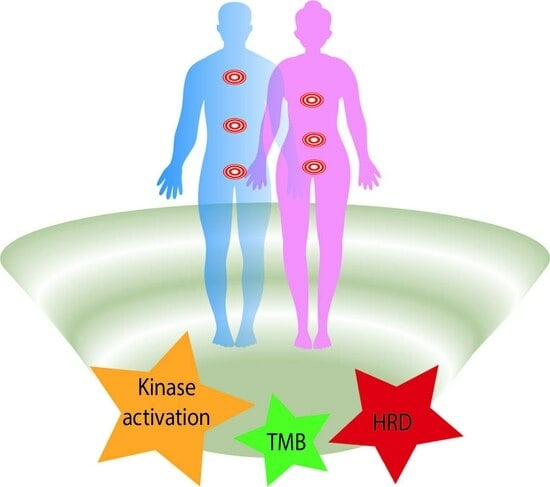
Graphical Abstract

1. Introduction

The first discoveries of oncogenes and suppressor genes in the 1980s revealed that many driver events, for example, TP53 or RAS mutations, are shared between tumors of different types [1]. These findings fueled hopes for the development of “universal” cancer drugs, which are capable of interfering with backbone molecular pathways across a diverse spectrum of malignancies. The invention of mutation-tailored targeted therapies and advent of high-throughput sequencing technologies confirmed that many actionable molecular targets, e.g., BRAF V600E substitutions, ALK/ROS1/RET/NTRK rearrangements, microsatellite instability (MSI-H), etc., occur at some frequency even in tumors belonging to entirely distinct histological lineages. There is a clear rationale for the use of relevant drugs in an agnostic manner, i.e., in all tumors carrying the appropriate target irrespective of their morphology [2,3,4,5].
Expectedly, the first case reports confirming the success of this approach were met with great optimism. The history of the agnostic targeting of neoplastic growth originates from attempts to utilize tamoxifen, a well-known breast cancer drug directed against the estrogen receptor (ER), for the treatment of ER+ pulmonary lymphangiomyomatosis [6]. Trastuzumab, an anti-HER2 antibody, was approved for the treatment of HER2-driven breast cancer in 1998 [7]. The first report demonstrating the agnostic utility of this drug was published just a year later: trastuzumab rendered substantial benefit to a patient suffering from a HER2-overexpressing germ cell tumor [8]. The EGFR inhibitors gefitinib and erlotinib were developed on the assumption that many cancers overexpress this receptor. Clinical trials for these drugs revealed tumor shrinkage only in a small subset of lung cancer patients, and subsequent analysis of tumor tissues obtained from the responders identified unknown drug-sensitizing EGFR mutations [9]. The first attempt of the agnostic use of gefitinib is exemplified by its administration to a patient with EGFR-mutated renal cell cancer, which resulted in an objective tumor response [10]. In contrast to gefitinib or erlotinib, the development of vemurafenib and dabrafenib relied on the knowledge of the occurrence of BRAF V600E mutations across multiple cancer types; therefore, many instances of the agnostic use of these kinase inhibitors were reported immediately after their invention [11,12].
The concept of agnostic drug administration fueled interest in tumor genomic profiling. This approach relies on comprehensive analysis of all known molecular targets in every cancer patient, followed by matching of available drugs to genetic findings. Although the overall promise of reflex DNA and RNA sequencing is beyond any reasonable doubt, some systematic studies brought disappointing results [13,14]. First, the probability of finding the “Achilles heel” in a given tumor is often vanishingly low, as most of the known druggable mutations are exceptionally rare in the agnostic setting [15]. Secondly, even rationally matched treatments sometimes produce limited, if any, benefit [16]. Only a few investigations have assessed real-world attitudes towards agnostic therapy and tumor genomic profiling among medical oncologists and cancer patients [17,18], and there is some contrast between the enthusiasm conveyed by drug manufacturers or genetic service providers and the caution expressed by many practicing physicians or healthcare regulators [15,19,20].

2. Approved Agnostic Targets and Drugs

2.1. Overview

Several agnostic targets and their matched drugs have been approved by the Food and Drug Administration (FDA) in the past few years [21,22,23]. This list includes microsatellite instability (MSI) and/or deficient mismatch DNA repair (MMR-D) coupled with immune therapy; high TMB (tumor mutation burden), which includes both MSI and some other instances of accumulation of multiple somatic genetic alterations, and is also associated with a response to immune checkpoint inhibitors (ICIs); NTRK1-3 rearrangements rendering tumor sensitivity to NTRK tyrosine kinase inhibitors (TKIs); RET rearrangements calling for the therapeutic use of RET TKI; and BRAF V600E mutations associated with tumor responsiveness to combined BRAF and MEK inhibition (Figure 1; Table 1). There are some nuances related to the process of drug approval. For example, both entrectinib and larotrectinib have been subjected to agnostic clinical trials, and, therefore, both of these drugs are permitted for use in NTRK-rearranged tumors irrespective of the cancer type. In contrast, only the dabrafenib plus trametinib combination has been successfully evaluated for BRAF V600E mutated tumors in the agnostic setting, while similar combinations, i.e., vemurafenib plus cobimetinib, or encorafenib plus binimetinib, have only histology-specific indications for the time being. The European Medicines Agency (EMA) has a more cautious attitude towards tissue-agnostic approvals than the FDA, so the list of agnostic drugs and targets is currently less extensive in Europe than in the USA [23,24,25] (Table 1).

2.2. Microsatellite Instability (MSI)/Mismatch Repair Deficiency (MMR-D)

MSI is a relatively straightforward DNA test, which identifies tumors with an accumulation of mutations in microsatellite repeats. These carcinomas have about one to two orders of magnitude more mutations than other malignancies; therefore, they are usually highly antigenic and responsive to immune therapy. Mismatch repair deficiency (MMR-D) is the only known cause of MSI. While MSI detection requires the utilization of molecular genetic tests, the analysis of MMR-D is based on immunohistochemical (IHC) detection of MLH1, MSH2, MSH6, and PMS2 proteins, and, therefore, is perfectly compatible with a standard morphological examination of the tumor tissue. Colorectal carcinomas are the most common tumors with an MSI/MMR-D phenotype. In colorectal cancers, MSI is almost always accompanied by MMR-D, and vice versa; therefore, DNA and IHC tests are interchangeable for this tumor type. This is not true for malignancies with low proliferation rate, in which MMR-D does not result in similarly high accumulation of mutations affecting microsatellites [26,27,28]. Importantly, agnostic clinical trials on pembrolizumab and dostarlimab considered MSI and MMR-D as equivalent tests [29,30]. There is a subset of tumors showing discordant results by MSI and MMR-D, and this discordance is particularly relevant to malignancies arising outside the gastrointestinal tract [31]. Some studies suggest that the use of MMR-D testing alone, i.e., without confirmation of the accumulation of mutations in tumor DNA, may require additional validation, at least in non-colorectal neoplasms [32]. The opposite applies to screening for Lynch syndrome, given that in some rare instances tumors attributed to germline mutations in the MLH1, MSH2, MSH6, or PMS2 genes may demonstrate MMR-D in the absence of MSI [26,28]. MSI is positioned as an agnostic target, however relevant clinical trials usually included only a few tumor types characterized by high or moderate frequency of MSI/MMR-D occurrence [29,30]. MSI is relatively common in colorectal, gastric, small bowel, and endometrial malignancies, occurs at low frequency in several other cancer types, e.g., biliary tract, pancreatic, or ovarian tumors, but is exceptionally rare in some of the most common oncological diseases, for example, breast or lung carcinomas [33,34,35,36,37]. The detection of MSI involves either the utilization of individually administered IHC or PCR tests, or the use of next-generation sequencing (NGS) panels consisting of several hundred genes [27]. Given that the probability of finding MSI/MMR-D in some cancer types is very small, it is questionable whether indeed all tumor categories deserve MSI/MMR-D testing, unless they arise in subjects with Lynch syndrome.

2.3. Tumor Mutation Burden (TMB)

High TMB is an FDA-approved agnostic indication for the use of pembrolizumab. The predictive role of TMB is particularly evident in carcinogen-induced tumors, e.g., lung carcinomas or melanomas. Still, the actual value of TMB determination in these tumor types may be questioned because the history of tumor development is highly indicative of its potential immunogenicity. Indeed, cigarette smoking is a reliable predictor of high TMB in lung cancer, while an excessive number of mutations is observed mainly in melanomas related to sunburns [38,39]. MSI is the most common cause of highly elevated TMB in carcinogen-unrelated tumors. Microsatellite-unstable tumors vary by approximately an order of magnitude with regard to the number of mutations per genome, and, expectedly, the response to immune therapy is as better as more mutations are observed [40,41]. The predictive value of TMB for tumors unrelated to carcinogen exposure or MSI is less proven when considering the commonly accepted threshold of 10 mutations per megabase. Indeed, the agnostic registration trial for pembrolizumab included only a few malignancies whose development was not related to smoking or ultraviolet mutagenic pressure. Strikingly, some relatively common tumor types, for example, breast carcinomas or glioblastomas, do not demonstrate an association between TMB > 10 mutations per megabase and responsiveness to immune therapy [42,43]. There are studies suggesting that increasing the TMB cut-off to >15 mutations/megabase may improve the performance of this marker in an agnostic setting [44]. Also, it is beyond discussion that tumors with ultra-high TMB caused by the deficiency of some DNA repair modules or alterations in DNA polymerase genes are to be prioritized towards the use of immune checkpoint inhibitors [45,46]. Actually, some controversy exists mainly for cancers with moderately increased TMB, with values falling within the range of 10–15 or 10–20 mutations per megabase [44]. Also, technical variations in TMB assays may affect clinical decisions in patients with borderline TMB scores [27,47].

2.4. NTRK1-3 Rearrangements

MSI and TMB reflect the integrative characteristics of the tumor genome and, being continuous variables, may have some borderline values. Individual gene rearrangements and mutations are more straightforward targets, as their presence or absence can usually be revealed with a significant level of reliability. NTRK1-3 fusions were the first categorical variables to receive the recognition of an agnostic target. Two NTRK inhibitors, entrectinib and larotrectinib, received an agnostic approval from the FDA based on the results of the studies, which included a wide spectrum of NTRK-rearranged tumors [48,49]. Indirect comparisons of clinical data suggest that larotrectinib may be more efficacious than entrectinib in NTRK-driven malignancies [50,51,52]. Although NTRK1-3 rearrangements are frequently presented as an example of agnostic medicine, their actual utility is limited by some factors. NTRK1-3 fusions are exceptionally rare in common cancer types; therefore, the feasibility of the use of individual NTRK1-3 tests is questionable. However, NTRK1-3 rearrangements are included in the majority of multigene diagnostic panels, so their detection is supported by the rapidly increasing utilization of NGS. While the incidence of NTRK activation in commonly occurring carcinomas affecting adults falls below 1:500–1:1000, NTRK1-3 translocations are detected at frequency of about 5% in pediatric tumors, sarcomas, and salivary gland carcinomas [53,54]. NTRK1-3 fusions are particularly common in microsatellite-unstable RAS/RAF mutation-negative colorectal carcinomas, while non-colorectal tumors with MSI rarely carry gene translocations [37]. Occasional instances of NTRK1-3 rearrangements were reported in KRAS mutation-negative pancreatic carcinomas and biliary tract cancers [55]. An elevated incidence of NTRK1-3 translocations was observed in RET/RAS/RAF mutation-negative thyroid malignancies and Spitz melanomas [56,57,58]. Current clinical experience of the use of entrectinib and larotrectinib includes no more than a few hundred patients with very diverse tumor types. The data are particularly scarce for NTRK2 rearrangements, which are significantly less frequent than NTRK1 or NTRK3 fusions [48,49,53,54].

2.5. RET Rearrangements

RET activation may be caused by amino acid substitutions or gene rearrangements. RET mutations are observed in about a half of medullary thyroid carcinomas and occur at some frequency in neuroendocrine tumors. RET translocations are particularly characteristic for papillary thyroid carcinomas, being detected in approximately 10–20% of these tumors [59,60,61]. RET fusions are also observed in 2–4% of lung adenocarcinomas [62]. Lung cancer is among the most common cancer types in the world, with the majority of subjects presenting with metastatic spread; therefore, this disease is definitively the main source of patients requiring treatment by RET inhibitors [63]. RET rearrangements are also common in colorectal tumors with microsatellite instability and Spitz melanomas. Occasional instances of RET rearrangements have been described in breast carcinomas, KRAS mutation-negative pancreatic cancers, and some other tumor types [37,59,60,64,65,66]. Two RET inhibitors, selpercatinib and pralsetinib, are the standard treatment option for RET-driven thyroid carcinomas as well as for RET-rearranged non-small cell lung cancers [63,66]. In addition, selpercatinib has already received an agnostic approval for all tumor types carrying RET fusion. A selpercatinib pan-cancer clinical trial included patients with 14 tumor types other than lung or thyroid carcinomas; an objective response was observed in 18/41 (44%) subjects [67]. Similar results were reported for pralsetinib, with 13/23 (57%) cases showing complete or partial tumor shrinkage [68].

2.6. BRAF V600E

BRAF V600E mutations occur in approximately half of patients with skin melanomas, papillary thyroid carcinomas, and Langerhans cell histiocytosis; 5–10% of colorectal cancers and KRAS mutation-negative pancreatic malignancies; 1.5–2% of lung carcinomas and biliary tract cancers; and almost all patients with hairy cell leukemia as well as in some other tumor types [12,69]. BRAF V600E inhibitors (BRAFi) were initially utilized as single-agents in their first clinical trials. It was quickly revealed that the duration of their effect can be increased by simultaneous inhibition of the downstream target of BRAF, i.e., MEK kinase. Consequently, combined administration of BRAF and MEK inhibitors (MEKi) is the most common approach for the treatment of cancers carrying a BRAF V600E substitution. While vemurafenib demonstrated high efficacy against BRAF-mutated melanomas, it failed to produce high response rates in colorectal carcinomas [70]. This failure is emphasized by virtually all publications devoted to agnostic therapy as an example of a limitation of the histology-independent consideration of molecular targets [12,16]. In contrast to melanomas, colorectal carcinomas express high amounts of EGFR, and this receptor initiates a collateral signaling cascade in response to BRAF inhibition. In fact, vemurafenib, dabrafenib, or encorafenib do render benefits to colorectal cancer patients, however their use needs to be supplemented by simultaneous administration of anti-EGFR antibodies to avoid feedback activation of the EGFR [71]. Still, there are some controversies which do not have satisfactory explanations. Colorectal, biliary tract, pancreatic, and lung carcinomas are similar with respect to their levels of EGFR expression and EGFR signaling; however, a combined inhibition of EGFR and BRAF is accepted mainly for colorectal malignancies, while the remaining cancer types are treated by BRAF and MEK antagonists. Feasibility of both of the above approaches has been exemplified, at least for gallbladder tumors [12,72,73,74]. Of note, the numerical difference in response rates for BRAFi/MEKi versus anti-EGFR/BRAFi combinations in colorectal cancer is moderate, being 12% for the former but only 26% for the latter [75,76]. The rarity of non-colorectal epithelial BRAF-mutated cancers compromises their clinical studies. It may also be easier to arrange a trial involving the combination of BRAF and MEK inhibitors, as both of them are produced by the same manufacturers, while the addition of anti-EGFR antibodies is likely to involve some additional negotiations. An agnostic trial has been carried out for the combination of dabrafenib and trametinib. It included 206 patients with rare varieties of BRAF-mutated malignancies. A sufficient number of patients were recruited for anaplastic thyroid cancer, biliary tract cancer, low-grade glioma, high-grade glioma, hairy cell leukemia, and multiple myeloma. A response rate of 33% was observed for high-grade gliomas, while this estimate was equal to or above 50% in the five remaining categories of patients [73]. Similar results were observed in a pan-cancer study utilizing a combination of vemurafenib and cobimetinib [77].

3. Other Mutated Oncogenes as Potential Agnostic Targets

Proper clinical evaluation of the agnostic significance of the majority of targets is highly complicated because they are either confined to a single histological entity or exceptionally rare. For example, EGFR exon 19 and 21 alterations or MET exon 14 skipping mutations occur almost exclusively in lung carcinomas, while BCR-ABL rearrangements are characteristic mainly of hematological malignancies [78]. Hence, it is actually impossible to collect a critical number of patients with other than the above tumor types, who have these druggable alterations and receive a matched targeted drug. ROS1 rearrangements are observed in approximately 1.5% of non-small cell lung cancers, and even for this well-known indication only a few available ROS1 inhibitors have been subjected to a proper registration procedure. The overall number of non-lung ROS1-rearranged tumors is vanishingly small, therefore it is highly unlikely that respective clinical experience will be gained in the near future [79,80].
There are several targets and drugs which did not receive formal approval; however, their agnostic significance seems self-explanatory. ALK fusions occur in approximately 5% of lung adenocarcinomas. They are also repeatedly found in anaplastic large cell lymphomas, inflammatory myofibroblastic tumors, microsatellite-unstable colorectal carcinomas, KRAS mutation-negative pancreatic cancers, thyroid carcinomas, melanomas lacking common oncogene mutations, etc. While the NCI-MATCH trial failed to recruit a significant number of non-lung cancer patients with ALK rearrangements [80], there is a critical mass of case reports and small patient series strongly supporting the agnostic utilization of ALK inhibitors [81,82]. ROS1 is highly similar to ALK with regard to biochemical properties and the spectrum of tumor types carrying actionable rearrangements, although its alterations are detected at significantly lower frequencies [79]. The agnostic use of ROS1 inhibitors was considered in the NCI-MATCH investigation; however, only one out of four recruited patients experienced an objective tumor response [80]. Still, several case reports have convincingly demonstrated the utility of ROS1 inhibitors in non-lung cancer types [83,84,85,86,87,88,89,90]. Although case reports are understandably biased towards “positive” findings, they apparently provide a correct picture while justifying a histology-independent use of ALK- and ROS1-targeted drugs.
PIK3CA, a catalytic subunit of PI3K kinase, is a highly attractive target as its activating mutations occur at a reasonable frequency across the most common cancer types, particularly breast, colorectal, and lung carcinomas. PI3K upregulation plays multiple roles in cancer progression. PI3K-driven signaling cascades enhance the survival of transformed cells and thus support their rescue from therapeutic pressure. This property explains the success of combined administration of PI3K inhibitors and endocrine therapy in PIK3CA-mutated estrogen receptor-positive breast cancer. Even in this setting, the use of alpelisib was associated mainly with an increase in progression-free survival (PFS), while the gain in overall survival did not cross the threshold for statistical significance [91]. The feasibility of a single-agent administration of PI3K antagonists is disputable. One breast cancer trial demonstrated substantial efficacy of alpelisib monotherapy [92]. In contrast, the use of the PI3K inhibitor taselisib did not render significant benefit to patients with the PIK3CA mutation either in a lung cancer study or in an agnostic clinical trial [93,94]. The contribution of PIK3CA mutations to cancer phenotype is distinct from other oncogenic drivers. While EGFR, HER2, ALK, ROS1, RET, MET, NTRK1-3, KRAS, BRAF, and other mutations activating the MAPK pathway are usually mutually exclusive and render clear-cut oncogenic addiction, PIK3CA alterations trigger a collateral signaling cascade and are very often coincident with MAPK upregulation [3,95]. KRAS-mutated tumors are characterized by decreased responsiveness to PI3K pathway downregulation [96]. The PI3K inhibitor copanlisib produced a 16% response rate in the agnostic NCI-MATCH trial; this trial excluded subjects with KRAS mutations and some other categories of patients [97].
KRAS G12C mutations occur mainly in lung carcinomas obtained from smokers as well as in colorectal carcinomas, although they are occasionally present in other cancer types [48,98]. Clinical trials involving the KRAS G12C inhibitors sotorasib and adagrasib demonstrated modest response rates and PFS in lung cancer patients carrying this mutation (28% and 43%, and 5.6 months and 6.5 months, respectively) [99,100]. However, these drugs showed even lower clinical activity when applied towards colorectal cancer (7% and 19%, and 4.0 months and 5.6 months, respectively) [48,101]. Similarly to BRAF V600 inhibitors, these drugs appear to be more efficacious for colorectal cancer when administered in combination with anti-EGFR antibodies [101,102]. Responses to sotorasib and adagrasib have also been observed in tumor types other than lung or colorectal; therefore, these drugs appear to have some agnostic potential [48,103].
The FGFR receptor family includes the FGFR1, FGFR2, FGFR3, and FGFR4 genes. Activating FGFR1-4 gene rearrangements and point mutations occur at variable frequencies across a diverse spectrum of tumor types. Similarly to other TKIs, FGFR inhibitors demonstrated signs of agnostic antitumor activity [104,105]. The AKT inhibitor capivasertib has demonstrated clinical efficacy in cancers harboring the AKT1 E17K mutation [106].

4. BRCA1/2 and Homologous Recombination Deficiency (HRD)

The BRCA1 and BRCA2 genes were discovered via research on breast–ovarian cancer predisposition. The analysis of BRCA1/2-deficient cell lines and tumors obtained from BRCA1/2 mutation carriers led to the identification of a therapeutic vulnerability, which was unknown at the time of BRCA1 and BRCA2 gene cloning. It has been revealed that BRCA1/2-inactive cells cannot properly repair DNA double-strand breaks due to homologous recombination deficiency (HRD). Consequently, these tumors are highly sensitive to conventional drugs like cisplatin or anthracyclines, which had been invented long before the BRCA1/2 discovery. The recognition of HRD as a therapeutic target led to the development of PARP inhibitors (PARPis) [107,108]. Germline BRCA1/2 mutations are particularly characteristic for breast, ovarian, prostate, and pancreatic malignancies. BRCA1/2 genes are somehow promiscuous with respect to the spectrum of associated tumors; therefore, many other cancer types are slightly enriched by BRCA1/2 mutation carriers [109]. In addition, the analysis of tumor DNA sometimes results in the finding of somatic mutatios, which occur in subjects with wild-type BRCA1/2 germ-line DNA [109,110]. For hereditary cancers, the approved PARPi indications for breast, ovarian, pancreatic, and prostate malignancies rely on the fact that, in these tumor types, the germline BRCA1/2 alteration is almost always accompanied by somatic second-hit inactivation of the remaining allele. In addition, some PARPis may be used for the treatment of phenocopies of the above categories of tumors, e.g., for sporadic prostate or ovarian carcinomas with presumed or established HRD [111].
Substantial limitations are applicable even for currently accepted attitudes towards the use of PARPis. Loss of the remaining BRCA1/2 allele is observed in the vast majority but still not all hereditary BRCA1/2-driven breast and ovarian carcinomas. Expectedly, tumors with preserved BRCA1/2 heterozygosity are not sensitive to conventional BRCA1/2-specific drugs [111,112,113]. BRCA1 and BRCA2 play similar roles in the above cancer types, however their predictive significance for prostate cancer is distinct: indeed, mainly BRCA2 but not BRCA1 germline mutations are accompanied by somatic loss of the wild-copy of the gene and consequent sensitivity to PARPis [114,115]. The detection of somatic BRCA1/2 mutations is sufficient for the administration of PARPis in ovarian and prostate cancers [111]. This indication has been taken from registration trials which permitted the analysis of the tumor tissue only, i.e., without consideration of the status of the germline DNA. Consequently, many subjects included in these trials were in fact hereditary cancer patients. Somatic BRCA1/2 mutations occur at some frequency in sporadic ovarian and prostate cancers; however, they are not necessarily accompanied by the inactivation of the remaining allele, and, hence, sensitivity to PARPis [113,115,116]. The use of PARPis in prostate cancer relies on the detection of mutations in several homologous recombination genes in addition to BRCA1 and BRCA2. While the predictive role of, for example, mutations in PALB2 or RAD51C appears to be similar to BRCA1/2, alterations in some other tested genes, particularly CHEK2 and ATM, are not associated with HRD [110,117,118,119,120].
This situation is getting even more controversial when other than the above tumor types are considered. BRCA1 and BRCA2 are included in almost all multigene NGS panels, so their alterations are commonly detected in various categories of malignancies. However, unlike breast and ovarian carcinomas, “non-canonical” malignancies arising in BRCA1/2 mutation carriers relatively rarely involve inactivation of the remaining gene allele [109,111]. Consequently, agnostic studies utilizing PARPis for the treatment of BRCA1/2-mutated cancers demonstrated lower response rates than breast, ovarian, prostate, or pancreatic clinical trials [109,110,111,120]. HRD appears to be a more informative test: it is based not on the detection of mutations in BRCA1/2 or similar genes, which may or may not render PARPi sensitivity, but on the phenotypic manifestation of deficient DNA double-strand break repair [27]. Although the HRD is very likely to have an agnostic value, this assumption has not been rigorously tested. Clinical trials on the utilization of HRD in the agnostic setting are complicated due to mutual cross-sensitivity and cross-resistance between platinum compounds, anthracyclines, PARPis, and some other drugs [121]. Histology-independent clinical studies usually consider pretreated patients; in contrast, HRD is likely to have a value mainly for the upfront administration of DNA double-strand-break-inducing drugs. HRD is a more sophisticated test than conventional NGS multigene analysis: it relies on pan-genomic scanning of various types of genetic alterations [27,122]. The thresholds for HRD scores appear to vary between different cancer types [109,123]. For example, BRCA2 mutations are generally associated with lower HRD scores in prostate versus ovarian carcinomas [124]. Overall, treatment decisions based on the results of BRCA1/2, HRD, or similar tests are associated with significantly more complexity as compared to relatively straightforward targeting of mutated kinases.

5. Oncogene Amplification and Overexpression

While the drug-target relationships discussed above are usually clear-cut, clinical decisions relying on quantitative differences between tumor and norm are more complicated. The increased dosage of oncogenes encoding for tyrosine kinases is frequently observed in various tumor types. Most current NGS-based multigene assays utilize DNA as a template, so these tests are capable of identifying gene amplifications. However, extra gene copies are not necessarily accompanied by overexpression of corresponding RNA transcripts as they may reflect general instability of the tumor genome or accompany amplification-driven activation of neighboring genes located at the same chromosomal locus [125]. Vice versa, gene or protein overexpression does not necessarily involves genomic changes, as it may be caused by epigenetic mechanisms. Furthermore, it is difficult to establish a meaningful threshold for increased gene dosage [4]. For example, different guidelines for the evaluation of HER2 oncogene activation are applied for breast and stomach cancer, with more strict requirements for the former and seemingly relaxed criteria for the latter [3,126,127]. Comprehensive tumor profiling may help in predicting the drug-target relationships by discriminating between driver and passenger events. Indeed, most clinically relevant amplification or overexpression events affect the MAPK signaling pathway, in which true activating alterations are mutually exclusive. Consequently, the analysis of mutations in RAS and RAF oncogenes as well as in some relevant receptor kinases may support clinical decisions for tumors with identified extra gene copies [125].
More or less systematic data with regard to agnostic clinical significance of increased gene dosage have been obtained mainly for HER2 receptor tyrosine kinase. HER2 amplification and overexpression occur at variable frequencies across a wide spectrum of cancer types [126]. Sweeney et al. [125] evaluated the agnostic activity of pertuzumab and trastuzumab in patients with evidence of HER2 activation (amplification and/or overexpression and/or mutation). This MyPathway trial demonstrated encouraging results. Expectedly, the highest clinical benefit was observed for tumors with combined amplification and overexpression (response rate: 39%; PFS: 5.3 months; duration of response: 7.3 months). The efficacy of this therapy was strikingly higher in KRAS wild-type versus KRAS-mutated tumors. Promising data have also been obtained in the DESTINI-PanTumor02 trial, which utilized trastuzumab deruxtecan for the agnostic treatment of IHC HER2 3+/2+ tumors. There was a strong trend towards improved efficacy of T-DXd in patients with 3+ versus 2+ IHC staining [128]. Overall, the available data suggest that HER2 is indeed an agnostic target, although efforts need to be invested to discriminate between “driver” and “passenger” amplification and/or overexpression.

6. PD-L1

PD-L1 is a ligand which interacts with T-cell PD1 receptors and suppresses the antitumor immune response. ICIs interacting with either PD-L1 or PD1 have revolutionized cancer treatment. Soon after the invention of the first ICIs, it was revealed that their effects may be correlated with the level of expression of PD-L1 [129]. Still, there are several controversies and uncertainties related to PD-L1 testing. Most of the currently accepted predictive markers are genetic alterations, i.e., they are categorical variables that are either present or absent in the tumor tissue. Furthermore, mutations are usually stable over time, as their status does not change during the natural history of tumor disease. In contrast, expression-based markers are continuous variables, so clinical decisions are based on the established thresholds [4]. The expression of a given molecule is a more flexible parameter than the presence of mutations. All these limitations are highly relevant to PD-L1 testing. Clinical trials, which demonstrated benefit from ICIs in PD-L1 expressors, utilized different cut-offs. Furthermore, some drugs are tailored to the staining of tumor cells, while others also consider the PD-L1 status of immune cells. The predictive value of PD-L1 may be distinct even for seemingly similar drugs, e.g., pembrolizumab and nivolumab, and these differences are observed even for patients with the same tumor type [130,131,132,133,134]. Importantly, there is no any biological or medical rationale explaining these controversies. Technical variations in PD-L1 testing, including the use of different IHC kits and subjective nature of scoring of PD-L1-positive cells, further compromise this approach [4,130,134]. PD-L1 status may change during chemotherapy, so it is questionable whether PD-L1 analysis of the primary tumor is capable of guiding the administration of ICIs in pretreated patients [135].
The use of PD-L1 testing is complicated even in tumor types with a well-established role for immune therapy. For example, there are many nuances regarding the use of PD-L1 status for the selection of treatment strategy in lung cancer. In fact, PD-L1 determination plays a critical role mainly in the choice of first-line therapy, because tumors with high expression (≥50% tumor cells) can be treated by ICIs without the addition of chemotherapy [131,132,133]. Combined administration of ICIs and cytotoxic drugs, as well as the use of ICIs in pretreated patients, generally do not require determination of PD-L1 status. PD-L1 testing is also not utilized in melanoma treatment, where immune therapy plays a primary role [136]. Despite all these caveats, PD-L1 testing is often included in comprehensive tumor profiling being considered as a potentially agnostic marker [137,138]. Systematic analysis of available data does not confirm the validity of this approach [139]. For the time being, PD-L1 testing may serve as an example of erroneous utilization of tissue-agnostic cancer medicine [4]. While MSI/MMR-D and high TMB have histology-independent significance for the administration of immune therapy, the use of PD-L1 IHC outside the approved clinical indications has to be discouraged [4,139].

7. Agnostic Administration of Anticancer Drugs Based on the Results of Comprehensive Tumor Profiling: Clinical Trials

There are several types of clinical trials, which can be categorized as tissue-agnostic investigations. The most straightforward are so-called basket trials: they either utilize well-proven targets, which are established for one or more cancer types, but deserve to be evaluated across a wide spectrum of tumors, or evaluate an agnostic activity of the drug upfront, i.e., without having a prior “success story” in a certain category of cancer patients. Some of these trials have already been referenced in the above sections of this paper. Overall, the majority of basket trials completed so far provided sound evidence for agnostic clinical benefit when dealing with indeed well-defined targets and indeed highly efficient drugs. The appropriate examples include the use of crizotinib in ALK-rearranged tumors, or dabrafenib plus trametinib in BRAF V600E-mutated malignancies [16]. In addition to histology-agnostic effects, these basket trials have revealed several examples of tumor-specific drug activity [140]. On the other hand, they expectedly often showed no benefit when the definition of target was less strict and/or the efficacy of the applied drug had not been previously demonstrated in at least one tumor type. For example, afatinib has shown clinical efficacy against lung cancers harboring exon 19 deletions, L858R substitutions, or some other EGFR activating mutations located in exons 18–21 of the EGFR gene [9]. Consequently, the attempt to use it against tumors with EGFR exon 13 R521K mutations appears to be poorly justified [141]. Of note, the same study demonstrated a clinical benefit from this drug in a patient with an EGFR L858R-mutated salivary gland tumor [141]. Afatinib has shown no benefit in patients with HER2-mutated lung carcinomas [142]. Not surprisingly, the use of this drug against the same target in an agnostic setting failed to render histology-independent benefit to cancer patients [143]. Amplifications of CCND1, CCND2, and CCND3 are frequently observed across multiple cancer types. Despite initial expectations, they have not been confirmed to be reliable predictive biomarkers for inhibitors of cyclin-dependent kinases (CDKs) in breast cancer studies [144]. Consequently, CDK inhibition has not demonstrated promising results in CCND-amplified malignancies [145].
Some trials utilized comprehensive genomic profiling for a certain tumor type. A pancreatic cancer trial demonstrated improved overall survival in patients who received therapy matched to genomic aberrations. Still, the most pronounced responses were observed either in patients with already approved organ-specific drug indications (e.g., olaparib in BRCA1/2-driven pancreatic cancer), or in subjects with overtly actionable lesions (ALK fusions, BRAF V600E mutations, microsatellite instability, etc.) [146]. Similarly, the vast majority of patients benefiting from genomically-tailored therapy in a recently reported breast cancer trial either had a PIK3CA mutation and received alpelisib, or had BRCA1/2 or similar alterations and received olaparib. There was no benefit from matched treatment in women whose tumors carried alterations with the level of actionability below I/II according to the ESMO Scale for Clinical Actionability of Molecular Targets (ESCAT) [113].
Earlier trials invited heavily pretreated patients with any tumor type, and attempted to match therapy both to clearly druggable and to apparently less actionable genetic alterations [147]. In contrast to target-centered or organ-centered trials (see above), in which drug-target matching was agreed at the start of the study, these investigations allowed some flexibility in treatment decisions on the case-by-case basis [13,137,148,149]. Some trials considered only genomic alterations, while others supplemented DNA testing with RNA expression profiling or IHC analysis of selected molecules [136,149,150]. Overall, these studies showed mixed results, as they included highly heterogeneous categories of patients and treatments, and were not easily amenable to quantitative assessment [15]. However, a subgroup analysis of some of these trials has provided encouraging data [151,152].

8. Conclusions and Perspectives

The mere feasibility of an agnostic approach has never been subjected to a doubt. However, many nuances related to the use of cancer drugs in a histology-independent manner remain to be investigated and discussed (Table 2). Obviously, the actual value of the molecular targets ranges from almost a panacea to a fiction. Some conflict-of-interest is probably involved in this field, as providers of genetic services are sometimes interested in the overinterpretation of genetic findings in order to increase the attractiveness of tumor genomic profiling. ESMO has developed the ESCAT criteria, which provide a framework for ranking the targets according to their potential druggability [153]. The feasibility of using ESCAT has been confirmed in clinical studies [133]. Still, treatment decisions for individual patients are almost always associated with significant level of complexity; therefore, the majority of cancer centers utilize Molecular Tumor Boards (MTBs) for the personalization of treatment choices [13,137,148,149,154]. MTBs are compatible with general attitudes in cancer medicine, which rely on tight interaction of physicians with complementary expertise. While conventional treatment discussions usually involve surgeons, medical oncologists, and radiologists, MTBs require an additional input from pathologists, molecular geneticists, and translational specialists, i.e., the professionals who are fluent in essential technical and fundamental aspects related to the personalization of targeted therapy. In many instances, the determination of the potential druggability of a given molecular feature looks relatively straightforward; however, it is more difficult to compare the chances of deriving benefit between experimental and standard treatment options. BRAF mutations may serve as a relevant example: indeed, BRAF V600E-driven malignancies demonstrate significant histology-specific variations in the efficacy of BRAFi/MEKi, and many of these tumors can be efficiently managed by other than BRAFi/MEKi therapies [75,76,77].
There are many examples in modern oncology when tumors have more than one molecular target, and in these cases there are difficult choices with regard to the sequence of the treatments and the feasibility of combined therapy [155]. The best-known examples include sun-induced melanomas with high tumor mutation burden and BRAF V600E mutations, smoking-related lung carcinomas with KRAS G12C substitutions, and microsatellite-unstable colorectal tumors with kinase-activating genetic alterations, which are potentially sensitive both to immune therapy and to inhibitors of the MAPK pathway [37,98,156]. For example, clinical studies on BRAF V600E mutant melanomas revealed that the overall survival is higher in patients who received BRAFi/MEKi after disease progression during immune therapy when compared to subjects treated with BRAFi/MEKi upfront and ICIs as a second-line [157,158]. Furthermore, combined administration of BRAFi/MEKi and ICIs seems feasible in patients who developed resistance to these drugs previously given as sequential treatment [159]. It is of interest whether the principles, which have been obtained for melanoma, are applicable towards other cancer types.
Agnostic clinical trials are highly demonstrative as a proof-of-concept, but obviously cannot cover the entire spectrum of clinical situations. By definition, even the most comprehensive studies are characterized by uneven histological distribution of target-containing tumors, with many cancer categories often represented by one patient each. Furthermore, these investigations often do not account for many important nuances: for example, subjects with brain metastases are usually considered as an especial entity in tumor-specific trials, however this issue has not received a proper attention in the pan-cancer evaluation of targeted drugs [16,29,30,42,44,48,49,67,68,73,77,80,94,97,103,104,105,125,128]. The accumulation of a real-world experience is critical for the progress of agnostic cancer medicine. The existing format of dissemination of treatment experience relies mainly on publications in peer-reviewed journals. However, medical journals usually discourage presentation of case observations, and the bias towards positive findings is a significant problem for research reports. It is probable that personalized cancer medicine requires more accessible and more flexible format of data dissemination; however, proper quality assurance and reliability are an issue for many internet-based data sources. For example, significant differences have been observed for mutation frequencies presented in databases and published clinical studies [16].
Despite all these controversies, comprehensive NGS-based genomic and transcriptomic profiling is very likely to become a mandatory procedure for tumor analysis in the future, similarly to a standard morphological examination or routine clinical tests. There are opinion leaders suggesting that the existing organ-based or histology-based approach needs to be replaced by target-based tumor classification [138]. It is necessary to keep in mind that the results of comprehensive molecular profiling cannot be properly interpreted irrespective of the clinical and tissue context, as the same molecular events demonstrate significant variations in druggability across different types of cancer [140]. Although some caveats and limitations exist, the histology-agnostic approach will probably become a prevailing attitude in targeted cancer treatment in the next few years.

Supplementary Materials

The following supporting information can be downloaded at: https://www.mdpi.com/article/10.3390/ijms25074094/s1.

Author Contributions

Conceptualization, E.N.I.; writing—original draft preparation, S.N.A., A.O.I. and E.N.I.; writing—review and editing, S.N.A. and E.N.I. All authors have read and agreed to the published version of the manuscript.

Funding

This research was supported by the Russian Science Foundation (grant number 24-45-10007).

Conflicts of Interest

The authors declare no conflicts of interest.

References

  1. Vogelstein, B.; Papadopoulos, N.; Velculescu, V.E.; Zhou, S.; Diaz, L.A., Jr.; Kinzler, K.W. Cancer genome landscapes. Science 2013, 339, 1546–1558. [Google Scholar] [CrossRef] [PubMed]
  2. Lacombe, D.; Burock, S.; Bogaerts, J.; Schoeffski, P.; Golfinopoulos, V.; Stupp, R. The dream and reality of histology agnostic cancer clinical trials. Mol. Oncol. 2014, 8, 1057–1063. [Google Scholar] [CrossRef] [PubMed]
  3. Pestana, R.C.; Sen, S.; Hobbs, B.P.; Hong, D.S. Histology-agnostic drug development-considering issues beyond the tissue. Nat. Rev. Clin. Oncol. 2020, 17, 555–568. [Google Scholar] [CrossRef] [PubMed]
  4. Tarantino, P.; Mazzarella, L.; Marra, A.; Trapani, D.; Curigliano, G. The evolving paradigm of biomarker actionability: Histology-agnosticism as a spectrum, rather than a binary quality. Cancer Treat. Rev. 2021, 94, 102169. [Google Scholar] [CrossRef] [PubMed]
  5. Adashek, J.J.; Subbiah, V.; Kurzrock, R. From Tissue-Agnostic to N-of-One Therapies: (R)Evolution of the Precision Paradigm. Trends Cancer 2021, 7, 15–28. [Google Scholar] [CrossRef] [PubMed]
  6. Luna, C.M.; Gené, R.; Jolly, E.C.; Nahmod, N.; Defranchi, H.A.; Patiño, G.; Elsner, B. Pulmonary lymphangiomyomatosis associated with tuberous sclerosis. Treatment with tamoxifen and tetracycline-pleurodesis. Chest 1985, 88, 473–475. [Google Scholar] [CrossRef] [PubMed]
  7. Sawyers, C.L. Herceptin: A First Assault on Oncogenes that Launched a Revolution. Cell 2019, 179, 8–12. [Google Scholar] [CrossRef] [PubMed]
  8. Kollmannsberger, C.; Pressler, H.; Mayer, F.; Kanz, L.; Bokemeyer, C. Cisplatin-refractory, HER2/neu-expressing germ-cell cancer: Induction of remission by the monoclonal antibody Trastuzumab. Ann. Oncol. 1999, 10, 1393–1394. [Google Scholar] [CrossRef] [PubMed]
  9. Imyanitov, E.N.; Iyevleva, A.G.; Levchenko, E.V. Molecular testing and targeted therapy for non-small cell lung cancer: Current status and perspectives. Crit. Rev. Oncol. Hematol. 2021, 157, 103194. [Google Scholar] [CrossRef]
  10. Iyevleva, A.G.; Novik, A.V.; Moiseyenko, V.M.; Imyanitov, E.N. EGFR mutation in kidney carcinoma confers sensitivity to gefitinib treatment: A case report. Urol. Oncol. 2009, 27, 548–550. [Google Scholar] [CrossRef]
  11. Protsenko, S.A.; Semionova, A.I.; Komarov, Y.I.; Aleksakhina, S.N.; Ivantsov, A.O.; Iyevleva, A.G.; Imyanitov, E.N. BRAF-mutated clear cell sarcoma is sensitive to vemurafenib treatment. Investig. New Drugs 2015, 33, 1136–1143. [Google Scholar] [CrossRef] [PubMed]
  12. Adashek, J.J.; Menta, A.K.; Reddy, N.K.; Desai, A.P.; Roszik, J.; Subbiah, V. Tissue-Agnostic Activity of BRAF plus MEK Inhibitor in BRAF V600-Mutant Tumors. Mol. Cancer Ther. 2022, 21, 871–878. [Google Scholar] [CrossRef] [PubMed]
  13. Trédan, O.; Wang, Q.; Pissaloux, D.; Cassier, P.; de la Fouchardière, A.; Fayette, J.; Desseigne, F.; Ray-Coquard, I.; de la Fouchardière, C.; Frappaz, D.; et al. Molecular screening program to select molecular-based recommended therapies for metastatic cancer patients: Analysis from the ProfiLER trial. Ann. Oncol. 2019, 30, 757–765. [Google Scholar] [CrossRef] [PubMed]
  14. Chen, A.P.; Kummar, S.; Moore, N.; Rubinstein, L.V.; Zhao, Y.; Williams, P.M.; Palmisano, A.; Sims, D.; O’Sullivan Coyne, G.; Rosenberger, C.L.; et al. Molecular Profiling-Based Assignment of Cancer Therapy (NCI-MPACT): A Randomized Multicenter Phase II Trial. JCO Precis. Oncol. 2021, 5, 133–144. [Google Scholar] [CrossRef]
  15. Tannock, I.F.; Hickman, J.A. Molecular screening to select therapy for advanced cancer? Ann. Oncol. 2019, 30, 661–663. [Google Scholar] [CrossRef] [PubMed]
  16. O’Dwyer, P.J.; Gray, R.J.; Flaherty, K.T.; Chen, A.P.; Li, S.; Wang, V.; McShane, L.M.; Patton, D.R.; Tricoli, J.V.; Williams, P.M.; et al. The NCI-MATCH trial: Lessons for precision oncology. Nat. Med. 2023, 6, 1349–1357. [Google Scholar] [CrossRef]
  17. Chen, A.; Flaherty, K.; O’Dwyer, P.J.; Giantonio, B.; Marinucci, D.M.; Lee, J.W.; Railey, E.; Smith, M.L.; White, C.; Conley, B. Tumor Genomic Profiling Practices and Perceptions: A Survey of Physicians Participating in the NCI-MATCH Trial. JCO Precis. Oncol. 2020, 4, 1207–1216. [Google Scholar] [CrossRef] [PubMed]
  18. Fortin Ensign, S.; Hrachova, M.; Chang, S.; Mrugala, M.M. Assessing the utility and attitudes toward molecular testing in neuro-oncology: A survey of the Society for Neuro-Oncology members. Neurooncol. Pract. 2021, 8, 310–316. [Google Scholar] [CrossRef] [PubMed]
  19. Mosele, F.; Remon, J.; Mateo, J.; Westphalen, C.B.; Barlesi, F.; Lolkema, M.P.; Normanno, N.; Scarpa, A.; Robson, M.; Meric-Bernstam, F.; et al. Recommendations for the use of next-generation sequencing (NGS) for patients with metastatic cancers: A report from the ESMO Precision Medicine Working Group. Ann. Oncol. 2020, 31, 1491–1505. [Google Scholar] [CrossRef]
  20. Hickman, J.A.; Tannock, I.F.; Meheus, L.; Hutchinson, L. The European Union and personalised cancer medicine. Eur. J. Cancer 2021, 150, 95–98. [Google Scholar] [CrossRef]
  21. Vranic, S.; Basu, G.D.; Hall, D.W.; Gatalica, Z. Tumor-Type Agnostic, Targeted Therapies: BRAF Inhibitors Join the Group. Acta Med. Acad. 2022, 51, 217–231. [Google Scholar] [CrossRef] [PubMed]
  22. Tateo, V.; Marchese, P.V.; Mollica, V.; Massari, F.; Kurzrock, R.; Adashek, J.J. Agnostic Approvals in Oncology: Getting the Right Drug to the Right Patient with the Right Genomics. Pharmaceuticals 2023, 16, 614. [Google Scholar] [CrossRef] [PubMed]
  23. Lu, C.C.; Beckman, R.A.; Li, X.N.; Zhang, W.; Jiang, Q.; Marchenko, O.; Sun, Z.; Tian, H.; Ye, J.; Yuan, S.S.; et al. Tumor-Agnostic Approvals: Insights and Practical Considerations. Clin. Cancer Res. 2024, 30, 480–488. [Google Scholar] [CrossRef] [PubMed]
  24. Du Rusquec, P.; Le Tourneau, C. Drug Development in Tissue-Agnostic Indications. Cancers 2021, 13, 2758. [Google Scholar] [CrossRef] [PubMed]
  25. Mansinho, A.; Fernandes, R.M.; Carneiro, A.V. Histology-Agnostic Drugs: A Paradigm Shift-A Narrative Review. Adv. Ther. 2023, 40, 1379–1392. [Google Scholar] [CrossRef] [PubMed]
  26. Shia, J. The diversity of tumours with microsatellite instability: Molecular mechanisms and impact upon microsatellite instability testing and mismatch repair protein immunohistochemistry. Histopathology 2021, 78, 485–497. [Google Scholar] [CrossRef] [PubMed]
  27. Imyanitov, E.; Sokolenko, A. Integrative Genomic Tests in Clinical Oncology. Int. J. Mol. Sci. 2022, 23, 13129. [Google Scholar] [CrossRef] [PubMed]
  28. Wang, C.; Zhang, L.; Vakiani, E.; Shia, J. Detecting mismatch repair deficiency in solid neoplasms: Immunohistochemistry, microsatellite instability, or both? Mod. Pathol. 2022, 35, 1515–1528. [Google Scholar] [CrossRef] [PubMed]
  29. Marabelle, A.; Le, D.T.; Ascierto, P.A.; Di Giacomo, A.M.; De Jesus-Acosta, A.; Delord, J.P.; Geva, R.; Gottfried, M.; Penel, N.; Hansen, A.R.; et al. Efficacy of Pembrolizumab in Patients with Noncolorectal High Microsatellite Instability/Mismatch Repair-Deficient Cancer: Results from the Phase II KEYNOTE-158 Study. J. Clin. Oncol. 2020, 38, 1–10. [Google Scholar] [CrossRef]
  30. André, T.; Berton, D.; Curigliano, G.; Sabatier, R.; Tinker, A.V.; Oaknin, A.; Ellard, S.; de Braud, F.; Arkenau, H.T.; Trigo, J.; et al. Antitumor Activity and Safety of Dostarlimab Monotherapy in Patients with Mismatch Repair Deficient Solid Tumors: A Nonrandomized Controlled Trial. JAMA Netw. Open 2023, 6, e2341165. [Google Scholar] [CrossRef]
  31. Rüschoff, J.; Schildhaus, H.U.; Rüschoff, J.H.; Jöhrens, K.; Bocker Edmonston, T.; Dietmaier, W.; Bläker, H.; Baretton, G.; Horst, D.; Dietel, M.; et al. Testing for deficient mismatch repair and microsatellite instability: A focused update. Pathologie 2023, 44, 61–70. [Google Scholar] [CrossRef] [PubMed]
  32. Quintanilha, J.C.F.; Graf, R.P.; Fisher, V.A.; Oxnard, G.R.; Ellis, H.; Panarelli, N.; Lin, D.I.; Li, G.; Huang, R.S.P.; Ross, J.S.; et al. Comparative Effectiveness of Immune Checkpoint Inhibitors vs Chemotherapy in Patients with Metastatic Colorectal Cancer with Measures of Microsatellite Instability, Mismatch Repair, or Tumor Mutational Burden. JAMA Netw. Open 2023, 6, e2252244. [Google Scholar] [CrossRef] [PubMed]
  33. Bonneville, R.; Krook, M.A.; Kautto, E.A.; Miya, J.; Wing, M.R.; Chen, H.Z.; Reeser, J.W.; Yu, L.; Roychowdhury, S. Landscape of Microsatellite Instability Across 39 Cancer Types. JCO Precis. Oncol. 2017, 2017, 1–15. [Google Scholar] [CrossRef] [PubMed]
  34. Le, D.T.; Durham, J.N.; Smith, K.N.; Wang, H.; Bartlett, B.R.; Aulakh, L.K.; Lu, S.; Kemberling, H.; Wilt, C.; Luber, B.S.; et al. Mismatch repair deficiency predicts response of solid tumors to PD-1 blockade. Science 2017, 357, 409–413. [Google Scholar] [CrossRef]
  35. Akagi, K.; Oki, E.; Taniguchi, H.; Nakatani, K.; Aoki, D.; Kuwata, T.; Yoshino, T. Real-world data on microsatellite instability status in various unresectable or metastatic solid tumors. Cancer Sci. 2021, 112, 1105–1113. [Google Scholar] [CrossRef] [PubMed]
  36. Bhamidipati, D.; Subbiah, V. Tumor-agnostic drug development in dMMR/MSI-H solid tumors. Trends Cancer 2023, 9, 828–839. [Google Scholar] [CrossRef] [PubMed]
  37. Mulkidjan, R.S.; Saitova, E.S.; Preobrazhenskaya, E.V.; Asadulaeva, K.A.; Bubnov, M.G.; Otradnova, E.A.; Terina, D.M.; Shulga, S.S.; Martynenko, D.E.; Semina, M.V.; et al. ALK, ROS1, RET and NTRK1-3 Gene Fusions in Colorectal and Non-Colorectal Microsatellite-Unstable Cancers. Int. J. Mol. Sci. 2023, 24, 13610. [Google Scholar] [CrossRef] [PubMed]
  38. Wang, X.; Ricciuti, B.; Alessi, J.V.; Nguyen, T.; Awad, M.M.; Lin, X.; Johnson, B.E.; Christiani, D.C. Smoking History as a Potential Predictor of Immune Checkpoint Inhibitor Efficacy in Metastatic Non-Small Cell Lung Cancer. J. Natl. Cancer Inst. 2021, 113, 1761–1769. [Google Scholar] [CrossRef] [PubMed]
  39. Dousset, L.; Poizeau, F.; Robert, C.; Mansard, S.; Mortier, L.; Caumont, C.; Routier, É.; Dupuy, A.; Rouanet, J.; Battistella, M.; et al. Positive Association Between Location of Melanoma, Ultraviolet Signature, Tumor Mutational Burden, and Response to Anti-PD-1 Therapy. JCO Precis. Oncol. 2021, 5, 1821–1829. [Google Scholar] [CrossRef]
  40. Le, D.T.; Uram, J.N.; Wang, H.; Bartlett, B.R.; Kemberling, H.; Eyring, A.D.; Skora, A.D.; Luber, B.S.; Azad, N.S.; Laheru, D.; et al. PD-1 Blockade in Tumors with Mismatch-Repair Deficiency. N. Engl. J. Med. 2015, 372, 2509–2520. [Google Scholar] [CrossRef]
  41. Chida, K.; Kawazoe, A.; Kawazu, M.; Suzuki, T.; Nakamura, Y.; Nakatsura, T.; Kuwata, T.; Ueno, T.; Kuboki, Y.; Kotani, D.; et al. A Low Tumor Mutational Burden and PTEN Mutations Are Predictors of a Negative Response to PD-1 Blockade in MSI-H/dMMR Gastrointestinal Tumors. Clin. Cancer Res. 2021, 27, 3714–3724. [Google Scholar] [CrossRef]
  42. Samstein, R.M.; Lee, C.H.; Shoushtari, A.N.; Hellmann, M.D.; Shen, R.; Janjigian, Y.Y.; Barron, D.A.; Zehir, A.; Jordan, E.J.; Omuro, A.; et al. Tumor mutational load predicts survival after immunotherapy across multiple cancer types. Nat. Genet. 2019, 51, 202–206. [Google Scholar] [CrossRef]
  43. McGrail, D.J.; Pilié, P.G.; Rashid, N.U.; Voorwerk, L.; Slagter, M.; Kok, M.; Jonasch, E.; Khasraw, M.; Heimberger, A.B.; Lim, B.; et al. High tumor mutation burden fails to predict immune checkpoint blockade response across all cancer types. Ann. Oncol. 2021, 32, 661–672. [Google Scholar] [CrossRef]
  44. Friedman, C.F.; Hainsworth, J.D.; Kurzrock, R.; Spigel, D.R.; Burris, H.A.; Sweeney, C.J.; Meric-Bernstam, F.; Wang, Y.; Levy, J.; Grindheim, J.; et al. Atezolizumab Treatment of Tumors with High Tumor Mutational Burden from MyPathway, a Multicenter, Open-Label, Phase IIa Multiple Basket Study. Cancer Discov. 2022, 12, 654–669. [Google Scholar] [CrossRef]
  45. Volkov, N.M.; Yanus, G.A.; Ivantsov, A.O.; Moiseenko, F.V.; Matorina, O.G.; Bizin, I.V.; Moiseyenko, V.M.; Imyanitov, E.N. Efficacy of immune checkpoint blockade in MUTYH-associated hereditary colorectal cancer. Investig. New Drugs 2020, 38, 894–898. [Google Scholar] [CrossRef]
  46. Rousseau, B.; Bieche, I.; Pasmant, E.; Hamzaoui, N.; Leulliot, N.; Michon, L.; de Reynies, A.; Attignon, V.; Foote, M.B.; Masliah-Planchon, J.; et al. PD-1 Blockade in Solid Tumors with Defects in Polymerase Epsilon. Cancer Discov. 2022, 12, 1435–1448. [Google Scholar] [CrossRef]
  47. Mishima, S.; Naito, Y.; Akagi, K.; Hayashi, N.; Hirasawa, A.; Hishiki, T.; Igarashi, A.; Ikeda, M.; Kadowaki, S.; Kajiyama, H.; et al. Japanese Society of Medical Oncology/Japan Society of Clinical Oncology/Japanese Society of Pediatric Hematology/Oncology-led clinical recommendations on the diagnosis and use of immunotherapy in patients with high tumor mutational burden tumors. Int. J. Clin. Oncol. 2023, 28, 941–955. [Google Scholar] [CrossRef]
  48. Hong, D.S.; DuBois, S.G.; Kummar, S.; Farago, A.F.; Albert, C.M.; Rohrberg, K.S.; van Tilburg, C.M.; Nagasubramanian, R.; Berlin, J.D.; Federman, N.; et al. Larotrectinib in patients with TRK fusion-positive solid tumours: A pooled analysis of three phase 1/2 clinical trials. Lancet Oncol. 2020, 21, 531–540. [Google Scholar] [CrossRef]
  49. Doebele, R.C.; Drilon, A.; Paz-Ares, L.; Siena, S.; Shaw, A.T.; Farago, A.F.; Blakely, C.M.; Seto, T.; Cho, B.C.; Tosi, D.; et al. Entrectinib in patients with advanced or metastatic NTRK fusion-positive solid tumours: Integrated analysis of three phase 1-2 trials. Lancet Oncol. 2020, 21, 271–282. [Google Scholar] [CrossRef] [PubMed]
  50. Carlson, J.J.; Italiano, A.; Brose, M.S.; Federman, N.; Lassen, U.; Kummar, S.; Sullivan, S.D. Comparative effectiveness of larotrectinib and entrectinib for TRK fusion cancer. Am. J. Manag. Care 2022, 28, S26–S32. [Google Scholar] [CrossRef] [PubMed]
  51. Garcia-Foncillas, J.; Bokemeyer, C.; Italiano, A.; Keating, K.; Paracha, N.; Fellous, M.; Marian, M.; Fillbrunn, M.; Gao, W.; Ayyagari, R.; et al. Indirect Treatment Comparison of Larotrectinib versus Entrectinib in Treating Patients with TRK Gene Fusion Cancers. Cancers 2022, 14, 1793. [Google Scholar] [CrossRef]
  52. Suh, K.; Carlson, J.J.; Xia, F.; Williamson, T.; Sullivan, S.D. Comparative effectiveness of larotrectinib versus entrectinib for the treatment of metastatic NTRK gene fusion cancers. J. Comp. Eff. Res. 2022, 11, 1011–1019. [Google Scholar] [CrossRef]
  53. O’Haire, S.; Franchini, F.; Kang, Y.J.; Steinberg, J.; Canfell, K.; Desai, J.; Fox, S.; IJzerman, M. Systematic review of NTRK 1/2/3 fusion prevalence pan-cancer and across solid tumours. Sci. Rep. 2023, 13, 4116. [Google Scholar] [CrossRef]
  54. Romanko, A.A.; Mulkidjan, R.S.; Tiurin, V.I.; Saitova, E.S.; Preobrazhenskaya, E.V.; Krivosheyeva, E.A.; Mitiushkina, N.V.; Shestakova, A.D.; Belogubova, E.V.; Ivantsov, A.O.; et al. Cost-Efficient Detection of NTRK1/2/3 Gene Fusions: Single-Center Analysis of 8075 Tumor Samples. Int. J. Mol. Sci. 2023, 24, 14203. [Google Scholar] [CrossRef]
  55. Klink, A.J.; Kavati, A.; Gassama, A.; Kozlek, T.; Gajra, A.; Antoine, R. Treatment Patterns of Real-World Patients with TRK Fusion Cancer Treated by US Community Oncologists. Target. Oncol. 2022, 17, 549–561. [Google Scholar] [CrossRef]
  56. Yeh, I.; Tee, M.K.; Botton, T.; Shain, A.H.; Sparatta, A.J.; Gagnon, A.; Vemula, S.S.; Garrido, M.C.; Nakamaru, K.; Isoyama, T.; et al. NTRK3 kinase fusions in Spitz tumours. J. Pathol. 2016, 240, 282–290. [Google Scholar] [CrossRef]
  57. Hang, J.F.; Chen, J.Y.; Kuo, P.C.; Lai, H.F.; Lee, T.L.; Tai, S.K.; Kuo, C.S.; Chen, H.S.; Li, W.S.; Li, C.F. A Shift in Molecular Drivers of Papillary Thyroid Carcinoma Following the 2017 World Health Organization Classification: Characterization of 554 Consecutive Tumors with Emphasis on BRAF-Negative Cases. Mod. Pathol. 2023, 36, 100242. [Google Scholar] [CrossRef]
  58. Mansour, B.; Vanecek, T.; Kastnerova, L.; Nosek, D.; Kazakov, D.V.; Donati, M. Spitz Tumor With SQSTM1::NTRK2 Fusion: A Clinicopathological Study of 5 Cases. Am. J. Dermatopathol. 2023, 45, 306–310. [Google Scholar] [CrossRef]
  59. Kohno, T.; Tabata, J.; Nakaoku, T. REToma: A cancer subtype with a shared driver oncogene. Carcinogenesis 2020, 41, 123–129. [Google Scholar] [CrossRef]
  60. Desilets, A.; Repetto, M.; Yang, S.R.; Sherman, E.J.; Drilon, A. RET-Altered Cancers-A Tumor-Agnostic Review of Biology, Diagnosis and Targeted Therapy Activity. Cancers 2023, 15, 4146. [Google Scholar] [CrossRef] [PubMed]
  61. Oliveira, L.C.B.; Mulligan, L.M. Selpercatinib: First approved selective RET inhibitor. Cell 2023, 186, 1517. [Google Scholar] [CrossRef]
  62. Tiurin, V.I.; Preobrazhenskaya, E.V.; Mitiushkina, N.V.; Romanko, A.A.; Anuskina, A.A.; Mulkidjan, R.S.; Saitova, E.S.; Krivosheyeva, E.A.; Kharitonova, E.D.; Shevyakov, M.P.; et al. Rapid and Cost-Efficient Detection of RET Rearrangements in a Large Consecutive Series of Lung Carcinomas. Int. J. Mol. Sci. 2023, 24, 10530. [Google Scholar] [CrossRef]
  63. Gouda, M.A.; Subbiah, V. Precision oncology with selective RET inhibitor selpercatinib in RET-rearranged cancers. Ther. Adv. Med. Oncol. 2023, 15, 17588359231177015. [Google Scholar] [CrossRef]
  64. Ross, D.S.; Liu, B.; Schram, A.M.; Razavi, P.; Lagana, S.M.; Zhang, Y.; Scaltriti, M.; Bromberg, J.F.; Ladanyi, M.; Hyman, D.M.; et al. Enrichment of kinase fusions in ESR1 wild-type, metastatic breast cancer revealed by a systematic analysis of 4854 patients. Ann. Oncol. 2020, 31, 991–1000. [Google Scholar] [CrossRef]
  65. Zhang, T.; Wang, H.; Cai, Z.; Zhang, S.; Jiang, C. RET rearrangement-positive pancreatic cancer has remarkable response to pralsetinib: A case report. Front. Oncol. 2023, 13, 1078076. [Google Scholar] [CrossRef] [PubMed]
  66. Hu, X.; Khatri, U.; Shen, T.; Wu, J. Progress and challenges in RET-targeted cancer therapy. Front. Med. 2023, 17, 207–219. [Google Scholar] [CrossRef]
  67. Subbiah, V.; Wolf, J.; Konda, B.; Kang, H.; Spira, A.; Weiss, J.; Takeda, M.; Ohe, Y.; Khan, S.; Ohashi, K.; et al. Tumour-agnostic efficacy and safety of selpercatinib in patients with RET fusion-positive solid tumours other than lung or thyroid tumours (LIBRETTO-001): A phase 1/2, open-label, basket trial. Lancet Oncol. 2022, 23, 1261–1273. [Google Scholar] [CrossRef] [PubMed]
  68. Subbiah, V.; Cassier, P.A.; Siena, S.; Garralda, E.; Paz-Ares, L.; Garrido, P.; Nadal, E.; Vuky, J.; Lopes, G.; Kalemkerian, G.P.; et al. Pan-cancer efficacy of pralsetinib in patients with RET fusion-positive solid tumors from the phase 1/2 ARROW trial. Nat. Med. 2022, 28, 1640–1645. [Google Scholar] [CrossRef]
  69. Tiacci, E.; Trifonov, V.; Schiavoni, G.; Holmes, A.; Kern, W.; Martelli, M.P.; Pucciarini, A.; Bigerna, B.; Pacini, R.; Wells, V.A.; et al. BRAF mutations in hairy-cell leukemia. N. Engl. J. Med. 2011, 364, 2305–2315. [Google Scholar] [CrossRef] [PubMed]
  70. Kopetz, S.; Desai, J.; Chan, E.; Hecht, J.R.; O’Dwyer, P.J.; Maru, D.; Morris, V.; Janku, F.; Dasari, A.; Chung, W.; et al. Phase II Pilot Study of Vemurafenib in Patients with Metastatic BRAF-Mutated Colorectal Cancer. J. Clin. Oncol. 2015, 33, 4032–4038. [Google Scholar] [CrossRef] [PubMed]
  71. Prahallad, A.; Sun, C.; Huang, S.; Di Nicolantonio, F.; Salazar, R.; Zecchin, D.; Beijersbergen, R.L.; Bardelli, A.; Bernards, R. Unresponsiveness of colon cancer to BRAF(V600E) inhibition through feedback activation of EGFR. Nature 2012, 483, 100–103. [Google Scholar] [CrossRef]
  72. Silkin, S.V.; Startsev, S.S.; Krasnova, M.E.; Raskin, G.A.; Mitiushkina, N.V.; Iyevleva, A.G.; Sokolenko, A.P.; Imyanitov, E.N. Complete Clinical Response of BRAF-Mutated Cholangiocarcinoma to Vemurafenib, Panitumumab, and Irinotecan. J. Gastrointest. Cancer 2016, 47, 502–505. [Google Scholar] [CrossRef] [PubMed]
  73. Subbiah, V.; Kreitman, R.J.; Wainberg, Z.A.; Gazzah, A.; Lassen, U.; Stein, A.; Wen, P.Y.; Dietrich, S.; de Jonge, M.J.A.; Blay, J.Y.; et al. Dabrafenib plus trametinib in BRAFV600E-mutated rare cancers: The phase 2 ROAR trial. Nat. Med. 2023, 29, 1103–1112. [Google Scholar] [CrossRef]
  74. Gouda, M.A.; Subbiah, V. Expanding the Benefit: Dabrafenib/Trametinib as Tissue-Agnostic Therapy for BRAF V600E-Positive Adult and Pediatric Solid Tumors. Am. Soc. Clin. Oncol. Educ. Book 2023, 43, e404770. [Google Scholar] [CrossRef]
  75. Corcoran, R.B.; Atreya, C.E.; Falchook, G.S.; Kwak, E.L.; Ryan, D.P.; Bendell, J.C.; Hamid, O.; Messersmith, W.A.; Daud, A.; Kurzrock, R.; et al. Combined BRAF and MEK Inhibition with Dabrafenib and Trametinib in BRAF V600-Mutant Colorectal Cancer. J. Clin. Oncol. 2015, 33, 4023–4031. [Google Scholar] [CrossRef]
  76. Kopetz, S.; Grothey, A.; Yaeger, R.; Van Cutsem, E.; Desai, J.; Yoshino, T.; Wasan, H.; Ciardiello, F.; Loupakis, F.; Hong, Y.S.; et al. Encorafenib, Binimetinib, and Cetuximab in BRAF V600E-Mutated Colorectal Cancer. N. Engl. J. Med. 2019, 381, 1632–1643. [Google Scholar] [CrossRef]
  77. Meric-Bernstam, F.; Rothe, M.; Mangat, P.K.; Garrett-Mayer, E.; Gutierrez, R.; Ahn, E.R.; Cannon, T.L.; Powell, S.; Krauss, J.C.; Reynolds, C.M.; et al. Cobimetinib Plus Vemurafenib in Patients with Solid Tumors with BRAF Mutations: Results from the Targeted Agent and Profiling Utilization Registry Study. JCO Precis. Oncol. 2023, 7, e2300385. [Google Scholar] [CrossRef]
  78. Tate, J.G.; Bamford, S.; Jubb, H.C.; Sondka, Z.; Beare, D.M.; Bindal, N.; Boutselakis, H.; Cole, C.G.; Creatore, C.; Dawson, E.; et al. COSMIC: The Catalogue of Somatic Mutations in Cancer. Nucleic Acids Res. 2019, 47, D941–D947. [Google Scholar] [CrossRef]
  79. Drilon, A.; Jenkins, C.; Iyer, S.; Schoenfeld, A.; Keddy, C.; Davare, M.A. ROS1-dependent cancers—Biology, diagnostics and therapeutics. Nat. Rev. Clin. Oncol. 2021, 18, 35–55. [Google Scholar] [CrossRef] [PubMed]
  80. Mansfield, A.S.; Wei, Z.; Mehra, R.; Shaw, A.T.; Lieu, C.H.; Forde, P.M.; Drilon, A.E.; Mitchell, E.P.; Wright, J.J.; Takebe, N.; et al. Crizotinib in patients with tumors harboring ALK or ROS1 rearrangements in the NCI-MATCH trial. NPJ Precis. Oncol. 2022, 6, 13. [Google Scholar] [CrossRef] [PubMed]
  81. Ross, J.S.; Ali, S.M.; Fasan, O.; Block, J.; Pal, S.; Elvin, J.A.; Schrock, A.B.; Suh, J.; Nozad, S.; Kim, S.; et al. ALK Fusions in a Wide Variety of Tumor Types Respond to Anti-ALK Targeted Therapy. Oncologist 2017, 22, 1444–1450. [Google Scholar] [CrossRef] [PubMed]
  82. Shreenivas, A.; Janku, F.; Gouda, M.A.; Chen, H.Z.; George, B.; Kato, S.; Kurzrock, R. ALK fusions in the pan-cancer setting: Another tumor-agnostic target? NPJ Precis. Oncol. 2023, 7, 101. [Google Scholar] [CrossRef] [PubMed]
  83. Liu, S.V.; Macke, L.A.; Colton, B.S.; Imran, S.S.; Christiansen, J.; Chow-Maneval, E.; Hornby, Z.; Multani, P.S. Response to Entrectinib in Differentiated Thyroid Cancer with a ROS1 Fusion. JCO Precis. Oncol. 2017, 1, 1–5. [Google Scholar] [CrossRef] [PubMed]
  84. Li, J.; Liu, L.; Zhang, Q.; Huang, Y.; Zhang, Y.; Gan, X.; Liu, S.; Yue, Z.; Wei, Y. A novel TJP1-ROS1 fusion in malignant peripheral nerve sheath tumor responding to crizotinib: A case report. Medicine 2020, 99, e20725. [Google Scholar] [CrossRef] [PubMed]
  85. Dong, D.; Shen, G.; Da, Y.; Zhou, M.; Yang, G.; Yuan, M.; Chen, R. Successful Treatment of Patients with Refractory High-Grade Serous Ovarian Cancer with GOPC-ROS1 Fusion Using Crizotinib: A Case Report. Oncologist 2020, 25, e1720–e1724. [Google Scholar] [CrossRef] [PubMed]
  86. Cao, J.; Yu, Y.; Zhou, Y.; Ji, Q.; Qian, W.; Jia, D.; Jin, G.; Qi, Y.; Li, X.; Li, N.; et al. Case report: Complete remission with crizotinib in ROS1 fusion-positive sinonasal mucosal melanoma. Front. Oncol. 2022, 12, 942258. [Google Scholar] [CrossRef] [PubMed]
  87. Xie, X.; You, M.; Meng, E.; Wang, S.; Niu, B.; Huang, W. Complete and durable response to crizotinib in a patient with malignant pleural mesothelioma harboring CD74-ROS1 fusion. J. Cancer Res. Clin. Oncol. 2022, 148, 2561–2566. [Google Scholar] [CrossRef]
  88. Papusha, L.; Zaytseva, M.; Panferova, A.; Druy, A.; Valiakhmetova, A.; Artemov, A.; Salnikova, E.; Kislyakov, A.; Imyanitov, E.; Karachunsky, A.; et al. Two clinically distinct cases of infant hemispheric glioma carrying ZCCHC8:ROS1 fusion and responding to entrectinib. Neuro Oncol. 2022, 24, 1029–1031. [Google Scholar] [CrossRef]
  89. Sommer, S.; Schmutz, M.; Schaller, T.; Mayr, P.; Dintner, S.; Märkl, B.; Huss, R.; Golas, M.M.; Kuhlen, M.; Jordan, F.; et al. Individualized targeted treatment in a case of a rare TFG::ROS1 fusion positive inflammatory myofibroblastic tumor (IMT). Cancer Rep. 2023, 11, e1916. [Google Scholar] [CrossRef]
  90. Reutter, T.; Fassunke, J.; Püsken, M.; Weber, J.P.; Binot, E.; Eisert, A.; Fischer, R.; Nogova, L.; Riedel, R.; Schaufler, D.; et al. Durable Response with Sequential Tyrosine Kinase Inhibitor Treatment in a Patient with ROS1 Fusion-Positive Pancreatic Adenocarcinoma: A Case Report. JCO Precis. Oncol. 2023, 7, e2200467. [Google Scholar] [CrossRef]
  91. André, F.; Ciruelos, E.M.; Juric, D.; Loibl, S.; Campone, M.; Mayer, I.A.; Rubovszky, G.; Yamashita, T.; Kaufman, B.; Lu, Y.S.; et al. Alpelisib plus fulvestrant for PIK3CA-mutated, hormone receptor-positive, human epidermal growth factor receptor-2-negative advanced breast cancer: Final overall survival results from SOLAR-1. Ann. Oncol. 2021, 32, 208–217. [Google Scholar] [CrossRef]
  92. Savas, P.; Lo, L.L.; Luen, S.J.; Blackley, E.F.; Callahan, J.; Moodie, K.; van Geelen, C.T.; Ko, Y.A.; Weng, C.F.; Wein, L.; et al. Alpelisib Monotherapy for PI3K-Altered, Pretreated Advanced Breast Cancer: A Phase II Study. Cancer Discov. 2022, 12, 2058–2073. [Google Scholar] [CrossRef]
  93. Langer, C.J.; Redman, M.W.; Wade, J.L., 3rd; Aggarwal, C.; Bradley, J.D.; Crawford, J.; Stella, P.J.; Knapp, M.H.; Miao, J.; Minichiello, K.; et al. SWOG S1400B (NCT02785913), a Phase II Study of GDC-0032 (Taselisib) for Previously Treated PI3K-Positive Patients with Stage IV Squamous Cell Lung Cancer (Lung-MAP Sub-Study). J. Thorac. Oncol. 2019, 14, 1839–1846. [Google Scholar] [CrossRef]
  94. Krop, I.E.; Jegede, O.A.; Grilley-Olson, J.E.; Lauring, J.D.; Mitchell, E.P.; Zwiebel, J.A.; Gray, R.J.; Wang, V.; McShane, L.M.; Rubinstein, L.V.; et al. Phase II Study of Taselisib in PIK3CA-Mutated Solid Tumors Other Than Breast and Squamous Lung Cancer: Results From the NCI-MATCH ECOG-ACRIN Trial (EAY131) Subprotocol I. JCO Precis. Oncol. 2022, 6, e2100424. [Google Scholar] [CrossRef]
  95. Yanus, G.A.; Belyaeva, A.V.; Ivantsov, A.O.; Kuligina, E.S.; Suspitsin, E.N.; Mitiushkina, N.V.; Aleksakhina, S.N.; Iyevleva, A.G.; Zaitseva, O.A.; Yatsuk, O.S.; et al. Pattern of clinically relevant mutations in consecutive series of Russian colorectal cancer patients. Med. Oncol. 2013, 30, 686. [Google Scholar] [CrossRef]
  96. Janku, F.; Hong, D.S.; Fu, S.; Piha-Paul, S.A.; Naing, A.; Falchook, G.S.; Tsimberidou, A.M.; Stepanek, V.M.; Moulder, S.L.; Lee, J.J.; et al. Assessing PIK3CA and PTEN in early-phase trials with PI3K/AKT/mTOR inhibitors. Cell Rep. 2014, 6, 377–387. [Google Scholar] [CrossRef]
  97. Damodaran, S.; Zhao, F.; Deming, D.A.; Mitchell, E.P.; Wright, J.J.; Gray, R.J.; Wang, V.; McShane, L.M.; Rubinstein, L.V.; Patton, D.R.; et al. Phase II Study of Copanlisib in Patients with Tumors with PIK3CA Mutations: Results From the NCI-MATCH ECOG-ACRIN Trial (EAY131) Subprotocol Z1F. J. Clin. Oncol. 2022, 40, 1552–1561. [Google Scholar] [CrossRef]
  98. Bhamidipati, D.; Subbiah, V. Cracking KRASG12C across all solid tumors: The new kid on the block for tissue-agnostic precision medicine. ESMO Open 2023, 8, 101591. [Google Scholar] [CrossRef]
  99. Jänne, P.A.; Riely, G.J.; Gadgeel, S.M.; Heist, R.S.; Ou, S.I.; Pacheco, J.M.; Johnson, M.L.; Sabari, J.K.; Leventakos, K.; Yau, E.; et al. Adagrasib in Non-Small-Cell Lung Cancer Harboring a KRASG12C Mutation. N. Engl. J. Med. 2022, 387, 120–131. [Google Scholar] [CrossRef] [PubMed]
  100. de Langen, A.J.; Johnson, M.L.; Mazieres, J.; Dingemans, A.C.; Mountzios, G.; Pless, M.; Wolf, J.; Schuler, M.; Lena, H.; Skoulidis, F.; et al. Sotorasib versus docetaxel for previously treated non-small-cell lung cancer with KRASG12C mutation: A randomised, open-label, phase 3 trial. Lancet 2023, 401, 733–746. [Google Scholar] [CrossRef] [PubMed]
  101. Yaeger, R.; Weiss, J.; Pelster, M.S.; Spira, A.I.; Barve, M.; Ou, S.I.; Leal, T.A.; Bekaii-Saab, T.S.; Paweletz, C.P.; Heavey, G.A.; et al. Adagrasib with or without Cetuximab in Colorectal Cancer with Mutated KRAS G12C. N. Engl. J. Med. 2023, 388, 44–54. [Google Scholar] [CrossRef]
  102. Fakih, M.G.; Salvatore, L.; Esaki, T.; Modest, D.P.; Lopez-Bravo, D.P.; Taieb, J.; Karamouzis, M.V.; Ruiz-Garcia, E.; Kim, T.W.; Kuboki, Y.; et al. Sotorasib plus Panitumumab in Refractory Colorectal Cancer with Mutated KRAS G12C. N. Engl. J. Med. 2023, 389, 2125–2139. [Google Scholar] [CrossRef]
  103. Bekaii-Saab, T.S.; Yaeger, R.; Spira, A.I.; Pelster, M.S.; Sabari, J.K.; Hafez, N.; Barve, M.; Velastegui, K.; Yan, X.; Shetty, A.; et al. Adagrasib in Advanced Solid Tumors Harboring a KRASG12C Mutation. J. Clin. Oncol. 2023, 41, 4097–4106. [Google Scholar] [CrossRef]
  104. Fujiwara, Y.; Kuboki, Y.; Furukawa, M.; Mizuno, N.; Hara, H.; Ioka, T.; Ueno, M.; Takahashi, Y.; Takahashi, S.; Takeuchi, S.; et al. FIGHT-102: A phase 1 study of pemigatinib in Japanese patients with advanced malignancies. Cancer Med. 2023, 12, 10597–10611. [Google Scholar] [CrossRef]
  105. Pant, S.; Schuler, M.; Iyer, G.; Witt, O.; Doi, T.; Qin, S.; Tabernero, J.; Reardon, D.A.; Massard, C.; Minchom, A.; et al. Erdafitinib in patients with advanced solid tumours with FGFR alterations (RAGNAR): An international, single-arm, phase 2 study. Lancet Oncol. 2023, 24, 925–935. [Google Scholar] [CrossRef]
  106. Kalinsky, K.; Hong, F.; McCourt, C.K.; Sachdev, J.C.; Mitchell, E.P.; Zwiebel, J.A.; Doyle, L.A.; McShane, L.M.; Li, S.; Gray, R.J.; et al. Effect of Capivasertib in Patients with an AKT1 E17K-Mutated Tumor: NCI-MATCH Subprotocol EAY131-Y Nonrandomized Trial. JAMA Oncol. 2021, 7, 271–278. [Google Scholar] [CrossRef]
  107. Imyanitov, E.N. Cytotoxic and targeted therapy for BRCA1/2-driven cancers. Hered. Cancer Clin. Pract. 2021, 19, 36. [Google Scholar] [CrossRef]
  108. Wicks, A.J.; Krastev, D.B.; Pettitt, S.J.; Tutt, A.N.J.; Lord, C.J. Opinion: PARP inhibitors in cancer-what do we still need to know? Open Biol. 2022, 12, 220118. [Google Scholar] [CrossRef]
  109. Jonsson, P.; Bandlamudi, C.; Cheng, M.L.; Srinivasan, P.; Chavan, S.S.; Friedman, N.D.; Rosen, E.Y.; Richards, A.L.; Bouvier, N.; Selcuklu, S.D.; et al. Tumour lineage shapes BRCA-mediated phenotypes. Nature 2019, 571, 576–579. [Google Scholar] [CrossRef]
  110. Joris, S.; Denys, H.; Collignon, J.; Rasschaert, M.; T’Kint de Roodenbeke, D.; Duhoux, F.P.; Canon, J.L.; Tejpar, S.; Mebis, J.; Decoster, L.; et al. Efficacy of olaparib in advanced cancers with germline or somatic mutations in BRCA1, BRCA2, CHEK2 and ATM, a Belgian Precision tumor-agnostic phase II study. ESMO Open 2023, 8, 102041. [Google Scholar] [CrossRef]
  111. Fanale, D.; Corsini, L.R.; Pedone, E.; Randazzo, U.; Fiorino, A.; Di Piazza, M.; Brando, C.; Magrin, L.; Contino, S.; Piraino, P.; et al. Potential agnostic role of BRCA alterations in patients with several solid tumors: One for all, all for one? Crit. Rev. Oncol. Hematol. 2023, 190, 104086. [Google Scholar] [CrossRef]
  112. Maxwell, K.N.; Wubbenhorst, B.; Wenz, B.M.; De Sloover, D.; Pluta, J.; Emery, L.; Barrett, A.; Kraya, A.A.; Anastopoulos, I.N.; Yu, S.; et al. BRCA locus-specific loss of heterozygosity in germline BRCA1 and BRCA2 carriers. Nat. Commun. 2017, 8, 319. [Google Scholar] [CrossRef]
  113. Andre, F.; Filleron, T.; Kamal, M.; Mosele, F.; Arnedos, M.; Dalenc, F.; Sablin, M.P.; Campone, M.; Bonnefoi, H.; Lefeuvre-Plesse, C.; et al. Genomics to select treatment for patients with metastatic breast cancer. Nature 2022, 610, 343–348. [Google Scholar] [CrossRef]
  114. Markowski, M.C.; Antonarakis, E.S. BRCA1 Versus BRCA2 and PARP Inhibitor Sensitivity in Prostate Cancer: More Different Than Alike? J. Clin. Oncol. 2020, 38, 3735–3739. [Google Scholar] [CrossRef]
  115. Mateo, J.; de Bono, J.S.; Fizazi, K.; Saad, F.; Shore, N.; Sandhu, S.; Chi, K.N.; Agarwal, N.; Olmos, D.; Thiery-Vuillemin, A.; et al. Olaparib for the Treatment of Patients with Metastatic Castration-Resistant Prostate Cancer and Alterations in BRCA1 and/or BRCA2 in the PROfound Trial. J. Clin. Oncol. 2023, 42, 571–583. [Google Scholar] [CrossRef]
  116. Sokolenko, A.P.; Gorodnova, T.V.; Bizin, I.V.; Kuligina, E.S.; Kotiv, K.B.; Romanko, A.A.; Ermachenkova, T.I.; Ivantsov, A.O.; Preobrazhenskaya, E.V.; Sokolova, T.N.; et al. Molecular predictors of the outcome of paclitaxel plus carboplatin neoadjuvant therapy in high-grade serous ovarian cancer patients. Cancer Chemother. Pharmacol. 2021, 88, 439–450. [Google Scholar] [CrossRef]
  117. de Bono, J.; Mateo, J.; Fizazi, K.; Saad, F.; Shore, N.; Sandhu, S.; Chi, K.N.; Sartor, O.; Agarwal, N.; Olmos, D.; et al. Olaparib for Metastatic Castration-Resistant Prostate Cancer. N. Engl. J. Med. 2020, 382, 2091–2102. [Google Scholar] [CrossRef]
  118. Preobrazhenskaya, E.V.; Shleykina, A.U.; Gorustovich, O.A.; Martianov, A.S.; Bizin, I.V.; Anisimova, E.I.; Sokolova, T.N.; Chuinyshena, S.A.; Kuligina, E.S.; Togo, A.V.; et al. Frequency and molecular characteristics of PALB2-associated cancers in Russian patients. Int. J. Cancer 2021, 148, 203–210. [Google Scholar] [CrossRef]
  119. Iyevleva, A.G.; Aleksakhina, S.N.; Sokolenko, A.P.; Baskina, S.V.; Venina, A.R.; Anisimova, E.I.; Bizin, I.V.; Ivantsov, A.O.; Belysheva, Y.V.; Chernyakova, A.P.; et al. Somatic loss of the remaining allele occurs approximately in half of CHEK2-driven breast cancers and is accompanied by a border-line increase of chromosomal instability. Breast Cancer Res. Treat. 2022, 192, 283–291. [Google Scholar] [CrossRef]
  120. Schram, A.M.; Colombo, N.; Arrowsmith, E.; Narayan, V.; Yonemori, K.; Scambia, G.; Zelnak, A.; Bauer, T.M.; Jin, N.; Ulahannan, S.V.; et al. Avelumab Plus Talazoparib in Patients with BRCA1/2- or ATM-Altered Advanced Solid Tumors: Results From JAVELIN BRCA/ATM, an Open-Label, Multicenter, Phase 2b, Tumor-Agnostic Trial. JAMA Oncol. 2023, 9, 29–39. [Google Scholar] [CrossRef]
  121. Imyanitov, E.; Sokolenko, A. Mechanisms of acquired resistance of BRCA1/2-driven tumors to platinum compounds and PARP inhibitors. World J. Clin. Oncol. 2021, 12, 544–556. [Google Scholar] [CrossRef]
  122. Pacheco-Barcia, V.; Muñoz, A.; Castro, E.; Ballesteros, A.I.; Marquina, G.; González-Díaz, I.; Colomer, R.; Romero-Laorden, N. The Homologous Recombination Deficiency Scar in Advanced Cancer: Agnostic Targeting of Damaged DNA Repair. Cancers 2022, 14, 2950. [Google Scholar] [CrossRef]
  123. Štancl, P.; Hamel, N.; Sigel, K.M.; Foulkes, W.D.; Karlić, R.; Polak, P. The Great Majority of Homologous Recombination Repair-Deficient Tumors Are Accounted for by Established Causes. Front. Genet. 2022, 13, 852159. [Google Scholar] [CrossRef]
  124. Lotan, T.L.; Kaur, H.B.; Salles, D.C.; Murali, S.; Schaeffer, E.M.; Lanchbury, J.S.; Isaacs, W.B.; Brown, R.; Richardson, A.L.; Cussenot, O.; et al. Homologous recombination deficiency (HRD) score in germline BRCA2- versus ATM-altered prostate cancer. Mod. Pathol. 2021, 3, 1185–1193. [Google Scholar] [CrossRef]
  125. Sweeney, C.J.; Hainsworth, J.D.; Bose, R.; Burris, H.A.; Kurzrock, R.; Swanton, C.; Friedman, C.F.; Spigel, D.R.; Szado, T.; Schulze, K.; et al. MyPathway Human Epidermal Growth Factor Receptor 2 Basket Study: Pertuzumab + Trastuzumab Treatment of a Tissue-Agnostic Cohort of Patients with Human Epidermal Growth Factor Receptor 2-Altered Advanced Solid Tumors. J. Clin. Oncol. 2024, 42, 258–265. [Google Scholar] [CrossRef]
  126. Cho, D.; Lord, S.J.; Simes, J.; Cooper, W.; Friedlander, M.; Bae, S.; Lee, C.K. Next-generation sequencing, should I use anti-HER2 therapy for HER2-amplified tumors off-label? Illustrating an extrapolation framework. Ther. Adv. Med. Oncol. 2022, 14, 17588359221112822. [Google Scholar] [CrossRef]
  127. Subasinghe, D.; Acott, N.; Kumarasinghe, M.P. A survival guide to HER2 testing in gastric/gastroesophageal junction carcinoma. Gastrointest. Endosc. 2019, 90, 44–54. [Google Scholar] [CrossRef]
  128. Meric-Bernstam, F.; Makker, V.; Oaknin, A.; Oh, D.Y.; Banerjee, S.; González-Martín, A.; Jung, K.H.; Ługowska, I.; Manso, L.; Manzano, A.; et al. Efficacy and Safety of Trastuzumab Deruxtecan in Patients with HER2-Expressing Solid Tumors: Primary Results From the DESTINY-PanTumor02 Phase II Trial. J. Clin. Oncol. 2024, 42, 47–58. [Google Scholar] [CrossRef]
  129. Topalian, S.L.; Hodi, F.S.; Brahmer, J.R.; Gettinger, S.N.; Smith, D.C.; McDermott, D.F.; Powderly, J.D.; Carvajal, R.D.; Sosman, J.A.; Atkins, M.B.; et al. Safety, activity, and immune correlates of anti-PD-1 antibody in cancer. N. Engl. J. Med. 2012, 366, 2443–2454. [Google Scholar] [CrossRef]
  130. Doroshow, D.B.; Bhalla, S.; Beasley, M.B.; Sholl, L.M.; Kerr, K.M.; Gnjatic, S.; Wistuba, I.I.; Rimm, D.L.; Tsao, M.S.; Hirsch, F.R. PD-L1 as a biomarker of response to immune-checkpoint inhibitors. Nat. Rev. Clin. Oncol. 2021, 18, 345–362. [Google Scholar] [CrossRef]
  131. Meshulami, N.; Tavolacci, S.; de Miguel-Perez, D.; Rolfo, C.; Mack, P.C.; Hirsch, F.R. Predictive Capability of PD-L1 Protein Expression for Patients with Advanced NSCLC: Any Differences Based on Histology? Clin. Lung Cancer 2023, 24, 401–406. [Google Scholar] [CrossRef]
  132. Pabst, L.; Lopes, S.; Bertrand, B.; Creusot, Q.; Kotovskaya, M.; Pencreach, E.; Beau-Faller, M.; Mascaux, C. Prognostic and Predictive Biomarkers in the Era of Immunotherapy for Lung Cancer. Int. J. Mol. Sci. 2023, 24, 7577. [Google Scholar] [CrossRef] [PubMed]
  133. Russano, M.; La Cava, G.; Cortellini, A.; Citarella, F.; Galletti, A.; Di Fazio, G.R.; Santo, V.; Brunetti, L.; Vendittelli, A.; Fioroni, I.; et al. Immunotherapy for Metastatic Non-Small Cell Lung Cancer: Therapeutic Advances and Biomarkers. Curr. Oncol. 2023, 30, 2366–2387. [Google Scholar] [CrossRef] [PubMed]
  134. Vranic, S.; Gatalica, Z. PD-L1 testing by immunohistochemistry in immuno-oncology. Biomol. Biomed. 2023, 23, 15–25. [Google Scholar] [CrossRef] [PubMed]
  135. Mesnage, S.J.L.; Auguste, A.; Genestie, C.; Dunant, A.; Pain, E.; Drusch, F.; Gouy, S.; Morice, P.; Bentivegna, E.; Lhomme, C.; et al. Neoadjuvant chemotherapy (NACT) increases immune infiltration and programmed death-ligand 1 (PD-L1) expression in epithelial ovarian cancer (EOC). Ann. Oncol. 2017, 28, 651–657. [Google Scholar] [CrossRef] [PubMed]
  136. Su, D.; Kluger, H.; Olino, K. Educational Review: Clinical Application of Immune Checkpoint Blockade for the Treatment of Melanoma. Ann. Surg. Oncol. 2024, 31, 1865–1879. [Google Scholar] [CrossRef] [PubMed]
  137. Sicklick, J.K.; Kato, S.; Okamura, R.; Schwaederle, M.; Hahn, M.E.; Williams, C.B.; De, P.; Krie, A.; Piccioni, D.E.; Miller, V.A.; et al. Molecular profiling of cancer patients enables personalized combination therapy: The I-PREDICT study. Nat. Med. 2019, 25, 744–750. [Google Scholar] [CrossRef] [PubMed]
  138. Andre, F.; Rassy, E.; Marabelle, A.; Michiels, S.; Besse, B. The way we name cancers needs to change. Nature 2024, 626, 26–29. [Google Scholar] [CrossRef] [PubMed]
  139. Fitzsimmons, T.S.; Singh, N.; Walker, T.D.J.; Newton, C.; Evans, D.G.R.; Crosbie, E.J.; Ryan, N.A.J. Immune checkpoint inhibitors efficacy across solid cancers and the utility of PD-L1 as a biomarker of response: A systematic review and meta-analysis. Front. Med. 2023, 10, 1192762. [Google Scholar] [CrossRef]
  140. Zhou, I.; Plana, D.; Palmer, A.C. Tumor-specific activity of precision medicines in the NCI-MATCH trial. Clin. Cancer Res. 2023, 30, 786–792. [Google Scholar] [CrossRef]
  141. Salawu, A.; Hansen, A.R.; Spreafico, A.; Al-Ezzi, E.; Webster, S.; Bedard, P.L.; Doi, J.; Wang, L.; Siu, L.L.; Abdul Razak, A.R. A Phase 2 Trial of Afatinib in Patients with Solid Tumors that Harbor Genomic Aberrations in the HER family: The MOBILITY3 Basket Study. Target. Oncol. 2022, 17, 271–281. [Google Scholar] [CrossRef] [PubMed]
  142. Fan, Y.; Chen, J.; Zhou, C.; Wang, H.; Shu, Y.; Zhang, J.; Hua, H.; Huang, D.C.; Zhou, C. Afatinib in patients with advanced non-small cell lung cancer harboring HER2 mutations, previously treated with chemotherapy: A phase II trial. Lung Cancer 2020, 147, 209–213. [Google Scholar] [CrossRef]
  143. Bedard, P.L.; Li, S.; Wisinski, K.B.; Yang, E.S.; Limaye, S.A.; Mitchell, E.P.; Zwiebel, J.A.; Moscow, J.A.; Gray, R.J.; Wang, V.; et al. Phase II Study of Afatinib in Patients with Tumors with Human Epidermal Growth Factor Receptor 2-Activating Mutations: Results from the National Cancer Institute-Molecular Analysis for Therapy Choice ECOG-ACRIN Trial (EAY131) Subprotocol EAY131-B. JCO Precis. Oncol. 2022, 6, e2200165. [Google Scholar] [CrossRef]
  144. Finn, R.S.; Crown, J.P.; Lang, I.; Boer, K.; Bondarenko, I.M.; Kulyk, S.O.; Ettl, J.; Patel, R.; Pinter, T.; Schmidt, M.; et al. The cyclin-dependent kinase 4/6 inhibitor palbociclib in combination with letrozole versus letrozole alone as first-line treatment of oestrogen receptor-positive, HER2-negative, advanced breast cancer (PALOMA-1/TRIO-18): A randomised phase 2 study. Lancet Oncol. 2015, 16, 25–35. [Google Scholar] [CrossRef] [PubMed]
  145. Clark, A.S.; Hong, F.; Finn, R.S.; DeMichele, A.M.; Mitchell, E.P.; Zwiebel, J.; Arnaldez, F.I.; Gray, R.J.; Wang, V.; McShane, L.M.; et al. Phase II Study of Palbociclib (PD-0332991) in CCND1, 2, or 3 Amplification: Results from the NCI-MATCH ECOG-ACRIN Trial (EAY131) Subprotocol Z1B. Clin. Cancer Res. 2023, 29, 1477–1483. [Google Scholar] [CrossRef]
  146. Pishvaian, M.J.; Blais, E.M.; Brody, J.R.; Lyons, E.; DeArbeloa, P.; Hendifar, A.; Mikhail, S.; Chung, V.; Sahai, V.; Sohal, D.P.S.; et al. Overall survival in patients with pancreatic cancer receiving matched therapies following molecular profiling: A retrospective analysis of the Know Your Tumor registry trial. Lancet Oncol. 2020, 2, 508–518. [Google Scholar] [CrossRef]
  147. Offin, M.; Liu, D.; Drilon, A. Tumor-Agnostic Drug Development. Am. Soc. Clin. Oncol. Educ. Book 2018, 38, 184–187. [Google Scholar] [CrossRef] [PubMed]
  148. Massard, C.; Michiels, S.; Ferté, C.; Le Deley, M.C.; Lacroix, L.; Hollebecque, A.; Verlingue, L.; Ileana, E.; Rosellini, S.; Ammari, S.; et al. High-Throughput Genomics and Clinical Outcome in Hard-to-Treat Advanced Cancers: Results of the MOSCATO 01 Trial. Cancer Discov. 2017, 7, 586–595. [Google Scholar] [CrossRef] [PubMed]
  149. Rodon, J.; Soria, J.C.; Berger, R.; Miller, W.H.; Rubin, E.; Kugel, A.; Tsimberidou, A.; Saintigny, P.; Ackerstein, A.; Braña, I.; et al. Genomic and transcriptomic profiling expands precision cancer medicine: The WINTHER trial. Nat. Med. 2019, 25, 751–758. [Google Scholar] [CrossRef]
  150. Le Tourneau, C.; Delord, J.P.; Gonçalves, A.; Gavoille, C.; Dubot, C.; Isambert, N.; Campone, M.; Trédan, O.; Massiani, M.A.; Mauborgne, C.; et al. Molecularly targeted therapy based on tumour molecular profiling versus conventional therapy for advanced cancer (SHIVA): A multicentre, open-label, proof-of-concept, randomised, controlled phase 2 trial. Lancet Oncol. 2015, 16, 1324–1334. [Google Scholar] [CrossRef]
  151. Verlingue, L.; Malka, D.; Allorant, A.; Massard, C.; Ferté, C.; Lacroix, L.; Rouleau, E.; Auger, N.; Ngo, M.; Nicotra, C.; et al. Precision medicine for patients with advanced biliary tract cancers: An effective strategy within the prospective MOSCATO-01 trial. Eur. J. Cancer 2017, 87, 122–130. [Google Scholar] [CrossRef] [PubMed]
  152. Debien, V.; Vignot, S.; Massard, C.; Malouf, G.; Hollebecque, A.; Scoazec, J.Y.; Michiels, S.; Verlingue, L. Molecular analysis for refractory rare cancers: Sequencing battle continues—Learnings for the MOSCATO-01 study. Crit. Rev. Oncol. Hematol. 2023, 181, 103888. [Google Scholar] [CrossRef] [PubMed]
  153. Mateo, J.; Chakravarty, D.; Dienstmann, R.; Jezdic, S.; Gonzalez-Perez, A.; Lopez-Bigas, N.; Ng, C.K.Y.; Bedard, P.L.; Tortora, G.; Douillard, J.Y.; et al. A framework to rank genomic alterations as targets for cancer precision medicine: The ESMO Scale for Clinical Actionability of molecular Targets (ESCAT). Ann. Oncol. 2018, 29, 1895–1902. [Google Scholar] [CrossRef] [PubMed]
  154. Irelli, A.; Chiatamone Ranieri, S.; Di Giacomo, D.; Malatesta, S.; Patruno, L.V.; Tessitore, A.; Alesse, E.; Cannita, K. Role of the Molecular Tumor Board for the Personalized Treatment of Patients with Metastatic Breast Cancer: A Focus on the State of the Art in Italy. Cancers 2023, 15, 1727. [Google Scholar] [CrossRef]
  155. Meric-Bernstam, F.; Ford, J.M.; O’Dwyer, P.J.; Shapiro, G.I.; McShane, L.M.; Freidlin, B.; O’Cearbhaill, R.E.; George, S.; Glade-Bender, J.; Lyman, G.H.; et al. National Cancer Institute Combination Therapy Platform Trial with Molecular Analysis for Therapy Choice (ComboMATCH). Clin. Cancer Res. 2023, 29, 1412–1422. [Google Scholar] [CrossRef]
  156. Aleksakhina, S.N.; Imyanitov, E.N. Cancer Therapy Guided by Mutation Tests: Current Status and Perspectives. Int. J. Mol. Sci. 2021, 22, 10931. [Google Scholar] [CrossRef]
  157. Atkins, M.B.; Lee, S.J.; Chmielowski, B.; Tarhini, A.A.; Cohen, G.I.; Truong, T.G.; Moon, H.H.; Davar, D.; O’Rourke, M.; Stephenson, J.J.; et al. Combination Dabrafenib and Trametinib Versus Combination Nivolumab and Ipilimumab for Patients with Advanced BRAF-Mutant Melanoma: The DREAMseq Trial-ECOG-ACRIN EA6134. J. Clin. Oncol. 2023, 41, 186–197. [Google Scholar] [CrossRef] [PubMed]
  158. Ascierto, P.A.; Casula, M.; Bulgarelli, J.; Pisano, M.; Piccinini, C.; Piccin, L.; Cossu, A.; Mandalà, M.; Ferrucci, P.F.; Guidoboni, M.; et al. Sequential immunotherapy and targeted therapy for metastatic BRAF V600 mutated melanoma: 4-year survival and biomarkers evaluation from the phase II SECOMBIT trial. Nat. Commun. 2024, 15, 146. [Google Scholar] [CrossRef]
  159. Albrecht, L.J.; Dimitriou, F.; Grover, P.; Hassel, J.C.; Erdmann, M.; Forschner, A.; Johnson, D.B.; Váraljai, R.; Lodde, G.; Placke, J.M.; et al. Anti-PD-(L)1 plus BRAF/MEK inhibitors (triplet therapy) after failure of immune checkpoint inhibition and targeted therapy in patients with advanced melanoma. Eur. J. Cancer 2024, 202, 113976. [Google Scholar] [CrossRef]
Figure 1. The concept of agnostic therapy. Some tumor vulnerabilities are shared across various cancer types; therefore, their targeting is potentially effective irrespective of the histological origin of a given malignancy. Activating mutations in kinases involved in the MAPK signaling pathway are the best established agnostic targets. High tumor mutation burden, which may be caused by external carcinogens, errors in DNA repair pathways, or alterations in DNA polymerases, is associated with increased tumor antigenicity and, therefore, its sensitivity to immune therapy. Homologous recombination deficiency may be attributed to the inactivation of BRCA1/2 or some other genes; it renders tumor responsiveness to DNA double-strand-break-inducing drugs.
Ijms 25 04094 g001
Table 1. Approved agnostic drugs and corresponding clinical indications.
DrugFDAEMA
PembrolizumabTumors with MSI/MMR-D or high TMB that have progressed following prior treatment and which have no satisfactory alternative treatment options Colorectal, endometrial, gastric, small intestine or biliary tumors with MSI/MMR-D, but not a truly agnostic approval
DostarlimabTumors with MMR-D that have progressed during or following prior treatment and who have no satisfactory alternative treatment options-
Entrectinib, larotrectinibNTRK-rearranged tumors that have progressed following treatment or have no satisfactory alternative treatment optionsNTRK-rearranged tumors that have no satisfactory alternative treatment options
SelpercatinibRET-rearranged tumors that have progressed following treatment or have no satisfactory alternative treatment options-
Dabrafenib plus trametinibTumors with BRAF V600E mutations that have progressed following prior treatment and which have no satisfactory alternative treatment options-
Data are taken from https://www.accessdata.fda.gov/ (last accessed on 17 February 2024) for FDA and from https://www.ema.europa.eu/en/medicines/human/EPAR/ for EMA (last assessed on 17 February 2024); explanations underlying gene names are given in Supplementary Table S1.
Table 2. Examples of established, controversial, and mistaken agnostic targets in cancer therapy.
TargetCurrent StatusOutstanding Issues
Approved target-drug matches
MSI/MMR-DApproved indication for pembrolizumab and dostarlimab (previously treated tumors only)Feasibility of the first-line administration of ICIs; interchangeability of different ICIs; interchangeability of MSI and MMR-D testing in non-gastrointestinal tumors
High TMBApproved indication for pembrolizumab (previously treated tumors only)Feasibility of the first-line administration of ICIs for tumors with ultra-high TMB; interchangeability of different ICIs; proper choice of the TMB threshold
NTRK1-3 rearrangementsApproved indication for entrectinib and larotrectinib
RET rearrangementsApproved indication for selpercatinib; similar tumor-agnostic efficacy has already been demonstrated for pralsetinib Feasibility of agnostic use of RET inhibitors for tumors with activating RET mutations
BRAF V600EApproved indication for dabrafenib plus trametinib (previously treated tumors only); similar tumor-agnostic efficacy has already been demonstrated for vemurafenib plus cobimetinibFeasibility of the combined use of BRAF inhibitors and anti-EGFR antibodies in other than non-colorectal tumors of epithelial origin
Highly convincing target-drug matches
ALK or ROS1 rearrangementsAlthough definite clinical trials have not been carried out, the available evidence indicate that the principles obtained for NTRK1-3 and RET rearrangements are applicable for ALK and ROS1 fusions
HER2 overexpression due to gene amplificationMultiple lines of evidence suggest benefit from anti-HER2 antibodies; exclusion of other activating events in MAPK pathway, particularly RAS hot-spot mutations, is a strong prerequisite for this therapyThe choice between anti-HER2 therapy alone versus combination of anti-HER2 antibodies with chemotherapy
Target-drug matches requiring additional consideration
PIK3CA mutationsLimited efficacy of PI3K inhibitors in agnostic clinical trialsThe benefit from PI3K inhibitors may depend on the molecular tumor context, e.g., the status of MAPK cascade; although having limited clinical efficacy as single agents, PI3K inhibitors may exert synergism with other therapeutic compounds
KRAS G12CModerate efficacy of sotorasib and adagrasib both in tumor-specific and agnostic clinical trials
FGFR1-4 rearrangements and mutationsFGFR inhibitors have demonstrated signs of pan-tumor activity in agnostic clinical trials, although more data remain to be accumulated
BRCA1/2 mutations, mutations in “BRCA-like” genes, HRDAgnostic clinical studies have been carried out for PARP inhibitors and produced controversial results; preclinical data do not support the agnostic significance of BRCA1/2 etc. mutations; despite these limitations, some genomic profiling services mistakenly use the analysis of mutations in BRCA1/2 and related genes as a standalone predictive test The analysis of somatic “second-hit” mutation in the tumor tissue is a mandatory requirement for the treatment decisions when considering carriers of BRCA1/2 pathogenic alleles. While some genes (PALB2, RAD51 family, etc.) are equivalent to BRCA1/2, several other genes (ATM, CHEK2, etc.) need to be removed from the drug-matching tumor genomic profiling. HRD is a highly promising agnostic marker deserving evaluation in clinical trials; HRD-tailored clinical studies should focus on treatment-naive but not chemotherapy-pretreated patients. Potential interchangeability of PARP inhibitors, platinum salts, metronomic oral cyclophosphamide, etc. requires consideration
Examples of mistaken target-drug matches
PD-L1 expressionNeither preclinical nor clinical data support the use of PD-L1 testing in agnostic setting; however, some diagnostic services mistakenly offer PD-L1 IHC as a pan-tumor assayFor the time being, the use of PD-L1 IHC for treatment decisions outside the approved indications has to be strongly discouraged
CCND1-3 gene amplificationAlthough the predictive significance of CCND1-3 gene status has not been proven, some genomic profiling services mistakenly interpret CCND1-3 gene amplification as a rationale for the use of CDK inhibitors
Explanations underlying gene names are given in Supplementary Table S1.
Disclaimer/Publisher’s Note: The statements, opinions and data contained in all publications are solely those of the individual author(s) and contributor(s) and not of MDPI and/or the editor(s). MDPI and/or the editor(s) disclaim responsibility for any injury to people or property resulting from any ideas, methods, instructions or products referred to in the content.