Article

Heteromers Formed by GPR55 and Either Cannabinoid CB1 or CB2 Receptors Are Upregulated in the Prefrontal Cortex of Multiple Sclerosis Patients

by
Carlota Menéndez-Pérez
1,2,3,
Rafael Rivas-Santisteban
4,5,6,
Eva del Valle
1,2,3,
Jorge Tolivia
1,2,3,
Ana Navarro
1,2,3,
Rafael Franco
4,5,† and
Eva Martínez-Pinilla
1,2,3,*,†
1
Department of Morphology and Cell Biology, University of Oviedo, 33006 Oviedo, Spain
2
Instituto de Neurociencias del Principado de Asturias (INEUROPA), 33006 Oviedo, Spain
3
Instituto de Investigación Sanitaria del Principado de Asturias (ISPA), 33006 Oviedo, Spain
4
Molecular Neurobiology Laboratory, Department of Biochemistry and Molecular Biomedicine, Faculty of Biology, University of Barcelona, 08028 Barcelona, Spain
5
CiberNed, Network Center for Neurodegenerative Diseases, National Spanish Health Institute Carlos III, 28031 Madrid, Spain
6
Laboratory of Computational Medicine, Biostatistics Unit, Faculty of Medicine, Autonomous University of Barcelona, Campus Bellaterra, 08193 Bellaterra, Spain
*
Author to whom correspondence should be addressed.
These authors contributed equally to this work.
Int. J. Mol. Sci. 2024, 25(8), 4176; https://doi.org/10.3390/ijms25084176
Submission received: 15 March 2024 / Revised: 3 April 2024 / Accepted: 8 April 2024 / Published: 10 April 2024

Abstract

:
Multiple sclerosis (MS) is an autoimmune, inflammatory, and neurodegenerative disease of the central nervous system for which there is no cure, making it necessary to search for new treatments. The endocannabinoid system (ECS) plays a very important neuromodulatory role in the CNS. In recent years, the formation of heteromers containing cannabinoid receptors and their up/downregulation in some neurodegenerative diseases have been demonstrated. Despite the beneficial effects shown by some phytocannabinoids in MS, the role of the ECS in its pathophysiology is unknown. The main objective of this work was to identify heteromers of cell surface proteins receptive to cannabinoids, namely GPR55, CB1 and CB2 receptors, in brain samples from control subjects and MS patients, as well as determining their cellular localization, using In Situ Proximity Ligation Assays and immunohistochemical techniques. For the first time, CB1R-GPR55 and CB2R-GPR55 heteromers are identified in the prefrontal cortex of the human brain, more in the grey than in the white matter. Remarkably, the number of CB1R-GPR55 and CB2R-GPR55 complexes was found to be increased in MS patient samples. The results obtained open a promising avenue of research on the use of these receptor complexes as potential therapeutic targets for the disease.

1. Introduction

Multiple sclerosis (MS) is a neurodegenerative, demyelinating, and inflammatory disease of the central nervous system (CNS) that affects more than 2.8 million people worldwide, with its prevalence three to four times higher in women than in men [1]. Its incidence is increasing; it is the second cause of disability among people between 20 and 30 years old, although it can occur in children and older adults [2]. Geographic area, as well as race and ethnicity, also appears to be related to the risk of developing the disease, with White people of European origin being the most affected [1]. MS is characterized by inflammation, progressive focal loss of oligodendrocytes, and demyelinating lesions known as plaques, in both the grey matter (GM) and white matter (WM), which compromise axonal transport and ultimately result in the typical symptomatology of the disease [3,4,5,6]. Symptoms include, but are not limited to, fatigue, tremors, spasticity, pain, bladder dysfunction, visual impairment, and cognitive deficits [7]. The heterogeneous clinical course delineates up to four MS types, i.e., clinically isolated syndrome (CIS), relapsing-remitting MS (RRMS), primary progressive MS (PPMS), and secondary progressive MS (SPMS). An early diagnosis of the initial episode is instrumental in prescribing individualized treatments and preventing multiple relapses [8]. To date, all attempts to identify a panel of biomarkers applicable to the disease have failed [3,9,10]. The exact cause of MS is unknown, but a variety of genetic and environmental factors, such as lifestyle and viral infections, may contribute to the onset of the disease [3,11]. While there is no cure for MS, translational research has provided effective treatments focused on reducing symptoms (e.g., glucocorticoids) or modifying the natural course of the disease (e.g., monoclonal antibodies and immunomodulatory agents) [12,13,14]. Despite symptomatic relief and, in some cases, the slowing of disease progression, all of these therapies fail in the long term and seriously compromise patients’ quality of life, both physically and cognitively. Cumulative clinical evidence has demonstrated that certain natural cannabinoids such as Δ9-tetrahydrocannabinol (THC) and cannabidiol (CBD) in a 1:1 mixture available and approved in the form of an oromucosal spray, Sativex® (nabiximols), reduce MS-related spasticity [15]. Therefore, cannabinoids have aroused great interest since they may offer potential new treatment options for patients suffering from MS [16,17,18].
Endogenous cannabinoids acting through cannabinoid receptors, CB1 and/or CB2, are relevant neuromodulators. They participate in several events occurring in the CNS, from neural development to neurotransmission and synaptic plasticity [19,20,21]. The CB1 receptor (CB1R) is the most abundant G-protein-coupled receptor (GPCR) superfamily member in mammalian CNS, it is particularly abundant in the olfactory bulb, hippocampus, basal ganglia, and cerebellum [22,23,24]. Moderate/low levels of this receptor have been found in the cerebral cortex, amygdala, hypothalamus, some areas of the brainstem, and the spinal cord [22,25]. Regarding CB2 receptor (CB2R) expression in the CNS, it is primarily identified in microglia but also in some neurons in diverse brain areas including the cerebral cortex, hippocampus, and globus pallidus [23,26,27,28,29,30,31]. At the neuronal level, these two receptors are preferentially located in the plasma membrane of pre- and post-synaptic terminals, but they can appear in the soma and dendrites of glutamatergic and serotonergic neurons, as well as in GABAergic interneurons [25,29,32,33]. Cannabinoids, endogenous, natural, or synthetic, may limit excitotoxic damage and enhance synaptic plasticity via CB1R or may exert anti-inflammatory and immunomodulatory actions via CB2R [17,34]. Despite responding to cannabinoids, GPR55 (G protein-coupled receptor 55) is still considered an orphan receptor. Although early studies proved the binding of CP55940, a synthetic cannabinoid, to GPR55 and activation of this receptor by anandamide [35], it is now assumed that the binding of cannabinoids to the receptor occurs at an allosteric site [36,37,38]. GPR55 is abundant in various regions within the CNS, both in neurons and glial cells; it has been described in the hippocampus, thalamic nuclei, and basal ganglia of rodent, primate, and human brains [25,30,39,40]. While CB1R and CB2R couple primarily to the adenylyl cyclase-inhibiting heterotrimeric Gi/o protein [41,42,43], GPR55 likely couples to Gq/G11 and G12/G13, showing a pleiotropic pharmacological profile that includes the activation of different Rho and ERK signal transduction pathways [41,44,45].
GPCRs are often expressed on the cell surface as dimers/oligomers, and the functionality of these receptor complexes, i.e., ligand binding and signaling characteristics, differ from when expressed as monomers [46,47,48]. Evidence of direct interactions between CB1R or CB2R and GPR55 has been obtained through the use of biophysical, biochemical, and pharmacological approaches. CB1R-GPR55 and CB2R-GPR55 receptor heteromers have been identified in in vitro models [49,50] and also in the rat striatum and in the caudate, putamen and accumbens nuclei of non-human primates [25,51]. Something that is noteworthy is that alterations in the brain levels of these heteromers have been demonstrated in animal models of Parkinson’s disease and in parkinsonian animals rendered dyskinetic by treatment with levodopa [30].
This study aimed to explore the possibility that CB1R-GPR55 and CB2R-GPR55 heteromers are expressed in the human prefrontal cortex. Taking into account that cannabinoids can ameliorate clinical signs of MS by exerting their effects through cannabinoid receptors [52,53], this study also determined whether the expression of these heteromers was altered in prefrontal cortex samples from patients affected by MS.

2. Results

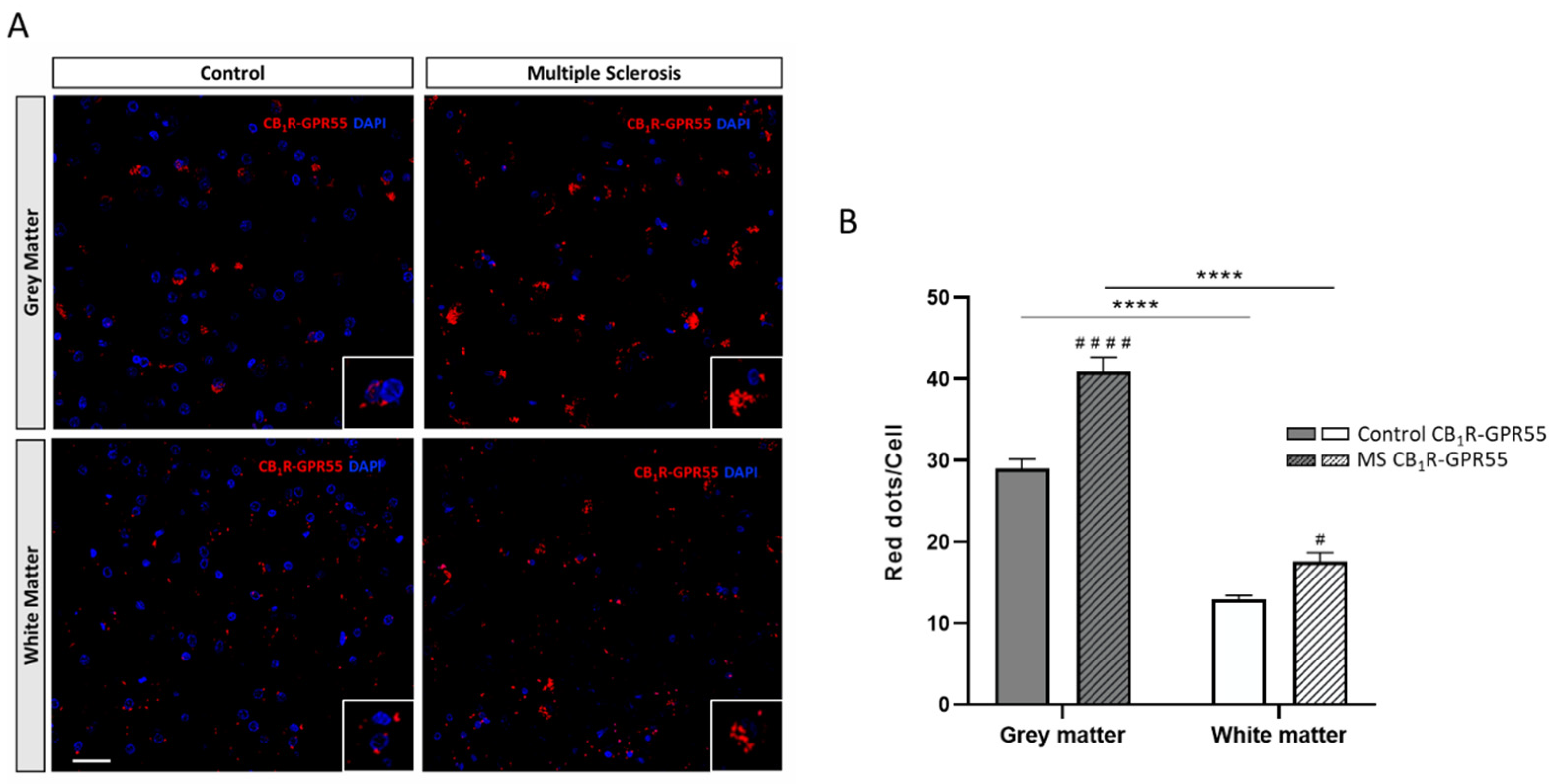
2.1. Expression of CB1R-GPR55 Heteromers in Controls and MS Patients

The In Situ Proximity Ligation Assay (PLA) is the most appropriate technique to detect the presence of complexes formed by two different receptors in a native system. Using specific antibodies against the CB1R and the GPR55 coupled to PLA oligonucleotide probes, a punctate fluorescent red signal was observed surrounding DAPI-counterstained nuclei, reflecting the formation of CB1R-GPR55 heteromers in the plasma membrane of cells in all prefrontal cortex samples analyzed (see Video S1), both in GM and WM (Figure 1A). Data analysis demonstrated that the number of red dots per cell, which reflects the number of CB1R-GPR55 heteromers, was significantly higher in the GM compared to the WM in the prefrontal cortex samples from the controls and MS patients (F1,596 = 8.23; p < 0.05) (Figure 1B).
Once the expression of CB1R-GPR55 heteromers in the human prefrontal cortex was shown, the next objective was to determine whether their expression was altered in samples from MS patients. The representative images in Figure 1A demonstrate an increase in the amount of red signal and, consequently, the expression of CB1R-GPR55 heteromers in samples from the MS patients (GM and WM). Quantitative analysis confirmed a significantly higher density of red clusters in the prefrontal cortex of MS patients compared to data obtained using samples from healthy controls (Figure 1B).

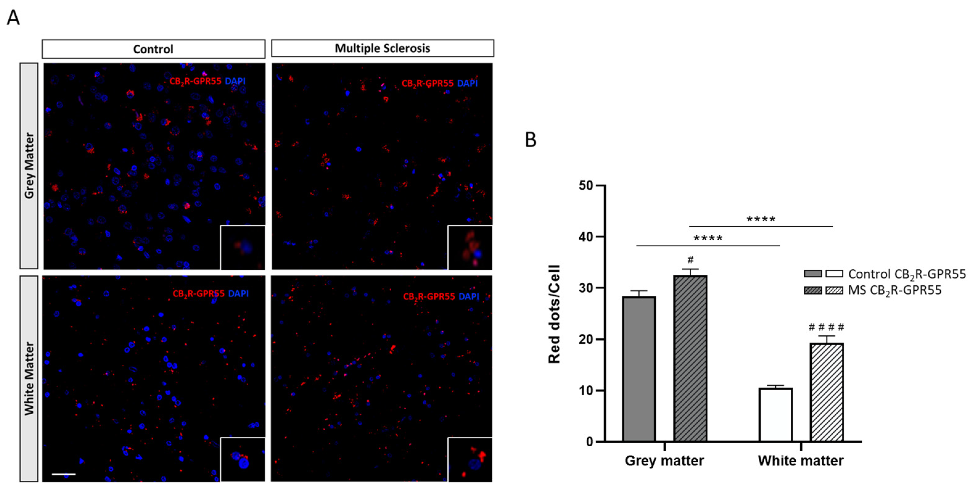
2.2. Expression of CB2R-GPR55 Heteromers in Controls and MS Patients

Similar findings were found when analogous assays addressing CB2R-GPR55 heteromer formation were performed using the same brain samples. Representative images and quantitative data from PLA assays (Figure 2A) revealed that CB2R and GPR55 formed heteromeric complexes in the plasma membrane of cells (see Video S2) in all brain areas and samples analyzed. When considering the total number of CB2R-GPR55 heteromers by assessing the number of red dots per cell, data analysis demonstrated higher expression in the GM of the prefrontal cortex compared to the WM, both in the control and MS patient samples (F1,572 = 4.63; p < 0.05) (Figure 2B).
Once again, significant differences were found when comparing the red fluorescent dots in the controls and individuals diagnosed with MS; the prefrontal cortex of patients showed a greater amount of CB2R-GPR55 heteromers compared to that observed in the controls (Figure 2A,B).

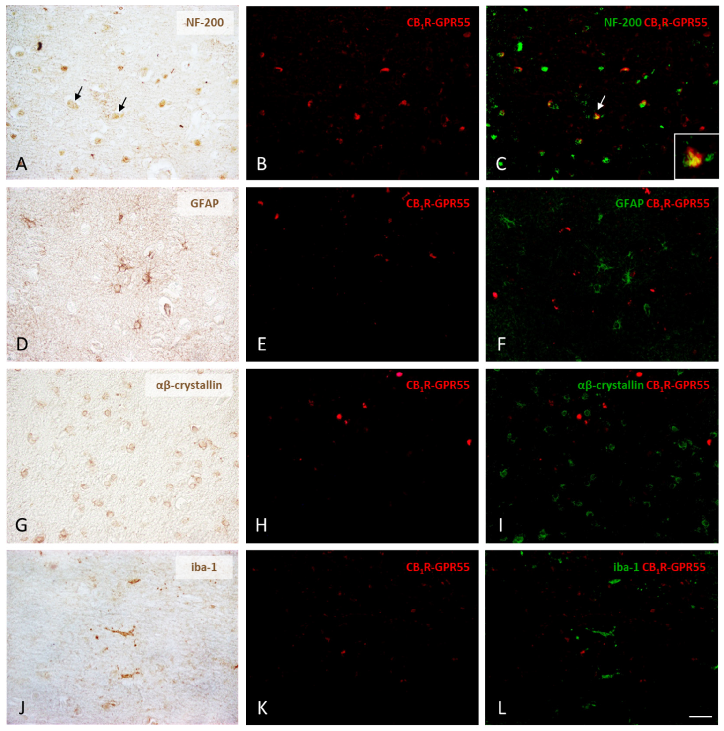
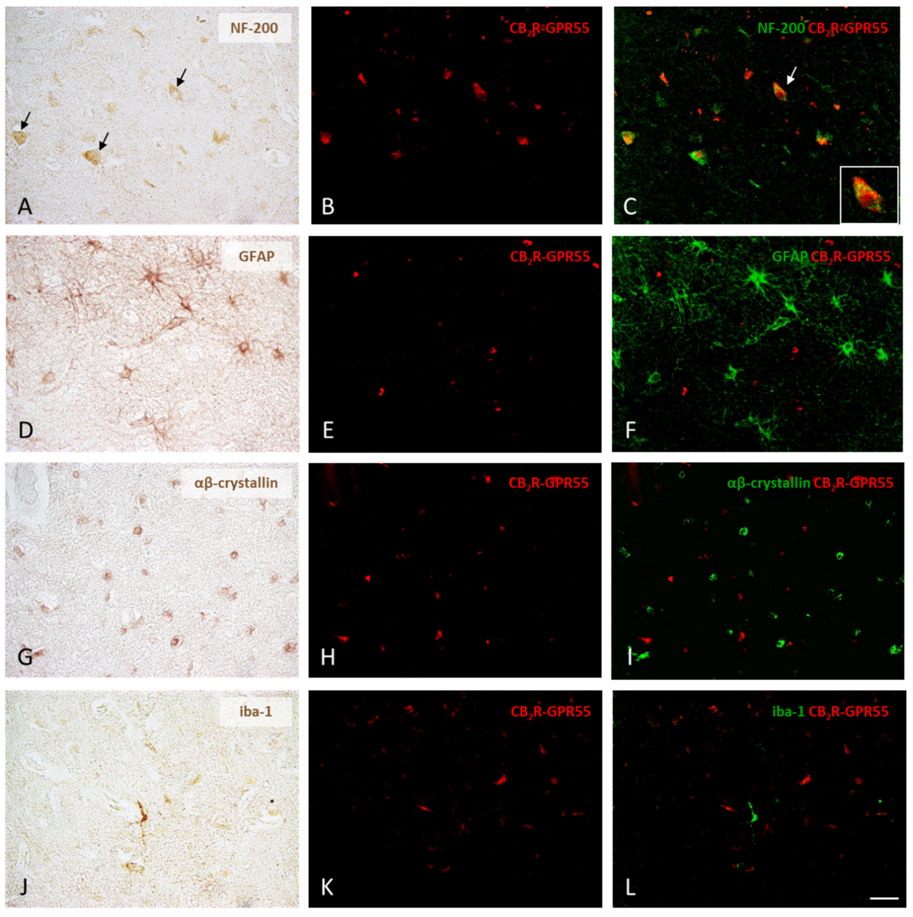
2.3. Expression of CB1R-GPR55 and CB2R-GPR55 Heteromers in Neurons and Different Types of Glial Cells

The final experimental approach was designed to identify those cells in the human prefrontal cortex that expressed CB1R-GPR55 or CB2R-GPR55 complexes. While the PLA was used to detect the presence of receptor–receptor interactions, neurons and glial cells were visualized using classical immunohistochemical staining. The combination of the two approaches made it possible to detect CB1R-GPR55 heteromers in a significant number of NF-200-labeled neurons in the samples of control individuals (Figure 3A–C). However, no co-staining was found when glial marker signals (GFAP, αβ-crystallin, and iba-1) were used (Figure 3D–L).
Regarding CB2R-GPR55 heteromers, the results were similar, i.e., the receptor complexes in the prefrontal cortex of the control subjects were identified in neurons (Figure 4A–C) but not in astrocytes, oligodendrocytes or microglia (Figure 4D–L).

3. Discussion

Understanding the modulatory role of the ECS in the CNS and its implication in neurodegenerative diseases has been the focus of considerable research efforts over the past few decades [19]. Despite the benefits of cannabinoids and the approval of cannabinoid-based medication, Sativex®, for some symptoms, little is known about the role of the ECS in the etiology and progression of MS. Recent evidence using animal models of this pathology sustained the therapeutic potential of cannabinoids in reducing certain symptoms of MS through the activation of cannabinoid receptors [54]. Preclinical studies in the experimental autoimmune encephalomyelitis (EAE) mouse model demonstrated that treatment with CBD and THC decreases axonal damage, inflammation, microglial activation, and T-cell recruitment, leading to a symptomatic improvement that seems to be related to direct action on the CB1R [55,56]. Spasticity increases rapidly after the administration of a CB1R antagonist, rimonabant, suggesting that alleviation of hind limb alteration is CB1R dependent [57]. A more recent study reported in a cuprizone-induced mouse model of MS that reducing the global amount of CB1R limits myelin repair potential [52]. Regarding CB2R, some studies have shown that JWH-133, a selective receptor agonist, ameliorates tremor and spasticity in EAE mice by promoting autophagy and inhibiting NLRP3 inflammasome activation [53]. Similarly, treatment with another CB2R agonist, HU-308, attenuated the development of the pathological condition. Consistent with these observations, CB2R−/− mice displayed greater vulnerability to neurofilament degeneration, inflammation, apoptosis, and axonal damage, common pathological features of the EAE [54]. Moreover, it is increasingly recognized that CBD and THC, administered together in controlled doses (Sativex®), reduce muscle spasms and spasticity in MS patients and even induce analgesia [15]. However, the pharmacology behind the receptor-mediated neuroprotective effects exerted by cannabinoids is not straightforward; it involves multiple targets and mechanisms that, collectively, unfold a particular pattern of cellular events. In this sense, dimer/oligomerization is now considered a relevant mechanism to induce diverse functional selectivity in signaling mediated by GPCRs [47]. Considering the ability of cannabinoid receptors to form heteromers that may constitute therapeutic targets, as already postulated for neurodegenerative diseases such as Alzheimer’s disease and Parkinson’s disease [30,58], this work aimed to adequately characterize the formation of complexes between CB1 or CB2 and GPR55 receptors in the CNS and to evaluate whether the expression of these receptor complexes is affected in MS.
The results presented herein constitute the first description of CB1R-GPR55 and CB2R-GPR55 heteromers in the human prefrontal cortex of control individuals and patients with MS. By taking advantage of the PLA technique and immunohistochemistry using antibodies against neuronal and glial markers, the expression of these receptor complexes was confirmed in neurons of the cerebral cortex, both in GM and WM, but not in glial cells labeled with antibodies against αβ-crystallin, GFAP or Iba-1. The possibility of direct interaction between CB1 or CB2 and GPR55 receptors has previously been demonstrated in cell cultures using energy transfer techniques [49,50] and cell biology techniques using brain samples of rats and non-human primates [25,30,51]. Indeed, CB1R-GPR55 heteromers were identified on the cell surface and in intracellular locations of striatal neuronal subtypes [25]. The expression of CB2R in the neurons of the CNS has been less well characterized, and the described changes in CB2R-GPR55 heteromer levels in the striatum of the Macaca fascicularis model of Parkinson’s disease were attributed to the upregulation of heteromers in activated microglia [30]. Interestingly, our data show that CB1R-GPR55 and CB2R-GPR55 heteromers are expressed almost exclusively at the level of the neuronal plasma membrane in the prefrontal cortex. Of note, the presence of these complexes is scarce in neuronal extensions which would explain the higher amount of heteromers found in GM compared to WM.
Remarkably, we identified an increase in the single-cell density of receptor complexes, for both CB1R-GPR55 and CB2R-GPR55, in the prefrontal cortex of MS patients (compared to control subjects). Changes in the expression of cannabinoid heteroreceptor complexes have been demonstrated in the brain of animal models of other neurodegenerative diseases [30,50]. An increase in the expression levels of CB1R-GPR55 and CB2R-GPR55 heteromers was found in basal ganglia input nuclei (i.e., caudate, putamen, and accumbens) of MPTP-treated parkinsonian primates; this increase was reverted through chronic treatment with levodopa only in those animals that became dyskinetic due to the chronic treatment [30]. The CB1R-CB2R heteromer is upregulated in activated microglia that, unlike resting microglia, are highly responsive to cannabinoids [58]. Interestingly, CB1R-CB2R heteromers were also upregulated in the hippocampus of a transgenic model of Alzheimer’s disease; it has been suggested that microglia in these animals display a neuroprotective phenotype that could explain why cognitive deficits do not appear until late in the life of the transgenic Alzheimer’s disease model [58]. The relevance of increased heteromer appearance to the pathophysiology of MS is unclear but could be part of a compensatory mechanism to restore homeostasis and brain integrity in response to neuronal damage. Cannabinoids are important players in neuroinflammation by regulating the release of neuropeptides and the activation of microglia [59,60]. In addition, these compounds may affect cellular energy production via GPR55-containing receptor complexes; mitochondrial dysfunctions observed in the disease could be caused by functional changes derived from differential heteromer expression [50,61]. All of this evidence supports the neuroprotective effect attributed to cannabinoids acting on cannabinoid and GPR55 receptors [50]. In this context, endocannabinoids and natural/synthetic cannabinoids capable of activating CB1R, CB2R, and GPR55 appear to offer protection against excitotoxic damage [17,38]. Furthermore, some studies in murine models have described a significant increase in endocannabinoid levels i.e., anandamide, palmitoylethanolamide, and 2-arachidonoylglycerol, as a part of an anti-inflammatory response resulting from axonal damage [19]. The presence of heteromers at significant levels in MS also opens up new possibilities in drug discovery, that is, targeting them for therapeutic benefit. The study has a limitation which is the small sample size derived from the difficulty in obtaining samples from patients. Although it may take time, validating MS-related changes in heteromer expression requires research with larger human cohort samples.
The potential of cannabinoids as drugs to combat or delay the progression of a neurological disease such as MS has gained interest in recent years. In the field of Parkinson’s disease research, it is increasingly recognized that targeting neuronal CB1R-GPR55 and CB2R-GPR55 heteromers with cannabinoids can be a successful therapeutic approach to both manage symptoms and delay disease progression [62,63]. The beneficial effect of some phytocannabinoids on MS symptoms [15] may be due to multiple molecular mechanisms. Cannabinoids can not only drive individualized responses through CB1, CB2, or GPR55 receptors but also, as this work suggests for the first time, act on functional units consisting of receptor heteromers. What is crucial to designing effective approaches is to consider the particular properties of the heteromers in terms of signaling. GPR55-mediated signaling is complex and this issue is delaying the discovery of selective compounds and the development of drugs targeting it. In contrast, the sustained interest in CB2R as a therapeutic target for neuroprotection has gained momentum over the last decade; agonists, antagonists, and allosteric modulators have been designed that, unlike CB1R targeting, lack unwanted psychotropic side effects [17,64]. Also interesting for the design of therapies to combat MS is the finding that CB1R-GPR55 and CB2R-GPR55 heteromers are expressed in neurons. This piece of information related to the CB2R-GPR55 heteromer is both intriguing and relevant since it is considered that the CB2R in the CNS is expressed more in the glia than in neurons. Finally, it should be noted that the existence of differences in the number of heteromers when comparing samples from control individuals and patients with MS offers a way forward for future research. In this sense, correlating changes in CB1R/CB2R-GPR55 heteromer levels with specific MS symptoms holds promise in using these complex receptors as therapeutic targets for personalized medicine approaches.

4. Materials and Methods

4.1. Subjects

In the present study, human prefrontal cortices from healthy subjects and patients with MS were used. It should be noted that brain samples from MS patients are very scarce. Human brain tissues were provided by different Spanish brain banks located at the University Hospital of Asturias, Central Nervous Tissue Bank Madrid (CIEN Foundation), the Center for Biomedical Research of Navarra (NAVARRABIOMED), and the Southern Galicia Health Research Institute (IISGS). In total, samples from eight individuals between 38 and 66 years old were obtained, including controls and those with histologically inflammatory demyelination consistent with MS, properly confirmed by a neuropathologist. Detailed information on the subjects and samples is given in Table 1.
Following retrieval, the brain specimens were fixed by immersion in 10% buffered formalin, dehydrated, cleared in butyl acetate, and embedded in paraffin. Tissue blocks containing the prefrontal cortex were sectioned at 7 µm, mounted on SuperFrost Plus (Mentzel-Glasse) slides, dried at 36 °C, and stored at room temperature until processed.
The ethics committees of each participating biobank reviewed and approved the study protocol. Moreover, all research procedures involving the manipulation of human samples were approved by the Comité de Ética de la Investigación del Principado de Asturias (CEImPA 23-174) and are in accordance with the ethical principles and guidelines established in the Declaration of Helsinki and in the Spanish laws: Ley de Investigaciones Biomédicas (ley 14/2007) and Ley de Protección de Datos Personales y Garantías de los Derechos Digitales (Ley Orgánica 3/2018).

4.2. In Situ Proximity Ligation Assay (PLA)

In Situ PLA, a technique instrumental for detecting receptor–receptor interactions and their precise anatomical localization, was used to test for the presence of CB1R-GPR55 and CB2R-GPR55 heteromers in the prefrontal cortex of brain samples from control individuals and patients diagnosed with MS. For this purpose, the tissue sections were incubated for 1 h at 37 °C with the blocking solution, followed by overnight incubation at 4 °C with the PLA probe-linked antibodies (at a final concentration of 75 µg/mL). Proximity probes consist of affinity-purified antibodies modified by covalent attachment of the 5′ end of various nucleotides to each primary antibody. In this case, PLA probes were prepared by conjugating a rabbit anti-CB1 antibody (PA1-743, Invitrogen, Paisley, UK) and a rabbit anti-CB2 antibody (101550, Cayman Chemical, Ann Arbor, MI, USA) with a PLUS oligonucleotide (Duolink® In Situ Probemaker PLUS ref: DUO92009; Sigma-Aldrich, St. Louis, MO, USA) and a rabbit anti-GPR55 antibody (10224; Cayman Chemicals, Ann Arbor, MI, USA), raised against the human 207–219 sequence, with a MINUS oligonucleotide (Duolink® In Situ Probemaker MINUS Ref: DUO92010; Sigma-Aldrich, St. Louis, MO, USA) according to the manufacturer’s guidelines. After washing with buffer A (DUO82047; Sigma-Aldrich, St. Louis, MO, USA), the GPCR heteromers were detected using the Duolink in Situ PLA detection kit (Duolink® In Situ Detection Reagents Red; DUO92008, Sigma-Aldrich, St. Louis, MO, USA). Then, the sections were washed with buffer A and incubated with the ligation solution for 1 h at 37 °C in a humidity chamber, washed with buffer A again, incubated with the amplification solution for 100 min at 37 °C, and finally washed with buffer B (wash buffer B; DUO82048; Sigma-Aldrich, St. Louis, MO, USA). The sections were finally mounted using an aqueous mounting medium with DAPI which allows for visualization of the cell nuclei (NB-23-00159-2, NeoBioTech, Nanterre, France). Appropriate negative control assays were carried out to ensure that there was a lack of non-specific labeling and amplification.
The quantification of PLA signals and cell nuclei was performed in images generated from a Leica sTCS-SP8X Spectral Confocal Laser Microscope (Leica Microsystems, Mannheim, Germany). Regarding selected regions of interest (ROIs), and for each field of view, a stack of two channels (one per staining) and 9–15 Z stacks with a step size of 1 μm were acquired with the 63× oil-immersion lens. Statistical analysis on the receptor heteromer densities was conducted according to a modification of the method of Tolivia et al. (see [65]). A quantification of cells containing one or more red spots versus total cells (blue nucleus) and the ratio r (number of red spots/cell) in cells containing spots were determined considering a total of 300–400 cells from ten different fields in both WM and GM for each prefrontal cortex section. The experiments were performed on a blind basis; the experimenter was not aware of the label and the conditions (control or MS) when images were taken. Moreover, the experimenter who made the analysis did not know the exact nature of the analyzed samples.

4.3. Co-Staining Combining PLA and Immunohistochemistry

Identification of the specific cell types (neurons, oligodendrocytes, astrocytes, and microglia) expressing CB1R-GPR55 and CB2R-GPR55 heteromers was carried out using immunohistochemistry followed by PLA. First, chromogenic immunodetection was performed using specific neuronal and glial markers. The immunohistochemistry process was conducted as follows. Prefrontal cortex sections of the control subjects were sequentially treated with Triton X-100 (0.01%, 5 min), washed with distilled water, treated with H2O2 (3%, 5 min), washed with distilled water again, and treated with PBS (2 min). Non-specific binding was blocked via incubation with 1% BSA (30 min). Then, incubation with a specific monoclonal antibody against NF-200, a neuronal marker (1:100; N-0142, Sigma-Aldrich, St. Louis, MO, USA), rabbit antibody against the glial fibrillary acidic protein (GFAP), an astrocytic marker (1:500; z0334, DAKO Agilent, Santa Clara, CA, USA), goat antibody against Iba-1, a microglia marker (1:1000; ab107159, Abcam, Cambridge, UK), and rabbit antibody against αβ-crystallin, a mature OLG marker (1:200; NCL-ABCrys-512, Novocastra, St. Gallen, Switzerland), was carried out overnight at 4 °C. After several washes in PBS, the sections were incubated at room temperature using a biotinylated horse universal antibody (1:40; PK-8800, Vector Laboratories Inc., Newark, NJ, USA) for 30 min. Afterward, the sections were treated with Extravidin labeled with horseradish peroxidase (HRP) (E2886, Sigma-Aldrich Extra-3, Sigma-Aldrich, St. Louis, MO, USA). Peroxidase activity was visualized with diaminobenzidine (DAB) (D4168, Sigma-Aldrich, St. Louis, MO, USA). After the immunohistochemical protocol, the PLA technique was used, as described in Section 4.2, to detect CB1R-GPR55 and CB2R-GPR55 heteromers.
The sections were observed using an Epi-Fl Nikon Eclipse E400 microscope (Nikon, Minato-ku, Tokyo, Japan) equipped with Plan Fluor objectives, and images were recorded using a digital camera (63×; NikonDN-100, Nikon, Minato-ku, Tokyo, Japan). Final images were obtained through the digital superposition of the corresponding DAB (NF-200, GFAP, αβ-crystallin, or Iba-1 signal) and red fluorescence (PLA signal) images of the same sections. The positive signal of each image was selected according to the method of Navarro et al. (2008) [66]; DAB signals were converted to green and saved as an RGB image. Merged images show the PLA fluorescence signal in red and the DAB label in green; the yellow color indicates the superposition of red and green colors.

4.4. Data Analysis

Data collected in samples from 4 subjects per group (control and MS) were the mean ± SEM. Statistical analysis was performed with GraphPad Prism 8 (San Diego, CA, USA). A two-way ANOVA followed by Bonferroni’s post hoc multiple comparison test were used to compare the values (r spots/cell) obtained for each pair of receptors. Data were tested for normality of populations and homogeneity of variances. Differences were considered significant when p < 0.05.

Supplementary Materials

The following supporting information can be downloaded at: https://www.mdpi.com/article/10.3390/ijms25084176/s1.

Author Contributions

Conceptualization was agreed by E.M.-P. and R.F., who also participated in the design of the project and analyzed the results; C.M.-P. and E.M.-P. performed the majority of the experiments; R.R.-S. participated in a significant number of experiments; A.N., E.d.V. and J.T. participated in the data analysis and preparing the final figures; C.M.-P., E.M.-P. and R.R.-S. performed many of the imaging assays using the confocal microscope, took photographs, and participated in the data analysis; E.M.-P. and R.F. wrote the first draft of the manuscript which was further edited by all co-authors, who agreed with the final submission. All authors have read and agreed to the published version of the manuscript.

Funding

This research was supported by a grant from Instituto de Investigación Sanitaria del Principado de Asturias (ISPA) (2022-073-INTRAMUR-MAPIE).

Institutional Review Board Statement

The study was conducted in accordance with the Declaration of Helsinki and the Spanish laws: Ley de Investigaciones Biomédicas (ley 14/2007) and Ley de Protección de Datos Personales y Garantías de los Derechos Digitales (Ley Orgánica 3/2018) and approved by the Comité de Ética de la Investigación del Principado de Asturias (CEImPA 23-174; 25 March 2023).

Informed Consent Statement

Informed consent was obtained from all subjects involved in the study.

Data Availability Statement

The datasets generated and/or analyzed during the current study are not publicly available due to privacy/ethical restrictions but are available from the corresponding author upon reasonable request.

Acknowledgments

We acknowledge the technical support of Marta Alonso from the Photonic Microscopy and Imaging Processing Unit of the Scientific and Technical Services of the University of Oviedo.

Conflicts of Interest

The authors declare no conflicts of interest.

References

  1. Walton, C.; King, R.; Rechtman, L.; Kaye, W.; Leray, E.; Marrie, R.A.; Robertson, N.; La Rocca, N.; Uitdehaag, B.; van der Mei, I.; et al. Rising prevalence of multiple sclerosis worldwide: Insights from the Atlas of MS, third edition. Mult. Scler. J. 2020, 26, 1816–1821. [Google Scholar] [CrossRef]
  2. McGinley, M.P.; Goldschmidt, C.H.; Rae-Grant, A.D. Diagnosis and Treatment of Multiple Sclerosis: A Review. JAMA 2021, 325, 765–779. [Google Scholar] [CrossRef]
  3. Reich, D.S.; Lucchinetti, C.F.; Calabresi, P.A. Multiple sclerosis. N. Engl. J. Med. 2018, 378, 169–180. [Google Scholar] [CrossRef]
  4. Cao, Y.; Diao, W.; Tian, F.; Zhang, F.; He, L.; Long, X.; Zhou, F.; Jia, Z. Gray Matter Atrophy in the Cortico-Striatal-Thalamic Network and Sensorimotor Network in Relapsing–Remitting and Primary Progressive Multiple Sclerosis. Neuropsychol. Rev. 2021, 31, 703–720. [Google Scholar] [CrossRef]
  5. Calabrese, M.; Magliozzi, R.; Ciccarelli, O.; Geurts, J.J.G.; Reynolds, R.; Martin, R. Exploring the origins of grey matter damage in multiple sclerosis. Nat. Rev. Neurosci. 2015, 16, 147–158. [Google Scholar] [CrossRef]
  6. Oh, J.; Vidal-Jordana, A.; Montalban, X. Multiple sclerosis: Clinical aspects. Curr. Opin. Neurol. 2018, 31, 752–759. [Google Scholar] [CrossRef]
  7. Ford, H. Clinical presentation and diagnosis of multiple sclerosis. Clin. Med. J. R. Coll. Physicians Lond. 2020, 20, 380–383. [Google Scholar] [CrossRef]
  8. Lublin, F.D.; Reingold, S.C.; Cohen, J.A.; Cutter, G.R.; Sørensen, P.S.; Thompson, A.J.; Wolinsky, J.S.; Balcer, L.J.; Banwell, B.; Barkhof, F.; et al. Defining the clinical course of multiple sclerosis: The 2013 revisions. Neurology 2014, 83, 278–286. [Google Scholar] [CrossRef]
  9. Hegen, H.; Berek, K.; Deisenhammer, F. Cerebrospinal fluid kappa free light chains as biomarker in multiple sclerosis—From diagnosis to prediction of disease activity. Wien. Med. Wochenschr. 2022, 172, 337–345. [Google Scholar] [CrossRef]
  10. Thompson, A.J.; Banwell, B.L.; Barkhof, F.; Carroll, W.M.; Coetzee, T.; Comi, G.; Correale, J.; Fazekas, F.; Filippi, M.; Freedman, M.S.; et al. Diagnosis of multiple sclerosis: 2017 revisions of the McDonald criteria. Lancet Neurol. 2018, 17, 162–173. [Google Scholar] [CrossRef]
  11. International Multiple Sclerosis Genetics Consortium; Harroud, A.; Stridh, P.; McCauley, J.L.; Saarela, J.; Bosch, A.M.R.v.D.; Engelenburg, H.J.; Beecham, A.H.; Alfredsson, L.; Alikhani, K.; et al. Locus for severity implicates CNS resilience in progression of multiple sclerosis. Nature 2023, 619, 323–331. [Google Scholar] [CrossRef]
  12. Goodin, D.S. Glucocorticoid treatment of multiple sclerosis. In Handbook of Clinical Neurology; Elsevier: Amsterdam, The Netherlands, 2014; Volume 122, pp. 455–464. [Google Scholar]
  13. Krajnc, N.; Bsteh, G.; Berger, T.; Mares, J.; Hartung, H.P. Monoclonal Antibodies in the Treatment of Relapsing Multiple Sclerosis: An Overview with Emphasis on Pregnancy, Vaccination, and Risk Management. Neurotherapeutics 2022, 19, 753–773. [Google Scholar] [CrossRef]
  14. McGinley, M.P.; Cohen, J.A. Sphingosine 1-phosphate receptor modulators in multiple sclerosis and other conditions. Lancet 2021, 398, 1184–1194. [Google Scholar] [CrossRef]
  15. Conte, A.; Silván, C.V. Review of Available Data for the Efficacy and Effectiveness of Nabiximols Oromucosal Spray (Sativex®) in Multiple Sclerosis Patients with Moderate to Severe Spasticity. Neurodegener. Dis. 2022, 21, 55–62. [Google Scholar] [CrossRef]
  16. Fernández-Ruiz, J.; Gómez-Ruiz, M.; García, C.; Hernández, M.; Ramos, J.A. Modeling Neurodegenerative Disorders for Developing Cannabinoid-Based Neuroprotective Therapies. Methods Enzymol. 2017, 593, 175–198. [Google Scholar]
  17. Mecha, M.; Carrillo-Salinas, F.J.; Feliú, A.; Mestre, L.; Guaza, C. Perspectives on Cannabis-Based Therapy of Multiple Sclerosis: A Mini-Review. Front. Cell. Neurosci. 2020, 14, 34. [Google Scholar] [CrossRef]
  18. Nouh, R.A.; Kamal, A.; Abdelnaser, A. Cannabinoids and Multiple Sclerosis: A Critical Analysis of Therapeutic Potentials and Safety Concerns. Pharmaceutics 2023, 15, 1151. [Google Scholar] [CrossRef]
  19. Cristino, L.; Bisogno, T.; Di Marzo, V. Cannabinoids and the expanded endocannabinoid system in neurological disorders. Nat. Rev. Neurol. 2020, 16, 9–29. [Google Scholar] [CrossRef]
  20. Giuffrida, A.; Seillier, A. New insights on endocannabinoid transmission in psychomotor disorders. Prog. Neuro-Psychopharmacol. Biol. Psychiatry 2012, 38, 51–58. [Google Scholar] [CrossRef]
  21. Aymerich, M.S.; Aso, E.; Abellanas, M.A.; Tolon, R.M.; Ramos, J.A.; Ferrer, I.; Romero, J.; Fernández-Ruiz, J. Cannabinoid pharmacology/therapeutics in chronic degenerative disorders affecting the central nervous system. Biochem. Pharmacol. 2018, 157, 67–84. [Google Scholar] [CrossRef]
  22. Shu-Jung Hu, S.; Mackie, K. Distribution of the endocannabinoid system in the central nervous system. In Handbook of Experimental Pharmacology; Springer: Berlin/Heidelberg, Germany, 2015; Volume 231, pp. 59–93. [Google Scholar]
  23. Glass, M.; Dragunow, M.; Faull, R.L.M. Cannabinoid receptors in the human brain: A detailed anatomical and quantitative autoradiographic study in the fetal, neonatal and adult human brain. Neuroscience 1997, 77, 299–318. [Google Scholar] [CrossRef]
  24. Chou, S.; Ranganath, T.; Fish, K.N.; Lewis, D.A.; Sweet, R.A. Cell type specific cannabinoid CB1 receptor distribution across the human and non-human primate cortex. Sci. Rep. 2022, 12, 9605. [Google Scholar] [CrossRef]
  25. Martínez-Pinilla, E.; Rico, A.J.; Rivas-Santisteban, R.; Lillo, J.; Roda, E.; Navarro, G.; Franco, R.; Lanciego, J.L. Expression of cannabinoid CB1 R–GPR55 heteromers in neuronal subtypes of the Macaca fascicularis striatum. Ann. N. Y. Acad. Sci. 2020, 1475, 34–42. [Google Scholar] [CrossRef]
  26. Cabral, G.A.; Ferreira, G.A.; Jamerson, M.J. Endocannabinoids and the immune system in health and disease. In Endocannabinoids. Handbook of Experimental Pharmacology; Springer: Cham, Switzerland, 2015; Volume 231, pp. 185–211. ISBN 9783319208251. [Google Scholar]
  27. Onaivi, E.S. Neuropsychobiological evidence for the functional presence and expression of cannabinoid CB2 receptors in the brain. Neuropsychobiology 2007, 54, 231–246. [Google Scholar] [CrossRef]
  28. Onaivi, E.S.; Ishiguro, H.; Gong, J.P.; Patel, S.; Perchuk, A.; Meozzi, P.A.; Myers, L.; Mora, Z.; Tagliaferro, P.; Gardner, E.; et al. Discovery of the presence and functional expression of cannabinoid CB2 receptors in brain. Ann. N. Y. Acad. Sci. 2006, 1074, 514–536. [Google Scholar] [CrossRef]
  29. Lanciego, J.L.; Barroso-Chinea, P.; Rico, A.J.; Conte-Perales, L.; Callén, L.; Roda, E.; Gómez-Bautista, V.; López, I.P.; Lluis, C.; Labandeira-García, J.L.; et al. Expression of the mRNA coding the cannabinoid receptor 2 in the pallidal complex of Macaca fascicularis. J. Psychopharmacol. 2011, 25, 97–104. [Google Scholar] [CrossRef]
  30. Martínez-Pinilla, E.; Rico, A.J.; Rivas-Santisteban, R.; Lillo, J.; Roda, E.; Navarro, G.; Lanciego, J.L.; Franco, R. Expression of GPR55 and either cannabinoid CB1 or CB2 heteroreceptor complexes in the caudate, putamen, and accumbens nuclei of control, parkinsonian, and dyskinetic non-human primates. Brain Struct. Funct. 2020, 225, 2153–2164. [Google Scholar] [CrossRef]
  31. Liu, Q.R.; Pan, C.H.; Hishimoto, A.; Li, C.Y.; Xi, Z.X.; Llorente-Berzal, A.; Viveros, M.P.; Ishiguro, H.; Arinami, T.; Onaivi, E.S.; et al. Species differences in cannabinoid receptor 2 (CNR2 gene): Identification of novel human and rodent CB2 isoforms, differential tissue expression and regulation by cannabinoid receptor ligands. Genes Brain Behav. 2009, 8, 519–530. [Google Scholar] [CrossRef]
  32. Brusco, A.; Tagliaferro, P.A.; Saez, T.; Onaivi, E.S. Ultrastructural localization of neuronal brain CB2 cannabinoid receptors. Ann. N. Y. Acad. Sci. 2008, 1139, 450–457. [Google Scholar] [CrossRef]
  33. Brusco, A.; Tagliaferro, P.; Saez, T.; Onaivi, E.S. Postsynaptic localization of CB2 cannabinoid receptors in the rat hippocampus. Synapse 2008, 62, 944–949. [Google Scholar] [CrossRef]
  34. Fernández-Ruiz, J.; Moro, M.A.; Martínez-Orgado, J. Cannabinoids in Neurodegenerative Disorders and Stroke/Brain Trauma: From Preclinical Models to Clinical Applications. Neurotherapeutics 2015, 12, 793–806. [Google Scholar] [CrossRef]
  35. Ryberg, E.; Larsson, N.; Sjögren, S.; Hjorth, S.; Hermansson, N.O.; Leonova, J.; Elebring, T.; Nilsson, K.; Drmota, T.; Greasley, P.J. The orphan receptor GPR55 is a novel cannabinoid receptor. Br. J. Pharmacol. 2007, 152, 1092–1101. [Google Scholar] [CrossRef]
  36. Baker, D.; Pryce, G.; Davies, W.L.; Hiley, C.R. In silico patent searching reveals a new cannabinoid receptor. Trends Pharmacol. Sci. 2006, 27, 1–4. [Google Scholar] [CrossRef]
  37. Oka, S.; Nakajima, K.; Yamashita, A.; Kishimoto, S.; Sugiura, T. Identification of GPR55 as a lysophosphatidylinositol receptor. Biochem. Biophys. Res. Commun. 2007, 362, 928–934. [Google Scholar] [CrossRef]
  38. Oyagawa, C.R.M.; Grimsey, N.L. Cannabinoid receptor CB1 and CB2 interacting proteins: Techniques, progress and perspectives. In Methods in Cell Biology; Elsevier: Amsterdam, The Netherlands, 2021; Volume 166, pp. 83–132. ISBN 9780128233511. [Google Scholar]
  39. Pietr, M.; Kozela, E.; Levy, R.; Rimmerman, N.; Lin, Y.H.; Stella, N.; Vogel, Z.; Juknat, A. Differential changes in GPR55 during microglial cell activation. FEBS Lett. 2009, 583, 2071–2076. [Google Scholar] [CrossRef]
  40. Wu, C.S.; Zhu, J.; Wager-Miller, J.; Wang, S.; O’Leary, D.; Monory, K.; Lutz, B.; MacKie, K.; Lu, H.C. Requirement of cannabinoid CB1 receptors in cortical pyramidal neurons for appropriate development of corticothalamic and thalamocortical projections. Eur. J. Neurosci. 2010, 32, 693–706. [Google Scholar] [CrossRef]
  41. Alexander, S.P.H.; Christopoulos, A.; Davenport, A.P.; Kelly, E.; Marrion, N.V.; Peters, J.A.; Faccenda, E.; Harding, S.D.; Pawson, A.J.; Sharman, J.L.; et al. THE CONCISE GUIDE TO PHARMACOLOGY 2017/18: G protein-coupled receptors. Br. J. Pharmacol. 2017, 174, S17–S129. [Google Scholar] [CrossRef]
  42. Krishna Kumar, K.; Shalev-Benami, M.; Robertson, M.J.; Hu, H.; Banister, S.D.; Hollingsworth, S.A.; Latorraca, N.R.; Kato, H.E.; Hilger, D.; Maeda, S.; et al. Structure of a Signaling Cannabinoid Receptor 1-G Protein Complex. Cell 2019, 176, 448–458.e12. [Google Scholar] [CrossRef]
  43. Lynch, D.L.; Hurst, D.P.; Reggio, P.H. The Nucleotide-Free State of the Cannabinoid CB2/Gi Complex. Cell 2020, 180, 603–604. [Google Scholar] [CrossRef]
  44. Henstridge, C.M.; Balenga, N.A.B.; Ford, L.A.; Ross, R.A.; Waldhoer, M.; Irving, A.J. The GPR55 ligand L-α-lysophosphatidylinositol promotes RhoA-dependent Ca2+ signaling and NFAT activation. FASEB J. 2009, 23, 183–193. [Google Scholar] [CrossRef]
  45. Henstridge, C.M.; Balenga, N.A.; Schröder, R.; Kargl, J.K.; Platzer, W.; Martini, L.; Arthur, S.; Penman, J.; Whistler, J.L.; Kostenis, E.; et al. GPR55 ligands promote receptor coupling to multiple signalling pathways. Br. J. Pharmacol. 2010, 160, 604–614. [Google Scholar] [CrossRef]
  46. Ferré, S.; Baler, R.; Bouvier, M.; Caron, M.G.; Devi, L.A.; Durroux, T.; Fuxe, K.; George, S.R.; Javitch, J.A.; Lohse, M.J.; et al. Building a new conceptual framework for receptor heteromers. Nat. Chem. Biol. 2009, 5, 131–134. [Google Scholar] [CrossRef]
  47. Franco, R.; Martínez-Pinilla, E.; Lanciego, J.L.; Navarro, G. Basic pharmacological and structural evidence for class A G-protein-coupled receptor heteromerization. Front. Pharmacol. 2016, 7, 76. [Google Scholar] [CrossRef]
  48. Franco, R.; Aguinaga, D.; Jiménez, J.; Lillo, J.; Martínez-Pinilla, E.; Navarro, G. Biased receptor functionality versus biased agonism in G-protein-coupled receptors. Biomol. Concepts 2018, 9, 143–154. [Google Scholar] [CrossRef]
  49. Balenga, N.A.; Martínez-Pinilla, E.; Kargl, J.; Schröder, R.; Peinhaupt, M.; Platzer, W.; Bálint, Z.; Zamarbide, M.; Dopeso-Reyes, I.G.; Ricobaraza, A.; et al. Heteromerization of GPR55 and cannabinoid CB2 receptors modulates signalling. Br. J. Pharmacol. 2014, 171, 5387–5406. [Google Scholar] [CrossRef]
  50. Martínez-Pinilla, E.; Aguinaga, D.; Navarro, G.; Rico, A.J.; Oyarzábal, J.; Sánchez-Arias, J.A.; Lanciego, J.L.; Franco, R. Targeting CB1 and GPR55 Endocannabinoid Receptors as a Potential Neuroprotective Approach for Parkinson’s Disease. Mol. Neurobiol. 2019, 56, 5900–5910. [Google Scholar] [CrossRef]
  51. Martínez-Pinilla, E.; Reyes-Resina, I.; Oñatibia-Astibia, A.; Zamarbide, M.; Ricobaraza, A.; Navarro, G.; Moreno, E.; Dopeso-Reyes, I.G.; Sierra, S.; Rico, A.J.; et al. CB1 and GPR55 receptors are co-expressed and form heteromers in rat and monkey striatum. Exp. Neurol. 2014, 261, 44–52. [Google Scholar] [CrossRef]
  52. Tomas-Roig, J.; Agbemenyah, H.Y.; Celarain, N.; Quintana, E.; Ramió-Torrentà, L.; Havemann-Reinecke, U. Dose-dependent effect of cannabinoid WIN-55,212-2 on myelin repair following a demyelinating insult. Sci. Rep. 2020, 10, 590. [Google Scholar] [CrossRef]
  53. Shao, B.Z.; Wei, W.; Ke, P.; Xu, Z.Q.; Zhou, J.X.; Liu, C. Activating cannabinoid receptor 2 alleviates pathogenesis of experimental autoimmune encephalomyelitis via activation of autophagy and inhibiting NLRP3 inflammasome. CNS Neurosci. Ther. 2014, 20, 1021–1028. [Google Scholar] [CrossRef]
  54. Paloczi, J.; Varga, Z.V.; Hasko, G.; Pacher, P. Neuroprotection in Oxidative Stress-Related Neurodegenerative Diseases: Role of Endocannabinoid System Modulation. Antioxid. Redox Signal. 2018, 29, 75–108. [Google Scholar] [CrossRef]
  55. Kozela, E.; Lev, N.; Kaushansky, N.; Eilam, R.; Rimmerman, N.; Levy, R.; Ben-Nun, A.; Juknat, A.; Vogel, Z. Cannabidiol inhibits pathogenic T cells, decreases spinal microglial activation and ameliorates multiple sclerosis-like disease in C57BL/6 mice. Br. J. Pharmacol. 2011, 163, 1507–1519. [Google Scholar] [CrossRef]
  56. Fujiwara, M.; Egashira, N. New perspectives in the studies on endocannabinoid and cannabis: Abnormal behaviors associate with CB1 cannabinoid receptor and development of therapeutic application. J. Pharmacol. Sci. 2004, 96, 362–366. [Google Scholar] [CrossRef]
  57. Pryce, G.; Ahmed, Z.; Hankey, D.J.R.; Jackson, S.J.; Croxford, J.L.; Pocock, J.M.; Ledent, C.; Petzold, A.; Thompson, A.J.; Giovannoni, G.; et al. Cannabinoids inhibit neurodegeneration in models of multiple sclerosis. Brain 2003, 126, 2191–2202. [Google Scholar] [CrossRef]
  58. Navarro, G.; Borroto-Escuela, D.; Angelats, E.; Etayo, I.; Reyes-Resina, I.; Pulido-Salgado, M.; Rodríguez-Pérez, A.; Canela, E.; Saura, J.; Lanciego, J.L.; et al. Receptor-heteromer mediated regulation of endocannabinoid signaling in activated microglia. Role of CB1 and CB2 receptors and relevance for Alzheimer’s disease and levodopa-induced dyskinesia. Brain. Behav. Immun. 2018, 67, 139–151. [Google Scholar] [CrossRef]
  59. McKenna, M.; McDougall, J.J. Cannabinoid control of neurogenic inflammation. Br. J. Pharmacol. 2020, 177, 4386–4399. [Google Scholar] [CrossRef]
  60. Tao, Y.; Li, L.; Jiang, B.; Feng, Z.; Yang, L.; Tang, J.; Chen, Q.; Zhang, J.; Tan, Q.; Feng, H.; et al. Cannabinoid receptor-2 stimulation suppresses neuroinflammation by regulating microglial M1/M2 polarization through the cAMP/PKA pathway in an experimental GMH rat model. Brain. Behav. Immun. 2016, 58, 118–129. [Google Scholar] [CrossRef]
  61. Martínez-pinilla, E.; Rubio-sardón, N.; Villar-conde, S.; Navarro, G.; Del Valle, E.; Tolivia, J.; Franco, R.; Navarro, A. Cuprizone-induced neurotoxicity in human neural cell lines is mediated by a reversible mitochondrial dysfunction: Relevance for demyelination models. Brain Sci. 2021, 11, 272. [Google Scholar] [CrossRef]
  62. Urbi, B.; Corbett, J.; Hughes, I.; Owusu, M.A.; Thorning, S.; Broadley, S.A.; Sabet, A.; Heshmat, S. Effects of Cannabis in Parkinson’s Disease: A Systematic Review and Meta-Analysis. J. Parkinsons. Dis. 2022, 12, 495–508. [Google Scholar] [CrossRef]
  63. Varshney, K.; Patel, A.; Ansari, S.; Shet, P.; Panag, S.S. Cannabinoids in Treating Parkinson’s Disease Symptoms: A Systematic Review of Clinical Studies. Cannabis Cannabinoid Res. 2023, 8, 716–730. [Google Scholar] [CrossRef]
  64. Christensen, D.B.; Roth, M.; Trygstad, T.; Byrd, J. Evaluation of a pilot medication therapy management project within the North Carolina State Health Plan. J. Am. Pharm. Assoc. 2007, 47, 471–483. [Google Scholar] [CrossRef]
  65. Tolivia, J.; Navarro, A.; Del Valle, E.; Perez, C.; Ordoñez, C.; Martínez, E. Application of photoshop and scion image analysis to quantification of signals in histochemistry, immunocytochemistry and hybridocytochemistry. Anal. Quant. Cytol. Histol. 2006, 28, 43–53. [Google Scholar]
  66. Navarro, A.; Ordóñez, C.; Martínez, E.; Pérez, C.; Astudillo, A.; Tolivia, J. Apolipoprotein d expression absence in degenerating neurons of human central nervous system. Histol. Histopathol. 2008, 23, 995–1001. [Google Scholar]
Figure 1. CB1R-GPR55 heteromers detected using the In Situ Proximity Ligation Assay (PLA) in the grey (GM) and white matter (WM) of the prefrontal cortex of samples from the controls and multiple sclerosis (MS) patients. Representative confocal images (40×) showing PLA label for CB1R-GPR55 heteromers as red dots in cells with DAPI-stained nuclei. Scale bar 50 µm (A). Quantification of CB1R-GPR55 heteromers as the number of red dots per cell in the GM and WM in the prefrontal cortex samples of the controls and MS patients. Data are the mean ± SEM (40 fields per section) (B). Significant differences were analyzed via two-way ANOVA followed by post hoc Bonferroni’s test. **** p < 0.0001 compared with GM; # p < 0.05, #### p < 0.0001 compared with the control.
Ijms 25 04176 g001
Figure 2. CB2R-GPR55 heteromers detected using the In Situ Proximity Ligation Assay (PLA) in the grey (GM) and white matter (WM) of the prefrontal cortex of samples from the controls and multiple sclerosis (MS) patients. Representative confocal images (40×) showing the PLA label for CB2R-GPR55 heteromers as red dots in cells with DAPI-stained nuclei. Scale bar 50 µm (A). Quantification of CB2R-GPR55 heteromers as the number of red dots per cell in the GM and WM in the prefrontal cortex samples of the controls and MS patients. Data are the mean ± SEM (40 fields per section) (B). Significant differences were analyzed via two-way ANOVA followed by post hoc Bonferroni’s test. **** p < 0.0001 compared with GM; # p < 0.05, #### p < 0.0001 compared with the control.
Ijms 25 04176 g002
Figure 3. Expression of CB1R-GPR55 heteromers in neurons, astrocytes, oligodendrocytes, and microglia in the prefrontal cortex of the control subjects. Following the chromogenic immunohistochemical detection of the four different types of cells, those positive for NF-200, GFAP, αβ-crystallin, and iba-1 (A,D,G,J), CB1R-GPR55 heteromers were identified as red dots in cells with DAPI-stained nuclei using the PLA method (B,E,H,K). Digital overlays of PLA images (red fluorescent signal) and DAB immunolabeling images (the DAB signal converted into a green color) clearly show that only neurons express CB1R-GPR55 receptor complexes (arrows), whereas oligodendrocytes, astrocytes, and microglia completely lack them (C,F,I,L). 40×. Detail: 100×. Scale bar 50 µm.
Ijms 25 04176 g003
Figure 4. Expression of CB2R-GPR55 heteromers in neurons, oligodendrocytes, astrocytes, and microglia in the prefrontal cortex of the control subjects. Following the chromogenic immunohistochemical detection of the four different types of cells, those positive for NF-200, GFAP, αβ-crystallin, and iba-1 (A,D,G,J), CB2R-GPR55 heteromers were identified as red dots in cells with DAPI-stained nuclei using the PLA method (B,E,H,K). Digital overlays of PLA images (red fluorescent signal) and DAB immunolabeling images (the DAB signal converted into a green fluorescent signal) clearly showed that only neurons express CB2R-GPR55 receptor complexes (arrows), whereas oligodendrocytes, astrocytes, and microglia completely lack them (C,F,I,L). 40×. Detail: 100×. Scale bar 50 µm.
Ijms 25 04176 g004
Table 1. Demographic data of cases.
CaseSexAgeType of MSBrain AreaPost-Mortem TimeBrain Bank
1Male58MSPrefrontal cortex6–12 hCentral Nervous Tissue Bank Madrid (CIEN Foundation)
2Male38SPMSPrefrontal cortex6–12 hUniversity Central Hospital of Asturias (HUCA)
3Female47PPMSPrefrontal cortex6–12 hCenter for Biomedical Research of Navarra (NAVARRABIOMED)
4Female48MSPrefrontal cortex6–12 hSouthern Galicia Health Research Institute (IISGS)
5Male38ControlPrefrontal cortex6–12 hUniversity Central Hospital of Asturias (HUCA)
6Male66ControlPrefrontal cortex6–12 hCenter for Biomedical Research of Navarra (NAVARRABIOMED)
7Male62ControlPrefrontal cortex6–12 hCenter for Biomedical Research of Navarra (NAVARRABIOMED)
8Female52ControlPrefrontal cortex6–12 hUniversity Central Hospital of Asturias (HUCA)
MS, multiple sclerosis; PPMS, primary progressive MS; SPMS, secondary progressive MS.
Disclaimer/Publisher’s Note: The statements, opinions and data contained in all publications are solely those of the individual author(s) and contributor(s) and not of MDPI and/or the editor(s). MDPI and/or the editor(s) disclaim responsibility for any injury to people or property resulting from any ideas, methods, instructions or products referred to in the content.

