

Low-Level Expression of p-S6 Is Associated with Nodal Metastasis in Patients with Head and Neck Cutaneous Squamous Cell Carcinoma
 , ,
, ,  , , , ,
  , and
, and
Abstract
:1. Introduction
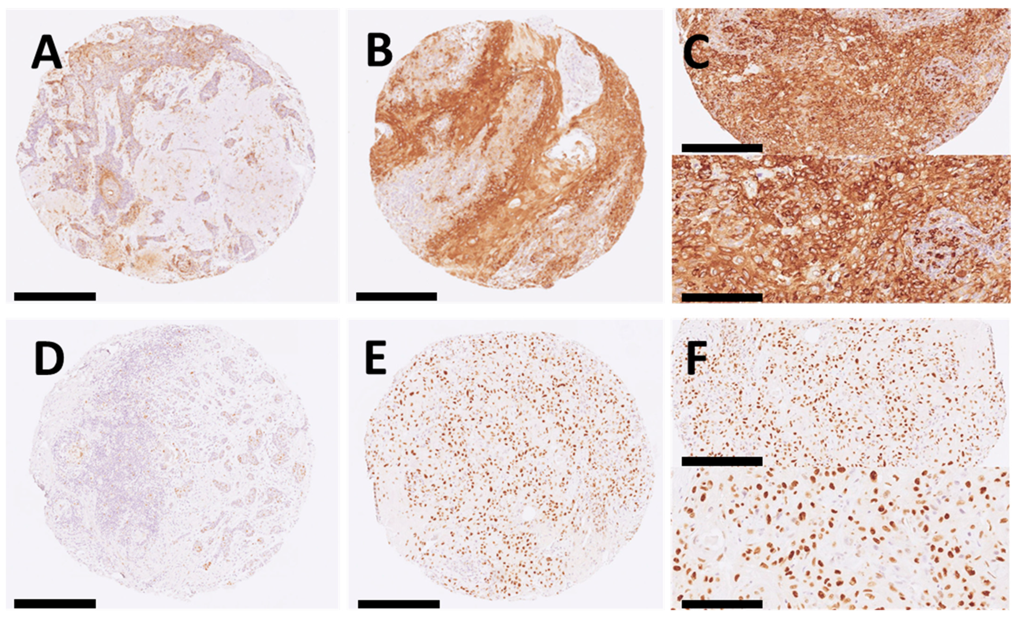
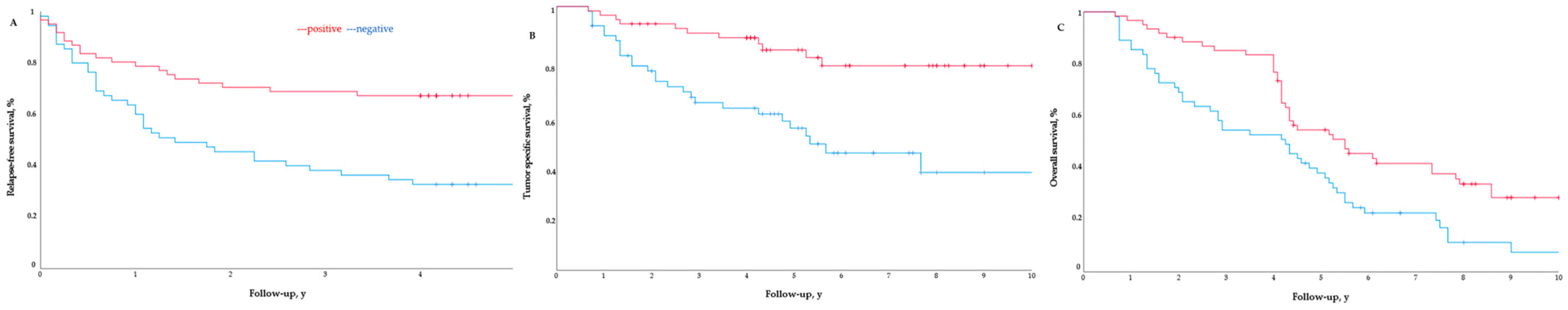
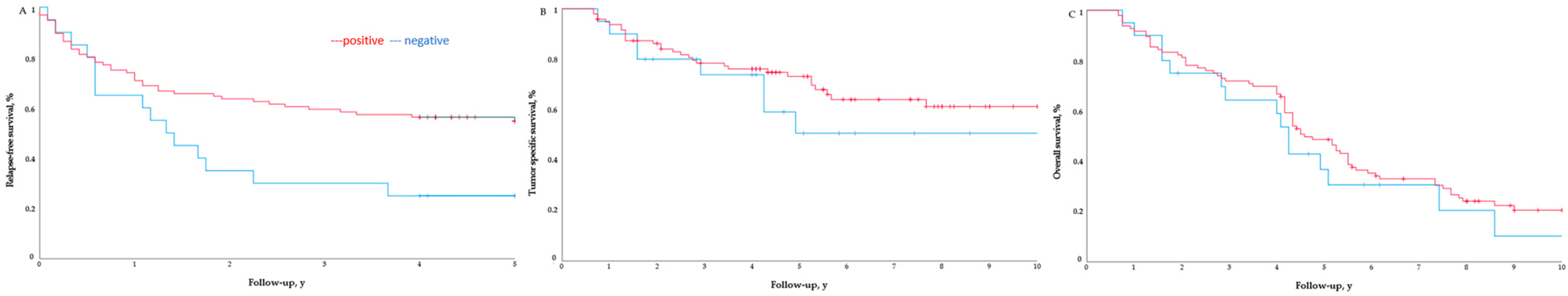
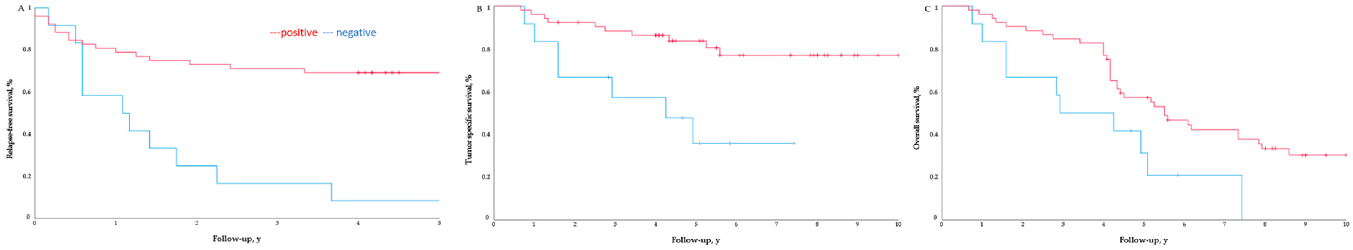
2. Results
Survival Curves
3. Discussion
4. Materials and Methods
4.1. Patients and Procedures
4.2. Histopathological Evaluation
4.3. Tissue Microarray Construction
4.4. Immunohistochemistry
4.5. Statistical Analysis
5. Conclusions
Author Contributions
Funding
Institutional Review Board Statement
Informed Consent Statement
Data Availability Statement
Conflicts of Interest
References
- Hooper, P.B.; Farberg, A.S.; Fitzgerald, A.L.; Siegel, J.J.; Rackley, B.B.; Prasai, A.; Kurley, S.J.; Goldberg, M.S.; Litchman, G.H. Real-World Evidence Shows Clinicians Appropriately Use the Prognostic 40-Gene Expression Profile (40-GEP) Test for High-Risk Cutaneous Squamous Cell Carcinoma (cSCC) Patients. Cancer Investig. 2022, 40, 911–922. [Google Scholar] [CrossRef]
- Waldman, A.; Schmults, C. Cutaneous Squamous Cell Carcinoma. Hematol. Oncol. Clin. N. Am. 2019, 33, 1–12. [Google Scholar] [CrossRef] [PubMed]
- Caudill, J.; Thomas, J.E.; Burkhart, C.G. The Risk of Metastases from Squamous Cell Carcinoma of the Skin. Int. J. Dermatol. 2023, 62, 483–486. [Google Scholar] [CrossRef] [PubMed]
- Zakhem, G.A.; Pulavarty, A.N.; Carucci, J.; Stevenson, M.L. Association of Patient Risk Factors, Tumor Characteristics, and Treatment Modality With Poor Outcomes in Primary Cutaneous Squamous Cell Carcinoma: A Systematic Review and Meta-Analysis. JAMA Dermatol. 2023, 159, 160–171. [Google Scholar] [CrossRef] [PubMed]
- Dessinioti, C.; Stratigos, A.J. Recent Advances in the Diagnosis and Management of High-Risk Cutaneous Squamous Cell Carcinoma. Cancers 2022, 14, 3556. [Google Scholar] [CrossRef] [PubMed]
- Tsang, D.A.; Tam, S.Y.C.; Oh, C.C. Molecular Alterations in Cutaneous Squamous Cell Carcinoma in Immunocompetent and Immunosuppressed Hosts-A Systematic Review. Cancers 2023, 15, 1832. [Google Scholar] [CrossRef]
- Llanos, S.; García-Pedrero, J.M.; Morgado-Palacin, L.; Rodrigo, J.P.; Serrano, M. Stabilization of P21 by mTORC1/4E-BP1 Predicts Clinical Outcome of Head and Neck Cancers. Nat. Commun. 2016, 7, 10438. [Google Scholar] [CrossRef] [PubMed]
- de Vicente, J.C.; Peña, I.; Rodrigo, J.P.; Rodríguez-Santamarta, T.; Lequerica-Fernández, P.; Suárez-Fernández, L.; Allonca, E.; García-Pedrero, J.M. Phosphorylated Ribosomal Protein S6 Correlation with P21 Expression and Inverse Association with Tumor Size in Oral Squamous Cell Carcinoma: PS6 and P21 in Oral Squamous Cell Carcinoma. Head Neck 2017, 39, 1876–1887. [Google Scholar] [CrossRef] [PubMed]
- García-Carracedo, D.; Villaronga, M.Á.; Álvarez-Teijeiro, S.; Hermida-Prado, F.; Santamaría, I.; Allonca, E.; Suárez-Fernández, L.; Gonzalez, M.V.; Balbín, M.; Astudillo, A.; et al. Impact of PI3K/AKT/mTOR Pathway Activation on the Prognosis of Patients with Head and Neck Squamous Cell Carcinomas. Oncotarget 2016, 7, 29780–29793. [Google Scholar] [CrossRef]
- Sonavane, K.; Phillips, J.; Ekshyyan, O.; Moore-Medlin, T.; Roberts Gill, J.; Rong, X.; Lakshmaiah, R.R.; Abreo, F.; Boudreaux, D.; Clifford, J.L.; et al. Topical Curcumin-Based Cream Is Equivalent to Dietary Curcumin in a Skin Cancer Model. J. Skin. Cancer 2012, 2012, 147863. [Google Scholar] [CrossRef]
- Chen, S.-J.; Nakahara, T.; Takahara, M.; Kido, M.; Dugu, L.; Uchi, H.; Takeuchi, S.; Tu, Y.-T.; Moroi, Y.; Furue, M. Activation of the Mammalian Target of Rapamycin Signalling Pathway in Epidermal Tumours and Its Correlation with Cyclin-Dependent Kinase 2. Br. J. Dermatol. 2009, 160, 442–445. [Google Scholar] [CrossRef]
- Khandelwal, A.R.; Ma, X.; Egan, P.; Kaskas, N.M.; Moore-Medlin, T.; Caldito, G.; Abreo, F.; Gu, X.; Aubrey, L.; Milligan, E.; et al. Biomarker and Pathologic Predictors of Cutaneous Squamous Cell Carcinoma Aggressiveness. Otolaryngol. Head Neck Surg. 2016, 155, 281–288. [Google Scholar] [CrossRef]
- Martins, F.; de Sousa, S.C.; Dos Santos, E.; Woo, S.-B.; Gallottini, M. PI3K-AKT-mTOR Pathway Proteins Are Differently Expressed in Oral Carcinogenesis. J. Oral. Pathol. Med. 2016, 45, 746–752. [Google Scholar] [CrossRef]
- Li, L.; Liu, D.; Qiu, Z.-X.; Zhao, S.; Zhang, L.; Li, W.-M. The Prognostic Role of mTOR and P-mTOR for Survival in Non-Small Cell Lung Cancer: A Systematic Review and Meta-Analysis. PLoS ONE 2015, 10, e0116771. [Google Scholar] [CrossRef]
- Beca, F.; Andre, R.; Martins, D.S.; Bilhim, T.; Martins, D.; Schmitt, F. P-mTOR Expression Is Associated with Better Prognosis in Luminal Breast Carcinoma. J. Clin. Pathol. 2014, 67, 961–967. [Google Scholar] [CrossRef] [PubMed]
- Marques, A.E.M.; Elias, S.T.; Porporatti, A.L.; Castilho, R.M.; Squarize, C.H.; De Luca Canto, G.; Guerra, E.N.S. mTOR Pathway Protein Immunoexpression as a Prognostic Factor for Survival in Head and Neck Cancer Patients: A Systematic Review and Meta-Analysis. J. Oral. Pathol. Med. 2016, 45, 319–328. [Google Scholar] [CrossRef] [PubMed]
- Wang, H.; Li, J. A Systematic Review and Meta-Analysis Protocol of Clinical Characteristics and Prognostic Significance of Mammalian Target of Rapamycin for Gastric Cancer Patients. Medicine 2020, 99, e21138. [Google Scholar] [CrossRef]
- McBride, S.M.; Perez, D.A.; Polley, M.-Y.; Vandenberg, S.R.; Smith, J.S.; Zheng, S.; Lamborn, K.R.; Wiencke, J.K.; Chang, S.M.; Prados, M.D.; et al. Activation of PI3K/mTOR Pathway Occurs in Most Adult Low-Grade Gliomas and Predicts Patient Survival. J. Neurooncol. 2010, 97, 33–40. [Google Scholar] [CrossRef] [PubMed]
- Pantuck, A.J.; Seligson, D.B.; Klatte, T.; Yu, H.; Leppert, J.T.; Moore, L.; O’Toole, T.; Gibbons, J.; Belldegrun, A.S.; Figlin, R.A. Prognostic Relevance of the mTOR Pathway in Renal Cell Carcinoma: Implications for Molecular Patient Selection for Targeted Therapy. Cancer 2007, 109, 2257–2267. [Google Scholar] [CrossRef]
- Chen, J.; Hu, C.-F.; Hou, J.-H.; Shao, Q.; Yan, L.-X.; Zhu, X.-F.; Zeng, Y.-X.; Shao, J.-Y. Epstein-Barr Virus Encoded Latent Membrane Protein 1 Regulates mTOR Signaling Pathway Genes Which Predict Poor Prognosis of Nasopharyngeal Carcinoma. J. Transl. Med. 2010, 8, 30. [Google Scholar] [CrossRef]
- Stelloo, S.; Sanders, J.; Nevedomskaya, E.; de Jong, J.; Peters, D.; van Leenders, G.J.L.H.; Jenster, G.; Bergman, A.M.; Zwart, W. mTOR Pathway Activation Is a Favorable Prognostic Factor in Human Prostate Adenocarcinoma. Oncotarget 2016, 7, 32916–32924. [Google Scholar] [CrossRef] [PubMed]
- Ahmed, N.U.; Ueda, M.; Ichihashi, M. p21WAF1/CIP1 Expression in Non-Melanoma Skin Tumors. J. Cutan. Pathol. 1997, 24, 223–227. [Google Scholar] [CrossRef] [PubMed]
- Bedir, R.; Güçer, H.; Şehitoğlu, İ.; Yurdakul, C.; Bağcı, P.; Üstüner, P. The Role of P16, P21, P27, P53 and Ki-67 Expression in the Differential Diagnosis of Cutaneous Squamous Cell Carcinomas and Keratoacanthomas: An Immunohistochemical Study. Balk. Med. J. 2016, 33, 121–127. [Google Scholar] [CrossRef] [PubMed]
- Kikuchi, A.; Amagai, M.; Hayakawa, K.; Ueda, M.; Hirohashi, S.; Shimizu, N.; Nishikawa, T. Association of EGF Receptor Expression with Proliferating Cells and of Ras P21 Expression with Differentiating Cells in Various Skin Tumours. Br. J. Dermatol. 1990, 123, 49–58. [Google Scholar] [CrossRef] [PubMed]
- Inohara, S.; Kitagawa, K.; Kitano, Y. Coexpression of p21Waf1/Cip1 and P53 in Sun-Exposed Normal Epidermis, but Not in Neoplastic Epidermis. Br. J. Dermatol. 1996, 135, 717–720. [Google Scholar] [CrossRef]
- Lu, S.; Tiekso, J.; Hietanen, S.; Syrjänen, K.; Havu, V.K.; Syrjänen, S. Expression of Cell-Cycle Proteins P53, P21 (WAF-1), PCNA and Ki-67 in Benign, Premalignant and Malignant Skin Lesions with Implicated HPV Involvement. Acta Derm. Venereol. 1999, 79, 268–273. [Google Scholar] [CrossRef] [PubMed]
- Healy, E.; Reynolds, N.J.; Smith, M.D.; Harrison, D.; Doherty, E.; Campbell, C.; Rees, J.L. Up-Regulation of p21WAF1/CIP1 in Psoriasis and after the Application of Irritants and Tape Stripping. J. Investig. Dermatol. 1995, 105, 274–279. [Google Scholar] [CrossRef] [PubMed]
- Kumar, S.; Singh, N.; Kataria, S.P.; Kandoi, S.; Verma, M.; Sen, R. Cyclin-Dependent Kinase Inhibitor P21 and Proliferative Marker Ki67 in Colonic Carcinoma. J. Cancer Res. Ther. 2022, 18, 915–920. [Google Scholar] [CrossRef]
- Sun, Y.; Yang, S.; Sun, N.; Chen, J. Differential Expression of STAT1 and P21 Proteins Predicts Pancreatic Cancer Progression and Prognosis. Pancreas 2014, 43, 619–623. [Google Scholar] [CrossRef]
- McClelland, R.A.; Gee, J.M.; O’Sullivan, L.; Barnes, D.M.; Robertson, J.F.; Ellis, I.O.; Nicholson, R.I. P21(WAF1) Expression and Endocrine Response in Breast Cancer. J. Pathol. 1999, 188, 126–132. [Google Scholar] [CrossRef]
- Balcere, A.; Sperga, M.; Čēma, I.; Lauskis, G.; Zolovs, M.; Rone Kupfere, M.; Krūmiņa, A. Expression of P53, P63, P16, Ki67, Cyclin D, Bcl-2, and CD31 Markers in Actinic Keratosis, In Situ Squamous Cell Carcinoma and Normal Sun-Exposed Skin of Elderly Patients. J. Clin. Med. 2023, 12, 7291. [Google Scholar] [CrossRef] [PubMed]
- Ruiz, E.S.; Karia, P.S.; Besaw, R.; Schmults, C.D. Performance of the American Joint Committee on Cancer Staging Manual, 8th Edition vs the Brigham and Women’s Hospital Tumor Classification System for Cutaneous Squamous Cell Carcinoma. JAMA Dermatol. 2019, 155, 819–825. [Google Scholar] [CrossRef] [PubMed]
- Gonzalez-Guerrero, M.; Martínez-Camblor, P.; Vivanco, B.; Fernández-Vega, I.; Munguía-Calzada, P.; Gonzalez-Gutierrez, M.P.; Rodrigo, J.P.; Galache, C.; Santos-Juanes, J. The Adverse Prognostic Effect of Tumor Budding on the Evolution of Cutaneous Head and Neck Squamous Cell Carcinoma. J. Am. Acad. Dermatol. 2017, 76, 1139–1145. [Google Scholar] [CrossRef] [PubMed]
- Menéndez, S.T.; Rodrigo, J.P.; Alvarez-Teijeiro, S.; Villaronga, M.Á.; Allonca, E.; Vallina, A.; Astudillo, A.; Barros, F.; Suárez, C.; García-Pedrero, J.M. Role of HERG1 Potassium Channel in Both Malignant Transformation and Disease Progression in Head and Neck Carcinomas. Mod. Pathol. 2012, 25, 1069–1078. [Google Scholar] [CrossRef] [PubMed]


| p-S6 Expression | ||||
|---|---|---|---|---|
| Clinicopathological Characteristics | N = 116 | Negative (≤50%) N = 55; n (%) | Positive (>50%) N = 61; n (%) | p |
| Sex, male | 89 | 42 (76.3) | 47 (77.0) | 1.000 |
| Age, mean ± SD, (min–max) | 78.74 ± 8.32 (50–97) | 79.11 ± 7.73 (51–93) | 78.41 ± 8.87 (50–97) | 0.713 |
| Tumor thickness, mm, mean ± SD | 8.91 ± 6.54 | 11.55 ± 7.38 | 6.54 ± 4.59 | <0.001 |
| >6 mm | 60 | 40 (72.7) | 20 (32.8) | <0.001 |
| Tumor horizontal size, mm, mean ± SD | 21.56 ± 13.89 | 25.27 ± 16.09 | 18.21 ± 10.63 | 0.007 |
| >20 mm | 42 | 29 (52.7) | 13 (21.3) | <0.001 |
| Tumor differentiation | 0.110 | |||
| Well differentiated | 61 (52.6%) | 27 (49.1) | 34 (55.7) | |
| Moderately differentiated | 48 (41.4%) | 22 (40.0) | 26 (42.6) | |
| Poorly differentiated | 7 (6.0%) | 6 (10.9) | 1 (1.6) | |
| Desmoplastic growth | 21 | 15 (27.3) | 6 (9.8) | 0.017 |
| Tumor site, ear | 26 | 12 (21.8) | 14 (22.9) | 1.000 |
| pTNM 8th edition | 0.007 | |||
| pT1 | 64 (55.2%) | 22 (40.0) | 42 (68.9) | |
| pT2 | 36 (31.0%) | 22 (40.0) | 14 (23.0) | |
| pT3 | 16 (13.8%) | 11 (20.0) | 5 (8.2) | |
| Inflammation | 0.609 | |||
| None | 29 | 15 (27.3) | 14 (23.0) | |
| Mild | 73 | 35 (63.6) | 38 (62.2) | |
| Strong | 14 | 5 (9.1) | 9 (14.8) | |
| Other SCC | 38 | 13 (23.6) | 25 (40.1) | 0.051 |
| Immunosuppression | 1.000 | |||
| Yes | 16 (13.8%) | 8 (14.5) | 8 (13.1) | |
| No | 100 (86.2%) | 47 (85.5) | 53 (86.9) | |
| Perineural invasion | 0.029 | |||
| Yes | 16 (13.8%) | 12 (21.8) | 4 (6.6) | |
| No | 100 (86.2%) | 43 (78.2) | 57 (93.4) | |
| Lymph-vascular invasion | 0.603 | |||
| Yes | 3 (1.6%) | 2 (3.6) | 1 (1.6) | |
| No | 113 (97.4%) | 53 (96.4) | 60 (98.4) | |
| Tumor buds present | <0.001 | |||
| Yes | 54 | 37 (67.3) | 17 (27.9) | |
| No | 62 | 18 (32.7) | 44 (72.1) | |
| ≥5 tumor buds | 0.258 | |||
| Yes | 24 | 14 (25.5) | 10 (16.4) | |
| No | 92 | 41 (74.5) | 51 (83.6) | |
| p21 Expression | ||||
| Clinicopathological Characteristics | N = 116 | Negative N = 20 n (%) | Positive N = 96 n (%) | p |
| Sex, male | 89 | 16 (18.0) | 73 (82.0) | 1.000 |
| Age, mean ± SD (min–max) | 78.74 ± 8.32 (50–97) | 78.35 ± 8.75 (54–96) | 78.82 ± 8.27 (50–97) | 0.818 |
| Tumor thickness, mm, mean ± SD | 8.91 ± 6.54 | 11.30 ± 6.02 | 8.42 ± 6.57 | 0.869 |
| >6 mm | 60 | 15 (75.0) | 45 (46.9) | 0.027 |
| Tumor horizontal size, mm, mean ± SD | 21.56 ± 13.89 | 21.05 ± 9.66 | 21.67 ± 14.66 | 0.113 |
| >20 mm | 42 | 8 (40.0) | 34 (35.4) | 0.799 |
| Tumor differentiation | 0.110 | |||
| Well differentiated | 61 | 27 (49.1) | 34 (55.7) | |
| Moderately differentiated | 48 | 22 (40.0) | 26 (42.6) | |
| Poorly differentiated | 7 | 6 (10.9) | 1 (1.6) | |
| Desmoplastic growth | 21 | 15 (27.3) | 6 (9.8) | 0.017 |
| Tumor site, ear | 26 | 3 (15.0) | 23 (23.9) | 0.558 |
| pTNM 8th edition | 0.329 | |||
| pT1 | 64 | 9 (45.0) | 55 (57.3) | |
| pT2 | 36 | 9 (45.0) | 27 (28.1) | |
| pT3 | 16 | 2 (10.0) | 14 (14.6) | |
| Inflammation | 0.368 | |||
| None | 29 | 7 (35.0) | 22 (22.9) | |
| Mild | 73 | 12 (60.0) | 61 (63.5) | |
| Strong | 14 | 1 (5.0) | 13 (13.5) | |
| Other SCC | 38 | 5 (25.0) | 33 (34.4) | 0.601 |
| Immunosuppression | 0.474 | |||
| Yes | 16 | 4 (20.0) | 12 (12.5) | |
| No | 100 | 16 (80.0) | 84 (87.5) | |
| Perineural invasion | 0.032 | |||
| Yes | 16 | 6 (30.0) | 10 (10.4) | |
| No | 100 | 14 (70.0) | 86 (89.6) | |
| Lymph-vascular invasion | 0.436 | |||
| Yes | 3 | 1 (5.0) | 2 (2.08) | |
| No | 113 | 19 (95.0) | 94 (97.9) | |
| Tumor buds present | 0.222 | |||
| Yes | 54 | 12 (60.0) | 42 (43.8) | |
| No | 62 | 8 (40.0) | 54 (56.2) | |
| ≥5 tumor buds | 0.361 | |||
| Yes | 24 | 6 (3.0) | 18 (18.8) | |
| No | 92 | 14 (70) | 78 (81.2) | |
| p-S6 | <10% | 10–50% | >50% | p (Chi-Square) |
|---|---|---|---|---|
| cSCCHN | 0 (0%) | 17 (29.3%) | 41 (70.7%) | <0.001 |
| McSCCHN | 4 (6.9%) | 34 (58.6%) | 20 (34.5%) | |
| p21 | 0–10% | >10% | p (chi-square) | |
| cSCCHN | 5 (8.6%) | 53 (91.4%) | 0.025 | |
| McSCCHN | 15 (25.9%) | 43 (74.1%) |
| p-S6 ≤ 50 | p-S6 > 50 | p (Chi-Square) | p (Cramer’s V) | |
|---|---|---|---|---|
| p21 (−) | 12 (60.0%) | 8 (40.0%) | 0.230 | 0.215 |
| p21 (+) | 43 (44.8%) | 53 (55.2%) |
| Metastasis | Tumor Mortality | All-Cause Mortality | ||||
|---|---|---|---|---|---|---|
| p-S6 | HR (95% CI) | p | HR (95% CI) | p | HR (95% CI) | p |
| Univariate | ||||||
| p-S6 | 2.63 (1.51–4.54) | <0.001 | 3.70 (1.78–7.69) | <0.001 | 1.75 (1.14–2.63) | 0.008 |
| Multivariate | ||||||
| p-S6 | 2.23 (1.01–4.91) | 0.047 | 0.99 (0.42–2.29) | 0.975 | 1.08 (0.66–1.77) | 0.766 |
| Age | 0.99 (0.65–2.31) | 0.526 | 1.01 (0.97–1.07) | 0.975 | 1.05 (1.01–1.09) | 0.005 |
| Sex | 0.79 (0.55–2.18) | 0.791 | 1.20 (0.49–2.91) | 0.684 | 0.88 (0.54–1.47) | 0.634 |
| Tumor thickness | 3.25 (1.55–6.85) | 0.002 | 4.03 (1.42–11.42) | 0.009 | 1.14 (0.66–1.99) | 0.642 |
| Tumor horizontal size | 1.12 (0.48–2.58) | 0.798 | 0.83 (0.27–2.52) | 0.743 | 0.97 (0.43–2.20) | 0.934 |
| Desmoplastic growth | 1.08 (0.55–2.13) | 0.812 | 0.88 (0.36–2.11) | 0.771 | 0.85 (0.45–1.63) | 0.632 |
| Perineural invasion | 1.27 (0.58–2.79) | 0.547 | 0.80 (0.28–2.29) | 0.802 | 1.12 (0.53–2.32) | 0.770 |
| Tumor buds | 6.72 (3.32–13.58) | <0.001 | 7.93 (3.12–20.17) | <0.001 | 3.36 (2.09–5.42) | <0.001 |
| pTNM 8th edition | 1.44 (0.87–2.36) | 0.152 | 2.13 (1.20–4.06) | 0.021 | 1.95 (1.18–3.24) | 0.009 |
| Metastasis | Tumor Mortality | All-Causes Mortality | ||||
|---|---|---|---|---|---|---|
| p21 | HR (95% CI) | p | HR (95% CI) | p | HR (95% CI) | p |
| Univariate | ||||||
| p21 | 4.76 (1.11–5.55) | 0.020 | 1.72 (0.77–3.84) | 0.185 | 1.31 (0.71–2.27) | 0.323 |
| Multivariate | ||||||
| p21 | 0.86 (0.45–1.66) | 0.656 | 1.14 (0.48–2.73) | 0.770 | 0.72 (0.39–1.33) | 0.720 |
| Age | 0.99 (0.97–1.03) | 0.945 | 1.03 (0.98–1.08) | 0.207 | 1.03 (1.01–1.07) | 0.014 |
| Sex | 1.13 (0.57–2.22) | 0.727 | 1.37 (0.57–3.33) | 0.482 | 0.96 (0.58–1.60) | 0.885 |
| Tumor thickness | 4.31 (2.23–8.32) | <0.001 | 7.00 (2.80–17.53) | <0.001 | 1.79 (1.12–2.84) | 0.014 |
| Desmoplastic growth | 1.48 (0.76–2.88) | 0.249 | 1.62 (0.69–3.79) | 0.266 | 1.00 (0.54–1.87) | 0.999 |
| Perineural invasion | 1.52 (0.76–3.05) | 0.234 | 1.16 (0.48–2.80) | 0.739 | 1.45 (0.73–2.87) | 0.285 |
| p-S6 | ≤10% | 10–50% | >50% | p (Chi-Squared) |
|---|---|---|---|---|
| cSCCHN | 0 (0%) | 1 (29.3%) | 10 (70.7%) | 0.007 |
| McSCCHN | 4 (6.9%) | 3 (58.6%) | 4 (34.5%) |
Disclaimer/Publisher’s Note: The statements, opinions and data contained in all publications are solely those of the individual author(s) and contributor(s) and not of MDPI and/or the editor(s). MDPI and/or the editor(s) disclaim responsibility for any injury to people or property resulting from any ideas, methods, instructions or products referred to in the content. |
© 2024 by the authors. Licensee MDPI, Basel, Switzerland. This article is an open access article distributed under the terms and conditions of the Creative Commons Attribution (CC BY) license (https://creativecommons.org/licenses/by/4.0/).
Share and Cite
Gómez-de Castro, C.; Santos-Juanes, R.; Nuñez-Gómez, B.; Fernández-Vega, I.; Vivanco, B.; Fernández-Velasco, A.; Reyes-García, S.; Carrero-Martín, J.; García-Pedrero, J.M.; Rodrigo, J.P.; et al. Low-Level Expression of p-S6 Is Associated with Nodal Metastasis in Patients with Head and Neck Cutaneous Squamous Cell Carcinoma. Int. J. Mol. Sci. 2024, 25, 4304. https://doi.org/10.3390/ijms25084304
Gómez-de Castro C, Santos-Juanes R, Nuñez-Gómez B, Fernández-Vega I, Vivanco B, Fernández-Velasco A, Reyes-García S, Carrero-Martín J, García-Pedrero JM, Rodrigo JP, et al. Low-Level Expression of p-S6 Is Associated with Nodal Metastasis in Patients with Head and Neck Cutaneous Squamous Cell Carcinoma. International Journal of Molecular Sciences. 2024; 25(8):4304. https://doi.org/10.3390/ijms25084304
Chicago/Turabian StyleGómez-de Castro, Celia, Raquel Santos-Juanes, Borja Nuñez-Gómez, Iván Fernández-Vega, Blanca Vivanco, Adela Fernández-Velasco, Sebastián Reyes-García, Jimena Carrero-Martín, Juana M. García-Pedrero, Juan P. Rodrigo, and et al. 2024. "Low-Level Expression of p-S6 Is Associated with Nodal Metastasis in Patients with Head and Neck Cutaneous Squamous Cell Carcinoma" International Journal of Molecular Sciences 25, no. 8: 4304. https://doi.org/10.3390/ijms25084304