Abstract
Neuroinflammation is involved in various neurological and neurodegenerative disorders in which the activation of microglia is one of the key factors. In this study, we examined the anti-inflammatory effects of the flavonoids nobiletin (5,6,7,8,3′,4′-hexamethoxyflavone) and eriodictyol (3′,4′,5,7-tetraxydroxyflavanone) on human microglia cell line activation stimulated by either lipopolysaccharide (LPS), severe acute respiratory syndrome coronavirus 2 (SARS-CoV-2) full-length Spike protein (FL-Spike), or the mycotoxin ochratoxin A (OTA). Human microglia were preincubated with the flavonoids (10, 50, and 100 µM) for 2 h, following which, they were stimulated for 24 h. The inflammatory mediators interleukin-1 beta (IL-1β), chemokine (C-X-C motif) ligand 8 (CXCL8), IL-6, and matrix metalloproteinase-9 (MMP-9) were quantified in the cell culture supernatant by enzyme-linked immunosorbent assay (ELISA). Both nobiletin and eriodictyol significantly inhibited the LPS, FL-Spike, and OTA-stimulated release of IL-1β, CXCL8, IL-6, and MMP-9 at 50 and 100 µM, while, in most cases, nobiletin was also effective at 10 µM, with the most pronounced reductions at 100 µM. These findings suggest that both nobiletin and eriodictyol are potent inhibitors of the pathogen-stimulated microglial release of inflammatory mediators, highlighting their potential for therapeutic application in neuroinflammatory diseases, such as long COVID.
Keywords:
CXCL8; flavonoids; IL-1β; IL-6; long COVID; luteolin; mast cells; microglia; MMP-9; neuroinflammation; nobiletin; SARS-CoV-2 spike protein 1. Introduction
Neuroinflammation, particularly pathogen-stimulated microglial activation, is a key pathological feature in numerous neurodegenerative diseases, including Alzheimer’s disease (AD) and Parkinson’s disease (PD) [1,2,3]. Microglia, the resident immune cells of the central nervous system (CNS), are essential for brain functions, especially the surveillance of the microenvironment and the maintenance of brain homeostasis, as well as defense against infections and injury [4]. When stimulated by pathogens, such as bacterial lipopolysaccharides (LPS), viral proteins, and other environmental toxins, microglia can initiate a cascade of responses aimed at protecting the brain, but prolonged activation can lead to the release of various inflammatory mediators, ultimately leading to neuronal damage and contributing to the development of chronic neuroinflammatory [5] and neurodegenerative disorders [6,7]. Infection with severe acute respiratory syndrome coronavirus 2 (SARS-CoV-2) causes the loss of microglial homeostasis [3]. The pro-inflammatory molecules evaluated in this study include the cytokines interleukin-1 beta (IL-1β) and IL-6, the chemokine (C-X-C motif) ligand 8 (CXCL8)/IL-8, and matrix metalloproteinase-9 (MMP-9), which disrupts neuronal connectivity [8,9,10,11]. Inflammatory molecules derived from microglia can impair the blood-brain barrier (BBB), allowing the entry of peripheral immune cells, activating and damaging glial cells and neurons, and contributing to neurodegenerative processes [12,13]. Perineural nets were reported to be phagocytosed by MMP-9-expressing microglia in the SOD1G83A mouse strain, an Amyotrophic lateral sclerosis (ALS) disease model [14].
A high level of MMP-9 has been shown to exacerbate neuroinflammatory processes, neurodegeneration, basement membrane degradation, and BBB disruption [8,9,11,15,16]. Serum levels of MMP-9 levels were elevated in patients with coronavirus disease 2019 (COVID-19) [17,18] and associated with severe symptoms [19,20]. Blood MMP-9 levels were also reported to be higher in the acute phase of neuro COVID patients [16].
Mycotoxins have been increasingly reported to have neurotoxic effects [21,22]. Ochratoxin A (OTA), a mycotoxin found in mold in the environment and on foods, has immunotoxic and neurotoxic properties since it can cross the BBB and accumulate in brain areas, leading to neurodegeneration [22,23]. In a recent study, we found that OTA stimulated the release of IL-1β, IL-18, and CXCL8 from the human SV-40 microglia cell line [24].
Recent studies have highlighted the potential of natural compounds, especially citrus peel flavonoids [25,26], to modulate microglial activation and attenuate neuroinflammation [13,27,28,29]. We previously reported that luteolin (3′,4′,5,7-tetrahydroxyflavone, Lut) and methoxy luteolin (3′,4′,5,7-tetramethoxyflavone, Methlut) inhibited IL-1β release from cultured human SV-40 microglia stimulated by neurotensin [30] and MMP-9 release stimulated by the SARS-CoV-2 Spike protein [31]. Moreover, luteolin induced an anti-inflammatory and neuroprotective phenotype in BV-2 microglia [32]. Luteolin has been explored for the treatment of various conditions [33,34,35,36] including brain disorders [37,38,39,40,41], such as long COVID [42,43,44,45,46,47] and autism spectrum disorder (ASD) [48,49].
Nobiletin (5,6,7, 8,3′,4′-hexamethoxyflavone) found in citrus fruits has emerged as a promising candidate due to its anti-inflammatory, antioxidant, and neuroprotective properties and promotion of neuronal survival [50,51]. Eriodictyol (3′,4′,5,7-tetraxydroxyflavanone) also has antioxidant, anti-inflammatory, and neuroprotective properties [52]. However, the specific role of nobiletin or eriodictyol in pathogen-stimulated microglial-dependent inflammation remains poorly understood.
In this study, we investigated the potential of nobiletin and eriodictyol to inhibit the release of IL-1β, IL-6, CXCL8, and MMP-9 from the cultured human microglia cell line stimulated by pathogen-derived LPS, full-length Spike (FL Spike), and OTA.
2. Results
2.1. Inhibitory Effects of Nobiletin and Eriodictyol on LPS-Stimulated Microglia
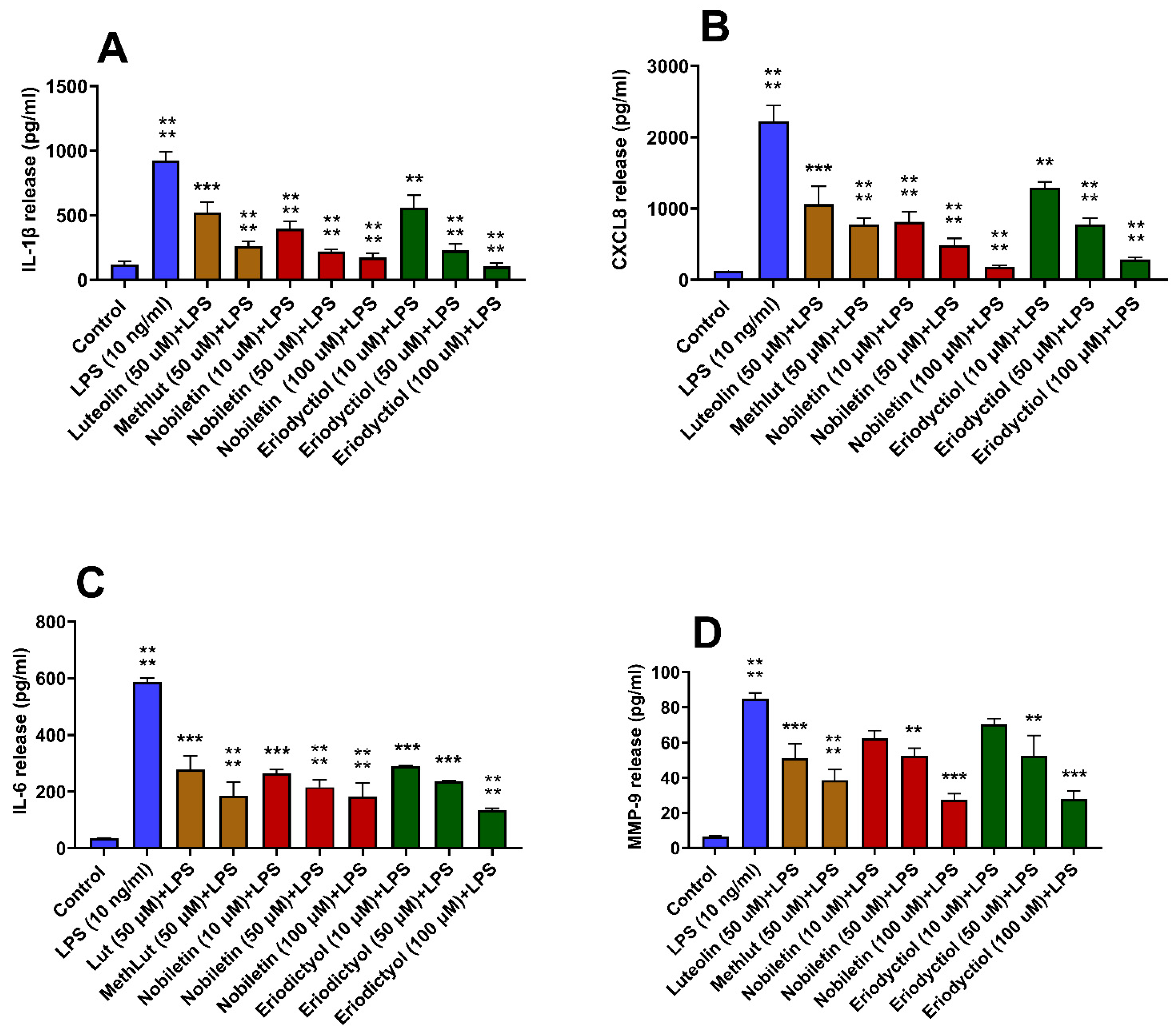
Pre-treatment of microglia with either nobiletin or eriodictyol at 10, 50, and 100 µM for 2 h significantly inhibited LPS-induced pro-inflammatory mediator release, with 100 µM showing the highest inhibition (Figure 1A–D). Both nobiletin and eriodictyol inhibited release even at 10 µM, with the exception of MMP-9 release. Pretreatment with either luteolin or methoxy luteolin at 50 μM also significantly inhibited the release of all the mediators (Figure 1A–D). Multivariant analysis among all flavonoids at 50 µM showed no significant difference.
Figure 1.
Inhibition of the release of inflammatory mediators from human microglia stimulated by LPS. Human microglia (0.5 × 10⁵ cells/1 mL/well in 24-well plates) were first preincubated with nobiletin (>98% purity; 10, 50, and 100 μM), eriodictyol (10, 50, and 100 μM), luteolin (50 μM), and methoxyluteolin (50 μM) for 2 h and then incubated with LPS (10 ng/mL) for 24 h. Control cells were treated with an equal volume of culture medium. Then, the cell culture supernatant fluids were collected and assayed for IL-1β (A), CXCL8 (B), IL-6 (C), and MMP-9 (D) by commercial ELISA kits. All assays were performed in triplicate. LPS was compared to the control; each of the other conditions was compared to LPS (n = 3, * p < 0.05, ** p < 0.01, *** p < 0.001, **** p < 0.0001).
2.2. Inhibitory Effects of Nobiletin and Eriodictyol on FL Spike-Stimulated Microglia
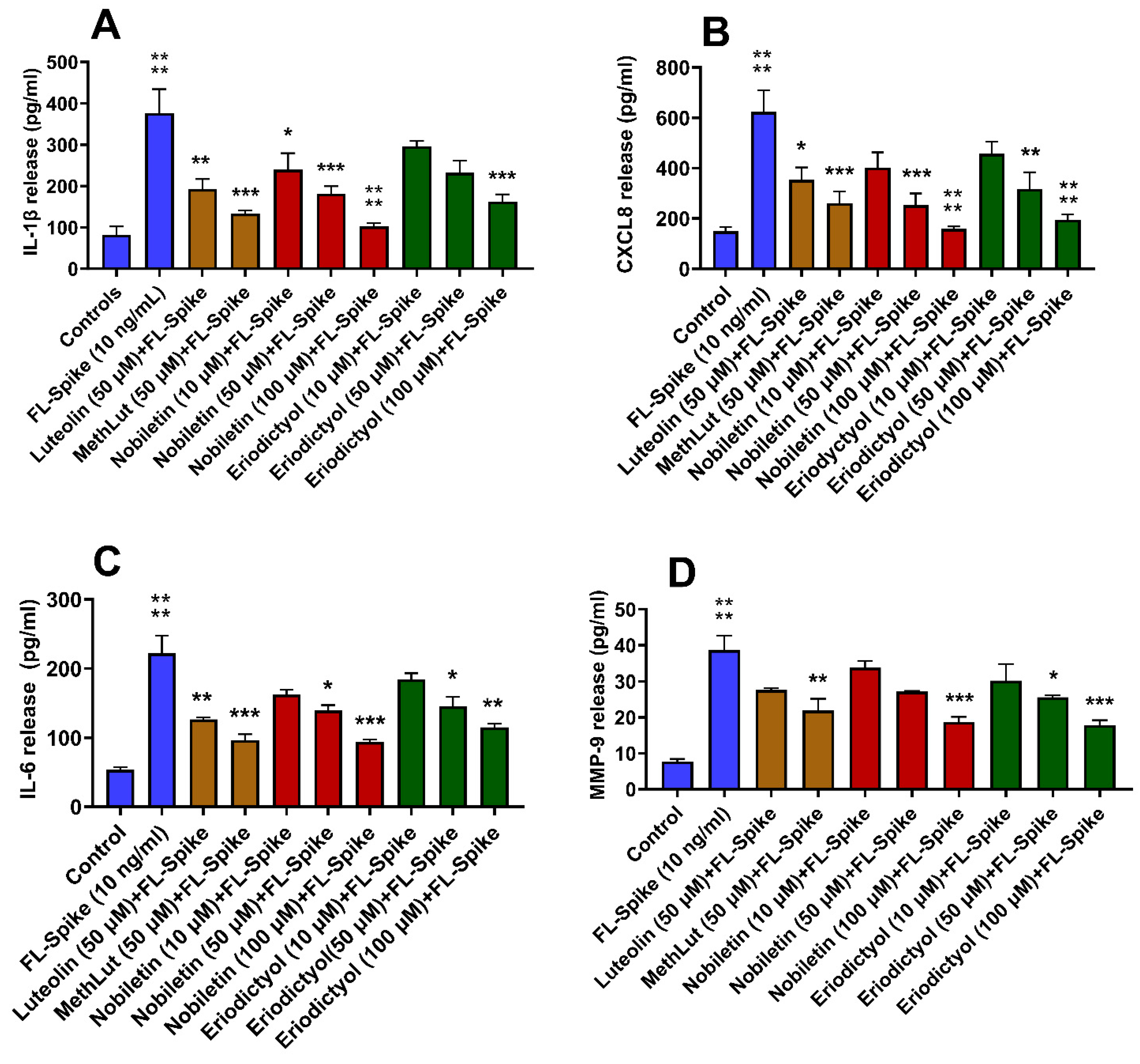
Pre-treatment of microglia with either nobiletin or eriodictyol at 50 and 100 µM for 2 h significantly inhibited the FL Spike-induced release of IL-1β, CXCL8, IL-6, and MMP-9 (Figure 2A–D). Nobiletin at 10 µM inhibited LPS induced IL-1β release but not eriodictyol. Pre-treatment with either luteolin or methoxy luteolin at 50 μM also significantly inhibited the release of all the mediators (Figure 2A–D). Multivariant analysis among all flavonoids at 50 µM showed no significant difference.
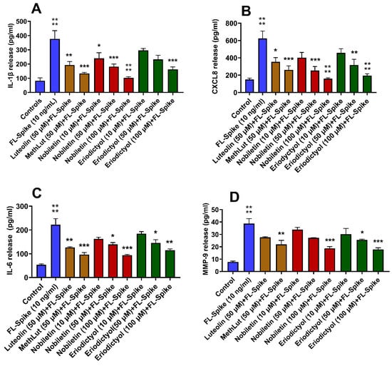
Figure 2.
Inhibition of the release of inflammatory mediators from human microglia stimulated by SARS-CoV-2 Spike protein. Human microglia (0.5 × 10⁵ cells/1 mL /well in 24-well plates) were preincubated with nobiletin (10, 50, and 100 μM), eriodictyol (10, 50, and 100 μM), luteolin (50 μM), and methoxy luteolin (50 μM) for 2 h and then incubated with SARS-CoV-2 Spike protein (10 ng/mL) for 24 h. Control cells were treated with an equal volume of culture medium. Following this, cell culture supernatant fluids were collected and assayed for IL-1β (A), CXCL8 (B), IL-6 (C), and MMP-9 (D) by commercial ELISA kits. FL Spike was compared to the control; each of the other conditions was compared to FL Spike (n = 3, * p < 0.05, ** p < 0.01, *** p < 0.001, **** p < 0.0001).
2.3. Inhibitory Effects of Nobiletin and Eriodyctiol on OTA-Stimulated Microglia
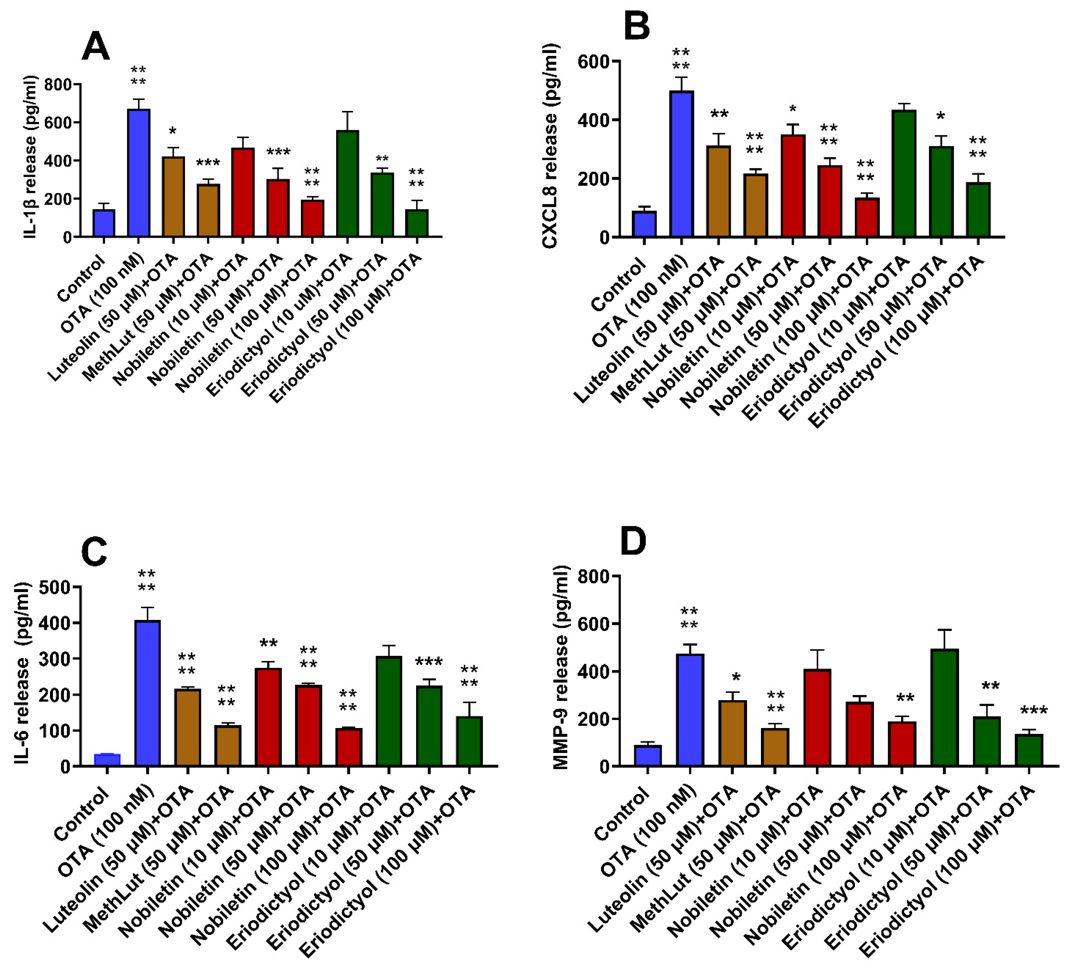
Pre-treatment of microglia with either nobiletin or eriodictyol at 50 and 100 µM for 2 h significantly inhibited the OTA-induced release of IL-1β, CXCL8, IL-6, and MMP-9 from microglia, with the 100 µM concentration showing the greatest inhibition (Figure 3A–D). Nobiletin, unlike eriodictyol, significantly inhibited the release of CXCL8 and IL-6, even at 10 µM, while neither inhibited the release of MMP-9 at this low concentration. Pretreatment with either luteolin or methoxy luteolin at 50 μM significantly inhibited the release of all the mediators (Figure 3A–D). Multivariant analysis among all flavonoids at 50 µM showed no significant difference.
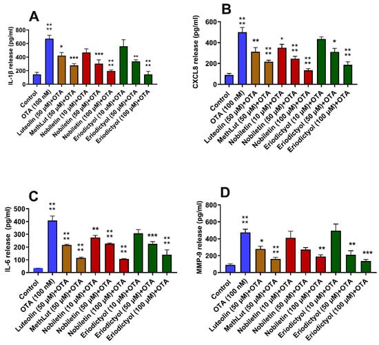
Figure 3.
Inhibition of the release of inflammatory mediators from human microglia stimulated by OTA. Human microglia (0.5 × 10⁵ cells/500 μL/well in 24-well plates) were preincubated with nobiletin (10, 50, and 100 μM), luteolin (50 μM), and methoxy luteolin (50 μM) for 2 h and then incubated with OTA (10 nM) for 24 h (n = 3). Control cells were treated with an equal volume of culture medium. Following this, cell culture supernatant fluids were collected and assayed for IL-1β (A), CXCL8 (B), IL-6 (C), and MMP-9 (D) by commercial ELISA kits. OTA was compared to the control; each of the other conditions was compared to OTA (n = 3, * p < 0.05, ** p < 0.01, *** p < 0.001, **** p < 0.0001).
3. Methods
3.1. Culture of Human Microglia
The immortalized human microglia SV-40 cell line (cat no. T10251), derived from primary human microglia, was obtained from Applied Biological Materials Inc. (ABM Inc., Richmond, BC, Canada) and maintained in Prigrow III medium (ABM Inc., Richmond, BC, Canada) supplemented with 10% fetal bovine serum (FBS) and 1% penicillin/streptomycin at 37 °C in a 5% CO2 incubator, as we have reported previously and recommended by the supplier [24]. Cells were cultured in type I collagen-coated T25 flasks (BD PureCoat™ ECM Mimetic Cultureware, Becton Dickinson, Bedford, MA, USA). The microglia-SV40 cell line retained its phenotype and proliferative capacity for over 10 passages. All the experiments were conducted using multiple cell thawings and subcultures that did not exceed 10 passages. Cell viability was assessed via the trypan blue (0.4%) exclusion method, and all experiments were performed in type I collagen-coated tissue culture plates (BD) with cultures over 95% viability.
3.2. Treatments of Microglia
Human microglia cell line (0.5 × 10⁵ cells in 1 mL complete medium/well in 24-well culture plate) were incubated for 24 h with the following stimuli: lipopolysaccharide (LPS; 10 ng/mL) (Sigma-Aldrich, St. Paul, MN, USA), full-length SARS-CoV-2 Spike protein (10 ng/mL), and OTA (10 ng/mL) (both from Abcam, Waltham, MA, USA). Luteolin, tetramethoxyluteolin (Methlut), hexamethoxyflavone (nobiletin), and tetrahydroxyflavanone (Eriodictyol), all >98% purity, were obtained from (CAS BioSciences, Costa Mesa, CA, USA). Levels of inflammatory mediators IL-1β, CXCL8, IL-6, and MMP-9 were quantified in the cell culture supernatant by enzyme-linked immunosorbent assay (ELISA) using commercial kits (BioTechne/R&D System, Minneapolis, MN, USA), following the manufacturer’s protocols, as we have reported previously [53,54]. Control cells were treated with an equivalent volume of culture medium in all experimental conditions. To investigate the inhibition of microglia activation, cells were pretreated with the various flavonoids at 10, 50, and 100 μM for 2 h.
3.3. Statistical Analysis
All experimental conditions were conducted in triplicate, and each experiment was repeated a minimum of three times (n = 3). Data are expressed as the mean ± standard deviation (SD). Multiple comparisons were performed using one-way analysis of variance (ANOVA) followed by Tukey’s multiple comparisons test. All statistical analyses were conducted using GraphPad Prism software version 10.0.3. A p-value of <0.05 was considered statistically significant in all comparisons.
4. Discussion
The present study demonstrates that both nobiletin (5,6,7,8,3′,4′-hexamethoxyflavone) and eriodictyol (3′,4′,5,7-tetraxydroxyflavanone) inhibited the LPS-, SARS-CoV-2-, or OTA-induced release of IL-1β, CXCL8, IL-6, and MMP-9 from human microglia stimulated by three different pathogenic agents: LPS, FL Spike, and OTA. Previous studies have reported that nobiletin inhibited the release of proinflammatory cytokines from LPS-stimulated mouse microglia [55,56]. Eriodictyol has also been shown to suppress LPS-stimulated BV-2 microglia [57]. There was no significant difference between nobiletin and eriodictyol at 100 μM. Multivariant analysis at 50 μM did not show any significance compared to luteolin (3′,4′,5,7-tetrahydroxyflavone) and methoxyluteolin (3′,4′,5,7-tetramethoxyflavone), which were used as “positive controls” because we previously reported them to inhibit IL-1β release from cultured human SV-40 microglia stimulated by neurotensin [30] and MMP-9 release stimulated by the FL Spike protein [31].
We previously reported that the SARS-CoV-2 Spike protein stimulated cultured human microglia SV-40 to secrete IL-1β, IL-18, MMP-9, and protein S100B, all of which are linked to brain damage [58]. Additional evidence indicates that the Spike protein can directly activate microglia [59,60,61], leading to proinflammatory effects. We also showed that SV-40 microglia release MMP-9 when stimulated by the Spike protein [58]. The Spike protein was also shown to induce neuroinflammation in mice via the activation of NLPR3 (nucleotide-binding oligomerization domain, Leucine-rich Repeat, and Pyrin domain-containing) and the BV-2 murine microglia cell line in vitro [62].
We recently reported that OTA stimulated the release of IL-1β, IL-18, and CXCL8 from the human SV-40 microglia cell line [24].
The present findings are supported by other previous studies showing that polyphenolic compounds can lower MMP-9 levels in both in vivo and in vitro conditions [63,64,65]. One recent study reported that methoxylated flavones inhibited the tumor necrosis factor (TNF)-mediated induction of MMP-9 [15].
The flavonoids examined in this study have been considered for therapeutic intervention for a number of neurodegenerative conditions [66]. For instance, nobiletin could serve as a potential inhibitor of beta amyloid (Aβ) toxicity [67]. In particular, nobiletin prevented Aβ 1-40 peptide-induced neuroinflammation and cognitive decline [68] and the Aβ 25-35 peptide-induced death of cultured primary neurons in vitro [69]. Nobiletin may also be beneficial in AD [70,71] by reducing neuroinflammation [56,72,73]. Nobiletin has also been shown to be useful in PD [72,73,74,75]. Moreover, nobiletin induced neurite outgrowth in PC12D cells, a rat pheochromocytoma cell line [76]. Eriodictyol was reported to reduce neuroinflammation induced by experimental stroke in rodents [77]. Eriodictyol also improved memory in Aβ 25-35-treated mice [78].
The mechanism of inhibition by the flavonoids is not entirely clear. Nobiletin was reported to inhibit mitogen-activated protein kinases (MAPK) and nuclear factor-kappa B (NF-kB) signaling pathways [55], as previously been reported for Methlut [79]. Both nobiletin [80,81] and eriodictyol [78,82] were reported to inhibit the NLP3 inflammasome, as did luteolin [83]. Such flavonoids could also inhibit activation of the cellular regulatory complex mammalian target of rapamycin (mTOR), as shown for for nobiletin [84] and Methlut [30,85,86].
Flavonoids are not soluble in aqueous media, making their administration and oral absorption in sufficient amounts problematic. An advantage of eriodictyol over other flavonoids is its planar configuration, which renders it partially water-soluble and, hence, easier to formulate in effective concentrations for delivery where other solvents may be irritating.
5. Conclusions
These findings indicate that both nobiletin and eriodictyol are potent inhibitors of the pathogen-stimulated microglial release of inflammatory mediators, highlighting their potential for therapeutic application in neuroinflammatory disorders.
Author Contributions
I.T. performed the studies, analyzed the results, and helped write the paper. T.C.T. designed the studies, analyzed the results, reviewed the literature, and wrote the manuscript. D.K. reviewed the literature, helped analyze the results, and edited the manuscript and figures. All authors have read and agreed to the published version of the manuscript.
Funding
This research was partly funded by CAS BioSciences (Costa Mesa, CA).
Institutional Review Board Statement
Not applicable.
Informed Consent Statement
Not Applicable.
Data Availability Statement
Data will be provided upon reasonable request.
Conflicts of Interest
On behalf of all authors, the corresponding author states that there are no conflicts of interest.
References
- Calderone, A.; Latella, D.; Cardile, D.; Gangemi, A.; Corallo, F.; Rifici, C.; Quartarone, A.; Calabro, R.S. The Role of Neuroinflammation in Shaping Neuroplasticity and Recovery Outcomes Following Traumatic Brain Injury: A Systematic Review. Int. J. Mol. Sci. 2024, 25, 11708. [Google Scholar] [CrossRef] [PubMed]
- Leng, F.; Edison, P. Neuroinflammation and microglial activation in Alzheimer disease: Where do we go from here? Nat. Rev. Neurol. 2021, 17, 157–172. [Google Scholar] [CrossRef] [PubMed]
- Villareal, J.A.B.; Bathe, T.; Hery, G.P.; Phillips, J.L.; Tsering, W.; Prokop, S. Deterioration of neuroimmune homeostasis in Alzheimer’s Disease patients who survive a COVID-19 infection. J. Neuroinflammation 2024, 21, 202. [Google Scholar] [CrossRef] [PubMed]
- Mayer, M.G.; Fischer, T. Microglia at the blood brain barrier in health and disease. Front. Cell Neurosci. 2024, 18, 1360195. [Google Scholar] [CrossRef] [PubMed]
- Bachiller, S.; Jimenez-Ferrer, I.; Paulus, A.; Yang, Y.; Swanberg, M.; Deierborg, T.; Boza-Serrano, A. Microglia in Neurological Diseases: A Road Map to Brain-Disease Dependent-Inflammatory Response. Front. Cell Neurosci. 2018, 12, 488. [Google Scholar] [CrossRef] [PubMed]
- Perry, V.H.; Nicoll, J.A.; Holmes, C. Microglia in neurodegenerative disease. Nat. Rev. Neurol. 2010, 6, 193–201. [Google Scholar] [CrossRef] [PubMed]
- Hickman, S.; Izzy, S.; Sen, P.; Morsett, L.; El, K.J. Microglia in neurodegeneration. Nat. Neurosci. 2018, 21, 1359–1369. [Google Scholar] [CrossRef] [PubMed]
- Vafadari, B.; Salamian, A.; Kaczmarek, L. MMP-9 in translation: From molecule to brain physiology, pathology, and therapy. J. Neurochem. 2016, 139 (Suppl. S2), 91–114. [Google Scholar] [CrossRef] [PubMed]
- Rempe, R.G.; Hartz, A.M.; Bauer, B. Matrix metalloproteinases in the brain and blood-brain barrier: Versatile breakers and makers. J. Cereb. Blood Flow. Metab. 2016, 36, 1481–14507. [Google Scholar] [CrossRef] [PubMed]
- Beroun, A.; Mitra, S.; Michaluk, P.; Pijet, B.; Stefaniuk, M.; Kaczmarek, L. MMPs in learning and memory and neuropsychiatric disorders. Cell Mol. Life Sci. 2019, 76, 3207–3228. [Google Scholar] [CrossRef] [PubMed]
- Hannocks, M.J.; Zhang, X.; Gerwien, H.; Chashchina, A.; Burmeister, M.; Korpos, E.; Song, J.; Sorokin, L. The gelatinases, MMP-2 and MMP-9, as fine tuners of neuroinflammatory processes. Matrix Biol. 2017, 75–76, 102–113. [Google Scholar] [CrossRef] [PubMed]
- Kempuraj, D.; Aenlle, K.K.; Cohen, J.; Mathew, A.; Isler, D.; Pangeni, R.P.; Nathanson, L.; Theoharides, T.C.; Klimas, N.G. COVID-19 and Long COVID: Disruption of the Neurovascular Unit, Blood-Brain Barrier, and Tight Junctions. Neuroscientist 2024, 30, 421–439. [Google Scholar] [CrossRef] [PubMed]
- Kempuraj, D.D.K.; Cohen, J.; Valladares, D.S.; Joshi, R.S.; Kothuru, S.P.; Anderson, T.; Chinnappan, B.; Cheema, A.K.; Klimas, N.G.; Theoharides, T.C. Neurovascular Unit, neuroinflammation and neurodegeneration markers in brain disorders. Front. Cell Neurosci. 2024, 18, 1491952. [Google Scholar] [CrossRef] [PubMed]
- Cheung, S.W.; Bhavnani, E.; Simmons, D.G.; Bellingham, M.C.; Noakes, P.G. Perineuronal nets are phagocytosed by MMP-9 expressing microglia and astrocytes in the SOD1(G93A) ALS mouse model. Neuropathol. Appl. Neurobiol. 2024, 50, e12982. [Google Scholar] [CrossRef] [PubMed]
- Kamiya, T.; Mizuno, N.; Hayashi, K.; Otsuka, T.; Haba, M.; Abe, N.; Oyama, M.; Hara, H. Methoxylated Flavones from Casimiroa edulis La Llave Suppress MMP9 Expression via Inhibition of the JAK/STAT3 Pathway and TNFalpha-Dependent Pathways. J. Agric. Food Chem. 2024, 72, 14678–14683. [Google Scholar] [CrossRef] [PubMed]
- Bonetto, V.; Pasetto, L.; Lisi, I.; Carbonara, M.; Zangari, R.; Ferrari, E.; Punzi, V.; Luotti, S.; Bottino, N.; Biagianti, B.; et al. Markers of blood-brain barrier disruption increase early and persistently in COVID-19 patients with neurological manifestations. Front. Immunol. 2022, 13, 1070379. [Google Scholar] [CrossRef] [PubMed]
- Ueland, T.; Holter, J.C.; Holten, A.R.; Muller, K.E.; Lind, A.; Bekken, G.K.; Dudman, S.; Aukrust, P.; Dyrhol-Riise, A.M.; Heggelund, L. Distinct and early increase in circulating MMP-9 in COVID-19 patients with respiratory failure. J. Infect. 2020, 81, e41–e43. [Google Scholar] [CrossRef] [PubMed]
- Stawarski, M.; Stefaniuk, M.; Wlodarczyk, J. Matrix metalloproteinase-9 involvement in the structural plasticity of dendritic spines. Front. Neuroanat. 2014, 8, 68. [Google Scholar] [CrossRef] [PubMed]
- Ding, L.; Guo, H.; Zhang, C.; Jin, H.; Guo, X.; Li, T. Elevated matrix metalloproteinase-9 expression is associated with COVID-19 severity: A meta-analysis. Exp. Ther. Med. 2023, 26, 545. [Google Scholar] [CrossRef] [PubMed]
- Savic, G.; Stevanovic, I.; Mihajlovic, D.; Jurisevic, M.; Gajovic, N.; Jovanovic, I.; Ninkovic, M. MMP-9/BDNF ratio predicts more severe COVID-19 outcomes. Int. J. Med. Sci. 2022, 19, 1903–1911. [Google Scholar] [CrossRef] [PubMed]
- Ratnaseelan, A.M.; Tsilioni, I.; Theoharides, T.C. Effects of Mycotoxins on Neuropsychiatric Symptoms and Immune Processes. Clin. Ther. 2018, 40, 903–917. [Google Scholar] [CrossRef] [PubMed]
- Obafemi, B.A.; Adedara, I.A.; Rocha, J.B.T. Neurotoxicity of ochratoxin A.; Molecular mechanisms and neurotherapeutic strategies. Toxicology 2023, 497–498, 153630. [Google Scholar] [CrossRef] [PubMed]
- von Tobel, J.S.; Antinori, P.; Zurich, M.G.; Rosset, R.; Aschner, M.; Gluck, F.; Scherl, A.; Monnet-Tschudi, F. Repeated exposure to Ochratoxin A generates a neuroinflammatory response, characterized by neurodegenerative M1 microglial phenotype. Neurotoxicology 2014, 44, 61–70. [Google Scholar] [CrossRef] [PubMed]
- Tsilioni, I.; Theoharides, T.C. Ochratoxin A stimulates release of IL-1beta, IL-18 and CXCL8 from cultured human microglia. Toxicology 2024, 502, 153738. [Google Scholar] [CrossRef] [PubMed]
- Ho, S.C.; Kuo, C.T. Hesperidin, nobiletin, and tangeretin are collectively responsible for the anti-neuroinflammatory capacity of tangerine peel (Citri reticulatae pericarpium). Food Chem. Toxicol. 2014, 71, 176–182. [Google Scholar] [CrossRef] [PubMed]
- Matsuzaki, K.; Nakajima, A.; Guo, Y.; Ohizumi, Y. A Narrative Review of the Effects of Citrus Peels and Extracts on Human Brain Health and Metabolism. Nutrients 2022, 14, 1847. [Google Scholar] [CrossRef] [PubMed]
- Trainor, A.R.; MacDonald, D.S.; Penney, J. Microglia: Roles and genetic risk in Parkinson’s disease. Front. Neurosci. 2024, 18, 1506358. [Google Scholar] [CrossRef] [PubMed]
- Samant, R.R.; Standaert, D.G.; Harms, A.S. The emerging role of disease-associated microglia in Parkinson’s disease. Front. Cell Neurosci. 2024, 18, 1476461. [Google Scholar] [CrossRef] [PubMed]
- Tan, Z.; Xia, R.; Zhao, X.; Yang, Z.; Liu, H.; Wang, W. Potential key pathophysiological participant and treatment target in autism spectrum disorder: Microglia. Mol. Cell Neurosci. 2024, 131, 103980. [Google Scholar] [CrossRef] [PubMed]
- Patel, A.B.; Tsilioni, I.; Leeman, S.E.; Theoharides, T.C. Neurotensin stimulates sortilin and mTOR in human microglia inhibitable by methoxyluteolin, a potential therapeutic target for autism. Proc. Natl. Acad. Sci. USA 2016, 113, E7049–E7058. [Google Scholar] [CrossRef] [PubMed]
- Kempuraj, D.; Tsilioni, I.; Aenlle, K.K.; Klimas, N.G.; Theoharides, T.C. Long COVID elevated MMP-9 and release from microglia by SARS-CoV-2 Spike protein. Transl. Neurosci. 2024, 15, 20220352. [Google Scholar] [CrossRef] [PubMed]
- Dirscherl, K.; Karlstetter, M.; Ebert, S.; Kraus, D.; Hlawatsch, J.; Walczak, Y.; Moehle, C.; Fuchshofer, R.; Langmann, T. Luteolin triggers global changes in the microglial transcriptome leading to a unique anti-inflammatory and neuroprotective phenotype. J. Neuroinflammation 2010, 7, 3. [Google Scholar] [CrossRef] [PubMed]
- Zhu, M.; Sun, Y.; Su, Y.; Guan, W.; Wang, Y.; Han, J.; Wang, S.; Yang, B.; Wang, Q.; Kuang, H. Luteolin: A promising multifunctional natural flavonoid for human diseases. Phytother. Res. 2024, 38, 3417–3443. [Google Scholar] [CrossRef] [PubMed]
- Muruganathan, N.; Dhanapal, A.R.; Baskar, V.; Muthuramalingam, P.; Selvaraj, D.; Aara, H.; Shiek Abdullah, M.Z.; Sivanesan, I. Recent Updates on Source, Biosynthesis, and Therapeutic Potential of Natural Flavonoid Luteolin: A Review. Metabolites 2022, 12, 1145. [Google Scholar] [CrossRef] [PubMed]
- Yousaf, S. Travel burnout: Exploring the return journeys of pilgrim-tourists amidst the COVID-19 pandemic. Tour. Manag. 2021, 84, 104285. [Google Scholar] [CrossRef] [PubMed]
- Huang, L.; Kim, M.Y.; Cho, J.Y. Immunopharmacological Activities of Luteolin in Chronic Diseases. Int. J. Mol. Sci. 2023, 24, 2136. [Google Scholar] [CrossRef] [PubMed]
- Singh, N.K.; Bhushan, B.; Singh, P.; Sahu, K.K. Therapeutic Expedition of Luteolin against Brain-related Disorders: An Updated Review. Comb. Chem. High. Throughput Screen. 2024, 28, 371–391. [Google Scholar] [CrossRef] [PubMed]
- Delgado, A.; Cholevas, C.; Theoharides, T.C. Neuroinflammation in Alzheimer’s disease and beneficial action of luteolin. Biofactors 2021, 47, 207–217. [Google Scholar] [CrossRef] [PubMed]
- Theoharides, T.C.; Cholevas, C.; Polyzoidis, K.; Politis, A. Long-COVID syndrome-associated brain fog and chemofog: Luteolin to the rescue. Biofactors 2021, 47, 232–241. [Google Scholar] [CrossRef] [PubMed]
- Goyal, A.; Solanki, K.; Verma, A. Luteolin: Nature’s promising warrior against Alzheimer’s and Parkinson’s disease. J. Biochem. Mol. Toxicol. 2024, 38, e23619. [Google Scholar] [CrossRef] [PubMed]
- Jayawickreme, D.K.; Ekwosi, C.; Anand, A.; Andres-Mach, M.; Wlaz, P.; Socala, K. Luteolin for neurodegenerative diseases: A review. Pharmacol. Rep. 2024, 76, 644–664. [Google Scholar] [CrossRef] [PubMed]
- Yang, J.Y.; Ma, Y.X.; Liu, Y.; Peng, X.J.; Chen, X.Z. A Comprehensive Review of Natural Flavonoids with Anti-SARS-CoV-2 Activity. Molecules 2023, 28, 2735. [Google Scholar] [CrossRef] [PubMed]
- Dissook, S.; Umsumarng, S.; Mapoung, S.; Semmarath, W.; Arjsri, P.; Srisawad, K.; Dejkriengkraikul, P. Luteolin-rich fraction from Perilla frutescens seed meal inhibits spike glycoprotein S1 of SARS-CoV-2-induced NLRP3 inflammasome lung cell inflammation via regulation of JAK1/STAT3 pathway: A potential anti-inflammatory compound against inflammation-induced long-COVID. Front. Med. 2022, 9, 1072056. [Google Scholar] [PubMed]
- Elkaeed, E.B.; Alsfouk, B.A.; Ibrahim, T.H.; Arafa, R.K.; Elkady, H.; Ibrahim, I.M.; Eissa, I.H.; Metwaly, A.M. Computer-assisted drug discovery of potential natural inhibitors of the SARS-CoV-2 RNA-dependent RNA polymerase through a multi-phase in silico approach. Antivir. Ther. 2023, 28, 13596535231199838. [Google Scholar] [CrossRef] [PubMed]
- Mokhtari, T.; Azizi, M.; Sheikhbahaei, F.; Sharifi, H.; Sadr, M. Plant-Derived Antioxidants for Management of COVID-19: A Comprehensive Review of Molecular Mechanisms. Tanaffos 2023, 22, 27–39. [Google Scholar] [PubMed]
- Almatroudi, A. Analysis of bioactive compounds of Olea europaea as potential inhibitors of SARS-CoV-2 main protease: A pharmacokinetics, molecular docking and molecular dynamics simulation studies. J. Biomol. Struct. Dyn. 2023, 1–12. [Google Scholar] [CrossRef] [PubMed]
- Majrashi, T.A.; El Hassab, M.A.; Mahmoud, S.H.; Mostafa, A.; Wahsh, E.A.; Elkaeed, E.B.; Hassan, F.E.; Eldehna, W.M.; Abdelgawad, S.M. In vitro biological evaluation and in silico insights into the antiviral activity of standardized olive leaves extract against SARS-CoV-2. PLoS ONE 2024, 19, e0301086. [Google Scholar] [CrossRef] [PubMed]
- Savino, R.; Medoro, A.; Ali, S.; Scapagnini, G.; Maes, M.; Davinelli, S. The Emerging Role of Flavonoids in Autism Spectrum Disorder: A Systematic Review. J. Clin. Med. 2023, 12, 3520. [Google Scholar] [CrossRef] [PubMed]
- Taliou, A.; Zintzaras, E.; Lykouras, L.; Francis, K. An open-label pilot study of a formulation containing the anti-inflammatory flavonoid luteolin and its effects on behavior in children with autism spectrum disorders. Clin. Ther. 2013, 35, 592–602. [Google Scholar] [CrossRef] [PubMed]
- Pang, Y.; Xiong, J.; Wu, Y.; Ding, W. A review on recent advances on nobiletin in central and peripheral nervous system diseases. Eur. J. Med. Res. 2023, 28, 485. [Google Scholar] [CrossRef] [PubMed]
- Huang, H.; Li, L.; Shi, W.; Liu, H.; Yang, J.; Yuan, X.; Wu, L. The Multifunctional Effects of Nobiletin and Its Metabolites In Vivo and In Vitro. Evid. Based Complement. Alternat Med. 2016, 2016, 2918796. [Google Scholar] [CrossRef] [PubMed]
- Islam, A.; Islam, M.S.; Rahman, M.K.; Uddin, M.N.; Akanda, M.R. The pharmacological and biological roles of eriodictyol. Arch. Pharm. Res. 2020, 43, 582–592. [Google Scholar] [CrossRef] [PubMed]
- Tsilioni, I.; Pantazopoulos, H.; Conti, P.; Leeman, S.E.; Theoharides, T.C. IL-38 inhibits microglial inflammatory mediators and is decreased in amygdala of children with autism spectrum disorder. Proc. Natl. Acad. Sci. USA 2020, 117, 16475–16480. [Google Scholar] [CrossRef] [PubMed]
- Tsilioni, I.; Theoharides, T.C. Recombinant SARS-CoV-2 Spike Protein Stimulates Secretion of Chymase, Tryptase, and IL-1beta from Human Mast Cells, Augmented by IL-33. Int. J. Mol. Sci. 2023, 24, 9487. [Google Scholar] [CrossRef] [PubMed]
- Qi, G.; Mi, Y.; Fan, R.; Li, R.; Liu, Z.; Liu, X. Nobiletin Protects against Systemic Inflammation-Stimulated Memory Impairment via MAPK and NF-kappaB Signaling Pathways. J. Agric. Food Chem. 2019, 67, 5122–5134. [Google Scholar] [CrossRef] [PubMed]
- Chai, W.; Zhang, J.; Xiang, Z.; Zhang, H.; Mei, Z.; Nie, H.; Xu, R.; Zhang, P. Potential of nobiletin against Alzheimer’s disease through inhibiting neuroinflammation. Metab. Brain Dis. 2022, 37, 1145–1154. [Google Scholar] [CrossRef] [PubMed]
- He, P.; Yan, S.; Zheng, J.; Gao, Y.; Zhang, S.; Liu, Z.; Liu, X.; Xiao, C. Eriodictyol Attenuates LPS-Induced Neuroinflammation, Amyloidogenesis, and Cognitive Impairments via the Inhibition of NF-kappaB in Male C57BL/6J Mice and BV2 Microglial Cells. J. Agric. Food Chem. 2018, 66, 10205–10214. [Google Scholar] [CrossRef] [PubMed]
- Tsilioni, I.; Theoharides, T.C. Recombinant SARS-CoV-2 Spike Protein and Its Receptor Binding Domain Stimulate Release of Different Pro-Inflammatory Mediators via Activation of Distinct Receptors on Human Microglia Cells. Mol. Neurobiol. 2023, 11, 6704–6714. [Google Scholar] [CrossRef] [PubMed]
- Jeong, G.U.; Lyu, J.; Kim, K.D.; Chung, Y.C.; Yoon, G.Y.; Lee, S.; Hwang, I.; Shin, W.H.; Ko, J.; Lee, J.Y.; et al. SARS-CoV-2 Infection of Microglia Elicits Proinflammatory Activation and Apoptotic Cell Death. Microbiol. Spectr. 2022, 10, e0109122. [Google Scholar] [CrossRef] [PubMed]
- Olajide, O.A.; Iwuanyanwu, V.U.; Adegbola, O.D.; Al-Hindawi, A.A. SARS-CoV-2 Spike Glycoprotein S1 Induces Neuroinflammation in BV-2 Microglia. Mol. Neurobiol. 2022, 59, 445–458. [Google Scholar] [CrossRef] [PubMed]
- Samudyata, N.; Oliveira, A.O.; Malwade, S.; Rufino de Sousa, N.; Goparaju, S.K.; Gracias, J.; Orhan, F.; Steponaviciute, L.; Schalling, M.; Sheridan, S.D.; et al. SARS-CoV-2 promotes microglial synapse elimination in human brain organoids. Mol. Psychiatry 2022, 27, 3939–3950. [Google Scholar] [CrossRef] [PubMed]
- Jiang, Q.; Li, G.; Wang, H.; Chen, W.; Liang, F.; Kong, H.; Chen, T.S.R.; Lin, L.; Hong, H.; Pei, Z. SARS-CoV-2 spike S1 protein induces microglial NLRP3-dependent neuroinflammation and cognitive impairment in mice. Exp. Neurol. 2025, 383, 115020. [Google Scholar] [CrossRef] [PubMed]
- Pagliara, V.; De Rosa, M.; Di Donato, P.; Nasso, R.; D’Errico, A.; Cammarota, F.; Poli, A.; Masullo, M.; Arcone, R. Inhibition of Interleukin-6-Induced Matrix Metalloproteinase-2 Expression and Invasive Ability of Lemon Peel Polyphenol Extract in Human Primary Colon Cancer Cells. Molecules 2021, 26, 7076. [Google Scholar] [CrossRef] [PubMed]
- Milton-Laskibar, I.; Trepiana, J.; Macarulla, M.T.; Gomez-Zorita, S.; Arellano-Garcia, L.; Fernandez-Quintela, A.; Portillo, M.P. Potential usefulness of Mediterranean diet polyphenols against COVID-19-induced inflammation: A review of the current knowledge. J. Physiol. Biochem. 2023, 79, 371–382. [Google Scholar] [CrossRef] [PubMed]
- Islam, M.T.; Jang, N.H.; Lee, H.J. Natural Products as Regulators against Matrix Metalloproteinases for the Treatment of Cancer. Biomedicines 2024, 12, 794. [Google Scholar] [CrossRef] [PubMed]
- Theoharides, T.C.; Tsilioni, I. Tetramethoxyluteolin for the treatment of neurodegenerative diseases. Curr. Top. Med. Chem. 2018, 18, 1872–1882. [Google Scholar] [CrossRef] [PubMed]
- Youn, K.; Lee, S.; Jun, M. Discovery of Nobiletin from Citrus Peel as a Potent Inhibitor of beta-Amyloid Peptide Toxicity. Nutrients 2019, 11, 2648. [Google Scholar] [CrossRef] [PubMed]
- Ghasemi-Tarie, R.; Kiasalari, Z.; Fakour, M.; Khorasani, M.; Keshtkar, S.; Baluchnejadmojarad, T.; Roghani, M. Nobiletin prevents amyloid beta(1-40)-induced cognitive impairment via inhibition of neuroinflammation and oxidative/nitrosative stress. Metab. Brain Dis. 2022, 37, 1337–1349. [Google Scholar] [CrossRef] [PubMed]
- Jing, X.; Shi, H.; Zhu, X.; Wei, X.; Ren, M.; Han, M.; Ren, D.; Lou, H. Eriodictyol Attenuates beta-Amyloid 25-35 Peptide-Induced Oxidative Cell Death in Primary Cultured Neurons by Activation of Nrf2. Neurochem. Res. 2015, 40, 1463–1471. [Google Scholar] [CrossRef] [PubMed]
- Kim, E.; Nohara, K.; Wirianto, M.; Escobedo, G., Jr.; Lim, J.Y.; Morales, R.; Yoo, S.H.; Chen, Z. Effects of the Clock Modulator Nobiletin on Circadian Rhythms and Pathophysiology in Female Mice of an Alzheimer’s Disease Model. Biomolecules 2021, 11, 1004. [Google Scholar] [CrossRef] [PubMed]
- Wirianto, M.; Wang, C.Y.; Kim, E.; Koike, N.; Gomez-Gutierrez, R.; Nohara, K.; Escobedo, G., Jr.; Choi, J.M.; Han, C.; Yagita, K.; et al. The clock modulator Nobiletin mitigates astrogliosis-associated neuroinflammation and disease hallmarks in an Alzheimer’s disease model. FASEB J. 2022, 36, e22186. [Google Scholar] [CrossRef] [PubMed]
- Braidy, N.; Behzad, S.; Habtemariam, S.; Ahmed, T.; Daglia, M.; Nabavi, S.M.; Sobarzo-Sanchez, E.; Nabavi, S.F. Neuroprotective Effects of Citrus Fruit-Derived Flavonoids, Nobiletin and Tangeretin in Alzheimer’s and Parkinson’s Disease. CNS Neurol. Disord. Drug Targets 2017, 16, 387–397. [Google Scholar] [CrossRef] [PubMed]
- Nakajima, A.; Ohizumi, Y. Potential Benefits of Nobiletin, A Citrus Flavonoid, against Alzheimer’s Disease and Parkinson’s Disease. Int. J. Mol. Sci. 2019, 20, 3380. [Google Scholar] [CrossRef] [PubMed]
- Yabuki, Y.; Ohizumi, Y.; Yokosuka, A.; Mimaki, Y.; Fukunaga, K. Nobiletin treatment improves motor and cognitive deficits seen in MPTP-induced Parkinson model mice. Neuroscience 2014, 259, 126–141. [Google Scholar] [CrossRef] [PubMed]
- Jeong, K.H.; Jeon, M.T.; Kim, H.D.; Jung, U.J.; Jang, M.C.; Chu, J.W.; Yang, S.J.; Choi, I.Y.; Choi, M.S.; Kim, S.R. Nobiletin protects dopaminergic neurons in the 1-methyl-4-phenylpyridinium-treated rat model of Parkinson’s disease. J. Med. Food 2015, 18, 409–414. [Google Scholar] [CrossRef] [PubMed]
- Nagase, H.; Yamakuni, T.; Matsuzaki, K.; Maruyama, Y.; Kasahara, J.; Hinohara, Y.; Kondo, S.; Mimaki, Y.; Sashida, Y.; Tank, A.W.; et al. Mechanism of neurotrophic action of nobiletin in PC12D cells. Biochemistry 2005, 44, 13683–13691. [Google Scholar] [CrossRef] [PubMed]
- Ferreira Ede, O.; Fernandes, M.Y.; Lima, N.M.; Neves, K.R.; Carmo, M.R.; Lima, F.A.; Fonteles, A.A.; Menezes, A.P.; Andrade, G.M. Neuroinflammatory response to experimental stroke is inhibited by eriodictyol. Behav. Brain Res. 2016, 312, 321–332. [Google Scholar] [CrossRef] [PubMed]
- Guo, P.; Zeng, M.; Wang, S.; Cao, B.; Liu, M.; Zhang, Y.; Jia, J.; Zhang, Q.; Zhang, B.; Wang, R.; et al. Eriodictyol and Homoeriodictyol Improve Memory Impairment in Abeta(25–35)-Induced Mice by Inhibiting the NLRP3 Inflammasome. Molecules 2022, 27, 2488. [Google Scholar] [CrossRef] [PubMed]
- Weng, Z.; Patel, A.B.; Panagiotidou, S.; Theoharides, T.C. The novel flavone tetramethoxyluteolin is a potent inhibitor of human mast cells. J. Allergy Clin. Immunol. 2015, 135, 1044–1052.e5. [Google Scholar] [CrossRef] [PubMed]
- Peng, Z.; Li, X.; Xing, D.; Du, X.; Wang, Z.; Liu, G.; Li, X. Nobiletin alleviates palmitic acid-induced NLRP3 inflammasome activation in a sirtuin 1-dependent manner in AML-12 cells. Mol. Med. Rep. 2018, 18, 5815–5822. [Google Scholar] [CrossRef] [PubMed]
- Wang, H.; Guo, Y.; Qiao, Y.; Zhang, J.; Jiang, P. Nobiletin Ameliorates NLRP3 Inflammasome-Mediated Inflammation Through Promoting Autophagy via the AMPK Pathway. Mol. Neurobiol. 2020, 57, 5056–5068. [Google Scholar] [CrossRef] [PubMed]
- Huang, W.; Wang, C.; Zhang, H. Eriodictyol inhibits the motility, angiogenesis and tumor growth of hepatocellular carcinoma via NLRP3 inflammasome inactivation. Heliyon 2024, 10, e24401. [Google Scholar] [CrossRef] [PubMed]
- Zhang, Z.H.; Liu, J.Q.; Hu, C.D.; Zhao, X.T.; Qin, F.Y.; Zhuang, Z.; Zhang, X.S. Luteolin Confers Cerebroprotection after Subarachnoid Hemorrhage by Suppression of NLPR3 Inflammasome Activation through Nrf2-Dependent Pathway. Oxid. Med. Cell Longev. 2021, 2021, 5838101. [Google Scholar] [CrossRef] [PubMed]
- El Tabaa, M.M.; El Tabaa, M.M.; Elgharabawy, R.M.; Abdelhamid, W.G. Suppressing NLRP3 activation and PI3K/AKT/mTOR signaling ameliorates amiodarone-induced pulmonary fibrosis in rats: A possible protective role of nobiletin. Inflammopharmacology 2023, 31, 1373–1386. [Google Scholar] [CrossRef] [PubMed]
- Patel, A.B.; Theoharides, T.C. Methoxyluteolin Inhibits Neuropeptide-stimulated Proinflammatory Mediator Release via mTOR Activation from Human Mast Cells. J. Pharmacol. Exp. Ther. 2017, 361, 462–471. [Google Scholar] [CrossRef] [PubMed]
- Patel, A.B.; Tsilioni, I.; Weng, Z.; Theoharides, T.C. TNF stimulates IL-6, CXCL8 and VEGF secretion from human keratinocytes via activation of mT.O.R.; inhibited by tetramethoxyluteolin. Exp. Dermatol. 2018, 27, 135–143. [Google Scholar] [CrossRef] [PubMed]
Disclaimer/Publisher’s Note: The statements, opinions and data contained in all publications are solely those of the individual author(s) and contributor(s) and not of MDPI and/or the editor(s). MDPI and/or the editor(s) disclaim responsibility for any injury to people or property resulting from any ideas, methods, instructions or products referred to in the content. |
© 2025 by the authors. Licensee MDPI, Basel, Switzerland. This article is an open access article distributed under the terms and conditions of the Creative Commons Attribution (CC BY) license (https://creativecommons.org/licenses/by/4.0/).

