Article

Biofilm Formation, Modulation, and Transcriptomic Regulation Under Stress Conditions in Halomicronema sp.

1
Department of Chemistry, Life Sciences and Environmental Sustainability, University of Parma, Parco Area delle Scienze 11/A, 43124 Parma, Italy
2
Interdepartmental Center SITEIA.PARMA, University of Parma, Parco Area delle Scienze 33, 43124 Parma, Italy
3
Department of Marine Microbiology and Biogeochemistry (MMB), NIOZ Royal Netherlands Institute for Sea Research, Landsdiep 4, 1797 SZ Den Hoorn, Texel, The Netherlands
*
Author to whom correspondence should be addressed.
Int. J. Mol. Sci. 2025, 26(2), 673; https://doi.org/10.3390/ijms26020673
Submission received: 19 December 2024 / Revised: 9 January 2025 / Accepted: 10 January 2025 / Published: 15 January 2025
(This article belongs to the Section Molecular Microbiology)

Abstract

:
In nature, bacteria often form heterogeneous communities enclosed in a complex matrix known as biofilms. This extracellular matrix, produced by the microorganisms themselves, serves as the first barrier between the cells and the environment. It is composed mainly of water, extracellular polymeric substances (EPS), lipids, proteins, and DNA. Cyanobacteria form biofilms and have unique characteristics such as oxygenic photosynthesis, nitrogen fixation, excellent adaptability to various abiotic stress conditions, and the ability to secrete a variety of metabolites and hormones. This work focused on the characterization of the cyanobacterium Halomicronema sp. strain isolated from a brackish environment. This study included microscopic imaging, determination of phenolic content and antioxidant capacity, identification of chemicals interfering with biofilm formation, and transcriptomic analysis by RNA sequencing and real-time PCR. Gene expression analysis was centered on genes related to the production of EPS and biofilm-related transcription factors. This study led to the identification of wza1 and wzt as EPS biomarkers and luxR-05665, along with genes belonging to the TetR/AcrR and LysR families, as potential biomarkers useful for studying and monitoring biofilm formation under different environmental conditions. Moreover, this work revealed that Halomicronema sp. can grow even in the presence of strong abiotic stresses, such as high salt, and has good antioxidant properties.

1. Introduction

Cyanobacteria are Gram-negative microbes widespread in numerous habitats, such as terrestrial, freshwater, and hypersaline environments [1]. All species belonging to this phylum are mostly obligate oxygenic photoautotrophs, capable of using carbon dioxide (CO2) as their main carbon source and light as their main energy source [2]. To support these functions, they contain phycobiliproteins, which act as accessory pigments in photosynthesis [3].
Due to their inherent ability to fix nitrogen and CO2 and their excellent adaptability to various abiotic stress conditions [4], cyanobacteria can be considered excellent biofertilizers since they improve soil fertility and texture, resulting in increased crop productivity. Their ability to release antimicrobials makes them ideal candidates for use as biocontrol agents and as an alternative to chemical pesticides [5]. Moreover, some cyanobacteria can detoxify or degrade various pollutants, both natural and xenobiotic, and can uptake heavy metals through the use of metallothioneins [6,7]. In addition, cyanobacteria play an important role in rhizospheric engineering [8]. These organisms are capable of secreting biologically active compounds such as phytohormones, amino acids, sugars, phenols, and vitamins, which act as signaling molecules for the synthesis of a series of plant secondary compounds [9]. Biocrusts are cyanobacteria-dominated biofilms that develop on the soil surface and comprise a complex community of other microorganisms. These biocrusts improve water-holding capacity, increase infiltration, and decrease erosion while maintaining soil moisture for a longer period [4,10,11]. The cyanobacteria are largely responsible for the formation of extracellular polymeric substances (EPS), and their composition varies according to the species, nutrient availability, growth stage, and environmental conditions [12,13,14,15]. Such substances consist mainly of polysaccharides and may remain associated with the cell surface or be released into the surrounding environment [15]. Besides playing a fundamental role in cell aggregation and adhesion, they can perform different functions, including protection from desiccation, viral attack, UV radiation, or sequestration of nutrients and metal ions [16,17].
Knowledge of EPS production in cyanobacteria is limited. The synthesis typically starts in the cytoplasm, where monosaccharides are converted into sugar nucleotides. These are then transferred by specific glycosyltransferases to transporter molecules in the plasma membrane. The subsequent steps of polymerization, assembly, and export are usually fairly conserved in bacteria [18]. Three main mechanisms have been described: the Wzy-dependent pathway, the ABC-dependent transporter pathway, and the synthase-dependent pathway [19]. Analysis at the phylum level shows that most cyanobacteria carry genes encoding proteins involved in one of the three pathways, but often not the full set that characterizes it. Moreover, EPS may be located loosely outside the cells (soluble fraction) or, in flocs or biofilms, closely associated with the cells [20]. Other important elements of EPS are lipopolysaccharides (LPS), which also play a key role in the stabilization of biocrusts [4,10]. Lipopolysaccharides are large and complex amphipathic molecules consisting of lipids and saccharide residues that constitute the outer leaflet of the cell membrane of Gram-negative bacteria [21].
Halomicronema sp. is a cyanobacterium found in brackish water and microbial mats, where it forms biofilms. In this study, the isolate Halomicronema sp. CCY15110 was considered; this strain was isolated from coastal microbial mats on the North Sea beach of Schiermonnikoog Island in the Netherlands [22] and its genome has been sequenced [23]. This study aimed to better characterize the biofilm formation of this bacteria, to identify chemicals that can inhibit this process, and to study gene expression regulating biofilm formation, including EPS genes.

2. Results and Discussion

2.1. Growth of Halomicronema sp. and Biofilm Formation

For imaging biofilm formation, cultures of Halomicronema sp. were grown on slides. ESEM images in low vacuum mode (Figure 1A,B) showed that Halomicronema sp. forms long tangled filaments, a characteristic that was also observed with classical light microscopy (Figure 1C). Using fluorescence staining with SYTO-9, which stains DNA, it was possible to visualize individual cells within these filaments (Figure 1D).
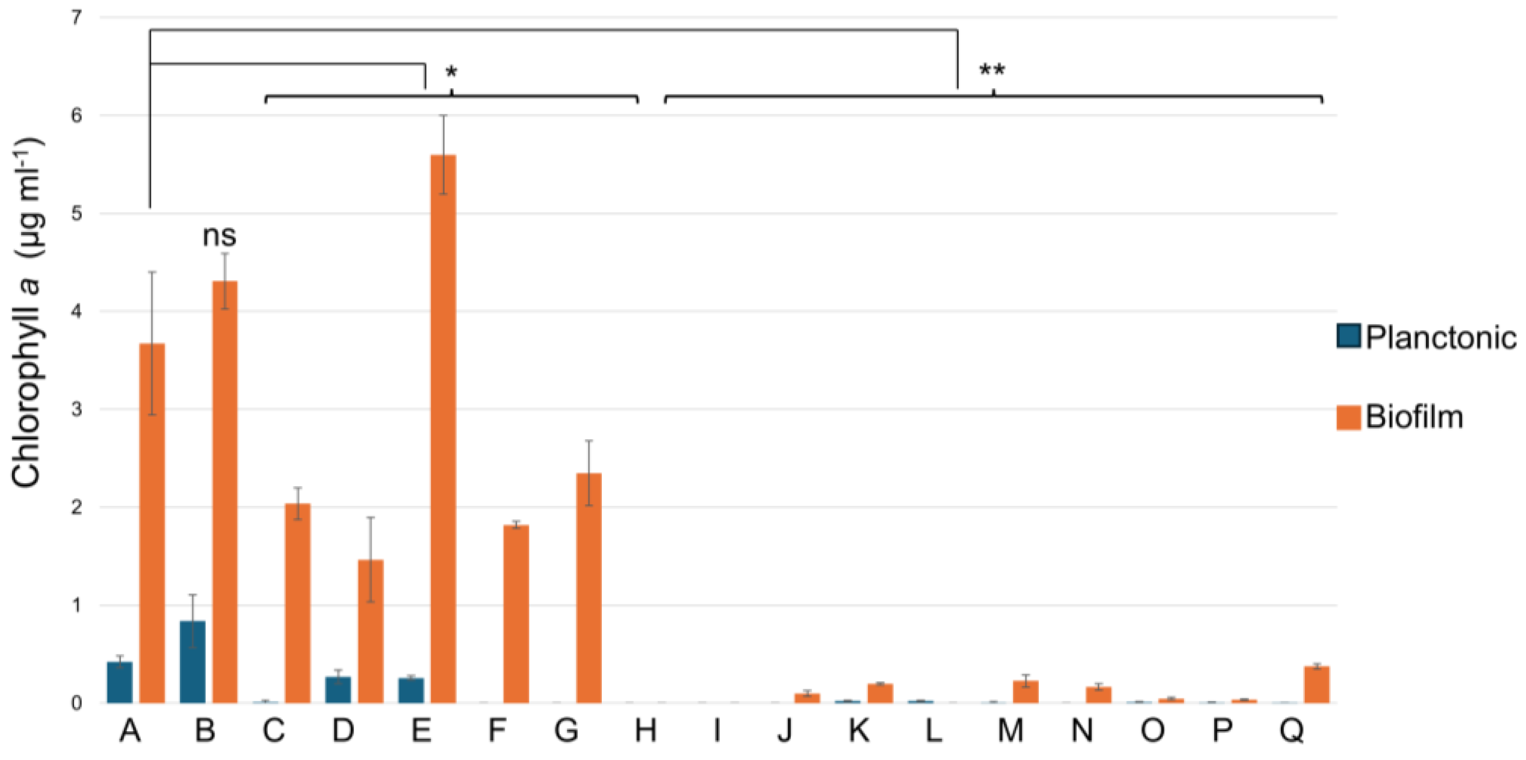
Next, cyanobacteria were grown in a liquid BA+ medium in 24-well plates. Here, cells of Halomicronema sp. grew predominantly in a biofilm attached to the bottom of the wells with few planktonic cells in the supernatant (Figure 2 condition A). This cyanobacterium is relatively insensitive to the highest concentration of salt tested; even with 1 M NaCl, it displays almost half the biomass with respect to the control condition (Figure 2, condition D). In some cyanobacteria, salt stress increases EPS production, a phenomenon that was suggested to be linked to a higher salt tolerance [24]. Higher tolerance to salt is a phenotype not present in cyanobacteria with a thick and firmly attached capsule [17]. The presence of any of the other tested chemicals disturbed its growth, except for MgSO4, which induces biofilm formation (Figure 2, condition E). The effect could be the result of both the presence of sulfate and Mg2+. Indeed, sulfur is an essential nutrient and is needed to support different functions such as photosynthesis, respiration, and redox metabolism [25], while Mg2+, as well as Ca2+, forms ion bridging that strengthens the EPS matrix [26,27]. Not surprisingly, the chelating agent EDTA at a 250 µM concentration did not have a strong influence, but at 2.5 mM, it completely inhibited growth (Figure 2, conditions G and L). EDTA destabilizes biofilms by sequestering calcium, iron, magnesium, and zinc [28].

2.2. Phenolic Content and Antioxidant Capacity

Phenolic compounds containing one or more hydroxyl groups (such as flavonoids, gallic acid, tannins, and hydroxybenzoic acid) are able to chelate metals, decompose hydrogen peroxide, and have the capacity to act as antioxidants. The total phenolic content, (0.7 mg GAE g−1, where GAE is Gallic Acid Equivalent, see Table 1) fell between previously reported values in other cyanobacteria (0.54 mg GAE g−1 extract measured in Merismopedia sp. and 2.45 mg GAE g−1 extract found in Synechocystis salina) [29,30]. The phenolic content was reported to be slightly positively correlated with the DPPH (1,1-diphenyl-2-picrylhydrazil) inhibition assay and the ABTS (2,2-azino-bis-3-ethylbenzothiazoline-6-sulphonic acid) antioxidant test [29]. The antioxidant capacity in these assays was high when compared to isolates of Arthrospira platensis, Lithothamnium calcareum, Synechocystis salina, Cyanobium sp., Tychonema sp., and similar to Nodosilinea nodulosa [30,31]. Overall, the data suggest that Halimicronema sp. has good antioxidant potential (Table 1).
EPS content was measured previously [32]. The results showed that soluble EPS ranged, depending on the growing conditions, between 47 and 188 mg L−1, while for the bound EPS, the range was 38–211 mg L−1. Similarly, the protein fraction of the EPS reached 2.4–12.4 mg L−1 within the soluble EPS and could go up to 72–100 mg L−1 within the bound EPS [32].

2.3. Expression Analysis of EPS-Related Genes

The EPS structure is a key element of a biofilm. After having listed all the components involved in the three main bacterial pathways of EPS assembly and export (Wzy-dependent, ABC transporter-dependent, and synthase-dependent) and those responsible for lipopolysaccharide export [10,17,19], their presence in the genome of Halomicronema sp. was verified using NCBI and txid 2767773 ([23], https://www.ncbi.nlm.nih.gov/bioproject/?term=PRJNA658956, accessed on 10 July 2023). We focused on the last stages of EPS biosynthesis as they are the best-known and most conserved among the various bacterial species. Table S3 shows the proteins that were identified in Halomicronema sp. As can be seen, most of the genes involved in these mechanisms encode for proteins characteristic of the Wzy-dependent pathway, suggesting that in Halomicronema sp., the assembly and export of EPS most often proceed through this system, but the complete assembly pathway is not present. The same can be observed for the lipopolysaccharides export pathway.
Halotolerance in cyanobacteria has been reviewed recently [33,34], while salt acclimation in Synechocystis sp. has been reviewed previously [35]. Data from Synechocystis sp showed that many of the factors involved in this response, such as the expression of the gppS gene and the accumulation of glucosylglycerol [36], the sensory histidine kinases that transduce salt stress signals [37], and heat shock proteins [38]. In Synechocystis sp., different stresses trigger similar stress-related genes [38]. In all these studies, information on EPS-related genes is missing.
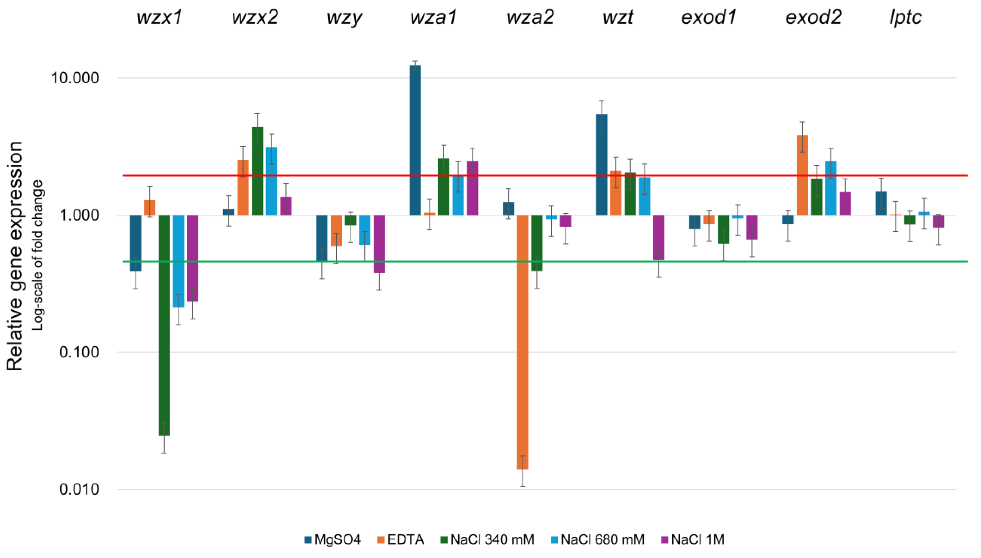
Gene expression of the identified EPS genes (Figure 3) was sensitive to the conditions tested. Specifically, wzx genes, involved in transferring the lipid-bound oligosaccharide repeat units to the periplasmic side of the plasma membrane [19,39], displayed opposite behaviors: wzx2 was overexpressed in the conditions tested, while wzx1 was repressed. wzy, a gene that polymerases polysaccharides units, is mildly downregulated in all the conditions tested. wza1, which exports polymer across the outer membrane [18,19], was over-expressed in most of the conditions tested except in the presence of EDTA, while wza2 was repressed by EDTA and at the lowest NaCl concentration. wzt, which transfers the polysaccharide that has been fully polymerized to the inner leaflet of the plasma membrane [19], was slightly induced, except at the highest concentrations of NaCl. exod1, whose function is still not clear, was over-expressed in all the conditions except when MgSO4 was present in the growing medium. wzy, which polymerizes polysaccharide units, was repressed when NaCl or EDTA were present. In the previous section, it was reported that MgSO4 was the only chemical that stimulated biofilm formation. This phenotype is supported also by gene expression. Specifically, Figure 3 shows that wza2 and exod2 have opposite expression profiles when MgSO4 is present in the medium or when any of the other chemicals. Moreover, the expression of wza1 and wzt is much higher when MgSO4 is present compared to the other conditions. All the genes studied were modulated except for exod1 and lptc, which maintained constant expression levels. This genetic distribution in Halomicronema sp. seems similar to what has been described in Synechocystis sp. strain PCC 6803 [40]; however, in Halimicronema sp., the kpsM gene was not identified. This gene was reported as the only EPS-related gene that, when mutated, impairs the secretion of EPS [40].

2.4. Transcriptomic Profile of Halomicronema sp.

Next, to know more about the general gene expression profile of Halomicronema sp., an RNA sequencing experiment was performed on the strain grown under control conditions. Here, about 20 million reads were obtained for each biological replicate with an average input read length of 292 bp and a mean quality score of 36 (Table S1). More than 76% of the reads aligned with the reference genome of Halomicronema sp. CCY15110. More specifically, a total of 5492 different genes were detected, of which 1268 (23%) encode for proteins with unknown function (or defined as “hypothetical proteins”). The most abundant transcript (Table S2), which had a value of 1,283,853 reads, is that for transfer-messenger RNA (tmRNA or SsrA) (values were normalized for gene length and total reads abundance per sample). The tmRNA is a bacterial RNA molecule that functions as both transfer and messenger RNA. In fact, it possesses paired and structured ends similar to a tRNA molecule, capable of loading alanine, while the central sequence of the molecule contains a short ORF coding for about ten amino acids, including various alanines, and ends with a stop codon [41]. It forms a ribonucleoprotein complex (tmRNP) along with Small Protein B (SmpB), elongation factor EF-Tu, and ribosomal protein S1. The key role of tmRNA is to bind stalled ribosomes and add a tag to the C-terminus of the unfinished polypeptide, inducing its proteolysis. This process assures translational quality and optimal regulation of gene expression. This process is known as a trans-translation reaction [42]. Given its importance, tmRNA is conserved and it is one of the most abundant RNAs in all bacteria [43]. As expected, analyzing the most commonly present transcripts (at least 5000 reads) showed that they code for proteins playing a role in photosynthesis (photosystem I and II related proteins, cytochrome), and among them, there are also genes involved in the synthesis of phycobilisome, phycocyanin, and allophycocyanin. These proteins form elaborate antennae capturing sunlight and transferring the energy to the photosynthetic reaction systems [44]. These protein complexes are located on the thylakoid and exhibit different abundance during their biogenesis and under light-activated heterotrophic growth [45,46]. Genes coding for transporters of iron, phosphate, and ammonium are also highly represented (Table S2). This is not surprising since it is known that the availability of those components can be a bottleneck for the growth of cyanobacteria in various ecosystems [47,48,49]. Interestingly, growth as entangled filaments (a phenotype seen in Halomicronema sp., see Figure 1) in Trichodesmium has been linked to an enhanced capacity to acquire iron from particulate sources such as dust, a physical structure that can facilitate the growth of a complex bacterial community [49,50]. Similarly, it is possible that Halomicronema sp. plays a similar role in the microbial mat where it was isolated. Finally, among the most abundant transcripts, we find genes coding for regulators of protein synthesis (sigma factors, elongation factors, and ribosomal proteins), other transcripts are linked to the production of ABC transporters and metalloproteases. In addition, there are numerous transcripts coding for transcription factors, transposases, methyltransferases, and various transporters. Considering the important regulatory work that transcription factors carry out, including regulating biofilm formation, the next analysis was performed on a subset of these proteins.

2.5. Expression Analysis of Main Transcription Regulators

Next, nine families of transcription regulators were studied as they are involved in virulence, biofilm formation, and development (Table 2). For each family (Table 2), the biological role and a selection of transcription factors further analyzed for each category are reported, along with their Gene_id, average reads value (considering the RNA-seq experiment), the Protein_id, and the average read values found in the RNA seq experiment. For each of these genes, specific primers were designed, and their expression was studied by real-time quantitative PCR (RTqPCR) in the presence of five different biofilm modulators: 340 mM NaCl, 680 mM NaCl, 1 M NaCl, 50 mM MgSO4, and 250 µM EDTA.
Factors belonging to the AbrB family and the YebC/PmpR family, which play an essential role in biofilm formation, show a lower expression level (Figure 4) when NaCl was added at the highest concentration [51,52]. TetR/AcrR regulates essential processes, including metabolism, virulence, resistance, and biofilm formation [53,54]. The selected genes belonging to the TetR/AcrR family exhibited opposite characteristics depending on the gene being considered (Figure 4); tetR-01335 and tetR-14575 were strongly overexpressed, while the expression of tetR-16580, tetR-19750, tetR-23190, tetR-11335, and tetR-13695 was repressed with respect to the control condition. All LuxR family genes, which play a key role in quorum sensing in Gram-negative bacteria, and regulate virulence, motility, and biofilm formation [55,56], were repressed. Similarly, those of the LysR, GntR, and Crp/Fnr protein families, all involved in virulence processes [57,58,59,60], were downregulated compared to the control. The XRE protein, which was suggested to be implicated in virulence [61], decreased with increasing NaCl concentration, remained nearly constant in the presence of 50 mM of MgSO4, and increased if 250 μM of EDTA was introduced into the culture medium. BolA factor, which regulates the transcription of several genes coding for proteins implicated in the transition between the planktonic stage and biofilm development, spore formation, and cell survival [62], was repressed in all the conditions tested when compared to the control. Differences in gene expression due to the presence of MgSO4 were displayed only for a few genes. Searching for the RNA-seq data for the EPS genes, only the expression of the gene lptc was clearly identified (average reads 2830). The other EPS-related genes were assigned general categories, such as the “ABC-related transporter”, and no clear correspondence was identified between this general category and the genes described within Section 2.3.

3. Materials and Methods

3.1. Growing Conditions

Halomicronema sp. (CCY15110) [22] was grown in 24-well plates in 1 mL of BA+ medium (https://www.dsmz.de/microorganisms/medium/pdf/DSMZ_Medium1677.pdf, accessed on 1 March 2023) per well at the constant temperature of 23 °C with a photoperiod of 16 h light and 8 h dark. The biomass was estimated by measuring the chlorophyll content [63,64]. Several chemicals have been reported to influence the formation of biofilms and EPS (i.e., nitrogen phosphate, sulfate, salt content, and light), as described in Pereira et al., 2015. The effects of different substances were tested by growing the bacteria in the presence of different growing conditions (340 mM NaCl, 680 mM NaCl, 1M NaCl, MgSO4 50 mM, MgCl2 250 mM, EDTA 250 µM and 2.5 mM, CaCl2 500 mM, Glucose 2%, KH2PO4 125 mM, CaCl2 250 mM, Na-acetate 50 mM, Na-citrate 50 mM, (NH4)2HPO4 100 mM, (NH4)HSO4 100 mM, and KH2PO4 50 mM), Biofilm biomass was tested 7 days after the incubation, following previously described protocols [63,64]. All the experiments were performed in triplicates.

3.2. Microscopy

To characterize the biofilm formed by Halomicronema sp. CCY15110, a microscope slide, was placed transversely in a plastic container (SacO2 Microbox O95/114 + OD95, Belgium) with the cyanobacterial strain and 25 mL of BA+ culture medium. After 10 days, the strain formed a biofilm at the bottom of the container and on the slide, which could be used for imaging. Images were taken using the AXIO fluorescence microscope Imager Z2 (Zeiss, Jena, Germany) by staining cells with SYTO-9 (Thermo Fischer Scientific Inc., Waltham, MA, USA) and by employing the environmental scanning electron microscope (ESEM) FEG2500 FEI (FEI Europe, Eindhoven, The Netherlands) operating in low vacuum mode (60 Pa) with LFD (large field detector) to enable optimal imaging of electron secondary electrons (SEs). The pressure-limited conical aperture (PLA) of 500 μm improved the signal available for the QUANTAX XFlash X-ray detector (Bruker, Billerica, MA, USA). SE imaging was performed at 10 keV with a beam size of 2.5 μm [65].

3.3. Total Phenolic Content, DPPH, and ABTS Assays

Lyophilized extracts were tested following published protocols to determine the total phenolic compounds [66,67], and the antioxidant profile by performing the DPPH inhibition assay [66,68] and the ABTS assay [31]. The experiments were performed in triplicates.

3.4. Gene Selection and Primer Design

To design the specific primers to study specific gene expression in cyanobacteria, the list of the genes involved in the last steps of EPS synthesis was first retrieved from the literature [10,17,19]. The search was focused on the EPS-related systems known to be conserved across species (Wzy transporter-dependent pathway, ABC transporter-dependent pathway, and the synthase-dependent pathway) along with the ones involved in lipopolysaccharide synthesis. Having identified from the literature all the members involved in the different systems, their presence within the genomes of Halomicronema sp. was checked using NCBI and the txid 2767773 [23] (https://www.ncbi.nlm.nih.gov/bioproject/?term=PRJNA658956, accessed on 10 July 2023). When a match was found, the gene sequence was retrieved, and specific primers were designed using NCBI Primer Blast (https://www.ncbi.nlm.nih.gov/tools/primer-blast/index.cgi, accessed on 20 July 2023) with the following specification: 60 °C optimal melting temperature, and amplicon size between 120 and 170 bp. Similarly, the primer design was also performed on the housekeeping gene secA. For a complete list, see Table S4.

3.5. Gene Expression Analysis by RTq-PCR

Total RNA was isolated from cells growing for one week in BA+ (T 23 °C, 16 h light and 8 h dark). During the light condition, chemicals (MgSO4 50 mM, EDTA 250 μM, NaCl 340 mM, NaCl 680 mM, and NaCl 1 M) were added and maintained for 2 h. One well was used as a control condition, and no chemicals were added. After 2 h of treatment, the cells were harvested for RNA extraction. RNA was recovered from one well using the RNeasy® Mini Kit (Qiagen GmbH, Hilden, Germany) according to the manufacturer’s instructions. RNA concentrations were determined using a spectrophotometer VARIAN Cary 50 UV-VIS (Agilent Technologies, Santa Clara, CA, USA). Total RNA (500 ng) was reverse transcribed into cDNA using a OneScript ® cDNA Synthesis Kit (which also contains AccuRT Genomic DNA Removal Kit purchased from Applied Biological Materials Inc., Richmond, BC, Canada) according to manufacturer instructions. The subsequent RTq-PCR was based on 20 ng of cDNA template, PowerUp SYBR Green Master Mix (ThermoFisher Scientific, Wilmington, NC, USA), with 250 nM of each forward and reverse primer. The reaction was conducted in the CFX96 Touch Real-Time PCR Detection System (Bio-Rad, Hercules, CA, USA), with the following program: 95° C for 5 min, followed by 40 cycles at 95 °C for 15 s, 60 °C for 60 s, immediately followed by melting curve analysis. The data were analyzed with the 2−ΔΔCt method as described [69] using secA as a housekeeping gene as it had stable expression in all the conditions tested. The experiment was performed with three biological replicates.

3.6. RNA Sequencing

Total RNA was isolated from cells using RNAprotect® Bacteria Reagent (Qiagen GmbH, Hilden, Germany), RNeasy® Mini Kit (Qiagen GmbH, Hilden, Germany), and DNase I RNase-free (ThermoFisher Scientific, Wilmington, NC, USA). After extraction, the concentration and purity of RNA were determined using the NanoDrop 2000 (ThermoFisher Scientific, Wilmington, NC, USA). The integrity of the RNA was assessed by gel electrophoresis on 1.5% agarose gel.
To perform RNA sequencing, three total RNA samples were sent to the GALSEQ S.r.l. Company (Milan, Italy). The concentration and purity were measured with a spectrophotometer, and the quantitative assessment of RNA integrity (RIN) was determined with a 4200 TapeStation system (RNA ScreenTape Assay kit) (Agilent Technologies, Santa Clara, CA, USA). RNA was processed using NEBNext® rRNA Depletion Kit (Bacteria) following the NEBNext® Ultra™ II Directional RNA Library protocol Prep Kit for Illumina® (New England Biolabs, Ipswich, MA, USA). RNA libraries were generated from 500 ng of total RNA, quantified and checked qualitatively with a 4200 TapeStation system (High Sensitivity D1000 kit), and then sequenced on an Illumina NovaSeq platform with paired-end reads 150 bp long and with an average depth of 20 million clusters per sample. The Fastq reads were checked with FastQC (https://www.bioinformatics.babraham.ac.uk/projects/fastqc/, accessed on 10 October 2022) to verify the overall quality of the reads and the presence of adaptor sequences. The paired reads were subsequently aligned to the genome of Halomicronema sp. CCY15110 (Assembly: GCF_016888165.1) using splice-aware aligner STAR v 2.7.9a [70] and indexed using Samtool [71]. Per-gene read counts were generated using the STAR quantMode geneCounts option [60].

3.7. Statistical Analysis

Data analysis was performed using SPSS (Ver.27, IBM Corp., Armonk, NY, USA). The Student’s t-test was performed to compare the effects of the different chemicals (see legend to Figure 2 for details).

4. Conclusions

This work represents an initial characterization of the cyanobacterium Halomicronema sp. in the field of biofilm formation and the causes that affect it. The expression of genes related to the production of EPS and transcription factors potentially involved in biofilm formation under different growth conditions were studied. This work identified possible biomarkers useful for Halomicronema sp. biofilm formation, including the EPS related genes wza1, wzt, along with luxR-05665, and some genes belonging to the TetR/AcrR and LysR families, as their expression is strongly sensitive to the growth conditions analyzed. In addition, given its antioxidant properties, as also observed in the case of Spirulina spp. it would also be useful to further study its antioxidant capacity. Of relevance was also the demonstration that Halomicronema sp can survive and grow under several stress conditions, including high salt. Therefore, an interesting application will be the possibility of testing if this bacterium has “Plant Growth Promoting” capacities, which can support crop growth in arable lands with high salinity.

Supplementary Materials

The following supporting information can be downloaded at https://www.mdpi.com/article/10.3390/ijms26020673/s1.

Author Contributions

Conceptualization: M.C.; methodology. M.C. and H.B.; formal analysis: M.C. and M.M.; investigation: M.C.; resources: N.M.; writing—original draft preparation: M.C.; writing—review and editing: N.M., M.C., M.M. and H.B.; funding acquisition: N.M. All authors have read and agreed to the published version of the manuscript.

Funding

This project received funding from the European Union’s Horizon 2020 research and innovation program under grant agreement No. 818431 (SIMBA), awarded to SITEIA.Parma. This output reflects only the author’s view, and the Research Executive Agency (REA) cannot be held responsible for any use that may be made of the information contained therein.

Institutional Review Board Statement

Not applicable.

Informed Consent Statement

Not applicable.

Data Availability Statement

The RNA sequencing data presented in this study are openly available in the NCBI database, BioProject, under accession number PRJNA1200597 (https://www.ncbi.nlm.nih.gov/bioproject/ PRJNA1200597, uploaded on 15 December 24).

Acknowledgments

The authors wish to thank C. Carlini who contributed to the practical work during her Masters programme.

Conflicts of Interest

The authors declare no conflicts of interest.

References

  1. Martens, S. Handbook of Cyanobacterial Monitoring and Cyanotoxin Analysis. Adv. Oceanogr. Limnol. 2017, 8, 7221. [Google Scholar] [CrossRef]
  2. Durall, C.; Lindblad, P. Mechanisms of carbon fixation and engineering for increased carbon fixation in cyanobacteria. Algal Res. 2015, 11, 263–270. [Google Scholar] [CrossRef]
  3. Zakar, T.; Laczko-Dobos, H.; Toth, T.N.; Gombos, Z. Carotenoids assist in cyanobacterial photosystem II assembly and function. Front. Plant Sci. 2016, 7, 295. [Google Scholar] [CrossRef]
  4. Chamizo, S. Soil Inoculation with Cyanobacteria: Reviewing Its’ Potential for Agriculture Sustainability in Drylands. Agric. Res. Technol. Open Access J. 2018, 18, 556046. [Google Scholar] [CrossRef]
  5. Dahms, H.U.; Ying, X.; Pfeiffer, C. Antifouling potential of cyanobacteria: A mini-review. Biofouling 2006, 22, 317–327. [Google Scholar] [CrossRef]
  6. Chakdar, H.; Thapa, S.; Srivastava, A.; Shukla, P. Genomic and proteomic insights into the heavy metal bioremediation by cyanobacteria. J. Hazard. Mater. 2022, 424, 127609. [Google Scholar] [CrossRef] [PubMed]
  7. Cui, J.; Xie, Y.; Sun, T.; Chen, L.; Zhang, W. Deciphering and engineering photosynthetic cyanobacteria for heavy metal bioremediation. Sci. Total Environ. 2021, 761, 144111. [Google Scholar] [CrossRef]
  8. Dang, H.; Lovell, C.R. Microbial Surface Colonization and Biofilm Development in Marine Environments. Microbiol. Mol. Biol. Rev. 2016, 80, 91–138. [Google Scholar] [CrossRef] [PubMed]
  9. Chittora, D.; Meena, M.; Barupal, T.; Swapnil, P. Cyanobacteria as a source of biofertilizers for sustainable agriculture. Biochem. Biophys. Rep. 2020, 22, 100737. [Google Scholar] [CrossRef] [PubMed]
  10. Cania, B.; Vestergaard, G.; Kublik, S.; Köhne, J.M.; Fischer, T.; Albert, A.; Winkler, B.; Schloter, M.; Schulz, S. Biological Soil Crusts from Different Soil Substrates Harbor Distinct Bacterial Groups with the Potential to Produce Exopolysaccharides and Lipopolysaccharides. Microb. Ecol. 2020, 79, 326–341. [Google Scholar] [CrossRef] [PubMed]
  11. Bolhuis, H.; Cretoiu, M.S.; Stal, L.J. Molecular ecology of microbial mats. FEMS Microbiol. Ecol. 2014, 90, 335–350. [Google Scholar] [PubMed]
  12. Flemming, H.; Wingender, J. The biofilm matrix. Nat. Rev. Microbiol. 2010, 8, 623–633. [Google Scholar] [CrossRef] [PubMed]
  13. Flemming, H.C.; van Hullebusch, E.D.; Neu, T.R.; Nielsen, P.H.; Seviour, T.; Stoodley, P.; Wingender, J.; Wuertz, S. The biofilm matrix: Multitasking in a shared space. Nat. Rev. Microbiol. 2023, 21, 70–86. [Google Scholar] [CrossRef]
  14. Delattre, C.; Pierre, G.; Laroche, C.; Michaud, P. Production, extraction and characterization of microalgal and cyanobacterial exopolysaccharides. Biotechnol. Adv. 2016, 34, 1159–1179. [Google Scholar] [CrossRef]
  15. Laroche, C. Exopolysaccharides from Microalgae and Cyanobacteria: Diversity of Strains, Production Strategies, and Applications. Mar. Drugs 2022, 20, 336. [Google Scholar] [CrossRef] [PubMed]
  16. Kehr, J.C.; Dittmann, E. Biosynthesis and function of extracellular glycans in cyanobacteria. Life 2015, 5, 164–180. [Google Scholar] [CrossRef] [PubMed]
  17. Pereira, S.; Zille, A.; Micheletti, E.; Moradas-Ferreira, P.; De Philippis, R.; Tamagnini, P. Complexity of cyanobacterial exopolysaccharides: Composition, structures, inducing factors and putative genes involved in their biosynthesis and assembly. FEMS Microbiol. Rev. 2009, 33, 917–941. [Google Scholar] [CrossRef] [PubMed]
  18. Reeves, P.R.; Hobbs, M.; Valvano, M.A.; Skurnik, M.; Whitfield, C.; Coplin, D.; Kido, N.; Klena, J.; Maskell, D.; Raetz, C.R.H.; et al. Bacterial polysaccharide synthesis and gene nomenclature. Trends Microbiol. 1996, 4, 495–503. [Google Scholar] [CrossRef]
  19. Pereira, S.B.; Mota, R.; Vieira, C.P.; Vieira, J.; Tamagnini, P. Phylum-wide analysis of genes/proteins related to the last steps of assembly and export of extracellular polymeric substances (EPS) in cyanobacteria. Sci. Rep. 2015, 5, 14835. [Google Scholar] [CrossRef] [PubMed]
  20. Comte, S.; Guibaud, G.; Baudu, M. Biosorption properties of extracellular polymeric substances (EPS) resulting from activated sludge according to their type: Soluble or bound. Process Biochem. 2006, 41, 815–823. [Google Scholar] [CrossRef]
  21. Whitfield, C.; Stephen Trent, M. Biosynthesis and export of bacterial lipopolysaccharides. Annu. Rev. Biochem. 2014, 83, 99–128. [Google Scholar] [CrossRef] [PubMed]
  22. Cardoso, D.C.; Cretoiu, M.S.; Stal, L.J.; Bolhuis, H. Seasonal development of a coastal microbial mat. Sci. Rep. 2019, 9, 9035. [Google Scholar] [CrossRef] [PubMed]
  23. Boden, J.S.; Grego, M.; Bolhuis, H.; Sánchez-Baracaldo, P. Draft genome sequences of three filamentous cyanobacteria isolated from brackish habitats. J. Genom. 2021, 9, 20–25. [Google Scholar] [CrossRef] [PubMed]
  24. Chen, L.; Li, D.; Liu, Y. Salt tolerance of Microcoleus vaginatus Gom., a cyanobacterium isolated from desert algal crust, was enhanced by exogenous carbohydrates. J. Arid Environ. 2003, 55, 645–656. [Google Scholar] [CrossRef]
  25. Kharwar, S.; Mishra, A.K. Unraveling the complexities underlying sulfur deficiency and starvation in the cyanobacterium Anabaena sp. PCC 7120. Environ. Exp. Bot. 2020, 172, 103966. [Google Scholar] [CrossRef]
  26. Wu, R.X.; Zhang, Y.; Guo, Z.Q.; Zhao, B.; Guo, J.S. Role of Ca2+ and Mg2+ in changing biofilm structure and enhancing biofilm formation of P. stutzeri strain XL-2. Colloids Surf. B Biointerfaces 2022, 220, 112972. [Google Scholar] [CrossRef]
  27. Shen, Y.; Huang, P.C.; Huang, C.; Sun, P.; Monroy, G.L.; Wu, W.; Lin, J.; Espinosa-Marzal, R.M.; Boppart, S.A.; Liu, W.T.; et al. Effect of divalent ions and a polyphosphate on composition, structure, and stiffness of simulated drinking water biofilms. npj Biofilms Microbiomes 2018, 4, 15. [Google Scholar] [CrossRef] [PubMed]
  28. Finnegan, S.; Percival, S.L. EDTA: An Antimicrobial and Antibiofilm Agent for Use in Wound Care. Adv. Wound Care 2015, 4, 415–421. [Google Scholar] [CrossRef] [PubMed]
  29. Wickramasinghe, M.; Katyana, K.; Sewwandi, K.; Rathnayaka, I.; Magana-Arachchi, D.; Jayawardana, B.; Liyanage, R. Exploration of antioxidant activities, microstructural properties, and fatty acid composition of three cyanobacteria species. Biocatal. Agric. Biotechnol. 2024, 56, 103015. [Google Scholar] [CrossRef]
  30. Morone, J.; Lopes, G.; Preto, M.; Vasconcelos, V.; Martins, R. Exploitation of filamentous and picoplanktonic cyanobacteria for cosmetic applications: Potential to improve skin structure and preserve dermal matrix components. Mar. Drugs 2020, 18, 486. [Google Scholar] [CrossRef] [PubMed]
  31. Frazzini, S.; Scaglia, E.; Dell’anno, M.; Reggi, S.; Panseri, S.; Giromini, C.; Lanzoni, D.; Rossi, C.A.S.; Rossi, L. Antioxidant and Antimicrobial Activity of Algal and Cyanobacterial Extracts: An In Vitro Study. Antioxidants 2022, 11, 992. [Google Scholar] [CrossRef] [PubMed]
  32. Lee, H.E.; Lee, J.H.; Park, S.M.; Kim, D.G. Symbiotic relationship between filamentous algae (Halomicronema sp.) and extracellular polymeric substance-producing algae (Chlamydomonas sp.) through biomimetic simulation of natural algal mats. Front. Microbiol. 2023, 14, 1176069. [Google Scholar] [CrossRef] [PubMed]
  33. Waditee-Sirisattha, R.; Kageyama, H. Halotolerance, stress mechanisms, and circadian clock of salt-tolerant cyanobacteria. Appl. Microbiol. Biotechnol. 2023, 107, 1129–1141. [Google Scholar] [CrossRef] [PubMed]
  34. Kageyama, H.; Waditee-Sirisattha, R. Halotolerance mechanisms in salt-tolerant cyanobacteria. In Advances in Applied Microbiology; Elsevier: Amsterdam, The Netherlands, 2023; Volume 124, pp. 55–117. ISBN 9780443192746. [Google Scholar]
  35. Hagemann, M. Molecular biology of cyanobacterial salt acclimation. FEMS Microbiol. Rev. 2011, 35, 87–123. [Google Scholar] [CrossRef] [PubMed]
  36. Marin, K.; Huckauf, J.; Fulda, S.; Hagemann, M. Salt-dependent expression of glucosylglycerol-phosphate synthase, involved in osmolyte synthesis in the cyanobacterium Synechocystis sp. strain PCC 6803. J. Bacteriol. 2002, 184, 2870–2877. [Google Scholar] [CrossRef]
  37. Marin, K.; Suzuki, I.; Yamaguchi, K.; Ribbeck, K.; Yamamoto, H.; Kanesaki, Y.; Hagemann, M.; Murata, N. Identification of histidine kinases that act as sensors in the perception of salt stress in Synechocystis sp. PCC 6803. Proc. Natl. Acad. Sci. USA 2003, 100, 9061–9066. [Google Scholar] [CrossRef] [PubMed]
  38. Sinetova, M.A.; Los, D.A. Systemic analysis of stress transcriptomics of Synechocystis reveals common stress genes and their universal triggers. Mol. Biosyst. 2016, 12, 3254–3258. [Google Scholar] [CrossRef] [PubMed]
  39. Soumya, M.P.; Nampoothiri, K.M. An overview of functional genomics and relevance of glycosyltransferases in exopolysaccharide production by lactic acid bacteria. Int. J. Biol. Macromol. 2021, 184, 1014–1025. [Google Scholar] [CrossRef] [PubMed]
  40. Santos, M.; Pereira, S.B.; Flores, C.; Príncipe, C.; Couto, N.; Karunakaran, E.; Cravo, S.M.; Oliveira, P.; Tamagnini, P. Absence of KpsM (Slr0977) Impairs the Secretion of Extracellular Polymeric Substances (EPS) and Impacts Carbon Fluxes in Synechocystis sp. PCC 6803. mSphere 2021, 6, 10–128. [Google Scholar] [CrossRef]
  41. Keiler, K.C.; Ramadoss, N.S. Bifunctional transfer-messenger RNA. Biochimie 2011, 93, 1993–1997. [Google Scholar] [CrossRef] [PubMed]
  42. Shimizu, Y.; Ueda, T. The role of SmpB protein in trans-translation. Proc. FEBS Lett. 2002, 514, 74–77. [Google Scholar] [CrossRef] [PubMed]
  43. Keiler, K.C.; Shapiro, L.; Williams, K.P. tmRNAs that encode proteolysis-inducing tags are found in all known bacterial genomes: A two-piece tmRNA functions in caulobacter. Proc. Natl. Acad. Sci. USA 2000, 97, 7778–7783. [Google Scholar] [CrossRef] [PubMed]
  44. Domínguez-Martín, M.A.; Sauer, P.V.; Kirst, H.; Sutter, M.; Bína, D.; Greber, B.J.; Nogales, E.; Polívka, T.; Kerfeld, C.A. Structures of a phycobilisome in light-harvesting and photoprotected states. Nature 2022, 609, 835–845. [Google Scholar] [CrossRef] [PubMed]
  45. Plohnke, N.; Seidel, T.; Kahmann, U.; Rögner, M.; Schneider, D.; Rexroth, S. The proteome and lipidome of Synechocystis sp. PCC 6803 cells grown under light-activated heterotrophic conditions. Mol. Cell. Proteom. 2015, 14, 572–584. [Google Scholar] [CrossRef]
  46. Huang, F.; Grauslys, A.; Huokko, T.; Caamaño Gutiérrez, E.; Jones, A.R.; Liu, L.N. Dynamic Changes in the Thylakoid Proteome of Cyanobacteria during Light-Regulated Thylakoid Membrane Development. Plants 2023, 12, 3967. [Google Scholar] [CrossRef]
  47. Tiwari, B. Phosphate metabolism in cyanobacteria: Fundamental prospective and applications. In Cyanobacteria: Metabolisms to Molecules; Elsevier: Amsterdam, The Netherlands, 2023; pp. 159–175. ISBN 9780443132315. [Google Scholar]
  48. Muro-Pastor, M.I.; Florencio, F.J. Regulation of ammonium assimilation in cyanobacteria. Plant Physiol. Biochem. 2003, 41, 595–603. [Google Scholar] [CrossRef]
  49. Qiu, G.W.; Koedooder, C.; Qiu, B.S.; Shaked, Y.; Keren, N. Iron transport in cyanobacteria—From molecules to communities. Trends Microbiol. 2022, 30, 229–240. [Google Scholar] [CrossRef]
  50. Rubin, M.; Berman-Frank, I.; Shaked, Y. Dust-and mineral-iron utilization by the marine dinitrogen-fixer Trichodesmium. Nat. Geosci. 2011, 4, 529–534. [Google Scholar] [CrossRef]
  51. Sullivan, D.M.; Bobay, B.G.; Kojetin, D.J.; Thompson, R.J.; Rance, M.; Strauch, M.A.; Cavanagh, J. Insights into the Nature of DNA Binding of AbrB-like Transcription Factors. Structure 2008, 16, 1702–1713. [Google Scholar] [CrossRef] [PubMed]
  52. Wei, L.; Wu, Y.; Qiao, H.; Xu, W.; Zhang, Y.; Liu, X.; Wang, Q. YebC controls virulence by activating T3SS gene expression in the pathogen Edwardsiella piscicida. FEMS Microbiol. Lett. 2018, 365, fny137. [Google Scholar] [CrossRef]
  53. Colclough, A.L.; Scadden, J.; Blair, J.M.A. TetR-family transcription factors in Gram-negative bacteria: Conservation, variation and implications for efflux-mediated antimicrobial resistance. BMC Genom. 2019, 20, 731. [Google Scholar] [CrossRef] [PubMed]
  54. Deng, W.; Li, C.; Xie, J. The underling mechanism of bacterial TetR/AcrR family transcriptional repressors. Cell. Signal. 2013, 25, 1608–1613. [Google Scholar] [CrossRef]
  55. Chen, J.; Xie, J. Role and regulation of bacterial LuxR-like regulators. J. Cell. Biochem. 2011, 112, 2694–2702. [Google Scholar] [CrossRef] [PubMed]
  56. Santos, C.L.; Correia-Neves, M.; Moradas-Ferreira, P.; Mendes, M.V. A Walk into the LuxR Regulators of Actinobacteria: Phylogenomic Distribution and Functional Diversity. PLoS ONE 2012, 7, 0046758. [Google Scholar] [CrossRef]
  57. Maddocks, S.E.; Oyston, P.C.F. Structure and function of the LysR-type transcriptional regulator (LTTR) family proteins. Microbiology 2008, 154, 3609–3623. [Google Scholar] [CrossRef]
  58. Liu, G.F.; Wang, X.X.; Su, H.Z.; Lu, G.T. Progress on the GntR family transcription regulators in bacteria. Yi Chuan = Hered. 2021, 43, 66–73. [Google Scholar]
  59. Körner, H.; Sofia, H.J.; Zumft, W.G. Phylogeny of the bacterial superfamily of Crp-Fnr transcription regulators: Exploiting the metabolic spectrum by controlling alternative gene programs. FEMS Microbiol. Rev. 2003, 27, 559–592. [Google Scholar] [CrossRef] [PubMed]
  60. Soberón-Chávez, G.; Alcaraz, L.D.; Morales, E.; Ponce-Soto, G.Y.; Servín-González, L. The transcriptional regulators of the CRP family regulate different essential bacterial functions and can be inherited vertically and horizontally. Front. Microbiol. 2017, 8, 959. [Google Scholar] [CrossRef] [PubMed]
  61. Hu, Y.; Hu, Q.; Wei, R.; Li, R.; Zhao, D.; Ge, M.; Yao, Q.; Yu, X. The XRE family transcriptional regulator SrtR in streptococcus suis is involved in oxidant tolerance and virulence. Front. Cell. Infect. Microbiol. 2019, 9, 00452. [Google Scholar] [CrossRef]
  62. Dressaire, C.; Moreira, R.N.; Barahona, S.; de Matos, A.P.A.; Arraiano, C.M. BolA is a transcriptional switch that turns off motility and turns on biofilm development. mBio 2015, 6, e02352-14. [Google Scholar] [CrossRef]
  63. Zavrel, T.; Sinetova, M.; Cerven, J. Measurement of Chlorophyll a and Carotenoids Concentration in Cyanobacteria. Bio-Protocol 2015, 5, 1467. [Google Scholar] [CrossRef]
  64. Sendersky, E.; Simkovsky, R.; Golden, S.; Schwarz, R. Quantification of Chlorophyll as a Proxy for Biofilm Formation in the Cyanobacterium Synechococcus elongatus. Bio-Protocol 2017, 7, 2406. [Google Scholar] [CrossRef]
  65. Marmiroli, M.; Caldara, M.; Pantalone, S.; Malcevschi, A.; Maestri, E.; Marmiroli, N. Building a risk matrix for the safety assessment of wood derived biochars. Sci. Total Environ. 2022, 15, 156265. [Google Scholar] [CrossRef] [PubMed]
  66. Guerreiro, A.; Andrade, M.A.; Menezes, C.; Vilarinho, F.; Dias, E. Antioxidant and Cytoprotective Properties of Cyanobacteria: Potential for Biotechnological Applications. Toxins 2020, 12, 548. [Google Scholar] [CrossRef] [PubMed]
  67. Erkan, N.; Ayranci, G.; Ayranci, E. Antioxidant activities of rosemary (Rosmarinus officinalis L.) extract, blackseed (Nigella sativa L.) essential oil, carnosic acid, rosmarinic acid and sesamol. Food Chem. 2008, 110, 76–82. [Google Scholar] [CrossRef]
  68. Andrade, M.A.; Ribeiro-Santos, R.; Costa Bonito, M.C.; Saraiva, M.; Sanches-Silva, A. Characterization of rosemary and thyme extracts for incorporation into a whey protein based film. LWT 2018, 92, 497–508. [Google Scholar] [CrossRef]
  69. Livak, K.J.; Schmittgen, T.D. Analysis of relative gene expression data using real-time quantitative PCR and the 2−ΔΔCT Method. Methods 2001, 25, 402–408. [Google Scholar] [CrossRef]
  70. Dobin, A.; Davis, C.A.; Schlesinger, F.; Drenkow, J.; Zaleski, C.; Jha, S.; Batut, P.; Chaisson, M.; Gingeras, T.R. STAR: Ultrafast universal RNA-seq aligner. Bioinformatics 2013, 29, 15–21. [Google Scholar] [CrossRef] [PubMed]
  71. Li, H.; Handsaker, B.; Wysoker, A.; Fennell, T.; Ruan, J.; Homer, N.; Marth, G.; Abecasis, G.; Durbin, R. The Sequence Alignment/Map format and SAMtools. Bioinformatics 2009, 25, 2078–2079. [Google Scholar] [CrossRef]
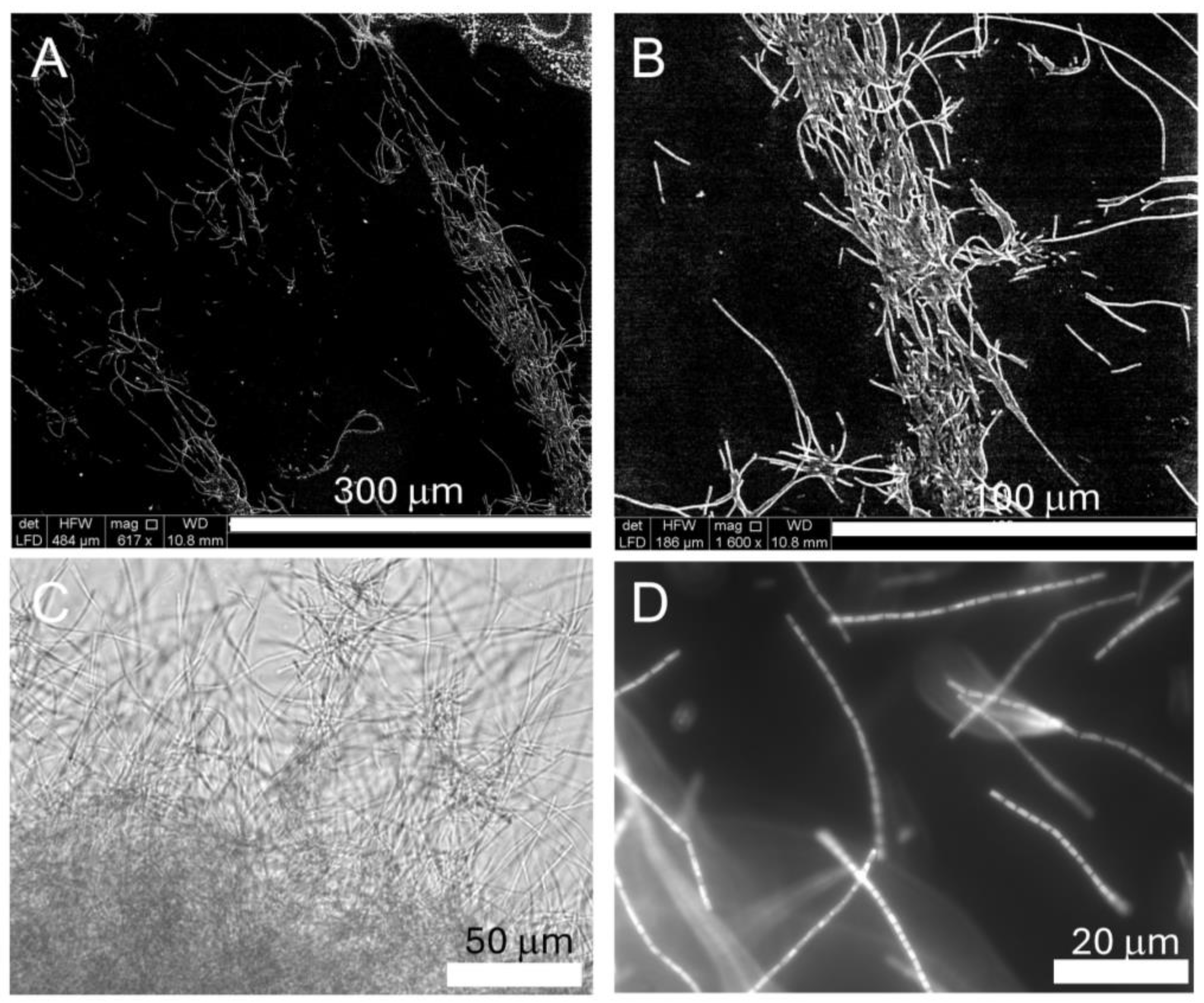
Figure 1. Imaging Halomicronema sp. with ESEM in low vacuum mode (A,B), light microscope (C), and with fluorescence microscopy after staining with SYTO-9 (D); for image (C), the magnification used was 40×, and for image (D), the magnification was 100×. For each image, the scale bar is reported at the bottom right.
Ijms 26 00673 g001
Figure 2. Biomass formation of cyanobacteria planktonic and biofilm fractions detected through chlorophyll measurements. Halomicronema sp was grown in BA+ medium for 7 days under 16 h light and 8 h dark; temperature was constant at 23 °C for all the conditions tested. The strain was also tested in the presence of different chemicals: A = CTR the control condition, B = NaCl 340 mM, C = NaCl 680 mM, D = NaCl 1 M, E = MgSO4 (50 mM), F = MgCl2 (250 mM), G = EDTA (250 µM), H = CaCl2 (500 mM), I = Glucose 110 mM (2%), J = KH2PO4 (125 mM), K = CaCl2 (250 mM), L = EDTA (2.5 mM), M = Na-acetate (50 mM), N = Na-citrate (50 mM), O = (NH4)2HPO4 (100 mM), P = (NH4)HSO4 (100 mM), Q = KH2PO4 (50 mM). The data represent the mean and the standard deviation of three independent biological replicates. A Student’s t-test was performed to compare the effects of the different chemicals. The effects were all significant, except for NaCl 340 mM (* was used for p-values < 0.05 and ** for p-values < 0.01; ns: not significant).
Ijms 26 00673 g002
Figure 3. Gene expression analysis of EPS-related genes from Halomicronema sp, cells were grown in six different growing conditions: control, and in the presence of EDTA 250 µM, or MgSO4 50 mM, NaCl 340 mM, NaCl 680 mM, and NaCl 1 M; expression is relative to the control condition using the 2−ΔΔCt method. Red line represents a 2 logfold increased expression, while the green line represents a 2 log-fold decrease in expression. Data outside these two boundaries were considered significant. The data represent the mean and the standard deviation of three independent biological replicates.
Ijms 26 00673 g003
Figure 4. Gene expression analysis of selected transcriptional regulators from Halomicronema sp, cells were grown in six different growing conditions: control, and in the presence of EDTA 250 µM, or MgSO4 50 mM, NaCl 340 mM, NaCl 680 mM, and NaCl 1 M. Expression is relative to the control condition using the 2−ΔΔCt method, and it represented with a heat-map obtained by using www.heatmapper.ca (accessed on 1 July 2024).
Ijms 26 00673 g004
Table 1. Total phenolic concentration and antioxidant profile. The data are the mean of three independent experiments, and the standard deviation (SD) is reported. PI is the Percentage of Inhibition.
ASSAYMean ± SD
Total Phenolic Compounds
(mg GAE g−1 extract DW)
0.7 ± 0.1
DPPH Inhibition
(PI%)
63 ± 5
ABTS assay
(PI%)
52 ± 4
Table 2. List of the transcription factors considered and their biological role.
Transcription Factor FamilyRole of the Transcription Factor FamilyProtein NameGene_IDAverage Value *Protein_ID
AbrB family
transcriptional regulator
Essential for cell survival, spore formation, and biofilm development [51]AbrB-06010JUK32_RS060105239WP_204137943.1
YebC/PmpR family
DNA-binding
transcriptional regulator
Probably involved in regulation of quorum sensing, and virulence, regulates EPS and LPS production to form biofilm [52]YebC-12275JUK32_RS12275283WP_204139153.1
TetR/AcrR family
transcriptional regulator
Regulation of essential processes (metabolism virulence, resistance, and biofilm formation) [53,54]TetR-01335JUK32_RS01335768WP_204137057.1
TetR-14575JUK32_RS14575167WP_204139591.1
TetR-16580JUK32_RS165801471WP_204139976.1
TetR-19750JUK32_RS19750517WP_204140582.1
TetR-23190JUK32_RS23190271WP_204141244.1
TetR-11335JUK32_RS11335702WP_239112565.1
TetR-13695JUK32_RS13695483WP_239112647.1
LuxR C-terminal-related transcriptional regulatorInvolved in quorum sensing, regulates virulence gene production, motility, plasmid transfer, and biofilm formation [55,56]LuxR-02440JUK32_RS02440445WP_204137261.1
LuxR-04290JUK32_RS04290935WP_204137618.1
LuxR-05665JUK32_RS05665653WP_239112365.1
LysR family
transcriptional regulator
Regulates virulence, metabolism, quorum sensing, and motility [57]LysR-07280JUK32_RS07280645WP_204138189.1
LysR-08635JUK32_RS086351034WP_204138452.1
LysR-09925JUK32_RS09925266WP_204138702.1
LysR-11580JUK32_RS11580654WP_204139027.1
LysR-12705JUK32_RS12705613WP_204139229.1
LysR-20905JUK32_RS209051630WP_204140807.1
GntR family
transcriptional regulator
Regulates motility, glucose metabolism, and virulence [58]GntR-03040JUK32_RS030402277WP_239112277.1
GntR-24680JUK32_RS246801551WP_239113044.1
Crp/Fnr family
transcriptional regulator
Regulates virulence, the enzymes that degrade aromatic rings, genes involved in nitrogen fixation and photosynthesis [59,60]Crp-04910JUK32_RS049104875WP_204137735.1
Crp-18565JUK32_RS185658743WP_204140370.1
Crp-20005JUK32_RS20005592WP_204140632.1
Crp-24180JUK32_RS241803763WP_204141438.1
XRE family
transcriptional regulator
Unknown function, probably involved in the expression of virulence genes [61]XRE-06870JUK32_RS068703544WP_204138108.1
BolA family
transcriptional regulator
Involved in stress response, cell growth, morphology and division, biofilm development, inhibit motility [62]BolA-25875JUK32_RS258752291WP_204141766.1
* Average value of the reads measured in the RNA-seq experiment under BA+ growing conditions.
Disclaimer/Publisher’s Note: The statements, opinions and data contained in all publications are solely those of the individual author(s) and contributor(s) and not of MDPI and/or the editor(s). MDPI and/or the editor(s) disclaim responsibility for any injury to people or property resulting from any ideas, methods, instructions or products referred to in the content.