Article

Exploring the Genetic and Functional Diversity of Porphyromonas gingivalis Survival Factor RagAB

by
Pauline G. Montz
1,
Evdokia Dafni
1,
Bernd Neumann
2,
Dongmei Deng
3,
Mohamed M. H. Abdelbary
1,† and
Georg Conrads
1,*
1
Division of Oral Microbiology and Immunology, Department of Operative Dentistry, Periodontology and Preventive Dentistry, Rheinisch-Westfälische Technische Hochschule (RWTH) University Hospital, 52074 Aachen, Germany
2
Institute of Clinical Microbiology, Infectious Diseases and Infection Control, Paracelsus Medical University, Klinikum Nürnberg, 90419 Nürnberg, Germany
3
Department of Preventive Dentistry, Academic Centre for Dentistry Amsterdam (ACTA), University of Amsterdam and VU University Amsterdam, 1081LA Amsterdam, The Netherlands
*
Author to whom correspondence should be addressed.
Current address: Division of Nosocomial Pathogens and Antibiotic Resistances, Department of Infectious Diseases, Robert Koch Institute, Wernigerode Branch, 38855 Wernigerode, Germany.
Int. J. Mol. Sci. 2025, 26(3), 1073; https://doi.org/10.3390/ijms26031073
Submission received: 2 January 2025 / Revised: 21 January 2025 / Accepted: 22 January 2025 / Published: 26 January 2025

Abstract

:
Porphyromonas gingivalis is a key pathogen in periodontitis. Its outer membrane contains the RagAB transport complex, which has been implicated in protein uptake, essential for a proteolytic species. RagA is a 22-stranded β-barrel, and RagB is the corresponding 4-TPR lid, together forming a TonB-dependent system acting as a “pedal bin”. Four different alleles were observed, of which ragAB-1 is more virulent than the others. Our aim was to map ragAB in 129 strains of P. gingivalis and related species available in our collection, supported by a newly introduced universal PCR for amplification/sequencing of all four ragA variants and to find reasons for the differences in virulence and/or fitness. Regarding the PCR method, by pairing established Long-PCR primers with our newly designed sequencing primers (ragA-F0, -F1, -R2, -R2a, -R4), it was possible to amplify and sequence all four ragA variants. The same was not possible for ragB due to high heterogeneity. The mapping allowed us to type all strains into ragAB-1-4. For each type, some strains (of mainly animal origin such as Porphyromonas gulae) with slightly different amino acid sequences were identified (designated ragAB-1a to -4a). In terms of function, the transfer of recently discovered SusCD information to the similar RagAB complex provided new insights. Substrate specificity as well as length of pedal could be the route to differential virulence (survival rate, fitness) as Rag-1 (closer related to Rag-3/4) and Rag-2 were found to be massively different here. In general, substrate–ligand-binding sites seem to be quite variable with the exception of Rag-1, probably indicating nutritional preferences. In addition, an insertion (8 aa long) found in loop L7 throughout RagA-2 could not only affect the dynamics of lid opening/closing but might also alter the associated substrate throughput rate.

1. Introduction

Porphyromonas gingivalis (P. gingivalis) is a key pathogen in severe forms of periodontitis, which is described as a chronic inflammatory disease of the teeth-surrounding tissues based on robust dysbiosis. P. gingivalis is an obligate anaerobic, Gram-negative, rod-shaped to pleomorphic, immobile, black-pigmented, and asaccharolytic/proteolytic bacterium [1,2]. Together with Treponema denticola and Tannerella forsythia, it forms the so-called “red complex” [3], which is partly responsible for the induction of inflammation, bleeding on probing, the formation of deep gingiva pockets, attachment loss, and, ultimately, the destruction of the periodontium. P. gingivalis has been linked with a number of systemic diseases, including Alzheimer’s disease [4,5], rheumatoid arthritis [6], vascular malfunctions [4], and preterm birth [7], as reviewed elsewhere [1]. The intensive study of its virulence factors, including capsule, outer-membrane vesicles, lipopolysaccharide, gingipains, and fimbriae, recently shown for long fimbriae by our group [8], has not yet yielded a comprehensive explanation of the differences in pathogenicity or fitness observed between different strains, which is why further genetic investigation of P. gingivalis is essential.
Receptor antigens RagA and RagB are major components of the outer membrane of P. gingivalis and have been linked with virulence because of their contribution to subcutaneous lesion development, epithelial cell invasion, and efficient growth of the pathogen by acting as a transport system for nutrients [9]. The RagAB importance has been confirmed in a murine model, as evidenced by the reduced virulence observed in a ragB mutant—and especially, a ragA mutant—in terms of murine lethality and lesion size [10]. Earlier studies have predicted that only some “more virulent” P. gingivalis strains such as W50, W83, or WPH35 possess the ragAB locus [11,12]. The lack of universal primers capable of amplifying all variants may contribute to this observation, as the ragAB locus (also abbreviated as rag and first described in 1999 [13]) exhibits considerable heterogeneity. Sequence variations of up to 30% in ragA and as high as 48–47% in ragB were observed [14]. Later studies have described four different variants of the rag locus, both in oral samples and in P. gingivalis strains, named rag-1 to rag-4, of which type rag-1 (including W50 or W83) appears to be more virulent [15,16,17]. Genes ragA and ragB are co-transcribed [13], and their expression is highly interdependent, in the sense that a ragA mutant significantly reduces the expression of RagB, and a ragB mutant leads to degraded RagA [14]. The ragA operon (3.1 kb) results in the production of RagA (PG_0185, GenBank: AAQ65420.1), a 115 kDa TonB-dependent 22-stranded β-barrel transporter (TBDT) with a plug domain blocking the substrate passage through the outer membrane [16,18]. RagA is covered by the 55 kDa substrate-binding lipoprotein RagB (PG_0186, GenBank: AAQ65421.1), encoded by the ragB operon (1.5 kb) 30 bp downstream [9,12,14,18]. Crystallography revealed the RagAB structure of strain W83 as a heterotetrameric complex (RagA1B1-RagA2B2 dimer) acting as a double “pedal bin” with monomers opening independently, enabling the uptake of the gingipain-cleaved proteinaceous nutrients/peptides [18,19]. The binding of an extracellular substrate (peptide) leads to a conformational change making it accessible for interaction with TonB, an inner membrane protein complex. This interaction allows the formation of a substrate transport channel in the periplasmic space [18]. The fact that the RagAB complex is similar to other TonB-dependent outer membrane transport systems, such as SusCD, which is involved in malto-oligosaccharide and starch uptake in Bacteroides thetaiotaomicron [20], will be used to critically transfer structural and functional knowledge. While other virulence factors of P. gingivalis have been extensively studied, the genetic and functional diversity of RagAB remains poorly understood.
The objectives of this study were the following: first, to demonstrate the presence of the rag locus in a maximum number of strains and to classify them accordingly; second, to investigate the genetic differences between the four rag types; third, to compare functional domains of the deduced amino acid sequence; and finally, to develop a universal PCR method capable of amplifying all rag variants.

2. Results

2.1. Developing a Universal RagA-Gene-Directed PCR Method

We begin by providing a brief overview of the achievements derived from the universal ragA-directed PCR. Attempts to develop a universal (type independent) ragB amplification were unsuccessful due to its high degree of heterogeneity. However, if ragA type is determined, the corresponding ragB can be amplified by type-specific primers as published by Hall et al. in 2005 [16]. Subsequently, we present a comprehensive summary of the mapping results.
Amplification of the ragA gene (for details and Figures see Section 4): We were able to sequence the ragA locus using the Long-PCR product as a template and the ragA-primers for sequencing. The LPCR primers of Hall et al. (2005) were able to amplify 6–8.5 kb products, which were visualized via agarose gel electrophoresis [16]. Type-1 led to the biggest product with approximately 8.5 kb (because of ISPg insertion and a long ragB-PG0188 spacer encoding a hypothetical peptide ID SEQF1064.1_00180), followed by type-4 of almost 8 kb (with an extra-long ragB-PG0188 spacer encoding the C-terminal domain of Arg- and Lys-gingipain proteinase, ID BAG32814.1). In contrast, type-2 and -3 LPCR products were notably shorter with 6 kb. Regarding sequencing results with the double cleaned LPCR product as template, type-1 and -2 strains produced ambiguity-free ragA-sequences with only about 40–50 bps missing at both ends. In contrast, for type-3 strains and primer ragA-R2 an ambiguity-rich sequence occurred, but this problem could be solved by applying Ripseq (Pathogenomix, Santa Cruz, CA, USA), an online platform which is able to resolve overlapping sequences [21], and where reference sequences of ragA-1-4 (W83-SEQF1064|AE015924.1, LyEC01-CP126309.1:280912-284052, KCOM 2796-CP024597.1:1253375-1256473, ATCC33277-SEQF1538|AP009380.1) were deposed and can be accessed on request. As primer ragA-R2 might fail for sequencing type ragA-4, a new primer (ragA-R2a) was designed which produced an almost ambiguity-free sequence. However, in the case of ambiguities, Ripseq can again be helpful in resolving them. As ragA and ragB types are always correlated—so far known and confirmed in our study—the universal ragA amplification/sequencing can also be used to predict the ragB type and select the corresponding type-directed sequencing primer published by Hall et al. (2005) [16].

2.2. Genome Mapping Results

First, we confirmed that all 129 P. gingivalis strains investigated contained the ragA and ragB genes. Additionally, ragA and ragB types were corresponding in every genome and were apparently transmitted in a dependent manner.
For ragA, interestingly, the first 450 bases (150 aa) were nearly identical for all four types and thus very conserved and type-independently essential. Contrarily, ragB was very heterogeneous throughout the entire gene. Supplementary Table S1 gives an overview of typing results of our isolates together with strain information (synonymous strain numbers, place and date of isolation, host if not human). Clearly, ragAB-2 was the most abundant with almost 50% (64 strains), followed by ragAB-1 with 17% (22 strains, confirming earlier PCR-based results [22], ragAB-3 with 17%, and ragAB-4 with 16% (21 strains). It is notable that isolates from Germany (cities of Aachen, Freiburg, Kiel, Mainz, and Nürnberg) were over-represented in our strain collection, which could introduce a potential bias.

2.3. Phylogenetic Tree Building

After importing all rag sequence data (most as a result of genome mapping, a few as a result of PCR amplification and sequencing) into MEGA11, the neighbor-joining phylogenetic trees with 100 bootstrap replications on the DNA-level, shown in Supplementary Figure S1, as well as—after in silico translation—on the protein level, shown in Figure S2, were created. Four clusters could be observed for genotypes as well as protein-/serotypes according to the four different ragAB/RagAB alleles. In every type, however, a few strains were identified with a slightly different aa sequence (based on a significantly different bp sequence) which we refer to as type-1a to type-4a. For instance, strains OMI 1072 and 1081 (both P. gulae isolated from monkeys) demonstrated a variant of rag/Rag-1 (designated rag/Rag-1a), OMI 1119 (synonymous no. HG3637, JKG6; matching also with HG1691 and LyEC01/02) a variant of rag/Rag-2 (designated rag/Rag-2a), OMI 1076 and 1128 (both P. gulae isolated from cat and dog) a variant of rag/Rag-3 (designated rag/Rag-3a), and—finally—OMI 1080 and 1160 (again P. gulae) a variant of rag/Rag-4 (designated rag/Rag-4a). Next, we performed a type-specific analysis.
Type-1: Cluster ragAB-1 included two out of five investigated P. gulae isolates (OMI 1072, OMI 1081, both from monkeys) that were slightly different by sequence (ragAB-1a) as outlined above and 20 human isolates. No geographical pattern was observed, as human isolates were from Europe, America, and Asia. Four strain pairs of Indonesian people that contain the same ragAB type during the span of eight years fall in this cluster (1994/2002: OMI 1079/1101, OMI 1084/1108, OMI 1078/1127, OMI 1087/1125). However, one Indonesian patient showed type-1 in 1994 (OMI 1068) but type-4 in 2002 (OMI 1062-2). This was confirmed by whole-genome comparison where OMI 1068 and 1062-2 had a 0.08 PhyloPhlAn-calculated distance while pairing strains were (almost) identical.
Type-2: Cluster ragAB-2 showed one minor outlier (OMI 1119 synonymous JKG6). Otherwise, we observed a very high similarity among the remaining 63 isolates which can only partially be explained by the fact that many of these strains came from Germany. Seven pairs of isolates from Indonesia belonged to ragAB-2. No geographical pattern was observed, as isolates were from Europe, America, Asia, and Africa. Notably, OMI 731 could not be included at first because of gaps in the mapping sequences, but these could later be edited using the PCR method described.
Type-3: Cluster ragAB-3 contained two P. gulae strains, OMI 1076 (from cat) and OMI 1128 (from dog) that were phylogenetically apart from the other isolates (variant rag-3a). In general, type-3 was more heterogeneous compared to type-1/2. Type-3 included isolates from Germany, Belgium, The Netherlands, Sweden, USA, and one pair of isolates (1994/2002: OMI 1088/OMI 1057) from the same Indonesian patient. The sequences between the related isolates, however, were distinct, which was confirmed on the whole-genome level. Thus, this Indonesian patient was either colonized by two different P. gingivalis strains or switched strains with the same rag type between 1994 and 2002.
Type-4: Cluster ragAB-4 included two proven P. gulae strains phylogenetically apart from the other isolates of type-4 (variant rag-4a). Type-4 included isolates from Germany, Japan, USA, Canada, and Indonesia. Among the Indonesian patients, two matching pairs were isolated with eight years of distance (1994/2002: OMI 1046/OMI 1104 and OMI 1054/OMI 1105). An additional Indonesian patient presented a ragAB type-1 isolate in 1994 (OMI 1068) but a type-4 isolate in 2002 (OMI 1062-2), both also not closely related on the whole-genome level. Besides the P. gulae rag-4a, at least one additional subcluster (rag-4b) was recognized.
Since ragAB type-1, the most virulent, was identified in monkeys, we propose here that it may represent the ancestral sequence of the ragAB gene cluster. However, according to our phylogenetic trees, several other animal lineages exist, and horizontal transfer (HGT) of the ragAB gene cluster between P. gulae and P. gingivalis may be possible on several independent occasions and may even be ongoing, as is known for other virulence genes in P. gingivalis/P. gulae, such as the fim genes [8].
We were also interested in any evidence of a possible separation/break of ragA and ragB co-evolution/transfer. In general, with little variation in tree topology, the assignment to the four different types remained the same (see Figures S1–S3 and S6). As a very minor exception of the strict co-evolution, strains OMI 1110, OMI 884, OMI 1126-14, OMI 1107, OMI 1091-1, and OMI 1085 formed different subclusters comparing ragB-2 with ragA-2. Whilst for ragA type-1 and -2 were grouping together, on all other levels (RagA, ragB/RagB) type-1, -3, and -4 were grouping together and were in contrast to type-2 (Figure S1).
Our next step was to analyze the extent to which the amino acid sequence differences had an effect on RagAB presumable function and ultimately on P. gingivalis virulence. By comparing RagAB with SusCD, where a substantial amount of information regarding functional domains is available, and by integrating the amino acid sequence data of 129 OMI strains with 100 top matching NCBI data, we were able to gain further insights into the essential structures and functions of the barrel and lid. Nevertheless, while SusCD is responsible for glycan uptake, RagAB-1-4 is responsible for peptide uptake, a distinction that must be acknowledged.

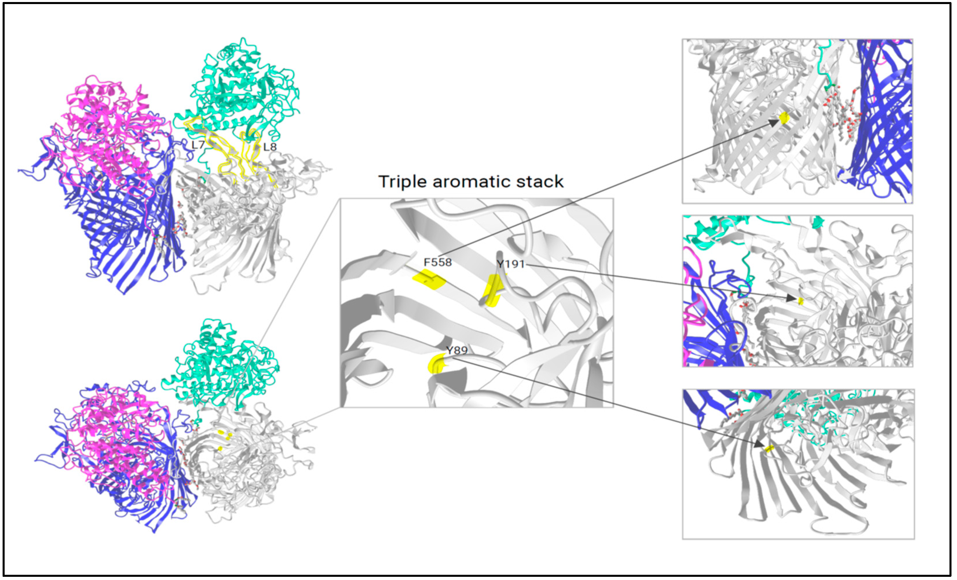
2.4. Analysis of RagA Functional Domains

First, a neighbor-joining phylogenetic tree based on the top 100 aligned and currently (date: 31 October 2024, source NCBI) available protein BLAST RagA sequences of P. gingivalis and related species was created (Figure S3). As some sequences, that are identical but observed in different strains, have been grouped into the same non-redundant RefSeq WP numbers, a translation into associated strain numbers is provided with Supplementary Information (“WP-strain numbers translation”). For RagA, the percentage identity of the W83 blastp results reached from ~97–96% (type-1) over ~69–67% (type-4 and -3) to ~64–58% (type-2). Even the outgroup had a percentage identity of ~53–49% to the reference W83.
Second, a maximum alignment, integrating NCBI and OMI RagA sequences, was presented (Figure S4A–C). The objectives were as follows: i. to identify areas that might explain the different virulence/fitness of the four types, ii. to further search for conserved domains representing structures of fundamental importance, and iii. to exclude highly variable regions as less important. Relevant domains (based on studies of Madej et al. 2020 and White et al. 2023 [18,23]) were therefore viewed from several angles: similarity to the reference W83 (thus to Rag-1, reference color blue), hydropathy (reference color blue = water soluble), RasMol (grouped by similar aa properties), and size of aa side chains (reference color blue = long).
The TonB box (X103-G108) (varieties shown in Figure S4A left) is located inside the barrel and is shifting in position between an open and closed state [18]. In fact, among the four RagA types, we did not find any difference at the protein level which confirms its immense importance (see homogenous colors over all strains, types, and aa features). Next, as part of the plug domain, region X211-A219 (Figure S4A right) represents an important area going through conformational changes during the process of substrate binding and passage [18]. It is interesting to note that, while otherwise conserved among P. gingivalis/P. gulae, serine (S211, polar, hydrophilic) instead of alanine (A211, non-polar, hydrophobic) is found here for type-2 and outgroups including Bacteroides thetaiotaomicron. Given the crucial role of the plug in facilitating substrate passage, this single position and its change in polarity may have played a pivotal role in the evolutionary trajectory towards a more (or contrarily less) virulent/fit phenotype. In contrast, the RagA loops that are positioned on the outside of the membrane show more sequence variability.
In total, 11 loops can be counted, of which loops L7 and L8 were assigned a major role by Madej et al. [18]. We further propose that loop L7 functions as a genuine “pedal” capable of raising the RagB lid while loop L8 is holding the RagB lid to limit/secure its movement. Figure 1, modified from iCn3D, shows the RagAB transport dimer in the open/closed state. Loops L7 and L8 are highlighted, showing the stable association with RagB. For loop L7, depicted in Figure S4B (left), there is an insert of usually eight amino acids (GNPEYYAH) in RagA-2 with a few sub-types (as in COT-052 P. gulae strains from Leicestershire, isolated 2012). Transferring this structural peculiarity to its role as “pedal”, a possibly larger opening of the lid could have a crucial influence on the absorption of the substrate and on the lid opening/closing dynamics. Loop 8 is not only involved in holding the lid but also in substrate transport. Figure S4B (right) shows L8 region X729-X753 to document the many differences among RagA -types, with type-4 showing both inter- and intra-type variations. Known from the literature (SusCD as model [23]), substrate binding leads to crucial conformational changes: W685 (on the base of loop 8, highly conserved in SusC and RagA-1-4) shifts inwards and pushes F583 up (conserved) and S193 down (polar uncharged serine in SusC is changed to alanine in RagA). As outlined above, without any ligand (apo), amino acid Y191 (conserved), which is part of the same plug loop, forms a triple aromatic stack with Y89 (nearby the TonB box, conserved) and F558 (barrel wall, conserved) that links the barrel wall, the TonB box, and the plug domain together (Figure 1). If a substrate is bound, Y191 shifts towards the periplasm, the aromatic lock is resolved, and the N-terminus is released, making the TonB box (energy transducer) able to interact with TonB, disrupting the plug and opening a channel into the periplasm [23]. Since we can transfer almost all the SusCD amino acids involved to the RagAB transport system, we can assume that the triple aromatic stack works in the same way here, resulting in an equal mechanism for plug triggering. Since these regions/positions are identical for SusC as well as for type RagA-1-4, we confirm their fundamental importance for the functionality of the nutrient acquisition protein complexes but conclude that they cannot explain the differences in virulence between the four heterogeneous RagAB types.
Next, we show potential RagA domains in contact with the proteinaceous substrate during uptake (Figure S4C for multi-alignment and Figure S5, partA, for RagA-1 W83 in a three-dimensional structure). The most virulent RagA-1 possesses an alanine at X399 (instead of polar serine for RagA-2-4 and SusC) and methionine at position X797 (instead of valine for RagA-2-4 and SusC, source: NCBI). Methionine in proteins fulfils an important antioxidant role, stabilizes the structure of proteins, participates in the sequence-independent recognition of protein surfaces, and can act as a regulatory switch through reversible redox reactions.
Furthermore, while X400-RagA-1 is aspartic acid (negatively charged, polar), X400-RagA-2 is glycine, and X400 in both RagA-3 and RagA-4 is a polar uncharged serine. Additionally, X405-RagA-1/A-2 is identified as tyrosine and alanine (both hydrophobic) versus polar and uncharged asparagine for RagA-3/A-4. At X408, RagA-3/A-4 is again presenting asparagine in contrast to glycine for RagA-1 and serine for RagA-2. Interestingly, the region X399-X409 in RagA-1 has four aromatic side chains (1xF, 3xY) and type-3 and -4 have three (1xF, 2xY), while type-2 has only two (1xF, 1xY). SusC, on the other hand, has none. If we consider that aromatic compounds like to assemble in stacks, importance for the overall structure should be considered. However, the spatial distance makes participation in the triple aromatic stack unlikely. While N800 and T803 are the same for RagA-1, RagA-4, and RagA-3, the type for RagA-2 differs with glycine (G800) and tyrosine (Y803) and is markedly different, as it is less hydrophobic (less red in Figure S4C, category hydropathy) and with a different pattern of side chains (less purple, more red or blue). Finally, and in contrast to all other substrate-binding regions, N893-Y898 —absent in SusC—is conserved among all RagA-1-4 types and thus excluded for variant or virulence definition but is probably instead important for switching from sugar to protein substrate.
While the aforementioned potential substrate-binding sites exclusively refer to proteinaceous ligands, it is necessary to screen for the fructo-oligosaccharide contact of SusC by F649, as proposed by White et al. [23]. Indeed, this region was found to be absent in all RagA.
Concluding here, the very conserved regions like the TonB box, the plug region (except position S211/A211), or the substrate-binding site N893-F898 can be excluded to explain the differences in virulence, but X399-X409, X799-X804, and the length of Loop 7 as the most expanding hinge (pedal) could be of interest here. Obviously, the virulent RagA-1 differs from RagA-2 with RagA-3/A-4 as possible intermediates. Figure 2 summarizes the peptide transport by RagAB.

2.5. Analysis of RagB Functional Domains

Like for RagA, a RagB phylogenetic tree based on the top 100 non-redundant RefSeq WP sequences was calculated (Figure S6), combined with all OMI-RagB sequences, and aligned by the NCBI Multiple Sequence Viewer 1.25.0 (Figure S7). In contrast to RagA, RagB was less conserved over the entire protein. The percentage identity, with W83 as reference, showed greater leaps from ~100–99% (type-1), ~57% (type-3), ~49–48% (type-4), ~48–47% (type-2), to finally ~33–27% (outgroups). While for RagA the outgroup included mainly P. uenonis, for RagB the outgroup contained a variety of Porphyromonas species (P. asaccharolytica, P. endodontalis, P. gingivicanis, P. uenonis).
Some relevant structures of RagB lid were analyzed more intensively. In accordance with the 3D model of RagAB on NCBI-iCn3D and Goulas [26], the anchor/hinge region (Figure S7, left), connecting the lid with the outer membrane, starts with a lipidated cysteine (C20, highly conserved even for outgroups but not for SusD). This is followed by a domain of 17 amino acids (X21-X38) whose C-terminal region presumably forms the hinge/pivot point that allows the “rigid-body movement of RagB” [18]. There are two amino acids (L22 and R24) that are the same for all RagB variants as well as for SusD and therefore structurally most essential. From position X27-X38, a colorful picture (again reflecting W83 similarity, hydropathy, RasMol visualization of properties, and side chain size of aa) emerges, which suggests that the hinge is constructed differently for the four types, with a potential impact on the opening and closing movement. A clear pattern linking type to practice is difficult. Madej et al. further depicted a RagB region close to the substrate (X76 and X83, here slightly expanded to X75-X84 for larger insight) (see Figure S5B for three-dimensional structure and Figure S7-middle for multi-sequence alignment). As D77 and G78 are conserved for all four RagB types, they might play an important role regarding substrate binding. For position X79, RagB-1, -3, and -4 possess a polar uncharged asparagine, while RagB-2 has a hydrophobic glycine. Another outlier is RagB-4 at position X83 with arginine (positively charged) instead of proline (hydrophobic) for RagB-1-3. However, in this case RagB-2 again shows the most differences, as the last shown amino acid (X84) is a tyrosine (polar) for type-2 instead of phenylalanine (hydrophobic). From a wider perspective, RagB-2 appears to be less hydrophobic in this region and especially in contrast to RagB-1, which is redder (=hydrophobic). As the substrate is absorbed solvent-free, the hydrophobicity could support absorption in RagB-1. In addition, a number of smaller side chains were identified in RagB-2, which could also affect substrate binding. Finally, the acidic loop insertion in RagB-1 (99DEDE102) and—in altering form—in RagB-3 (99DED101) mentioned by Madej et al. [18] was studied in detail (Figure S7, right). Interestingly, our amino acid analysis showed that the RagB-1 P. gulae strains differ from the human strains by one amino acid: instead of asparagine (D101), glycine (G101) was found. This could potentially indicate a gain of the acidic characteristic (with preference for alkaline substrates) by human P. gingivalis strains in the sense of an evolutionary event.

3. Discussion

3.1. Benefits and Limits of Universal RagA-Gene-Directed PCR Method

Because periodontitis is a widespread chronic inflammatory disease, not only affecting the oral cavity but also general health of patients [3], the importance of efficient diagnostic tools for the application of targeted therapeutic strategies is apparent. Research into the bacteria that cause periodontitis and in particular their ability to damage cells or ensure their own survival—as virulence factors do—is therefore essential. In contrast to other virulence factors like fimbrial adhesion proteins Fim/Mfa or gingipains, RagAB and the ragAB locus are not yet that well studied. Additionally, the four different rag alleles (with importance for virulence) make identification difficult.
As RagA is a transmembrane transporter, there are a whole series of homologous regions distributed across the entire genome. This hindered a direct ragA amplification as—even after testing several primer pairs—cross-reactions occurred, and presumptive ragA amplicons contained a mixture of transporter sequences. Instead, for the selective and type-independent sequencing of the ragA gene, an extended Long-PCR-product of 6–8.5 kb was amplified, including ragA (PG_0185) and ragB (PG_0186) as well as the ragAB genetic environment. This Long-PCR (LPCR) was introduced by Hall et al. in 2005, and for architecture of the different PCR products see Figure 2 of the same publication [16]. As IS elements can be found upstream of ragA and thus downstream of PG0183 (encoding for minor fimbriae ancillary tip subunit Mfa5), they might influence gene expression, an assumption that needs to be verified in future experiments. Of note here, the duplication of mfa5, found in a few strains [27], could be due to such IS elements. In fact, rag-1 strains A7436 and HG66 both have an ISPg element and show an mfa5 duplication, with the mfa5-2 variant being longer, while strains ATCC 33277 (rag-4) and TDC60 (rag-4) neither have ISPg elements nor show an mfa5 duplication. The LPCR products provided the baseline for the development of universal, type-independent ragA primers during our study and—by combining our five ragA with the eight ragB sequencing primers of Hall et al. [16]—both genes can be amplified and sequenced for any strain.
Limiting or aggravating factors are that double purification might be needed, as the smallest residuals of LPCR primers were found to interfere with sequencing. The principal reason could be secondary structures of the Long-PCR product impeding access of sequencing primers, a matter that has been discussed in previous publications [28]. In our experiments, this was necessary for the two reversed primers ragA-R4 and -R2, most likely due to their many wobbling bases. Additionally, the increase in sequencing primer’s concentration might help to reduce the impediment described above. Since ragA-R2 could not provide satisfactory results for type-4, ragA-R2a was added, which starts 80 bases further away from the ragA end but shows fewer wobbles and thus less cross annealing.
Nevertheless, this new way of detecting all types of ragA contributes to improving P. gingivalis rag locus detection and thus therapy-relevant information acquisition. Knowing the type of ragAB and the corresponding level of virulence can help determine the appropriate therapeutic approach, for example, by treating type-1 more aggressively with antibiotics such as metronidazole [29].
As genome-derived (mapped) sequences might have gaps (such as the rag locus of OMI 731 in our data set) or sequencing errors, the LPCR-rag amplification and sequencing approach we developed here can be used to resolve ambiguities. In addition, re-sequencing a single or a few bacterial genomes is very expensive and takes weeks or months to complete. So, the LPCR-rag sequencing approach here saves money and time.
In the future, the introduction of deoxy-inosine may allow type-independent ragB amplification by substituting the most wobbled positions in primer sequences, thus solving the problem of heterogeneity. Inosine primers have already demonstrated their advantages in the broad amplification and sequencing of the taxonomically important but highly heterogeneous rpoB gene, allowing a novel assay for broader bacterial identification in clinical microbiology [30].

3.2. New Insights into RagAB Phylogeny and Evolution

Hall et al. investigated the diversity of the rag locus and showed that there are at least four different variants (rag-1 to rag-4) [16]. Since the ragA and ragB genes were found in all P. gingivalis strains investigated, we were able to confirm the universal presence of rag in P. gingivalis, but different types may be present at different frequencies. Our results differed slightly from the study of Hall et al. [16], where rag-4 was 14%, rag-1 was 26%, and rag-3 was 25%, whereas we found about the same percentage (16–17%) for all three. However, their calculation for rag-2 being the most common (36%) was confirmed by our data (almost 50%). In conclusion here, geographical differences in rag-type prevalence might exist, but these results need to be interpreted with caution.
Regarding the within-patient evolution of P. gingivalis, we confirm that most patients sampled twice over eight years (1994 and 2002) showed the same strain and rag type. However, in other cases different strains with the same or different rag types were isolated from the same patient. Taken together, this pattern of in-host evolution is similar to that described for Stenotrophomonas maltophilia genotypes in cystic fibrosis [31].
All of these evolutionary processes and adaptations can have a variety of possible causes, including not only natural selection for the fittest but also host-specific selective pressures including biological, environmental, and behavioral factors. In particular, our immune system may select for traits that enable the survival and persistence of certain strains or communities [32]. Furthermore, the host environment can not only provide different mobile genetic elements but also modulate both recombination rates and mutational signatures [33].

3.3. RagAB as Factors of Virulence and Fitness

Diard and Hardt discussed different definitions of “virulence”, one at the level of individual host–pathogen interactions, where virulence increases colonization, exploitation, and damage, and one at the level of co-evolution, where virulence increases the fitness of a pathogen in the population [34]. In both definitions, “survival” is central; thus, most virulence factors are no more than survival factors that stimulate growth in the host environment and ensure persistence [35]. As an asaccharolytic species, the growth and survival of P. gingivalis depends on protein uptake through RagAB, modified type-specifically. The variability in colonization time, which can range from a few days to more than a week, is a well-documented phenomenon among P. gingivalis researchers. From the study of Madej et al., it is known that W83-RagAB-1 grows well on even minimal medium (supplemented with vitamin K1/K3, L-cysteine, and hemin), but ATCC 33277-RagAB-4 needed much more time, a deficiency which could be complemented by just replacing gene ragAB-4 with ragAB-1 [18].
The rag locus, as well as other virulence genes, is independently (uncorrelated) horizontally transferred between strains of the same species or even between different species [11,13]. However, Hall et al. found a limited degree of correlation between the k-capsular serotypes k1–k6 (plus k0 as un-capsuled) as k3 and k5 correspond to Rag-3, k4 to Rag-1, and k0–2 to two different rag alleles [16]. Furthermore, combining earlier results about long fimbriae (FimA) from of our group with the results about Rag type here, a few strains seem to accumulate more virulent types of both genes as ragAB-1 and fimA-IV are found together in strains of very different origin such as OMI 629 (W83, Germany), OMI 1079/OMI 1101 (Indonesia), and OMI 1049 (USA) [8].
In the same study, it was found that the animal P. gulae fimA typeA is closely related to human P. gingivalis strains of cluster Ib, potentially representing an ancestor genotype. Since ragAB type-1, the most virulent, has been identified in P. gulae from monkeys, we propose here that it may represent the ancestral sequence of the ragAB gene cluster (designated rag-1a). However, according to our phylogenetic trees, several other animal lineages exist, and horizontal transfer of the ragAB gene cluster between P. gulae and P. gingivalis may be possible on several independent occasions and may even be ongoing, as suggested by Meyer et al. and Fujiwara-Takahashi et al. for the fim type [8,36].
Another aspect that can be discussed is the N-terminal highly conserved part of ragA. This N-terminus contained a 20 aa signal peptide (MKRMTLFFLCLLTSIGWAMA) and a carboxypeptidase D_ regulatory-like domain (CarbopepD_reg_2, PF13715) found in bacteria, archaea, and eukaryotes, of approximately 90 aa in length, also known as N-terminal extension (NTE)—as preceding the TonB box—but with an unknown function so far. However, homologues are known to be collagen-binding [37] and might have proteolytic activity. Three-dimensional structures of RagA (deduced from W83) suggest that this domain is not part of the mature protein, but this has to be confirmed.
Still, there are no molecular explanations on why rag-1 leads to a higher virulence than the other three rag alleles, leading us to the next section.

3.4. Substrate Specificity and Binding/Uptake Capacity as Explanation for Virulence

Recent analysis on the structure of the RagAB protein complex proved that this transporter is responsible for the uptake of proteinaceous nutrients [18]. The exact substrate-binding sites can be revealed when sites of different substrates (glycans and proteins) are compared, as we carried out in this study here. Our protein alignment of RagA and SusC confirmed the hypothesis that for RagA a potential glycan-binding site is missing while different (type-specific) peptide-binding sites appear instead. In total, RagA interacts via minimal 26 aa residues and RagB via minimal 8 aa residues with the bound peptide [18]. This allows type- and peptide-specific variation in nutrient uptake. However, ragA type (barrel) and ragB type (lid) are corresponding and thus dependent, and both together define not only the substrate preferences but also the uptake capacity and speed. For instance, the sheer size of the cavity between lid and barrel could also be decisive for the preferred volume of substrate. According to Glenwright et al. [38], for BT2263 (SusD in B. thetaiotaomicron) and BT2264 (SusC), a deca-glycine or even several peptides can fit into this hollow space. With a contact area of ~3800 Å2 [38] and a volume of 11,500 Å3 [18], the calculated height of 3.0 Å results in only one layer of peptides. However, these assumptions have yet to be proven for RagAB.
Related to uptake speed, our hypothesis is that this may depend on how high the lid can open (with loop L7 as pedal) before loop L8 limits the range. In RagA-2, loop L7 contains more amino acids, and such a longer pedal may modulate the dynamics either by allowing a higher substrate capacity or by slowing down the uptake process compared to the other types. The latter might be more likely, as the substrate/ligand binding itself leads—like a zipper—to the bin closing, and this might take more time if the aperture angle between lid and barrel is larger. In conclusion here, the virulence might just be driven by P. gingivalis strain-dependent “appetite” and how important the preferred substrates (proteins) are for periodontal integrity and/or immunological defense.
For RagB, a meaningful transfer of SusD information—as performed above for RagA/SusC—was much more difficult. Corresponding to Pollet and coauthors addressing TBDT in Bacteroides, the C-terminal region containing the ligand-binding region is much more variable [39]. The length of the different SusD/RagB proteins varies fundamentally among different members of the SusD/RagB family, with most variables at the C-terminal region, making it even harder to draw conclusions on the substrate-binding site. The RagB/SusD variability itself could even be the principle for keeping the choice of food as flexible as possible. Finally, RagB could just be a rigid lid (or trap) that forms a cavity (of about 11,500 Å3) together with RagA and opens (by about 35 Å) while waiting for the ligand, a hypothesis that needs to be proven, of course. Nevertheless, the immunological function of RagB as a pro-inflammatory signal transducer as well as toll-like receptor 2 (TLR2) and TLR4 agonist [26,40] should not be disregarded.
According to new findings by White et al., additional outer membrane components (namely a surface glycan-binding protein, SGBP, and a corresponding glycoside hydrolase, GH) assemble on the core SusCD transporter in Bacteroides, forming a stable, octameric glycan-utilizing machine that they termed utilisome [23]. In the future, it will be exciting to discover if RagAB forms not only tetrameric structures but such octameric utilisomes. It is plausible that RagAB does not stand alone but that it is interacting with (a variety) of surface protein-binding molecules (SBPs), attracting the substrate and (a variety) of peptidases digesting it before. The only question is how distant these two other partners are, either floating in the outer membrane (such as earlier speculated for SusCD [18,41]) or directly attached to RagAB, as proven for SusCD recently [23]. In the latter model, there is need for binding of much bigger molecules such as SBPs and enzymes and thus for different lid-opening angles as we found some evidence for in the hinge/pivot region of our sequences. Nonetheless, confirmation needs sophisticated quantitative proteomics and single-particle cryo-EM investigations.
As a limitation of our study, it should be added that a direct comparison of SusCD and RagAB, even if performed critically, must be treated with caution. Although both TBDTs have a similar structure, they occur in different species with very different substrate preferences. We are very confident that a few key structures were identified on species and type levels. However, how essential these domains are can only be proven by knock-out mutations and/or animal trials.
In conclusion, despite some limitations, our analysis of the 129 P. gingivalis/P. gulae genomes from our own collection and the 100 best-matching WP sequences imported from NCBI enabled us to further explore the genetic and functional diversity of RagAB, encoded by four rag alleles. A way to detect all four ragA variants was realized by combining an already known Long-PCR with our new sequencing primers. In addition, a potential plug-opening mechanism for RagA in the form of the aromatic triplet was discovered through the transfer of SusC information. The essential TBDT functions are more dependent on the RagA barrel than on the RagB lid. In general, the complex seems to be quite flexible with respect to the substrate, with the exception of RagA-1, which probably indicates a “special diet”. In addition, an insertion (8 aa long) found in loop L7 throughout RagA-2 might not only affect the dynamics of lid opening/closing but also alter the associated substrate throughput rate.

4. Material and Methods

This study can be divided into two parts: In the first part, phylogenetic trees based on genome mapping and corresponding amino acid sequences of our collection, further expanded by GenBank (NCBI) entries, were constructed and domain-specifically analyzed. In the second part, an attempt was made to develop a new PCR method for universal ragA and ragB amplification and sequencing. Figure 3 represents a graphical overview of our study.

4.1. Bacterial Strains and Growth Conditions

The basis of this study was a P. gingivalis collection (Division of Oral Microbiology and Immunology (OMI)) at the RWTH Aachen University reflecting a wide variety of strains (human and animal hosts, geographical origin, capsule type, fimbriae type). Further information on the isolates is shown in Table S1. It is noteworthy that some species closely related to P. gingivalis, such as Porphyromonas gulae (P. gulae), P. loveana, P. macacae, or P. somerae, were also added to the mapping for broader and reliable insight into ragAB evolution. All isolates were stored in cryotubes at −72 °C. The samples were cultivated on tryptone soya blood agar (TSBA) or Brucella blood agar and put under anaerobic conditions via BD GasPakTM Gas Generating System bags (<1% oxygen, ≥13% CO2) for 3–7 days at 37 °C.

4.2. Genomic DNA (gDNA) Extraction

After cultivation, colonies were suspended in 1 mL 0.9% NaCl, washed, and bacterial genomic DNA was extracted using the spin column method in accordance with the manufacturer’s instructions (QIAamp DNA Mini Kit; Qiagen; Venlo, The Netherlands). Spectrophotometrically, the DNA concentration and purity were evaluated (NanoVue Plus Spectrophotometer, GE Healthcare Europe GmbH, Freiburg, Germany). The gDNA concentration was between 25 ng/µL and 91 ng/µL, and the purity was in a regular range from 1.8 to 2.0 (260 nm/280 nm). The extracted gDNA was either subjected to the whole-genome sequencing (WGS), using the Illumina NovaSeq 6000 platform (San Diego, CA, USA) generating 250 base paired (bp) end reads or to a ragA-type independent (universal) PCR.

4.3. Comparative Genomics and Phylogeny of RagAB

Mapping with 129 isolates of P. gingivalis/P. gulae and a few related species of the collection was performed. The genomic DNA was subjected to whole-genome sequencing (WGS) using the Illumina NovaSeq 6000 platform (San Diego, CA, USA) generating 250 base paired (bp) end reads. Demultiplexing of all libraries and for all sequencing data was performed using Illumina bcl2fastq software v2.20, and reads with a final length of less than 20 bases were discarded. For mapping, FASTQ files were aligned against the ragAB-type-specific reference genes using the Snippy pipeline (Table S2). If the genome sequence data of the rag locus were not complete, a universal PCR was used (see Section 4.4). After mapping, the strains were assigned to one of the four different ragAB types. Figure S8 shows two partial alignments (1100 bp each) of representatives of all four types and in both ragA and ragB.
Analysis of ragAB/RagAB phylogeny and RagAB functional domains: Alignments for each type were created by Seaview, version 5.0.5 [43], and phylogenetic trees were generated on DNA (nucleic acids) as well as—after in silico translation—protein (amino acids) level using the neighbor-joining model with 100 bootstrap replications applying MEGA11, version 11.0.13 [44] (DNA level: Figure S1; protein-level: Figure S2). Furtherly, for each of the four Rag types, one representative was chosen to run a protein BLAST on NCBI database (Rag-1: strain W83 [RagA-1: AAQ65420.1; RagB-1: AAQ65421.1]; Rag-2: strain 11A [RagA-2: SJL25370.1; RagB-2: SJL25368.1]; Rag-3: strain SU60 [RagA-3: SJL29627.1; RagB-3: SJL29621.1]; Rag-4: strain ATCC33277 [RagA-4: AUR48986.1; RagB-4: AUR49279.1]). Comprehensive alignments including the first 100 protein BLAST results for RagA and RagB covered all four types, and the related phylogenetic trees were calculated (phylogenetic trees shown in Figures S3 and S6) (MEGA11). Moreover, additional alignments with functional domains (RagA: TonB box, plug region, hinge-associated loops 7 and 8, substrate-binding sites; RagB: anchor/hinge, substrate-binding sites including X75-X84 and an acid loop), combining NCBI and our own strain data, were generated by the NCBI Multiple Sequence Alignment Viewer (1.25.0) (Figures S4A–C and S7). The three-dimensional crystal structure, deposed by White, Ranson, and van den Berg, and based on data of Madej et al. (2020) [18], which can be viewed at https://www.rcsb.org/structure/6SMQ (accessed on 30 November 2024) (W83, open/closed state), was also taken into account here.

4.4. Development of Universal RagA-Gene-Directed PCR Method

Since approaches for ragB universal amplification failed due to its high heterogeneity, the methods are limited to ragA. For the ragA universal PCR design, we used one representative for each rag type: type-1: P. gingivalis strain OMI 629, original name W83, isolated in Bonn, Germany, 1950s; type-2: OMI 1090, original name HW24D-2, isolated in Quebec, Canada, before 1993; type-3: OMI 1088, original name 59Pg1, isolated in Indonesia, 1994; type-4: OMI 1132, type strain ATCC 33277, isolated in USA, before 1981.
PCR amplification of ragA: For ragA, the Long-PCR protocol introduced by Hall et al. (2005) was used [16]. The ingredient composition and the cycling parameters were adjusted as follows. First, we created a mastermix of 49 µL for each sample that contained 34 µL nuclease-free water (Life Technologies Corporation, Austin, TX, USA), 10 µL of 5X PCR buffer (2 mM MgCl2; Roche Diagnostik GmbH; Mannheim, Germany), 3.0 µL of nucleoside triphosphate set (dATP, dCTP, dGTP, dTTP; Roche Diagnostics GmbH; Mannheim, Germany), 0.5 µL of forward primer, 0.5 µL of reverse primer (each 1 µM, synthesized by TIB Molbiol Syntheselabor GmbH, Berlin; for sequences see Table 1), and 2.0 µL of LONG-Taq-DNA-Polymerase (New England Biolabs, Frankfurt am Main, Germany). Second, the mastermix was combined with 1 µL of undiluted gDNA. The cycling parameters used were the following: initial denaturation at 95 °C for 5 min; 25 cycles; per cycle denaturation at 95 °C for 1 min, primer annealing at 60 °C for 30 s, and for elongation 68 °C for 8 min; final elongation at 72 °C for 10 min. A positive control (DNA of reference strains) and negative control (water without any DNA) were always included. MicroAmp™ reaction tubes (Applied Biosystems, Waltham, MA, USA) were used, and the reaction was performed in a PCR Express Thermal Cycler (Thermo Hybaid, Ashford, UK). Via agarose gel electrophoresis, the Long-PCR products were visualized. Therefore, 1% gels were used by heating 0.6 g of agarose (Top Vision Agarose, Thermo Fisher Scientific Inc., Waltham, MA, USA) and 60 mL of TAE buffer (TRIS-Acetate-EDTA-buffer, SERVA Electrophoresis GmbH, Heidelberg, Germany) until the agarose was completely dissolved. As fluorescent colorant, 3.5 µL of Midori Green Advance (NIPPON Genetics EUROPE, Düren, Germany) was added. A volume of 8 µL of LPCR products was mixed with 3 µL of Blue Marker (40% saccharose and 0.1% Bromophenol blue) and pipetted in the gel pockets. For every run, a MassRuler High Range DNA Ladder (Thermo Fisher Scientific Inc., Waltham, MA, USA) was subjoined. The parameters for electrophoresis were 100 V and 250 mA for 60–90 min. With the help of UV light (GelStudio SA, Analytik Jena GmbH, Jena, Deutschland), the bands became visible. The Long-PCR products were purified in accordance with the manufacturer’s instructions (NucleoSpin® Gel and PCR Clean-up, Macherey + Nagel, Düren, Germany). The purified product was sent for Sanger sequencing (Eurofins Genomics, Ebersberg, Germany). For this sequencing reaction, 5 µL of each LPCR product (20–80 ng/µL) and 5 µL of the newly designed five ragA-type-independent primers (ragA-F0 and -F1 each 5 µM, all others 20 µM) were mixed (Table 1). The ragA primers were synthesized by TIB Molbiol. The sequencing results were aligned/merged by the ClustalO algorithm using Seaview [43] to obtain an almost complete and ambiguity-free sequence for every type (allele). Figure 4 visualizes the LPCR product, the ragAB loci, the coverage of ragA primers, and flanking genes of P. gingivalis strain OMI 629 (W83, type ragA-1) as an example. In Figure S9, representative results of the other ragA types are shown, namely type-2: OMI 1090 (HW24D-2), type-3: OMI 1088 (59Pg1), and type-4: OMI 1132 (ATCC 33277).

Supplementary Materials

The supporting information can be downloaded at: https://www.mdpi.com/article/10.3390/ijms26031073/s1.

Author Contributions

P.G.M. performed most experiments, collected and analyzed the data, and wrote the manuscript. E.D. helped with performing the LPCR and ragA-sequencing experiments and interpreted the data. B.N. and D.D. collected and provided clinical strains. M.M.H.A. produced genome data, delivered the mapping results, secured funding, interpreted the data, and corrected the manuscript. G.C. conceived the idea, secured funding, supervised authors P.G.M. and E.D., interpreted the data, and corrected the manuscript. All authors have read and agreed to the published version of the manuscript.

Funding

This study was supported by the IZKF (Interdisciplinary Center for Clinical Research, grant no. OC1-06) of the Medical Faculty, RWTH Aachen, Germany.

Data Availability Statement

The data that support the findings of this study are available from G.C. upon request. Genome data of ragAB were deposited by NCBI-BankIt under Accession-Nos. PQ658417-PQ658670.

Acknowledgments

We would like to thank the many scientists worldwide who helped collect strains and data. We thank Sarah Böcher for inspiring this study and Beate Melzer-Krick for her excellent technical assistance.

Conflicts of Interest

The authors declare no conflicts of interest.

References

  1. Conrads, G.; Nagy, E.; Könönen, E. Bacteroides, Porphyromonas, Prevotella, Fusobacterium, and Other Anaerobic Gram-Negative Rods. In Manual of Clinical Microbiology, 12th ed.; Caroll, K., Pfaller, M.A., Eds.; ASM Press: Washington, DC, USA, 2019. [Google Scholar]
  2. Henry, L.G.; McKenzie, R.M.; Robles, A.; Fletcher, H.M. Oxidative stress resistance in Porphyromonas gingivalis. Future Microbiol. 2012, 7, 497–512. [Google Scholar] [CrossRef] [PubMed]
  3. Socransky, S.S.; Haffajee, A.D.; Cugini, M.A.; Smith, C.; Kent, R.L., Jr. Microbial complexes in subgingival plaque. J. Clin. Periodontol. 1998, 25, 134–144. [Google Scholar] [CrossRef] [PubMed]
  4. Carter, C.J.; France, J.; Crean, S.; Singhrao, S.K. The Porphyromonas gingivalis/host interactome shows enrichment in GWASdb genes related to Alzheimer’s Disease, Diabetes and Cardiovascular Diseases. Front. Aging Neurosci. 2017, 9, 408. [Google Scholar] [CrossRef] [PubMed]
  5. Dominy, S.S.; Lynch, C.; Ermini, F.; Benedyk, M.; Marczyk, A.; Konradi, A.; Nguyen, M.; Haditsch, U.; Raha, D.; Griffin, C.; et al. Porphyromonas gingivalis in Alzheimer’s disease brains: Evidence for disease causation and treatment with small-molecule inhibitors. Sci. Adv. 2019, 5, eaau3333. [Google Scholar] [CrossRef]
  6. Potempa, J.; Mydel, P.; Koziel, J. The case for periodontitis in the pathogenesis of rheumatoid arthritis. Nat. Rev. Rheumatol. 2017, 13, 606–620. [Google Scholar] [CrossRef]
  7. Miyauchi, M.; Ao, M.; Furusho, H.; Chea, C.; Nagasaki, A.; Sakamoto, S.; Ando, T.; Inubushi, T.; Kozai, K.; Takata, T. Galectin-3 plays an important role in preterm birth caused by dental infection of Porphyromonas gingivalis. Sci. Rep. 2018, 8, 2867. [Google Scholar] [CrossRef]
  8. Meyer, H.L.; Abdelbary, M.M.H.; Buhl, E.M.; Kuppe, C.; Conrads, G. Exploring the genetic and functional diversity of Porphyromonas gingivalis long fimbriae. Mol. Oral. Microbiol. 2023, 38, 408–423. [Google Scholar] [CrossRef] [PubMed]
  9. Potempa, J.; Madej, M.; Scott, D.A. The RagA and RagB proteins of Porphyromonas gingivalis. Mol. Oral. Microbiol. 2021, 36, 225–232. [Google Scholar] [CrossRef]
  10. Shi, X.; Hanley, S.A.; Faray-Kele, M.C.; Fawell, S.C.; Aduse-Opoku, J.; Whiley, R.A.; Curtis, M.A.; Hall, L.M. The rag locus of Porphyromonas gingivalis contributes to virulence in a murine model of soft tissue destruction. Infect. Immun. 2007, 75, 2071–2074. [Google Scholar] [CrossRef] [PubMed]
  11. Frandsen, E.V.; Poulsen, K.; Curtis, M.A.; Kilian, M. Evidence of recombination in Porphyromonas gingivalis and random distribution of putative virulence markers. Infect. Immun. 2001, 69, 4479–4485. [Google Scholar] [CrossRef]
  12. Hanley, S.A.; Aduse-Opoku, J.; Curtis, M.A. A 55-kilodalton immunodominant antigen of Porphyromonas gingivalis W50 has arisen via horizontal gene transfer. Infect. Immun. 1999, 67, 1157–1171. [Google Scholar] [CrossRef] [PubMed]
  13. Curtis, M.A.; Hanley, S.A.; Aduse-Opoku, J. The rag locus of Porphyromonas gingivalis: A novel pathogenicity island. J. Periodontal Res. 1999, 34, 400–405. [Google Scholar] [CrossRef]
  14. Nagano, K.; Murakami, Y.; Nishikawa, K.; Sakakibara, J.; Shimozato, K.; Yoshimura, F. Characterization of RagA and RagB in Porphyromonas gingivalis: Study using gene-deletion mutants. J. Med. Microbiol. 2007, 56, 1536–1548. [Google Scholar] [CrossRef] [PubMed]
  15. Dashper, S.G.; Mitchell, H.L.; Seers, C.A.; Gladman, S.L.; Seemann, T.; Bulach, D.M.; Chandry, P.S.; Cross, K.J.; Cleal, S.M.; Reynolds, E.C. Porphyromonas gingivalis uses specific domain rearrangements and allelic exchange to generate diversity in surface virulence factors. Front. Microbiol. 2017, 8, 48. [Google Scholar] [CrossRef]
  16. Hall, L.M.; Fawell, S.C.; Shi, X.; Faray-Kele, M.C.; Aduse-Opoku, J.; Whiley, R.A.; Curtis, M.A. Sequence diversity and antigenic variation at the rag locus of Porphyromonas gingivalis. Infect. Immun. 2005, 73, 4253–4262. [Google Scholar] [CrossRef] [PubMed]
  17. Liu, Y.; Zhang, Y.; Wang, L.; Guo, Y.; Xiao, S. Prevalence of Porphyromonas gingivalis four rag locus genotypes in patients of orthodontic gingivitis and periodontitis. PLoS ONE 2013, 8, e61028. [Google Scholar] [CrossRef] [PubMed]
  18. Madej, M.; White, J.B.R.; Nowakowska, Z.; Rawson, S.; Scavenius, C.; Enghild, J.J.; Bereta, G.P.; Pothula, K.; Kleinekathoefer, U.; Basle, A.; et al. Structural and functional insights into oligopeptide acquisition by the RagAB transporter from Porphyromonas gingivalis. Nat. Microbiol. 2020, 5, 1016–1025. [Google Scholar] [CrossRef] [PubMed]
  19. Kadowaki, T.; Nakayama, K.; Okamoto, K.; Abe, N.; Baba, A.; Shi, Y.; Ratnayake, D.B.; Yamamoto, K. Porphyromonas gingivalis proteinases as virulence determinants in progression of periodontal diseases. J. Biochem. 2000, 128, 153–159. [Google Scholar] [CrossRef] [PubMed]
  20. Reeves, A.R.; Wang, G.R.; Salyers, A.A. Characterization of four outer membrane proteins that play a role in utilization of starch by Bacteroides thetaiotaomicron. J. Bacteriol. 1997, 179, 643–649. [Google Scholar] [CrossRef] [PubMed]
  21. Kommedal, O.; Karlsen, B.; Saebo, O. Analysis of mixed sequencing chromatograms and its application in direct 16S rRNA gene sequencing of polymicrobial samples. J. Clin. Microbiol. 2008, 46, 3766–3771. [Google Scholar] [CrossRef] [PubMed]
  22. Böcher, S.; Meyer, H.L.; Dafni, E.; Conrads, G. Prevalence and phylogenetic analysis of lipoprotein-Gene ragB-1 of Porphyromonas gingivalis-A pilot study. Antibiotics 2023, 12, 1458. [Google Scholar] [CrossRef] [PubMed]
  23. White, J.B.R.; Silale, A.; Feasey, M.; Heunis, T.; Zhu, Y.; Zheng, H.; Gajbhiye, A.; Firbank, S.; Basle, A.; Trost, M.; et al. Outer membrane utilisomes mediate glycan uptake in gut Bacteroidetes. Nature 2023, 618, 583–589. [Google Scholar] [CrossRef] [PubMed]
  24. Montz, P. 2025. Figure 1: RagAB Functional Domains During Substrate Transport. Created in BioRender. Available online: https://BioRender.com/o50b721 (accessed on 21 January 2025).
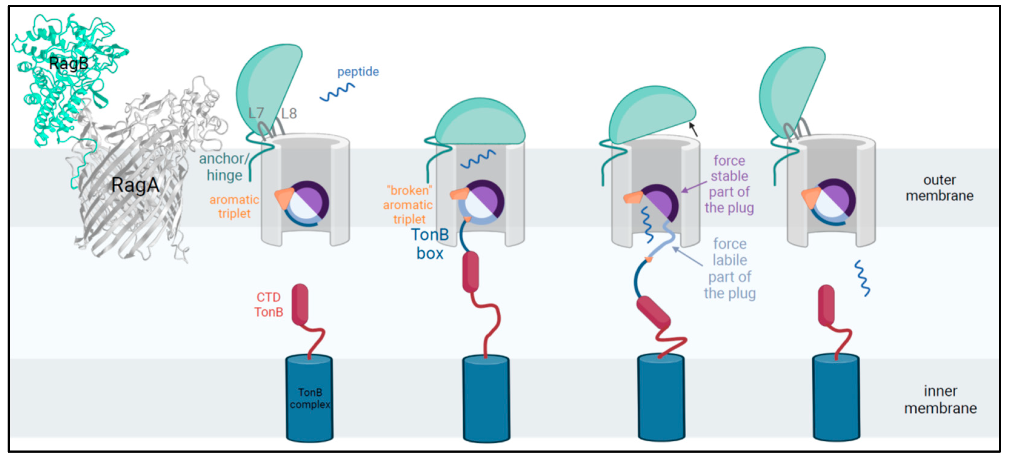
  25. Montz, P. 2025. Figure 2: TonB-Dependent Peptide-Substrate Transport in Porphyromonas gingivalis. Created in BioRender. Available online: https://BioRender.com/u76z058 (accessed on 21 January 2025).
  26. Goulas, T.; Garcia-Ferrer, I.; Hutcherson, J.A.; Potempa, B.A.; Potempa, J.; Scott, D.A.; Gomis-Ruth, F.X. Structure of RagB, a major immunodominant outer-membrane surface receptor antigen of Porphyromonas gingivalis. Mol. Oral. Microbiol. 2016, 31, 472–485. [Google Scholar] [CrossRef] [PubMed]
  27. Fujita, M.; Chiu, C.H.; Nagano, K. Transcriptional analysis of the mfa-cluster genes in Porphyromonas gingivalis strains with one and two mfa5 genes. Mol. Oral. Microbiol. 2023, 38, 41–47. [Google Scholar] [CrossRef]
  28. Snyder, T.M.; Tse, B.N.; Liu, D.R. Effects of template sequence and secondary structure on DNA-templated reactivity. J. Am. Chem. Soc. 2008, 130, 1392–1401. [Google Scholar] [CrossRef]
  29. Conrads, G.; Klomp, T.; Deng, D.; Wenzler, J.S.; Braun, A.; Abdelbary, M.M.H. The Antimicrobial Susceptibility of Porphyromonas gingivalis: Genetic Repertoire, Global Phenotype, and Review of the Literature. Antibiotics 2021, 10, 1438. [Google Scholar] [CrossRef] [PubMed]
  30. Bivand, J.M.; Dyrhovden, R.; Sivertsen, A.; Tellevik, M.G.; Patel, R.; Kommedal, O. Broad-range amplification and sequencing of the rpoB gene: A novel assay for bacterial identification in clinical microbiology. J. Clin. Microbiol. 2024, 62, e0026624. [Google Scholar] [CrossRef] [PubMed]
  31. Izydorczyk, C.; Waddell, B.J.; Thornton, C.S.; Conly, J.M.; Rabin, H.R.; Somayaji, R.; Surette, M.G.; Church, D.L.; Parkins, M.D. Stenotrophomonas maltophilia natural history and evolution in the airways of adults with cystic fibrosis. Front. Microbiol. 2023, 14, 1205389. [Google Scholar] [CrossRef]
  32. Baishya, J.; Wakeman, C.A. Selective pressures during chronic infection drive microbial competition and cooperation. NPJ Biofilms Microbiomes 2019, 5, 16. [Google Scholar] [CrossRef]
  33. Abdelbary, M.M.H.; Hatting, M.; Dahlhausen, A.; Bott, A.; Conrads, G. Insights into Within-Host Evolution and Dynamics of Oral and Intestinal Streptococci Unveil Niche Adaptation. Int. J. Mol. Sci. 2024, 25, 13507. [Google Scholar] [CrossRef] [PubMed]
  34. Diard, M.; Hardt, W.D. Evolution of bacterial virulence. FEMS Microbiol. Rev. 2017, 41, 679–697. [Google Scholar] [CrossRef] [PubMed]
  35. Dahlen, G.; Basic, A.; Bylund, J. Importance of virulence factors for the persistence of oral bacteria in the inflamed gingival crevice and in the pathogenesis of Periodontal Disease. J. Clin. Med. 2019, 8, 1339. [Google Scholar] [CrossRef]
  36. Fujiwara-Takahashi, K.; Watanabe, T.; Shimogishi, M.; Shibasaki, M.; Umeda, M.; Izumi, Y.; Nakagawa, I. Phylogenetic diversity in fim and mfa gene clusters between Porphyromonas gingivalis and Porphyromonas gulae, as a potential cause of host specificity. J. Oral. Microbiol. 2020, 12, 1775333. [Google Scholar] [CrossRef]
  37. Kim, Y.H.; O’Neill, H.M.; Whitehead, J.P. Carboxypeptidase X-1 (CPX-1) is a secreted collagen-binding glycoprotein. Biochem. Biophys. Res. Commun. 2015, 468, 894–899. [Google Scholar] [CrossRef] [PubMed]
  38. Glenwright, A.J.; Pothula, K.R.; Bhamidimarri, S.P.; Chorev, D.S.; Basle, A.; Firbank, S.J.; Zheng, H.; Robinson, C.V.; Winterhalter, M.; Kleinekathofer, U.; et al. Structural basis for nutrient acquisition by dominant members of the human gut microbiota. Nature 2017, 541, 407–411. [Google Scholar] [CrossRef] [PubMed]
  39. Pollet, R.M.; Martin, L.M.; Koropatkin, N.M. TonB-dependent transporters in the Bacteroidetes: Unique domain structures and potential functions. Mol. Microbiol. 2021, 115, 490–501. [Google Scholar] [CrossRef]
  40. Hutcherson, J.A.; Bagaitkar, J.; Nagano, K.; Yoshimura, F.; Wang, H.; Scott, D.A. Porphyromonas gingivalis RagB is a proinflammatory signal transducer and activator of transcription 4 agonist. Mol. Oral. Microbiol. 2015, 30, 242–252. [Google Scholar] [CrossRef]
  41. Gray, D.A.; White, J.B.R.; Oluwole, A.O.; Rath, P.; Glenwright, A.J.; Mazur, A.; Zahn, M.; Basle, A.; Morland, C.; Evans, S.L.; et al. Insights into SusCD-mediated glycan import by a prominent gut symbiont. Nat. Commun. 2021, 12, 44. [Google Scholar] [CrossRef] [PubMed]
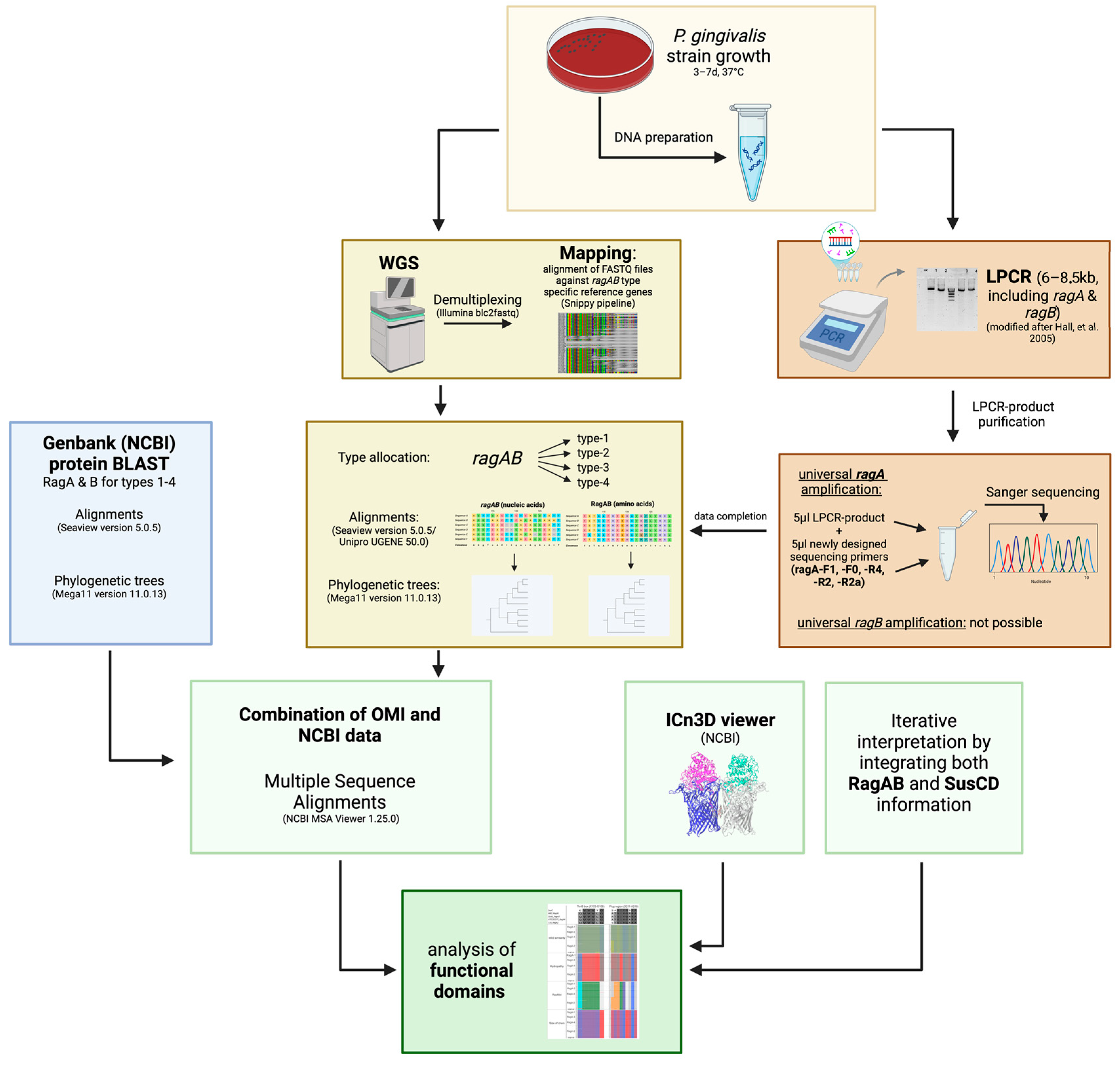
  42. Montz, P. 2025. Figure 3. Graphical Abstract of Methodological Flow of Study. Created in BioRender. Available online: https://BioRender.com/m93c482 (accessed on 21 January 2025).
  43. Gouy, M.; Tannier, E.; Comte, N.; Parsons, D.P. Seaview Version 5: A multiplatform software for multiple sequence alignment, molecular phylogenetic analyses, and tree reconciliation. Methods Mol. Biol. 2021, 2231, 241–260. [Google Scholar] [CrossRef] [PubMed]
  44. Tamura, K.; Stecher, G.; Kumar, S. MEGA11: Molecular Evolutionary Genetics Analysis Version 11. Mol. Biol. Evol. 2021, 38, 3022–3027. [Google Scholar] [CrossRef]
  45. Copley, R.R.; Russell, R.B.; Ponting, C.P. Sialidase-like Asp-boxes: Sequence-similar structures within different protein folds. Protein Sci. 2001, 10, 285–292. [Google Scholar] [CrossRef]
Figure 1. RagAB functional domains during substrate transport: loops L7 and L8 are highlighted in yellow in image upper left. Comparing the conformation (movement) in the open and closed states, L7 appears to be important for pedaling, while L8 is important for lid holding. At the bottom left, an overview of the position of the triple aromatic stack is given with a zoomed image as well as each participant from a different perspective: F558 from the outside and at the back of the barrel, Y191 from the inside as part of the plug loop, and Y89 from below following the TonB box. Modified from iCn3D and created in BioRender [24].
Ijms 26 01073 g001
Figure 2. TonB-dependent peptide-substrate transport in Porphyromonas gingivalis. Only in substrate-loaded status, when the aromatic triple is disrupted, is the shifting TonB box able to interact with TonB-CTD, located in the inner membrane. TonB transduces the energy stored in the proton gradient to exert a force on the mechanically labile subdomain (N-terminal, light blue) in contrast to the solid domain (C-terminal, purple). Once the plug has been removed from the β-barrel lumen, substrates are able to diffuse to the periplasmatic space and subsequently gain access to the cytoplasm via ABC transporters. Modified after Madej et al. 2020 and White et al. 2023 [18,23] and created in BioRender [25].
Ijms 26 01073 g002
Figure 3. Graphical abstract of methodological flow of study. Abbreviation explanation: Long-PCR (LPCR), whole-genome sequencing (WGS), Division of Oral Microbiology and Immunology (OMI). Created in BioRender [16,42].
Ijms 26 01073 g003
Figure 4. By the Long-PCR, the rag locus was amplified with flanking regions such as ISPg transposase (facultative), partial PG0183 (3′ end with only 48 bp, encoding for minor fimbriae subunit Mfa5), and partial PG0188 (5′ end with 408 bp, encoding for a conserved protein of unknown function containing a sialidase-like Asp-box; [45]). With the double-cleaned Long-PCR product, ragA could be sequenced type-independently and, subsequently, the ragB type-dependently. Note that this approach does not cover the first and last 50 bp of ragA.
Ijms 26 01073 g004
Table 1. Primers used for Long-PCR amplification and subsequent sequencing to cover all four ragA alleles (ragA-1 to ragA-4). Primer ragA-R2a was applied in case ragA-R2 did not generate any sequences, especially occurring in type-4 strains. R: purine G or A; Y: pyrimidine C/T; M: amino A/C; W: weak A/T; N: all nucleotides A/T/G/C.
PrimerSequenceLength
PCR LPCR-F5′ CAA AGT CCT GCC ACG AGT AGC 3′6–8.5 kb
primerLPCR-R5′ CGT TTT CTC GCC ACT TTC GTC 3′
Position Start/End
SequencingragA-F15′ ATG AAA AGA ATG ACG CTA TTC TTC C 3′50/1170
PrimerragA-F05′ GGT CAG GTA GCC GGT ATG CAG GTT AT 3′480/1540
ragA-R45′ CCR GGR ACA TAC CAC A 3′2500/1370
ragA-R25′ TTA RAA MGA MAN YTG RAT ACC 3′3030/2000
ragA-R2a5′ GGG TCR AAR CCT TTR WAC TT 3′2980/1900 (type-4)
Disclaimer/Publisher’s Note: The statements, opinions and data contained in all publications are solely those of the individual author(s) and contributor(s) and not of MDPI and/or the editor(s). MDPI and/or the editor(s) disclaim responsibility for any injury to people or property resulting from any ideas, methods, instructions or products referred to in the content.

